Piscirickettsia salmonis Induces Apoptosis in Macrophages and Monocyte-Like Cells From Rainbow Trout Vero ´nica Rojas, 1 Norbel Galanti, 2 * Niels C. Bols, 3 Vero ´ nica Jime ´nez, 2 Rodolfo Paredes, 2 and Sergio H. Marshall 1 1 Laboratorio de Gene ´tica e Inmunologı ´a Molecular, Instituto de Biologı ´a, Pontificia Universidad Cato ´lica de Valparaı ´so, Valparaı ´so, Chile 2 Programa de Biologı ´a Celular y Molecular, Instituto de Ciencias Biome ´dicas, Facultad de Medicina, Universidad de Chile, Santiago, Chile 3 Department of Biology, University of Waterloo, Waterloo, Ontario, Canada ABSTRACT Piscirickettsia salmonis is the etiologic agent of the salmonid rickettsial septicemia (SRS) which causes significant losses in salmon production in Chile and other and in other regions in the southern hemisphere. As the killing of phagocytes is an important pathogenic mechanism for other bacteria to establish infections in vertebrates, we investigated whether P. salmonis kills trout macrophages by apoptosis. Apoptosis in infected macrophages was demonstrated by techniques based on morphological changes and host cell DNA fragmentation. Transmission electron microcopy showed classic apoptotic characteristics and terminal deoxynucleotidyl transferase-mediated dUTP nick end labeling showed fragmented DNA. Programmed cell death type I was further confirmed by increased binding of annexin V to externalized phosphatidylserine in infected macrophages. Moreover, significant increases of caspase 3 activation were detected in infected cells and treatment with caspase inhibitor caused a decrease in levels of apoptosis. This is the first evidence that P. salmonis induces cell death in trout macrophages. This could lead to bacterial survival and evasion of the host immune response and play an important role in the establishment of infection in the host. J. Cell. Biochem. 110: 468–476, 2010. ß 2010 Wiley-Liss, Inc. KEY WORDS: P. SALMONIS; APOPTOSIS; RTS11 CELL LINE; MACROPHAGES; MONOCYTES P iscirickettsia salmonis is the etiologic agent of the salmonid rickettsial septicemia (SRS), or piscirickettsiosis [Fryer et al., 1990, 1992; Fryer and Hedrick, 2003], a systemic infection which affects all cultured salmon fish species causing significant losses in salmon production all around the world [Mauel and Miller, 2002; Fryer and Hedrick, 2003]. P. salmonis was described as a non-motile, not encapsulated, pleomorphic but generally coccoid bacteria, with a diameter ranging from 0.2 to 1.5 mm [Bravo and Campos, 1989; Rojas et al., 2007]. It is a Gram-negative intracellular bacterium that replicates within membrane-bound cytoplasmic vacuoles in tissues from infected fish and in cell cultures derived from fish [Cvitanich et al., 1991; Fryer and Hedrick, 2003]. However, two recent reports have suggested that the bacterium may be able to grow in an artificial cell-free media [Mauel et al., 2008; Mikalsen et al., 2008]. Nevertheless, mainly due to experimental limitations imposed by the intracellular nature of the bacterium, little is known about its infective strategy. In a previous study we found that P. salmonis is able of infect, survive, and propagate inside salmonid macrophages and monocyte- like cells without inducing cytopathic effect and maintaining its expression capacity [Rojas et al., 2009]. These properties may be part of a strategy leading to the survival of this bacterium which may include resistance to killing by macrophages. Therefore, a combination of intracellular growth of the bacteria and regulation of macrophage cell death may be considered as the basis for P. salmonis pathogenesis. Numerous experiments with a variety of microbes demonstrate that apoptosis of professional phagocytes is a common event in pathogenesis and plays a pivotal role in the initiation of the infection, survival of the pathogens, and evasion of the first line of defense of the immune system [Hilbi et al., 1997]. Bacteria Journal of Cellular Biochemistry ARTICLE Journal of Cellular Biochemistry 110:468–476 (2010) 468 Grant sponsor: FONDEF; Grant number: 1038; Grant sponsor: DI-PUCV; Grant number: 122785; Grant sponsor: RTPD-SIDA/SAREC. *Correspondence to: Prof. Norbel Galanti, Programa de Biologı ´a Celular y Molecular, Instituto de Ciencias Biome ´dicas, Facultad de Medicina, Universidad de Chile, Avenida Independencia 1027, Casilla 70061, Correo 7, Santiago, Chile. E- mail: [email protected] Received 29 January 2010; Accepted 1 February 2010 DOI 10.1002/jcb.22560 ß 2009 Wiley-Liss, Inc. Published online 26 March 2010 in Wiley InterScience (www.interscience.wiley.com).

Welcome message from author
This document is posted to help you gain knowledge. Please leave a comment to let me know what you think about it! Share it to your friends and learn new things together.
Transcript
Journal of CellularBiochemistry
ARTICLEJournal of Cellular Biochemistry 110:468–476 (2010)
Piscirickettsia salmonis Induces Apoptosis in Macrophagesand Monocyte-Like Cells From Rainbow Trout
GR
*Fm
R
P
Veronica Rojas,1 Norbel Galanti,2* Niels C. Bols,3 Veronica Jimenez,2 Rodolfo Paredes,2
and Sergio H. Marshall1
1Laboratorio de Genetica e Inmunologıa Molecular, Instituto de Biologıa,Pontificia Universidad Catolica de Valparaıso, Valparaıso, Chile
2Programa de Biologıa Celular y Molecular, Instituto de Ciencias Biomedicas, Facultad de Medicina,Universidad de Chile, Santiago, Chile
3Department of Biology, University of Waterloo, Waterloo, Ontario, Canada
ABSTRACTPiscirickettsia salmonis is the etiologic agent of the salmonid rickettsial septicemia (SRS) which causes significant losses in salmon production
in Chile and other and in other regions in the southern hemisphere. As the killing of phagocytes is an important pathogenic mechanism for
other bacteria to establish infections in vertebrates, we investigated whether P. salmonis kills trout macrophages by apoptosis. Apoptosis in
infected macrophages was demonstrated by techniques based on morphological changes and host cell DNA fragmentation. Transmission
electron microcopy showed classic apoptotic characteristics and terminal deoxynucleotidyl transferase-mediated dUTP nick end labeling
showed fragmented DNA. Programmed cell death type I was further confirmed by increased binding of annexin V to externalized
phosphatidylserine in infected macrophages. Moreover, significant increases of caspase 3 activation were detected in infected cells and
treatment with caspase inhibitor caused a decrease in levels of apoptosis. This is the first evidence that P. salmonis induces cell death in trout
macrophages. This could lead to bacterial survival and evasion of the host immune response and play an important role in the establishment of
infection in the host. J. Cell. Biochem. 110: 468–476, 2010. � 2010 Wiley-Liss, Inc.
KEY WORDS: P. SALMONIS; APOPTOSIS; RTS11 CELL LINE; MACROPHAGES; MONOCYTES
P iscirickettsia salmonis is the etiologic agent of the salmonid
rickettsial septicemia (SRS), or piscirickettsiosis [Fryer et al.,
1990, 1992; Fryer and Hedrick, 2003], a systemic infection which
affects all cultured salmon fish species causing significant losses in
salmon production all around the world [Mauel and Miller, 2002;
Fryer and Hedrick, 2003].
P. salmonis was described as a non-motile, not encapsulated,
pleomorphic but generally coccoid bacteria, with a diameter ranging
from 0.2 to 1.5mm [Bravo and Campos, 1989; Rojas et al., 2007]. It is
a Gram-negative intracellular bacterium that replicates within
membrane-bound cytoplasmic vacuoles in tissues from infected fish
and in cell cultures derived from fish [Cvitanich et al., 1991; Fryer
and Hedrick, 2003]. However, two recent reports have suggested that
the bacterium may be able to grow in an artificial cell-free media
[Mauel et al., 2008; Mikalsen et al., 2008]. Nevertheless, mainly due
rant sponsor: FONDEF; Grant number: 1038; Grant sponsor: DI-PUCV;TPD-SIDA/SAREC.
Correspondence to: Prof. Norbel Galanti, Programa de Biologıa Celular y Macultad de Medicina, Universidad de Chile, Avenida Independencia 1027, Cail: [email protected]
eceived 29 January 2010; Accepted 1 February 2010 � DOI 10.1002/jcb
ublished online 26 March 2010 in Wiley InterScience (www.interscience
to experimental limitations imposed by the intracellular nature of
the bacterium, little is known about its infective strategy.
In a previous study we found that P. salmonis is able of infect,
survive, and propagate inside salmonid macrophages and monocyte-
like cells without inducing cytopathic effect and maintaining its
expression capacity [Rojas et al., 2009]. These properties may be part
of a strategy leading to the survival of this bacterium which may
include resistance to killing by macrophages. Therefore, a combination
of intracellular growth of the bacteria and regulation of macrophage
cell death may be considered as the basis for P. salmonis pathogenesis.
Numerous experiments with a variety of microbes demonstrate
that apoptosis of professional phagocytes is a common event in
pathogenesis and plays a pivotal role in the initiation of the
infection, survival of the pathogens, and evasion of the first line of
defense of the immune system [Hilbi et al., 1997]. Bacteria
468
Grant number: 122785; Grant sponsor:
olecular, Instituto de Ciencias Biomedicas,asilla 70061, Correo 7, Santiago, Chile. E-
.22560 � � 2009 Wiley-Liss, Inc.
.wiley.com).
promoting the destruction of phagocytic cells by apoptosis include
Shigella flexneri [Zychlinsky et al., 1992], Legionella pneumophilia,
and Legionella longbeachae [Muller et al., 1996; Arakaki et al., 2002],
Yersinia enterocolitica [Monack et al., 1997; Ruckdeschel et al.,
1997; Zhang and Bliska, 2005], Listeria monocytogenes [Rogers
et al., 1996], Salmonella typhimurium [Lindgren et al., 1996; Hersh
et al., 1999; Valle and Guiney, 2005], and Chlamydia psittaci [Ojcius
et al., 1998; Byrne and Ojcius, 2004; Miyairi and Byrne, 2006]. This
act serves to eliminate key defense cells that are necessary to
eradicate the pathogens and results in a decrease in the effectiveness
of the immune response and in a further spread of the pathogens to
other tissues, often epithelial cells. Nevertheless, other pathogens
including Mycobacterium tuberculosis, Chlamydia pneumoniae, and
Listeria monocytogenes [Cornelsen et al., 2003; Byrne and Ojcius,
2004; Miyairi and Byrne, 2006] have a totally opposite strategy and
prevent the programmed cell death in host cells.
Macrophages are professional phagocytes serving as sentinels in
the innate immune response against invading microorganisms. The
innate ability of these phagocytes to kill bacteria is crucial for host
defense because it is immediate, non-specific, and not dependent on
previous pathogen exposure. Nevertheless, although phagocytes are
highly adapted at destroying bacterial pathogens, modulation of
phagocyte cell death has emerged as a mechanism of pathogenesis
[Hilbi et al., 1997; Zychlinsky and Sansonetti, 1997a,b; Weinrauch
and Zychlinsky, 1999; Zychlinsky and Sansonetti, 1997a,b; Gao and
Abu Kwaik, 2000; Navarre and Zychlinsky, 2000; DeLeo, 2004].
Apoptosis is an evolutionarily conserved and genetically controlled
multistep process of cell death in response to a wide variety of stimuli
that can be signaled from external environment or from within the cell
[Arnoult et al., 2002; Koonin and Aravind, 2002]. It occurs in isolated
single cells by controlled autodigestion, which is induced by the
activation of specific endogenous cysteine proteases, the caspases.
Programmed cell death progresses through a series of morphological
and biochemical changes including cytoskeleton disruption, cell
shrinkage, membrane blebbing, and cell fragmentation into apoptotic
bodies. Though apoptotic cells maintain their plasma membrane
integrity alterations such as phosphatidylserine (PS) exposition at
the cell surface signal neighboring phagocytic cells to engulf them.
In addition, apoptosis also involves chromatin condensation and
cleavage into oligonucleosomes size fragments [Vaux and Strasser,
1996; Nagata, 1997, 2000; Song and Steller, 1999; Hengarter, 2000;
Fan et al., 2005; Yan and Shi, 2005; Elmore, 2007].
In this study we report that P. salmonis induces apoptosis in
salmonid macrophages in vitro. The macrophages were the rainbow
trout monocyte/macrophage cell line RTS11 [Ganassin and Bols,
1998], which in response to the viral mimic poly IC, more readily
undergoes apoptosis than fibroblasts and epithelial cells [DeWitte-
Orr et al., 2005]. We propose that P. salmonis induces cell death as
part of a strategy to modulate host immune response and to establish
infection in the host.
MATERIALS AND METHODS
PISCIRICKETTSIA SALMONIS
The prototype strain LF-89 (ATCC VR 1361) was propagated in
CHSE-214 cells in MEM medium (Gibco BRL) supplemented with
JOURNAL OF CELLULAR BIOCHEMISTRY
15 mM HEPES, 10 mM sodium bicarbonate, and 10% FBS (Gibco
BRL) [Rojas et al., 2007, 2009].
RTS11
RTS11 was cultured at 208C in Leibovitz’s L-15 medium (Sigma)
supplemented with 15% FBS (Gibco BRL). Cells were replicated
every 15 days by dividing the cells into two 25 cm2 flasks (Falcon)
along with their spent medium, and adding an equivalent volume of
fresh growth medium [Rojas et al., 2009].
In this original condition, RTS11 cell line presents two different
cell types: small, round, and non-adherent cells named monocyte-
like cells, and large and adherent cells with typical morphology of
macrophages [Ganassin and Bols, 1998; Rojas et al., 2009]. In order
to increase macrophage population, RTS11 cells were cultivated
with L-15 medium supplemented with 7.5% FBS.
INFECTING RTS11 CULTURES
RTS11 cells were seeded onto poly-L-lysine-coated coverslips in six-
well plates with L-15 medium supplemented with 7.5% FBS and then
cultivated for 7 days to 50–70% confluence.
Monolayers containing adherent macrophages-like cells were
infected for 1 h with P. salmonis at a multiplicity of infection (MOI)
of 50 [Rojas et al., 2009]. Then, adherent macrophages were rinsed
with PBS and incubated with fresh medium until 2, 5, or 10 days
post-infection. Conditioned medium containing round non-adher-
ent cells detached by effect of infection were centrifuged at 300g for
20 min, cells were suspended in PBS, recovered onto microscope
slides using a cytocentrifuge Hettich Mikro 22, and analyzed as
macrophages cells.
IMMUNOFLUORESCENCE STAINING
Macrophages adhered onto coverslips and non-adherent monocyte-
like cells recovered by cytospin were fixed and permeabilized at 2, 5,
and 10 days post-infection with methanol–acetic acid (3:1, v/v) for
10 min at 208C.
For P. salmonis detection cells were incubated in the dark for 1 h
at 208C with a 1:75 (v/v) dilution in BSA 1%, saponin 0.1% in PBS of the
oligoclonal antibody anti-P. salmonis (SRS Inmunotest Bios Chile)
conjugated to FITC. Alternatively infected cells were incubated with
1:10 (v/v) of an anti-P. salmonis antibody for 1 h at 208C and then
incubated in the same conditions with 1:200 (v/v) of an anti-rabbit
secondary antibody conjugated to Alexa Fluor 532 (Molecular
Probes). Afterwards, cells were washed three times with PBS, mounted
with Vectashield mounting medium (Vector Lab., Inc.) and viewed
with a Nikon Eclipse 400 fluorescence microscope. Color photography
was performed with a Nikon Coolpix 4500 digital camera.
TRANSMISSION ELECTRON MICROSCOPY
Macrophages RTS11 were grown in 10-mm culture dishes (Orange)
and infected as described above. Cells were fixed for 4–8 h at 48Cwith 2.5% glutaraldehyde–0.1 M phosphate buffer (pH 7.4) and post-
fixed for 1 h with 1% osmium tetroxide. Samples were dehydrated
by incubations in a graded ethanol series, embedding with 1.5%
agarose and then, infiltrated with Epon 812 (Shell Chemical Co.).
Serial sections were analyzed with a Zeiss-EM-109 transmission
electron microscope (TEM).
P. salmonis INDUCES APOPTOSIS IN TROUT CELLS 469
TUNEL ASSAY
DNA double-strand ruptures occurring during apoptosis were
detected in situ by TUNEL assay according to manufacturer’s
protocol (DeadEndTM Fluorometric TUNEL System, Promega). All
growing, infection, and fixation–permeabilization cell procedures
were carried out at 2, 5, and 10 days post-infection as described
previously. Macrophages incubated for 30 h with 2mg/ml of
actinomycin D were used as a positive control for apoptotis
[DeWitte-Orr et al., 2005]. Macrophages and detached monocytes-
like cells were incubated with a reaction mix containing dUTP-FITC.
Nuclei were counterstained with DAPI (1mg/ml) for 10 min.
P. salmonis infection was evaluated by indirect immunofluorescence
as described previously. Fluorescence was observed in a Nikon Eclipse
E 400 microscopy and color photography was performed with a
digital camera Nikon Coolpix 4500. Results were quantified counting
about 500 cells in triplicate from two independent experiments. The
means and standard deviations (SD) were calculated.Caspase-3 activation. Activated caspase-3 was detected by immu-
nocytochemistry with an anti-active caspase-3 polyclonal antibody
(BD Pharmingen). All growing, infection, and fixation–permeabi-
lization procedures were carried out at 2, 5, and 10 days post-
infection as described previously. Infected, non-infected and
actinomycin D induced macrophages were blocked for 12 h at
48C with CASBlock (Zymed). Cells were incubated for 2 h at 378Cwith 1:50 (v/v) dilution of the primary antibody in blocking
solution. After washing with PBS, cells were subsequently labeled
for 1 h at 378C with 1:400 (v/v) dilution of an anti-rabbit IgG Alexa
Fluor 532-conjugated antibody. Afterwards, cells were labeled with
the anti-P. salmonis-FITC antibody and then incubated with DAPI.
Assays were also performed in the presence of the pan-caspase
inhibitor Z-VAD-FMK (benzyloxycarbonyl-Val-Ala-Asp (OMe)
fluoromethyl ketone) (BD Pharmingen). Briefly, macrophages were
pre-incubated for 1 h at a final concentration of 50 and 100mM of
the inhibitor and after replacing with fresh medium, cells were
infected with P. salmonis as previously described. Fluorescence was
observed and results were quantified counting about 500 cells in
triplicate from three independent experiments.Flow cytometric analysis. Exposure of PS to the outer leaflet of the
plasma membrane was determined by flow cytometry with FITC-
conjugated annexin V, a Caþ2-dependent protein with high affinity
for PS. Viable cells exclude propidium iodide (PI), then dual
fluorescent labeling allow specific detection and quantification of
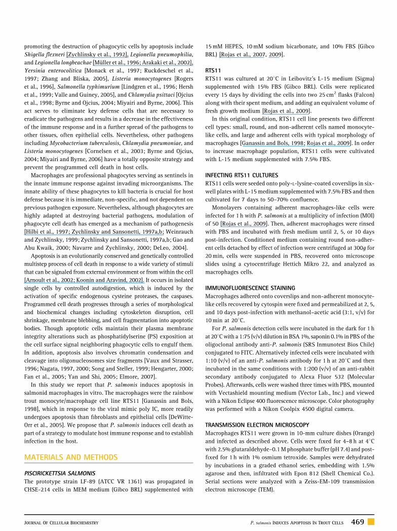
Fig. 1. Transmission electron micrographs of RTS11 macrophages with time after P. sal
magnification of an infected cell. Arrows indicate the presence of P. salmonis. Scale b
470 P. salmonis INDUCES APOPTOSIS IN TROUT CELLS
apoptosis by fluorescence-activated cell sorter (FACS) analysis in a
FACSort equipment (Becton Dickinson & Co). All growing,
infection, and fixation cell procedures were carried out at 5 days
post-infection as described previously. Infected, non-infected, and
actinomycin D induced cells were trypsinized, harvested by
centrifugation, washed once with ice-cold PBS, and resuspended
in binding buffer (140 mM NaCl, 5 mM CaCl2, 10 mM HEPES–Na, pH
7.4) to a concentration of 1� 106 cells/ml. Next, aliquots of 100,000
cells in 0.1 ml of binding buffer were transferred to a 5-ml FACS
tubes and incubated with 5ml of annexin V-FITC (BD Pharmingen
556420) and 50mg/ml of PI (Molecular Probes) for 15 min at room
temperature in the dark. Finally, additional 400ml of binding buffer
was added, and samples were analyzed within 30 min. All
experiments were performed three times in duplicate. Analysis
was performed using a WinMDi 2.8 software. The means and the SD
were calculated and statistical analysis was performed using the
non-parametric Mann–Whitney test for independent means. Results
described as different imply a significant difference from control,
P< 0.05.
RESULTS
MORPHOLOGICAL FEATURES OF APOPTOSIS IN INFECTED
MACROPHAGES
Macrophages infected with P. salmonis displayed morphological
changes characteristic of apoptosis including chromatin condensa-
tion at the nuclear boundary, fragmentation of the nucleus as well as
cytoplasm vacuolization, and apoptotic body formation (Fig. 1B–E).
In contrast, non-infected macrophages present a normal appearance
(Fig. 1A).
DNA FRAGMENTATION IN INFECTED MACROPHAGES
A significant number of infected macrophages showed fragmen-
ted DNA at different time points after infection, whereas a low
level of DNA fragmentation in non-infected RTS11 cells was
observed (Fig. 2A). The pattern of DNA cleavage in infected
macrophages was similar to that in cells treated with actinomycin D,
which is known to induce apoptosis [Jimenez et al., 2008]. By 2, 5,
and 10 days post-infection 25–32% of the nuclei were TUNEL-
positive (Fig. 2B).
monis infection. A: Non-infected, (B–D) 2, 5, and 10 days post-infection, (E): a higher
ar in A–D: 4.5mm; E: 3mm.
JOURNAL OF CELLULAR BIOCHEMISTRY
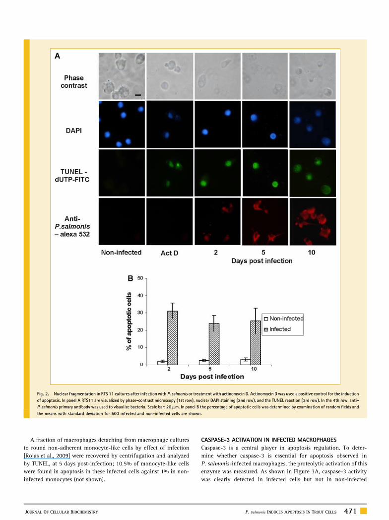
Fig. 2. Nuclear fragmentation in RTS 11 cultures after infection with P. salmonis or treatment with actinomycin D. Actinomycin D was used a positive control for the induction
of apoptosis. In panel A RTS11 are visualized by phase-contrast microscopy (1st row), nuclear DAPI staining (2nd row), and the TUNEL reaction (3rd row). In the 4th row, anti-
P. salmonis primary antibody was used to visualize bacteria. Scale bar: 20mm. In panel B the percentage of apoptotic cells was determined by examination of random fields and
the means with standard deviation for 500 infected and non-infected cells are shown.
A fraction of macrophages detaching from macrophage cultures
to round non-adherent monocyte-like cells by effect of infection
[Rojas et al., 2009] were recovered by centrifugation and analyzed
by TUNEL, at 5 days post-infection; 10.5% of monocyte-like cells
were found in apoptosis in these infected cells against 1% in non-
infected monocytes (not shown).
JOURNAL OF CELLULAR BIOCHEMISTRY
CASPASE-3 ACTIVATION IN INFECTED MACROPHAGES
Caspase-3 is a central player in apoptosis regulation. To deter-
mine whether caspase-3 is essential for apoptosis observed in
P. salmonis-infected macrophages, the proteolytic activation of this
enzyme was measured. As shown in Figure 3A, caspase-3 activity
was clearly detected in infected cells but not in non-infected
P. salmonis INDUCES APOPTOSIS IN TROUT CELLS 471
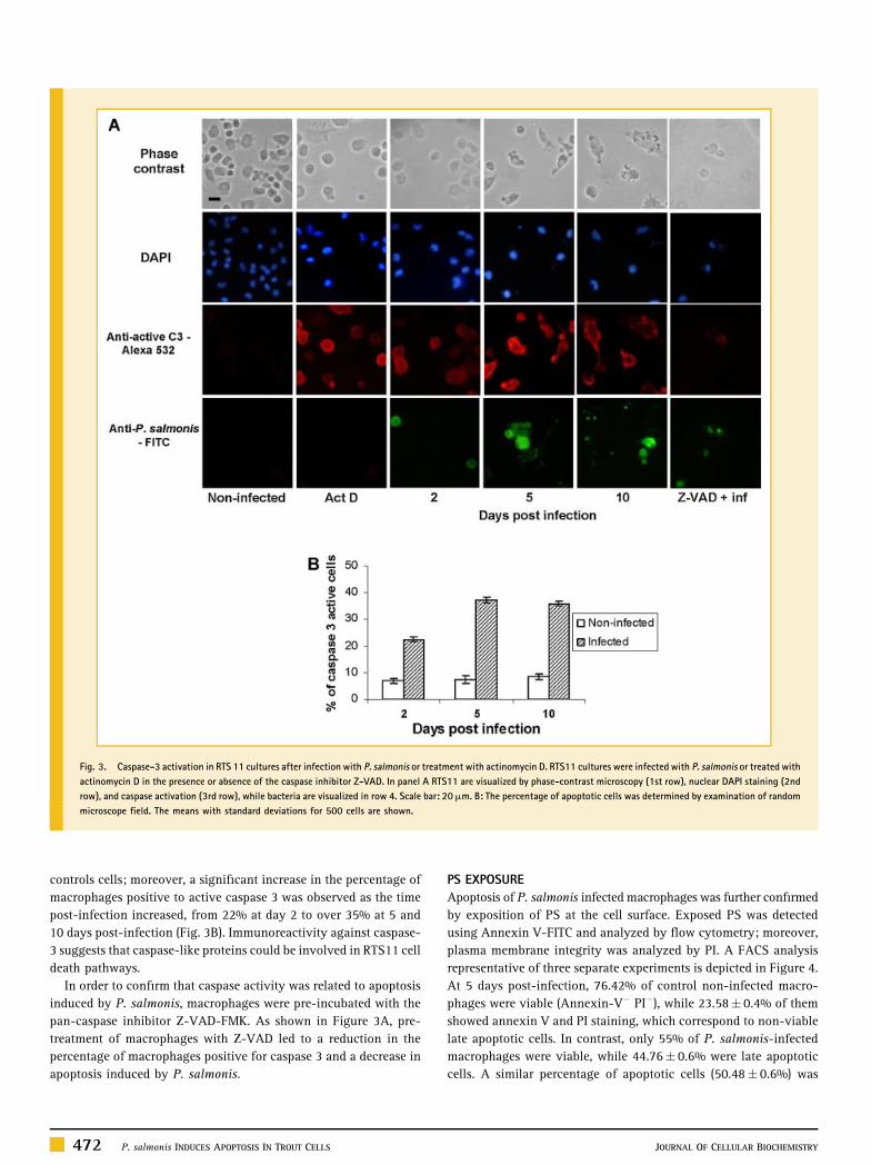
Fig. 3. Caspase-3 activation in RTS 11 cultures after infection with P. salmonis or treatment with actinomycin D. RTS11 cultures were infected with P. salmonis or treated with
actinomycin D in the presence or absence of the caspase inhibitor Z-VAD. In panel A RTS11 are visualized by phase-contrast microscopy (1st row), nuclear DAPI staining (2nd
row), and caspase activation (3rd row), while bacteria are visualized in row 4. Scale bar: 20mm. B: The percentage of apoptotic cells was determined by examination of random
microscope field. The means with standard deviations for 500 cells are shown.
controls cells; moreover, a significant increase in the percentage of
macrophages positive to active caspase 3 was observed as the time
post-infection increased, from 22% at day 2 to over 35% at 5 and
10 days post-infection (Fig. 3B). Immunoreactivity against caspase-
3 suggests that caspase-like proteins could be involved in RTS11 cell
death pathways.
In order to confirm that caspase activity was related to apoptosis
induced by P. salmonis, macrophages were pre-incubated with the
pan-caspase inhibitor Z-VAD-FMK. As shown in Figure 3A, pre-
treatment of macrophages with Z-VAD led to a reduction in the
percentage of macrophages positive for caspase 3 and a decrease in
apoptosis induced by P. salmonis.
472 P. salmonis INDUCES APOPTOSIS IN TROUT CELLS
PS EXPOSURE
Apoptosis of P. salmonis infected macrophages was further confirmed
by exposition of PS at the cell surface. Exposed PS was detected
using Annexin V-FITC and analyzed by flow cytometry; moreover,
plasma membrane integrity was analyzed by PI. A FACS analysis
representative of three separate experiments is depicted in Figure 4.
At 5 days post-infection, 76.42% of control non-infected macro-
phages were viable (Annexin-V� PI�), while 23.58� 0.4% of them
showed annexin V and PI staining, which correspond to non-viable
late apoptotic cells. In contrast, only 55% of P. salmonis-infected
macrophages were viable, while 44.76� 0.6% were late apoptotic
cells. A similar percentage of apoptotic cells (50.48� 0.6%) was
JOURNAL OF CELLULAR BIOCHEMISTRY
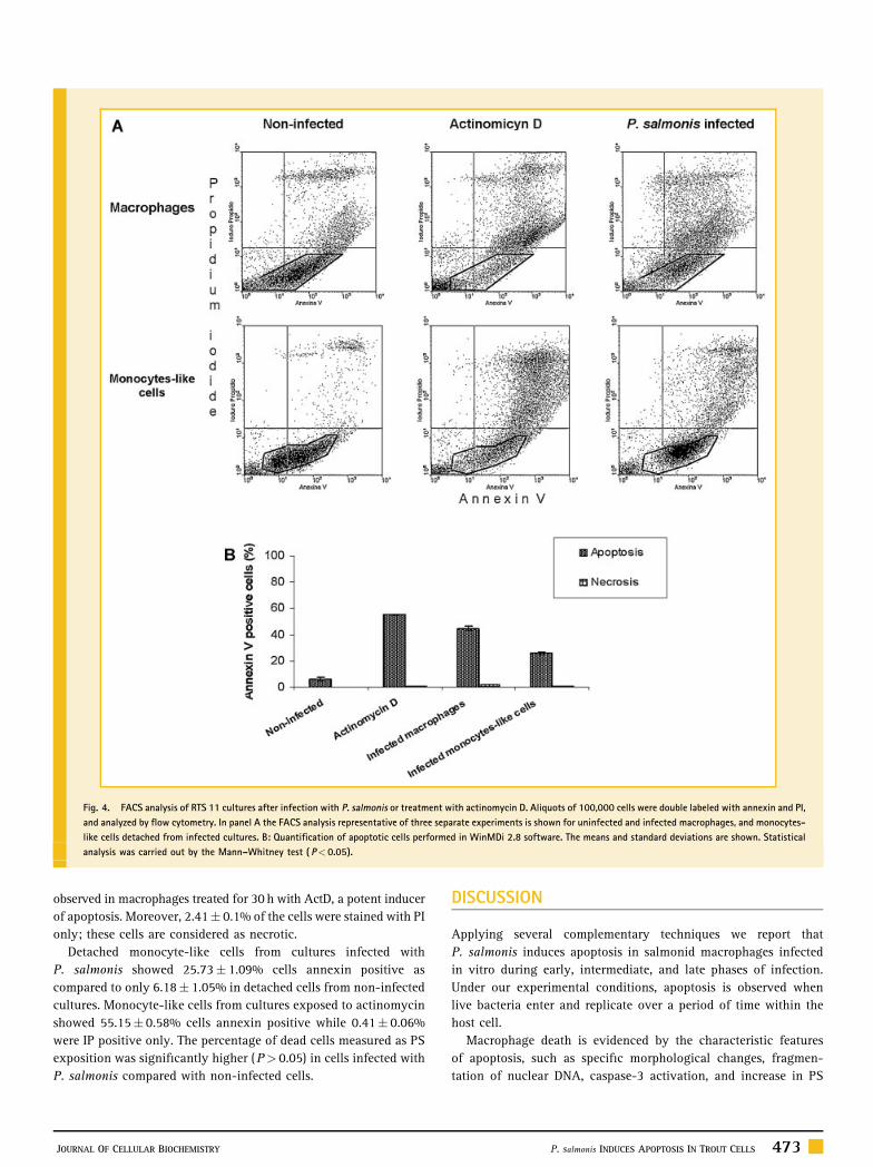
Fig. 4. FACS analysis of RTS 11 cultures after infection with P. salmonis or treatment with actinomycin D. Aliquots of 100,000 cells were double labeled with annexin and PI,
and analyzed by flow cytometry. In panel A the FACS analysis representative of three separate experiments is shown for uninfected and infected macrophages, and monocytes-
like cells detached from infected cultures. B: Quantification of apoptotic cells performed in WinMDi 2.8 software. The means and standard deviations are shown. Statistical
analysis was carried out by the Mann–Whitney test ( P< 0.05).
observed in macrophages treated for 30 h with ActD, a potent inducer
of apoptosis. Moreover, 2.41� 0.1% of the cells were stained with PI
only; these cells are considered as necrotic.
Detached monocyte-like cells from cultures infected with
P. salmonis showed 25.73� 1.09% cells annexin positive as
compared to only 6.18� 1.05% in detached cells from non-infected
cultures. Monocyte-like cells from cultures exposed to actinomycin
showed 55.15� 0.58% cells annexin positive while 0.41� 0.06%
were IP positive only. The percentage of dead cells measured as PS
exposition was significantly higher (P> 0.05) in cells infected with
P. salmonis compared with non-infected cells.
JOURNAL OF CELLULAR BIOCHEMISTRY
DISCUSSION
Applying several complementary techniques we report that
P. salmonis induces apoptosis in salmonid macrophages infected
in vitro during early, intermediate, and late phases of infection.
Under our experimental conditions, apoptosis is observed when
live bacteria enter and replicate over a period of time within the
host cell.
Macrophage death is evidenced by the characteristic features
of apoptosis, such as specific morphological changes, fragmen-
tation of nuclear DNA, caspase-3 activation, and increase in PS
P. salmonis INDUCES APOPTOSIS IN TROUT CELLS 473
externalization. Apoptosis induction by P. salmonis was
observed in both, infected macrophages and monocyte-like
cells. Surprisingly, in three independent experiments each one in
duplicate, non-infected trout macrophages also showed a
significant exposition of PS, while a parallel analysis of Vero
cells displayed the expected PS distribution of viable mammalian
cells. Interestingly, P. salmonis infection of the salmonid
epithelial cell line CHSE-214 did not cause apoptotic morpho-
logical changes or fragmentation of nuclear DNA (data not
shown); yet these cells and control CHSE-214 cells also showed
an significant exposition of PS, which may be an indicative of a
unique feature of healthy fish cells.
Interestingly, only 24–31% of infected macrophages show
apoptosis as detected by TUNEL and this agrees with 22–37% of
cells showing active caspase-3. These results suggest that the
induction of apoptosis by P. salmonis is restricted to some
monocytes/macrophages. An alternative explanation is that we
have measured apoptotic cells at specific days post-infection and
dead cells may further increase as a function of time. In agreement
with this, monocyte-like cells detached from infected RTS11
cultures also showed only 10–12% of apoptotic cells, both by
TUNEL and active caspase-3 (data not shown). Our hypothesis is
that the induction of apoptosis by P. salmonis in a fraction of the
macrophages allows the major part of the macrophage population
to be productively infected by the bacteria. These two outcomes
are not mutually exclusive and may be influenced by the
particular physiological state of both the pathogen and the
macrophage.
Several proteins produced by pathogenic bacteria are capable of
specifically initiating apoptosis in macrophages [Navarre and
Zychlinsky, 2000]. Moreover, there are a variety of reasons to
explain the particular susceptibility of macrophages to pathogen-
induced apoptosis. As for example, the expression of surface
receptors that recognize highly conserved bacterial components,
such as lipopolysaccharide (LPS) and bacterial lipoproteins, have
been shown to activate pro-apoptotic signaling pathways [Fukui
et al., 2008; Navarre and Zychlinsky, 2000].
In fish, apoptosis of macrophages due to bacterial infection has
not been reported. Nevertheless, viruses as IPNV induces apoptosis
in the zebrafish cell line (ZLE) [Hong et al., 2005; Santi et al., 2005]
and in the salmon cell line CHSE-214 [Hong and Wu, 2002].
Moreover, characteristic apoptotic DNA fragmentation was reported
in SHK-1 and CHSE 214 cells infected with ISAV [Joseph et al.,
2004]. By contrast, another fish virus, chum salmon reovirus (CSV),
caused apoptosis in CHSE-214 and RTG-2 but not in RTS11
[DeWitte-Orr and Bols, 2007].
The exact relevance of apoptotic cell death to the pathogenesis of
SRS is still undefined. Thus, in addition to other strategies to evade
host immune responses, such as enclosing inside vacuoles,
induction of macrophage apoptosis may represent a mechanism
by which P. salmonis escape the host immune response and establish
productive infections.
We propose that the ability of P. salmonis to promote early
apoptosis in a fraction of phagocytic cells may be important for
the initiation of infection. Bacteria survive and evade the host
immune response, allowing them to spread to other tissues,
474 P. salmonis INDUCES APOPTOSIS IN TROUT CELLS
which are targeted by the pathogen to establish a productive
infection.
Like other pathogens, P. salmonis efficiently induce apoptosis in
macrophages but not in epithelial cells [Navarre and Zychlinsky,
2000]. For example, the fungal toxin, gliotoxin, induced apoptosis
in the monocyte/macrophage cell line RTS11 but not in adherent
cells of the salmonid epithelium cell line CHSE-214 [DeWitte-Orr
and Bols, 2005]. Therefore, we infected CHSE-214 under the same
conditions as RTS11, but no clear evidence of apoptosis was seen as
judged by MET, TUNEL, caspase-3 activation, and PS exposition.
Probably P. salmonis has evolved pathways by which they can
selectively kill part of the macrophages, diminishing cellular attack,
while residing within a population of epithelial cells that are
resistant to apoptosis.
Further studies are necessary to understand the mechanism by
which P. salmonis triggers apoptosis. It is possible that the
bacteria either activates the macrophage intrinsic death program
or interferes with factors that inhibit the apoptosis program. As
other pathogenic bacteria, P. salmonis may produce toxins that
can induce macrophage apoptosis. Indeed we also detected cells
without positive signal for anti-P. salmonis antibody (non-
infected cells) that die through apoptosis. Nevertheless, this
observation may be explained by a lower loading of bacteria not
detectable by immunocytochemistry or that infected cells may
elaborate soluble factors that induce apoptosis of neighboring
non-infected cells.
The existence in P. salmonis of virulence factors analog to IpaB of
Shigella [Chen et al., 1996] or SipB of Salmonella [Hersh et al., 1999],
with structural and functional conserved mechanism to induce
apoptosis, or Yop(s) proteins of Yersinia [Mills et al., 1997] remains
to be elucidated. It has been demonstrated that a type III secretion
system is used by those and other Gram-negative pathogens to
deliver virulence factors to the intracellular compartment of
eukaryotic cells, which are required for the induction of apoptosis
in macrophages; nevertheless, this secretion system had not yet
described in P. salmonis.
In summary, we report for the first time that P. salmonis
induce apoptosis in salmon phagocytic cells but not in epithelial
cells. Further analysis in order to understand the molecular
mechanisms of apoptosis induced by this pathogen could be
important in the development of new potential approaches
against piscirickettsiosis.
ACKNOWLEDGMENTS
We thank Italo Costa for figures preparation. We thank NancyOlea and Marta Gacitua for technical assistance in electronicmicroscopy.
REFERENCES
Arakaki N, Higa F, Koide M, Tateyama M, Saito A. 2002. Induction ofapoptosis of human macrophages in vitro by Legionella longbeachaethrough activation of the caspase pathway. J Med Microbiol 51: 159–168.
JOURNAL OF CELLULAR BIOCHEMISTRY
Arnoult D, Akarid K, Grodet A, Petit PX, Estaquier J, Ameisen JC. 2002. Onthe evolution of programmed cell death: Apoptosis of the unicellulareukaryote Leishmania major involves cysteine proteinase activation andmitochondrion permeabilization. Cell Death Differ 9: 65–81.
Bravo S, Campos M. 1989. Coho salmon syndrome in Chile. Am Fish Soc FishHealth Sect News 17: 3.
Byrne GI, Ojcius DM. 2004. Chlamydia and apoptosis: Life and deathdecisions of an intracellular pathogen. Nat Rev Microbiol 2: 802–808.
Chen Y, Smith MR, Thirumalai K, Zychlinsky A. 1996. A bacterial invasininduces macrophages apoptosis by binding directly to ICE. EMBO J 15: 3853–3860.
Cornelsen MM, Tamm M, Glanville A, Perruchoud AP, Roth M. 2003.Chlamydia pneumoniae activates epithelial cell proliferation via NF-kband the glucocorticoid receptor. Infect Immun 71: 5814–5822.
Cvitanich JD, Garate NO, Smith CF. 1991. The isolation of a rickettsia-likeorganism causing disease and mortality in chilean salmonids and its con-firmation by Koch’s postulate. J Fish Dis 14: 121–145.
DeLeo FR. 2004. Modulation of phagocyte apoptosis by bacterial pathogens.Apoptosis 9: 399–413.
DeWitte-Orr SJ, Bols NC. 2005. Gliotoxin-induced cytotoxicity in threesalmonid cell lines: Cell death by apoptosis and necrosis. Comp BiochemPhysiol C 141: 157–167.
DeWitte-Orr SJ, Bols NC. 2007. Cytopathic effects of chum salmon reovirus tosalmonid epithelial, fibroblast and macrophage cell lines. Virus Res 126:159–171.
DeWitte-Orr SJ, Zorzitto JR, Sutton LP, Bols NC. 2005. Preferential inductionof apoptosis in the rainbow trout macrophage cell line, RTS11, by actino-mycin D, cycloheximide and double stranded RNA. Fish Shellfish Immunol18: 279–295.
Elmore S. 2007. Apoptosis: A review of programmed cell death. ToxicolPathol 35: 495–516.
Fan T-J, Han L-H, Cong R-S, Liang J. 2005. Caspase family proteases andapoptosis. Acta Bioch Biophys Sin 37: 719–727.
Fryer JL, Hedrick R. 2003. Piscirickettsia salmonis: A Gram-negative intra-cellular bacterial pathogen of fish. J Fish Dis 26: 251–262.
Fryer JL, Lannan CN, Garces LH, Larenas JJ, Smith PA. 1990. Isolation of arickettsiales-like organism from diseased coho salmon (Oncorhynchus) inChile. Fish Pathol 25: 107–114.
Fryer JL, Lannan CN, Giovannoni J, Wood ND. 1992. Piscirickettsia salmonisgen. Nov., sp. Nov., the causative agent of an epizootic disease in salmonidfishes. Int J Syst Bacteriol 42: 120–126.
Fukui M, Imamura R, Umemura M, Kawabe T, Suda T. 2008. Pathogen-associated molecular patterns sensitize macrophages to Fas ligand-inducedapoptosis and IL-1b release. J Immunol 171:1868–1874.
Ganassin RC, Bols NC. 1998. Development of a monocyte/macrophage-like cellline, RTS11, from rainbow trout spleen. Fish Shellfish Immunol 8: 457–476.
Gao Ll-Y, Abu Kwaik Y. 2000. The modulation of host cell apoptosis byintracellular bacterial pathogens. Trends Microbiol 8: 306–313.
Hengarter MO. 2000. The biochemistry of the apoptosis. Nature 407: 770–776.
Hersh D, Monack D, Smith M, Ghori N, Falkow S, Zychlinsky A. 1999. TheSalmonella invasin SipB induces macrophage apoptosis by binding tocaspase-1. Proc Natl Acad Sci USA 96: 2396–2401.
Hilbi H, Zychlinsky A, Sansonetti PJ. 1997. Macrophage apoptosis inmicrobial infections. Parasitology 115: S79–S87.
Hong J-R, Wu J-L. 2002. Induction of apoptotic death in cells via Bad geneexpression by infectious pancreatic necrosis virus infection. Cell Death Differ9: 113–124.
JOURNAL OF CELLULAR BIOCHEMISTRY
Hong J-R, Huang L-J, Wu J-L. 2005. Aquatic birnavirus induces apoptosisthrough activated caspase-8 and -3 in a zebrafish cell line. J Fish Dis 28: 133–140.
Jimenez V, Paredes R, Sosa MA, Galanti N. 2008. Natural programmed celldeath in T. cruzi epimastigotes maintained in axenic cultures. J Cell Biochem105: 688–698.
Joseph T, Cepica A, Brown L, Ikede BO, Kibenge FSB. 2004. Mechanism of celldeath during infectious salmon anemia virus infection is cell type-specific. JGen Virol 85: 3027–3036.
Koonin EV, Aravind L. 2002. Origin and evolution of eukaryotic apoptosis:The bacterial connection. Cell Death Diff 9: 394–404.
Lindgren SW, Stojiljkovic I, Heffron F. 1996. Macrophage killing is anessential virulence mechanism of Salmonella typhimurium. Proc Natl AcadSci USA 93: 4197–4201.
Mauel MJ, Miller DL. 2002. Piscirickettsiosis and piscirickettsiosis-likeinfections of fish: A review. Vet Microbiol 87: 279–289.
Mauel MJ, Ware C, Smith PA. 2008. Culture of Piscirickettsia salmonis onenriched blood agar. J Vet Diagn Invest 20: 213–214.
Mikalsen J, Skjaervik O, Wiik-Nielsen J, Wasmuth MA, Colquhoun DJ. 2008.Agar culture of Piscirickettsia salmonis, a serious pathogen of farmedsalmonid and marine fish. FEMS Microbiol Lett 278: 43–47.
Mills S, Boland A, Sory M-P, Van Der Smissen P, Kerbourch C, Finlay B,Cornelis G. 1997. Yersinia enterocolitica induces apoptosis in macrophagesby a process requiring functional type III secretion and translocationmechanisms and involving YopP, presumably acting as an effector protein.Proc Natl Acad Sci USA 94: 12638–12643.
Miyairi I, Byrne GI. 2006. Chlamydia and programmed cell death. Curr OpinMicrobiol 9: 102–108.
Monack D, Mecsas J, Ghori N, Falkow S. 1997. Yersinia signals macrophagesto undergo apoptosis and YopJ is necessary for this cell death. Proc Natl AcadSci USA 94: 10385–10390.
Muller A, Hacker J, Brand BC. 1996. Evidence for apoptosis of humanmacrophage-like HL-60cells by Legionella pneumophilia infection. InfectImmun 64: 4900–4906.
Nagata S. 1997. Apoptosis by death factor. Cell 88: 355–365.
Nagata S. 2000. Apoptotic DNA fragmentation. Exp Cell Res 256:12–18.
Navarre WW, Zychlinsky A. 2000. Pathogen-induced apoptosis of macro-phages: A common end for different pathogenic strategies. Cell Microbiol 2:265–273.
Ojcius D, Souque P, Perfettini J-L, Dautry-Varsat A. 1998. Apoptosis ofepithelial cells and macrophages due to infection with the obligate intra-cellular pathogen Chlamydia psittaci. J Immunol 161: 4220–4226.
Rogers HW, Callery MP, Deck B, Unanue ER. 1996. Listeria monocytogenesinduces apoptosis of infected hepatocytes. J Immunol 156: 679–684.
Rojas MV, Olivares J, Del Rıo R, Marshall SH. 2007. Characterization of anovel and genetically different small infective form of Piscirickettsia sal-monis. Microb Pathog 44: 370–378.
Rojas V, Galanti N, Bols NC, Marshall SH. 2009. Productive infection ofPiscirickettsia salmonis in macrophages and monocyte-like cells fromrainbow trout, a possible survival strategy. J Cell Biochem 108: 631–637.
Ruckdeschel KA, Roggenkamp V, Lafont P, Mangeat P, Heesemann J,Rouot B. 1997. Interaction of Yersinia enterocolitica with macrophagesleads to macrophage cell death through apoptosis. Infect Immun 65:4813–4821.
Santi N, Sandtro A, Sindre H, Song H, Hong H, Hong J-R, Thu B, Wu J-L,Vakharia VN, Evensen O. 2005. Infectious pancreatic necrosis virus inducesapoptosis in vitro and in vivo independent of VP5 expression. Virology 342:13–25.
P. salmonis INDUCES APOPTOSIS IN TROUT CELLS 475

Song Z, Steller H. 1999. Death by design: Mechanism and control ofapoptosis. Trends Cell Biol 9: 49–52.
Valle E, Guiney DG. 2005. Characterization of Salmonella-induced cell deathin human macrophage-like THP-1 cells. Infect Immun 73: 2835–2840.
Vaux DL, Strasser A. 1996. The molecular biology of apoptosis. Proc NatlAcad Sci USA 93: 2239–2244.
Weinrauch Y, Zychlinsky A. 1999. The induction of apoptosis by bacterialpathogens. Annu Rev Microbiol 53: 155–187.
Yan N, Shi Y. 2005. Mechanisms of apoptosis through structural biology.Annu Rev Cell Dev Biol 21: 35–56.
476 P. salmonis INDUCES APOPTOSIS IN TROUT CELLS
Zhang J, Bliska J. 2005. Role of macrophages apoptosis in the pathogenesis ofYersinia. Curr Topics Microbiol Immunol 289: 151–173.
Zychlinsky A, Sansonetti P. 1997a. Apoptosis as a proinflammatory event:What can we learn from bacteria-induced cell death? Trends Microbiol 5:201–204.
Zychlinsky A, Sansonetti P. 1997b. Apoptosis in bacterial pathogenesis. JClin Invest 100: 493–495.
Zychlinsky A, Prevost MC, Sansonetti P. 1992. Shigella flexneriinduces apoptosis in infected macrophages. Nature 358: 167–169.
JOURNAL OF CELLULAR BIOCHEMISTRY
Related Documents