FORUM ORIGINAL RESEARCH COMMUNICATION Oxidant Production, oxLDL Uptake, and CD36 Levels in Human Monocyte–Derived Macrophages Are Downregulated by the Macrophage-Generated Antioxidant 7,8-Dihydroneopterin Steven P. Gieseg, Zunika Amit, Ya-Ting Yang, Anastasia Shchepetkina, and Hanadi Katouah Abstract The severity of atheroma burden in patients strongly correlates to increasing levels of plasma neopterin, the oxidation product of 7,8-dihydroneopterin. Interferon-g stimulation of macrophages causes the synthesis of 7,8- dihydroneopterin, a potent antioxidant that inhibits oxidative damage to cells, and the cytotoxicity of oxidized low-density lipoprotein (oxLDL) to monocyte-like U937 cells but not THP-1 cells. With human monocyte– derived macrophages (HMDMs), oxLDL triggered a large oxidative stress, causing the rapid loss of cellular glutathione, glyceradehyde-3-phosphate dehydrogenase (GAPDH) inhibition, and eventual loss of viability without caspase-3 activation. Inhibition of oxLDL cytotoxicity to HMDMs occurred at 7,8-dihydroneopterin concentrations >100 mM. The oxLDL-mediated glutathione loss and GAPDH inactivation was inhibited by 7,8- dihydroneopterin. 7,8-Dihydroneopterin rapidly entered the HMDMs, suggesting that much of the protective effect was scavenging of intracellular oxidants generated in response to oxLDL. OxLDL uptake by HMDMs was reduced by 30% by 7,8-dihydroneopterin. Immunoblot analysis suggests that this decrease in oxLDL uptake was due to a significant downregulation in the levels of CD36. These results imply that 7,8-dihydroneopterin protects human macrophages both by scavenging oxidants generated in response to oxLDL and by decreasing CD36- mediated uptake of oxLDL. Antioxid. Redox Signal. 13, 1525–1534. Introduction I ncreased clinical use of plasma neopterin is seen as a marker of a patient’s level of inflammation in conditions as diverse as cancer and tuberculosis infection. Neopterin is the oxidation product of 7,8-dihydroneopterin, a pterin synthe- sized by macrophages when stimulated by g-interferon re- leased by Th-1 cells (17, 52). The increase of plasma neopterin levels has been strongly correlated with increased athero- sclerotic burden (1, 43, 47). Significant amounts of neopterin and 7,8-dihydroneopterin have been reported in the interior of an atherosclerotic plaque (13, 17), which represents local- ized sites of chronic inflammation (36). Although neopterin is a sensitive marker of inflammation, it is becoming increas- ingly apparent that 7,8-dihydroneopterin and neopterin have distinct biologic properties. They may have significant influ- ence on the inflammatory process, especially in chronic in- flammation, such as atherosclerosis. In vitro studies have shown that 7,8-dihydroneopterin is a potent antioxidant that can protect a range of biomolecules and cells from various oxidants, including hydroxyl and peroxyl radicals (21, 24, 25, 31). Oxidized low-density lipo- protein (OxLDL) formation with copper, peroxyl, or cellular oxidants is effectively inhibited by scavenging of the chain carrying lipid-peroxyl radical within the LDL particle by 7,8- dihydroneopterin (13, 15, 23, 24). 7,8-Dihydroneopterin ac- celerates copper-mediated oxidation if added after the lag phase because of increased metal reduction (26). 7,8-Dihydroneopterin is effective in protecting the mono- cyte-like U937 cells from the cytotoxic effects of oxidized low- density lipoprotein (oxLDL) (5). Cell death due to oxLDL cytotoxicity is considered to be a major factor in the develop- ment of a necrotic core in advanced atherosclerotic plaques (28, 30). Our previous studies have shown 7,8-dihydroneopterin protects U937 cells from oxLDL by preventing the loss of intracellular glutathione and the initiation of necrosis (5). Free Radical Biochemistry Laboratory, School of Biological Sciences, University of Canterbury, Christchurch, New Zealand. ANTIOXIDANTS & REDOX SIGNALING Volume 13, Number 10, 2010 ª Mary Ann Liebert, Inc. DOI: 10.1089=ars.2009.3065 1525

Welcome message from author
This document is posted to help you gain knowledge. Please leave a comment to let me know what you think about it! Share it to your friends and learn new things together.
Transcript

FORUM ORIGINAL RESEARCH COMMUNICATION
Oxidant Production, oxLDL Uptake, and CD36 Levelsin Human Monocyte–Derived Macrophages
Are Downregulated by the Macrophage-GeneratedAntioxidant 7,8-Dihydroneopterin
Steven P. Gieseg, Zunika Amit, Ya-Ting Yang, Anastasia Shchepetkina, and Hanadi Katouah
Abstract
The severity of atheroma burden in patients strongly correlates to increasing levels of plasma neopterin, theoxidation product of 7,8-dihydroneopterin. Interferon-g stimulation of macrophages causes the synthesis of 7,8-dihydroneopterin, a potent antioxidant that inhibits oxidative damage to cells, and the cytotoxicity of oxidizedlow-density lipoprotein (oxLDL) to monocyte-like U937 cells but not THP-1 cells. With human monocyte–derived macrophages (HMDMs), oxLDL triggered a large oxidative stress, causing the rapid loss of cellularglutathione, glyceradehyde-3-phosphate dehydrogenase (GAPDH) inhibition, and eventual loss of viabilitywithout caspase-3 activation. Inhibition of oxLDL cytotoxicity to HMDMs occurred at 7,8-dihydroneopterinconcentrations >100 mM. The oxLDL-mediated glutathione loss and GAPDH inactivation was inhibited by 7,8-dihydroneopterin. 7,8-Dihydroneopterin rapidly entered the HMDMs, suggesting that much of the protectiveeffect was scavenging of intracellular oxidants generated in response to oxLDL. OxLDL uptake by HMDMs wasreduced by 30% by 7,8-dihydroneopterin. Immunoblot analysis suggests that this decrease in oxLDL uptake wasdue to a significant downregulation in the levels of CD36. These results imply that 7,8-dihydroneopterin protectshuman macrophages both by scavenging oxidants generated in response to oxLDL and by decreasing CD36-mediated uptake of oxLDL. Antioxid. Redox Signal. 13, 1525–1534.
Introduction
Increased clinical use of plasma neopterin is seen as amarker of a patient’s level of inflammation in conditions as
diverse as cancer and tuberculosis infection. Neopterin is theoxidation product of 7,8-dihydroneopterin, a pterin synthe-sized by macrophages when stimulated by g-interferon re-leased by Th-1 cells (17, 52). The increase of plasma neopterinlevels has been strongly correlated with increased athero-sclerotic burden (1, 43, 47). Significant amounts of neopterinand 7,8-dihydroneopterin have been reported in the interiorof an atherosclerotic plaque (13, 17), which represents local-ized sites of chronic inflammation (36). Although neopterin isa sensitive marker of inflammation, it is becoming increas-ingly apparent that 7,8-dihydroneopterin and neopterin havedistinct biologic properties. They may have significant influ-ence on the inflammatory process, especially in chronic in-flammation, such as atherosclerosis.
In vitro studies have shown that 7,8-dihydroneopterin is apotent antioxidant that can protect a range of biomoleculesand cells from various oxidants, including hydroxyl andperoxyl radicals (21, 24, 25, 31). Oxidized low-density lipo-protein (OxLDL) formation with copper, peroxyl, or cellularoxidants is effectively inhibited by scavenging of the chaincarrying lipid-peroxyl radical within the LDL particle by 7,8-dihydroneopterin (13, 15, 23, 24). 7,8-Dihydroneopterin ac-celerates copper-mediated oxidation if added after the lagphase because of increased metal reduction (26).
7,8-Dihydroneopterin is effective in protecting the mono-cyte-like U937 cells from the cytotoxic effects of oxidized low-density lipoprotein (oxLDL) (5). Cell death due to oxLDLcytotoxicity is considered to be a major factor in the develop-ment of a necrotic core in advanced atherosclerotic plaques (28,30). Our previous studies have shown 7,8-dihydroneopterinprotects U937 cells from oxLDL by preventing the loss ofintracellular glutathione and the initiation of necrosis (5).
Free Radical Biochemistry Laboratory, School of Biological Sciences, University of Canterbury, Christchurch, New Zealand.
ANTIOXIDANTS & REDOX SIGNALINGVolume 13, Number 10, 2010ª Mary Ann Liebert, Inc.DOI: 10.1089=ars.2009.3065
1525
Although oxLDL itself is not capable of directly generatingoxidants, exposure to U937 cells causes a large intracellularoxidant flux, which can be scavenged or inhibited by 7,8-dihydroneopterin.
Surprisingly, 7,8-dihydroneopterin cannot protect the re-lated monocyte-like THP-1 cells from oxLDL-induced cyto-toxicity (5). In THP-1 cells, oxLDL triggers caspase activationand apoptosis without the loss of glutathione (4). This non-oxidative stress mechanism in THP-1 cells does not appear tobe inhibited by 7,8-dihydroneopterin. OxLDL-induced celldeath occurs through a number of mechanisms, depending onthe type of cell under study and the level to which the LDL hasoxidized (22). The proposed mechanisms include calpainactivation through oxysterol-induced alterations in lipid rafts–associated calcium channels (6), dephosphorylation and inhi-bition of protein kinase B, leading to mitochondrial dysfunctionthrough Bcl-2 and MAPK (9, 45), lysozyme destabilization (7,34), and CD36-linked NADPH oxidase activation (33, 37).
Human macrophages derived from peripheral bloodmonocytes, usually described as human monocyte–derivedmacrophages (HMDMs) have been described undergoingboth caspase and caspase-independent cell death (3, 22, 53).We have observed oxLDL causing HMDMs to undergo acaspase-independent cell death characterized by a rapid lossof glutathione, caspase inactivation, and the appearance ofnecrotic cell morphology (22). In this study, we examinedwhether 7,8-dihydroneopterin can inhibit oxLDL-induceddeath in HMDMs and whether the mechanism involves thereduction of intracellular oxidative stress, as observed withU937 cells.
Materials and Methods
All reagents and chemicals were AR grade or better andobtained from either the Sigma Chemical Company (St. Louis,MO) or BDH Chemicals New Zealand Limited. Solutionswere prepared by using high-purity water generated from anNANOpure ultrapure water system from Barnstead=Thermolyne (Dubuque, IA). Cells were grown in Nunc platessupplied by In Vitro New Zealand Ltd., and the remainingplastic ware was supplied by Greiner Bio-one through RaylabNew Zealand Ltd. Neopterin and 7,8-dihydroneopterin wereobtained from Schirck’s Laboratories, Jona, Switzerland.Phosphate-buffered saline (PBS) solution consisted of 150 mMsodium chloride and 10 mM sodium phosphate, pH 7.4.
LDL was purified by buoyant density-gradient ultracen-trifugation by using a Beckman SW41 rotor from EDTA-treated plasma collected by venipuncture from healthy maleand female donors after an overnight fast, as previously de-scribed (5, 10, 23). LDL concentration (total mass) was deter-mined by enzymatic cholesterol assay with the Chol MPR 2 kitsupplied by Roche Chemicals (Bern, Switzerland) (20).Heavily oxidized oxLDL with the properties previously de-scribed (5) was prepared by incubating LDL at a concentra-tion of 3 mg=ml (600mg=ml protein) with 400 mM CuCl2solution for 24 h at 378C before stirring with chelex-100 resinfor 2 h to remove copper ions. The oxLDL was concentratedby using a Vivapore membrane concentrator (Millipore,Billerica, MA) before filter sterilization through a 0.22-mmmembrane filter and then stored at 48C.
Human monocyte–derived macrophages (HMDMs) wereprepared from whole blood donated by clinically healthy
hemochromatosis patients at the NZ Blood Services(Christchurch) under ethics approval CTY=98=07=069 gran-ted by the New Zealand Upper South B Ethics Committee.The monocytes were purified by centrifugation over Lym-phoprep and differentiated into adherent macrophages inadherent 12-well plates (14). The cells were maintained inRPMI 1640 supplemented with 10% heat-inactivated humanserum plus 100 U=ml penicillin and 100 mg=ml streptomycin.Cell viability was determined by MTT reduction (38). Cellprotein concentration was determined by using the BCAProtein Determination kit supplied by Pierce, (Rockford, IL)with bovine serum albumin as the standard.
Cellular glutathione levels were measured by deriva-tion with monobromobimane and reverse-phase C18 high-performance liquid chromatography (HPLC) analysis ofthe fluorescent derivative by using monobromobimane-derivatized GSH as a standard (11). Neopterin and 7,8-dihydroneopterin were measured with reverse-phase HPLCanalysis of cell lysates after removal of proteins by precipi-tation with trichloroacetic acid (13, 21). GAPDH activity wasmeasured in cell lysates by monitoring the rate of conversionof NADPþ to NADPH in the presence of glyceraldehyde-3-phosphate over a 4-min period (49). OxLDL uptake byHMDMs was measured by using DiI-labelled oxLDL (12, 50).The DiI uptake was measured by lysing cells into isopropanoland measuring the native DiI fluorescence intensity. The cel-lular protein concentration was determined by analysis of cellpellets collected after centrifugation of the isopropanol. In-tracellular superoxide release was detected with dihy-droethidium (DHE) staining (8) of cells grown on sterile glasscoverslips. The cells were incubated for 20 min with DHEbefore examination with fluorescence microscopy.
Caspase-3, CD36, and scavenger receptor-A (SCR-A) weredetected by immunoblotting after SDS-PAGE electrophoresiscells were harvested by scraping in lysis buffer (40 mMHEPES, 50 mM NaCl, 1 mM EDTA, and 1 mM EGTA, plus onetablet of Roche Complete mini protease inhibitor per 10 ml).Immunoblots were probed with either anti-CD36 rabbitpolyclonal (NB400-145, Novus Biologicals, Cambridge, UK),diluted 1:1,000, followed by peroxidase-conjugated goatpolyclonal anti-rabbit IgG (NB730-H, Novus Biologicals) at1:2,000 dilution; or anti-caspase-3 (E-8) (SC-7272, Santa CruzBiotechnology, Santa Cruz, CA) followed by conjugated goatanti-mouse IgG (Fc) (31434; Pierce Biotechnology); or anti-SR-A (I-20) (SC-20441, Santa Cruz Biotechnology) followed byperoxidase-conjugated donkey anti-goat IgG (SC-2020, SantaCruz Biotechnology). Lane loading was accessed by reblottingwith anti-b-actin (A5316, Sigma-Aldrich Chemical, St. Louis,MO) followed by peroxidase-conjugated sheep anti-mouseIgG (RPN4401, Amersham Biosciences, Amersham, England).Immunoblots were visualised by using Supersignal WestDura chemiluminescence from Pierce. The image was re-corded on a Syngene Chemigenius-2 bioimaging system byusing Genesnap software (Global, Aotearoa, NZ).
Phosphatidylserine (PS) translocation to the cell surface as amarker of apoptosis was measured by staining cells grown oncoverslips with annexin-V-FITC by using the Annexin VApoptosis Kit form Santa Cruz Biotechnology and viewing byfluorescence microscope by using Zeiss AxioImager.M1 epi-fluorescent microscope.
The data were analyzed by using the Prism softwarepackage supplied by Graphpad Software Inc. (San Diego,
1526 GIESEG ET AL.
CA). Comparisons among treatments were performed byusing one-way analysis of variance (ANOVA). All results areexpressed as the mean� SD of triplicate treatments. Resultsshown are from single experiments, representative of a min-imum of three. Where appropriate, significance is indicated as(***)p� 0.001; (**)p� 0.01, or (*)p� 0.05.
Results
OxLDL induced cell death in the HMDMs only after 12 h ofincubation (Fig. 1A). Up to the 12-h time point, the cell mor-phology appeared normal, but after this, increasing numbersof lysed cells were observed. The appearance of lysed cellscorresponded with the loss of cell viability, as measured bythe ability to reduce MTT to the purple formazan product. TheoxLDL-induced loss of viability was significantly inhibitedby 50 mM 7,8-dihydroneopterin added 10 min before theoxLDL treatment (Fig. 1B). One hundred micromolar 7,8-dihydroneopterin reduced the loss of viability to a mean valueof 20%, with the majority of the cells displaying normalmorphology.
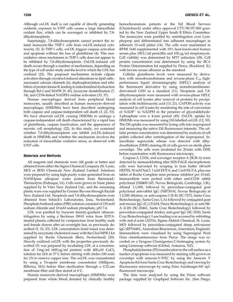
To understand the nature of the 7,8-dihydroneopterinprotection, the mechanism of the oxLDL-induced death in theHMDMs was further examined, as conflicting reports in theliterature exist on the oxLDL-induced death mechanism (22).Western blot analysis constantly failed to show any sign ofproteolytic activation of caspase-3 in the oxLDL-treatedHMDMs. The 32-kDa band of procaspase-3 was clearly seen(Fig. 2A), but no active 17- or 20-kDa band was evident. Thisconfirms our earlier investigations, in which no caspase en-zyme activity was observed with oxLDL exposure (22).Phosphatidylserine externalization in the plasma membraneappeared to increase, as indicated by staining with annexin-Vafter 3 and 6 h after the treatment with oxLDL (Fig. 2B–E). Thisprocess was significantly slowed and reduced in the presenceof 7,8-dihydroneopterin (Fig. 2F–H). Although the effectorcaspase-3 appears not to be activated by oxLDL exposure,phosphatidylserine translocation, another classic marker ofapoptosis, occurs.
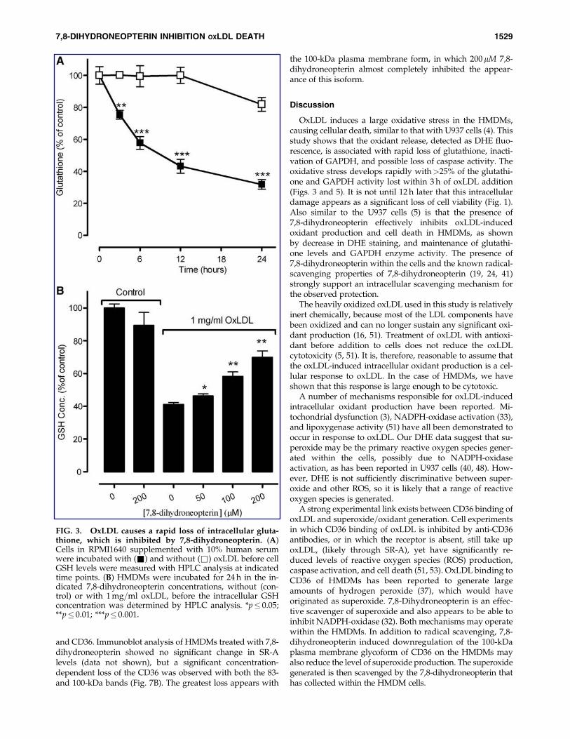
We previously observed the loss of caspase activity duringoxLDL-induced death of U937 cells (4). This was also accom-panied by a large loss of glutathione, suggesting inhibition ofcaspase-3 activity through oxidative loss of key thiols withinthe enzyme. With the HMDMs, we observed a similar and veryrapid loss of glutathione (Fig. 3A). Within 6 h of adding theoxLDL, nearly 50% of the glutathione was lost, with the leveldropping to 36% of the control by 24 h. Generally, no signifi-cant loss was observed in the absence of oxLDL, although asmall loss was observed at 24 h in this experiment. As withprevious studies using 7,8-dihydroneopterin on macrophage-like cells (5, 25), addition of 7,8-dihydroneopterin alone had nosignificant effect on the levels of glutathione, but in the pres-ence of oxLDL, a dose-dependent protection of the glutathionelevels was noted at the end of a 24-h incubation (Fig. 3B). The200mM 7,8-dihydroneopterin maintained the glutathione levelat 68% of the control value.
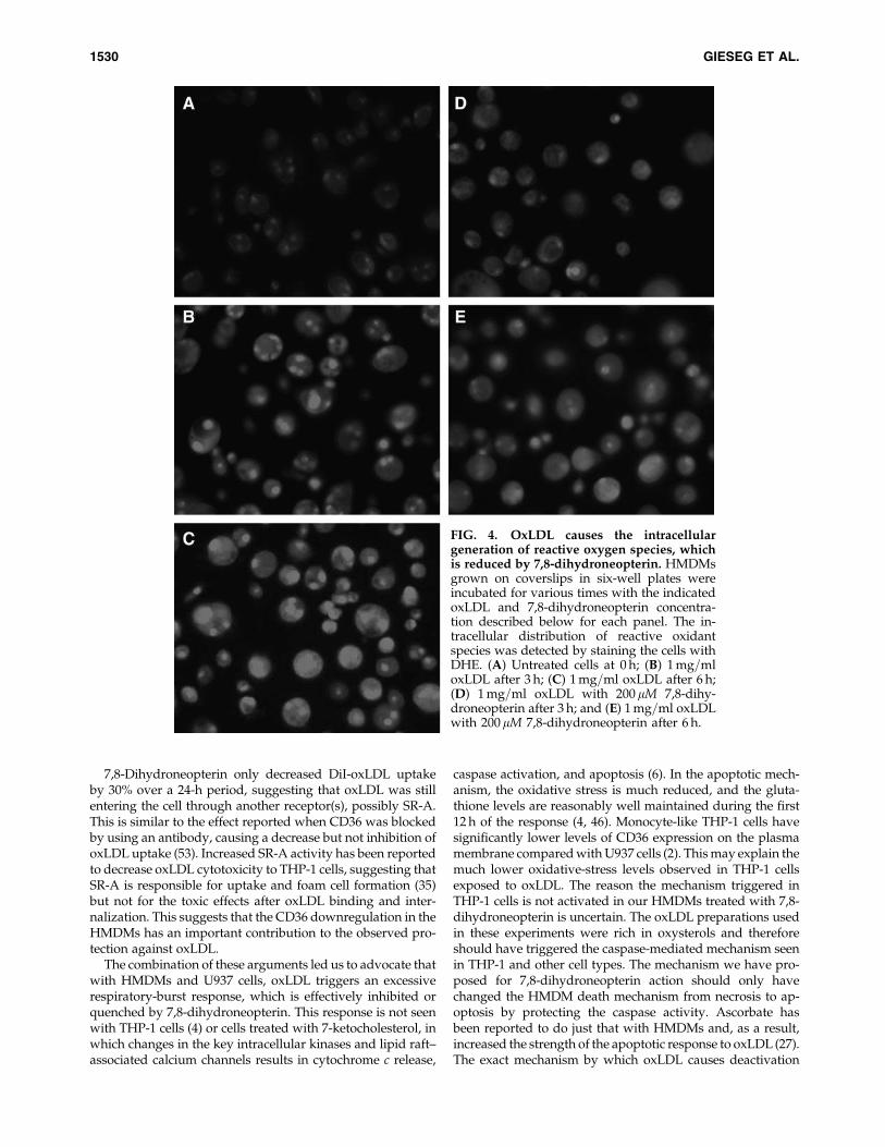
Based on studies with monocyte-like U937 cells (4, 29) andendothelial cells (51), we suspect that oxLDL induced theHMDM cells to generate reactive oxidants that depletedthe intracellular glutathione levels. Treatment of cells with thefluorescent dye DHE confirmed that oxLDL was inducing anoxidative stress over the first 6 h (Fig. 4A–C), which was re-
duced by the addition of 7,8-dihydroneopterin (Fig. 4D andE). This suggested that the 7,8-dihydroneopterin was eitherinhibiting the oxidant production or scavenging the oxidantdirectly.
Like the caspases, the glycolytic enzyme glyceraldehyde3-phosphate dehydrogenase (GAPDH) has a critical thiolwithin its active site that, when oxidized, results in the loss of
FIG. 1. OxLDL loss of HMDM cell viability is inhibitedby 7,8-dihydroneopterin. (A) HMDMs were incubated with1 mg=ml oxLDL in RPMI-1640 supplemented with 10%human serum. At indicated time points, the medium wasremoved, and the cell viability was determined by measur-ing MTT reduction. (B) Increasing concentrations of 7,8-dihydroneopterin inhibited oxLDL-induced loss of HMDMcell viability during a 24-h incubation with 1 mg=ml oxLDL.The 7,8-dihydroneopterin was added 10 min before the oxLDL.*p� 0.05; **p� 0.01; ***p� 0.001.
7,8-DIHYDRONEOPTERIN INHIBITION OXLDL DEATH 1527
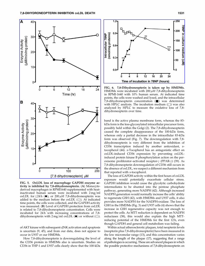
enzymatic activity. In HMDMs, oxLDL was observed to causea rapid reduction of GAPDH activity, which paralleled theloss of glutathione (Fig. 5A). Within 6 h of oxLDL addition, theGAPDH activity had been reduced to 50% of the control. Noloss in GAPDH activity was observed in the absence ofoxLDL or in the presence of only 7,8-dihydroneopterin. At50 mM 7,8-dihydroneopterin, a small but statistically signifi-cant protection of the GAPDH activity steadily increased with7,8-dihydroneopterin concentration. 7,8-Dihydroneopterin at200 mM completely prevented the oxLDL-induced loss ofGAPDH activity (Fig. 5B).
The data strongly suggest that 7,8-dihydroneopterin maybe scavenging oxidants generated by the cell in responseto the oxLDL binding or internalization. Such a mode ofactivity would require 7,8-dihydroneopterin to enter thecells. This was examined by incubating HMDMs with 7,8-dihydroneopterin and analyzing cell lysates at various timepoints. A gradual loss of 7,8-dihydroneopterin was observedin the media, with the concentration decreasing by 50% over a24-h period (Fig. 6). This loss is due to oxidation of the 7,8-
dihydroneopterin to 7,8-dihydroxanthopterin and neopterin(13, 18). Intracellularly, the 7,8-dihydroneopterin level in-creased rapidly, with half the maximum concentrationreached within 4 h and maximum at 12 h (Fig. 6). No signifi-cant loss of intracellular 7,8-dihydroneopterin was noted overthe following 12-h period, even though the media concen-tration dropped by 25%. As the cells were not stimulated withg-interferon, the intracellular generation was negligible.
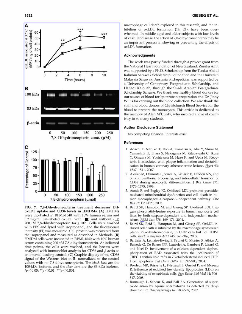
Although intracellular oxidant scavenging by 7,8-dihydroneopterin appeared to explain the cellular protectionfrom oxLDL, reduced uptake of oxLDL into the cells was alsoexplored. By using DiI-labeled oxLDL, we measured oxLDLuptake by HMDMs and found that 7,8-dihydroneopterin re-duced the uptake, but only by a third when compared withcells without 7,8-dihydroneopterin (Fig. 7A). The concentra-tion of oxLDL used was below the cytotoxic concentration,thus removing the compounding factor of cell death and lysisfrom the analysis. A simple explanation for this decrease inoxLDL uptake is downregulation in the levels of the mainscavenger receptors of oxLDL, scavenger receptor-A (SR-A)
FIG. 2. OxLDL does not activate HMDM cell caspase-3 but causes phosphatidylserine translocation to the plasmamembrane, which is inhibited by 7,8-dihydroneopterin. (A) HMDMs were incubated under various conditions beforewashing, and cell lysates were collected for immunoblot staining for caspase-3 activation and b-actin as a loading control.Lane 1: control cells at zero time; lane 2: with 1 mg=ml oxLDL for 12 h; lane 3: with 2 mg=ml oxLDL for 12 h; lane 4: with1 mg=ml oxLDL for 24 h; lane 5: with 2 mg=ml oxLDL for 24 h. Cells were grown on coverslips before exposure to 1 mg=mloxLDL for 0 h. (B) 3 h (C, F), 6 h (D, G), and 12 h (E–H) with (F–H) or without 200mM 7,8-dihydroneopterin (C–E). Phos-phatidylserine exposure on the cells surface was visualized by staining with annexin-V under fluorescence microscopy.
1528 GIESEG ET AL.
and CD36. Immunoblot analysis of HMDMs treated with 7,8-dihydroneopterin showed no significant change in SR-Alevels (data not shown), but a significant concentration-dependent loss of the CD36 was observed with both the 83-and 100-kDa bands (Fig. 7B). The greatest loss appears with
the 100-kDa plasma membrane form, in which 200 mM 7,8-dihydroneopterin almost completely inhibited the appear-ance of this isoform.
Discussion
OxLDL induces a large oxidative stress in the HMDMs,causing cellular death, similar to that with U937 cells (4). Thisstudy shows that the oxidant release, detected as DHE fluo-rescence, is associated with rapid loss of glutathione, inacti-vation of GAPDH, and possible loss of caspase activity. Theoxidative stress develops rapidly with>25% of the glutathi-one and GAPDH activity lost within 3 h of oxLDL addition(Figs. 3 and 5). It is not until 12 h later that this intracellulardamage appears as a significant loss of cell viability (Fig. 1).Also similar to the U937 cells (5) is that the presence of7,8-dihydroneopterin effectively inhibits oxLDL-inducedoxidant production and cell death in HMDMs, as shownby decrease in DHE staining, and maintenance of glutathi-one levels and GAPDH enzyme activity. The presence of7,8-dihydroneopterin within the cells and the known radical-scavenging properties of 7,8-dihydroneopterin (19, 24, 41)strongly support an intracellular scavenging mechanism forthe observed protection.
The heavily oxidized oxLDL used in this study is relativelyinert chemically, because most of the LDL components havebeen oxidized and can no longer sustain any significant oxi-dant production (16, 51). Treatment of oxLDL with antioxi-dant before addition to cells does not reduce the oxLDLcytotoxicity (5, 51). It is, therefore, reasonable to assume thatthe oxLDL-induced intracellular oxidant production is a cel-lular response to oxLDL. In the case of HMDMs, we haveshown that this response is large enough to be cytotoxic.
A number of mechanisms responsible for oxLDL-inducedintracellular oxidant production have been reported. Mi-tochondrial dysfunction (3), NADPH-oxidase activation (33),and lipoxygenase activity (51) have all been demonstrated tooccur in response to oxLDL. Our DHE data suggest that su-peroxide may be the primary reactive oxygen species gener-ated within the cells, possibly due to NADPH-oxidaseactivation, as has been reported in U937 cells (40, 48). How-ever, DHE is not sufficiently discriminative between super-oxide and other ROS, so it is likely that a range of reactiveoxygen species is generated.
A strong experimental link exists between CD36 binding ofoxLDL and superoxide=oxidant generation. Cell experimentsin which CD36 binding of oxLDL is inhibited by anti-CD36antibodies, or in which the receptor is absent, still take upoxLDL, (likely through SR-A), yet have significantly re-duced levels of reactive oxygen species (ROS) production,caspase activation, and cell death (51, 53). OxLDL binding toCD36 of HMDMs has been reported to generate largeamounts of hydrogen peroxide (37), which would haveoriginated as superoxide. 7,8-Dihydroneopterin is an effec-tive scavenger of superoxide and also appears to be able toinhibit NADPH-oxidase (32). Both mechanisms may operatewithin the HMDMs. In addition to radical scavenging, 7,8-dihydroneopterin induced downregulation of the 100-kDaplasma membrane glycoform of CD36 on the HMDMs mayalso reduce the level of superoxide production. The superoxidegenerated is then scavenged by the 7,8-dihydroneopterin thathas collected within the HMDM cells.
FIG. 3. OxLDL causes a rapid loss of intracellular gluta-thione, which is inhibited by 7,8-dihydroneopterin. (A)Cells in RPMI1640 supplemented with 10% human serumwere incubated with (&) and without (&) oxLDL before cellGSH levels were measured with HPLC analysis at indicatedtime points. (B) HMDMs were incubated for 24 h in the in-dicated 7,8-dihydroneopterin concentrations, without (con-trol) or with 1 mg=ml oxLDL, before the intracellular GSHconcentration was determined by HPLC analysis. *p� 0.05;**p� 0.01; ***p� 0.001.
7,8-DIHYDRONEOPTERIN INHIBITION OXLDL DEATH 1529
7,8-Dihydroneopterin only decreased DiI-oxLDL uptakeby 30% over a 24-h period, suggesting that oxLDL was stillentering the cell through another receptor(s), possibly SR-A.This is similar to the effect reported when CD36 was blockedby using an antibody, causing a decrease but not inhibition ofoxLDL uptake (53). Increased SR-A activity has been reportedto decrease oxLDL cytotoxicity to THP-1 cells, suggesting thatSR-A is responsible for uptake and foam cell formation (35)but not for the toxic effects after oxLDL binding and inter-nalization. This suggests that the CD36 downregulation in theHMDMs has an important contribution to the observed pro-tection against oxLDL.
The combination of these arguments led us to advocate thatwith HMDMs and U937 cells, oxLDL triggers an excessiverespiratory-burst response, which is effectively inhibited orquenched by 7,8-dihydroneopterin. This response is not seenwith THP-1 cells (4) or cells treated with 7-ketocholesterol, inwhich changes in the key intracellular kinases and lipid raft–associated calcium channels results in cytochrome c release,
caspase activation, and apoptosis (6). In the apoptotic mech-anism, the oxidative stress is much reduced, and the gluta-thione levels are reasonably well maintained during the first12 h of the response (4, 46). Monocyte-like THP-1 cells havesignificantly lower levels of CD36 expression on the plasmamembrane compared with U937 cells (2). This may explain themuch lower oxidative-stress levels observed in THP-1 cellsexposed to oxLDL. The reason the mechanism triggered inTHP-1 cells is not activated in our HMDMs treated with 7,8-dihydroneopterin is uncertain. The oxLDL preparations usedin these experiments were rich in oxysterols and thereforeshould have triggered the caspase-mediated mechanism seenin THP-1 and other cell types. The mechanism we have pro-posed for 7,8-dihydroneopterin action should only havechanged the HMDM death mechanism from necrosis to ap-optosis by protecting the caspase activity. Ascorbate hasbeen reported to do just that with HMDMs and, as a result,increased the strength of the apoptotic response to oxLDL (27).The exact mechanism by which oxLDL causes deactivation
FIG. 4. OxLDL causes the intracellulargeneration of reactive oxygen species, whichis reduced by 7,8-dihydroneopterin. HMDMsgrown on coverslips in six-well plates wereincubated for various times with the indicatedoxLDL and 7,8-dihydroneopterin concentra-tion described below for each panel. The in-tracellular distribution of reactive oxidantspecies was detected by staining the cells withDHE. (A) Untreated cells at 0 h; (B) 1 mg=mloxLDL after 3 h; (C) 1 mg=ml oxLDL after 6 h;(D) 1 mg=ml oxLDL with 200 mM 7,8-dihy-droneopterin after 3 h; and (E) 1 mg=ml oxLDLwith 200mM 7,8-dihydroneopterin after 6 h.
1530 GIESEG ET AL.
of AKT kinase with subsequent cJNK activation and apoptosisis uncertain (9, 45), and from our data, does not appear tooccur in U937 or our HMDMs.
How 7,8-dihydroneopterin is downregulating the level ofthe CD36 protein in HMDMs also is uncertain. Studies onCD36 in THP-1 and U937 cells clearly show that the 100-kDa
band is the active plasma membrane form, whereas the 83-kDa form is the less-glycosylated intracellular precursor form,possibly held within the Golgi (2). The 7,8-dihydroneopterincaused the complete disappearance of the 100-kDa form,whereas only a partial decrease in the intracellular 83-kDaform was observed (Fig. 7). The downregulation with 7,8-dihydroneopterin is very different from the inhibition ofCD36 transcription induced by another antioxidant, a-tocopherol (44). a-Tocopherol has an antagonistic effect onoxLDL-induced CD36 expression by preventing oxLDL-induced protein kinase B phosphorylation action on the per-oxisome proliferator–activated receptor-g (PPAR-g) (39). As7,8-dihydroneopterin downregulation of CD36 still occurs inthe absence of oxLDL, we suspect a different mechanism fromthat reported with a-tocopherol.
The loss of GAPDH activity within the first hours of oxLDLexposure would potentially exacerbate cellular stress.GAPDH inhibition would cause the glycolytic carbohydrateintermediates to be shunted into the pentose phosphatepathway, generating more NADPH (42). Although increasedNADPH generation would normally increase the cells’ abilityto regenerate GSH (42), with HMDMs and U937 cells, it alsoprovides more NADPH for the NADPH-oxidase. The loss ofGSH in the HMDMs (Fig. 3) and U937 cells (4) shows that theincrease in GSH regenerative capacity was not enough toprotect the cells. As MTT reduction is dependent on NADPHreductases (38), this would also explain the high MTT-reducing potential of the HMDMs for the first 12 h, eventhough GAPDH and general cell metabolism was failing.
Within actual atherosclerotic plaques, total neopterin levels(neopterin plus 7,8-dihydroneopterin) have been measured inthe low micromolar range (13), and this concentration variesalong the length of the plaque (17), suggesting that a rangeof pathologies is occurring. These are advanced plaques in whichthe possible protective mechanisms of 7,8-dihydroneopterin on
FIG. 5. OxLDL loss of macrophage GAPDH enzyme ac-tivity is inhibited by 7,8-dihydroneopterin. (A) Monocyte-derived macrophages in RPMI1640 supplemented with heat-inactivated human serum were incubated with 2 mg=mloxLDL for�24 h (�), or 200mM 7,8-dihydroneopterin wasadded to the medium before the oxLDL (*). At indicatedtime points, the cells were collected, and the GAPDH activitywas measured. (B) Level of GAPDH protection from oxLDLis related to 7,8-dihydroneopterin concentration. Cells wereincubated for 24 h with increasing concentrations of 7,8-dihydroneopterin with 2 mg=ml oxLDL (�) or without (*).
FIG. 6. 7,8-Dihydroneopterin is taken up by HMDMs.HMDMs were incubated with 200mM 7,8-dihydroneopterinin RPMI-1640 with 10% human serum. At indicated timepoints, the cells were washed and lysed, and the intracellular7,8-dihydroneopterin concentration (&) was determinedwith HPLC analysis. The incubation medium (&) was alsoanalyzed by HPLC to measure the oxidative loss of 7,8-dihydroneopterin over time.
7,8-DIHYDRONEOPTERIN INHIBITION OXLDL DEATH 1531
macrophage cell death explored in this research, and the in-hibition of oxLDL formation (14, 24), have been over-whelmed. In middle-aged and older subjects with low levelsof vascular disease, the action of 7,8-dihydroneopterin may bean important process in slowing or preventing the effects ofoxLDL formation.
Acknowledgments
The work was partly funded through a project grant fromthe National Heart Foundation of New Zealand. Zunika Amitwas supported by a Ph.D. Scholarship from the Tunku AbdulRahman Sarawak Scholarship Foundation and the UniversitiMalaysia Sarawak. Anstasia Shchepetkina was supported bya University of Canterbury Postgraduate Scholarship, andHanadi Katouah, through the Suadi Arabian PostgraduateScholarship Scheme. We thank our healthy blood donors forour source of blood for lipoprotein preparation and Dr. JinnyWillis for carrying out the blood collection. We also thank thestaff and blood donors of Christchurch Blood Service for theblood to prepare the monocytes. This article is dedicated tothe memory of Alan McCurdy, who inspired a love of chem-istry in so many students.
Author Disclosure Statement
No competing financial interests exist.
References
1. Adachi T, Naruko T, Itoh A, Komatsu R, Abe Y, Shirai N,Yamashita H, Ehara S, Nakagawa M, Kitabayashi C, IkuraY, Ohsawa M, Yoshiyama M, Haze K, and Ueda M. Neop-terin is associated with plaque inflammation and destabili-sation in human coronary atherosclerotic lesions. Heart 93:1537–1541, 2007.
2. Alessio M, Demonte L, Scirea A, Gruarin P, Tandon NN, andSitia R. Synthesis, processing, and intracellular transport ofCD36 during monocytic differentiation. J Biol Chem 271:1770–1775, 1996.
3. Asmis R and Begley JG. Oxidized LDL promotes peroxide-mediated mitochondrial dysfunction and cell death in hu-man macrophages: a caspase-3-independent pathway. CircRes 92: E20–E29, 2003.
4. Baird SK, Hampton M, and Gieseg SP. Oxidised LDL trig-gers phosphatidylserine exposure in human monocyte celllines by both caspase-dependent and independent mecha-nisms. FEBS Lett 578: 169–174, 2004.
5. Baird SK, Reid L, Hampton M, and Gieseg SP. OxLDL in-duced cell death is inhibited by the macrophage synthesisedpterin, 7,8-dihydroneopterin, in U937 cells but not THP-1cells. Biochim Biophys Act 1745: 361–369, 2005.
6. Berthier A, Lemaire-Ewing S, Prunet C, Monier S, Athias A,Bessede G, De Barros JPP, Laubriet A, Gambert P, Lizard G,and Neel D. Involvement of a calcium-dependent dephos-phorylation of BAD associated with the localization ofTRPC-1 within lipid rafts in 7-ketocholesterol-induced THP-1 cell apoptosis. Cell Death Differ 11: 897–905, 2004.
7. Brodeur MR, Brissette L, Falstrault L, Ouellet P, and MoreauR. Influence of oxidized low-density lipoproteins (LDL) onthe viability of osteoblastic cells. Free Radic Biol Med 44: 506–517, 2008.
8. Burnaugh L, Sabeur K, and Ball BA. Generation of super-oxide anion by equine spermatozoa as detected by dihy-droethidium. Theriogenology 67: 580–589, 2007.
FIG. 7. 7,8-Dihydroneopterin treatment decreases DiI-oxLDL uptake and CD36 levels in HMDMs. (A) HMDMswere incubated in RPMI-1640 with 10% human serum and0.2 mg=ml DiI-labeled oxLDL with (&) and without (&)200 mM 7,8-dihydroneopterin for� 10 h. Cells were washedwith PBS and lysed with isopropanol, and the fluorescenceintensity (FI) was measured. Cell protein was recovered fromthe isopropanol and measured as described in Methods. (B)HMDM cells were incubated in RPMI-1640 with 10% humanserum containing 200mM 7,8-dihydroneopterin. At indicatedtime points, the cells were washed, and the lysates wereanalyzed with immunoblot analysis for CD36 and b-actin asan internal loading control. (C) Graphic display of the CD36signal of the Western blot in B, normalized to the controlvalues with no 7,8-dihydroneopterin. The black bars are the100-kDa isoform, and the clear bars are the 83-kDa isoform.*p� 0.05; **p� 0.01; ***p� 0.001.
1532 GIESEG ET AL.

9. Choi YK, Kim YS, Choi IY, Kim SW, and Kim WK. 25-Hydroxycholesterol induces mitochondria-dependent apo-ptosis via activation of glycogen synthase kinase-3 beta inPC12 cells. Free Radic Res 42: 544–553, 2008.
10. Chung BH, Segrest JP, Ray MJ, Brunzell JD, Hokanson JE,Krauss RM, Beaudrie K, and Cone JT. Single vertical spindensity gradient centrifugation. Methods Enzymol 128: 181–209, 1986.
11. Cotgreave IA and Moldeus P. Methodologies for the appli-cation of monobromobimane to the simultaneous analysis ofsoluble and protein thiol components of biological systems. JBiochem Biophys Methods 13: 231–249, 1986.
12. Devaraj S, Hugou I, and Jialal I. Alpha-Tocopherol decreasesCD36 expression in human monocyte-derived macrophages.J Lipid Res 42: 521–527, 2001.
13. Firth CA, Crone EM, Flavall EA, Roake J, and Gieseg SP.Macrophage mediated protein hydroperoxide formation andlipid oxidation in low density lipoprotein is inhibited by theinflammation marker 7,8 dihydroneopterin. Biochim BiophysActa 1783: 1095–1101, 2008.
14. Firth CA, Yang Y, and Gieseg SP. Lipid oxidation predom-inates over protein hydroperoxide formation in humanmonocyte-derived macrophages exposed to aqueous perox-yl radicals. Free Radic Res 41: 839–848, 2007.
15. Gieseg SP and Cato S. Inhibition of THP-1 cell-mediatedlow-density lipoprotein oxidation by the macrophage-synthesised pterin, 7,8-dihydroneopterin. Redox Rep 8: 113–119, 2003.
16. Gieseg SP, Crone E, and Amit Z. Oxidised low density li-poprotein cytotoxicity and vascular disease. In EndogenousToxins: Diet, Genetics, Disease and Treatment, edited byO’Brien PJ and Bruce WR. Weinheim, Germany: Wiley-VCH2009, pp. 620–645.
17. Gieseg SP, Crone EM, Flavall EA, and Amit Z. Potential toinhibit growth of atherosclerotic plaque development throughmodulation of macrophage neopterin=7,8-dihydroneopterinsynthesis. Br J Pharmacol 153: 627–635, 2008.
18. Gieseg SP, Cruz T, Glubb D, Muzghal D, and Whybrow J.7,8 Dihydroneopterin can protect cells from free radicalmediated damage. Free Radic Biol Med 25: 32, 1998.
19. Gieseg SP, Duggan S, Rait C, and Platt A. Protein and thioloxidation in cells exposed to peroxyl radicals, is inhibited bythe macrophage synthesised pterin 7,8-dihydroneopterin.Biochim Biophys Acta 1591: 139–145, 2002.
20. Gieseg SP and Esterbauer H. Low density lipoprotein is sat-urable by pro-oxidant copper. FEBS Lett 343: 188–194, 1994.
21. Gieseg SP, Glubb D, and Maghzal G. Protection of eryth-rocytes by the macrophage synthesized antioxidant 7,8 di-hydroneopterin. Free Radic Res 34: 123–136, 2001.
22. Gieseg SP, Leake DS, Flavall EM, Amit Z, Reid L, and YangY. Macrophage antioxidant protection within atheroscleroticplaques. Frontiers Biosci 14: 1230–1246, 2009.
23. Gieseg SP, Pearson J, and Firth CA. Protein hydroperoxidesare a major product of low density lipoprotein oxidationduring copper, peroxyl radical and macrophage-mediatedoxidation. Free Radic Res 37: 983–991, 2003.
24. Gieseg SP, Reibnegger G, Wachter H, and Esterbauer H. 7,8-Dihydroneopterin inhibits low density lipoprotein oxidationin vitro: evidence that this macrophage secreted pteridine isan antioxidant. Free Radic Res 23: 123–136, 1995.
25. Gieseg SP, Whybrow J, Glubb D, and Rait C. Protection ofU937 cells from free radical damage by the macrophagesynthesized antioxidant 7,8 dihydroneopterin. Free Radic Res35: 311–318, 2001.
26. Greilberger J, Oettl K, Cvirn G, Reibnegger G, and JurgensG. Modulation of LDL oxidation by 7,8-dihydroneopterin.Free Radic Res 38: 9–17, 2004.
27. Harris LK, Mann GE, Ruiz E, Mushtaq S, and Leake DS.Ascorbate does not protect macrophages against apoptosisinduced by oxidised low density lipoprotein. Arch BiochemBiophys 455: 68–76, 2006.
28. Hutter R, Valdiviezo C, Sauter BV, Savontaus M, ChereshnevI, Carrick FE, Bauriedel G, Luderitz B, Fallon JT, Fuster V, andBadimon JJ. Caspase-3 and tissue factor expression in lipid-rich plaque macrophages - Evidence for apoptosis as linkbetween inflammation and atherothrombosis. Circulation 109:2001–2008, 2004.
29. Kappler M, Gerry AJ, Brown E, Reid L, Leake DS, andGieseg SP. Aqueous peroxyl radical exposure to THP-1 cellscauses glutathione loss followed by protein oxidation andcell death without increased caspase-3 activity. Biochim Bio-phys Acta 1773: 945–953, 2007.
30. Kockx MM, De Meyer GRY, Muhring J, Bult H, Bultinck J,and Herman AG. Distribution of cell replication and apo-ptosis in atherosclerotic plaques of cholesterol fed rabbits.Atherosclerosis 120: 115–124, 1996.
31. Kojima S, Icho T, Kajiwara Y, and Kubota K. Neopterin as anendogenous antioxidant. FEBS Lett 304: 163–166, 1992.
32. Kojima S, Nomura T, Icho T, Kajiwara Y, Kitabatake K,and Kubota K. Inhibitory effects of neopterin on NADPH-dependent superoxide-generating oxidase of rat peritonealmacrophages. FEBS Lett 329: 125–128, 1993.
33. Leonarduzzi G, Vizio B, Sottero B, Verde V, Gamba P,Mascia C, Chiarpotto E, Poli G, and Biasi F. Early involve-ment of ROS overproduction in apoptosis induced by 7-ketocholesterol. Antioxidants Redox Signal 8: 375–380, 2006.
34. Li W, Yuan XM, and Brunk UT. OxLDL-induced macrophagecytotoxicity is mediated by lysosomal rupture and modified byintralysosomal redox-active iron. Free Radic Res 29: 389, 1998.
35. Liao HS, Kodama T, and Geng YJ. Expression of class Ascavenger receptor inhibits apoptosis of macrophages trig-gered by oxidized low density lipoprotein and oxysterol.Arterioscler Thromb Vasc Biol 8: 1968–1975, 2000.
36. Libby P, Ridker PM, and Maseri A. Inflammation and ath-erosclerosis. Circulation 105: 1135–1143, 2002.
37. Maxeiner H, Husemann J, Thomas CA, Loike JD, El Khoury J,and Silverstein SC. Complementary roles for scavenger receptora and CD36 of human monocyte-derived macrophages in ad-hesion to surfaces coated with oxidized low-density lipopro-teins and in secretion of H2O2. J Exp Med 188: 2257–2265, 1998.
38. Mosmann T. Rapid colorimetric assay for cellular growthand survival: application to proliferation and cytotoxicityassays. J Immunol Methods 65: 55–63, 1983.
39. Munteanu A, Taddei M, Tamburini I, Bergamini E, Azzi A,and Zingg JM. Antagonistic effects of oxidized low densitylipoprotein and alpha-tocopherol on CD36 scavengerreceptor expression in monocytes: involvement of proteinkinase B and peroxisome proliferator-activated receptor-gamma. J Biol Chem 281: 6489–6497, 2006.
40. Nguyen-Khoa T, Massy ZA, Witko-Sarsat V, Canteloup S,Kebede M, Lacour B, Drueke T, and Descamps-Latscha B.Oxidized low-density lipoprotein induces macrophage respi-ratory burst via its protein moiety: a novel pathway in ath-erogenesis? Biochem Biophys Res Commun 263: 804–809, 1999.
41. Oettl K, Dikalov S, Freisleben HJ, Mlekusch W, and Re-ibnegger G. Spin trapping study of antioxidant properties ofneopterin and 7,8-dihydroneopterin. Biochem Biophys ResCommmun 234: 774–778, 1997.
7,8-DIHYDRONEOPTERIN INHIBITION OXLDL DEATH 1533

42. Ralser M, Wamelink MM, Kowald A, Gerisch B, Heeren G,Struys EA, Klipp E, Jakobs C, Breitenbach M, Lehrach H,and Krobitsch S. Dynamic rerouting of the carbohydrate fluxis key to counteracting oxidative stress. J Biol 6: 10, 2007.
43. Ray KK, Morrow DA, Sabatine MS, Shui A, Rifai N, CannonCP, and Braunwald E. Long-term prognostic value of neop-terin a novel marker of monocyte activation in patients withacute coronary syndrome. Circulation 115: 3071–3078, 2007.
44. Ricciarelli R, Zingg JM, and Azzi A. Vitamin E reduces theuptake of oxidized LDL by inhibiting Cd36 scavenger re-ceptor expression in cultured aortic smooth muscle cells.Circulation 102: 82–87, 2000.
45. Rusinol AE, Thewke D, Liu J, Freeman N, Panini SR, andSinensky MS. AKT=Protein kinase B regulation of BCLfamily members during oxysterol-induced apoptosis. J BiolChem 279: 1392–1399, 2004.
46. Ryan L, O’Callaghan YC, and O’Brien NM. Involvement ofcalcium in 7 beta-hydroxycholesterol and cholesterol-5 beta, 6beta-epoxide-induced apoptosis. Int J Toxicol 25: 35–39, 2006.
47. Schumacher M, Eder B, Tatzber F, Kaufmann P, EsterbauerH, and Klein W. Neopterin levels in patients with coronaryartery disease. Atherosclerosis 94: 87–88, 1992.
48. Sellmayer A, Obermeier H, Danesch U, Aepfelbacher M, andWeber P. Arachidonic acid increases activation of NADPHoxidase in monocytic U937 cells by accelerating transloca-tion of p47-phox and Co-stimulation of protein kinase C. CellSignal 8: 397–402, 1996.
49. Steck TL and Kant JA. Preparation of impermeable ghostsand inside out vesicles from human erythrocyte membranes.Methods Enzymol 31: 172–180, 1974.
50. Stephan ZF and Yurachek EC. Rapid fluorometric assayof LDL receptor activity by Dil-labeled LDL. J Lipid Res 34:325–330, 1993.
51. Sukhanov S, Higashi Y, Shai SY, Itabe H, Ono K, Partha-sarathy S, and Delafontaine P. Novel effect of oxidizedlow-density lipoprotein: cellular ATP depletion via down-
regulation of glyceraldehyde-3-phosphate dehydrogenase.Circ Res 99: 191–200, 2006.
52. Wachter H, Fuchs D, Hausen A, Reibnegger G, and WernerER. Neopterin as marker for activation of cellular immunity:immunologic basis and clinical application. Adv Clin Chem27: 81–141, 1989.
53. Wintergerst ES, Jelk J, Rahner C, and Asmis R. Apoptosisinduced by oxidized low density lipoprotein in humanmonocyte-derived macrophages involves CD36 and activa-tion of caspase-3. Eur J Biochem 267: 6050–6058, 2000.
Address correspondence to:Dr. Steven P. Gieseg
School of Biological Sciences of CanterburyPrivate Bag 4800
ChristchurchNew Zealand
E-mail: [email protected]
Date of first submission to ARS Central, December 20, 2009;date of acceptance, March 9, 2010.
Abbreviations Used
DHE¼dihydroethidiumGAPDH¼ glyceradehyde-3-phosphate dehydrogenaseHMDMs¼human monocyte–derived macrophages
oxLDL¼ oxidized low-density lipoproteinPBS¼phosphate-buffered saline
PPAR-g¼peroxisome proliferator–activated receptor-gPS¼phosphatidylserine
SR-A¼ scavenger receptor-A
1534 GIESEG ET AL.

This article has been cited by:
1. Samir Samman . 2010. Antioxidants and Public HealthAntioxidants and Public Health. Antioxidants & Redox Signaling 13:10,1513-1515. [Citation] [Full Text] [PDF] [PDF Plus]
Related Documents