ORIGINAL ARTICLE Systemic Inflammation Disrupts the Developmental Program of White Matter Ge ´ raldine Favrais, MD, PhD, 1,2,3,4 Yohan van de Looij, PhD, 5,6 Bobbi Fleiss, PhD, 7 Nelina Ramanantsoa, PhD, 1,2,3 Philippe Bonnin, MD, PhD, 2,3,4,5,6,7,8 Gisela Stoltenburg-Didinger, MD, 9 Adrien Lacaud, BS, 10 Elie Saliba, MD, PhD, 4 Olaf Dammann, MD, 11,12,13 Jorge Gallego, PhD, 1,2,3 Ste ´ phane Sizonenko, MD, PhD, 5 Henrik Hagberg, MD, PhD, 7,14 Vincent Lelie ` vre, PhD, 1,2,3,9 and Pierre Gressens, MD, PhD 1,2,3,14 Objective: Perinatal inflammation is a major risk factor for neurological deficits in preterm infants. Several experimental studies have shown that systemic inflammation can alter the programming of the developing brain. However, these studies do not offer detailed pathophysiological mechanisms, and they rely on relatively severe infectious or inflammatory stimuli that most likely do not reflect the levels of systemic inflammation observed in many human preterm infants. The goal of the present study was to test the hypothesis that moderate systemic inflammation is sufficient to alter white matter development. Methods: Newborn mice received twice-daily intraperitoneal injections of interleukin-1b (IL-1b) over 5 days and were studied for myelination, oligodendrogenesis, and behavior and with magnetic resonance imaging (MRI). Results: Mice exposed to IL-1b had a long-lasting myelination defect that was characterized by an increased number of nonmyelinated axons. They also displayed a reduction of the diameter of the myelinated axons. In addition, IL-1b induced a significant reduction of the density of myelinating oligodendrocytes accompanied by an increased density of oligodendrocyte progenitors, suggesting a partial blockade in the oligodendrocyte maturation process. Accordingly, IL-1b disrupted the coordinated expression of several transcription factors known to control oligodendrocyte maturation. These cellular and molecular abnormalities were correlated with a reduced white matter fractional anisotropy on diffusion tensor imaging and with memory deficits. Interpretation: Moderate perinatal systemic inflammation alters the developmental program of the white matter. This insult induces a long-lasting myelination deficit accompanied by cognitive defects and MRI abnormalities, further supporting the clinical relevance of the present data. ANN NEUROL 2011;70:550–565 T he persistently high incidence of neurological adverse outcomes after preterm birth 1 warrants an intensified search for neuroprotective options. Designing such neuro- protectants requires a detailed knowledge of clinical phe- notype and pathophysiology. Major changes have recently been observed in the panorama of brain damage and neurological consequen- ces observed in preterm infants. At the clinical level, severe motor deficits are less frequent, 2 whereas fine motor deficits, cognitive and learning impairments, behavioral disturbances, and sensory deficits have become more prominent. 3 At the structural level, cystic periventricular leukomalacia is less frequent than subtle white matter abnormalities. 4 On the pathophysiologic level, the purely View this article online at wileyonlinelibrary.com. DOI: 10.1002/ana.22489 Received Sep 23, 2010, and in revised form May 11, 2011. Accepted for publication May 13, 2011. Address correspondence to Dr Gressens, Inserm U676, Ho ˆ pital Robert Debre ´ , 48 Blvd Serurier, F-75019 Paris, France. E-mail: [email protected] From the 1 Inserm U676, Paris, France; 2 Denis Diderot Faculty of Medicine, University of Paris 7, Paris, France; 3 PremUP, Paris, France; 4 Department of Pediatric and Neonatal Reanimation, Clocheville Hospital, Unversity Hospital Center of Tours, Franc ¸ ois Rabelais University, Tours, France; 5 Division of Child Development and Growth, Department of Pediatrics, University of Geneva, Geneva, Switzerland; 6 Laboratory for Functional and Metabolic Imaging, Lausanne Federal Polytechnic School, Lausanne, Switzerland; 7 Perinatal Center, Department of Physiology and Neuroscience, Sahlgrenska Academy, Gothenburg University, Goteborg, Sweden; 8 Inserm, U965, Paris, France; 9 Institute of Cell Biology and Neurobiology, Charite ´ University Clinic, Berlin, Germany; 10 CNRS UPR3212, University of Strasbourg, Strasbourg, France; 11 Department of Newborn Medicine, Floating Hospital for Children at Tufts Medical Center, Boston, MA; 12 Perinatal Neuroepidemiology Unit, Hannover Medical School, Hanover, Germany; 13 Neuroepidemiology Unit, Children’s Hospital, Boston, MA; and 14 Institute for Reproductive and Developmental Biology, Imperial College, Hammersmith Campus, London, United Kingdom. Additional supporting information can be found in the online version of this article. 550 V C 2011 American Neurological Association

Welcome message from author
This document is posted to help you gain knowledge. Please leave a comment to let me know what you think about it! Share it to your friends and learn new things together.
Transcript

ORIGINAL ARTICLE
Systemic Inflammation Disrupts theDevelopmental Program of White Matter
Geraldine Favrais, MD, PhD,1,2,3,4 Yohan van de Looij, PhD,5,6 Bobbi Fleiss, PhD,7
Nelina Ramanantsoa, PhD,1,2,3 Philippe Bonnin, MD, PhD,2,3,4,5,6,7,8
Gisela Stoltenburg-Didinger, MD,9 Adrien Lacaud, BS,10 Elie Saliba, MD, PhD,4
Olaf Dammann, MD,11,12,13 Jorge Gallego, PhD,1,2,3 Stephane Sizonenko, MD, PhD,5
Henrik Hagberg, MD, PhD,7,14 Vincent Lelievre, PhD,1,2,3,9
and Pierre Gressens, MD, PhD1,2,3,14
Objective: Perinatal inflammation is a major risk factor for neurological deficits in preterm infants. Severalexperimental studies have shown that systemic inflammation can alter the programming of the developing brain.However, these studies do not offer detailed pathophysiological mechanisms, and they rely on relatively severeinfectious or inflammatory stimuli that most likely do not reflect the levels of systemic inflammation observed inmany human preterm infants. The goal of the present study was to test the hypothesis that moderate systemicinflammation is sufficient to alter white matter development.Methods: Newborn mice received twice-daily intraperitoneal injections of interleukin-1b (IL-1b) over 5 days and werestudied for myelination, oligodendrogenesis, and behavior and with magnetic resonance imaging (MRI).Results: Mice exposed to IL-1b had a long-lasting myelination defect that was characterized by an increased numberof nonmyelinated axons. They also displayed a reduction of the diameter of the myelinated axons. In addition, IL-1binduced a significant reduction of the density of myelinating oligodendrocytes accompanied by an increased densityof oligodendrocyte progenitors, suggesting a partial blockade in the oligodendrocyte maturation process.Accordingly, IL-1b disrupted the coordinated expression of several transcription factors known to controloligodendrocyte maturation. These cellular and molecular abnormalities were correlated with a reduced white matterfractional anisotropy on diffusion tensor imaging and with memory deficits.Interpretation: Moderate perinatal systemic inflammation alters the developmental program of the white matter.This insult induces a long-lasting myelination deficit accompanied by cognitive defects and MRI abnormalities, furthersupporting the clinical relevance of the present data.
ANN NEUROL 2011;70:550–565
The persistently high incidence of neurological adverse
outcomes after preterm birth1 warrants an intensified
search for neuroprotective options. Designing such neuro-
protectants requires a detailed knowledge of clinical phe-
notype and pathophysiology.
Major changes have recently been observed in the
panorama of brain damage and neurological consequen-
ces observed in preterm infants. At the clinical level,
severe motor deficits are less frequent,2 whereas fine motor
deficits, cognitive and learning impairments, behavioral
disturbances, and sensory deficits have become more
prominent.3 At the structural level, cystic periventricular
leukomalacia is less frequent than subtle white matter
abnormalities.4 On the pathophysiologic level, the purely
View this article online at wileyonlinelibrary.com. DOI: 10.1002/ana.22489
Received Sep 23, 2010, and in revised form May 11, 2011. Accepted for publication May 13, 2011.
Address correspondence to Dr Gressens, Inserm U676, Hopital Robert Debre, 48 Blvd Serurier, F-75019 Paris, France. E-mail: [email protected]
From the 1Inserm U676, Paris, France; 2Denis Diderot Faculty of Medicine, University of Paris 7, Paris, France; 3PremUP, Paris, France; 4Department of
Pediatric and Neonatal Reanimation, Clocheville Hospital, Unversity Hospital Center of Tours, Francois Rabelais University, Tours, France; 5Division of Child
Development and Growth, Department of Pediatrics, University of Geneva, Geneva, Switzerland; 6Laboratory for Functional and Metabolic Imaging,
Lausanne Federal Polytechnic School, Lausanne, Switzerland; 7Perinatal Center, Department of Physiology and Neuroscience, Sahlgrenska Academy,
Gothenburg University, Goteborg, Sweden; 8Inserm, U965, Paris, France; 9Institute of Cell Biology and Neurobiology, Charite University Clinic, Berlin,
Germany; 10CNRS UPR3212, University of Strasbourg, Strasbourg, France; 11Department of Newborn Medicine, Floating Hospital for Children at Tufts
Medical Center, Boston, MA; 12Perinatal Neuroepidemiology Unit, Hannover Medical School, Hanover, Germany; 13Neuroepidemiology Unit, Children’s
Hospital, Boston, MA; and 14Institute for Reproductive and Developmental Biology, Imperial College, Hammersmith Campus, London, United Kingdom.
Additional supporting information can be found in the online version of this article.
550 VC 2011 American Neurological Association
hypoxic–ischemic paradigm has been replaced by a mul-
tifactorial hypothesis where systemic inflammation
appears to play a key role.5
Most epidemiologic studies suggest a strong associa-
tion between fetal infection/inflammation (chorioamnioni-
tis) and brain damage, especially white matter injury, in the
premature newborn and neurological disability in survivors,6
although a few studies did not confirm this association.7,8
Experimental studies suggest a sensitizing effect of systemic
inflammation that makes the perinatal brain more vulnera-
ble to hypoxic–ischemic or excitotoxic insults.9,10
In addition, relatively low levels of systemic inflam-
mation even in the absence of a second insult may alter
the programs of brain development, which will result in
lasting neurological deficits without leading to frank
brain lesions.11,12 Indeed, systemic infection/inflamma-
tion during pregnancy can induce brain abnormalities in
offspring, including diffuse white matter cell death after
injection of Escherichia coli to pregnant rabbits,13 myelin
defects and loss of interneurons after injection of preg-
nant mice with Ureaplasma parvum,14 and myelination
defects in offspring of pregnant rats injected with lipo-
polysaccharide (LPS).15
Although such research provides the proof of concept
that systemic inflammation can alter the programming of
the developing brain, it does not clarify pathophysiological
mechanisms, or provide imaging or behavioral data that
allow for a link to the human situation. Furthermore, stud-
ies have employed infectious or inflammatory stimuli that
are probably much more severe than those observed in
most human preterm infants.
Our goal was to test the hypothesis that mild sys-
temic inflammation induced by intraperitoneal injections
of interleukin-1b (IL-1b) is sufficient to disrupt oligoden-
drocyte maturation, myelin formation, and axonal develop-
ment in newborn mice. In addition, we hypothesized that
these white matter changes would lead to later diffusion
tensor imaging (DTI) abnormalities and cognitive defects
similar to those observed in human preterm infants. Finally,
we wanted to elucidate molecular mechanisms related to
the transcription of genes controlling oligodendrogenesis,
myelin formation, and/or axonal maturation.
Materials and Methods
Animals and Drug AdministrationExperimental protocols were approved by the institutional
review committee and were carried out in accordance with the
Guide for the Care and Use of Laboratory Animals of the
National Institutes of Health (NIH).
Sex was determined at birth, and confirmed by abdomi-
nal examination at sacrifice. To avoid any potential variability
linked to sex differences, only male Swiss pups were used. A
5ll volume of phosphate-buffered saline (PBS) containing
10lg/kg/injection of recombinant mouse IL-1b (R&D Systems,
Minneapolis, MN) or of PBS alone (control) was injected intra-
peritoneally (i.p.) twice a day on days P1 to P4 and once a day
on day P5. A second group of pups was similarly treated with
IL-1b or PBS between P6 and P10.
The timing of IL-1b injections was chosen to mimic a
chronic exposure to circulating cytokines at a developmental
stage corresponding to human preterm (P1–P4) and term birth
(P6–P10). The dose of injected IL-1b was selected on the basis
of what was previously shown to be sufficient to induce sensiti-
zation to a secondary excitotoxic insult in newborn mice.10
Supplementary Figure 1 gives a schematic timeline of the exper-
imental protocol and performed analyses.
Physiological Parameters Assessment
BODY WEIGHT. All litters were culled to 8 pups on the
day of birth by random selection. Each pup was weighed daily
before physiological tests.
BODY TEMPERATURE. Body temperature was measured by
placing a thermocouple probe at the level of the interscapular
region, which contains heat-producing brown adipose tissue. Fur-
thermore, brain temperature was measured by inserting a needle
thermocouple microprobe (52-1666; Harvard Apparatus, Hollis-
ton, MA) through the skull to a depth of 2mm inside the brain.
BREATHING VARIABLES. Breathing variables were meas-
ured noninvasively at P5, 1 hour after the last injection, using a
battery of 4 whole-body flow barometric plethysmographs
allowing the simultaneous measurement of breathing variables
as previously described.16 Each plethysmograph was composed
of two 50ml Plexiglas chambers, which were immersed in a
water bath set that maintained their temperature at 33�C. A200ml per minute flow of dry air (Brooks airflow stabilizer,
Urlo, Holland) was divided into two 100ml per minute flows
through the chambers. The differential pressure between the
chambers (DRUCK-EFFA transducer, Asnieres, France) was fil-
tered (bandwidth, 0.15–20 Hz at �3dB), converted into a digi-
tal signal at a sampling rate of 100Hz, and processed (Labview;
National Instruments, Austin, TX). Calibration was done before
each session using a built-in pump incorporating a microsyringe
(Ito Corporation, Fuji, Japan), which injected a sinusoidal air-
flow with maximal amplitude of 2ll and frequency of 8Hz into
the animal chamber. The limitations of the plethysmographic
method in newborn mice have been discussed elsewhere.17
Because of these limitations, the absolute values of tidal volume
(VT) and ventilation (VE) are indicative only, whereas breath
duration absolute values (TTOT) and apnea durations are
reliable. TTOT (seconds (sec)), VT (ll/g), and VE (calculated as
VT � T�1TOT and expressed in ll/sec/g) were calculated during
apnea-free periods. Apneas were defined as ventilatory pauses
longer than twice the duration of the preceding breath.18
Following a 2-minute period of familiarization in the plethys-
mograph chamber, breathing variables were recorded for 10
minutes, and averaged over this period.
Favrais et al: Systemic IL-1� and WM
October 2011 551

HEART RATE. The plethysmographic chambers were
equipped with recording platforms composed of 4 rectangular
gold electrodes insulated from each other and embedded in the
floor of the chamber, as previously described.19 Conduction was
enhanced using electrode hydrogel (Sekisui Plastics Company,
Nara, Japan). The signals were digitized at a sample rate of 1000
Hz (16 bits, PCI-6229, National Instruments). An electrocardio-
graphic (ECG) signal was obtained when at least 3 paws contacted
3 electrodes and, occasionally, when the pup laid on the floor.
Heart rate was determined from the R-R wave peaks after visual
selection of continuous, 1-second or longer ECG segments with
clearly defined QRS waves. Heart rates were averaged over the
entire recording period.
BLOOD GASES AND HEMOGLOBIN. PaCO2, pH,
bicarbonate (HCO�3 ), and hemoglobin were analyzed on P5
mice, immediately after the baseline ventilation measurement,
using a clinical blood gas analyzer (ABL80; Radiometer Amer-
ica, Cleveland, OH). The pups were decapitated, and the blood
samples were collected from the neck in heparinized capillary
tubes and immediately analyzed, as previously described in
infant rats.20
Cerebral Arterial Blood-Flow Monitoring byColor-Coded Pulsed Doppler UltrasoundImagingP5 mice were subjected to ultrasound measurements using an
echocardiograph (Vivid 7; GE Ultrasound, Horten, Norway)
equipped with a 12MHz linear transducer (12L) as previously
reported.21 With the ultrasound device applied on the occipital
part of the head, a medial sagittal cross-sectional view gave access
to the basilar trunk, a parasagittal cross-sectional view to the
right or left intracranial internal carotid arteries just at their
emergence through the base of the skull. Arteries were localized
by color-coded Doppler imaging and revealed by a red color
Doppler signal. A pulsed Doppler sample was then placed on
the longitudinal axis of each vessel upstream from the circle of
Willis. Spectral analysis of the Doppler signal was then recorded
with an optimal angle correction of the Doppler beam. Heart
rate, peak systolic, end diastolic, and time-average mean blood
flow velocities were measured from the recorded blood flow ve-
locity waveforms.
Behavioral AssessmentAll mice were weaned at P25. Their global psychomotor func-
tions were evaluated in an open field paradigm, performed at P28.
Animals were placed in a 36 � 36 � 10cm arena with a 6cm2
grid floor, located in a quiet room with lights dimmed. Their
activities were recorded for 10 minutes. Locomotor activities (ie,
outer, inner, and total squares crossed per minute), general behav-
ior (ie, freezing and grooming times), and the number of jumps
were assessed during 10 minutes of recorded activities.
Temporal and spatial memory functions were assessed at P29
and P30 through the novel object recognition (NOR) and the object
location memory (OLM) tests, respectively. For these tests, the explo-
ration time of 2 objects placed in a 36 � 36 � 10cm square arena
was measured twice for 4 minutes, 30 minutes apart. First, 2 identical
objects were placed in 2 distinct corners of the box. Second, 1 of the
2 cues was either displaced or replaced by a new cue for OLM or
NOR assessments, respectively. Exploration time was defined as the
duration an animal spent either pointing its nose toward the object
at a distance of <1cm and/or touching it with the nose, whereas
turning around, climbing, and sitting on a cue were not considered
as exploration. Recognition of the familiar object was scored by pref-
erential exploration of the novel object using a discrimination index
(novel object interaction/total interaction with both objects, range
from 0 to 100%; 50%¼ no preference).
DTIMice treated with IL-1b or PBS between P1 and P5 were sacri-
ficed at P35. Brains were fixed in 4% paraformaldehyde. Ex
vivo magnetic resonance imaging (MRI) analyses were per-
formed on an actively shielded 9.4T/31cm magnet (Magnex
Scientific, Abington, UK) interfaced to an INOVA console
(Varian, Palo Alto, CA) with 12cm gradient coils (400mT/m,
120 milliseconds) with a transceiver 25mm birdcage radiofre-
quency coil. Spin-echo sequence with addition of the Stejskal-Tan-
ner diffusion gradients was used. A dual gradient diffusion gradi-
ent sampling scheme was used as well as the 6 opposite directions
to cancel b value cross terms. Intensity, duration, and diffusion
time were set to 22G/cm, 3 milliseconds, and 19 milliseconds,
respectively, given a b value of 1,185s/mm2. A field of view of 20
� 20mm2 was sampled on a 128 � 128 Cartesian grid, given an
in-plane pixel size of 156lm, then zero-filled to 256 � 256.
Twenty slices of 0.8mm thickness were acquired in the axial plane.
Scan were averaged 6� with echo time and repetition time ¼ 30
and 5,000 milliseconds, respectively. Using Matlab software
(Mathworks, Natick, MA), diffusivity values (apparent diffusion
coefficient [ADC], axial diffusivity [D//], and radial diffusivity
[D?]) as well as fractional anisotropy (FA) were derived from the
tensor. To maximally avoid partial volume effect, regions of inter-
ests were delimited on direction encoded color maps in the corpus
callosum (CC), cingulum (Cg), external capsule (EC), superficial
layers of the motor cortex (Cx), and basal ganglia (BG) at 4 differ-
ent image planes from the splenium to the genu of the CC
(Fig 1). Diffusivity as well as FA values were averaged on these 4
different image planes to obtain 1 data set per structure (CC, Cg,
EC, and Cx) for each mouse.
Immunohistochemistry, Immunofluorescence,and Bodian-Luxol StainingPrimary antibodies used for immunohistochemistry and immu-
nofluorescence are listed in Supplementary Table 1. A counter-
stain by DAPI (1:10,000; Sigma-Aldrich, St Louis, MO)
labeling the nucleus was performed at the end of the immuno-
fluorescence protocol.
After the intervention, brains were collected at P5, P10,
P15, and P30. For paraffin sections, brains were immersed im-
mediately after sacrifice in 4% formaldehyde for 4 days at
room temperature, prior to dehydration and paraffin embedding.
For frozen sections, mice were intracardially perfused with 4%
paraformaldehyde–0.12M phosphate buffer solution under
ANNALS of Neurology
552 Volume 70, No. 4

isoflurane anesthesia. Brains were then postfixed in 4% parafor-
maldehyde overnight at 4�C. After 2 days in 10% sucrose–0.12M
phosphate buffer solution, brains were embedded in 10% sucrose–
7.5% gelatin solution before freezing at �80�C for storage until
sectioning on a cryostat. Immunohistochemistry, immunofluores-
cence, and Bodian-Luxol staining were performed as previously
described.22,23
The intensity of the myelin protein immunostainings was
assessed by a densitometry analysis through NIH ImageJ Soft-
ware. Optical density was deduced from grayscale standardized
to the photomicrograph background. Four measurements per
brain (2 in each hemisphere) were performed in each assessed
brain region. Cell counts were performed in duplicate by a
blinded experimenter within a defined brain structure on 4 sec-
tions per animal through NIH ImageJ Software. Results are
expressed as positive cells per square millimeter.
Electron Microscopy and AnalysisMice treated with IL-1b (n ¼ 3) or PBS (n ¼ 3) between P1
and P5 were sacrificed at P30 by intracardial perfusion with 2%
paraformaldehyde–0.25% glutaraldehyde for 10 minutes under
anesthesia by inhaled isoflurane. Brains were postfixed overnight
in 4% paraformaldehyde at 4�C, and the Cg regions were cut
coronally to obtain 100lm-thick sections. The region of interest
was located after toluidine blue staining. Then, ultrathin sections
were processed for transmission electron microscopy by standard
procedures.
The myelinated axon diameter was measured on axons
that were cut coronally. Three areas were explored for each ani-
mal, and an average of 200 measurements of myelinated axon
diameter per animal were performed, using NIH Image J soft-
ware. The thickness of the myelin sheath was assessed by deter-
mining the G ratio (axon diameter/total fiber diameter).
Quantitative Reverse Transcription PolymeraseChain ReactionCortex and underlying white matter at the level of the anterodor-
sal sensorimotor cortex were dissected at P0 and, after the i.p.
injection schedule, at P5, P10, P15, and P30 in each experimental
group. Sample preparations, primer design, and polymerase chain
reaction (PCR) protocol were similar to that previously
described.24
Primer sequences are given in Supplementary Table 2.
GAPDH (glyceraldehyde-3-phosphate dehydrogenase gene) was
chosen to standardize all the quantitative experiments. The differ-
ences between samples were calculated as the specific ratio of the
gene of interest/housekeeping gene.
Statistical AnalysisQuantitative data are expressed as mean 6 standard deviation
values for each treatment group. Comparisons of results were
conducted by using either nonparametric Mann-Whitney test
or a 2-way analysis of variance with Treatment and Time as fac-
tors (Prism version 4.01 for Windows; GraphPad Software, San
Diego, CA). When a main effect of Treatment or Time was
found to be significant, we conducted pairwise comparisons
between treatment groups using a Mann-Whitney test.
Results
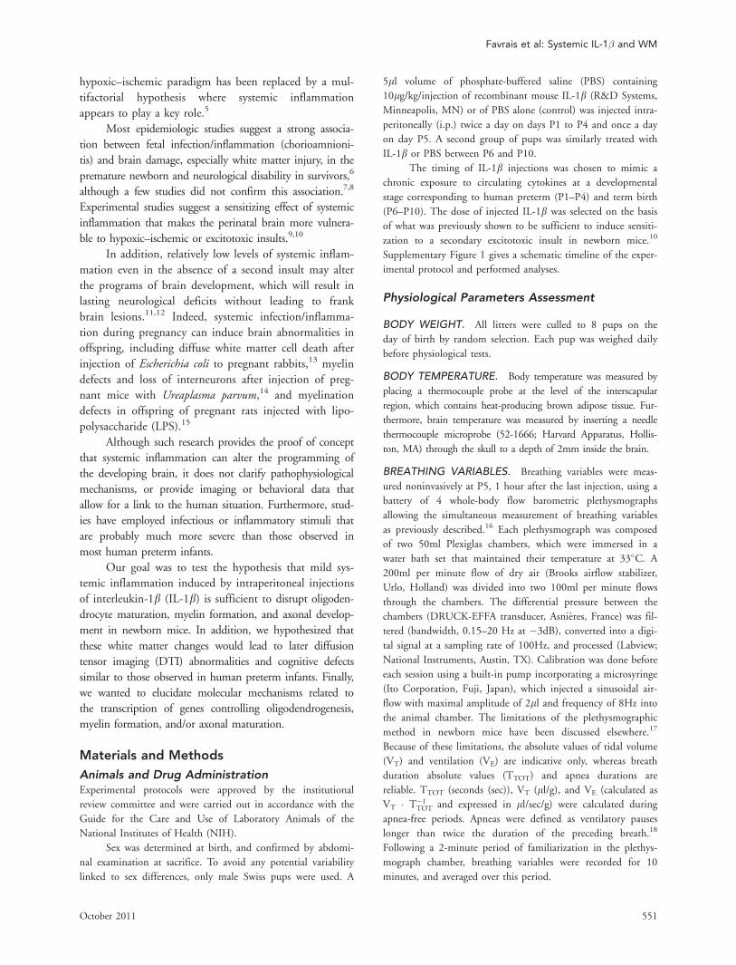
Intraperitoneal IL-1b–Induced SystemicInflammation without Altering Survival, Weight,Cerebral Blood Flow, and Brain AnatomyIL-1b injections between P1 and P5 induced systemic inflam-
mation as demonstrated by increased blood concentration of
IL-1b and tumor necrosis factor a (Fig 2A). As previously
reported,10 IL-1b injections between P1 and P5, when com-
pared to PBS injections, had no effect on mortality (which
was <1% in both experimental groups and which occurred
during the treatment period). At P1, P5, and P30, body
weights were similar in both groups (see Fig 2B).
IL-1b injections between P1 and P5 induced a
moderate decrease in minute ventilation, mainly due to
the increase in breathing cycle duration (see Fig 2). In
addition, animals exposed to IL-1b had an increase in
apnea duration, although total duration of apneas
remained low. These respiratory changes were accompa-
nied by a small increase in blood HCO�3 but had no sig-
nificant impact on blood pH, PCO2, PO2, hemoglobin,
or heart rate. IL-1b administration induced a moderate
and transient drop in body and brain temperatures.
By comparison, IL-1b injections between P6 and
P10 had no detectable effect on body weight and body
temperature (Supplementary Fig 2).
Ultrasound imaging performed in P5 mice on the 3
pre-Willis arteries (2 internal carotid arteries and basilar
trunk) did not reveal any difference in systolic, diastolic, or
time-average mean blood flow velocities between mice
injected with IL-1b or with PBS between P1 and P5 (Sup-
plementary Fig 3). This strongly suggested that IL-1b injec-
tions had no detectable impact on cerebral blood flow.
In addition, cresyl violet staining did not reveal any
gross anatomical abnormality or destructive lesions in
brains of P510 and P30 (data not shown) animals
exposed to IL-1b between P1 and P5 or between P6 and
P10, compared to PBS.
Intraperitoneal IL-1b Altered White MatterAnisotropy and Induced a Long-TermMemory DeficitAt P29 to P30, mice exposed to IL-1b between P1 and
P5 showed a severe deficit in memory, as they failed to
recognize novel or misplaced objects in NOR and OLM
memory tests, respectively (Fig 3A, B and Supplementary
Table 3). This cognitive impairment was absent in P29
to P30 mice treated by IL-1b between P6 and P10 (see
Fig 3A, B). The memory deficit observed in mice
exposed to IL-1b between P1 and P5 was not related to
Favrais et al: Systemic IL-1� and WM
October 2011 553

FIGURE 1: The neonatal exposure to interleukin (IL)-1b–induced microstructural abnormalities within the white matter at P35.(A) Magnetic resonance images (T2W) and diffusion tensor imaging (DTI)-derived maps (fractional anisotropy map [FA], direc-tion encoded color map [DEC]) were obtained from the ex vivo brains of P35 mice subjected to phosphate-buffered saline(PBS) (n 5 5) or IL-1b injections (n 5 5) from P1 to P5. (B–D) The axial diffusivity (D//), the radial diffusivity (D?), the apparentdiffusion coefficient (ADC) (C), and the FA (D) were derived from the DTI data and measured within the corpus callosum (CC),external capsule (EC), cingulum (Cg, Cing.), basal ganglia (BG), and cortex (Cx) in the PBS (n 5 5, white bar) and the IL-1b (n 55, black bar) groups as demonstrated in B. Results are expressed as mean 6 standard deviation. Asterisks indicate statisticallysignificant difference from respective white bar (C, D), *p < 0.05, **p < 0.01, ***p < 0.001 by Mann-Whitney test.
ANNALS of Neurology
554 Volume 70, No. 4

FIGURE 2: Physiological effects of systemic injections of interleukin (IL)-1b. Comparison of mice treated by IL-1b (black bars) orphosphate-buffered saline (PBS) (white bars) from P1 to P5. (A) Measurement of blood cytokine levels by enzyme-linked immuno-sorbent assay at P5 (n 5 6 for each group). TNF 5 tumor necrosis factor. (B) Weight gain from birth to adulthood (n 5 25 in PBSgroup and n 5 35 in IL-1b group). (C–E) Baseline ventilation measured in plethysmograph at P5, 1 hour after the last injection (n 5
23 in PBS group and n 5 25 in IL-1b group). (F, G) Apnea duration (F) and mean heart rate (G) calculated over the 10-minute re-cording of baseline ventilation (n 5 23 in PBS group and n 5 25 in IL-1b group). (H–L) Blood pH, gases, and hemoglobin measuredat P5, immediately after the baseline ventilation measurement (n 5 23 in PBS group and n 5 25 in IL-1b group). (M) Body tempera-ture measured at interscapular level just before and 1 hour after the last injection at P5 (n 5 23 in PBS group and n 5 25 in IL-1bgroup). (N) Brain temperature measured immediately after baseline ventilation measurement (n 5 23 in PBS group and n 5 25 inIL-1b group). Results are expressed in means 6 standard deviation. *p < 0.05, **p < 0.01, ***p < 0.001 by Mann-Whitney test.
Favrais et al: Systemic IL-1� and WM
October 2011 555

FIGURE 3: The systemic injection of interleukin (IL)-1b from P1 to P5 induced a long-lasting behavioral impairment. (A, B) Micewere subjected to the (A) novel object recognition (NOR) and (B) the object location memory (OLM) tests at P29 and P30.Mice treated with intraperitoneal injections of phosphate-buffered saline (PBS) (white bars) or IL-1b (black bars) from P1 to P5(n 5 21 and n 5 29 for NOR; n 5 20 and n 5 28 for OLM, respectively) or from P6 to P10 (n 5 10 and n 5 11 for both tests,respectively) were considered. The time spent to explore 1 object during the first round (T0) and the novel or misplaced objectduring the second round 30 minutes later (T30) was expressed in percentage of the overall exploration time. Asterisks indicatestatistically significant difference in percentage of exploration time between groups during the T30 period: **p < 0.01 and***p < 0.001 by nonparametric Mann-Whitney test performed after 2-way analysis of variance. (C) Mice treated with PBS (n 530, dotted line) or IL-1b (n 5 41, solid line) from P1 to P5 were subjected to the open field test at P28, including the quantifi-cation of squares crossed per minute for 10 minutes. Results are expressed as mean 6 standard deviation.
FIGURE 4: The systemic injection of interleukin (IL)-1b from P1 to P5 led to a diffuse reduction of the myelin proteins at P15and P30. (A–D) Immunostainings of 3 major myelin proteins myelin basic protein (MBP), proteolipid protein (PLP), and myelin-associated protein (MAG) were performed on P15 (A–C) and P30 (A–D) brains embedded in paraffin. Mice were treated withphosphate-buffered saline (PBS) or IL-1b from P1 to P5 (A–D) and from P6 to P10 (D) (n 5 6 in each group). (A) Photomicro-graphs of MBP immunostaining of P30 brains at original magnification 31.5 (left panel) and 340 (right panel) focusing on thesensorimotor cortex; scale bars 5 2,000lm and 50lm, respectively. (B) Photomicrographs of PLP immunostaining of P30 brainsat original magnification 31.5 (left panel) and 320 (right panel) focusing on the white matter; scale bars 5 2,000lm and100lm, respectively. (C) Photomicrographs of MAG immunostaining of P30 brains at original magnification 31.5 (left panel)and 320 (right panel) focusing on the corpus callosum; scale bars 5 2,000lm and 100lm, respectively. (D) Photomicrographsof MBP immunostaining focusing on the sensorimotor cortex of P30 mice treated with PBS or IL-1b from P1 to P5 and from P6to P10; original magnification 310, scale bar 5 20lm. The optical densities of stainings were determined in each experimentalcondition within the sensorimotor cortex for MBP (A, D), the white matter for PLP (B), and the corpus callosum for MAG (C).White bars 5 PBS group, black bars 5 IL-1b group. Results are expressed as means 6 standard deviation. Asterisks indicatestatistically significant difference from white bar, *p < 0.05 and p < 0.001 by Mann-Whitney test.
3
ANNALS of Neurology
556 Volume 70, No. 4

a deficit in exploratory behavior, as these mice performed
as PBS controls in the open field test (see Fig 3C).
T2W sequence analysis of P35 brains exposed to
systemic IL-1b between P1 and P5 confirmed the
absence of macroscopic abnormalities (see Fig 1A). How-
ever, microstructural changes were detected with an
increase of the D? in the CC, EC, Cg, BG, and Cx,
whereas the D// and the ADC were increased in the BG
FIGURE 4.
Favrais et al: Systemic IL-1� and WM
October 2011 557

only (see Fig 1A–C). In addition, FA was significantly
lower in all white matter structures analyzed: CC and
EC as well as Cg of IL-1b animals compared to controls
(see Fig 1D). Similar diffusion abnormalities in multiple
white matter structures confirmed diffuse white matter
injuries in IL-1b mice.
IL-1b–Induced White Matter AlterationsResulted from a Combined Myelinopathyand Axonopathy
To elucidate the mechanisms by which intraperitoneal
IL-1b between P1 and P5 could impact white matter,
immunohistochemical and electron microscopy studies
were performed. Densitometric analysis performed on
P15 and P30 brains showed an overall reduction of
myelin proteins including myelin basic protein (MBP)
(at the level of Cg, cortex and BG), proteolipid protein
(PLP) (at the level of Cg, CC, EC, and anterior com-
missure), and myelin-associated protein (MAG) (at the
level of Cg and CC) in IL-1b–treated animals when
compared to controls (Fig 4A–C and Supplementary
Fig 4). This reduction was time dependent, with a
MBP reduction observed as early as P15 and a subse-
quent PLP and MAG reduction observed at P30 (see
Fig 4A-C). This reduction in density of myelin markers
was associated with a striking disorganization in the ori-
entation (see Fig 4A) and a reduction of the length of
penetration (data not shown) of MBP-positive fibers
within the sensorimotor cortex.
Interestingly, MBP density within the sensorimotor cor-
tex of P30 animals treated with IL-1b between P6 and P10
was similar to control levels (see Fig 4D), in keeping with the
lack of effects of such a treatment on memory tests.
Bodian-Luxol fast blue staining of P1 to P5 treated
brains did not reveal any reduction of axonal density but fur-
ther confirmed the myelin reduction at P30 (Fig 5). Immu-
nolabeling with SMI-32, a marker of nonmyelinated axons,
showed an increased labeling in the IL-1b–exposed animals.
Electron microscopy confirmed our observation of fewer my-
elinated axons in IL-1b–exposed animals. Interestingly, in IL-
1b–exposed animals where axons were myelinated, the axons
had a normal myelin sheath that appeared appropriately
compacted, as confirmed by the G ratio analysis.
We also performed a morphometric analysis of the
axonal diameter of myelinated fibers crossing in the Cg
(see Fig 5E). This study revealed a reduction in fibers
exhibiting the largest axonal diameters (>0.5lm) that
was counterbalanced by an increase in numbers of small
fibers with diameters between 0.2 and 0.4lm. This
reduction in radial axon diameter could result from
impaired axonal outgrowth. In support of this hypothe-
sis, we observed a decrease of Wnt7a transcripts (see Fig
5F) encoding a peptide involved in the axonal
growth25,26 at P5 in the IL-1b–treated group. In con-
trast, no detectable changes were observed in the expres-
sion of light and medium chain neurofilament transcripts
(see Fig 5G, H).
Interestingly, when IL-1b was injected between P6 and
P10, we observed a reduction of the expression of Wnt7a
and light chain neurofilament in P10 IL-1b–treated pups,
when compared to control pups (Supplementary Fig 5).
IL-1b Impaired Oligodendrocyte DevelopmentThe myelination defect observed in the P1 to P5 IL-1b–treated animals could be due to a reduction of mature
myelinating oligodendrocytes. Olig2 immunolabeling, a
marker of oligodendrocytes irrespective of their stage of
maturation, did not reveal any difference between IL-1b–treated animals and controls (Fig 6A). Supporting these
data, IL-1b had no detectable effect on the density of
cleaved caspase 3-positive cells (a marker of cell death),
of Ki67-positive cells (a marker of proliferation), or of
Olig2/Ki67 double-positive cells (see Fig 6B–D).
In contrast, IL-1b–treated animals displayed an
increased density of NG2- and PDGFRa-positive cells
(markers of oligodendrocyte progenitors), but a decreased
FIGURE 5: The systemic injection of interleukin (IL)-1b from P1 to P5 induced a reduction of the axon diameter and ensheath-ment without altering the myelin sheath morphology. (A) Photomicrographs of Bodian-Luxol histochemistry performed on10lm- thick paraffin sections within the cingulum of P30 mice treated with phosphate-buffered saline (PBS) or IL-1b from P1 toP5; original magnification 340 and 363, scale bars 5 20lm and 5lm, respectively. (B) Immunofluorescent staining of SMI-32, amarker of nonmyelinated axons, performed on 10lm-thick frozen sections within the cingulum at P30; original magnification340, scale bars 5 20lm. (C) Electron microscopy (EM) processed on P30 brains of mice treated with PBS or IL-1b from P1 to P5focusing on the cingulum; original magnification 320,000 and 310,000; scale bars 5 1lm and 0.5lm, respectively. (D) Assess-ment of the myelin sheath thickness in reference to axon diameter by the G ratio measurement (axon diameter/fiber diameter)within the cingulum at P30 (PBS [n 5 3, white bars] and IL-1b [n 5 3, black bars]). (E) Classification of myelinated axons accordingto their diameter (mean, 0.1lm; range, 0–1.2lm; interval, 0.1lm) within the cingulum at P30 (PBS [n 5 3, dotted line] and IL-1b[n 5 3, solid line]). Axon diameter was measured on coronally sectioned axons from EM photomicrographs. (F–H) Relative Wnt7a(F), medium neurofilament (NF-M) (G), and light neurofilament (NF-L) (H) expressions by quantitative polymerase chain reaction atP0 (n 5 4, gray bar) and at P5, P10, P15, and P30 in PBS (n 5 4 at each age, white bars) and IL-1b (n 5 4 at each age, blackbars) groups within cortex and white matter. Results are expressed as mean 6 standard deviation. Asterisks indicate statisticallysignificant difference from white bar (D, F, G, H) or from dotted line (E), *p < 0.05 and ***p < 0.001 in Mann-Whitney test.
3
ANNALS of Neurology
558 Volume 70, No. 4

density of O4-positive cells (marker of oligodendrocyte
precursors), a decreased expression of CNPase (2030cyclicnucleotide 30 phosphohydrolase, a marker of premyeli-
nating oligodendrocytes), and a reduced density of
Adenomatosis Polyposis Coli (APC)-positive cells (a
marker of myelinating oligodendrocytes) (Fig 7).
FIGURE 5.
Favrais et al: Systemic IL-1� and WM
October 2011 559

These data suggest that IL-1b does not affect oligo-
dendrocyte proliferation or survival but rather affects mat-
uration, with potentially a partial blockade at the transi-
tion between oligodendrocyte progenitor and precursor.
FIGURE 6: The systemic injection of interleukin (IL)-1b from P1 to P5 did not affect the total number of oligodendrocytes, celldeath, and proliferation. (A) Quantification of fluorescent Olig21 cells/mm2 in the cingulate white matter of mice treated withphosphate-buffered saline (PBS) (n 5 6 at each age, white bars) or IL1-b (n 5 6 at each age, black bars) at P5, P10, P15, andP30. Staining was performed on 10lm-thick paraffin sections. (B) Quantification of fluorescent cleaved caspase 31 cells/mm2 atP5 and P10 within the cingulate white matter of mice treated with PBS (n 5 6 at each age, white bars) or IL-1b (n 5 6 at eachage, black bars). Cleaved caspase 3 staining was performed on 10lm-thick frozen sections. (C, D) Quantification of Ki671 (C)and of double-stained Ki67/Olig21 (D) cells/mm2 within the subventricular zone of mice treated with PBS (n 5 6 at each age,white bars) or IL-1b (n 5 6 at each age, black bars) at P5, P10, P15, and P30. Stainings were performed on 10lm-thick paraffinsections. Results are expressed as mean 6 standard deviation.
FIGURE 7: The systemic injection of interleukin (IL)-1b from P1 to P5 led to a significant increase of oligodendrocyte progeni-tors associated with a reduction of mature oligodendrocytes. (A) Immunofluorescent staining of NG2/DAPI, performed on fro-zen sections, within the external capsule of P5 mice treated with phosphate-buffered saline (PBS) or IL-1b from P1 to P5(original low magnification 340, scale bar 5 50lm; high magnification 363, scale bar 5 10lm). (B) Quantification of fluores-cent NG21 cells/mm2 at P5, P10, P15, and P30 within the external capsule of mice treated with PBS (n 5 6 at each age, whitebars) or IL-1b (n 5 6 at each age, black bars). (C) Relative quantification of the PDGFRa transcript within cortex and white mat-ter at P0 (n 5 4, gray bar) and at P5, P10, P15, and P30 in mice treated with PBS (n 5 4 at each age, white bars) or IL-1b (n 5
4 at each age, black bars). (D, E) Immunofluorescent stainings of PDGFRa (green)/DAPI (D) and of O4 (red)/DAPI (E) on 10lm-thick frozen sections, within the external capsule of P5 mice treated with either PBS or IL-1b from P1 to P5 (original low magni-fication 340, scale bar 5 50lm; original high magnification 363, scale bar 5 10lm). Quantification of fluorescent PDGFRa1 (D)and O41 (E) cells/mm2 within the external capsule of mice treated with PBS (n 5 6, white bars) or IL- 1b (n 5 6, black bars) atP5. (F) Assessment of 2030cyclic nucleotide 30 phosphohydrolase (CNPase) gene expression by quantitative polymerase chainreaction within cortex and white matter at P0 (n 5 4, gray bar) and at P5, P10, P15, and P30 in mice treated with PBS (n 5 4at each age, white bars) or IL-1b (n 5 4 at each age, black bars). (G) Photomicrographs of the Adenomatosis Polyposis Coli(APC) immunostaining at the external capsule level at P15 performed on 10lm-thick paraffin sections (original magnification320, scale bars 5 100lm). (H) Quantification of APC1 cells/mm2 within the external capsule at P10, P15, and P30 in micetreated with PBS (n 5 6 at each age, white bars) or IL-1b (n 5 6 at each age, black bars) from P1 to P5. Results are expressedas mean 6 standard deviation. Asterisks indicate statistically significant difference from white bar, *p < 0.05, **p < 0.01, and***p < 0.001 by Mann-Whitney test.
3
ANNALS of Neurology
560 Volume 70, No. 4

FIGURE 7.
Favrais et al: Systemic IL-1� and WM
October 2011 561

IL-1b Disrupted the Machinery ControllingOligodendrocyte MaturationTo explore this further, we performed real time reverse
transcription PCR (RT-PCR) of transcription factors
known to be involved in the maturation/differentiation
process of oligodendrocytes. Wheras expression of Olig1,
Olig2, Sox10, Tcf4, Axin2, HDAC1, and HDAC3 was
increased, that of Nkx2.2, Sox8, and P27Kip1 was inhib-
ited after IL-1b treatment (Fig 8).
The expression of factors known to mediate the inter-
actions between axons and oligodendrocytes was also meas-
ured by real time RT-PCR. No significant effect of IL-1bwas detected on the expression of SemaphorinA3, PlexinA4,
EphrinB2, EphrinB3, Fyn, Lingo1, and Neuregulin1 and
its receptors ErbB2 and ErbB4 (Supplementary Fig 6).
IL-1b Did Not Have a Major Impact on OtherCell TypesIL-1b injections between P1 and P5 had no detectable
effect on neocortical cell death (cleaved caspase 3 immuno-
staining), neuronal density (NeuN immunostaining), and
astrocyte density (glial fibrillary acidic protein immunostain-
ing) (Supplementary Fig 7A–C). In contrast, IL-1b injections
between P1 and P5 induced a transient increase in the density
of microglia in the neopallium (MAC1 and Iba-1 immuno-
staining) (Supplementary Fig 7D–E).
Discussion
We have shown that a moderate systemic inflammatory
stimulus disrupts oligodendrocyte and axon maturation,
and impairs myelination in newborn mice. This long-
lasting myelination defect is accompanied by abnormal
FA on DTI and cognitive behavioral defects.
Neonatal Systemic Inflammation Disrupts WhiteMatter Programming
In the present model, no obvious brain lesion or neural
cell death was detected, strongly suggesting that white
matter abnormalities were the result of alterations of the
developmental program of the brain. In keeping with
this hypothesis, the expression of several factors involved
in oligodendrocyte and axon maturation was altered fol-
lowing systemic inflammation. In addition, myelination
defects were observed when pups were exposed to sys-
temic inflammation between P1 and P5 but not when
they were exposed between P6 and P10, suggesting a pe-
riod of vulnerability of the developing white matter. As
developmental events occurring at P1 are quite different
from those occurring at P5, further studies will be neces-
sary to potentially refine the window of vulnerability,
specifically, to determine if repeated exposures in the P1
to P5 period are required to produce this myelination
defect or if exposure to IL-1b at a given day is sufficient.
White Matter Disease Is Both an Oligopathyand an AxonopathyAlthough research has identified damage to oligodendro-
cytes as the cause of periventricular white matter damage
in the human preterm infant,27 the potential specific
contribution of axonopathy to white matter abnormalities
and dysfunction remains to be clarified.28 This is critical,
as axonopathies have been described in a variety of other
human diseases affecting the white matter, such as multi-
ple sclerosis29 and leukodystrophies.30
In the present model, a combination of markers
suggests a block of oligodendrocyte maturation at the
progenitor stage, whereas proliferation and survival
remained unaffected. Although the precise mechanisms
linking systemic inflammation and blockade of oligoden-
drocyte maturation will require further studies, the present
data suggest an imbalance between transcription factors
controlling oligodendrocyte maturation. Indeed, some
transcriptional factors known to play a role during oligo-
dendrocyte maturation/differentiation, such as Olig1,31
Olig2,32 Sox10,33 Tcf4,34 Axin2, HDAC1,35 and
HDAC3,36 are increased by systemic IL-1b, whereas othertranscription factors also involved in oligodendrocyte mat-
uration, such as Nkx2.237 and Sox8,38 are reduced.
In addition to the oligopathy, we observed a clear axon-
opathy by electron microscopy, with reduced diameter of
myelinated axons, and altered water diffusivity on DTI in IL-
1b-exposed animals. IL-1b also significantly inhibited the
expression of Wnt7a, a transcription factor involved in axo-
nal maturation,25,26 supporting evidence of an axonopathy.
Interactions between oligodendrocytes and axons are
important for oligodendrocyte maturation, axonal growth,
and myelination.39 Thus, inflammation-induced axonopathy
due to systemic IL-1b may disrupt these interactions, leading
to abnormal white matter. The analysis at the transcription
level of some factors known to mediate these interactions,
such as Semaphorin3a, PlexinA4, EphrinB2,40 EphrinB3,41
Fyn,42 Lingo1,43 and Neuregulin1 and its receptors ErbB2
and ErbB4,44,45 did not reveal any significant effect of IL-1b.Interestingly, although IL-1b exposure between P6 and P10
did not interfere with myelination, it had a significant impact
on the expression of Wnt7a and light chain neurofilament,
suggesting that the effects of IL-1b on myelination and on
axonal growth might be partly dissociated.
Interestingly, at the electron microscopic level in
IL-1b–treated animals, a large number of axons were
totally deprived of myelin, whereas other axons had nor-
mally compacted myelin. The mechanisms and
ANNALS of Neurology
562 Volume 70, No. 4

functional significance of this phenomenon remain elu-
sive and warrant further investigation.
Clinical Relevance of the Present ModelThe relevance of our model for understanding white
matter disease of human preterm infants is supported by
several observations from this study.
First, the apparently moderate intensity of this
inflammatory insult compared to other models based on
LPS or E. coli administration13,46 adds to its relevance to
clinically silent or minor chorioamnionitis often observed
in preterm infants.
Second, the systemic inflammation is accompanied
by moderate but significant effects on ventilation and
temperature, which are reminiscent of what is observed
in preterm infants exposed to chorioamnionitis.47
Third, our MRI findings are consistent with white
matter abnormalities described in recent imaging studies in
FIGURE 8: The systemic injection of interleukin (IL)-1b from P1 to P5 perturbed the expression of key factors involved in oligo-dendrocyte maturation. Relative gene expression of Olig1 (A), Olig2 (B), Nkx2.2 (C), Sox8 (D), Sox10 (E), Tcf4 (F), Axin2 (G),HDAC1 (H), HDAC3 (I), HDAC4 (J), and P27Kip1 (K) were assessed by quantitative polymerase chain reaction within cortex andwhite matter at P0 (n 5 4, gray bars) and at P5, P10, P15, and P30 in mice treated with phosphate-buffered saline (n 5 4 ateach age, white bars) and with IL-1b (n 5 4 at each age, black bars). Results are expressed as mean 6 standard deviation.Asterisks indicate statistically significant difference from white bar, *p < 0.05 by Mann-Whitney test.
Favrais et al: Systemic IL-1� and WM
October 2011 563

very preterm infants also without detectable tissue destruc-
tion on anatomical sequences.48 In the present study, the
observed FA reduction, mainly due to an increase of D?,suggests a selective alteration of white matter, as a result of
myelination deficit and/or of myelin sheath damage.
Additionally, behavioral testing of adult animals
exposed to neonatal inflammation revealed significant cog-
nitive deficits but no motor impairment, similar to the
neurobehavioral profile observed in recent follow-up
cohorts of very preterm infants.3 Of note, although the
novel object and displaced object recognition tests are gen-
erally considered as testing hippocampal functions, memory
requires structures beyond the hippocampus. Illustrating
this, we have shown that cortical and white matter lesions
induced by glutamate analog injection into the neopallium
are sufficient to impair performances in these tests.49
Despite this relevance of our model, when extrapo-
lating the present observations to the human situation it
is important to remember species differences in inflam-
matory mechanisms. Nevertheless, increased IL-1b is a
common hallmark of inflammation/infection in humans
and mice,50,51 supporting the use of our model to further
the understanding of white matter disease.
To our knowledge, this is the first study showing
that a moderate systemic inflammation occurring at a
specific time during the perinatal period can alter the de-
velopmental programs of the white matter. This insult
leads to a long-lasting myelination deficit accompanied
by cognitive defects, mimicking MRI abnormalities and
neurological handicaps observed in some human preterm
infants. The impact of perinatal inflammation on pro-
grams of brain development could also have long-term
consequences in terms of susceptibility to adult brain
diseases.
Acknowledgments
This study was supported by grants from Inserm (P.G.),
Paris Diderot University (P.G.), Assistance Publique des
Hopitaux de Paris (Public Parisian Hospitals; Interface
contract to P.G.), PremUP Foundation (P.G.), Sixth
Framework Program of the European Commission (con-
tract No. LSHM-CT-2006-036534/NEOBRAIN; P.G.,
O.D., H.H.), Seventh Framework Program of the Euro-
pean Union (contract No. HEALTH-F2-2009-241778/
NEUROBID; P.G., O.D.), Leducq Foundation (P.G.,
H.H.), European Leukodystrophy Association (G.F.,
P.G.), Societe Francaise de Pediatrie (French Society for
Pediatrics; G.F.), Journees Francophones de Recherche en
Neonatologie (G.F.), Fondation pour le Recherche Medi-
cale (Medical Research Foundation), Swiss National
Fund (31003A-112233; S.S.), Biomedical Imaging
Centre of the Geneva University (UNIGE), Lausanne
University (UNIL), Geneva University Hospital (HUG),
Lausanne University Hospital (CHUV), Lausanne Poly-
technic School (EPFL), Leenards and Jeantet Founda-
tions (Y.v.d.L., S.S.), Swedish Medical Research Council
(VR 2006-3396; H.H.), Swedish governmental grants to
researchers in the public health service (ALFGBG2863;
H.H.), Medical Research Council UK (P19381; H.H.),
Medical Research Council Sweden (H.H.), and Action
Medical Research UK (SP4506; H.H.).
We thank F. Cluzeaud for excellent assistance with
the electron microscopy.
Authorship
V.L. and P.G. contributed equally to the work.
Potential Conflicts of Interest
Nothing to report.
References1. Larroque B, Ancel PY, Marret S, et al. Neurodevelopmental dis-
abilities and special care of 5-year-old children born before 33weeks of gestation (the EPIPAGE study): a longitudinal cohortstudy. Lancet 2008;371:813–820.
2. Wilson-Costello D, Friedman H, Minich N, et al. Improved neuro-developmental outcomes for extremely low birth weight infants in2000–2002. Pediatrics 2007;119:37–45.
3. Delobel-Ayoub M, Arnaud C, White-Koning M, et al. Behavioralproblems and cognitive performance at 5 years of age after verypreterm birth: the EPIPAGE Study. Pediatrics 2009;123:1485–1492.
4. Volpe JJ. Cerebral white matter injury of the premature infant—more common than you think. Pediatrics 2003;112:176–180.
5. Dammann O, Leviton A. Inflammatory brain damage in pretermnewborns—dry numbers, wet lab, and causal inferences. EarlyHum Dev 2004;79:1–15.
6. Wu YW, Colford JM Jr. Chorioamnionitis as a risk factor for cere-bral palsy: a meta-analysis. JAMA 2000;284:1417–1424.
7. Nelson KB, Grether JK, Dambrosia JM, et al. Neonatal cytokinesand cerebral palsy in very preterm infants. Pediatr Res 2003;53:600–607.
8. Bartha AI, Foster-Barber A, Miller SP, et al. Neonatal encephalop-athy: association of cytokines with MR spectroscopy and outcome.Pediatr Res 2004;56:960–966.
9. Eklind S, Mallard C, Leverin AL, et al. Bacterial endotoxin sensi-tizes the immature brain to hypoxic–ischaemic injury. Eur J Neuro-sci 2001;13:1101–1106.
10. Dommergues MA, Patkai J, Renauld JC, et al. Proinflammatorycytokines and interleukin-9 exacerbate excitotoxic lesions of thenewborn murine neopallium. Ann Neurol 2000;47:54–63.
11. Malaeb S, Dammann O. Fetal inflammatory response and braininjury in the preterm newborn. J Child Neurol 2009;24:1119–1126.
12. Volpe JJ. Brain injury in premature infants: a complex amalgam ofdestructive and developmental disturbances. Lancet Neurol 2009;8:110–124.
13. Debillon T, Gras-Leguen C, Verielle V, et al. Intrauterine infectioninduces programmed cell death in rabbit periventricular whitematter. Pediatr Res 2000;47:736–742.
ANNALS of Neurology
564 Volume 70, No. 4

14. Normann E, Lacaze-Masmonteil T, Eaton F, et al. A novel mousemodel of Ureaplasma-induced perinatal inflammation: effects onlung and brain injury. Pediatr Res 2009;65:430–436.
15. Rousset CI, Chalon S, Cantagrel S, et al. Maternal exposure toLPS induces hypomyelination in the internal capsule and pro-grammed cell death in the deep gray matter in newborn rats.Pediatr Res 2006;59:428–433.
16. Bollen B, Bouslama M, Matrot B, et al. Cold stimulates the behav-ioral response to hypoxia in newborn mice. Am J Physiol RegulIntegr Comp Physiol 2009;296:R1503–R1511.
17. Lofaso F, Dauger S, Matrot B, et al. Inhibitory effects of repeatedhyperoxia on breathing in newborn mice. Eur Respir J 2007;29:18–24.
18. Matrot B, Durand E, Dauger S, et al. Automatic classification ofactivity and apneas using whole body plethysmography in new-born mice. J Appl Physiol 2005;98:365–370.
19. Ramanantsoa N, Vaubourg V, Matrot B, et al. Effects of tempera-ture on ventilatory response to hypercapnia in newborn mice het-erozygous for transcription factor Phox2b. Am J Physiol RegulIntegr Comp Physiol 2007;293:R2027–R2035.
20. Dingley J, Hobbs C, Ferguson J, et al. Xenon/hypothermia neuro-protection regimes in spontaneously breathing neonatal rats afterhypoxic-ischemic insult: the respiratory and sedative effects.Anesth Analg 2008;106:916–923.
21. Bonnin P, Debbabi H, Mariani J, et al. Ultrasonic assessment ofcerebral blood flow changes during ischemia-reperfusion in 7-day-old rats. Ultrasound Med Biol 2008;34:913–922.
22. Olivier P, Baud O, Evrard P, et al. Prenatal ischemia and whitematter damage in rats. J Neuropathol Exp Neurol 2005;64:998–1006.
23. Deprez M, Ceuterick-de Groote C, Fumal A, et al. A new com-bined Bodian-Luxol technique for staining unmyelinated axons insemithin, resin-embedded peripheral nerves: a comparison withelectron microscopy. Acta Neuropathol 1999;98:323–329.
24. Favrais G, Schwendimann L, Gressens P, Lelievre V. Cyclooxygen-ase-2 mediates the sensitizing effects of systemic IL-1-beta onexcitotoxic brain lesions in newborn mice. Neurobiol Dis 2007;25:496–505.
25. Shimogori T, VanSant J, Paik E, Grove EA. Members of the Wnt,Fz, and Frp gene families expressed in postnatal mouse cerebralcortex. J Comp Neurol 2004;473:496–510.
26. Hirabayashi Y, Itoh Y, Tabata H, et al. The Wnt/beta-catenin path-way directs neuronal differentiation of cortical neural precursorcells. Development 2004;131:2791–2801.
27. Volpe JJ. Neurobiology of periventricular leukomalacia in the pre-mature infant. Pediatr Res 2001;50:553–562.
28. Leviton A, Gressens P. Neuronal damage accompanies perinatalwhite-matter damage. Trends Neurosci 2007;30:473–478.
29. Piaton G, Gould RM, Lubetzki C. Axon-oligodendrocyte interac-tions during developmental myelination, demyelination and repair.J Neurochem 2010;114:1243–1260.
30. Hofling AA, Kim JH, Fantz CR, et al. Diffusion tensor imagingdetects axonal injury and demyelination in the spinal cord andcranial nerves of a murine model of globoid cell leukodystrophy.NMR Biomed 2009;22:1100–1106.
31. Xin M, Yue T, Ma Z, et al. Myelinogenesis and axonal recognitionby oligodendrocytes in brain are uncoupled in Olig1-null mice.J Neurosci 2005;25:1354–1365.
32. Liu Z, Hu X, Cai J, et al. Induction of oligodendrocyte differentia-tion by Olig2 and Sox10: evidence for reciprocal interactions anddosage-dependent mechanisms. Dev Biol 2007;302:683–693.
33. Wei Q, Miskimins WK, Miskimins R. Sox10 acts as a tissue-specific tran-scription factor enhancing activation of the myelin basic protein genepromoter by p27Kip1 and Sp1. J Neurosci Res 2004;78:796–802.
34. Fancy SP, Baranzini SE, Zhao C, et al. Dysregulation of the Wntpathway inhibits timely myelination and remyelination in the mam-malian CNS. Genes Dev 2009;23:1571–1585.
35. Ye F, Chen Y, Hoang T, et al. HDAC1 and HDAC2 regulate oligo-dendrocyte differentiation by disrupting the beta-catenin-TCFinteraction. Nat Neurosci 2009;12:829–838.
36. Shen S, Li J, Casaccia-Bonnefil P. Histone modifications affect tim-ing of oligodendrocyte progenitor differentiation in the develop-ing rat brain. J Cell Biol 2005;169:577–589.
37. Zhou Q, Choi G, Anderson DJ. The bHLH transcription factorOlig2 promotes oligodendrocyte differentiation in collaborationwith Nkx2.2. Neuron 2001;31:791–807.
38. Stolt CC, Lommes P, Friedrich RP, Wegner M. Transcriptionfactors Sox8 and Sox10 perform non-equivalent roles during oli-godendrocyte development despite functional redundancy. Devel-opment 2004;131:2349–2358.
39. Nave KA, Trapp BD. Axon-glial signaling and the glial support ofaxon function. Annu Rev Neurosci 2008;31:535–561.
40. Yiu G, He Z. Glial inhibition of CNS axon regeneration. Nat RevNeurosci 2006;7:617–627.
41. Xiao Q, Chen J, Wu YQ. Expression of ephrin-B3 mRNA andcellular apoptosis in the brain of neonatal rats with periventricu-lar leukomalacia. Zhongguo Dang Dai Er Ke Za Zhi 2007;9:321–323.
42. Sperber BR, Boyle-Walsh EA, Engleka MJ, et al. A unique role forFyn in CNS myelination. J Neurosci 2001;21:2039–2047.
43. Mi S, Lee X, Shao Z, et al. LINGO-1 is a component of the Nogo-66receptor/p75 signaling complex. Nat Neurosci 2004;7:221–228.
44. Brinkmann BG, Agarwal A, Sereda MW, et al. Neuregulin-1/ErbBsignaling serves distinct functions in myelination of the peripheraland central nervous system. Neuron 2008;59:581–595.
45. Dammann O, Bueter W, Leviton A, et al. Neuregulin-1: a potentialendogenous protector in perinatal brain white matter damage.Neonatology 2008;93:182–187.
46. Young RS, Yagel SK, Towfighi J. Systemic and neuropathologiceffects of E. coli endotoxin in neonatal dogs. Pediatr Res 1983;17:349–353.
47. Abu-Shaweesh JM, Martin RJ. Neonatal apnea: what’s new?Pediatr Pulmonol 2008;43:937–944.
48. Brown NC, Inder TE, Bear MJ, et al. Neurobehavior at term andwhite and gray matter abnormalities in very preterm infants. JPediatr 2009;155:32–38, 38.e31.
49. Titomanlio L, Bouslama M, Verche VL, et al. Implanted neuro-sphere-derived precursors promote recovery after neonatal excito-toxic brain injury. Stem Cells Dev 2011;20:865–879.
50. Gotsch F, Romero R, Kusanovic JP, et al. The fetal inflammatoryresponse syndrome. Clin Obstet Gynecol 2007;50:652–683.
51. Feuerstein GZ, Wang X, Barone F. Inflammatory gene expressionin cerebral ischemia and trauma. Ann N Y Acad Sci 1997;825:179–193.
Favrais et al: Systemic IL-1� and WM
October 2011 565
Related Documents