BioMed Central Page 1 of 11 (page number not for citation purposes) BMC Genomics Open Access Research article Spatial and sex-specific dissection of the Anopheles gambiae midgut transcriptome Emma Warr, Ruth Aguilar, Yuemei Dong, Vassiliki Mahairaki and George Dimopoulos* Address: W. Harry Feinstone Department of Molecular Microbiology and Immunology, Bloomberg School of Public Health, Johns Hopkins University, 615 N. Wolfe Street, Baltimore, MD 21205-2179, USA Email: Emma Warr - [email protected]; Ruth Aguilar - [email protected]; Yuemei Dong - [email protected]; Vassiliki Mahairaki - [email protected]; George Dimopoulos* - [email protected] * Corresponding author Abstract Background: The midgut of hematophagous insects, such as disease transmitting mosquitoes, carries out a variety of essential functions that mostly relate to blood feeding. The midgut of the female malaria vector mosquito Anopheles gambiae is a major site of interactions between the parasite and the vector. Distinct compartments and cell types of the midgut tissue carry out specific functions and vector borne pathogens interact and infect different parts of the midgut. Results: A microarray based global gene expression approach was used to compare transcript abundance in the four major female midgut compartments (cardia, anterior, anterior part of posterior and posterior part of posterior midgut) and between the male and female Anopheles gambiae midgut. Major differences between the female and male midgut gene expression relate to digestive processes and immunity. Each compartment has a distinct gene function profile with the posterior midgut expressing digestive enzyme genes and the cardia and anterior midgut expressing high levels of antimicrobial peptide and other immune gene transcripts. Interestingly, the cardia expressed several known anti-Plasmodium factors. A parallel peptidomic analysis of the cardia identified known mosquito antimicrobial peptides as well as several putative short secreted peptides that are likely to represent novel antimicrobial factors. Conclusion: The A. gambiae sex specific midgut and female midgut compartment specific transcriptomes correlates with their known functions. The significantly greater functional diversity of the female midgut relate to hematophagy that is associated with digestion and nutrition uptake as well as exposes it to a variety of pathogens, and promotes growth of its endogenous microbial flora. The strikingly high proportion of immunity related factors in the cardia tissue most likely serves the function to increase sterility of ingested sugar and blood. A detailed characterization of the functional specificities of the female mosquito midgut and its various compartments can greatly contribute to our understanding of its role in disease transmission and generate the necessary tools for the development of malaria control strategies. Published: 29 January 2007 BMC Genomics 2007, 8:37 doi:10.1186/1471-2164-8-37 Received: 29 August 2006 Accepted: 29 January 2007 This article is available from: http://www.biomedcentral.com/1471-2164/8/37 © 2007 Warr et al; licensee BioMed Central Ltd. This is an Open Access article distributed under the terms of the Creative Commons Attribution License (http://creativecommons.org/licenses/by/2.0 ), which permits unrestricted use, distribution, and reproduction in any medium, provided the original work is properly cited.

Welcome message from author
This document is posted to help you gain knowledge. Please leave a comment to let me know what you think about it! Share it to your friends and learn new things together.
Transcript
BioMed CentralBMC Genomics
ss
Open AcceResearch articleSpatial and sex-specific dissection of the Anopheles gambiae midgut transcriptomeEmma Warr, Ruth Aguilar, Yuemei Dong, Vassiliki Mahairaki and George Dimopoulos*Address: W. Harry Feinstone Department of Molecular Microbiology and Immunology, Bloomberg School of Public Health, Johns Hopkins University, 615 N. Wolfe Street, Baltimore, MD 21205-2179, USA
Email: Emma Warr - [email protected]; Ruth Aguilar - [email protected]; Yuemei Dong - [email protected]; Vassiliki Mahairaki - [email protected]; George Dimopoulos* - [email protected]
* Corresponding author
AbstractBackground: The midgut of hematophagous insects, such as disease transmitting mosquitoes,carries out a variety of essential functions that mostly relate to blood feeding. The midgut of thefemale malaria vector mosquito Anopheles gambiae is a major site of interactions between theparasite and the vector. Distinct compartments and cell types of the midgut tissue carry out specificfunctions and vector borne pathogens interact and infect different parts of the midgut.
Results: A microarray based global gene expression approach was used to compare transcriptabundance in the four major female midgut compartments (cardia, anterior, anterior part ofposterior and posterior part of posterior midgut) and between the male and female Anophelesgambiae midgut. Major differences between the female and male midgut gene expression relate todigestive processes and immunity. Each compartment has a distinct gene function profile with theposterior midgut expressing digestive enzyme genes and the cardia and anterior midgut expressinghigh levels of antimicrobial peptide and other immune gene transcripts. Interestingly, the cardiaexpressed several known anti-Plasmodium factors. A parallel peptidomic analysis of the cardiaidentified known mosquito antimicrobial peptides as well as several putative short secretedpeptides that are likely to represent novel antimicrobial factors.
Conclusion: The A. gambiae sex specific midgut and female midgut compartment specifictranscriptomes correlates with their known functions. The significantly greater functional diversityof the female midgut relate to hematophagy that is associated with digestion and nutrition uptakeas well as exposes it to a variety of pathogens, and promotes growth of its endogenous microbialflora. The strikingly high proportion of immunity related factors in the cardia tissue most likelyserves the function to increase sterility of ingested sugar and blood. A detailed characterization ofthe functional specificities of the female mosquito midgut and its various compartments can greatlycontribute to our understanding of its role in disease transmission and generate the necessarytools for the development of malaria control strategies.
Published: 29 January 2007
BMC Genomics 2007, 8:37 doi:10.1186/1471-2164-8-37
Received: 29 August 2006Accepted: 29 January 2007
This article is available from: http://www.biomedcentral.com/1471-2164/8/37
© 2007 Warr et al; licensee BioMed Central Ltd. This is an Open Access article distributed under the terms of the Creative Commons Attribution License (http://creativecommons.org/licenses/by/2.0), which permits unrestricted use, distribution, and reproduction in any medium, provided the original work is properly cited.
Page 1 of 11(page number not for citation purposes)
BMC Genomics 2007, 8:37 http://www.biomedcentral.com/1471-2164/8/37
BackgroundThe midgut of the female Anopheles mosquito is responsi-ble for the digestion of the blood meal and is the entrypoint and site of extensive interaction with disease-caus-ing pathogens. Female mosquitoes are capable of trans-mitting a variety of pathogens and as such the midgut hasbecome an important focus for studies that aim at under-standing the transmission biology of vector-borne dis-eases [1-10].
A variety of studies, including microarray-based geneexpression analyses, have addressed the effect of infectionor blood-meal ingestion on the female mosquito. Thesestudies have established the immune responsive capacityof the midgut and provide detailed information about avariety of biological processes that are regulated uponblood feeding [1-3,5-12]. However, these studies havelargely focused on the overall mosquito midgut transcrip-tome, ignoring the spatial specialization of different func-tions in the various compartments of this tissue (Figure1a).
Adult female mosquitoes feed on both blood and sugar,and the storage and processing of these two foods are spa-tially different. The ingested sugar is first stored in theventral diverticulum (crop) and then delivered to the mid-gut. Its digestion and absorption occurs within both theanterior and posterior midgut [13,14]. The alpha-glucosi-dase activity responsible for sugar digestion is detected inboth compartments, although regulation does differbetween the two; the enzyme activity is constitutivelyexpressed in the anterior midgut, and blood-feeding doesnot influence its levels [15]. In contrast to sugar digestion,the blood meal is exclusively digested and absorbed in theposterior region [13,16]. Aminopeptidases and glucosi-dases are also detected in the posterior region, where theirlevels rise after blood-feeding. Digestion of the proteincontent of blood is mainly carried out by serine proteasessuch as trypsins and chymotrypsins that display complexpatterns of regulation in relation to blood feeding.Trypsin activity is limited to the posterior region of themidgut [15,17-19]. The majority of the cells in the poste-rior midgut are digestive epithelial cells, which produceand secrete the enzymes needed for blood-meal digestion[14]. Regenerative and endocrine cells are also present inthe posterior midgut, although their function is currentlyunknown [14]. In addition to their essential role inblood-meal digestion, trypsins have also been shown tobe required for the activation of an ookinete-secreted chi-tinase, which enables the parasite to penetrate the peri-trophic matrix of the mosquito midgut [20-22]. A varietyof transporters responsible for maintaining osmotic bal-ance and nutritient uptake are also expressed in the mid-gut and are regulated upon feeding [9].
The peritrophic matrix of mosquitoes is a semi-permeablechitinous matrix that lines the digestive tract and therebyacts as a barrier to direct contact with the blood meal andto microbial and parasitic infections [23,24]. The peri-trophic matrix is synthesized by midgut epithelial cells indirect response to the ingestion of a blood meal. The per-itrophic matrix precursors are stored in secretory vesiclesof midgut epithelial cells, and the secretion of these pro-teins is triggered by blood-feeding, releasing the vesiclecontents into the lumen of the mosquito midgut [25]. Themalarial parasite Plasmodium has to cross the peritrophicmatrix before it can invade the midgut epithelium and, assuch, the peritrophic matrix and the genes involved in itsformation have therefore become a focus for studies thataim to block parasite transmission [25-28].
In Plasmodium-infected mosquitoes, oocysts are primarilyfound in the very posterior region of the midgut [29]. Thereason(s) for this spatial distribution is still unclear. Anearly suggestion was that the parasites preferentiallyinvade vesicular ATPase (V-ATPase)-overexpressing cellsthat are located at the very posterior region of the midgut[29,30]. More recently, it has been shown that not all Plas-modium species specifically invade this particular type ofcell [31].
The close association between transcript abundance andgene function makes it possible to infer biological func-tion and physiological conditions from gene expressionprofiles. We have performed a global microarray-basedgene expression analysis to study the midgut sex- andcompartment-(cardia, anterior, anterior-posterior andposterior-posterior) specific transcriptomes. In conjunc-tion with this analysis, we have utilized a peptidomicapproach to identify putative short secreted peptides thatare enriched in the cardia and anterior compartment,where the majority of the mosquito's antimicrobial pep-tides are found to be produced. The elucidation of thesespatial profiles (both transcriptomic and peptidomic) willprovide a broadened basis for understanding the physio-logical significance and functional attributes of the A.gambiae midgut, and may lead to the identification ofcomponents that can be used to develop novel strategiesto disrupt pathogen transmission.
Results and discussionThe Anopheles gambiae sex-specific midgut transcriptomesand the female midgut compartment-(cardia, anterior,anterior-posterior and posterior-posterior) specific tran-scriptomes were characterized using microarrays that rep-resented the entire mosquito transcriptome [4]. For themidgut compartment-specific transcription assays,labeled samples made from RNA from each compartmentwere hybridized against a labeled sample made from RNAof the entire midgut. Genes that exhibited differential
Page 2 of 11(page number not for citation purposes)
BMC Genomics 2007, 8:37 http://www.biomedcentral.com/1471-2164/8/37
Page 3 of 11(page number not for citation purposes)
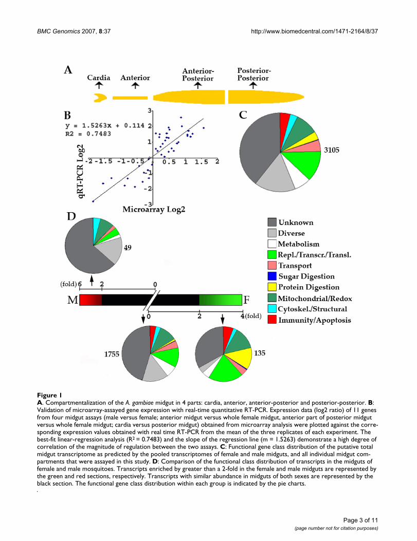
A. Compartmentalization of the A. gambiae midgut in 4 parts: cardia, anterior, anterior-posterior and posterior-posteriorFigure 1A. Compartmentalization of the A. gambiae midgut in 4 parts: cardia, anterior, anterior-posterior and posterior-posterior. B: Validation of microarray-assayed gene expression with real-time quantitative RT-PCR. Expression data (log2 ratio) of 11 genes from four midgut assays (male versus female; anterior midgut versus whole female midgut, anterior part of posterior midgut versus whole female midgut; cardia versus posterior midgut) obtained from microarray analysis were plotted against the corre-sponding expression values obtained with real time RT-PCR from the mean of the three replicates of each experiment. The best-fit linear-regression analysis (R2 = 0.7483) and the slope of the regression line (m = 1.5263) demonstrate a high degree of correlation of the magnitude of regulation between the two assays. C: Functional gene class distribution of the putative total midgut transcriptome as predicted by the pooled transcriptomes of female and male midguts, and all individual midgut com-partments that were assayed in this study. D: Comparison of the functional class distribution of transcripts in the midguts of female and male mosquitoes. Transcripts enriched by greater than a 2-fold in the female and male midguts are represented by the green and red sections, respectively. Transcripts with similar abundance in midguts of both sexes are represented by the black section. The functional gene class distribution within each group is indicated by the pie charts.
BMC Genomics 2007, 8:37 http://www.biomedcentral.com/1471-2164/8/37
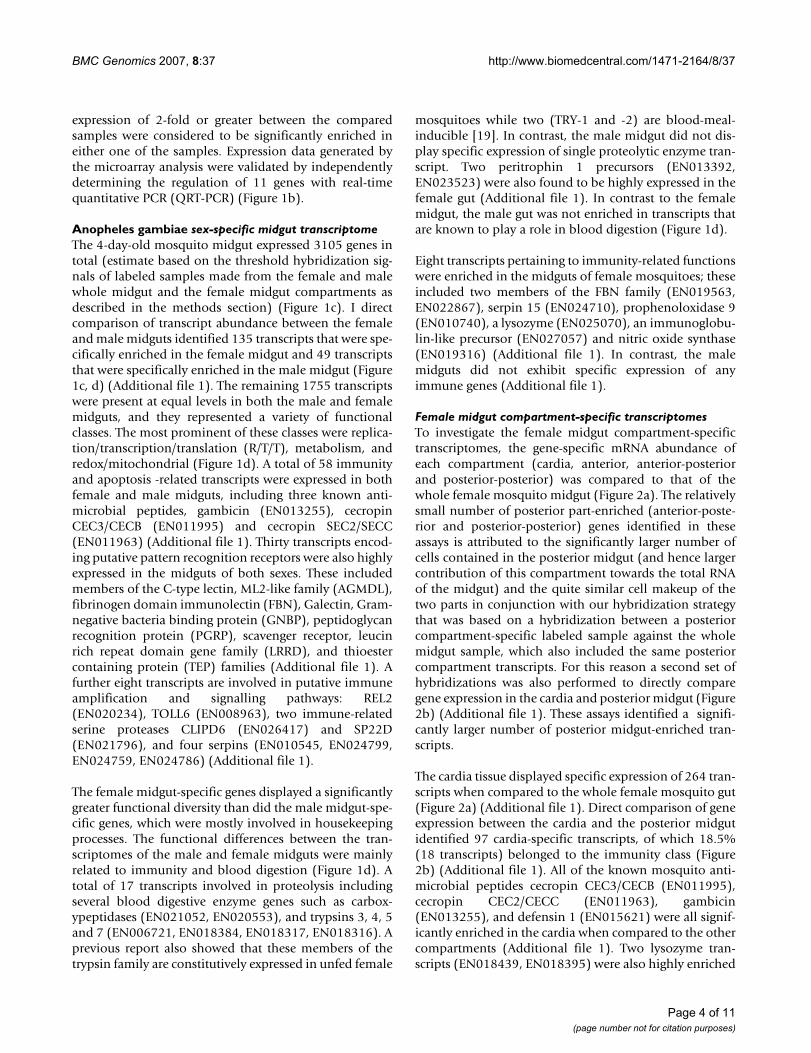
expression of 2-fold or greater between the comparedsamples were considered to be significantly enriched ineither one of the samples. Expression data generated bythe microarray analysis were validated by independentlydetermining the regulation of 11 genes with real-timequantitative PCR (QRT-PCR) (Figure 1b).
Anopheles gambiae sex-specific midgut transcriptomeThe 4-day-old mosquito midgut expressed 3105 genes intotal (estimate based on the threshold hybridization sig-nals of labeled samples made from the female and malewhole midgut and the female midgut compartments asdescribed in the methods section) (Figure 1c). I directcomparison of transcript abundance between the femaleand male midguts identified 135 transcripts that were spe-cifically enriched in the female midgut and 49 transcriptsthat were specifically enriched in the male midgut (Figure1c, d) (Additional file 1). The remaining 1755 transcriptswere present at equal levels in both the male and femalemidguts, and they represented a variety of functionalclasses. The most prominent of these classes were replica-tion/transcription/translation (R/T/T), metabolism, andredox/mitochondrial (Figure 1d). A total of 58 immunityand apoptosis -related transcripts were expressed in bothfemale and male midguts, including three known anti-microbial peptides, gambicin (EN013255), cecropinCEC3/CECB (EN011995) and cecropin SEC2/SECC(EN011963) (Additional file 1). Thirty transcripts encod-ing putative pattern recognition receptors were also highlyexpressed in the midguts of both sexes. These includedmembers of the C-type lectin, ML2-like family (AGMDL),fibrinogen domain immunolectin (FBN), Galectin, Gram-negative bacteria binding protein (GNBP), peptidoglycanrecognition protein (PGRP), scavenger receptor, leucinrich repeat domain gene family (LRRD), and thioestercontaining protein (TEP) families (Additional file 1). Afurther eight transcripts are involved in putative immuneamplification and signalling pathways: REL2(EN020234), TOLL6 (EN008963), two immune-relatedserine proteases CLIPD6 (EN026417) and SP22D(EN021796), and four serpins (EN010545, EN024799,EN024759, EN024786) (Additional file 1).
The female midgut-specific genes displayed a significantlygreater functional diversity than did the male midgut-spe-cific genes, which were mostly involved in housekeepingprocesses. The functional differences between the tran-scriptomes of the male and female midguts were mainlyrelated to immunity and blood digestion (Figure 1d). Atotal of 17 transcripts involved in proteolysis includingseveral blood digestive enzyme genes such as carbox-ypeptidases (EN021052, EN020553), and trypsins 3, 4, 5and 7 (EN006721, EN018384, EN018317, EN018316). Aprevious report also showed that these members of thetrypsin family are constitutively expressed in unfed female
mosquitoes while two (TRY-1 and -2) are blood-meal-inducible [19]. In contrast, the male midgut did not dis-play specific expression of single proteolytic enzyme tran-script. Two peritrophin 1 precursors (EN013392,EN023523) were also found to be highly expressed in thefemale gut (Additional file 1). In contrast to the femalemidgut, the male gut was not enriched in transcripts thatare known to play a role in blood digestion (Figure 1d).
Eight transcripts pertaining to immunity-related functionswere enriched in the midguts of female mosquitoes; theseincluded two members of the FBN family (EN019563,EN022867), serpin 15 (EN024710), prophenoloxidase 9(EN010740), a lysozyme (EN025070), an immunoglobu-lin-like precursor (EN027057) and nitric oxide synthase(EN019316) (Additional file 1). In contrast, the malemidguts did not exhibit specific expression of anyimmune genes (Additional file 1).
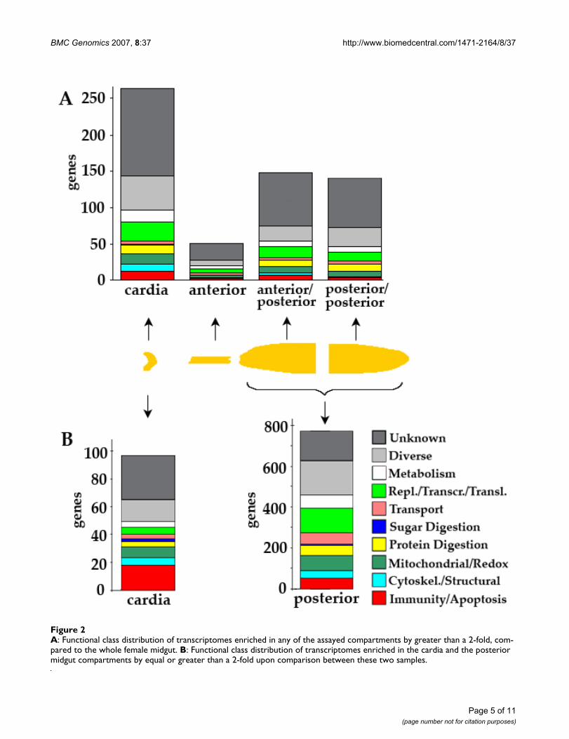
Female midgut compartment-specific transcriptomesTo investigate the female midgut compartment-specifictranscriptomes, the gene-specific mRNA abundance ofeach compartment (cardia, anterior, anterior-posteriorand posterior-posterior) was compared to that of thewhole female mosquito midgut (Figure 2a). The relativelysmall number of posterior part-enriched (anterior-poste-rior and posterior-posterior) genes identified in theseassays is attributed to the significantly larger number ofcells contained in the posterior midgut (and hence largercontribution of this compartment towards the total RNAof the midgut) and the quite similar cell makeup of thetwo parts in conjunction with our hybridization strategythat was based on a hybridization between a posteriorcompartment-specific labeled sample against the wholemidgut sample, which also included the same posteriorcompartment transcripts. For this reason a second set ofhybridizations was also performed to directly comparegene expression in the cardia and posterior midgut (Figure2b) (Additional file 1). These assays identified a signifi-cantly larger number of posterior midgut-enriched tran-scripts.
The cardia tissue displayed specific expression of 264 tran-scripts when compared to the whole female mosquito gut(Figure 2a) (Additional file 1). Direct comparison of geneexpression between the cardia and the posterior midgutidentified 97 cardia-specific transcripts, of which 18.5%(18 transcripts) belonged to the immunity class (Figure2b) (Additional file 1). All of the known mosquito anti-microbial peptides cecropin CEC3/CECB (EN011995),cecropin CEC2/CECC (EN011963), gambicin(EN013255), and defensin 1 (EN015621) were all signif-icantly enriched in the cardia when compared to the othercompartments (Additional file 1). Two lysozyme tran-scripts (EN018439, EN018395) were also highly enriched
Page 4 of 11(page number not for citation purposes)
BMC Genomics 2007, 8:37 http://www.biomedcentral.com/1471-2164/8/37
Page 5 of 11(page number not for citation purposes)
A: Functional class distribution of transcriptomes enriched in any of the assayed compartments by greater than a 2-fold, com-pared to the whole female midgutFigure 2A: Functional class distribution of transcriptomes enriched in any of the assayed compartments by greater than a 2-fold, com-pared to the whole female midgut. B: Functional class distribution of transcriptomes enriched in the cardia and the posterior midgut compartments by equal or greater than a 2-fold upon comparison between these two samples.
BMC Genomics 2007, 8:37 http://www.biomedcentral.com/1471-2164/8/37
in the cardia. Expression of Gambicin and defensin haspreviously been localized to the cardia [17], and boththese genes have been shown to be induced in the midgutupon invasion by malarial parasites [1,2]. Defensin hasbeen shown to be expressed by the anterior midgut ofanother hematophagous insect, Stomoxys calcitrans [32].The cardia showed highly enriched expression of sevenanti-Plasmodium factors including Tep1 (EN016857),LRRD7 (EN021822), FBN8 (EN011252), FBN9(EN011248), SP CLIP1 (EN020158) and Gambicin(EN013255) [4,33,34](Dimopoulos lab, unpublisheddata).
The anterior compartment, which mainly serves as a tubeto allow the blood to enter the posterior section, dis-played the smallest number of enriched genes: only 51transcripts of varying functions (Figure 2a) (Additionalfile 1). Three of these genes encoded proteins involved inimmune defense: TEP3 (EN016283), TEP16 (EN021282),and PGRPLB (EN013948) (Additional file 1).
The anterior part of the posterior midgut displayedenriched expression of 148 transcripts with quite diversefunctions (Figure 2a). Transcripts that play a role in prote-olysis, basic metabolism, and immunity represented a sig-nificant portion of these transcripts (Figure 2b, Additionalfile 1). Among 7 immunity-related transcripts were 3 anti-Plasmodium factors, TEP1 (EN016857), NOS (EN019316)and FBN7 (EN008282) [3,33](Dimopoulos lab, unpub-lished data). The most posterior compartment, the poste-rior part of the posterior midgut, was enriched for 142transcripts that had at least a two-fold higher expression inthis compartment than in the whole female gut and had afunctional class distribution with similar proportions tothat of the anterior part of the posterior midgut, while theactual genes are different (Figure 2a, b). Only 4 transcriptsencoding potential immune-related factors were morespecifically expressed in this compartment and none ofthose represented known anti-Plasmodium factors, sug-gesting that the anterior parts of the midgut is potentiallymore active in anti-Plasmodium gene expression (Addi-tional file 1).
A direct comparison of gene expression between the car-dia and the posterior midgut identified 867 differentiallyexpressed genes, of which 770 were enriched in the poste-rior midgut (Figure 2b). A major functional group of theposterior midgut was represented by 49 transcripts encod-ing proteolytic enzymes; many of those are know to beinvolved in blood digestion. (Additional file 1). It has pre-viously been shown that several blood digestive proteasesare expressed prior to feeding in order to be immediatelypresent upon blood ingestion [17-19]. Several of theseproteolytic enzymes may be involved in diverse processessuch as signal transduction, extracellular matrix process-
ing and immunity. The posterior midgut displayed ele-vated expression of 52 transcripts involved in immunity(6.7% of total posterior midgut specific genes) (Figure 2b,Additional file 1). As many as 34 of these putativeimmune genes encoded putative pattern recognitionreceptors, including two Plasmodium protective c-typelectins CTL4 (EN021138) and CTLMA2 (EN020902)(Additional file 1).
Peptidomic analysis of peptides within the cardia and posterior compartmentsThe transcription analyses indicated the cardia as a majorsite of antimicrobial gene expression. In order to identifypotential novel antimicrobial peptides, we employed apepdidomic approach using 2-dimensional liquid chro-matography tandem mass spectrometry (LC/MS/MS) toscreen for putative secreted short peptides that areenriched in the cardia compartments of the female mos-quito midgut. The selected approach allowed the sampleto be fractionated into several reversed-phase runs, givingbetter coverage by virtue of less complexity in any givenfraction. This was, however, not a comprehensive analysisof all the proteins/peptides that are present in the cardiasince a protein extraction method that is biased towardsshort peptides was used.
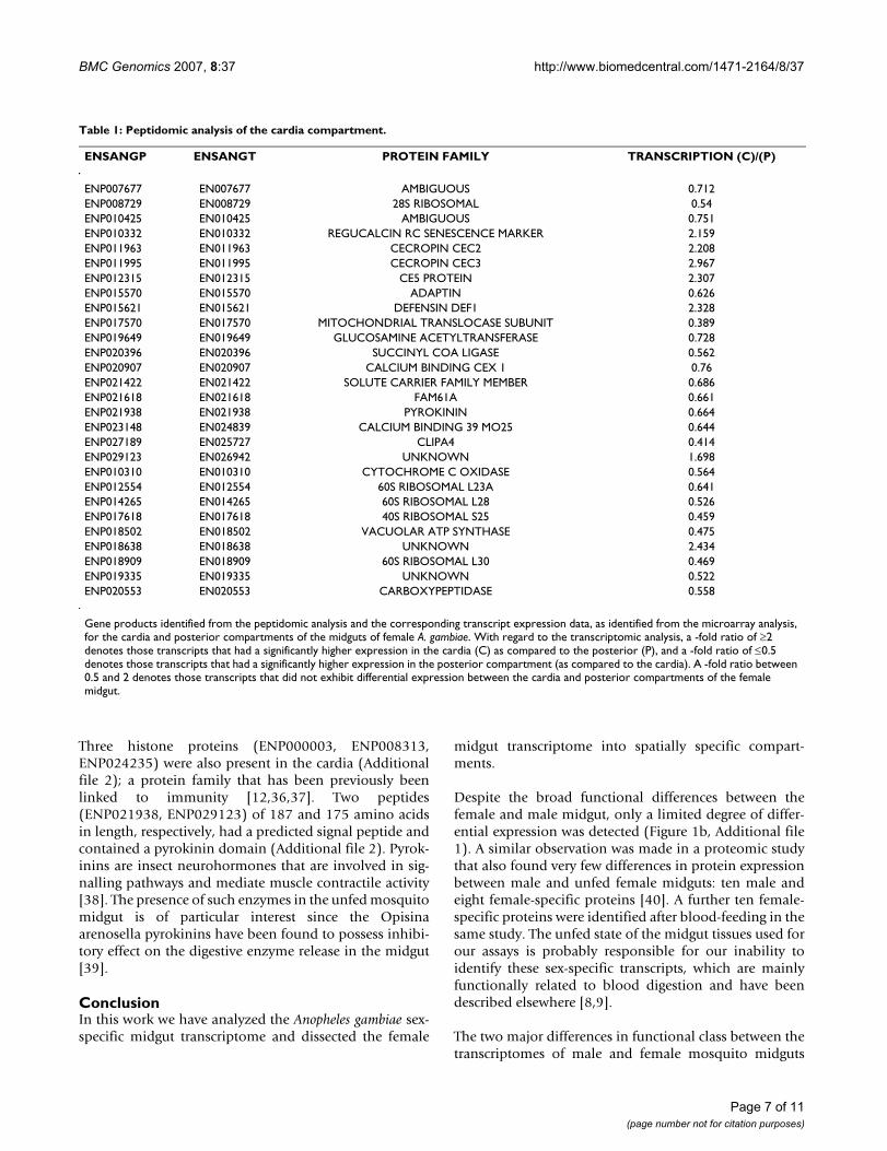
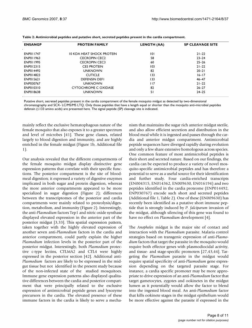
A total of 94 proteins were identified in the cardia peptideextract (Additional file 2). The peptidomic analysis wasoverlaid with the transcriptomic data to identify thosegene products that have corresponding data: 19.6% of theidentified peptides had corresponding transcript expres-sion data (Table 1). The relatively small overlap betweentranscriptome and peptidome data is most likely attrib-uted to differences between mRNA and protein turnoverrates and abundance that therefore does not permit detec-tion by either one of the two methods. Ten peptides wereidentified in the cardia that had both a length of less than133 amino acids and a signal peptide, as predicted by Sig-nalP 3.0 server (Technical University of Denmark DTU)(Table 2). Seven of these peptides are annotated inEnsembl and include three of the known mosquito anti-microbial peptides (cecropins CEC2, CEC3, and defensin)(Table 2). A heat shock protein (ENP011747), a cuticleprotein (ENP014823), and a salivary gland transcript ofunknown function CE5 (ENP012315) were also amongthese putative short secreted peptides (Table 2). BLASTanalysis of the CE5 amino acid sequence revealed that ithas a 52% identity to an anti-thrombin protein found inthe salivary gland of Anopheles stephensi [35]. Cuticle pro-teins have previously been linked to immune defensereactions in female A. gambiae [12]. The remaining threeputative short secreted peptides (ENP014492,ENP030767, ENP018638) are of unknown function.
Page 6 of 11(page number not for citation purposes)
BMC Genomics 2007, 8:37 http://www.biomedcentral.com/1471-2164/8/37
Three histone proteins (ENP000003, ENP008313,ENP024235) were also present in the cardia (Additionalfile 2); a protein family that has been previously beenlinked to immunity [12,36,37]. Two peptides(ENP021938, ENP029123) of 187 and 175 amino acidsin length, respectively, had a predicted signal peptide andcontained a pyrokinin domain (Additional file 2). Pyrok-inins are insect neurohormones that are involved in sig-nalling pathways and mediate muscle contractile activity[38]. The presence of such enzymes in the unfed mosquitomidgut is of particular interest since the Opisinaarenosella pyrokinins have been found to possess inhibi-tory effect on the digestive enzyme release in the midgut[39].
ConclusionIn this work we have analyzed the Anopheles gambiae sex-specific midgut transcriptome and dissected the female
midgut transcriptome into spatially specific compart-ments.
Despite the broad functional differences between thefemale and male midgut, only a limited degree of differ-ential expression was detected (Figure 1b, Additional file1). A similar observation was made in a proteomic studythat also found very few differences in protein expressionbetween male and unfed female midguts: ten male andeight female-specific proteins [40]. A further ten female-specific proteins were identified after blood-feeding in thesame study. The unfed state of the midgut tissues used forour assays is probably responsible for our inability toidentify these sex-specific transcripts, which are mainlyfunctionally related to blood digestion and have beendescribed elsewhere [8,9].
The two major differences in functional class between thetranscriptomes of male and female mosquito midguts
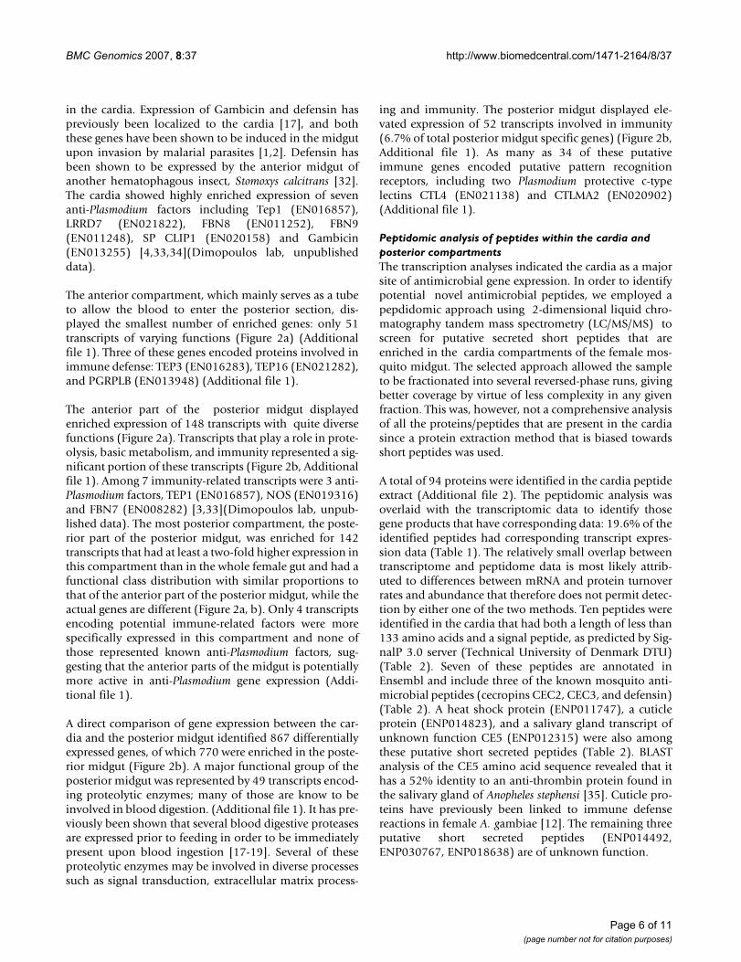
Table 1: Peptidomic analysis of the cardia compartment.
ENSANGP ENSANGT PROTEIN FAMILY TRANSCRIPTION (C)/(P)
ENP007677 EN007677 AMBIGUOUS 0.712ENP008729 EN008729 28S RIBOSOMAL 0.54ENP010425 EN010425 AMBIGUOUS 0.751ENP010332 EN010332 REGUCALCIN RC SENESCENCE MARKER 2.159ENP011963 EN011963 CECROPIN CEC2 2.208ENP011995 EN011995 CECROPIN CEC3 2.967ENP012315 EN012315 CE5 PROTEIN 2.307ENP015570 EN015570 ADAPTIN 0.626ENP015621 EN015621 DEFENSIN DEF1 2.328ENP017570 EN017570 MITOCHONDRIAL TRANSLOCASE SUBUNIT 0.389ENP019649 EN019649 GLUCOSAMINE ACETYLTRANSFERASE 0.728ENP020396 EN020396 SUCCINYL COA LIGASE 0.562ENP020907 EN020907 CALCIUM BINDING CEX 1 0.76ENP021422 EN021422 SOLUTE CARRIER FAMILY MEMBER 0.686ENP021618 EN021618 FAM61A 0.661ENP021938 EN021938 PYROKININ 0.664ENP023148 EN024839 CALCIUM BINDING 39 MO25 0.644ENP027189 EN025727 CLIPA4 0.414ENP029123 EN026942 UNKNOWN 1.698ENP010310 EN010310 CYTOCHROME C OXIDASE 0.564ENP012554 EN012554 60S RIBOSOMAL L23A 0.641ENP014265 EN014265 60S RIBOSOMAL L28 0.526ENP017618 EN017618 40S RIBOSOMAL S25 0.459ENP018502 EN018502 VACUOLAR ATP SYNTHASE 0.475ENP018638 EN018638 UNKNOWN 2.434ENP018909 EN018909 60S RIBOSOMAL L30 0.469ENP019335 EN019335 UNKNOWN 0.522ENP020553 EN020553 CARBOXYPEPTIDASE 0.558
Gene products identified from the peptidomic analysis and the corresponding transcript expression data, as identified from the microarray analysis, for the cardia and posterior compartments of the midguts of female A. gambiae. With regard to the transcriptomic analysis, a -fold ratio of ≥2 denotes those transcripts that had a significantly higher expression in the cardia (C) as compared to the posterior (P), and a -fold ratio of ≤0.5 denotes those transcripts that had a significantly higher expression in the posterior compartment (as compared to the cardia). A -fold ratio between 0.5 and 2 denotes those transcripts that did not exhibit differential expression between the cardia and posterior compartments of the female midgut.
Page 7 of 11(page number not for citation purposes)
BMC Genomics 2007, 8:37 http://www.biomedcentral.com/1471-2164/8/37
mainly reflect the exclusive hematophagous nature of thefemale mosquito that also exposes it to a greater spectrumand level of microbes [41]. These gene classes, relatedlargely to blood digestion and immunity, and are highlyenriched in the female midgut (Figure 1b, Additional file1).
Our analysis revealed that the different compartments ofthe female mosquito midgut display distinctive geneexpression patterns that correlate with their specific func-tions. The posterior compartment is the site of blood-meal digestion; it expressed a variety of digestive enzymesimplicated in both sugar and protein digestion, whereasthe more anterior compartments appeared to be morespecialized in sugar digestion (Figure 2); differencesbetween the transcriptomes of the posterior and cardiacompartments were mainly related to proteolysis/diges-tion, metabolism, and immunity (Figure 2). Interestingly,the anti-Plasmodium factors Tep1 and nitric oxide synthasedisplayed elevated expression in the anterior part of theposterior midgut [3,33]. This spatial expression pattern,taken together with the highly elevated expression ofanother seven anti-Plasmodium factors in the cardia andanterior compartment, could partly explain the higherPlasmodium infection levels in the posterior part of theposterior midgut. Interestingly, both Plasmodium protec-tive c-type lectins, CTLMA2 and CTL4 were highlyexpressed in the posterior section [42]. Additional anti-Plasmodium factors are likely to be expressed in the mid-gut tissue but not identified in the present study becauseof the non-infected state of the studied mosquitoes.Immune gene expression patterns also displayed qualita-tive differences between the cardia and posterior compart-ment that were principally related to the exclusiveexpression of antimicrobial peptide genes and lysozymeprecursors in the cardia. The elevated presence of theseimmune factors in the cardia is likely to serve a mecha-
nism that maintains the sugar rich anterior midgut sterile,and also allow efficient secretion and distribution in theblood meal while it is ingested and passes through the car-dia and anterior midgut compartment. Antimicrobialpeptide sequences have diverged rapidly during evolutionand only a few share extensive homologous across species.One common feature of most antimicrobial peptides istheir short and secreted nature. Based on our findings, thecardia can be expected to produce a variety of novel mos-quito-specific antimicrobial peptides and has therefore apotential to serve as a useful source for their identificationand further study. Four cardia-enriched transcripts(EN004315, EN014362, EN009630, EN016194) and twopeptides identified in the cardia proteome (ENP014492,ENP030767) encode such short and secreted peptides(Additional file 1, Table 2). One of these (EN009630) hasrecently been identified as a putative short immune pep-tide that is strongly induced by P. falciparum invasion ofthe midgut, although silencing of this gene was found tohave no effect on Plasmodium development [4].
The Anopheles midgut is the major site of contact andinteraction with the Plasmodium parasite. Malaria controlstrategies based on transgenic expression of anti-Plasmo-dium factors that target the parasite in the mosquito wouldrequire both effector genes with plasmodiocidal activity,and tissue- and stage-specific promoters [27,43,44]. Tar-geting the Plasmodium parasite in the midgut wouldrequire spatial specificity of anti-Plasmodium gene expres-sion depending on the targeted parasite stage. Forinstance, a cardia specific promoter may be more appro-priate to drive expression of an anti-Plasmodium factor thattarget gametocytes, zygotes and ookinetes in the midgutlumen as it potentially would allow the factor to blendinto the ingested blood meal. An anti-Plasmodium factorthat kills ookinete stages in the midgut epithelium wouldbe more effective against the parasite if expressed in the
Table 2: Antimicrobial peptides and putative short, secreted peptides present in the cardia compartment.
ENSANGP PROTEIN FAMILY LENGTH (AA) SP CLEAVAGE SITE
ENP011747 10 KDA HEAT SHOCK PROTEIN 101 21–22ENP011963 CECROPIN CEC2 58 23–24ENP011995 CECROPIN CEC3 60 25–26ENP012315 CE5 PROTEIN 103 21–22ENP014492 UNKNOWN 82 20–21ENP014823 CUTICLE 133 16–17ENP015621 DEFENSIN DEF1 133 46–47ENP030767 UNKNOWN 117 21–22ENP010310 CYTOCHROME C OXIDASE 82 26–27ENP018638 UNKNOWN 51 24–25
Putative short, secreted peptides present in the cardia compartment of the female mosquito midgut as detected by two-dimensional chromatography and SCX- LC/MS/MS LTQ. Only those peptides that have a length equal or shorter than the mosquito anti-microbial peptides defensin (≤133 amino acids) are presented here. The signal peptide (SP) cleavage site is indicated.
Page 8 of 11(page number not for citation purposes)
BMC Genomics 2007, 8:37 http://www.biomedcentral.com/1471-2164/8/37
posterior compartment which is invaded. This compre-hensive study and other studies have the potential to pro-vide such promoters and anti-Plasmodium factors.
MethodsMosquito rearing and sample collectionA. gambiae Keele strain mosquitoes were raised at 27°Cand 70% humidity, and adults were maintained on a 10%sucrose solution. Midguts from 4-day-old adult mosqui-toes were dissected on ice in PBS (0.6 mM MgCl2, 4 mMKCl, 1.8 mM NaHCO3, 150 mM NaCl, 25 mM HEPES, 1.7mM CaCl2, pH 7) and immediately frozen with dry ice.Total RNA was extracted with either the Mini RNA isola-tion kit (Zymo Research, Orange, CA) or the RNeasy minikit (Qiagen, Valencia, CA) according to the manufac-turer's instructions.
Microarray assaysDouble-stranded cDNA primed with an oligo d(T)-T7promoter, produced from total RNA (2 µg), was used tosynthesize complementary RNA (cRNA) with incorpo-rated Cy-3-dUTP and Cy-5-dUTP fluorescent nucleotides,using the Agilent Low RNA input Fluorescent LinearAmplification Kit (Agilent Technologies, Palo Alto, CA).Unincorporated dye-labeled nucleotides were removedwith the Qiagen PCR purification kit (Qiagen, Chats-worth, CA). A 60-mer oligonucleotide microarray repre-senting the entire A. gambiae transcriptome was used forthese assays [4]. To compare the transcriptomes of themale and female midgut, Cy-5-labeled cDNA targets madefrom the midgut RNA of male mosquitoes were hybrid-ized against a Cy-3-labeled reference probe made from themidgut RNA of female mosquitoes. To assay the transcrip-tomes of the individual compartments of the gut, Cy-5-labeled cDNA targets made from the RNA of female mid-gut compartments (cardia, anterior, anterior-posterior,and posterior-posterior) were hybridized against a Cy-3-labeled reference probe made from the midgut RNA ofwhole female midguts. To compare the gene expression ofthe cardia and the posterior compartments of the midgut,Cy-5-labeled cDNA targets made from the RNA of thefemale cardia midgut compartment were hybridizedagainst a Cy-3-labeled reference probe made from theRNA of the female posterior midgut. Three biological rep-lica assays were performed for each experiment.
Data analysisSpot intensities were measured with a GenePix 4200ALautoloader scanner (Axon Instruments). Images wereinspected manually using GenePix Pro 6.0 software (AxonInstruments), and any spots that were covered withhybridization artefacts were removed and were notincluded in the further analysis. The TIGR MIDAS soft-ware was used to filter the data set using a hybridizationsignal cut-off of 100 units to remove low intensity spots
from the analysis, and Loc-Fit normalization (LOWESS)was performed for all data sets independently to adjust fordye-specific biases. The normalized Cy5/Cy3 ratios fromreplicate assays were subjected to t-tests at a significancelevel of p ≤ 0.05 using TIGR MIDAS and MeV software.The replicate Cy5/Cy3 ratios for each transcript were aver-aged using the GEPAS (Gene Expression Pattern AnalysisSuite v1.1, available free online http://gepas.bioinfo.cipf.es/) after the data set had been processed and fil-tered. Microarray data sets have been submitted to theGEO-NCBI with the following accession numbers:GSM146402, GSM146403, GSM146404, GSM146405,GSM146406, GSM146407, GSM146408, GSM146409,GSM146410, GSM146411, GSM146412, GSM146413,GSM146414, GSM146415, GSM146416, GSM146417,GSM146418, GSM146419.
Real time quantitative PCRRNA samples were treated with Turbo DNase (Ambion)and reverse-transcribed using Superscript III (Invitrogen)with random hexamers. Real-time quantification was per-formed using the QuantiTect SYBR Green PCR Kit (Qia-gen) and ABI Detection System ABI Prism 7000. All PCRreactions were performed in triplicate. Specificity of thePCR reactions was assessed by analysis of melting curvesfor each data point. The ribosomal protein S7 gene wasused for normalization of cDNA templates. Primersequences are listed in table Additional file 4.
2-dimensional chromatography and SCX-LC/MS/MS LTQ analysisMidguts were dissected on a chilled platform, and the car-dia and posterior compartments were collected separatelyon dry ice. The tissue was homogenized in methanol/water/acetic acid (87:8:5 v:v:v), then centrifuged (8000 gfor 10 min at 4°C). Peptides present in the supernatantwere collected, and the pellet was re-homogenized andcentrifuged. The combined supernatants were lyophilizedand stored at -20°C until required. The 2-dimensionalchromatography method used employs a strong cationexchange separation (SCX) (PolyLC 300 um × 100 mm200A 5 um) in the first dimension, followed by reversed-phase chromatography in the second dimension. An addi-tional reversed-phase cleanup step was used to rid thesample of any residual salts that might affect retention onthe SCX column. Samples were bound and eluted from aC18 cartridge (Sep-Pak, Waters), elute, and dried downbefore being re-suspended in 24 µl of 0.1% formic acid.Each sample was then loaded onto the SCX column asthree 8-µl aliquots, back-to-back. Three 8-µl salt elutionsteps were performed with each fraction at 0 mM, 20 mM,30 mM, 40 mM, 125 mM, 250 mM and 1000 mM KCl in0.1% formic acid. Each salt fraction was trapped andeluted onto the reversed-phase column (a C18 7-µM col-umn packed with YMC ODS-AQ 5 µm particle size, 120A
Page 9 of 11(page number not for citation purposes)
BMC Genomics 2007, 8:37 http://www.biomedcentral.com/1471-2164/8/37
pore size). The DBParser program was used to collate thedatabase search results and to remove redundant proteinsand peptide hits that did not meet the data-set signifi-cance threshold. The criteria for protein identificationwere based on more than one peptide hit with the individ-ual peptide scores set above the data-set threshold.
AbbreviationsENSEMBL gene IDs have been abbreviated by omittingthe "NSANG00000" portion.
ENSEMBL protein IDs have been abbreviated by replacingthe "SANGP00000" portion with P.
Authors' contributionsEW, RA and YD dissected the mosquito midgut samplesand prepared RNA. EW performed microarray analysesand real time PCR assays. VM contributed with prelimi-nary data sets of the mosquito midgut transcriptome. EWand GD wrote the manuscript. All authors read andapproved the final manuscript. All authors read andapproved the final manuscript.
Additional material
AcknowledgementsWe thank other members of Dimopoulos lab for fruitful discussions. We thank the Johns Hopkins MRI/MMI Array Core Facility for providing the microarray equipment and the Johns Hopkins Malaria Research Institute insectary core facility for assistance with mosquito rearing. We thank the Johns Hopkins School of Medicine Proteomics Facility for assistance with peptidomic analyses. This work has been supported by the NIH/NIAID 1R01AI061576-01A1, the Ellison Medical Foundation, the WHO/TDR, a Johns Hopkins School of Public Health Faculty Innovation Grant and the Johns Hopkins Malaria Research Institute.
References1. Richman AM, Dimopoulos G, Seeley D, Kafatos FC: Plasmodium
activates the innate immune response of Anopheles gam-biae mosquitoes. Embo J 1997, 16(20):6114-6119.
2. Dimopoulos G, Richman A, Muller HM, Kafatos FC: Molecularimmune responses of the mosquito Anopheles gambiae tobacteria and malaria parasites. Proc Natl Acad Sci U S A 1997,94(21):11508-11513.
3. Luckhart S, Vodovotz Y, Cui L, Rosenberg R: The mosquitoAnopheles stephensi limits malaria parasite developmentwith inducible synthesis of nitric oxide. Proc Natl Acad Sci U S A1998, 95(10):5700-5705.
4. Dong Y, Aguilar R, Xi Z, Warr E, Mongin E, Dimopoulos G: Anoph-eles gambiae immune responses to human and rodent Plas-modium parasite species. PLoS Pathog 2006, 2(6):e52.
5. Aguilar R, Dong Y, Warr E, Dimopoulos G: Anopheles infectionresponses; laboratory models versus field malaria transmis-sion systems. Acta Trop 2005, 95(3):285-291.
6. Vlachou D, Schlegelmilch T, Christophides GK, Kafatos FC: Func-tional genomic analysis of midgut epithelial responses inAnopheles during Plasmodium invasion. Curr Biol 2005,15(13):1185-1195.
7. Xu X, Dong Y, Abraham EG, Kocan A, Srinivasan P, Ghosh AK, SindenRE, Ribeiro JM, Jacobs-Lorena M, Kafatos FC, et al.: Transcriptomeanalysis of Anopheles stephensi-Plasmodium berghei inter-actions. Mol Biochem Parasitol 2005, 142(1):76-87.
8. Dana AN, Hong YS, Kern MK, Hillenmeyer ME, Harker BW, LoboNF, Hogan JR, Romans P, Collins FH: Gene expression patternsassociated with blood-feeding in the malaria mosquitoAnopheles gambiae. BMC Genomics 2005, 6(1):5.
9. Sanders HR, Evans AM, Ross LS, Gill SS: Blood meal induces glo-bal changes in midgut gene expression in the disease vector,Aedes aegypti. Insect Biochem Mol Biol 2003, 33(11):1105-1122.
10. Marinotti O, Nguyen QK, Calvo E, James AA, Ribeiro JM: Microar-ray analysis of genes showing variable expression following ablood meal in Anopheles gambiae. Insect Mol Biol 2005,14(4):365-373.
11. Dimopoulos G, Seeley D, Wolf A, Kafatos FC: Malaria infection ofthe mosquito Anopheles gambiae activates immune-respon-sive genes during critical transition stages of the parasite lifecycle. Embo J 1998, 17(21):6115-6123.
12. Aguilar R, Jedlicka AE, Mintz M, Mahairaki V, Scott AL, DimopoulosG: Global gene expression analysis of Anopheles gambiaeresponses to microbial challenge. Insect Biochem Mol Biol 2005,35(7):709-719.
Additional File 1The midgut transcriptome. Processed microarray generated comparative gene expression ratios for male versus female comparisons, female midgut compartment (cardia, anterior, anterior-posterior, posterior-posterior) versus whole female midgut, and female midgut cardia compartment ver-sus posterior midgut.Click here for file[http://www.biomedcentral.com/content/supplementary/1471-2164-8-37-S1.xls]
Additional File 2The cardia peptidome. Peptidomic analysis of the cardia tissue identified 94 putative proteins. Transcript (ENSANGT) gene (ENSANGG) and protein (ENSANGP) identifiers are indicated. The length (AA), pre-dicted signal peptide (SP) and putative protein family/homology are indi-cated.Click here for file[http://www.biomedcentral.com/content/supplementary/1471-2164-8-37-S2.xls]
Additional File 4Real time RT-PCR primers. DNA sequences of primers used for real time RT-PCR analyses.Click here for file[http://www.biomedcentral.com/content/supplementary/1471-2164-8-37-S4.pdf]
Additional File 3Microarray data validation with real time RT-PCR. Validation of micro-array-assayed gene expression with real-time quantitative RT-PCR. Expression data (log2 ratio) of 11 genes from four midgut assays (male versus female; versus whole female midgut, anterior-posterior versus whole female midgut; cardia versus posterior midgut) obtained from microarray analysis were validated with 3 replica real time RT-PCR assays and pre-sented in figure 1A. m, male gut; f, female gut; c, cardia; p, posterior; a, anterior; a/p, anterior-posterior.Click here for file[http://www.biomedcentral.com/content/supplementary/1471-2164-8-37-S3.pdf]
Page 10 of 11(page number not for citation purposes)
http://www.ncbi.nlm.nih.gov/entrez/query.fcgi?cmd=Retrieve&db=PubMed&dopt=Abstract&list_uids=9321391
http://www.ncbi.nlm.nih.gov/entrez/query.fcgi?cmd=Retrieve&db=PubMed&dopt=Abstract&list_uids=9321391
http://www.ncbi.nlm.nih.gov/entrez/query.fcgi?cmd=Retrieve&db=PubMed&dopt=Abstract&list_uids=9321391
http://www.ncbi.nlm.nih.gov/entrez/query.fcgi?cmd=Retrieve&db=PubMed&dopt=Abstract&list_uids=9326640
http://www.ncbi.nlm.nih.gov/entrez/query.fcgi?cmd=Retrieve&db=PubMed&dopt=Abstract&list_uids=9326640
http://www.ncbi.nlm.nih.gov/entrez/query.fcgi?cmd=Retrieve&db=PubMed&dopt=Abstract&list_uids=9326640
http://www.ncbi.nlm.nih.gov/entrez/query.fcgi?cmd=Retrieve&db=PubMed&dopt=Abstract&list_uids=9576947
http://www.ncbi.nlm.nih.gov/entrez/query.fcgi?cmd=Retrieve&db=PubMed&dopt=Abstract&list_uids=9576947
http://www.ncbi.nlm.nih.gov/entrez/query.fcgi?cmd=Retrieve&db=PubMed&dopt=Abstract&list_uids=9576947
http://www.ncbi.nlm.nih.gov/entrez/query.fcgi?cmd=Retrieve&db=PubMed&dopt=Abstract&list_uids=9799221
http://www.ncbi.nlm.nih.gov/entrez/query.fcgi?cmd=Retrieve&db=PubMed&dopt=Abstract&list_uids=9799221
BMC Genomics 2007, 8:37 http://www.biomedcentral.com/1471-2164/8/37
Publish with BioMed Central and every scientist can read your work free of charge
"BioMed Central will be the most significant development for disseminating the results of biomedical research in our lifetime."
Sir Paul Nurse, Cancer Research UK
Your research papers will be:
available free of charge to the entire biomedical community
peer reviewed and published immediately upon acceptance
cited in PubMed and archived on PubMed Central
yours — you keep the copyright
Submit your manuscript here:http://www.biomedcentral.com/info/publishing_adv.asp
BioMedcentral
13. Okuda K, Caroci A, Ribolla P, Marinotti O, de Bianchi AG, BijovskyAT: Morphological and enzymatic analysis of the midgut ofAnopheles darlingi during blood digestion. J Insect Physiol 2005,51(7):769-776.
14. Pennington DA, Clegg W, Coles SJ, Harrington RW, Hursthouse MB,Hughes DL, Light ME, Schormann M, Bochmann M, Lancaster SJ: Thesynthesis, structure and ethene polymerisation catalysis ofmono(salicylaldiminato) titanium and zirconium complexes.Dalton Trans 2005:561-571.
15. Billingsley PF, Hecker H: Blood digestion in the mosquito,Anopheles stephensi Liston (Diptera: Culicidae): activity anddistribution of trypsin, aminopeptidase, and alpha-glucosi-dase in the midgut. J Med Entomol 1991, 28(6):865-871.
16. Okuda K, de Souza Caroci A, Ribolla PE, de Bianchi AG, Bijovsky AT:Functional morphology of adult female Culex quinquefascia-tus midgut during blood digestion. Tissue Cell 2002,34(3):210-219.
17. Vizioli J, Catteruccia F, della Torre A, Reckmann I, Muller HM: Blooddigestion in the malaria mosquito Anopheles gambiae:molecular cloning and biochemical characterization of twoinducible chymotrypsins. Eur J Biochem 2001,268(14):4027-4035.
18. Muller HM, Crampton JM, della Torre A, Sinden R, Crisanti A: Mem-bers of a trypsin gene family in Anopheles gambiae areinduced in the gut by blood meal. Embo J 1993,12(7):2891-2900.
19. Muller HM, Catteruccia F, Vizioli J, della Torre A, Crisanti A: Consti-tutive and blood meal-induced trypsin genes in Anophelesgambiae. Exp Parasitol 1995, 81(3):371-385.
20. Huber M, Cabib E, Miller LH: Malaria parasite chitinase and pen-etration of the mosquito peritrophic membrane. Proc NatlAcad Sci U S A 1991, 88(7):2807-2810.
21. Shahabuddin M, Toyoshima T, Aikawa M, Kaslow DC: Transmis-sion-blocking activity of a chitinase inhibitor and activationof malarial parasite chitinase by mosquito protease. Proc NatlAcad Sci U S A 1993, 90(9):4266-4270.
22. Shahabuddin M, Kaslow DC: Plasmodium: parasite chitinase andits role in malaria transmission. Exp Parasitol 1994, 79(1):85-88.
23. Tellam RL, Wijffels G, Willadsen P: Peritrophic matrix proteins.Insect Biochem Mol Biol 1999, 29(2):87-101.
24. Villalon JM, Ghosh A, Jacobs-Lorena M: The peritrophic matrixlimits the rate of digestion in adult Anopheles stephensi andAedes aegypti mosquitoes. J Insect Physiol 2003, 49(10):891-895.
25. Devenport M, Fujioka H, Jacobs-Lorena M: Storage and secretionof the peritrophic matrix protein Ag-Aper1 and trypsin inthe midgut of Anopheles gambiae. Insect Mol Biol 2004,13(4):349-358.
26. Filho BP, Lemos FJ, Secundino NF, Pascoa V, Pereira ST, Pimenta PF:Presence of chitinase and beta-N-acetylglucosaminidase inthe Aedes aegypti. a chitinolytic system involving peri-trophic matrix formation and degradation. Insect Biochem MolBiol 2002, 32(12):1723-1729.
27. Abraham EG, Donnelly-Doman M, Fujioka H, Ghosh A, Moreira L,Jacobs-Lorena M: Driving midgut-specific expression andsecretion of a foreign protein in transgenic mosquitoes withAgAper1 regulatory elements. Insect Mol Biol 2005,14(3):271-279.
28. Shao L, Devenport M, Fujioka H, Ghosh A, Jacobs-Lorena M: Identi-fication and characterization of a novel peritrophic matrixprotein, Ae-Aper50, and the microvillar membrane protein,AEG12, from the mosquito, Aedes aegypti. Insect Biochem MolBiol 2005, 35(9):947-959.
29. Cociancich SO, Park SS, Fidock DA, Shahabuddin M: VesicularATPase-overexpressing cells determine the distribution ofmalaria parasite oocysts on the midguts of mosquitoes. J BiolChem 1999, 274(18):12650-12655.
30. Shahabuddin M: Plasmodium ookinete development in themosquito midgut: a case of reciprocal manipulation. Parasitol-ogy 1998, 116(Suppl):S83-93.
31. Shahabuddin M: Do Plasmodium ookinetes invade a specificcell type in the mosquito midgut? Trends Parasitol 2002,18(4):157-161.
32. Lehane MJ, Wu D, Lehane SM: Midgut-specific immune mole-cules are produced by the blood-sucking insect Stomoxyscalcitrans. Proc Natl Acad Sci U S A 1997, 94(21):11502-11507.
33. Blandin S, Shiao SH, Moita LF, Janse CJ, Waters AP, Kafatos FC,Levashina EA: Complement-like protein TEP1 is a determi-nant of vectorial capacity in the malaria vector Anophelesgambiae. Cell 2004, 116(5):661-670.
34. Vizioli J, Bulet P, Hoffmann JA, Kafatos FC, Muller HM, DimopoulosG: Gambicin: a novel immune responsive antimicrobial pep-tide from the malaria vector Anopheles gambiae. Proc NatlAcad Sci U S A 2001, 98(22):12630-12635.
35. Valenzuela JG, Francischetti IM, Pham VM, Garfield MK, Ribeiro JM:Exploring the salivary gland transcriptome and proteome ofthe Anopheles stephensi mosquito. Insect Biochem Mol Biol 2003,33(7):717-732.
36. Oduol F, Xu J, Niare O, Natarajan R, Vernick KD: Genes identifiedby an expression screen of the vector mosquito Anophelesgambiae display differential molecular immune response tomalaria parasites and bacteria. Proc Natl Acad Sci U S A 2000,97(21):11397-11402.
37. Patat SA, Carnegie RB, Kingsbury C, Gross PS, Chapman R, Schey KL:Antimicrobial activity of histones from hemocytes of thePacific white shrimp. Eur J Biochem 2004, 271(23–24):4825-4833.
38. Nachman RJ, Holman GM, Haddon WF, Vensel WH: An activepseudopeptide analog of the leucokinin insect neuropeptidefamily. Int J Pept Protein Res 1991, 37(3):220-223.
39. Harshini S, Nachman RJ, Sreekumar S: Inhibition of digestiveenzyme release by neuropeptides in larvae of Opisinaarenosella (Lepidoptera: Cryptophasidae). Comp Biochem Phys-iol B Biochem Mol Biol 2002, 132(2):353-358.
40. Prevot GI, Laurent-Winter C, Rodhain F, Bourgouin C: Sex-specificand blood meal-induced proteins of Anopheles gambiae mid-guts: analysis by two-dimensional gel electrophoresis. MalarJ 2003, 2:1.
41. Demaio J, Pumpuni CB, Kent M, Beier JC: The midgut bacterialflora of wild Aedes triseriatus, Culex pipiens, and Psoro-phora columbiae mosquitoes. Am J Trop Med Hyg 1996,54(2):219-223.
42. Osta MA, Christophides GK, Kafatos FC: Effects of mosquitogenes on Plasmodium development. Science 2004,303(5666):2030-2032.
43. Moreira LA, Ghosh AK, Abraham EG, Jacobs-Lorena M: Genetictransformation of mosquitoes: a quest for malaria control.Int J Parasitol 2002, 32(13):1599-1605.
44. Moreira LA, Ito J, Ghosh A, Devenport M, Zieler H, Abraham EG,Crisanti A, Nolan T, Catteruccia F, Jacobs-Lorena M: Bee venomphospholipase inhibits malaria parasite development intransgenic mosquitoes. J Biol Chem 2002, 277(43):40839-40843.
Page 11 of 11(page number not for citation purposes)
http://www.ncbi.nlm.nih.gov/entrez/query.fcgi?cmd=Retrieve&db=PubMed&dopt=Abstract&list_uids=1770523
http://www.ncbi.nlm.nih.gov/entrez/query.fcgi?cmd=Retrieve&db=PubMed&dopt=Abstract&list_uids=1770523
http://www.ncbi.nlm.nih.gov/entrez/query.fcgi?cmd=Retrieve&db=PubMed&dopt=Abstract&list_uids=1770523
http://www.ncbi.nlm.nih.gov/entrez/query.fcgi?cmd=Retrieve&db=PubMed&dopt=Abstract&list_uids=8335004
http://www.ncbi.nlm.nih.gov/entrez/query.fcgi?cmd=Retrieve&db=PubMed&dopt=Abstract&list_uids=8335004
http://www.ncbi.nlm.nih.gov/entrez/query.fcgi?cmd=Retrieve&db=PubMed&dopt=Abstract&list_uids=8335004
http://www.ncbi.nlm.nih.gov/entrez/query.fcgi?cmd=Retrieve&db=PubMed&dopt=Abstract&list_uids=7498434
http://www.ncbi.nlm.nih.gov/entrez/query.fcgi?cmd=Retrieve&db=PubMed&dopt=Abstract&list_uids=7498434
http://www.ncbi.nlm.nih.gov/entrez/query.fcgi?cmd=Retrieve&db=PubMed&dopt=Abstract&list_uids=7498434
http://www.ncbi.nlm.nih.gov/entrez/query.fcgi?cmd=Retrieve&db=PubMed&dopt=Abstract&list_uids=2011589
http://www.ncbi.nlm.nih.gov/entrez/query.fcgi?cmd=Retrieve&db=PubMed&dopt=Abstract&list_uids=2011589
http://www.ncbi.nlm.nih.gov/entrez/query.fcgi?cmd=Retrieve&db=PubMed&dopt=Abstract&list_uids=8483942
http://www.ncbi.nlm.nih.gov/entrez/query.fcgi?cmd=Retrieve&db=PubMed&dopt=Abstract&list_uids=8483942
http://www.ncbi.nlm.nih.gov/entrez/query.fcgi?cmd=Retrieve&db=PubMed&dopt=Abstract&list_uids=8483942
http://www.ncbi.nlm.nih.gov/entrez/query.fcgi?cmd=Retrieve&db=PubMed&dopt=Abstract&list_uids=7914174
http://www.ncbi.nlm.nih.gov/entrez/query.fcgi?cmd=Retrieve&db=PubMed&dopt=Abstract&list_uids=7914174
http://www.ncbi.nlm.nih.gov/entrez/query.fcgi?cmd=Retrieve&db=PubMed&dopt=Abstract&list_uids=9695113
http://www.ncbi.nlm.nih.gov/entrez/query.fcgi?cmd=Retrieve&db=PubMed&dopt=Abstract&list_uids=9695113
http://www.ncbi.nlm.nih.gov/entrez/query.fcgi?cmd=Retrieve&db=PubMed&dopt=Abstract&list_uids=9326639
http://www.ncbi.nlm.nih.gov/entrez/query.fcgi?cmd=Retrieve&db=PubMed&dopt=Abstract&list_uids=9326639
http://www.ncbi.nlm.nih.gov/entrez/query.fcgi?cmd=Retrieve&db=PubMed&dopt=Abstract&list_uids=9326639
http://www.ncbi.nlm.nih.gov/entrez/query.fcgi?cmd=Retrieve&db=PubMed&dopt=Abstract&list_uids=1678383
http://www.ncbi.nlm.nih.gov/entrez/query.fcgi?cmd=Retrieve&db=PubMed&dopt=Abstract&list_uids=1678383
http://www.ncbi.nlm.nih.gov/entrez/query.fcgi?cmd=Retrieve&db=PubMed&dopt=Abstract&list_uids=1678383
http://www.ncbi.nlm.nih.gov/entrez/query.fcgi?cmd=Retrieve&db=PubMed&dopt=Abstract&list_uids=8619452
http://www.ncbi.nlm.nih.gov/entrez/query.fcgi?cmd=Retrieve&db=PubMed&dopt=Abstract&list_uids=8619452
Related Documents