RESEARCH Open Access Short-chain fatty acids can improve lipid and glucose metabolism independently of the pig gut microbiota Hua Zhou 1,2 , Bing Yu 1,2 , Jing Sun 3,4 , Zuohua Liu 3,4 , Hong Chen 5 , Liangpeng Ge 3,4* and Daiwen Chen 1,2* Abstract Background: Previous studies have shown that exogenous short-chain fatty acids (SCFAs) introduction attenuated the body fat deposition in conventional mice and pigs. However, limited studies have evaluated the effects of exogenously introduced SCFAs on the lipid and glucose metabolism independently of the gut microbiota. This study was to investigate the effects of exogenous introduction of SCFAs on the lipid and glucose metabolism in a germ-free (GF) pig model. Methods: Twelve hysterectomy-derived newborn pigs were reared in six sterile isolators. All pigs were hand-fed with sterile milk powder for 21 d, then the sterile feed was introduced to pigs for another 21 d. In the second 21-d period, six pigs were orally administrated with 25 mL/kg sterile saline per day and considered as the GF group, while the other six pigs were orally administrated with 25 mL/kg SCFAs mixture (acetic, propionic, and butyric acids, 45, 15, and 11 mmol/L, respectively) per day and regarded as FA group. Results: Orally administrated with SCFAs tended to increase the adiponectin concentration in serum, enhance the CPT-1 activity in longissimus dorsi, and upregulate the ANGPTL4 mRNA expression level in colon (P < 0.10). Meanwhile, the mRNA abundances of ACC, FAS, and SREBP-1C in liver and CD36 in longissimus dorsi of the FA group were decreased (P < 0.05) compared with those in the GF group. Besides, the mRNA expression of PGC-1α in liver and LPL in longissimus dorsi tended to (P < 0.10) upregulate and downregulate respectively in the FA group. Moreover, oral administration of SCFAs tended to increase the protein level of GPR43 (P < 0.10) and decrease the protein level of ACC (P < 0.10) in liver. Also, oral administration of SCFAs upregulated the p-AMPK/AMPK ratio and the mRNA expressions of GLUT-2 and GYS2 in liver (P < 0.05). In addition, the metabolic pathway associated with the biosynthesis of unsaturated fatty acids was most significantly promoted (P < 0.05) by oral administration of SCFAs. Conclusions: Exogenous introduction of SCFAs might attenuate the fat deposition and to some extent improve the glucose control in the pig model, which occurred independently of the gut microbiota. Keywords: Germ-free, Glucose metabolism, Lipid metabolism, Pig model, Short-chain fatty acids © The Author(s). 2021 Open Access This article is licensed under a Creative Commons Attribution 4.0 International License, which permits use, sharing, adaptation, distribution and reproduction in any medium or format, as long as you give appropriate credit to the original author(s) and the source, provide a link to the Creative Commons licence, and indicate if changes were made. The images or other third party material in this article are included in the article's Creative Commons licence, unless indicated otherwise in a credit line to the material. If material is not included in the article's Creative Commons licence and your intended use is not permitted by statutory regulation or exceeds the permitted use, you will need to obtain permission directly from the copyright holder. To view a copy of this licence, visit http://creativecommons.org/licenses/by/4.0/. The Creative Commons Public Domain Dedication waiver (http://creativecommons.org/publicdomain/zero/1.0/) applies to the data made available in this article, unless otherwise stated in a credit line to the data. * Correspondence: [email protected]; [email protected] 3 Key Laboratory of Pig Industry Sciences, Rongchang 402460, Chongqing, China 1 Key Laboratory of Animal Disease-Resistance Nutrition, Chengdu 611130, Sichuan, China Full list of author information is available at the end of the article Zhou et al. Journal of Animal Science and Biotechnology (2021) 12:61 https://doi.org/10.1186/s40104-021-00581-3

Welcome message from author
This document is posted to help you gain knowledge. Please leave a comment to let me know what you think about it! Share it to your friends and learn new things together.
Transcript
RESEARCH Open Access
Short-chain fatty acids can improve lipidand glucose metabolism independently ofthe pig gut microbiotaHua Zhou1,2, Bing Yu1,2, Jing Sun3,4, Zuohua Liu3,4, Hong Chen5, Liangpeng Ge3,4* and Daiwen Chen1,2*
Abstract
Background: Previous studies have shown that exogenous short-chain fatty acids (SCFAs) introduction attenuatedthe body fat deposition in conventional mice and pigs. However, limited studies have evaluated the effects ofexogenously introduced SCFAs on the lipid and glucose metabolism independently of the gut microbiota. Thisstudy was to investigate the effects of exogenous introduction of SCFAs on the lipid and glucose metabolism in agerm-free (GF) pig model.
Methods: Twelve hysterectomy-derived newborn pigs were reared in six sterile isolators. All pigs were hand-fedwith sterile milk powder for 21 d, then the sterile feed was introduced to pigs for another 21 d. In the second 21-dperiod, six pigs were orally administrated with 25 mL/kg sterile saline per day and considered as the GF group,while the other six pigs were orally administrated with 25 mL/kg SCFAs mixture (acetic, propionic, and butyric acids,45, 15, and 11 mmol/L, respectively) per day and regarded as FA group.
Results: Orally administrated with SCFAs tended to increase the adiponectin concentration in serum, enhance theCPT-1 activity in longissimus dorsi, and upregulate the ANGPTL4 mRNA expression level in colon (P < 0.10).Meanwhile, the mRNA abundances of ACC, FAS, and SREBP-1C in liver and CD36 in longissimus dorsi of the FAgroup were decreased (P < 0.05) compared with those in the GF group. Besides, the mRNA expression of PGC-1α inliver and LPL in longissimus dorsi tended to (P < 0.10) upregulate and downregulate respectively in the FA group.Moreover, oral administration of SCFAs tended to increase the protein level of GPR43 (P < 0.10) and decrease theprotein level of ACC (P < 0.10) in liver. Also, oral administration of SCFAs upregulated the p-AMPK/AMPK ratio andthe mRNA expressions of GLUT-2 and GYS2 in liver (P < 0.05). In addition, the metabolic pathway associated with thebiosynthesis of unsaturated fatty acids was most significantly promoted (P < 0.05) by oral administration of SCFAs.
Conclusions: Exogenous introduction of SCFAs might attenuate the fat deposition and to some extent improvethe glucose control in the pig model, which occurred independently of the gut microbiota.
Keywords: Germ-free, Glucose metabolism, Lipid metabolism, Pig model, Short-chain fatty acids
© The Author(s). 2021 Open Access This article is licensed under a Creative Commons Attribution 4.0 International License,which permits use, sharing, adaptation, distribution and reproduction in any medium or format, as long as you giveappropriate credit to the original author(s) and the source, provide a link to the Creative Commons licence, and indicate ifchanges were made. The images or other third party material in this article are included in the article's Creative Commonslicence, unless indicated otherwise in a credit line to the material. If material is not included in the article's Creative Commonslicence and your intended use is not permitted by statutory regulation or exceeds the permitted use, you will need to obtainpermission directly from the copyright holder. To view a copy of this licence, visit http://creativecommons.org/licenses/by/4.0/.The Creative Commons Public Domain Dedication waiver (http://creativecommons.org/publicdomain/zero/1.0/) applies to thedata made available in this article, unless otherwise stated in a credit line to the data.
* Correspondence: [email protected]; [email protected] Laboratory of Pig Industry Sciences, Rongchang 402460, Chongqing,China1Key Laboratory of Animal Disease-Resistance Nutrition, Chengdu 611130,Sichuan, ChinaFull list of author information is available at the end of the article
Zhou et al. Journal of Animal Science and Biotechnology (2021) 12:61 https://doi.org/10.1186/s40104-021-00581-3
BackgroundEmerging evidence indicated that gut microbiota plays acritical contributor to the host’s health [1]. Noteworthy,the beneficial effects of gut microbiota are at least partlyattributed to the short-chain fatty acids (SCFAs), whichare the end-products produced from the fermentation ofdietary fiber and resistant starch [2, 3]. SCFAs acts aspivotal roles in various biological activities, such as lipidand glucose metabolism [4–6]. Recent studies have re-ported that oral administration of SCFAs increased theenergy expenditure and fat oxidation in obese mice [7],prevented body weight gains, and enhanced insulin sen-sitivity in high-fat diet-fed mice [8]. Moreover, exogen-ously introduced SCFAs reduced the fat deposition inweaned and growing pigs by decreasing lipogenesis andpromoting lipolysis in different tissues [9, 10]. Humanintervention reports found that SCFAs could regulatewhole-body substrates and energy metabolism by in-creasing fasting fat oxidation and resting energy expend-iture [11]. However, controversy still exists consideringthe role of SCFAs in lipid metabolism. Previous workdemonstrated that SCFAs were thought to contributeadditional calories in the obese, thus resulting in add-itional weight gain [12]. Conflicting literature indicatedthat enhanced acetate turnover aggravated the develop-ment of obesity and insulin resistance in rodents [13].The inconsistent effects of SCFAs on lipid and glucosemetabolism might affect by gut microbiota, which playsa vital role in the development and progression of obes-ity [14, 15]. It has been observed that the Christensenellagenus could prevent weight gain in mice [16], and theAkkermansia genus was reported to correlate with lowervisceral fat deposits [17]. Decreased the abundances ofBacteroides and Prevotella were indicated positively cor-related with energy intake and adiposity [18]. Moreover,the numbers of microbiota are closely associated withSCFAs concentrations [19]. Thus, gut microbiota mayinterfere with the effects of exogenous introduction ofSCFAs on the host’s health, and further well-controlledstudies are urgently needed. Germ-free (GF) animals arefree from living microorganisms, including bacteria, vi-ruses, fungi, protozoa, and parasites throughout theirlife, and are reared in sterile environments [20, 21]. Thedomestic pig (Sus scrofa) is a preferable model of humanhealth, which is similar in anatomy, physiology, and gen-etics to humans [22, 23]. Accordingly, the pig with anabsence of gut microbes is the valid experimental modelfor dissecting the effects of exogenously introducedSCFAs on lipid and glucose metabolism. Moreover, thesystematic crosstalk of exogenous SCFAs and the host’slipid and glucose metabolism has been rarely investi-gated in the absence of gut microbiota. Therefore, thisstudy was to take biochemistry and metabolomics ana-lysis to explore the effects of oral administration of
SCFAs on the lipid and glucose metabolism in a GF pigmodel, which may help us to further understand theunderlying mechanisms of SCFAs for alleviating fat de-position and promoting the host health.
Materials and methodsThe experiment was conducted at the ExperimentalSwine Engineering Center of the Chongqing Academy ofAnimal Sciences (CMA No. 162221340234; Chongqing,China).
AnimalsTwelve neonatal GF pigs were delivered via hysterec-tomy from four multiparous Bama sows (a native breedof China). On 112 d of gestation (full-term, 114 d), preg-nant sows were anesthetized with 4% isoflurane, and theuterus was excised from the sow and transferred into asterile isolator (DOSSY Experimental Animals Co., Ltd.,Chengdu, China) through a tank containing 120 L of0.1% peracetic acid for decontamination. Then, 12 neo-natal pigs (male and female in half) were taken from theuterus and transferred to six rearing isolators (Class Bio-logically Clean Ltd., Madison, WI, USA). The rearingisolator contains a checkboard, two pigs per isolator,and fed separately. The rearing isolators had been per-formed by spraying with 1% peracetic acid in advanceand preserved in sterile environments as described previ-ously [21]. The pig’s skin, sterile environments, rectalswabs, and oral mucosa were detected via anaerobic(thioglycollate medium) and aerobic (brain heart infu-sion broth) culture of samples at least every week in ac-cordance with the Chinese National Standard (GB/T14926-41-2001). Colonic digesta was collected at the endof the experiment for further confirmation of sterilestatus.
Experimental design and dietAmong the six rearing isolators, three of them were des-ignated as the FA group, and the other three isolatorswere treated as the GF group. These pigs in the FA andGF groups were hand-fed 60Co-γ-irradiated sterile milkpowder (Table S1) and diluted with sterile water 1:4 for21 d. A corn-soybean feed (Table S2) was formulated ac-cording to the requirements of Chinese FeedingStandards (2004) for local pigs. It was sterilized byCo60-γ-radiation and introduced to the GF and FA pigsfor another 21 d. In the second 21-d period, the FAgroup was orally administrated with sterile 25 mL/kgSCFAs mixture (acetic, propionic, and butyric acids, 45,15, and 11 mmol/L, respectively) per day, and the GFgroup was orally administrated with 25 mL/kg sterile sa-line per day. The introduction volume of SCFAs mixtureor sterile saline for each pig per day is presented inTable S3. Furthermore, the SCFAs mixture was
Zhou et al. Journal of Animal Science and Biotechnology (2021) 12:61 Page 2 of 14
confected in the laminar airflow clean benches, and theacetic, propionic, and butyric acids (analytically pure)was filtered through a 0.22-μm membrane and mixingwith sterile water. During the two 21-d periods, pigswere allowed ad libitum access to sterile water. To main-tain the sterile environment in the present study, whenthe SCFAs, water, milk, and feed in the rearing isolatorswere consumed, the replacement containers for sterileSCFAs, water, milk, and feed were delivered into therearing isolator via the transfer port. Before transferredinto the transfer port, the containers were preliminarilydecontaminated with 0.5% peracetic acid and then spray-ing with 1% peracetic acid.
Sample collectionsIn the morning of d 42, blood was drawn from anteriorvena cava, centrifuged at 3,000×g for 15 min, and storedat − 80 °C for further measurements. After blooding, pigswere euthanized by isoflurane anesthesia. The abdomenwas opened in the laminar airflow clean benches, andthe samples of the colon, liver, and longissimus dorsiwere immediately collected in liquid nitrogen and storedat − 80 °C for further analysis.
Serum parameters measurementThe concentrations of adiponectin, insulin, glucagon,glucagon-like peptide 1, and leptin in serum were de-tected by commercial enzyme-linked immunosorbentassay (ELISA) kits from Chenglin Co. Ltd. (Beijing,China) in accordance with the manufacturer’s instruc-tions. The levels of total cholesterol (TC), triglyceride(TG), high-density lipoprotein-cholesterol (HDL-c), low-density lipoprotein-cholesterol (LDL-c), and glucose inserum were measured using commercial kits (NanjingJiancheng Bioengineering Institute, Nanjing, China) fol-lowing with the manufacturer’s instructions. Each par-ameter was simultaneously measured in triplicate on thesame plate. The differences among parallels should besmall (coefficient of variation was less than 10%) to guar-antee the reproducibility of repeated measurements.
Determination of enzyme activityThe frozen sample of the liver and longissimus dorsi(approximately 1.0 g) was homogenized in ice-cold salinesolution (1:9, wt/vol), centrifuged at 3,000×g for 15 minat 4 °C, and stored at − 80 °C for further analysis. The ac-tivities of carnitine palmitoyltransferase 1 (CPT-1), lipo-protein lipase (LPL), hepatic lipase (HL), and malatedehydrogenase (MDH) in liver and longissimus dorsiwere determined using commercial kits (Nanjing Jian-cheng Bioengineering Institute, Nanjing, China) in ac-cordance with the manufacturer’s instructions. The totalprotein concentration of liver and longissimus dorsi ho-mogenates was measured by the Bradford brilliant blue
method [24]. Each parameter was simultaneously deter-mined in triplicate on the same plate. The differencesamong parallels should be small (coefficient of variationwas less than 10%) to guarantee the reproducibility ofrepeated measurements.
Detection of mRNA expressionTotal RNA was isolated from the frozen colon, liver, andlongissimus dorsi using Trizol reagent (TaKaRa) in ac-cordance with the manufacturer’s instructions. The pur-ity and concentration of the RNA were detected using aNanoDrop ND-2000 spectrophotometer (NanoDrop,Germany). The OD260:OD280 ratios ranging from 1.8 to2.0 in all samples were considered as suitable for furthermeasurement. The integrity of RNA was measured byagarose gel electrophoresis, and the 28S:18S ribosomalRNA band ratio was evaluated to be ≥1.8. RNA was re-verse transcribed into cDNA by the PrimeScriptTM RTreagent kit (TaKaRa) following the manufacturer’s guide-lines. Primers for the associated genes (Table S4) weredesigned via Primer 6 software (PREMIER Biosoft Inter-national, Palo Alto, CA, USA) and commercially synthe-sized by Sangon Biotech Ltd. (Shanghai, China). Thequantitative real-time PCR was analyzed on an ABIPrism 7000 detection system in a two-step protocol withSYBR Green (Applied Biosystems, Foster City, CA,USA). Each reaction was contained in a volume of 5 μLSYBR Premix Ex Taq TM (2×), 1 μL cDNA, 0.4 μL ofeach forward and reverse primer, 0.2 μL ROX referencedye (50×), and 3 μL PCR-grade water. The PCR condi-tions were the initial denaturation at 95 °C for 30 s,followed by 40 cycles of denaturation at 95 °C for 10 s,then annealing at 60 °C for 25 s, and a 72 °C extensionstep for 5 min. The melting curve was formed followingeach quantitative real-time PCR determination to verifythe specificity of the reactions. β-actin (housekeepinggene) was selected as the reference gene to normalizethe mRNA expression of target genes. Gene abundancevalues of the replicate samples were computed by the2–ΔΔCT method [25]. The relative abundance of the tar-get genes in the GF group was treat a to be 1.0. Eachsample was determined in triplicate.
Determination of protein levelsThe antibodies against the β-actin, GPR43, p-AMPK,AMPK, CPT-1B, and ACC were brought from Abcam(Cambridge, MA, USA), Cell Signaling Technology(Davers, MA), and Santa Cruz Biotechnology Inc. (SantaCruz, CA, USA), respectively. Protein levels for the β-actin, GPR43, p-AMPK, AMPK, CPT-1B, and ACC inthe liver were measured by western blot analysis in ac-cordance with the instructions described by Suryawanet al. [26].
Zhou et al. Journal of Animal Science and Biotechnology (2021) 12:61 Page 3 of 14
Ultrahigh-performance liquid chromatography equippedwith quadrupole time of flight mass spectrometry(UHPLC-Q-TOF/MS) analysisSerum samples were separated by the ultra-high-performance liquid chromatography (UHPLC) system(Agilent 1290, Agilent Technologies, Palo Alto, USA) in-corporating a hydrophilic interaction liquid chromatog-raphy (HILIC) column (2.1 mm × 100mm, 1.7 μm;Waters, Milford, MA, USA). The samples were analyzedusing a triple time-of-flight (TOF) mass spectrometer(ESI/Triple TOF 5600; AB Sciex, Concord, Canada)equipped with an electrospray ionization source used inpositive and negative ion modes. The pretreatment, ex-traction, and identification of serum samples were ac-cording to the procedure described by Hu et al. [27].The raw data (whiff scan files) were converted intomzXML format using ProteoWizard [28] and wereimported to the XCMS software for peak matching, re-tention time alignment, and peak area extraction [29].Metabolite structure identification was performed bycomparing the accuracy of m/z values (< 25 ppm), andMS/MS spectra were interpreted with an in-house data-base (Shanghai Applied Protein Technology Co. Ltd.,China) established with authentic standards. For theXCMS data, the ion peaks that were missing valuesgreater than 50% in the group were filtered and excludedand data were normalized to total peak intensity. Then,the pattern recognition was analyzed by SIMCA-P soft-ware (version 14.1, Umetrics, Umea, Sweden), wherecould performed to multivariate data measurement, in-cluding unsupervised principal component analysis(PCA), supervised partial least squares discriminant ana-lysis (PLS-DA), and supervised orthogonal partial leastsquares discriminant analysis (OPLS-DA), which werecarried out to uncover and extract the statistically sig-nificant metabolite variations. The PLS-DA and OPLS-DA models were validated based on multiple correlationcoefficient (R2) and forecast ability according to themodel (Q2) in cross-validation and permutation test byapplying 200 iterations [30]. The R2 value in the permu-tated plot described how well the data fit the derivedmodel, whereas the Q2 value described the predictiveability of the constructed model and was a measure ofmodel quality [31]. Volcano Plot measurement synthe-sized t-test and Fold Change (FC) evaluation were tohelp identify potential metabolites. Metabolites with thehighest variable importance in the projection (VIP) scoreare the most powerful group discriminators, VIP score >1 are significant [32]. The significantly differential me-tabolites were ranked using the VIP score (> 1) based onthe OPLS-DA model and P < 0.10. The instructions ofmetabolites identification and Kyoto Encyclopedia ofGenes and Genomes (KEGG) pathway analysis were ac-cording to Wang et al.[30].
Statistical analysisData were analyzed in SAS 9.2 (SAS Institute, Inc., Cary,NC, USA) and analyzed using Student’s t-test, and pre-sented as means ± SEMs. The individual pig was thestatistical unit. P < 0.05 was considered to be statisticallysignificant, and tendency was declared with 0.05 < P <0.10.
ResultsThere were no differences in growth performance, nutri-ent digestibility, and relative organs weight between theGF and FA groups [33].
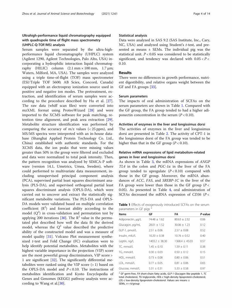
Serum parametersThe impacts of oral administration of SCFAs on theserum parameters are shown in Table 1. Compared withthe GF group, the FA group tended to had a higher adi-ponectin concentration in the serum (P < 0.10).
Activities of enzymes in the liver and longissimus dorsiThe activities of enzymes in the liver and longissimusdorsi are presented in Table 2. The activity of CPT-1 inthe longissimus dorsi of the FA group was tended to behigher than that in the GF group (P < 0.10).
Relative mRNA expressions of lipid metabolism-relatedgenes in liver and longissimus dorsiAs shown in Table 3, the mRNA expressions of ANGPTL4 in the colon and PGC-1α in the liver of the FAgroup tended to upregulate (P < 0.10) compared withthose in the GF group. Moreover, the mRNA abun-dances of ACC, FAS, and SREBP-1C in the liver of theFA group were lower than those in the GF group (P <0.05). As presented in Table 4, oral administration ofSCFAs decreased the mRNA expression of CD36 (P <
Table 1 Effects of exogenously introduced SCFAs on the serumparameters in GF pigs a
Items GF FA P-value
Adiponectin, μg/L 74.48 ± 1.62 80.63 ± 2.32 0.06
Glucagon, pg/mL 28.01 ± 1.52 30.06 ± 1.23 0.12
GLP-1, pmol/L 2.51 ± 0.06 2.57 ± 0.08 0.52
Insulin, mIU/L 10.20 ± 0.38 10.76 ± 0.52 0.40
Leptin, ng/L 1403.2 ± 38.30 1368.4 ± 45.03 0.57
TC, mmol/L 1.45 ± 0.10 1.59 ± 0.11 0.38
TG, mmol/L 0.30 ± 0.03 0.50 ± 0.12 0.15
HDL, mmol/L 0.73 ± 0.08 0.80 ± 0.06 0.51
LDL, mmol/L 0.77 ± 0.05 0.81 ± 0.06 0.65
Glucose, mmol/L 5.31 ± 0.31 5.33 ± 0.58 0.97a GF germ-free, FA short-chain fatty acids, GLP-1 Glucagon like peptide 1, TCtotal cholesterol, TG triglyceride, HDL-c high-density lipoprotein-cholesterol,LDL-c low-density lipoprotein-cholesterol. Values are means ±SEMs, n = 6/group
Zhou et al. Journal of Animal Science and Biotechnology (2021) 12:61 Page 4 of 14
0.05) and tended to downregulate the mRNA expressionof LPL (P < 0.10) in the longissimus dorsi.
Relative mRNA expressions of glucose metabolism-relatedgenes in liver and longissimus dorsiAs presented in Table 5, the oral administration ofSCFAs upregulated the mRNA expressions of GLU-2and GYS2 in the liver (P < 0.05). In addition, the mRNA
abundances of genes related to glucose metabolism inthe longissimus dorsi were no differences between theFA and GF groups (P > 0.10) (Table 6).
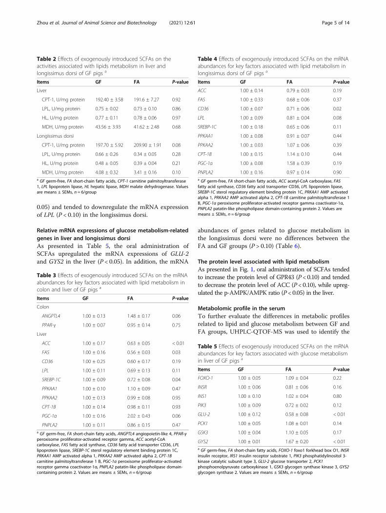
The protein level associated with lipid metabolismAs presented in Fig. 1, oral administration of SCFAs tendedto increase the protein level of GPR43 (P < 0.10) and tendedto decrease the protein level of ACC (P < 0.10), while upreg-ulated the p-AMPK/AMPK ratio (P < 0.05) in the liver.
Metabolomic profile in the serumTo further evaluate the differences in metabolic profilesrelated to lipid and glucose metabolism between GF andFA groups, UHPLC-QTOF-MS was used to identify the
Table 2 Effects of exogenously introduced SCFAs on theactivities associated with lipids metabolism in liver andlongissimus dorsi of GF pigs a
Items GF FA P-value
Liver
CPT-1, U/mg protein 192.40 ± 3.58 191.6 ± 7.27 0.92
LPL, U/mg protein 0.75 ± 0.02 0.73 ± 0.10 0.86
HL, U/mg protein 0.77 ± 0.11 0.78 ± 0.06 0.97
MDH, U/mg protein 43.56 ± 3.93 41.62 ± 2.48 0.68
Longissimus dorsi
CPT-1, U/mg protein 197.70 ± 5.92 209.90 ± 1.91 0.08
LPL, U/mg protein 0.66 ± 0.26 0.34 ± 0.05 0.28
HL, U/mg protein 0.48 ± 0.05 0.39 ± 0.04 0.21
MDH, U/mg protein 4.08 ± 0.32 3.41 ± 0.16 0.10a GF germ-free, FA short-chain fatty acids, CPT-1 carnitine palmitoyltransferase1, LPL lipoprotein lipase, HL hepatic lipase, MDH malate dehydrogenase. Valuesare means ± SEMs, n = 6/group
Table 3 Effects of exogenously introduced SCFAs on the mRNAabundances for key factors associated with lipid metabolism incolon and liver of GF pigs a
Items GF FA P-value
Colon
ANGPTL4 1.00 ± 0.13 1.48 ± 0.17 0.06
PPAR-γ 1.00 ± 0.07 0.95 ± 0.14 0.75
Liver
ACC 1.00 ± 0.17 0.63 ± 0.05 < 0.01
FAS 1.00 ± 0.16 0.56 ± 0.03 0.03
CD36 1.00 ± 0.25 0.60 ± 0.17 0.19
LPL 1.00 ± 0.11 0.69 ± 0.13 0.11
SREBP-1C 1.00 ± 0.09 0.72 ± 0.08 0.04
PPKAA1 1.00 ± 0.10 1.10 ± 0.09 0.47
PPKAA2 1.00 ± 0.13 0.99 ± 0.08 0.95
CPT-1B 1.00 ± 0.14 0.98 ± 0.11 0.93
PGC-1α 1.00 ± 0.16 2.02 ± 0.43 0.06
PNPLA2 1.00 ± 0.11 0.86 ± 0.15 0.47a GF germ-free, FA short-chain fatty acids, ANGPTL4 angiopoietin-like 4, PPAR-γperoxisome proliferator-activated receptor gamma, ACC acetyl-CoAcarboxylase, FAS fatty acid synthase, CD36 fatty acid transporter CD36, LPLlipoprotein lipase, SREBP-1C sterol regulatory element binding protein 1C,PRKAA1 AMP activated alpha 1, PRKAA2 AMP activated alpha 2, CPT-1Bcarnitine palmitoyltransferase 1 B, PGC-1α peroxisome proliferator-activatedreceptor gamma coactivator-1α, PNPLA2 patatin-like phospholipase domain-containing protein 2. Values are means ± SEMs, n = 6/group
Table 4 Effects of exogenously introduced SCFAs on the mRNAabundances for key factors associated with lipid metabolism inlongissimus dorsi of GF pigs a
Items GF FA P-value
ACC 1.00 ± 0.14 0.79 ± 0.03 0.19
FAS 1.00 ± 0.33 0.68 ± 0.06 0.37
CD36 1.00 ± 0.07 0.71 ± 0.06 0.02
LPL 1.00 ± 0.09 0.81 ± 0.04 0.08
SREBP-1C 1.00 ± 0.18 0.65 ± 0.06 0.11
PPKAA1 1.00 ± 0.08 0.91 ± 0.07 0.44
PPKAA2 1.00 ± 0.03 1.07 ± 0.06 0.39
CPT-1B 1.00 ± 0.15 1.14 ± 0.10 0.44
PGC-1α 1.00 ± 0.08 1.58 ± 0.39 0.19
PNPLA2 1.00 ± 0.16 0.97 ± 0.14 0.90a GF germ-free, FA short-chain fatty acids, ACC acetyl-CoA carboxylase, FASfatty acid synthase, CD36 fatty acid transporter CD36, LPL lipoprotein lipase,SREBP-1C sterol regulatory element binding protein 1C, PRKAA1 AMP activatedalpha 1, PRKAA2 AMP activated alpha 2, CPT-1B carnitine palmitoyltransferase 1B, PGC-1α peroxisome proliferator-activated receptor gamma coactivator-1α,PNPLA2 patatin-like phospholipase domain-containing protein 2. Values aremeans ± SEMs, n = 6/group
Table 5 Effects of exogenously introduced SCFAs on the mRNAabundances for key factors associated with glucose metabolismin liver of GF pigs a
Items GF FA P-value
FOXO-1 1.00 ± 0.05 1.09 ± 0.04 0.22
INSR 1.00 ± 0.06 0.81 ± 0.06 0.16
INS1 1.00 ± 0.10 1.02 ± 0.04 0.80
PIK3 1.00 ± 0.09 0.72 ± 0.02 0.12
GLU-2 1.00 ± 0.12 0.58 ± 0.08 < 0.01
PCK1 1.00 ± 0.05 1.08 ± 0.01 0.14
GSK3 1.00 ± 0.04 1.10 ± 0.05 0.17
GYS2 1.00 ± 0.01 1.67 ± 0.20 < 0.01a GF germ-free, FA short-chain fatty acids, FOXO-1 foxo1 forkhead box O1, INSRinsulin receptor, IRS1 insulin receptor substrate 1, PIK3 phosphatidylinositol 3-kinase catalytic subunit type 3, GLU-2 glucose transporter 2, PCK1phosphoenolpyruvate carboxykinase 1, GSK3 glycogen synthase kinase 3, GYS2glycogen synthase 2. Values are means ± SEMs, n = 6/group
Zhou et al. Journal of Animal Science and Biotechnology (2021) 12:61 Page 5 of 14
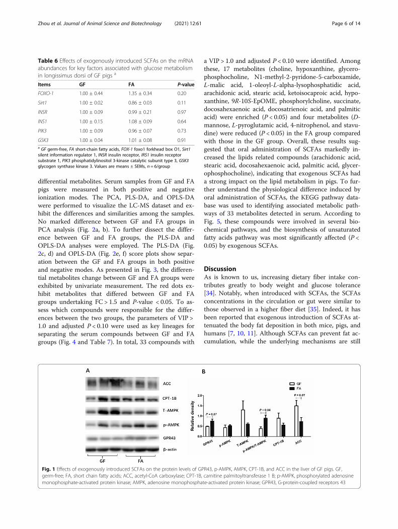
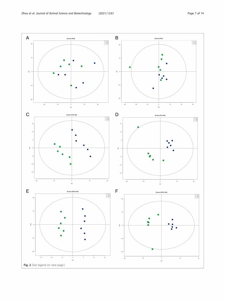
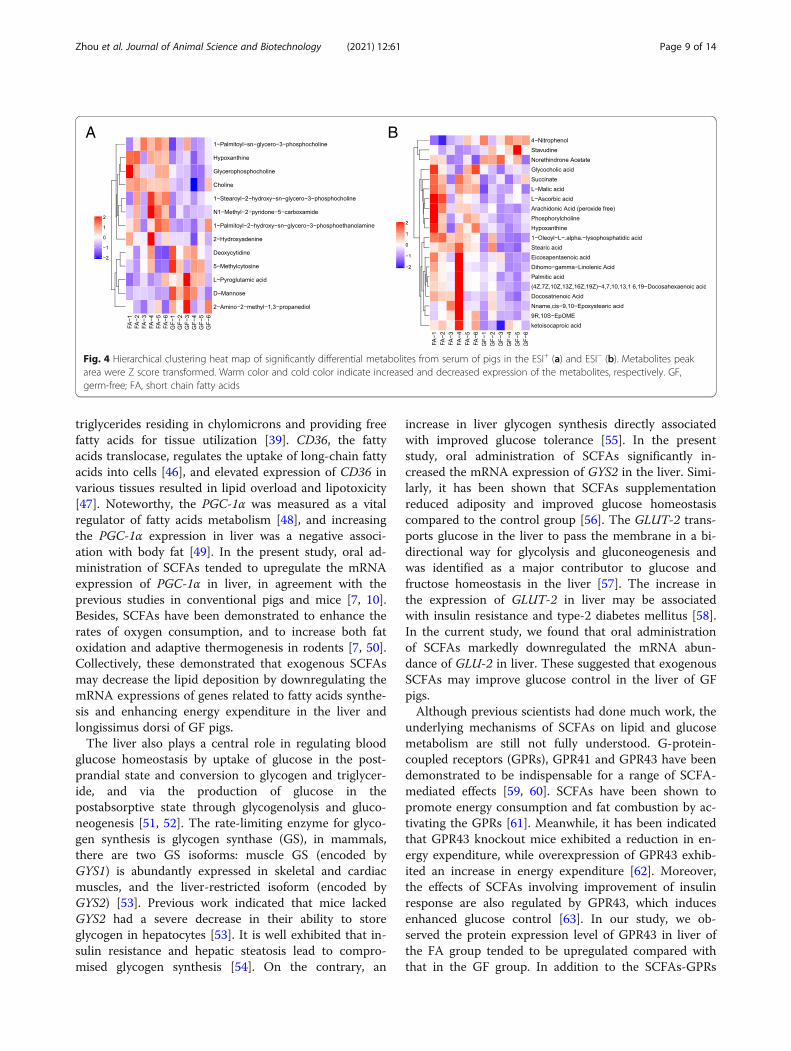
differential metabolites. Serum samples from GF and FApigs were measured in both positive and negativeionization modes. The PCA, PLS-DA, and OPLS-DAwere performed to visualize the LC-MS dataset and ex-hibit the differences and similarities among the samples.No marked difference between GF and FA groups inPCA analysis (Fig. 2a, b). To further dissect the differ-ence between GF and FA groups, the PLS-DA andOPLS-DA analyses were employed. The PLS-DA (Fig.2c, d) and OPLS-DA (Fig. 2e, f) score plots show separ-ation between the GF and FA groups in both positiveand negative modes. As presented in Fig. 3, the differen-tial metabolites change between GF and FA groups wereexhibited by univariate measurement. The red dots ex-hibit metabolites that differed between GF and FAgroups undertaking FC > 1.5 and P-value < 0.05. To as-sess which compounds were responsible for the differ-ences between the two groups, the parameters of VIP >1.0 and adjusted P < 0.10 were used as key lineages forseparating the serum compounds between GF and FAgroups (Fig. 4 and Table 7). In total, 33 compounds with
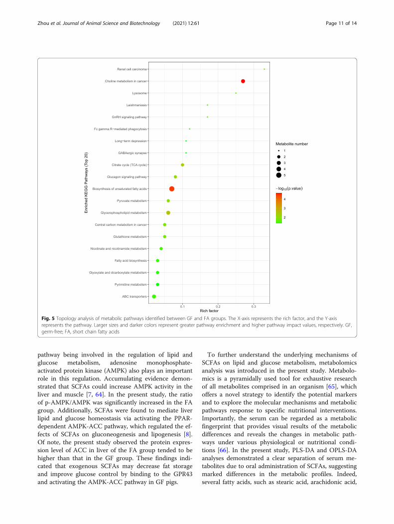
a VIP > 1.0 and adjusted P < 0.10 were identified. Amongthese, 17 metabolites (choline, hypoxanthine, glycero-phosphocholine, N1-methyl-2-pyridone-5-carboxamide,L-malic acid, 1-oleoyl-L-alpha-lysophosphatidic acid,arachidonic acid, stearic acid, ketoisocaproic acid, hypo-xanthine, 9R-10S-EpOME, phosphorylcholine, succinate,docosahexaenoic acid, docosatrienoic acid, and palmiticacid) were enriched (P < 0.05) and four metabolites (D-mannose, L-pyroglutamic acid, 4-nitrophenol, and stavu-dine) were reduced (P < 0.05) in the FA group comparedwith those in the GF group. Overall, these results sug-gested that oral administration of SCFAs markedly in-creased the lipids related compounds (arachidonic acid,stearic acid, docosahexaenoic acid, palmitic acid, glycer-ophosphocholine), indicating that exogenous SCFAs hada strong impact on the lipid metabolism in pigs. To fur-ther understand the physiological difference induced byoral administration of SCFAs, the KEGG pathway data-base was used to identifying associated metabolic path-ways of 33 metabolites detected in serum. According toFig. 5, these compounds were involved in several bio-chemical pathways, and the biosynthesis of unsaturatedfatty acids pathway was most significantly affected (P <0.05) by exogenous SCFAs.
DiscussionAs is known to us, increasing dietary fiber intake con-tributes greatly to body weight and glucose tolerance[34]. Notably, when introduced with SCFAs, the SCFAsconcentrations in the circulation or gut were similar tothose observed in a higher fiber diet [35]. Indeed, it hasbeen reported that exogenous introduction of SCFAs at-tenuated the body fat deposition in both mice, pigs, andhumans [7, 10, 11]. Although SCFAs can prevent fat ac-cumulation, while the underlying mechanisms are still
Table 6 Effects of exogenously introduced SCFAs on the mRNAabundances for key factors associated with glucose metabolismin longissimus dorsi of GF pigs a
Items GF FA P-value
FOXO-1 1.00 ± 0.44 1.35 ± 0.34 0.20
Sirt1 1.00 ± 0.02 0.86 ± 0.03 0.11
INSR 1.00 ± 0.09 0.99 ± 0.21 0.97
INS1 1.00 ± 0.15 1.08 ± 0.09 0.64
PIK3 1.00 ± 0.09 0.96 ± 0.07 0.73
GSK3 1.00 ± 0.04 1.01 ± 0.08 0.91a GF germ-free, FA short-chain fatty acids, FOX-1 foxo1 forkhead box O1, Sirt1silent information regulator 1, INSR insulin receptor, IRS1 insulin receptorsubstrate 1, PIK3 phosphatidylinositol 3-kinase catalytic subunit type 3, GSK3glycogen synthase kinase 3. Values are means ± SEMs, n = 6/group
Fig. 1 Effects of exogenously introduced SCFAs on the protein levels of GPR43, p-AMPK, AMPK, CPT-1B, and ACC in the liver of GF pigs. GF,germ-free; FA, short chain fatty acids; ACC, acetyl-CoA carboxylase; CPT-1B, carnitine palmitoyltransferase 1 B; p-AMPK, phosphorylated adenosinemonophosphate-activated protein kinase; AMPK, adenosine monophosphate-activated protein kinase; GPR43, G-protein-coupled receptors 43
Zhou et al. Journal of Animal Science and Biotechnology (2021) 12:61 Page 6 of 14
A B
C D
E F
Fig. 2 (See legend on next page.)
Zhou et al. Journal of Animal Science and Biotechnology (2021) 12:61 Page 7 of 14
not fully understood. SCFAs can be produced naturallyby host metabolic pathways particularly in the liver, butthe major site of SCFAs production is the colon whichrequires the presence of specific bacteria [36]. Moreover,the numbers and diversity of microbiota are positivelyassociated with SCFAs concentrations [19], and severalspecific microbes species were closely related to hostlipid metabolism [16, 17]. However, whether the SCFAsregulate lipid and glucose metabolism independent ofthe gut microbiota are largely unknown. Therefore, thisstudy was conducted to explore the effects of exogenousintroduction of SCFAs on lipid and glucose metabolismin a GF pig model and to further dissect the underlyingmechanisms of exogenously introduced SCFAs on thehost’s health. To determine the concentration and doseof the SCFAs mixture used in the present experiment,we conducted a preliminary experiment on the conven-tional Bama piglets (n = 12). In the preliminary trial,when pigs fed with 1.25 S0 (acetic, propionic, and butyricacids, 75, 25, and 19 mmol/L, respectively) and 25mL/kgor 1.0 S0 (acetic, propionic, and butyric acids, 60, 20, and15mmol/L, respectively) and 25mL/kg SCFAs mixtureled to diarrhea and death. Meanwhile, the feed intakewas not decreased and without diarrhea after 7 d whenpigs fed with 0.75 S0 (acetic, propionic, and butyric acids,45, 15, and 11 mmol/L, respectively) and 25 mL/kgSCFAs mixture. However, when pigs fed with 0.75 S0and 35 mL/kg SCFAs also led to diarrhea and reducedfeed intake. Taken together, we determined the mixtureconcentrations of acetic, propionic, and butyric acids at
45, 15, and 11mmol/L, respectively, and a dose of 25mL/kg in the present study.Acting in peripheral tissues, adiponectin could regulate
lipid metabolism and promote energy expenditure [37].Serum adiponectin concentration was reduced in indi-viduals with obesity and obesity-related diseases [38]. Inthe present study, oral administration of SCFAs tendedto increase the concentration of adiponectin in serum.CPT-1 is the rate-limiting enzyme that determines fattyacids oxidation [39]. ANGPTL4 is a valid inhibitor oflipoprotein lipase to regulate cellular uptake of triglycer-ides and promote fatty acids oxidation [40, 41]. In ourstudy, oral administration of SCFAs tended to increasethe activity of CPT-1 in longissimus dorsi and themRNA abundance of ANGPTL4 in colon. Meanwhile,we found the mRNA expressions of FAS, ACC, andSREBP-1C in liver of the FA group markedly downregu-lated compared with those in the GF group. Consist-ently, previous studies reported similar results in liver,longissimus dorsi, and adipose tissues of conventionalpigs [9, 10, 42]. Notably, FAS is the pivotal enzyme thatcatalyzes fatty acids synthesis [43]. ACC modulates fattyacids metabolism, and its product (e.g. malonyl-CoA)serves as a building block for de novo fatty acids synthe-sis [44]. The SREBPs increases the transcription of genesthat encode the enzymes of fatty acids biosynthesis andcholesterol uptake [45]. In addition, the current studyobserved the mRNA abundance of CD36 was apparentlydecreased, and LPL tended to be reduced in longissimusdorsi of the FA group. LPL catalyzes the hydrolysis of
(See figure on previous page.)Fig. 2 The PCA, PLS-DA, and OPLS-DA score plots comparing GF (blue rotundities) and FA (green squares) pigs in positive electrospray ionizationmode (ESI+) and negative electrospray ionization mode (ESI−) metabolomics profiles of serum. Panels a (ESI+) and b (ESI−) are PCA score plots.Panels c (ESI+, R2X = 0.459, R2Y = 0.996, Q2 = 0.649) and d (ESI−, R2X = 0.514, R2Y = 0.999, Q2 = 0.818) are PLS-DA score plots. Panels e (ESI+, R2X =0.317, R2Y = 0.976, Q2 = 0.009) and f (ESI−, R2X = 0.307, R2Y = 0.926, Q2 = 0.201) are OPLS-DA score plots. GF, germ-free; FA, short chain fatty acids;PCA, principal component analysis; PLS-DA, Partial least squares discriminant; OPLS-DA, orthogonal partial least-squares discriminant
A B
Fig. 3 Volcano plots showing the distribution of all metabolites based on their fold-change values (X-axis, on a logarithmic scale), P-value (Y-axis,on a logarithmic scale). Panel a is ESI+, Panel b is ESI−, respectively (FC > 1.50 and P-value < 0.05). GF, germ-free; FA, short chain fatty acids
Zhou et al. Journal of Animal Science and Biotechnology (2021) 12:61 Page 8 of 14
triglycerides residing in chylomicrons and providing freefatty acids for tissue utilization [39]. CD36, the fattyacids translocase, regulates the uptake of long-chain fattyacids into cells [46], and elevated expression of CD36 invarious tissues resulted in lipid overload and lipotoxicity[47]. Noteworthy, the PGC-1α was measured as a vitalregulator of fatty acids metabolism [48], and increasingthe PGC-1α expression in liver was a negative associ-ation with body fat [49]. In the present study, oral ad-ministration of SCFAs tended to upregulate the mRNAexpression of PGC-1α in liver, in agreement with theprevious studies in conventional pigs and mice [7, 10].Besides, SCFAs have been demonstrated to enhance therates of oxygen consumption, and to increase both fatoxidation and adaptive thermogenesis in rodents [7, 50].Collectively, these demonstrated that exogenous SCFAsmay decrease the lipid deposition by downregulating themRNA expressions of genes related to fatty acids synthe-sis and enhancing energy expenditure in the liver andlongissimus dorsi of GF pigs.The liver also plays a central role in regulating blood
glucose homeostasis by uptake of glucose in the post-prandial state and conversion to glycogen and triglycer-ide, and via the production of glucose in thepostabsorptive state through glycogenolysis and gluco-neogenesis [51, 52]. The rate-limiting enzyme for glyco-gen synthesis is glycogen synthase (GS), in mammals,there are two GS isoforms: muscle GS (encoded byGYS1) is abundantly expressed in skeletal and cardiacmuscles, and the liver-restricted isoform (encoded byGYS2) [53]. Previous work indicated that mice lackedGYS2 had a severe decrease in their ability to storeglycogen in hepatocytes [53]. It is well exhibited that in-sulin resistance and hepatic steatosis lead to compro-mised glycogen synthesis [54]. On the contrary, an
increase in liver glycogen synthesis directly associatedwith improved glucose tolerance [55]. In the presentstudy, oral administration of SCFAs significantly in-creased the mRNA expression of GYS2 in the liver. Simi-larly, it has been shown that SCFAs supplementationreduced adiposity and improved glucose homeostasiscompared to the control group [56]. The GLUT-2 trans-ports glucose in the liver to pass the membrane in a bi-directional way for glycolysis and gluconeogenesis andwas identified as a major contributor to glucose andfructose homeostasis in the liver [57]. The increase inthe expression of GLUT-2 in liver may be associatedwith insulin resistance and type-2 diabetes mellitus [58].In the current study, we found that oral administrationof SCFAs markedly downregulated the mRNA abun-dance of GLU-2 in liver. These suggested that exogenousSCFAs may improve glucose control in the liver of GFpigs.Although previous scientists had done much work, the
underlying mechanisms of SCFAs on lipid and glucosemetabolism are still not fully understood. G-protein-coupled receptors (GPRs), GPR41 and GPR43 have beendemonstrated to be indispensable for a range of SCFA-mediated effects [59, 60]. SCFAs have been shown topromote energy consumption and fat combustion by ac-tivating the GPRs [61]. Meanwhile, it has been indicatedthat GPR43 knockout mice exhibited a reduction in en-ergy expenditure, while overexpression of GPR43 exhib-ited an increase in energy expenditure [62]. Moreover,the effects of SCFAs involving improvement of insulinresponse are also regulated by GPR43, which inducesenhanced glucose control [63]. In our study, we ob-served the protein expression level of GPR43 in liver ofthe FA group tended to be upregulated compared withthat in the GF group. In addition to the SCFAs-GPRs
A B
Fig. 4 Hierarchical clustering heat map of significantly differential metabolites from serum of pigs in the ESI+ (a) and ESI− (b). Metabolites peakarea were Z score transformed. Warm color and cold color indicate increased and decreased expression of the metabolites, respectively. GF,germ-free; FA, short chain fatty acids
Zhou et al. Journal of Animal Science and Biotechnology (2021) 12:61 Page 9 of 14
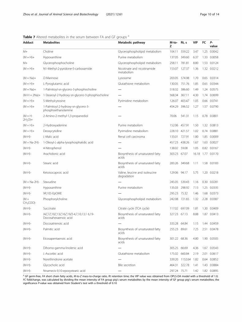
Table 7 Altered metabolites in the serum between FA and GF groups a
Adduct Metabolites Metabolic pathway M-to-Z
Rt, s VIP FC P-value
M+ Choline Glycerophospholipid metabolism 104.11 559.22 3.47 1.25 0.0042
(M + H)+ Hypoxanthine Purine metabolism 137.05 349.60 6.37 1.33 0.0058
M+ Glycerophosphocholine Glycerophospholipid metabolism 258.11 781.81 8.80 1.53 0.0124
(M + H)+ N1-Methyl-2-pyridone-5-carboxamide Nicotinate and nicotinamidemetabolism
153.07 127.37 1.36 1.32 0.0212
(M + Na)+ D-Mannose Lysosome 203.05 574.98 1.70 0.65 0.0314
(M + H)+ L-Pyroglutamic acid Glutathione metabolism 130.05 751.76 1.85 0.65 0.0344
(M + Na)+ 1-Palmitoyl-sn-glycero-3-phosphocholine ― 518.32 386.60 1.49 1.24 0.0575
(M-H + 2Na)+ 1-Stearoyl-2-hydroxy-sn-glycero-3-phosphocholine ― 568.34 367.11 4.30 1.74 0.0699
(M + H)+ 5-Methylcytosine Pyrimidine metabolism 126.07 403.47 1.05 0.66 0.0741
(M + H)+ 1-Palmitoyl-2-hydroxy-sn-glycero-3-phosphoethanolamine
― 454.29 396.52 1.27 1.57 0.0790
(M + H-2H2O)+
2-Amino-2-methyl-1,3-propanediol ― 70.06 541.31 1.15 0.78 0.0801
(M + H)+ 2-Hydroxyadenine Purine metabolism 152.06 457.91 1.50 1.32 0.0813
(M + H)+ Deoxycytidine Pyrimidine metabolism 228.10 421.57 1.02 0.74 0.0881
(M-H)- L-Malic acid Renal cell carcinoma 133.01 727.91 1.80 1.85 0.0009
(M + Na-2H)- 1-Oleoyl-L-alpha-lysophosphatidic acid ― 457.23 438.26 1.67 1.63 0.0027
(M-H)- 4-Nitrophenol ― 138.02 59.08 1.05 0.82 0.0167
(M-H)- Arachidonic acid Biosynthesis of unsaturated fattyacids
303.23 67.07 16.18 1.77 0.0170
(M-H)- Stearic acid Biosynthesis of unsaturated fattyacids
283.26 349.68 1.11 1.58 0.0183
(M-H)- Ketoisocaproic acid Valine, leucine and isoleucinedegradation
129.06 94.17 5.75 1.20 0.0218
(M + Na-2H)- Stavudine ― 245.05 539.43 1.16 0.30 0.0281
(M-H)- Hypoxanthine Purine metabolism 135.03 298.92 7.13 1.25 0.0335
(M-H)- 9R,10S-EpOME ― 295.23 75.32 1.46 1.68 0.0373
(M +CH3COO)-
Phosphorylcholine Glycerophospholipid metabolism 242.08 721.65 1.32 2.28 0.0387
(M-H)- Succinate Citrate cycle (TCA cycle) 117.02 697.09 1.81 1.30 0.0409
(M-H)- (4Z,7Z,10Z,13Z,16Z,19Z)-4,7,10,13,1 6,19-Docosahexaenoic acid
Biosynthesis of unsaturated fattyacids
327.23 67.13 8.88 1.87 0.0413
(M-H)- Docosatrienoic acid ― 333.28 64.84 1.15 1.44 0.0459
(M-H)- Palmitic acid Biosynthesis of unsaturated fattyacids
255.23 89.61 7.25 2.51 0.0478
(M-H)- Eicosapentaenoic acid Biosynthesis of unsaturated fattyacids
301.22 68.36 4.80 1.90 0.0505
(M-H)- Dihomo-gamma-linolenic acid ― 305.25 66.69 4.36 1.67 0.0543
(M-H)- L-Ascorbic acid Glutathione metabolism 175.02 660.84 2.19 2.01 0.0617
(M-H)- Norethindrone acetate ― 339.20 1152.64 1.82 0.64 0.0852
(M-H)- Glycocholic acid Bile secretion 464.31 322.78 1.41 1.43 0.0884
(M-H)- Nnamecis-9,10-epoxystearic acid ― 297.24 75.71 1.42 1.82 0.0895a GF germ-free, FA short chain fatty acids, M-to-Z mass-to-charge ratio, Rt retention time; the VIP value was obtained from OPLS-DA model with a threshold of 1.0;FC foldchange, was calculated by dividing the mean intensity of FA group pig’s serum metabolites by the mean intensity of GF group pig’s serum metabolites; thesignificance P-value was obtained from Student’s test with a threshold of 0.10
Zhou et al. Journal of Animal Science and Biotechnology (2021) 12:61 Page 10 of 14
pathway being involved in the regulation of lipid andglucose metabolism, adenosine monophosphate-activated protein kinase (AMPK) also plays an importantrole in this regulation. Accumulating evidence demon-strated that SCFAs could increase AMPK activity in theliver and muscle [7, 64]. In the present study, the ratioof p-AMPK/AMPK was significantly increased in the FAgroup. Additionally, SCFAs were found to mediate liverlipid and glucose homeostasis via activating the PPAR-dependent AMPK-ACC pathway, which regulated the ef-fects of SCFAs on gluconeogenesis and lipogenesis [8].Of note, the present study observed the protein expres-sion level of ACC in liver of the FA group tended to behigher than that in the GF group. These findings indi-cated that exogenous SCFAs may decrease fat storageand improve glucose control by binding to the GPR43and activating the AMPK-ACC pathway in GF pigs.
To further understand the underlying mechanisms ofSCFAs on lipid and glucose metabolism, metabolomicsanalysis was introduced in the present study. Metabolo-mics is a pyramidally used tool for exhaustive researchof all metabolites comprised in an organism [65], whichoffers a novel strategy to identify the potential markersand to explore the molecular mechanisms and metabolicpathways response to specific nutritional interventions.Importantly, the serum can be regarded as a metabolicfingerprint that provides visual results of the metabolicdifferences and reveals the changes in metabolic path-ways under various physiological or nutritional condi-tions [66]. In the present study, PLS-DA and OPLS-DAanalyses demonstrated a clear separation of serum me-tabolites due to oral administration of SCFAs, suggestingmarked differences in the metabolic profiles. Indeed,several fatty acids, such as stearic acid, arachidonic acid,
Fig. 5 Topology analysis of metabolic pathways identified between GF and FA groups. The X-axis represents the rich factor, and the Y-axisrepresents the pathway. Larger sizes and darker colors represent greater pathway enrichment and higher pathway impact values, respectively. GF,germ-free; FA, short chain fatty acids
Zhou et al. Journal of Animal Science and Biotechnology (2021) 12:61 Page 11 of 14
docosahexaenoic acid, and palmitic acid in the FA groupapparently increased compared with those in the GFgroup. Increases in serum fatty acids levels implied thatlipid metabolisms have been altered. Taken KEGG path-way analysis, we observed these fatty acids (stearic acid,arachidonic acid, docosahexaenoic acid, palmitic acid)were involved in the biosynthesis of unsaturated fattyacids pathway. Of note, oral administration of SCFAshas the most significant impact on this metabolicpathway. Intake of unsaturated fatty acids, which con-sist of monounsaturated fatty acids and polyunsatur-ated fatty acids, has been associated with favorablecardiac diastolic function and body composition inobese patients [67]. Moreover, increasing unsaturatedfatty acids in the diet also prevented weight gain andcardiac dysfunction in a mouse model [68]. Conse-quently, these suggested that exogenous introductionof SCFAs may alleviate the lipid deposition via acti-vating the metabolic pathway of biosynthesis of unsat-urated fatty acids in GF pigs.
ConclusionsIn summary, this study demonstrated that SCFAs mayattenuate fat deposition and to some extent improve glu-cose control in the liver and longissimus dorsi, whichoccur independently of the gut microbiota. The possiblemechanisms of exogenous SCFAs on lipid reduction andglucose tolerance improvement may be via binding tothe GPR43 and activating the AMPK-ACC pathway, andstimulating the metabolic pathway of biosynthesis of un-saturated fatty acids in GF pigs. The current work fur-ther suggested the importance of the presence of gutmicrobes and provided novel evidence that exogenousintroduction of SCFAs may be a possible therapeuticstrategy to prevent metabolic disorders and to counter-act the gut microbiota deficiency or imbalance.
AbbreviationsACC: Acetyl-CoA carboxylase; AMPK: Adenosine monophosphate-activatedprotein kinase; ANGPTL4: Angiopoietin-like 4; CD36: Fatty acid transporterCD36; CPT-1B: Carnitine palmitoyltransferase 1 B; FAS: Fatty acid synthase;FOXO-1: Foxo1 forkhead box O1; G6PC: Glucose-6-phosphatase; GF: Germ-free; GLU-2: Glucose transporter 2; GPR43: G-protein-coupled receptors 43;GSK3: Glycogen synthase kinase 3; GYS2: Glycogen synthase 2; HDL-c: Highdensity lipoprotein-cholesterol; HL: Hepatic lipase; INSR: Insulin receptor;IRS1: Insulin receptor substrate 1; LDL-c: Low density lipoprotein-cholesterol;LPL: Lipoprotein lipase; MDH: Malate dehydrogenase; OPLS-DA: Orthogonalpartial least squares discriminant analysis; p-AMPK: Phosphorylated adenosinemonophosphate-activated protein kinase; PCK1: Phosphoenolpyruvatecarboxykinase 1; PIK3: Phosphatidylinositol 3-kinase catalytic subunit type 3;PGC-1α: Peroxisome proliferator-activated receptor gamma coactivator-1α;PLS-DA: Partial least squares discriminant analysis; PNPLA2: Adiposetriglyceride lipase; PPAR-γ: Peroxisome proliferator-activated receptor gamma;PRKAA1: AMP activated alpha 1; PRKAA2: AMP activated alpha 2; Sirt1: Silentinformation regulator 1; SREBP-1C: Sterol regulatory element binding protein1C; TC: Total cholesterol; TG: Triglyceride; UHPLC-Q-TOF/MS: Ultrahigh-performance liquid chromatography equipped with quadrupole time of-flight mass spectrometry; VIP: Variable importance in the projection
Supplementary InformationThe online version contains supplementary material available at https://doi.org/10.1186/s40104-021-00581-3.
Additional file 1 : Table S1. Ingredient composition of the milkpowder (as-fed basis). Table S2. Ingredient composition of the basal diet(as-fed basis). Table S3. Infusion volume of sterile saline or SCFAsmixture for each pig per day. Table S4. Primer sequences used for real-time quantitative PCR.
AcknowledgementsNot applicable.
Authors’ contributionsH.Z. conducted the animal work and the laboratory work, and wrote themanuscript. H.Z., L.G. and D.C. designed the experiment. B.Y. and J.S. gaveadvice on the experiment design. H.Z. analyzed the study data and wrotethe manuscript. Z.L. and H.C. helped to revise the manuscript. All the authorshave read and approved the final manuscript.
FundingThis study was supported by National Natural Science Foundation of China(31730091) and the National Key Research and Development Program ofChina (2017YFD0500503).
Availability of data and materialsThe data were exhibited in the main manuscript and supplemental materials.
Declarations
Ethics approval and consent to participateExperimental protocols and procedures used in the present experiment wereapproved by the Animal Care and Use Committee of Sichuan AgriculturalUniversity (Chengdu, China) under permit number DKY-B20131704.
Consent for publicationNot applicable.
Competing interestsThe authors declare that they have no conflict of interest.
Author details1Key Laboratory of Animal Disease-Resistance Nutrition, Chengdu 611130,Sichuan, China. 2Animal Nutrition Institute, Sichuan Agricultural University,Chengdu 611130, Sichuan, China. 3Key Laboratory of Pig Industry Sciences,Rongchang 402460, Chongqing, China. 4Chongqing Academy of AnimalSciences, Rongchang 402460, Chongqing, China. 5College of Food Science,Sichuan Agricultural University, Ya’an 625014, Sichuan, China.
Received: 2 November 2020 Accepted: 8 March 2021
References1. Delzenne NM, Cani PD, Everard A, Neyrinck AM, Bindels LB. Gut
microorganisms as promising targets for the management of type 2diabetes. Diabetologia. 2015;58(10):2206–17. https://doi.org/10.1007/s00125-015-3712-7.
2. Canfora EE, Jocken JW, Blaak EE. Short-chain fatty acids in control of bodyweight and insulin sensitivity. Nat Rev Endocrinol. 2015;11(10):577–91.https://doi.org/10.1038/nrendo.2015.128.
3. DeVadder F, Kovatcheva-Datchary P, Zitoun C, Duchampt A, Bäckhed F,Mithieux G. Microbiota-produced succinate improves glucose homeostasisvia intestinal gluconeogenesis. Cell Metab. 2016;24(1):151–7. https://doi.org/10.1016/j.cmet.2016.06.013.
4. Byrne CS, Chambers ES, Morrison DJ, Frost G. The role of short chain fattyacids in appetite regulation and energy homeostasis. Int J Obes. 2015;39(9):1331–8. https://doi.org/10.1038/ijo.2015.84.
5. DeVadder F, Kovatcheva-Datchary P, Goncalves D, Vinera J, Zitoun C,Duchampt A, et al. Microbiota-generated metabolites promote metabolic
Zhou et al. Journal of Animal Science and Biotechnology (2021) 12:61 Page 12 of 14
benefits via gut-brain neural circuits. Cell. 2014;156(1-2):84–96. https://doi.org/10.1016/j.cell.2013.12.016.
6. Sahuri-Arisoylu M, Brody LP, Parkinson JR, Parkes H, Navaratnam N, MillerAD, et al. Reprogramming of hepatic fat accumulation and ‘browning’ ofadipose tissue by the short-chain fatty acid acetate. Int J Obes. 2016;40(6):955–63. https://doi.org/10.1038/ijo.2016.23.
7. Gao Z, Yin J, Zhang J, Ward RE, Martin RJ, Lefevre M, et al. Butyrateimproves insulin sensitivity and increases energy expenditure in mice.Diabetes. 2009;58(7):1509–17. https://doi.org/10.2337/db08-1637.
8. den Besten G, Bleeker A, Gerding A, van Eunen K, Havinga R, van Dijk TH,et al. Short-chain fatty acids protect against high-fat diet-induced obesityvia a PPARγ-dependent switch from lipogenesis to fat oxidation. Diabetes.2015;64(7):2398–408. https://doi.org/10.2337/db14-1213.
9. Jiao AR, Diao H, Yu B, He J, Yu J, Zheng P, et al. Oral administration of shortchain fatty acids could attenuate fat deposition of pigs. PLoS One. 2018;13(5):e0196867. https://doi.org/10.1371/journal.pone.0196867.
10. Jiao A, Yu B, He J, Yu J, Zheng P, Luo Y, et al. Short chain fatty acids couldprevent fat deposition in pigs via regulating related hormones and genes.Food Funct. 2020;11(2):1845–55. https://doi.org/10.1039/C9FO02585E.
11. Canfora EE, van der Beek CM, Jocken JWE, Goossens GH, Holst JJ, OldeDamink SWM, et al. Colonic infusions of short-chain fatty acid mixturespromote energy metabolism in overweight/obese men: a randomizedcrossover trial. Sci Rep. 2017;7(1):2360. https://doi.org/10.1038/s41598-017-02546-x.
12. Turnbaugh PJ, Ley RE, Mahowald MA, Magrini V, Mardis ER, Gordon JI. Anobesity-associated gut microbiome with increased capacity for energyharvest. Nature. 2006;444(7122):1027–31. https://doi.org/10.1038/nature05414.
13. Perry RJ, Peng L, Barry NA, Cline GW, Zhang D, Cardone RL, et al. Acetatemediates a microbiome-brain-β-cell axis to promote metabolic syndrome.Nature. 2016;534(7606):213–7. https://doi.org/10.1038/nature18309.
14. Lambeth SM, Carson T, Lowe J, Ramaraj T, Leff JW, Luo L, et al.Composition, diversity and abundance of gut microbiome in prediabetesand type 2 diabetes. J Diabetes Obes. 2015;2(3):1–7. https://doi.org/10.15436/2376-0949.15.031.
15. Menni C, Lin C, Cecelja M, Mangino M, Matey-Hernandez ML, Keehn L, et al.Gut microbial diversity is associated with lower arterial stiffness in women.Eur Heart J. 2018;39(25):2390–7. https://doi.org/10.1093/eurheartj/ehy226.
16. Goodrich JK, Waters JL, Poole AC, Sutter JL, Koren O, Blekhman R, et al.Human genetics shape the gut microbiome. Cell. 2014;159(4):789–99.https://doi.org/10.1016/j.cell.2014.09.053.
17. Beaumont M, Goodrich JK, Jackson MA, Yet I, Davenport ER, Vieira-Silva S,et al. Heritable components of the human fecal microbiome are associatedwith visceral fat. Genome Biol. 2016;17(1):189. https://doi.org/10.1186/s13059-016-1052-7.
18. Furet JP, Kong LC, Tap J, Poitou C, Basdevant A, Bouillot JL, et al. Differentialadaptation of human gut microbiota to bariatric surgery-induced weightloss: links with metabolic and low-grade inflammation markers. Diabetes.2010;59(12):3049–57. https://doi.org/10.2337/db10-0253.
19. Moran CP, Shanahan F. Gut microbiota and obesity: role in aetiology andpotential therapeutic target. Best Pract Res Clin Gastroenterol. 2014;28(4):585–97. https://doi.org/10.1016/j.bpg.2014.07.005.
20. Delzenne NM, Cani PD. Interaction between obesity and the gutmicrobiota: relevance in nutrition. Annu Rev Nutr. 2011;31:1–7.
21. Meyer R, Bohl E, Kohler E. Procurement and maintenance of germ-freeswine for microbiological investigations. Appl Microbiol. 1964;12(4):295–300.https://doi.org/10.1128/AM.12.4.295-300.1964.
22. Meurens FO, Summerfield A, Nauwynck H, Saif L, Gerdts V. The pig: a modelfor human infectious diseases. Trends Microbiol. 2012;20(1):50–7. https://doi.org/10.1016/j.tim.2011.11.002.
23. Odle J, Lin X, Jacobi SK, Kim SW, Stahl CH. The suckling piglet as anagrimedical model for the study of pediatric nutrition and metabolism.Annu Rev Anim Biosci. 2011;2:419–44.
24. Chen J, Li Y, Yu B, Chen D, Mao X, Zheng P, et al. Dietary chlorogenic acidimproves growth performance of weaned pigs through maintainingantioxidant capacity and intestinal digestion and absorption function. JAnim Sci. 2018;96(3):1108–18. https://doi.org/10.1093/jas/skx078.
25. Pfaffl MW. A new mathematical model for relative quantification in real-timeRT-PCR. Nucleic Acids Researc. 2001;29:900–5.
26. Suryawan A, Nguyen HV, Bush JA, Davis TA. Developmental changes in thefeeding-induced activation of the insulin-signaling pathway in neonatal
pigs. Am J Physiol Endocrinol Metab. 2001;281(5):E908–15. https://doi.org/10.1152/ajpendo.2001.281.5.E908.
27. Hu L, Che L, Wu C, Curtasu MV, Wu F, Fang Z. Metabolomic profiling revealsthe difference on reproductive performance between high and lowlactational weight loss sows. Metabolomics. 2019;9:295.
28. Chambers MC, Maclean B, Burke R, Amodei D, Ruderman DL, Neumann S,et al. A cross-platform toolkit for mass spectrometry and proteomics. NatBiotechnol. 2012;30(10):918–20. https://doi.org/10.1038/nbt.2377.
29. Jia H, Shen X, Guan Y, Xu M, Tu J, Mo M, et al. Predicting the pathologicalresponse to neoadjuvant chemoradiation using untargeted metabolomicsin locally advanced rectal cancer. Radiother Oncol. 2018;128(3):548–56.https://doi.org/10.1016/j.radonc.2018.06.022.
30. Wang H, Liu Z, Wang S, Cui D, Zhang X, Liu Y, Zhang Y. UHPLC-Q-TOF/MSbased plasma metabolomics reveals the metabolic perturbations bymanganese exposure in rat models. Metallomics. 2017;9(2):192–203. https://doi.org/10.1039/C7MT00007C.
31. Feng JH, Wu HF, Chen Z. Metabolic responses of HeLa cells to silicananoparticles by NMR-based metabolomic analyses. Metabolomics. 2013;9(4):874–86. https://doi.org/10.1007/s11306-013-0499-8.
32. Dervishi E, Zhang G, Dunn SM, Mandal R, Wishart DS, Ametaj BN. GC-MSmetabolomics identifies metabolite alterations that precede subclinicalmastitis in the blood of transition dairy cows. J Proteome Res. 2016;16(2):433–46. https://doi.org/10.1021/acs.jproteome.6b00538.
33. Zhou H, Sun J, Ge L, Liu Z, Chen H, Yu B, Chen D. Exogenous infusionof short-chain fatty acids can improve intestinal functionsindependently of the gut microbiota. J Anim Sci. 2020;98(12). https://doi.org/10.1093/jas/skaa371.
34. Petia KD, Anne N, Rozita A, Ying SL, Filipe DV, Tulika A, et al. Dietary fiber-induced improvement in glucose metabolism is associated with increasedabundance of prevotella. Cell Metab. 2015;22:971–82.
35. Liu S, Willett W, Manson J, Hu F, Rosner B, Colditz G. Relation betweenchanges in intakes of dietary fiber and grain products and changes inweight and development of obesity among middle-aged women. Am JClin Nutr. 2003;78(5):920–7. https://doi.org/10.1093/ajcn/78.5.920.
36. Høverstad T, Midtvedt T. Short-chain fatty acids in germfree mice and rats. JNutr. 1986;116(9):1772–6. https://doi.org/10.1093/jn/116.9.1772.
37. Liu M, Liu F. Regulation of adiponectin multimerization, signaling andfunction. Best Pract Res Clin Endocrinol Metab. 2014;28(1):25–31. https://doi.org/10.1016/j.beem.2013.06.003.
38. Arita Y, Kihara S, Ouchi N, Takahashi M, Maeda K, Miyagawa J, et al.Paradoxical decrease of an adipose-specific protein, adiponectin, in obesity.Biochem Biophys Res Commun. 1999;257(1):79–83. https://doi.org/10.1006/bbrc.1999.0255.
39. Snel M, Jonker JT, Schoones J, Lamb H, Jazet IM. Ectopic fat and insulinresistance: pathophysiology and effect of diet and lifestyle interventions. IntJ Endocrinol. 2012;7:983814.
40. Bäckhed F, Manchester JK, Semenkovich CF, Gordon JI. Mechanismsunderlying the resistance to diet-induced obesity in germ-free mice. Proc NatlAcad Sci U S A. 2007;104(3):979–84. https://doi.org/10.1073/pnas.0605374104.
41. Bäckhed F, Ding H, Wang T, Hooper LV, Koh GY, Nagy A, et al. The gutmicrobiota as an environmental factor that regulates fat storage. Proc NatlAcad Sci U S A. 2004;101(44):15718–23. https://doi.org/10.1073/pnas.0407076101.
42. Yu S, Ren E, Xu J, Su Y, Zhu W. Effects of early intervention with sodiumbutyrate on lipid metabolism-related gene expression and liver metaboliteprofiles in neonatal piglets. Livest Sci. 2017;195:80–6. https://doi.org/10.1016/j.livsci.2016.11.013.
43. Yan H, Zheng P, Yu B, Yu J, Mao X, He J, Huang Z, Chen D. Postnatal high-fat diet enhances ectopic fat deposition in pigs with intrauterine growthretardation. Eur J Nutr. 2017;56(2):483–90. https://doi.org/10.1007/s00394-015-1093-9.
44. Kim KH. Regulation of mammalian acetyl-coenzyme A carboxylase. AnnuRev Nutr. 2003;17:77–99.
45. Shimomura I, Bashmakov Y, Ikemoto S, Horton JD, Brown MS, Goldstein JL.Insulin selectively increases SREBP-1c mRNA in the livers of rats withstreptozotocin-induced diabetes. Proc Natl Acad Sci U S A. 1999;96(24):13656–61. https://doi.org/10.1073/pnas.96.24.13656.
46. Febbraio M, Hajjar DP, Silverstein RL. CD36: a class B scavenger receptorinvolved in angiogenesis, atherosclerosis, inflammation, and lipidmetabolism. J Clin Invest. 2001;108(6):785–91. https://doi.org/10.1172/JCI14006.
Zhou et al. Journal of Animal Science and Biotechnology (2021) 12:61 Page 13 of 14
47. Bonen A, Parolin ML, Steinberg GR, Calles-Escandon J, Tandon NN, Glatz JF,et al. Triacylglycerol accumulation in human obesity and type 2 diabetes isassociated with increased rates of skeletal muscle fatty acid transport andincreased sarcolemmal FAT/CD36. FASEB J. 2004;18(10):1144–6. https://doi.org/10.1096/fj.03-1065fje.
48. Vega RB, Huss JM, Kelly DP. The coactivator PGC-1 cooperates withperoxisome proliferator-activated receptor alpha in transcriptionalcontrol of nuclear genes encoding mitochondrial fatty acid oxidationenzymes. Mol Cell Biol. 2000;20(5):1868–76. https://doi.org/10.1128/MCB.20.5.1868-1876.2000.
49. Balampanis K, Chasapi A, Kourea E, Tanoglidi A, Hatziagelaki E, Lambadiari V,et al. Inter-tissue expression patterns of the key metabolic biomarker PGC-1α in severely obese individuals: implication in obesity-induced disease. HellJ Cardiol. 2019;60(5):282–93. https://doi.org/10.1016/j.hjc.2018.08.002.
50. Kimura I, Inoue D, Maeda T, Hara T, Ichimura A, Miyauchi S, et al. Short-chain fatty acids and ketones directly regulate sympathetic nervous systemvia G protein-coupled receptor 41 (GPR41). Proc Natl Acad Sci U S A. 2011;108(19):8030–5. https://doi.org/10.1073/pnas.1016088108.
51. Wasserman DH. Four grams of glucose. Am J Physiol Endocrinol Metab.2009;296(1):E11–21. https://doi.org/10.1152/ajpendo.90563.2008.
52. Agius L. Physiological control of liver glycogen metabolism: lessons fromnovel glycogen phosphorylase inhibitors. Mini Rev Med Chem. 2010;10(12):1175–87. https://doi.org/10.2174/1389557511009011175.
53. Irimia JM, Meyer CM, Peper CL, Zhai L, Bock CB, Previs SF, et al. Impairedglucose tolerance and predisposition to the fasted state in liver glycogensynthase knock-out mice. J Biol Chem. 2010;285(17):12851–61. https://doi.org/10.1074/jbc.M110.106534.
54. Petersen KF, Dufour S, Savage DB, Bilz S, Solomon G, Yonemitsu S, et al. Therole of skeletal muscle insulin resistance in the pathogenesis of themetabolic syndrome. Proc Natl Acad Sci U S A. 2007;104(31):12587–94.https://doi.org/10.1073/pnas.0705408104.
55. Ros S, Zafra D, Valles-Ortega J, García-Rocha M, Forrow S, Domínguez J,et al. Hepatic overexpression of a constitutively active form of liverglycogen synthase improves glucose homeostasis. J Biol Chem. 2010;285(48):37170–7. https://doi.org/10.1074/jbc.M110.157396.
56. Chambers ES, Viardot A, Psichas A, Morrison DJ, Murphy KG, Zac-VargheseSE, et al. Effects of targeted delivery of propionate to the human colon onappetite regulation, body weight maintenance and adiposity in overweightadults. Gut. 2015;64(11):1744–54. https://doi.org/10.1136/gutjnl-2014-307913.
57. Wood IS, Trayhurn P. Glucose transporters (GLUT and SGLT): expandedfamilies of sugar transport proteins. Br J Nutr. 2003;89(1):3–9. https://doi.org/10.1079/BJN2002763.
58. Narasimhan A, Chinnaiyan M, Karundevi B. Ferulic acid regulates hepaticGLUT2 gene expression in high fat and fructose-induced type-2 diabeticadult male rat. Eur J Pharmacol. 2015;761:391–7. https://doi.org/10.1016/j.ejphar.2015.04.043.
59. Maslowski KM, Vieira AT, Ng A, Kranich J, Sierro F, Yu D, et al. Regulation ofinflammatory responses by gut microbiota and chemoattractant receptorGPR43. Nature. 2009;461(7268):1282–6. https://doi.org/10.1038/nature08530.
60. Samuel BS, Shaito A, Motoike T, Rey FE, Backhed F, Manchester JK, et al.Effects of the gut microbiota on host adiposity are modulated by the short-chain fatty-acid binding G protein-coupled receptor, Gpr41. Proc Natl AcadSci U S A. 2008;105(43):16767–72. https://doi.org/10.1073/pnas.0808567105.
61. Ichimura A, Hirasawa A, Hara T, Tsujimoto G. Free fatty acid receptors act asnutrient sensors to regulate energy homeostasis. Prostaglandins Other LipidMediat. 2009;89(3-4):82–8. https://doi.org/10.1016/j.prostaglandins.2009.05.003.
62. Kimura I, Ozawa K, Inoue D, Imamura T, Kimura K, Maeda T, et al. The gutmicrobiota suppresses insulin-mediated fat accumulation via the short-chainfatty acid receptor GPR43. Nat Commun. 2013;4(1):1829. https://doi.org/10.1038/ncomms2852.
63. Tang C, Ahmed K, Gille A. Loss of FFA2 and FFA3 increases insulin secretionand improves glucose tolerance in type 2 diabetes. Nat Med. 2015;21(2):173–7. https://doi.org/10.1038/nm.3779.
64. Hiromi Y, Katsuhiko F, Erina I, Seika I, Nobuyo K, Kimoto M, et al.Improvement of obesity and glucose tolerance by acetate in type 2 diabeticOtsuka Long-Evans Tokushima Fatty (OLETF) rats. Biosci Biotechnol Biochem.2007;71:1236–43.
65. Lau SK, Lam CW, Curreem SO, Lee KC, Lau CC, Chow WN, et al.Identification of specific metabolites in culture supernatant ofmycobacterium tuberculosis using metabolomics: exploration of potentialbiomarkers. Emerg Microbes Infect. 2015;4:e6.
66. Ramsay TG, Stoll MJ, Shannon AE, Blomberg LA. Metabolomic analysis oflongissimus from underperforming piglets relative to piglets with normalpreweaning growth. J Anim Sci Biotechnol. 2018;9(1):36. https://doi.org/10.1186/s40104-018-0251-3.
67. Carbone S, Canada JM, Buckley LF, Trankle CR, Billingsley HE, Dixon DL, et al.Dietary fat, sugar consumption, and cardiorespiratory fitness in patients withheart failure with preserved ejection fraction. JACC Basic Transl Sci. 2017;2(5):513–25. https://doi.org/10.1016/j.jacbts.2017.06.009.
68. Carbone S, Mauro AG, Mezzaroma E, Kraskauskas D, Marchetti C, Buzzetti R,et al. A high-sugar and high-fat diet impairs cardiac systolic and diastolicfunction in mice. Int J Cardiol. 2015;198:66–9. https://doi.org/10.1016/j.ijcard.2015.06.136.
Zhou et al. Journal of Animal Science and Biotechnology (2021) 12:61 Page 14 of 14
Related Documents