JOURNAL OF BACTERIOLOGY, Oct. 1996, p. 5636–5643 Vol. 178, No. 19 0021-9193/96/$04.0010 Copyright q 1996, American Society for Microbiology Sequence Heterogeneities of Genes Encoding 16S rRNAs in Paenibacillus polymyxa Detected by Temperature Gradient Gel Electrophoresis ULRICH NU ¨ BEL, 1 ² BERT ENGELEN, 1 ANDREAS FELSKE, 1 ‡ JIRI SNAIDR, 2 ALOIS WIESHUBER, 2 RUDOLF I. AMANN, 2 WOLFGANG LUDWIG, 2 AND HORST BACKHAUS 1 * Biologische Bundesanstalt fu ¨r Land- und Forstwirtschaft, Institut fu ¨r Biochemie und Pflanzenvirologie, 38104 Braunschweig, 1 and Lehrstuhl fu ¨r Mikrobiologie, Technische Universita ¨t Mu ¨nchen, 80290 Mu ¨nchen, 2 Germany Received 22 January 1996/Accepted 23 July 1996 Sequence heterogeneities in 16S rRNA genes from individual strains of Paenibacillus polymyxa were detected by sequence-dependent separation of PCR products by temperature gradient gel electrophoresis (TGGE). A fragment of the 16S rRNA genes, comprising variable regions V6 to V8, was used as a target sequence for amplifications. PCR products from P. polymyxa (type strain) emerged as a well-defined pattern of bands in the gradient gel. Six plasmids with different inserts, individually demonstrating the migration characteristics of single bands of the pattern, were obtained by cloning the PCR products. Their sequences were analyzed as a representative sample of the total heterogeneity. An amount of 10 variant nucleotide positions in the fragment of 347 bp was observed, with all substitutions conserving the relevant secondary structures of the V6 and V8 regions in the RNA molecules. Hybridizations with specifically designed probes demonstrated different chro- mosomal locations of the respective rRNA genes. Amplifications of reverse-transcribed rRNA from ribosome preparations, as well as whole-cell hybridizations, revealed a predominant representation of particular se- quences in ribosomes of exponentially growing laboratory cultures. Different strains of P. polymyxa showed not only remarkably differing patterns of PCR products in TGGE analysis but also discriminative whole-cell labeling with the designed oligonucleotide probes, indicating the different representation of individual se- quences in active ribosomes. Our results demonstrate the usefulness of TGGE for the structural analysis of heterogeneous rRNA genes together with their expression, stress problems of the generation of meaningful data for 16S rRNA sequences and probe designs, and might have consequences for evolutionary concepts. The characterization of bacterial genes for small-subunit rRNA is widely used in evolutionary, taxonomic, and ecologi- cal studies. Sequence information has phylogenetic meaning, can be interpreted in the context of large databases, and can be obtained independently from cultivation techniques (20, 42). By PCR, rRNA sequences can be retrieved from small amounts of genomic DNA extracted from laboratory cultures or natural environments (16, 17, 31, 34). The separation of PCR-amplified segments of 16S rRNA genes (rDNA) different in sequence by denaturing gradient gel electrophoresis for characterization of bacterial communities was described by Muyzer et al. (23). We have combined PCR and temperature gradient gel electrophoresis (TGGE) to study the complexity of microbial communities in soils and their variation in a similar approach (unpublished data). TGGE can be used to separate DNA molecules identical in length but different in sequence (28). A linear gel temperature gradient results in drastic mobility shifts of DNA molecules at some position during electrophoresis due to sequence-dependent melting of molecule domains. With a GC-rich sequence (GC clamp) attached to one end of the molecules of interest via PCR, complete strand dissociation during electrophoresis is prevented and the other positions of the sequence, becoming parts of the melting domain(s), may contribute to the extent of mobility shifts. Within a short fragment, almost 100% of all single base pair substitutions could be detected by using this technique (33). TGGE analysis of PCR-amplified segments of 16S rDNAs from pure cultures of several strains of bacteria resulted in a number of bands each, indicating their heterogeneity. The significance of this observation was studied in some detail because it might interfere not only with analyses of denaturing gel patterns and other methods in microbial ecology but also with concepts in bacterial taxonomy and evolution. For Paeni- bacillus polymyxa (basonym: Bacillus polymyxa [3a]) DSM 36 T (T 5 type strain), it is shown that the reason for this phenom- enon is the existence of several cistrons encoding 16S rRNAs with different sequences within a single genome. The abundance and extent of such sequence heterogeneities have not been studied systematically. When more than a single 16S rRNA gene from one eubacterial genome has been ana- lyzed, the sequences determined have been identical or dif- fered from each other by less than 1% of their nucleotide positions (8, 12, 13, 26). Bigger insertions are an exception to these microheterogeneities; such “intervening sequences,” thought to be excised from the primary transcripts during rRNA processing, have been reported for rRNA genes from several species (6). However, a higher level of divergence has been described for small-subunit rRNA genes from archaebac- teria (24) and eukaryotes (18). A recent comparative analysis of sequences deposited in GenBank revealed a level of in- * Corresponding author. Mailing address: Biologische Bundesanstalt fu ¨r Land- und Forstwirtschaft, Institut fu ¨r Biochemie und Pflanzenvirologie, Messeweg 11/12, 38104 Braunschweig, Germany. Phone: 49 531 299 3806. Fax: 49 531 299 3013. Electronic mail address: [email protected]. ² Present address: Max Planck Institute for Marine Microbiology, 28359 Bremen, Germany. ‡ Present address: Department of Microbiology, Agricultural University of Wageningen, 6703 CT Wageningen, The Netherlands. 5636 on August 10, 2015 by guest http://jb.asm.org/ Downloaded from

Welcome message from author
This document is posted to help you gain knowledge. Please leave a comment to let me know what you think about it! Share it to your friends and learn new things together.
Transcript

JOURNAL OF BACTERIOLOGY, Oct. 1996, p. 5636–5643 Vol. 178, No. 190021-9193/96/$04.0010Copyright q 1996, American Society for Microbiology
Sequence Heterogeneities of Genes Encoding 16S rRNAs inPaenibacillus polymyxa Detected by Temperature Gradient
Gel ElectrophoresisULRICH NUBEL,1† BERT ENGELEN,1 ANDREAS FELSKE,1‡ JIRI SNAIDR,2 ALOIS WIESHUBER,2
RUDOLF I. AMANN,2 WOLFGANG LUDWIG,2 AND HORST BACKHAUS1*
Biologische Bundesanstalt fur Land- und Forstwirtschaft, Institut fur Biochemie und Pflanzenvirologie, 38104 Braunschweig,1
and Lehrstuhl fur Mikrobiologie, Technische Universitat Munchen, 80290 Munchen,2 Germany
Received 22 January 1996/Accepted 23 July 1996
Sequence heterogeneities in 16S rRNA genes from individual strains of Paenibacillus polymyxa were detectedby sequence-dependent separation of PCR products by temperature gradient gel electrophoresis (TGGE). Afragment of the 16S rRNA genes, comprising variable regions V6 to V8, was used as a target sequence foramplifications. PCR products from P. polymyxa (type strain) emerged as a well-defined pattern of bands in thegradient gel. Six plasmids with different inserts, individually demonstrating the migration characteristics ofsingle bands of the pattern, were obtained by cloning the PCR products. Their sequences were analyzed as arepresentative sample of the total heterogeneity. An amount of 10 variant nucleotide positions in the fragmentof 347 bp was observed, with all substitutions conserving the relevant secondary structures of the V6 and V8regions in the RNA molecules. Hybridizations with specifically designed probes demonstrated different chro-mosomal locations of the respective rRNA genes. Amplifications of reverse-transcribed rRNA from ribosomepreparations, as well as whole-cell hybridizations, revealed a predominant representation of particular se-quences in ribosomes of exponentially growing laboratory cultures. Different strains of P. polymyxa showed notonly remarkably differing patterns of PCR products in TGGE analysis but also discriminative whole-celllabeling with the designed oligonucleotide probes, indicating the different representation of individual se-quences in active ribosomes. Our results demonstrate the usefulness of TGGE for the structural analysis ofheterogeneous rRNA genes together with their expression, stress problems of the generation of meaningful datafor 16S rRNA sequences and probe designs, and might have consequences for evolutionary concepts.
The characterization of bacterial genes for small-subunitrRNA is widely used in evolutionary, taxonomic, and ecologi-cal studies. Sequence information has phylogenetic meaning,can be interpreted in the context of large databases, and can beobtained independently from cultivation techniques (20, 42).By PCR, rRNA sequences can be retrieved from smallamounts of genomic DNA extracted from laboratory culturesor natural environments (16, 17, 31, 34).The separation of PCR-amplified segments of 16S rRNA
genes (rDNA) different in sequence by denaturing gradient gelelectrophoresis for characterization of bacterial communitieswas described by Muyzer et al. (23). We have combined PCRand temperature gradient gel electrophoresis (TGGE) to studythe complexity of microbial communities in soils and theirvariation in a similar approach (unpublished data). TGGE canbe used to separate DNA molecules identical in length butdifferent in sequence (28). A linear gel temperature gradientresults in drastic mobility shifts of DNA molecules at someposition during electrophoresis due to sequence-dependentmelting of molecule domains. With a GC-rich sequence (GCclamp) attached to one end of the molecules of interest via
PCR, complete strand dissociation during electrophoresis isprevented and the other positions of the sequence, becomingparts of the melting domain(s), may contribute to the extent ofmobility shifts. Within a short fragment, almost 100% of allsingle base pair substitutions could be detected by using thistechnique (33).TGGE analysis of PCR-amplified segments of 16S rDNAs
from pure cultures of several strains of bacteria resulted in anumber of bands each, indicating their heterogeneity. Thesignificance of this observation was studied in some detailbecause it might interfere not only with analyses of denaturinggel patterns and other methods in microbial ecology but alsowith concepts in bacterial taxonomy and evolution. For Paeni-bacillus polymyxa (basonym: Bacillus polymyxa [3a]) DSM 36T
(T 5 type strain), it is shown that the reason for this phenom-enon is the existence of several cistrons encoding 16S rRNAswith different sequences within a single genome.The abundance and extent of such sequence heterogeneities
have not been studied systematically. When more than a single16S rRNA gene from one eubacterial genome has been ana-lyzed, the sequences determined have been identical or dif-fered from each other by less than 1% of their nucleotidepositions (8, 12, 13, 26). Bigger insertions are an exception tothese microheterogeneities; such “intervening sequences,”thought to be excised from the primary transcripts duringrRNA processing, have been reported for rRNA genes fromseveral species (6). However, a higher level of divergence hasbeen described for small-subunit rRNA genes from archaebac-teria (24) and eukaryotes (18). A recent comparative analysisof sequences deposited in GenBank revealed a level of in-
* Corresponding author. Mailing address: BiologischeBundesanstalt fur Land- und Forstwirtschaft, Institut fur Biochemieund Pflanzenvirologie, Messeweg 11/12, 38104 Braunschweig,Germany. Phone: 49 531 299 3806. Fax: 49 531 299 3013. Electronicmail address: [email protected].† Present address: Max Planck Institute for Marine Microbiology,
28359 Bremen, Germany.‡ Present address: Department of Microbiology, Agricultural
University of Wageningen, 6703 CT Wageningen, The Netherlands.
5636
on August 10, 2015 by guest
http://jb.asm.org/
Dow
nloaded from
traspecific and intrastrain sequence variation that cannot beexplained exclusively by errors in laboratory procedures (9).Our results demonstrate a surprisingly high degree of se-
quence divergence among rRNA cistrons within strains of P.polymyxa and the suitability of the combination of PCR andTGGE as a method for its detection. Potential consequencesfor sequencing strategies, the design of taxon-specific probes,functional studies, and the taxonomic and phylogenetic inter-pretation of rRNA sequence data are discussed.
MATERIALS AND METHODS
Organisms and culture techniques. Strains of P. polymyxa (DSM 36T, DSM292, DSM 356, and DSM 365) and B. subtilis (DSM 402) were obtained from theDeutsche Sammlung von Mikroorganismen und Zellkulturen, Braunschweig,Germany. Escherichia coli NM 522 was obtained from Promega, Heidelberg,Germany. Cells from single colonies, obtained from a second restreaking of asingle colony suspension on solid LB medium, were used for inoculation of liquidcultures and were grown aerobically at 308C (378C for E. coli) in LB medium(32).DNA extraction. Cell cultures (2 ml) in the late exponential growth phase were
harvested by centrifugation and lysed by incubation for 15 min in 150 ml ofglucose buffer (50 mM glucose, 25 mM Tris-Cl [pH 8.0], 10 mM EDTA) (20)containing lysozyme at 4 mg/ml and subsequent addition of sodium dodecylsulfate (SDS) to a final concentration of 1% (wt/vol). Chromosomal DNA wasextracted as described by Ausubel et al. (5).PCR. PCR primers F-968-GC (59-CGC CCG GGG CGC GCC CCG GGC
GGG GCG GGG GCA CGG GGG GAA CGC GAA GAA CCT TAC-39) andR-1346 (59-TAG CGA TTC CGA CTT CA-39) or R-1401 (59-CGG TGT GTACAA GAC CC-39) were combined to amplify the segment of eubacterial 16SrDNA from nucleotide 968 to nucleotide 1346 or 1401, respectively (E. colinumbering [7]). The 40-nucleotide GC-rich sequence at the 59 end of primerF-968-GC improves the detection of sequence variations of amplified DNAfragments by subsequent TGGE (33). The GC clamp of Muyzer et al. (23) wasmodified at positions 7 and 8 and positions 15 and 16 to avoid some comple-mentarity to other primers in use. PCR amplification was performed as follows.Twenty picomoles of each primer, 5 nmol of each deoxyribonucleoside triphos-phate, 50 nmol of MgCl2, 2.5 ml of dimethyl sulfoxide, 5 ml of 103 PCR buffer(100 mM Tris-HCl [pH 8.3], 100 mM KCl), and 0.5 U of Taq DNA polymerase(Stoffel fragment; Perkin Elmer Cetus, Norwalk, Conn.) were combined withH2O to a volume of 50 ml in a 0.5-ml test tube and overlaid with 50 ml of mineraloil. After addition of 10 ng of template DNA, the samples were incubated in aHybaid OmniGene Temperature Cycler (Hybaid, Teddington, United Kingdom)programmed as follows: initial denaturation of double-stranded DNA for 5 minat 948C; 35 cycles each consisting of 1 min at 948C, 1 min at 638C, and 1 min at728C; and extension of incomplete products for 5 min at 728C. Amplificationproducts were analyzed by electrophoresis in 1.5% (wt/vol) agarose gels andstored at 2208C until they were used.rRNA extraction. A protocol employing ribosome harvesting by differential
centrifugation steps and subsequent RNA isolation was used. Cells from a 40-mlculture in the late exponential growth phase were harvested by centrifugation,resuspended in 4 ml of TM buffer (10 mM Tris-Cl [pH 7.5], 10 mM MgCl2), andtransferred to a cell homogenizer vial containing 4 g of glass beads (diameter, 170to 180 mm; Braun, Melsungen, Germany). Cell lysis was performed by treatmentin a cell homogenizer (Braun) for 90 s at 4,000 rpm. The suspension wascentrifuged for 15 min at 15,000 3 g (Sorvall RC-5B, SM 24 rotor). Followingtransfer of the supernatant, centrifugation was repeated for 30 min at 30,000 3g. Ribosomes were then pelleted by centrifugation for 2 h at 100,000 3 g in aCentrikon K-2080 ultracentrifuge using a Kontron TFT 80.13 rotor and subse-quently resuspended in 500 ml of TN150 buffer (10 mM Tris-Cl [pH 8.0], 150 mMNaCl, 2 mM vanadyl ribonucleoside complexes [Fluka GmbH, Neu-Ulm, Ger-many]) by agitation with a small stirring bar at 08C. Following extraction with anequal volume of water-saturated phenol-chloroform-isoamyl alcohol (25:24:1),rRNA was precipitated by addition of 50 ml of 3 M sodium acetate (pH 5.0) and2 volumes of ethanol (2208C) to the aqueous phase. Following sedimentation ina microcentrifuge for 10 min at 12,000 3 g, the RNA pellet was rinsed with 1 mlof 70% ethanol, dried, and redissolved in 100 ml of TMC buffer (10 mM Tris-Cl[pH 7.5], 5 mM MgCl2, 0.1 mM CsCl) containing 5 U of RNase-free DNase(Promega) to remove traces of DNA. After incubation for 15 min at 378C, theextraction and precipitation procedures were repeated as described above andthe RNA isolated was redissolved in 100 ml of TE buffer (10 mM Tris-Cl [pH8.0], 1 mM EDTA).RT-PCR. Segments of 16S rRNA were reverse transcribed and subsequently
amplified by applying the rTth DNA polymerase (Perkin Elmer Cetus) and theprimers described for PCR. For reverse transcription (RT), 15 pmol of reverseprimer R-1346, 10 nmol of each deoxyribonucleoside triphosphate, 1 ml ofdimethyl sulfoxide, 2 ml of 103 RT buffer (100 mM Tris-Cl [pH 8.3], 900 mMKCl), 20 nmol of MnCl2, and 5 U of rTth DNA polymerase were combined withH2O to a volume of 20 ml in a 0.5-ml test tube and overlaid with 50 ml of mineraloil. Template RNA (200 ng) was added, and the samples were incubated at 708C
for 15 min. Afterwards, 80 ml of a solution containing 13 chelating buffer [10mM Tris-Cl (pH 8.3), 100 mM KCl, 0.75 mM ethylene glycol-bis (b-aminoethylether)-N,N,N9,N9-tetraacetic acid (EGTA), 0.05% (wt/vol) Tween 20, 5% (vol/vol) glycerol], 3 mM MgCl2, each deoxyribonucleoside triphosphate at 50 mM,and 15 pmol of primer F-968-GC was added and the samples were incubated ina Hybaid OmniGene Temperature Cycler programmed as follows: initial dena-turation for 1 min at 948C; 35 cycles each consisting of 10 s at 948C, 1 min at 608C,and 1 min at 688C; and extension of incomplete products for 7 min at 688C.Amplification products were analyzed by electrophoresis in 1.5% (wt/vol) aga-rose gels and stored at 2208C until they were used. A PCR without an RT stepwas performed to verify the absence of DNA.TGGE. TGGE was performed on the Diagen TGGE system (Diagen, Dussel-
dorf, Germany) by using horizontal polyacrylamide gels (6% [wt/vol]) acryl-amide, 0.1% [wt/vol] bisacrylamide, 8 M urea, 20% [vol/vol] formamide, 2%[vol/vol] glycerol) prepared in electrophoresis buffer (20 mM morpholinepro-panesulfonic acid, 1 mM EDTA, pH 8.0). After 4 h of electrophoresis at 350 Vwith a temperature gradient of 35 to 508C parallel to the electric field, DNAbands were visualized by silver staining (22).Cloning of PCR products. The PCR product was purified by using the QIA-
quick PCR Purification Kit (Diagen) and ligated into a pGEM-T plasmid vector(Promega) in accordance with the manufacturers’ instructions. Transformationof E. coli NM 522 and small-scale preparations of plasmid DNA were performedas described by Sambrook et al. (32).Sequence analysis of plasmid inserts. Extracted plasmid DNA was purified by
using the QIAGEN Plasmid Kit (Diagen) and used as a template in sequencingreactions by applying the Taq DyeDeoxy Terminator Cycle Sequencing Kit(Applied Biosystems). Sequences of both strands of plasmid inserts were deter-mined by using primers complementary to the T7 and SP6 promoters, flankingthe multiple cloning site of the vector. Products of sequencing reactions wereanalyzed by using an Applied Biosystems 373A DNA Sequencer.Oligonucleotide probes. The sequences of the probes used for hybridization
experiments are shown in Table 1. Oligonucleotides were synthesized with aC6-TFA aminolinker [6-(trifluoroacetylamino)-hexyl-(2-cyanoethyl)-(N,N-diiso-propyl)phosphoramidite] at the 59 end (MWG Biotech, Ebersberg, Germany).For hybridizations to nucleic acids fixed on nylon membranes, they were labeledwith digoxigenin (43). Labeling with the fluorescent dye tetramethylrhodamineisothiocyanate (Molecular Probes, Eugene, Oreg.) for whole-cell hybridizationswas performed as described previously (2).Southern blot hybridization. A 5-mg sample of genomic DNA from P. poly-
myxa DSM 36T was digested with 50 U of restriction endonucleases (Promega)for 2 h at 378C. Separation of restriction fragments by agarose gel electrophoresis(0.8% [wt/vol] in 0.53 TBE) and capillary transfer to nylon membranes (Amer-sham Buchler, Braunschweig, Germany) were performed as described by Sam-brook et al. (32). Nucleic acids were immobilized by baking for 2 h at 808C. Themembranes were prehybridized with 10 ml of hybridization solution (53 SSC[13 SSC is 0.15 M NaCl plus 0.015 M sodium citrate], 2% [wt/vol] blockingreagent [Boehringer, Mannheim, Germany], 0.1% [wt/vol] N-lauroylsarcosine,0.02% [wt/vol] SDS) for 3 h at a hybridization temperature which depended onthe sequence of the respective digoxigenin-labeled oligonucleotide subsequentlyused as a probe (50, 54, 56, and 588C for probes 1114, 1/2 and 3/4, Pp, and 5).Hybridizations were performed at the hybridization temperature for at least 12 hwith 1 ml of a hybridization solution containing 10 pmol of a digoxigenin-labeledprobe. After washing the membranes twice for 5 min at the hybridization tem-perature in washing buffer (23 SSC, 0.1% [wt/vol] SDS) hybridized probes weredetected by using antidigoxigenin antibodies coupled with alkaline phosphataseand the chemiluminescent substrate disodium 3-(4-methoxyspiro{1,2-dioxetane-3,29-(59-chloro)tricyclo[3.3.1.13,7]decan}-4-yl)phenyl phosphate in accordancewith the manufacturer’s (Boehringer) instructions. After removal of the boundprobe by incubation in 50 ml of 0.13 SSC plus 0.5% (wt/vol) SDS for 10 min at808C, filters could be hybridized with another probe.Whole-cell hybridization. Cell fixation with a paraformaldehyde solution, hy-
bridization of fixed cells with tetramethylrhodamine-isothiocyanate-labeled oli-gonucleotides, and epifluorescence microscopy have been described previously(1).Nucleotide sequence accession numbers. The GenBank accession numbers of
sequences 1 to 6 determined in this study are U60654 to U60659, respectively.
TABLE 1. Sequences of oligonucleotide probes used in this study
Probe Sequencea Complementarysequence(s)
1114 CAA CGA GCG CAA CCC 16S rDNA1/2 CAG ATC GCT CCT TCG CT 1, 2; D16276b
3/4 CAC CTC GCG ATT TCG CT 3, 45 CAC CTC GCG GCT TCG CT 5Pp CAC CTC GCT CCT TCG CT X60632b X57308b
a Variant positions in P. polymyxa sequences are boxed.b Accession number of sequence in the EMBL database.
VOL. 178, 1996 P. POLYMYXA 16S rRNA GENE SEQUENCE HETEROGENEITY 5637
on August 10, 2015 by guest
http://jb.asm.org/
Dow
nloaded from

RESULTS
Analysis of PCR-amplified segments of 16S rDNA by TGGE.The segment of bacterial 16S rDNA containing rapidly evolv-ing regions V6, V7, and V8 (25) was amplified and attached toa GC clamp via PCR. Figure 1 shows the patterns of bandsobtained by TGGE of PCR products derived from various P.polymyxa strains and B. subtilis 168 (DSM 402). The P. poly-myxa patterns are reproducible and characteristic for the re-spective strains under study. The clonality of each strain wasensured by conventional microbiological techniques as de-scribed in Materials and Methods. It was independently sup-ported by microscopic analysis during the whole-cell hybrid-izations described below.To separate DNA molecules with differences in electro-
phoretic migration behavior in TGGE gels, the PCR prod-uct(s) derived from P. polymyxa DSM 36T (type strain) werecloned into a pGEM-T plasmid vector. Six different plasmidinserts were identified by TGGE analysis of PCR productsobtained by amplification with the same primers as before.They can be tentatively assigned to bands of the complex pat-tern (Fig. 2). PCR products derived from templates with singlesequences as inserts of plasmids frequently emerge as two oreven more bands in the TGGE (lanes 2, 3, 5, and 6). Thisobservation indicates their composition of DNA moleculeswith slightly different migration behavior. In this case, it is verylikely due to abortion of the elongation reaction during PCRcaused by the GC clamp (hairpin formation). Sequencing re-
vealed incomplete extension and the position of elongationtermination within the GC clamp in some of the clones. Theapparent missing of respective double or triple bands in thepattern of bacterial strains might be explained by the diver-gence of PCR conditions, e.g., with respect to target sequenceabundance (lane 7 in Fig. 2). In a TGGE analysis of plasmidinserts (obtained by restriction endonuclease digestion), thefragments appeared as single bands (results not shown).Sequence analysis of cloned PCR products. Ten positions of
variable nucleotides were detected by comparing the se-quences of the plasmid inserts. In pairwise comparisons, one toeight transitions and transversions are equivalent to sequencedifferences of 0.3 to 2.3% within the DNA segment analyzed,which is 347 bp long (Table 2). A theoretical calculation of therelative gel mobilities of these fragment sequences with thecomputer program POLAND (36) was done to check for theconsistency of the results of sequencing with respect to migra-tion behavior (data not shown).The variable nucleotide positions detected correspond to
nucleotides in rapidly evolving regions V6 and V8 of the re-spective 16S rRNA molecules. The potential secondary struc-
FIG. 1. TGGE separation patterns of PCR-amplified segments of 16S rRNAgenes derived from P. polymyxa DSM 36T (lane 1), DSM 365 (lane 2), DSM 356(lane 3), DSM 292 (lane 4), and B. subtilis DSM 402 (lane 5). Primers F-968-GCand R-1401 were used for amplification. ss, single-stranded DNA, characterizedby its reddish color after silver staining.
FIG. 2. TGGE separation patterns of PCR-amplified segments of 16S rRNAgenes derived from P. polymyxa DSM 36T. PCR products derived from totalgenomic DNA emerged as a complex pattern (lane 7). Bands were singularizedby cloning of PCR products. Subsequent PCR amplification of cloned inserts andanalysis of products demonstrated their migration as shown in lanes 1 to 6. Thepartially multiple bands (see text) correspond to sequences designated 1 to 6 inthe text. They can tentatively be assigned to the following (individual) bands ofthe pattern: lane 1, a; lane 2, b, c, and d; lane 3, e; lane 4, f; lane 5, h; lane 6, iand k. ss, single-stranded DNA. Primers F-968-GC and R-1346 were used foramplification.
TABLE 2. Binary comparisons of sequences determined for asegmenta of 16S rRNA genes from P. polymyxa
SequenceNo. of nucleotides different from sequence:
1 2 3 4 5
2 23 8 84 8 6 25 7 5 4 26 8 6 5 3 1
a Nucleotides 984 to 1330.
5638 NUBEL ET AL. J. BACTERIOL.
on August 10, 2015 by guest
http://jb.asm.org/
Dow
nloaded from
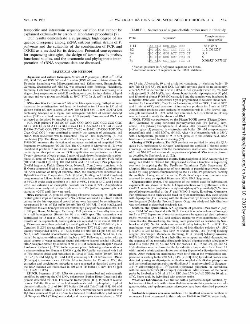
tures of these regions in biologically active rRNAs (19), asinferred from alignment with database sequences, are shown inFig. 3 and 4. One variable nucleotide in region V6 is thoughtnot to be involved in helix formation; other base differences areeither due to covariation of pairing ribonucleotides (transitionsand transversions) or of the transition type U:G7U:A/C:G.Thus, the exchanges do not significantly interfere with second-ary structure formation.
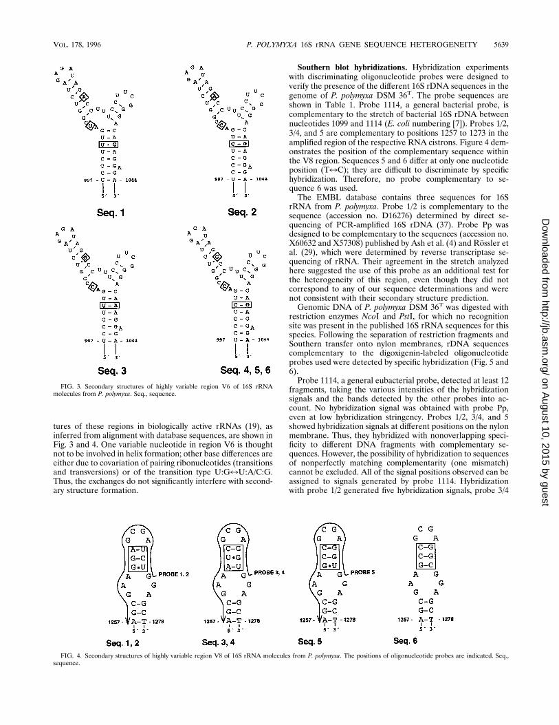
Southern blot hybridizations. Hybridization experimentswith discriminating oligonucleotide probes were designed toverify the presence of the different 16S rDNA sequences in thegenome of P. polymyxa DSM 36T. The probe sequences areshown in Table 1. Probe 1114, a general bacterial probe, iscomplementary to the stretch of bacterial 16S rDNA betweennucleotides 1099 and 1114 (E. coli numbering [7]). Probes 1/2,3/4, and 5 are complementary to positions 1257 to 1273 in theamplified region of the respective RNA cistrons. Figure 4 dem-onstrates the position of the complementary sequence withinthe V8 region. Sequences 5 and 6 differ at only one nucleotideposition (T7C); they are difficult to discriminate by specifichybridization. Therefore, no probe complementary to se-quence 6 was used.The EMBL database contains three sequences for 16S
rRNA from P. polymyxa. Probe 1/2 is complementary to thesequence (accession no. D16276) determined by direct se-quencing of PCR-amplified 16S rDNA (37). Probe Pp wasdesigned to be complementary to the sequences (accession no.X60632 and X57308) published by Ash et al. (4) and Rossler etal. (29), which were determined by reverse transcriptase se-quencing of rRNA. Their agreement in the stretch analyzedhere suggested the use of this probe as an additional test forthe heterogeneity of this region, even though they did notcorrespond to any of our sequence determinations and werenot consistent with their secondary structure prediction.Genomic DNA of P. polymyxa DSM 36T was digested with
restriction enzymes NcoI and PstI, for which no recognitionsite was present in the published 16S rRNA sequences for thisspecies. Following the separation of restriction fragments andSouthern transfer onto nylon membranes, rDNA sequencescomplementary to the digoxigenin-labeled oligonucleotideprobes used were detected by specific hybridization (Fig. 5 and6).Probe 1114, a general eubacterial probe, detected at least 12
fragments, taking the various intensities of the hybridizationsignals and the bands detected by the other probes into ac-count. No hybridization signal was obtained with probe Pp,even at low hybridization stringency. Probes 1/2, 3/4, and 5showed hybridization signals at different positions on the nylonmembrane. Thus, they hybridized with nonoverlapping speci-ficity to different DNA fragments with complementary se-quences. However, the possibility of hybridization to sequencesof nonperfectly matching complementarity (one mismatch)cannot be excluded. All of the signal positions observed can beassigned to signals generated by probe 1114. Hybridizationwith probe 1/2 generated five hybridization signals, probe 3/4
FIG. 3. Secondary structures of highly variable region V6 of 16S rRNAmolecules from P. polymyxa. Seq., sequence.
FIG. 4. Secondary structures of highly variable region V8 of 16S rRNA molecules from P. polymyxa. The positions of oligonucleotide probes are indicated. Seq.,sequence.
VOL. 178, 1996 P. POLYMYXA 16S rRNA GENE SEQUENCE HETEROGENEITY 5639
on August 10, 2015 by guest
http://jb.asm.org/
Dow
nloaded from

generated two, and probe 5 generated four, totalling a mini-mum of 11 fragments hybridizing to these probes in each of thedigests. The sequences determined and used for probe designdo not represent the heterogeneity of the region completely.This is most significantly apparent in theNcoI digest, where thesmallest fragment detected by general probe 1114 did nothybridize to any of the other probes (Fig. 5).An unequivocal counting of fragment numbers in the differ-
ent lanes is confronted with several factors contributing tosignal strength distributions: (i) the efficiency of DNA transferby blotting, (ii) the degree of matching of probes with targetsequences, and (iii) the potential presence of minor bandsresulting from incomplete restriction digestion. However, theinterpretation of Fig. 5 and 6 can be summarized by the state-ment of clearly separable specificity of the probes to targetsequences of different operons and the existence of at least 12operons with 16S rRNA sequences in this strain. A more pre-cise description of the organization of the rRNA operons,which was not intended here, would require much more de-tailed restriction analysis and hybridizations.RT-PCR of 16S rRNA. rRNA was extracted from cultures of
P. polymyxaDSM 36T and E. coli in the late exponential growthphase (optical density at 600 nm of 0.55 after incubation for69 h). Following RT, segments of small-subunit rRNA fromnucleotide 968 to nucleotide 1346 (E. coli numbering) wereamplified and attached to a GC clamp via PCR. Comparedwith the respective PCR product obtained by amplification ofrDNA, the RT-PCR product derived from the type strain of P.polymyxa emerged as a less complex pattern of bands afterTGGE (Fig. 7). No band corresponding to sequence 5 (band hof the strain pattern in lane 1) was detected in the RT-PCR
products from these ribosomes. In contrast, respective ampli-fication products derived from E. coli RNA and rDNA lookedfairly similar, with a slight difference in density distribution.Whole-cell hybridizations with different strains of P. poly-
myxa. Cells from P. polymyxa cultures were fixed with parafor-maldehyde solution and hybridized with fluorescent-dye-la-beled oligonucleotide probes with the sequences described inTable 1. Results obtained with cells from exponentially grow-ing cultures (optical density at 600 nm of 0.06 to 0.15 afterincubation for 20 h) are presented in Table 3. Ribosomes ofthe type strain simultaneously contained 16S rRNA with se-quences 1/2 and 5. The apparent conflict with the result of therRNA analysis presented in Fig. 7 was (partly) resolved byanalyzing cells from older cultures of DSM 36 (optical densi-ties at 600 nm of 0.45 and 0.55 after incubation for 48 and69 h). These demonstrated a less intense hybridization signalwith probe 5, whereas the signal intensity remained unchangedwith probe 1/2 (data not shown in Table 3). No rRNA with thesequences designated 3 and 4 was detected in the type strain.However, all of the different sequences of region V8 repre-sented in the complementary probes were proven to be presentin biologically active ribosomes, but in different strains. Probe3/4 yielded strong hybridization signals with strains 292 and 356only. In ribosomes of strain 365, 16S rRNA complementary toprobe 5 seemed to be predominant. Probe Pp, complementaryto previously published sequences, gave no hybridization signalwith any of the strains. As mentioned for Southern hybridiza-tion, binding of probes to RNA with single mismatches cannotbe excluded with certainty. However, probes 3/4 and 5, differ-ing at two nucleotide positions, discriminate their target se-quences in strains DSM 36, DSM 292, and DSM 356.
FIG. 5. Chromosomal DNA from P. polymyxa DSM 36T was digested withNcoI. Fragments were separated by agarose gel electrophoresis. Southern blot-ted, and hybridized to oligonucleotide probes 1114 (lane a), 1/2 (lane b), 3/4(lane c), 5 (lane d), and Pp (no signals obtained).
FIG. 6. Chromosomal DNA from P. polymyxa DSM 36T was digested withPstI. Fragments were separated by agarose gel electrophoresis. Southern blotted,and hybridized to oligonucleotide probes 1114 (lane a), 1/2 (lane b), 3/4 (lane c),5 (lane d), and Pp (no signals obtained).
5640 NUBEL ET AL. J. BACTERIOL.
on August 10, 2015 by guest
http://jb.asm.org/
Dow
nloaded from

DISCUSSION
Sequence heterogeneity of 16S rRNA genes from P. poly-myxa. Sequences of cloned PCR-amplified segments of 16SrDNA (347 bp corresponding to E. coli positions 984 to 1330)from the type strain of P. polymyxa differ from each other byone to eight nucleotides at 10 sites in total. The analyzedsequences of region V8 were demonstrated to be present in thegenome of the organism under study by Southern blot hybrid-ization. Most likely, the sequences in total, including the V6regions, represent segments of single 16S rRNA genes, al-though the possibility of errors introduced by PCR cannot beexcluded.So far, such a degree of heterogeneity in the small-subunit
rRNA genes of one strain has not been described for bacteria.Although most bacteria have multiple rrn operons, only a few
studies have addressed questions of their identity in structureand function (10). Most investigations so far support the no-tion of a high degree of sequence identity of rrn genes withinone organism. In the seven genes encoding 16S rRNA in thegenome of E. coli, 16 variable sites have been detected (8).Nine substitutions and seven deletions-insertions are scatteredover the rapidly evolving sections. The segment analyzed inthis work contains only three of them (nucleotides 1071 and1074, 1280, and 1321), at positions different from the 10 variantpositions in P. polymyxa. The 16S rRNA genes in four adjacentoperons from B. subtilis 168 (DSM 402) differ at less than 0.2%of their nucleotide positions (26). The three 16S rRNA se-quences of Rhodobacter sphaeroides are identical (12), as arethe six sequences in Haemophilus influenzae Rd, which areorganized in two kinds of operons differing only with respect tothe spacer between the 16S and 23S rRNA genes (13).In contrast, two rrn operons with an overall sequence diver-
gence of 5% and a pronounced uneven distribution of variantpositions were described to exist in the archaebacterium Halo-arcula marismortui (24). In a systematic comparison of pub-lished 16S rRNA sequences of prokaryotic species, a ratherhigh level of sequence variation was observed recently (9). Inaddition to our experimental data, these observations maysupport the notion of more interoperon variability in singlespecies than previously acknowledged.In addition, the distinct band patterns derived from different
P. polymyxa strains indicate interstrain (intraspecific) variabil-ity in the abundance of heterogeneous sequences. (Note thatidentical band positions do not unequivocally demonstratetheir sequence identity. In a segment of some 400 bp, such asthat analyzed here, compensating effects of exchanges in dif-ferent melting domains may account for similar mobility shiftsof nonidentical sequences.) Some bands clearly demonstratethe existence of sequences not found in the type strain. Con-versely, missing bands could be due to the absence of respec-tive variants or to deletions of rrn operons. Such deletionscaused by intrachromosomal recombination events have beenreported for B. subtilis (41).Heterogeneity of rRNA. TGGE analysis of reverse-tran-
scribed and amplified rRNA and whole-cell hybridizations witholigonucleotide probes complementary to sequenced rRNAcistrons indicates that their transcripts are not evenly repre-sented in ribosomes of P. polymyxa cells growing in the labo-ratory (Fig. 7 and Table 3). In addition, some results indicatea variation of the composition of ribosomes, depending ongrowth conditions (culture age). With cells from older cultures,a decreased signal intensity with probe 5 was observed, whichcorresponds to the absence of the respective band in the RT-PCR product analyzed by TGGE (Fig. 7).The results suggest a distinct pattern of use of 16S rrn
operon sequences for the assembly of ribosomes and theirspecific regulation. In B. subtilis, some observations indicategrowth rate-dependent differential expression of rrn operons(30). In some cases, a different pattern of regulation of rrnoperons may also be inferred from differences in upstreamsequence regions (12, 15, 21, 38). Assays of rrn gene transcrip-tion studies rely on more or less indirect methods, and it isdifficult to meet the requirements of a detailed analysis ofindividual operon regulation beyond the role of gene dosageeffects also influenced by the location with respect to the originof replication (11). It may be considered to be meaningful,however, to investigate whether differential regulation of indi-vidual operons, inducing the possibility of uneven selectiveforces acting on them, does in some cases play the biologicalrole which could reasonably be assigned to it (39). The differ-entiation of heterogeneous transcripts by RT-PCR and TGGE
FIG. 7. TGGE separation patterns of PCR products obtained by amplifica-tion of segments of 16S rRNA genes (lanes 1 and 3) and reverse-transcribedrRNA (lanes 2 and 4) from cells in the late exponential growth phase (see text).Amplification products derived from P. polymyxa DSM 36T (lanes 1 and 2) andE. coli NM 522 (lanes 3 and 4) are compared. For designations of individualbands, see the legend to Fig. 2. Primers F-968-GC and R-1346 were used foramplification. ss, single-stranded DNA.
TABLE 3. Results of whole-cell hybridizations with variousstrains of P. polymyxa
StrainHybridizationa with probe:
1/2 3/4 5 Pp
DSM 36T 1 2 1 2DSM 292 2 1 2 2DSM 356 2 1 2 2DSM 365 2 (1) 1 2
a 1, strong hybridization signal; (1), weak hybridization signal; 2, no hybrid-ization signal.
VOL. 178, 1996 P. POLYMYXA 16S rRNA GENE SEQUENCE HETEROGENEITY 5641
on August 10, 2015 by guest
http://jb.asm.org/
Dow
nloaded from

or specific probes in P. polymyxa offers convenient methods tofurther investigate the regulation of individual operons.Evolutionary aspects. The degree of identity of most of the
bacterial operons analyzed so far is thought to be maintainedby some “concerted evolution,” the mechanism of which isunclear in detail. Selective forces on RNA function contributeto a fixation and spread of sequence variants by some “biasedgene conversion” as demonstrated by a sequence conferringantibiotic tolerance (34a). To reconcile the particular degreeof diversity analyzed in P. polymyxa with such a model, one hasto assume that gene conversion and/or selection may act indifferent manners or frequencies in different species. Also,horizontal gene transfer has been implicated as a potentialcontribution to diversities in ribosomal operons of bacteria (24,40). A differential regulation of transcription of rRNA operonswith respect to life cycle or growth conditions would then alsoallow selection and functional adaptations to act on individualoperons.The highly divergent ribosomal sequences in the archaebac-
terium H. marismortui apparently are both represented in ac-tive ribosome structures of laboratory cultures (24). In eu-karyotes, however, differential use of ribosomal componentshas been reported. For example, in Plasmodium spp., correla-tion of the use of heterogeneous 16S rRNAs in ribosomes withthe life cycle in its different hosts has been described (18). Thedistinct abundance of 5S rRNA species in oocytes and somaticcells of Xenopus laevis may be considered to be an analogousobservation (27). It is conceivable that the coding for tRNAs inmany rRNA operons may contribute to some modulation oftranslation activities, but it is a matter of speculation whetherthis may be true for the particular structure of rRNAs. Thus, itremains unclear which structural differences of rRNAs mightreflect a modulation of ribosome function. The nature of an-alyzed exchanges (Fig. 3 and 4) and the presence of the dif-ferent sequence variants in active ribosomes of laboratory-grown cultures of different strains of P. polymyxa indicate thatall versions of the sequences analyzed are derived from func-tional operons. Also, these observations do not support a func-tional significance of the sequence variations observed.Consequences for prokaryotic systematics. The sequence
analysis of small-subunit rRNA is of great importance formodern bacterial taxonomy. The phylogenetic classification ofprokaryotes is based on the assumption that sequences of mac-romolecules such as 16S rRNA reflect the evolution of theorganisms they have been extracted from. The interpretationof these sequence data might be complicated by the presenceof several equivalent molecules with some degree of individu-ally different evolution within a single organism.If single clones of (amplified) rDNA are sequenced, se-
quence heterogeneities of gene copies remain undetected. Thisis also valid for the direct sequencing of rRNA if products ofgenes different in sequence are not evenly represented in ri-bosomes. The direct sequencing of PCR-amplified DNA mayresult in ambiguities due to interoperon variabilities whoseextent is unknown. TGGE offers a convenient tool for detec-tion of these heterogeneities and could thus be used routinelyto support sequence determinations for phylogenetic analyses.The determination of bacterial genera, which often is the
first step of identification procedures, probably is not confusedby the sequence heterogeneities observed here. 16S rRNAsequences are of limited value for measurement of closer phy-logenetic relationships of bacteria. Available data suggest thatthere is no correlation between the results of DNA-DNA re-association experiments and comparisons of small-subunitrRNA sequences at a sequence similarity exceeding 97% (35).Almost identical 16S rRNA sequences have been reported for
phenotypically divergent bacteria (14). Our results lead to theconverse argument that a difference of 2.3% observed in par-tial sequences is not sufficient to prove the presence of differ-ent clones. This stresses problems especially for biodiversityestimates if rRNA sequences are retrieved from uncharacter-ized mixtures of microorganisms. The genetic diversity ofrRNA sequences in a microbial population of an environmen-tal sample may exceed its organismic diversity. Problems alsoarise for the design of rRNA-specific oligonucleotide probesfor detection and identification of prokaryotes. New probesoften fail in practical applications (3). In addition to higher-order structures of rRNA molecules and associated proteins aspotential reasons, the observed sequence heterogeneity mightcontribute to this phenomenon with a frequency which cannotbe estimated with any confidence. However, it is evident fromour observations that sequences determined for rRNA genesdo not necessarily match the primary structure of biologicallyactive rRNA.
ACKNOWLEDGMENTS
Special thanks to Ed Moore and coworkers, GBF, Braunschweig,Germany, for sequencing support.This work was funded in part by the Federal Ministry of Education,
Science, Research and Technology (grant 0310582A).
REFERENCES1. Amann, R. I., B. J. Binder, R. J. Olson, S. W. Chisholm, R. Devereux, andD. A. Stahl. 1990. Combination of 16S rRNA-targeted oligonucleotideprobes with flow cytometry for analyzing mixed microbial populations. Appl.Environ. Microbiol. 56:1919–1925.
2. Amann, R. I., L. Krumholz, and D. A. Stahl. 1990. Fluorescent-oligonucle-otide probing of whole cells for determinative, phylogenetic, and environ-mental studies in microbiology. J. Bacteriol. 172:762–770.
3. Amann, R. I., W. Ludwig, and K. H. Schleifer. 1995. Phylogenetic identifi-cation and in situ detection of individual microbial cells without cultivation.Microbiol. Rev. 59:143–169.
3a.Anonymous. 1994. Validation of the publication of new names and newcombinations previously effectively published outside the IJSB. List no. 51.Int. J. Syst. Bateriol. 44:852.
4. Ash, C., J. A. E. Farrow, S. Wallbanks, and M. D. Collins. 1991. Phylogeneticheterogeneity of the genus Bacillus revealed by comparative analysis ofsmall-subunit-ribosomal RNA sequences. Lett. Appl. Microbiol. 13:202–206.
5. Ausubel, F. M., R. Brent, R. E. Kingston, D. D. Moore, J. A. Smith, J. G.Seidman, and K. Struhl. 1988. Preparation and analysis of DNA, p. 2.4.1.-2.4.5. In F. M. Ausubel (ed.), Current protocols in molecular biology. JohnWiley & Sons, Inc., New York.
6. Belfort, M., M. E. Reaban, T. Coetzee, and J. Z. Dalgaard. 1995. Prokaryoticintrons and inteins: a panoply of form and function. J. Bacteriol. 177:3897–3903.
7. Brosius, J., T. Dull, D. D. Sleeter, and H. F. Noller. 1981. Gene organizationand primary structure of a ribosomal RNA operon from Escherichia coli.Proc. Natl. Acad. Sci. USA 75:4801–4805.
8. Carbon, P., C. Ehresmann, B. Ehresmann, and J.-P. Ebel. 1979. The com-plete nucleotide sequence of the ribosomal 16S rRNA from Escherichia coli.Eur. J. Biochem. 100:399–410.
9. Clayton, R. A., G. Sutton, P. S. Hinkle, Jr., C. Bult, and C. Fields. 1995.Intraspecific variation in small-subunit rRNA sequences in GenBank: whysingle sequences may not adequately represent prokaryotic taxa. Int. J. Syst.Bacteriol. 45:595–599.
10. Cole, S. T., and I. Saint Girons. 1994. Bacterial genomics. FEMS Microbiol.Rev. 14:139–160.
11. Condon, C., D. Liveris, C. Squires, I. Schwartz, and C. L. Squires. 1995.rRNA operon multiplicity in Escherichia coli and the physiological implica-tions of rrn inactivation. J. Bacteriol. 177:4152–4156.
12. Dryden, S., and S. Kaplan. 1990. Localization and structural analysis of theribosomal RNA operons of Rhodobacter sphaeroides. Nucleic Acids Res.18:7267–7277.
13. Fleischmann, R. D., M. D. Adams, O. White, R. A. Clayton, E. F. Krikness,A. R. Kerlavage, C. J. Bult, J.-F. Tomb, B. A. Dougherty, J. M. Merrick, K.McKenney, G. Sutton, W. FitzHugh, C. Fields, J. D. Gocayne, J. Scott, R.Shirley, L.-I. Liu, A. Glodek, J. M. Kelley, J. F. Weidman, C. A. Philips, T.Spreiggs, E. Hedblom, M. D. Cotton, T. R. Utterback, M. C. Hanna, D. T.Nguyen, D. M. Saudek, R. C. Brandon, L. D. Fine, J. L. Fritchman, J. L.Fuhrmann, N. S. M. Geoghagen, C. L. Gnehm, L. A. McDonald, K. V. Small,C. M. Fraser, H. O. Smith, and J. C. Venter. 1995. Whole-genome randomsequencing and assembly of Haemophilus influenzae Rd. Science 269:496–512.
5642 NUBEL ET AL. J. BACTERIOL.
on August 10, 2015 by guest
http://jb.asm.org/
Dow
nloaded from

14. Fox, G. E., J. D. Wisotzkey, and P. Jurtshuk, Jr. 1992. How close is close: 16SrRNA sequence identity may not be sufficient to guarantee species identity.Int. J. Syst. Bacteriol. 42:166–170.
15. Garnier, T., B. Canard, and S. T. Cole. 1991. Cloning, mapping, and molec-ular characterization of the rRNA operons of Clostridium perfringens. J.Bacteriol. 173:5431–5438.
16. Giovannoni, S. J. 1991. The polymerase chain reaction, p. 178–203. In E.Stackebrandt and M. Goodfellow (ed.), Nucleic acid techniques in bacterialsystematics. John Wiley & Sons, Chichester, England.
17. Giovannoni, S. J., T. B. Britschgi, C. L. Moyer, and K. G. Field. 1990.Genetic diversity in Sargasso Sea bacterioplancton. Nature (London) 345:60–63.
18. Gunderson, J. H., M. L. Sogin, G. Wollett, M. Hollingdale, V. F. de la Cruz,A. P. Waters, and T. F. McCutchan. 1987. Structurally distinct, stage-specificribosomes occur in Plasmodium. Science 238:933–937.
19. Gutell, R. R., N. Larsen, and C. R. Woese. 1994. Lessons from an evolvingrRNA: 16S and 23S rRNA structures from a comparative perspective. Mi-crobiol. Rev. 58:10–26.
20. Larsen, N., G. J. Olsen, B. L. Maidak, M. J. McCaughey, R. Overbeek, T. J.Macke, T. L. Marsh, and C. R. Woese. 1993. The ribosomal database project.Nucleic Acids Res. 21:3021–3023.
21. Liesack, W., and E. Stackebrandt. 1989. Evidence for unlinked rrn operonsin the planctomycete Pirellula marina. J. Bacteriol. 171:5025–5030.
22. Meyer, C. G., E. Tannich, J. Harders, K. Henco, and R. D. Horstmann. 1991.Direct sequencing of variable HLA gene segments after in vitro amplificationand allele separation by temperature-gradient gel electrophoresis. J. Immu-nol. Methods 142:251–256.
23. Muyzer, G., E. C. de Waal, and A. G. Uitterlinden. 1993. Profiling of complexmicrobial populations by denaturing gradient gel electrophoresis analysis ofpolymerase chain reaction-amplified genes coding for 16S rRNA. Appl.Environ. Microbiol. 59:695–700.
24. Mylvaganam, S., and P. P. Dennis. 1992. Sequence heterogeneity betweenthe two genes encoding 16S rRNA from the halophilic archaebacteriumHaloarcula marismortui. Genetics 130:399–410.
25. Neefs, J. M., Y. V. de Peer, L. Hendriks, and R. de Wachter. 1990. Compi-lation of small ribosomal subunit RNA sequences. Nucleic Acids Res. 18:2237–2242.
26. Ogasawara, N., S. Nakai, and H. Joshikawa. 1994. Systematic sequencing ofthe 180 kilobase region of the Bacillus subtilis chromosome containing thereplication origin. DNA Res. 1:1–14.
27. Peterson, R. C., J. L. Doering, and D. D. Brown. 1980. Characterization oftwo Xenopus somatic 5S DNAs and one minor oocyte-specific 5S DNA. Cell20:131–141.
28. Rosenbaum, V., and D. Riesner. 1987. Temperature-gradient gel electro-phoresis; thermodynamic analysis of nucleic acids and proteins in purifiedform and in cellular extracts. Biophys. Chem. 26:235–246.
29. Rossler, D., W. Ludwig, K. H. Schleifer, C. Lin, T. J. McGill, J. D. Wisotzkey,P. Jurtshuk, Jr., and G. E. Fox. 1991. Phylogenetic diversity in the genusBacillus as seen by 16S rRNA sequencing studies. Syst. Appl. Microbiol.14:266–269.
30. Rudner, R., A.-M. White, B. Studamire, D. Liu, W. Lam, W. Samarrai, andC. J. Barangan. 1993. Heterogeneity in expression of ribosomal RNA oper-ons in Bacillus subtilis, abstr. H-257, p. 236. In Abstracts of the 93rd GeneralMeeting of the American Society for Microbiology 1993. American Societyfor Microbiology, Washington, D.C.
31. Saiki, R. K., D. H. Gelfand, S. Stoffel, S. J. Scharf, R. Higuchi, G. T. Horn,K. B. Mullis, and H. A. Erlich. 1988. Primer-directed enzymatic amplifica-tion of DNA with a thermostable DNA polymerase. Science 239:487–491.
32. Sambrook, J., E. F. Fritsch, and T. Maniatis. 1989. Molecular cloning: alaboratory manual, 2nd ed. Cold Spring Harbor Laboratory Press, ColdSpring Harbor, N.Y.
33. Sheffield, V. C., D. R. Cox, L. S. Lerman, and R. M. Myers. 1989. Attachmentof a 40-base-pair G1C-rich sequence (GC-clamp) to genomic DNA frag-ments by the polymerase chain reaction results in improved detection ofsingle-base changes. Proc. Natl. Acad. Sci. USA 86:232–236.
34. Sogin, M. L. 1990. Amplification of ribosomal RNA genes for molecularevolution studies, p. 307–314. In M. A. Innis, D. H. Gelfand, J. J. Sninsky,and T. J. White (ed.), PCR protocols, a guide to methods and applications.Academic Press, San Diego, Calif.
34a.Soller, R., E. Stackebrandt, and G. Krupp. Personal communication.35. Stackebrandt, E., and B. M. Goebel. 1994. Taxonomic note: a place for
DNA-DNA reassociation and 16S rRNA sequence analysis in the presentspecies definition in bacteriology. Int. J. Syst. Bacteriol. 44:846–849.
36. Steger, G. 1994. Thermal denaturation of double-stranded nucleic acids:prediction of temperatures critical for gradient gel electrophoresis and poly-merase chain reaction. Nucleic Acids Res. 22:2760–2768.
37. Suzuki, T., and K. Yamasato. 1994. Phylogeny of spore-forming lactic acidbacteria based on 16S rRNA gene sequences. FEMS Microbiol. Lett. 115:13–17.
38. van Wezel, G. P., E. Vijgenboom, and L. Bosch. 1991. A comparative studyof the ribosomal RNA operons of Streptomyces coelicolor A3(2) and se-quence analysis of rRNA. Nucleic Acids Res. 19:4399–4403.
39. Wagner, R. 1994. The regulation of ribosomal RNA synthesis and bacterialcell growth. Arch. Microbiol. 161:100–109.
40. Wich, G., L. Siebold, and A. Bock. 1987. Divergent evolution of 5S rRNAgenes in Methanococcus. Z. Naturforsch. 42c:373–380.
41. Widom, R. L., E. D. Jarvis, G. LaFauci, and R. Rudner. 1988. Instability ofrRNA operons in Bacillus subtilis. J. Bacteriol. 170:605–610.
42. Woese, C. R. 1987. Bacterial evolution. Microbiol. Rev. 51:221–271.43. Zarda, B., R. Amann, G. Wallner, and K.-H. Schleifer. 1991. Identification of
single bacterial cells using digoxigenin-labeled, rRNA-targeted oligonucleo-tides. J. Gen. Microbiol. 137:2823–2830.
VOL. 178, 1996 P. POLYMYXA 16S rRNA GENE SEQUENCE HETEROGENEITY 5643
on August 10, 2015 by guest
http://jb.asm.org/
Dow
nloaded from
Related Documents