Bacillus and Paenibacillus spp. associated with extended shelf life milk By Mugadza Desmond Tichaona Submitted in partial fulfillment of the requirements for the degree PhD (Food Science) In the Department of Food Science Faculty of Natural and Agricultural Sciences University of Pretoria Republic of South Africa November, 2017

Welcome message from author
This document is posted to help you gain knowledge. Please leave a comment to let me know what you think about it! Share it to your friends and learn new things together.
Transcript
Bacillus and Paenibacillus spp. associated with extended shelf life milk
By
Mugadza Desmond Tichaona
Submitted in partial fulfillment of the requirements for the degree
PhD (Food Science)
In the
Department of Food Science
Faculty of Natural and Agricultural Sciences
University of Pretoria
Republic of South Africa
November, 2017
i
DECLARATION
I, Desmond Tichaona Mugadza declare that the thesis, which I hereby submit for the degree PhD
Food Science at the University of Pretoria, is my own work and has not previously been
submitted by me for a degree at this or any other tertiary institution.
November, 2017
ii
ABSTRACT
Bacillus and Paenibacillus spp. associated with extended shelf life milk
By
Mugadza Desmond Tichaona
Supervisor: Prof. E. M. Buys
Degree: PhD Food Science
Extended shelf life (ESL) milk combines longer shelf life and better organoleptic characteristics;
a combination that is absent in both pasteurised and ultra-heat treated (UHT) milk. Bacterial
spoilage remains the main cause of food loss worldwide, which also includes milk and dairy
products, despite advances in food preservation technology. The objectives of this study were to
identify and characterise the spore-forming population associated with ESL milk during
processing and chilled storage as well as characterising Bacillus cereus isolates obtained from
ESL milk processing and during storage.
Characterisation of spore-formers associated with ESL milk was done by analysing
bacteriological quality of milk samples collected at various processing stages and during storage.
Isolates were identified with MALDI-TOF-MS. B. cereus strains obtained from ESL milk and
filler nozzles were characterised using (GTG)5 Rep PCR fingerprinting; the presence of virulence
genes; cytotoxin K (cytK), nonhemolytic enterotoxin A (nheA), emetic toxin cereulide (cer) and
enterotoxin hemolysin BL (hblA). The isolates were further discriminated as psychrotrophic and
iii
mesophilic strains using 16S rDNA. Furthermore, B. cereus isolates were selected for 16S partial
sequencing. Some of the B. cereus strains obtained from ESL milk and filler nozzles were further
characterised using rpoB partial sequencing and multilocus sequence typing (MLST).
Milk had spore counts < 2 log10 cfu/ml and 4 log10 cfu/ml during processing and storage,
respectively. Bacillus pumilus dominated the bacterial population. In addition B. subtilis, B.
cereus, B. sonorensis, B. licheniformis and Paenibacillus spp. were among the main spore-
formers identified in the study. Bacterial species were inoculated in sterile milk for a shelf life
study and population change observed over 42 days at 7 oC. Despite high prevalence of cer, hblA
and nheA; cytK was not widely distributed. There was 100% and 8% prevalence of mesophilic
and psychrotrophic signatures, respectively in B. cereus isolates. Although ESL milk process
was effective in the reduction of bacterial counts and species diversity, the presence of B. cereus
shows a potential safety problem in ESL milk. Despite the large diversity of the B. cereus strains
in this study, there is evidence that biofilms associated with filler nozzles and raw milk are a
source of contamination of B. cereus in ESL milk. Furthermore, the study has also shown that
rpoB partial sequencing and MLST can be used as a tool for source tracking in ESL milk
processing.

iv
DEDICATION
I dedicate this work to my late grandparents Mr and Mrs S.H Chiware. It is through the firm
foundation you laid in my life that I fought to see this dream turn into reality. May your souls
rest in eternal peace.
v
ACKNOWLEDGEMENTS
I give glory to God for the gift of life, the strength and opportunity He granted me to successfully
pursue my studies. I am very grateful to my supervisor and mentor Prof Elna Buys for her
excellent guidance, immeasurable dedication to my research and for believing in my abilities. I
would like to thank her for taking care and looking out for me throughout this journey, the
opportunities she availed for me as well as advice and encouragement she gave me, Thank you
Prof.
Many thanks go to the University of Pretoria for funding my studies and Department of Food
Science for the support and constructive criticism they provided throughout my research. I would
like to extend my gratitude to the Food Microbiology team for their immeasurable assistance
during my studies, in particular I want to appreciate the assistance and ideas I got from Victor,
Rodney, Sandile, Gabriel, Thulani, Mathew, Dr Fayemi, Dr Dlamini and Dr Njage. I would like
to express my gratitude to the Midlands State University for allowing me to pursue my studies.
Many thanks are due to The Department of Food Science and Nutrition for their support
throughout my studies especially Mr. Manhokwe, Mrs Makamure, Ruth, Victor and Beulah. I
would like to thank my wife Natasha and daughter Unathi for their emotional support and
prayers as well as giving me joy all the time. I love you guys. Many thanks are due to my mom,
Pr. Mzapi, Pr. Chimone and Sibonginkosi for their emotional support as well as everlasting
prayers. I am also grateful to my brother Tatenda and his family for always being there for me. I
am grateful to my family and friends for their support especially Reginald, Talknice, Amiel,
Ronald, Bernard, Munyaradzi, Leonard, Lindelwe, Edgar, James and Takudzwa.
vi
TABLE OF CONTENTS
DECLARATION ............................................................................................................................. i
ABSTRACT .................................................................................................................................... ii
DEDICATION ............................................................................................................................... iv
ACKNOWLEDGEMENTS ............................................................................................................ v
TABLE OF CONTENTS ............................................................................................................... vi
LIST OF TABLES ........................................................................................................................ xii
LIST OF FIGURES ..................................................................................................................... xiii
CHAPTER ONE ............................................................................................................................. 1
GENERAL INTRODUCTION ....................................................................................................... 1
CHAPTER TWO ............................................................................................................................ 5
LITERATURE REVIEW ............................................................................................................... 5
2.1 Milk production and consumption trends ........................................................................ 6
2.2 Extended shelf life milk ................................................................................................... 8
2.2.1 Milk chilling............................................................................................................ 10
2.2.2 Heat treatment of milk ............................................................................................ 11
2.2.2.1 Pasteurisation of milk ...................................................................................... 12
2.2.3 Microfiltration of milk ............................................................................................ 14
2.2.4 Bactofugation of milk ............................................................................................. 15
2.2.5 Aseptic packaging ................................................................................................... 16
vii
2.3 Bacteria associated with milk ......................................................................................... 17
2.3.1 Raw milk ................................................................................................................. 17
2.3.2 Heat treated milk ..................................................................................................... 19
2.3.2.1 Pasteurised milk ............................................................................................... 19
2.3.2.2 ESL milk .......................................................................................................... 20
2.4 Contamination routes in milk processing ....................................................................... 21
2.4.1 Milk at the farm ...................................................................................................... 21
2.4.1.1 Interior of the udder ......................................................................................... 21
2.4.1.2 Exterior of the udder ........................................................................................ 22
2.4.1.3 Water ............................................................................................................... 22
2.4.1.4 Milk handling equipment................................................................................. 23
2.4.2 Milk at the processing plant .................................................................................... 23
2.4.2.1 Processing equipment ...................................................................................... 23
2.4.2.2 Air in the processing plant ............................................................................... 24
2.4.2.3 Packaging materials ......................................................................................... 24
2.5 Adaptation mechanisms of bacteria during milk processing ......................................... 25
2.5.1 Bacterial endospores ............................................................................................... 26
2.5.2 Biofilms................................................................................................................... 27
2.5.3 Cold shock response ............................................................................................... 28
2.6 Spore-forming bacteria associated with ESL milk ......................................................... 30
2.6.1 Paenibacillus spp. ................................................................................................... 30
2.6.2 Bacillus spp. ............................................................................................................ 31
2.6.2.1 B. pumilus ........................................................................................................ 32
viii
2.6.2.2 B. subtilis ......................................................................................................... 33
2.6.2.3 B. cereus .......................................................................................................... 34
2.6.2.3.1 B. cereus pathogenicity and outbreaks ........................................................ 35
2.7 Advances in detection and identification of microorganisms in the food industry ........ 39
2.7.1 Protein based methods ............................................................................................ 39
2.7.1.1 Matrix Assisted Laser Desorption / Ionization Time of Flight Mass
Spectrometry (MALDI-TOF-MS) .................................................................................... 39
2.7.2 DNA based methods ............................................................................................... 40
2.7.2.1 End point PCR ................................................................................................. 40
2.7.2.2 rep-PCR ........................................................................................................... 40
2.7.2.3 16S sequencing ................................................................................................ 41
2.7.3.4 Multilocus sequence typing (MLST) ............................................................... 42
2.7.3.5 Whole genome sequencing (WGS) ................................................................. 43
CHAPTER THREE ...................................................................................................................... 45
HYPOTHESES AND OBJECTIVES ........................................................................................... 45
3.1 Hypothesis 1 ................................................................................................................... 46
3.1.1 Objective ........................................................................................................................ 46
3.2 Hypothesis 2 ................................................................................................................... 47
3.2.1 Objective ........................................................................................................................ 47
CHAPTER FOUR ......................................................................................................................... 48
Bacillus and Paenibacillus spp. associated with extended shelf life milk during processing and
storage ....................................................................................................................................... 48
ix
4.1.1 Abstract ................................................................................................................... 49
4.1.2 Introduction ............................................................................................................. 49
4.1.3 Materials and methods ............................................................................................ 51
4.1.3.1 Milk sampling .................................................................................................. 51
4.1.3.2 Isolation and identification of Bacillus spp. and Paenibacillus spp. ............... 52
4.1.3.3 Determination of heamolysis, proteolytic and lipolytic enzyme activity ........ 53
4.1.3.4 Shelf life studies .............................................................................................. 53
4.1.3.5 Growth profiles at 7 oC .................................................................................... 53
4.1.3.6 Isolates identification by MALDI-TOF-MS.................................................... 54
4.1.4 Results ..................................................................................................................... 55
4.1.4.1 Bacterial counts of milk during ESL milk processing and shelf life ............... 55
4.1.4.2 Bacterial identification, species distribution and enzymatic activity of isolates
from ESL milk processing and storage ............................................................................. 57
4.1.4.3 B. pumilus MALDI-TOF-MS cluster analysis ................................................ 59
4.1.4.4 B. subtilis MALDI-TOF-MS cluster analysis .................................................. 59
4.1.4.5 B. cereus MALDI-TOF-MS cluster analysis ................................................... 59
4.1.4.6 Paenibacillus spp. MALDI-TOF-MS cluster analysis .................................... 63
4.1.4.7 Bacillus spp. and Paenibacillus spp. growth profiles at 7 oC .......................... 64
4.1.5 Discussion ............................................................................................................... 65
4.1.6 Conclusions ............................................................................................................. 69
CHAPTER FIVE .......................................................................................................................... 70
Diversity of Bacillus cereus strains in extended shelf life milk ................................................ 70
x
5.1.1 Abstract......................................................................................................................... 71
5.1.2 Introduction ............................................................................................................. 71
5.1.3 Materials and methods ............................................................................................ 73
5.1.3.1 Isolates and DNA extraction............................................................................ 73
5.1.3.2 (GTG)5 Rep PCR Fingerprinting .......................................................................... 73
5.1.3.3 PCR to determine virulence genes and discriminate psychrotrophic from
mesophilic B. cereus ......................................................................................................... 74
5.1.3.4 B. cereus 16S rRNA Sequencing .......................................................................... 75
5.1.4 Results ..................................................................................................................... 77
5.1.4.1 (GTG)5 Rep PCR Fingerprinting of B. cereus strains isolated from ESL milk
processing and during storage ........................................................................................... 77
5.1.4.2 Detection of virulence, mesophilic and psychrotrophic genes in B. cereus strains
isolated from ESL milk processing and during storage .................................................... 78
5.1.4.3 B. cereus 16S Sequencing ............................................................................... 83
5.1.5 Discussion ............................................................................................................... 85
5.1.6 Conclusion .............................................................................................................. 88
CHAPTER SIX ............................................................................................................................. 90
Source tracking Bacillus cereus in an extended shelf life milk processing plant using partial
sequencing of rpoB and multilocus sequence typing ................................................................ 90
6.1.1 Abstract ................................................................................................................... 91
6.1.2 Introduction ............................................................................................................. 91
6.1.3 Materials and methods ............................................................................................ 93
6.1.3.1 Bacteria strains and DNA preparation ............................................................. 93
6.1.4 Results and discussion ............................................................................................ 97
xi
6.1.6 Conclusions ........................................................................................................... 101
CHAPTER SEVEN .................................................................................................................... 102
GENERAL DISCUSSION ......................................................................................................... 102
7.1 Methodological considerations .................................................................................... 103
7.2 Isolation of spore-formers in ESL milk and characterisation of B. cereus from ESL milk
processing and during storage ................................................................................................. 113
7.3 Future research ............................................................................................................. 117
CHAPTER EIGHT ..................................................................................................................... 119
CONCLUSIONS AND RECOMMENDATIONS ..................................................................... 119
CHAPTER NINE ........................................................................................................................ 121
REFERENCES ........................................................................................................................... 121
CHAPTER TEN.......................................................................................................................... 170
PUBLICATIONS AND AWARDS ............................................................................................ 170
10.1 Peer reviewed journal articles ...................................................................................... 171
10.2 Popular publications ..................................................................................................... 171
10.3 Conference presentations ............................................................................................. 171
10.4 Awards ......................................................................................................................... 173
xii
LIST OF TABLES
Table 2.1: Effects of the tripartite cytotoxins on different cells ................................................... 37
Table 4.1.1: Methodology for enumeration of bacterial organisms in raw milk, pasteurised milk
and packaged extended shelf life milk...................................................................... 52
Table 4.1.2: Enzymatic activity (%) of Bacillus pumilus, B. subtilis, B. cereus and Paenibacillus
species isolated from ESL milk processing and storage ........................................... 57
Table 4.1.3: Prevalence of spore forming bacteria isolated during extended shelf life milk
processing and storage .............................................................................................. 58
Table 5.1.1: Primers used to detect virulence, 16S mesophilic and psychrotrophic genes in B.
cereus strains isolated from extended shelf life milk processing and during shelf life
.................................................................................................................................. 76
Table 5.1.2: 16S Primers sequences used for B. cereus 16S sequencing ..................................... 77
Table 5.1.3: Detection of virulence, mesophilic and psychrotrophic genes in B. cereus strains
isolated from raw milk, ESL milk processing and during shelf life ......................... 81
Table 6.1.1: Description of virulence, mesophilic and psychrotrophic genes in B. cereus strains
isolated from raw milk, ESL milk processing and during shelf life (Khoza 2016*;
Mugadza and Buys 2017b) ....................................................................................... 94
Table 6.1.2: Sequence description and annealing temperatures for MLST and rpoB genes used
for sequencing B. cereus strains isolated from raw milk, ESL milk processing and
during shelf life. ........................................................................................................ 95
xiii
LIST OF FIGURES
Figure 2.1: Percentage composition of liquid milk in South Africa (Lacto data 2015) .................. 7
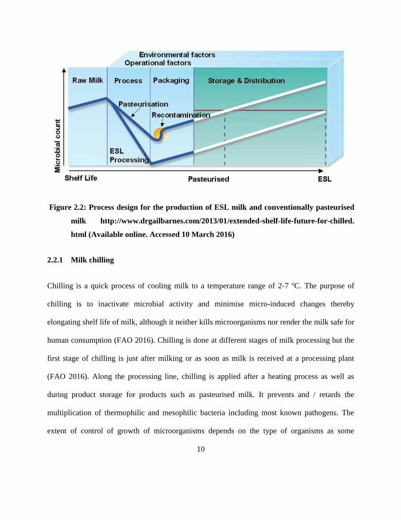
Figure 2.2: Process design for the production of ESL milk and conventionally pasteurised milk
http://www.drgailbarnes.com/2013/01/extended-shelf-life-future-for-chilled. html
(Available online. Accessed 10 March 2016) ............................................................ 10
Figure 2.3: Relative changes in time temperature profiles for the destruction of microorganisms
and vitamins in milk during pasteurisation.
https://www.uoguelph.ca/foodscience/book-page/thermal-destruction-
microorganisms. (Available online. Accessed 13 March 2016) ................................ 14
Figure 2.4: Contamination route of Bacillus cereus in a production chain (Heyndrickx 2011) ... 25
Figure 2.5: Stages that a bacterial cell goes through in a sporulation cycle
http://zf2t.dromibi.top/c/spore-formation/ (Available online. Accessed 27/03/2016) 27
Figure 4.1.1: Schematic diagram of the extended shelf life milk manufacturing process and the
sampling points ........................................................................................................... 51
Figure 4.1.2: Bacterial counts of milk at different sampling points during extended shelf life
milk processing. Sampling points with a different letter for the same bacterial group
have means that are significantly different (p < 0.05) (n = 6) .................................... 55
Figure 4.1.3: Effect of storage temperature on the bacterial composition of ESL milk during
storage for 21 days at 4 and 7 oC. Storage days with a different letter for the same
bacterial group have means that are significantly different (p < 0.05) (n = 72) ......... 56
Figure 4.1.4: Bacillus pumilus cluster analysis of MALDI-TOF-MS data derived from isolates
obtained in extended shelf life milk processing, extended shelf life milk stored at 4 oC
and 7 oC over a period of 21 days and extended shelf life milk filler nozzles after CIP
process. ....................................................................................................................... 60
Figure 4.1.5: Bacillus subtilis cluster analysis of MALDI-TOF-MS data derived from isolates
obtained in extended shelf life milk processing, extended shelf life milk stored at 4 oC
and 7 oC over a period of 21 days and Extended Shelf Life milk filler nozzles after
CIP process. ................................................................................................................ 61

xiv
Figure 4.1.6: Bacillus cereus cluster analysis of MALDI-TOF-MS data derived from isolates
obtained in extended shelf life milk processing, extended shelf life milk stored at 4
oC and 7 oC over a period of 21 days and extended shelf life milk filler nozzles after
CIP process. ............................................................................................................. 62
Figure 4.1.7: Paenibacillus spp. cluster analysis of MALDI-TOF-MS data derived from isolates
obtained in extended shelf life milk processing extended shelf life milk stored at 4
oC and 7 oC over a period of 21 days and extended shelf life milk filler nozzles after
CIP process. ............................................................................................................. 63
Figure 4.1.8: Changes in the population of inoculated B. cereus, B. subtilis, Paenibacillus spp.
B. pumilus and bacteria cocktail of B. cereus, B. subtilis, Paenibacillus spp. and B.
pumilus in milk over a 42 day shelf life at 7 oC. ..................................................... 64
Figure 5.1.1: Dendrogram of Rep-PCR (GTG)5 fingerprint patterns in B. cereus isolates from
raw milk, ESL milk processing and during shelf life. ............................................. 80
Figure 5.1.2: Agglomerative Hierarchical clustering of B. cereus isolates from ESL milk
processing and during shelf life based on presence of virulent genes, 16S
mesophilic and psychrotrophic signatures. ............................................................. 82
Figure 5.1.3: Principal component analysis of virulent genes, 16S mesophilic and psychrotrophic
signature data set consisting of 49 B. cereus isolates obtained from ESL milk
processing and during storage. ................................................................................ 84
Figure 5.1.4: 16S rDNA sequence based maximum likelihood phylogenetic tree of B. cereus
from ESL milk processing and during shelf life and other B. cereus strains.
. ................................................................................................................................ 84
Figure 6.1.1: MLST profiles using five housekeeping alleles showing relationship among B.
cereus strains isolated from raw milk, ESL milk processing and during shelf life. 97
Figure 6.1.2: Neighbor-joining rpoB dendrogram representing the phylogenetic relationships of
B. cereus strains isolated from raw milk, ESL milk processing and during shelf life.
................................................................................................................................. 99
Figure 6.1.3: Routes and sources of ESL milk contamination by B. cereus in a processing plant
based on similar MLST sequence type……………………………………………97
1
CHAPTER ONE
GENERAL INTRODUCTION
2
Extended shelf life (ESL) milk bridges the gap between high temperature short time (HTST)
pasteurised milk, with a shelf life of around 10 days under refrigeration, and ultra-heat treated
(UHT) milk with a shelf life of at least 3 months at ambient storage temperatures (Fitzgerald
2012). Currently, there are two methods used in the production of ESL milk, ever since its
existence (about 6 years) in the South African market. In the first method, milk is subjected to
bactofugation, pasteurised and finally packaged aseptically, while, in the second method milk is
subjected to UHT temperatures (135 oC) for about 0.5 s and packaged. The objective of the
product is to combine longer shelf life and better organoleptic characteristics, a combination that
is absent in both pasteurised and UHT milk (Rysstad and Kolstad 2006). Bacterial spoilage
remains the main cause of food loss worldwide, including milk and dairy products, despite these
and other advances in food preservation technology (Gram et al. 2002; Ranieri et al. 2012).
Previous studies have also indicated that most customer complaints emanate from microbial
spoilage compared to other factors in processed fluid milk (Hayes et al. 2002; Fromm and Boor
2004; Ranieri et al. 2012). The production of thermostable proteases and lipases that can remain
active even after the elimination of the vegetative microorganisms by heat treatments applied has
been reported as one of the biggest hurdle in extending the shelf life of milk (Júnior et al. 2017).
The ability of Bacillus and Paenibacillus spp. among other bacteria to form endospores has also
emerged as another great obstacle in extending the shelf life of milk. Most endospores are heat
resistant and upon germination the organisms are able to grow under a wide range of
temperatures and pH, (Huck et al. 2007). Psychrotrophic bacteria have been recognised as a
pertinent problem in the dairy industry and they contribute to about 25% of shelf life problems in
conventionally pasteurised milk and greatly limit shelf life extension of fluid milk and related
3
products (Francis et al. 1998; Stenfors and Granum 2001; Huck et al. 2007; Huck et al. 2008).
Research has documented that Bacillus spp. dominate the endospore forming population in milk
(Coorevits et al. 2008; De Jonghe et al. 2010; Schmidit et al. 2012; Aoudhi et al. 2014), while
Paenibacillus spp. increase in population during storage of pasteurised milk under refrigeration
to outnumber the previously dominating Bacillus spp. at the beginning of the shelf life of
pasteurised milk (Ranieri and Boor 2009; Ranieri et al. 2012).
Despite the high diversity of Bacillus spp. in milk (Fromm and Boor 2004; Aouadhi et al. 2014),
Bacillus cereus attracts great attention in food processing. In addition to causing spoilage
problems in milk, B. cereus has also been reported to be an opportunistic human pathogen
(Bartoszewicz et al. 2008) that causes two principal types of food poisoning, which are, the
emetic and diarrhea (Hansen and Hendriksen 2000). Although it is regarded as a mesophile,
some researchers have reported the existence of psychrotolerant strains of B. cereus (Stenfors
and Granum 2001). These strains have been reported to have the ability to germinate at
temperatures between 4-6 oC and grow well at temperatures below 10 oC (Larsen and Jørgensen
1997).
Although raw milk has been implicated as an important source of endospores in milk products
(Miller et al. 2015), other studies have shown that a different population of endospore forming
microorganisms exists in raw milk and other milk products and has been attributed to a number
of reasons including post heat treatment contamination by processing equipment. (Scott et al.
2007; Burgess et al. 2010; Hill and Smythe 2012). B. cereus contamination has been linked to
4
processing equipment such as milk fillers (Khoza 2016) since its spores are highly adhesive
(Anersen 2007).
Although several studies on ESL milk have been reported, nothing has been documented on the
bactofugation based ESL milk product. The objective of this study was to characterise the
Bacillus and Paenibacillus spp. associated with ESL milk spoilage, during processing and chilled
storage, with the aim of validating the effectiveness of heat and bactofugation based ESL milk
process on the spore-formers as well as understanding the route of ESL milk contamination in a
processing plant.
5
CHAPTER TWO
LITERATURE REVIEW
6
2.1 Milk production and consumption trends
Global milk production has increased by 50% in the past 3 decades from 482 million tonnes in
1982 to 754 million tonnes in 2012 (FAO 2016). While the greatest expansion in milk production
has been in South Asia since 1971 and other countries such as USA and New Zealand, little
growth has been observed in Africa due to poverty and adverse climatic conditions in other
countries (Hemme et al. 2010). World milk production declined by 9% in 2005 indicating that
the world milk production has not kept pace with increase in world population, despite the
increase in global milk production the per capita (Knips 2005).
Asia is the highest consuming region with 42% of total dairy demand, followed by Europe
(26%). Asia still has large growth potential as its per capita consumption (75 kg per person per
year) is still low compared to other areas, with the exception of Africa (49.2 kg). While UN
estimates a 16% global population increase by 2030, the OECD/FAO agricultural outlook,
projects that the global average per capita dairy consumption should increase by 13.7% between
2013 and 2023 (FAO 2016). The main drivers remain the growth in the global population,
income levels and urbanization. Faster growth is expected in developing countries with current
low per capita consumption.
While the average global cost of milk production is US$ 46/100 kg, the average cost of
production of milk in South Africa lies slightly above US$ 35 per 100 kg of milk, which is at par
with the New Zealand cost level but lower than most other dairy countries. Similar to global
trends, the South African dairy industry has also seen growth in production with a 22% increase
of milk in between 2009 and 2016 (Lacto data 2016). South African dairy market is divided into
7
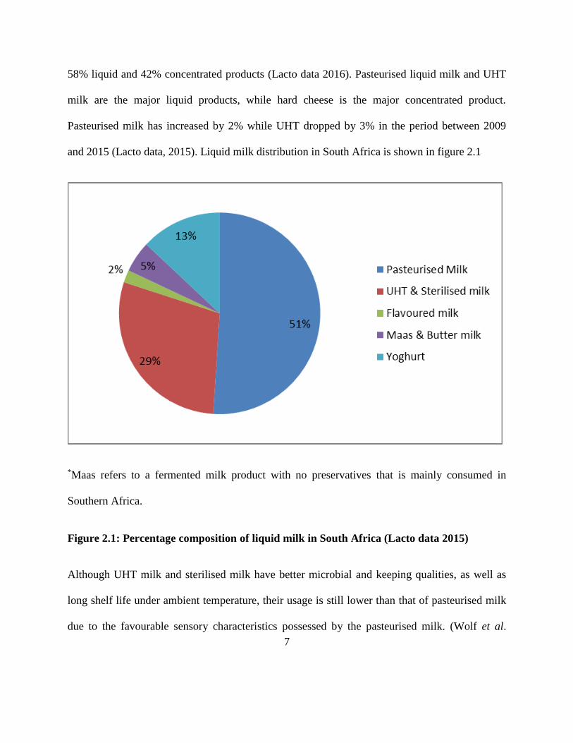
58% liquid and 42% concentrated products (Lacto data 2016). Pasteurised liquid milk and UHT
milk are the major liquid products, while hard cheese is the major concentrated product.
Pasteurised milk has increased by 2% while UHT dropped by 3% in the period between 2009
and 2015 (Lacto data, 2015). Liquid milk distribution in South Africa is shown in figure 2.1
*Maas refers to a fermented milk product with no preservatives that is mainly consumed in
Southern Africa.
Figure 2.1: Percentage composition of liquid milk in South Africa (Lacto data 2015)
Although UHT milk and sterilised milk have better microbial and keeping qualities, as well as
long shelf life under ambient temperature, their usage is still lower than that of pasteurised milk
due to the favourable sensory characteristics possessed by the pasteurised milk. (Wolf et al.
8
2013). The classification of pasteurised milk includes recently introduced extended shelf life
(ESL) milk which comprises of pasteurisation coupled with bactofugation as a non-thermal
hurdle.
2.2 Extended shelf life milk
ESL milk bridges the gap between high temperature short time (HTST) pasteurised milk, with a
shelf life of around 10 days under refrigeration, and ultra-heat treated (UHT) milk with a shelf
life of at least 3 months at ambient temperature storage (Fitzgerald 2012). Although a generally
accepted definition of ESL does not exist, the term has been used to refer to fresh milk with an
extended shelf life regardless of the process used(Buckenhüskes 2014). The objective of the
product is to combine longer shelf life and better organoleptic characteristics, a combination
which lacks in both pasteurised and UHT milk (Rysstad and Kolstad 2006). The milk undergoes
treatment in a manner that reduces the microbial count beyond normal pasteurisation, is
packaged under extreme hygiene conditions, and has a defined prolonged shelf life under
refrigeration conditions (Rysstad and Kolstad 2006; Lorenzen et al. 2011). Since the same
conditions may be achieved by different temperature/time profiles, ‘ESL’ is an umbrella term for
many different types of milk which also vary with regard to composition and flavor (Grabowski
et al. 2013). Various processing schemes of ESL milk have been described by several authors
ranging from high heat treatment for a few seconds to coupling pasteurisation with a non-thermal
process. Buckenhüskes (2014), listed five available methods of ESL milk processing, while other
researchers have only classified them as two methods (Rysstad and Kolstad 2006; Lorenzen et
al. 2011; Grabowski et al. 2013). Apart from bacterial count reduction due to various techniques
9
applied in ESL milk, the longer shelf life of ESL milk is also a result of reduced post process
contamination due to the use of aseptic packaging as illustrated in Figure 2.2.
Literature has indicated that the high heat treatment method of ESL milk is based on 123-127 oC
for 1-5 s or 135 oC for 0.5 s (Mayr et al. 2004a; Britz and Robinson 2008; Lorenzen et al. 2011).
In South Africa, milk is subjected to 135 oC for about 0.5 s and packaged in the conventional
manner. Although nothing has been published on the South African ESL milk produced using
this method, it has been reported that generally this method causes sensory characteristics
problems in the final product (Shmidt et al. 2012). Apart from high heat treatment another
commonly used method is a combination of HTST pasteurisation and a non-thermal process such
as microfiltration (Hoffman 2006) and bactofugation (Fox and McSweeney 1998; Fox et al.
2015) coupled with aseptic packaging. The main steps in ESL processing using pasteurisation
and a non-thermal step for bacteria removal start with the chilling of raw milk, followed by heat
treatment, homogenisation, bactofugation or microfiltration and lastly aseptic packaging. In
South Africa the dairy industry use the bactofugation based process.
10
Figure 2.2: Process design for the production of ESL milk and conventionally pasteurised
milk http://www.drgailbarnes.com/2013/01/extended-shelf-life-future-for-chilled.
html (Available online. Accessed 10 March 2016)
2.2.1 Milk chilling
Chilling is a quick process of cooling milk to a temperature range of 2-7 oC. The purpose of
chilling is to inactivate microbial activity and minimise micro-induced changes thereby
elongating shelf life of milk, although it neither kills microorganisms nor render the milk safe for
human consumption (FAO 2016). Chilling is done at different stages of milk processing but the
first stage of chilling is just after milking or as soon as milk is received at a processing plant
(FAO 2016). Along the processing line, chilling is applied after a heating process as well as
during product storage for products such as pasteurised milk. It prevents and / retards the
multiplication of thermophilic and mesophilic bacteria including most known pathogens. The
extent of control of growth of microorganisms depends on the type of organisms as some
11
organisms like Staphylococcus spp. do not grow below 10 oC. Growth stops for most type of
bacteria such as Escherechia coli, Bacillus proteus and Micrococcus spp. between 0 and 5 oC.
However, chilling is ineffective on psychrotrophs like Paenibacillus spp. which continue to grow
at temperatures below 8 oC (Ranieri et al. 2012), hence milk stored at low temperature for too
long can be undesirable due to increased psychrotrophic organisms which may produce
extremely heat resistant lipases and proteases that will subsequently have an effect on the
product quality.
2.2.2 Heat treatment of milk
Depending on the specifications of different organisations some processes may have a preheating
which usually ranges from thermisation temperatures to pasteurisation temperatures, followed by
final heating which ranges from pasteurisation to UHT temperatures. Thermisation is a mild heat
treatment of milk at a temperature range of 57-68 oC for 15 s with the ultimate goal of shelf life
extension by reduction of psychotrophic microorganisms followed by refrigeration in raw milk
that is to be stored for some time before use (FAO/WHO 2000; Smit 2003; McSweeney 2007).
Thermisation inactivates psychrotrophic bacteria in milk, preventing the growth of heat-resistant
enzymes and allowing the milk to be stored below 8 oC for three days (Lewis 2006) or stored at
0-1 oC for seven days (McSweeney 2007). Many experts are of the opinion that thermisation has
a favourable effect on certain spore-forming bacteria. The heat treatment causes many spores to
revert to the vegetative state, which means that they are destroyed when the milk is subsequently
pasteurised (Lewis 2006). However, some manufactures prefer tyndallisation, a process that
inactivates spores by sequential heat treatments (Smit 2003; Tammine 2009). Tyndallisation is
12
heat sterilisation by steaming the food or medium for a few minutes at atmospheric pressure on
three or four successive occasions, separated by 12-18 h intervals of incubation at a temperature
favorable for bacterial growth (Gould 2006). The process is based on inactivating spore-formers
using high heat then incubating the milk at temperatures that allow germination of spores so they
can be inactivated in their vegetative form at the second or third heating stage. Such milk is
mostly used for UHT milk because of the altered sensory characteristics.
2.2.2.1 Pasteurisation of milk
Named after Louis Pasteur a French chemist and microbiologist after doing a follow up on
Nicolas Apert’s discoveries. Pasteurisation has been defined by IDF as, “a process applied to a
milk product with the objective of minimising possible health hazards arising from pathogenic
microorganisms associated with milk by heat treatment which is consistent with minimal
chemical, physical and organoleptic change of the product,” (Staal 1986). Pasteurisation aims to
reduce the number of pathogens to a level where they do not constitute a significant health
hazard, reduce the level of undesirable enzymes and spoilage bacteria, thereby increasing the
keeping quality and achieving the preceding two goals without destroying the original
characteristics of the product. The original type of heat treatment was a batch process in which
milk was heated to 63 oC in open vats for 30 min. This method is called the holder method or
low temperature, long time (LTLT) method (Tammine 2009). Currently, milk is heat treated in
continuous processes known as high temperature short time (HTST) pasteurisation. HTST
process involves heating milk to 72-75 oC with a hold of 15-20 s before it is cooled (FAO/WHO
13
2000; Tammine 2009). The phosphatase enzyme is destroyed by this time/temperature
combination hence the phosphatase test is used to check that milk has been properly pasteurised.
Without pasteurising, food poisoning through milk is a possibility, with diseases such as
tuberculosis, salmonellosis sand listeriosis dominating. However, pasteurisation cannot destroy
spores and some of these spore-forming microorganisms such as Bacillus spp. are able to grow at
temperatures below 8 oC (Tammine, 2009; Ranieri et al. 2012). Pasteurisation can affect the
nutrient composition and flavor of milk. HTST causes less damage to the nutrient composition
and sensory characteristics of foods as compared to LTLT. The mandate of the manufacturer is
to choose the time/temperature combination that will be effective on microorganisms while
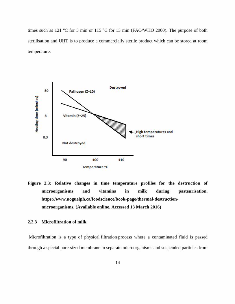
preserving heat sensitive nutrients and sensory characteristics. Figure 2.3 shows the relationship
of the pathogen destruction, nutrient loss and pasteurisation temperature.
2.2.2.2 UHT treatment and sterilisation of milk
To destroy most or all heat resistant microbes UHT is applied, where milk is pumped through a
plate exchanger for 2-5 s at 135-140 oC and rapidly cooled to prevent the Maillard reaction (Smit
2003). The processing of milk at high temperatures is aimed at destroying vegetative cells as
well as endospores present in raw milk so that it can be stored for prolonged periods, generally
several months, without refrigeration (Tabit 2010). Another high heat treatment is sterilisation,
which is a high-temperature/long-time heat treatment aimed at producing a commercially sterile
product which can be stored at room temperature. Sterilisation destroys all microorganisms and
any residual microorganisms are unlikely to cause spoilage under normal storage condition. The
temperatures for sterilisation should be 110 to 125 oC in combination with appropriate holding
14
times such as 121 oC for 3 min or 115 oC for 13 min (FAO/WHO 2000). The purpose of both
sterilisation and UHT is to produce a commercially sterile product which can be stored at room
temperature.
Figure 2.3: Relative changes in time temperature profiles for the destruction of
microorganisms and vitamins in milk during pasteurisation.
https://www.uoguelph.ca/foodscience/book-page/thermal-destruction-
microorganisms. (Available online. Accessed 13 March 2016)
2.2.3 Microfiltration of milk
Microfiltration is a type of physical filtration process where a contaminated fluid is passed
through a special pore-sized membrane to separate microorganisms and suspended particles from
15
process liquid. The filters used in the microfiltration process are specially designed to prevent
particles such as bacteria from passing through. The process was first implemented in the milk
and cheese production in the late 80s (Hoffman et al. 2006; Schmidt et al. 2012). The principle
of this technique during milk processing is to remove bacterial cells and spores from milk
mechanically using ceramic membrane with pore diameter of 0.8-1.4 mm (Rysstad and Kolstad
2006). Most experiences with microfiltered ESL milk are based on the patented Bactocatch®
system (Holm et al. 1986). This process and its variants comprise microfiltration of separated
skim milk resulting in a permeate. The permeate is added with or without subsequent HTST
pasteurisation to the highly heated (115-130 oC, 4-6 s) mixture of microfiltration retentate and
required an amount of cream. Finally, the recombined and fat-adjusted milk is filled aseptically
(Hoffman et al. 2006).
2.2.4 Bactofugation of milk
It is a process used to eliminate the bacteria contained in the milk by means of centrifugal force.
Its effectiveness increases with increase in temperature. Bactofugation compliments
pasteurisation and does not replace it (Fondation de technologie laitière du Québec 1985; Lund et
al. 2000). Effectiveness of bactofugation varies according to size and type of bacteria because
sedimentation by centrifugal force is greater for larger and denser bacterial cells. The process is
believed to reduce 90-99% of bacterial cells and clostridal spores which cause late blowing of
Swiss cheese (Fox and McSweeney 1998; Faccia et al. 2013). Bactofugation has proved to be an
efficient way of reducing the number of spores in milk. This method is claimed to be effective at
16
removing bacterial spores but it can be plagued by problems with recontamination (Lund et al.
2000a; Faccia et al. 2013).
2.2.5 Aseptic packaging
Aseptic filling is important to control contamination during packaging in order to achieve the
goal of extended shelf life milk processing. It is the process by which a sterile product is
packaged in a sterile container in a way that maintains sterility. Burton (1988), points out that in
order to achieve aseptic packaging the process must satisfy the following conditions;
Container and method of closure must not allow passage for microorganisms.
The part of the container that is in contact with milk must be sterilised when formed and
before filling.
Container must be filled without contamination from equipment and surrounding
atmosphere.
If closure is needed it must be sterilised before application.
Closure must be applied and sealed in place to avoid or prevent the passage of
contaminants.
Saturated steam has been used for container sterilisation although it has economic challenges in
setting up and energy costs. Dry heat in form of hot gas or hot non aqueous liquid such as glycol
is also used although it desiccates microorganisms and makes them more resistant. The heating
processes normally achieve temperatures between 91-146 oC. Apart from heat treatments,
17
hydrogen peroxide (H2O2) has been successfully used for aseptic packaging of UHT milk
(Ansari and Datta 2003), while UV light at a wavelength of 250 nm has also proved to be
effective (Burton 1988). UV light, however, has practicality difficulties of ensuring uniformity
during application. Ionising radiation such as gamma rays has been used to sterilise the interior
of sealed but empty containers, particularly those made of materials which cannot withstand
temperatures needed for thermal sterilisation. The most common packaging for aseptically
packed milk is laminates of paperboard cartons coated internally and externally with
polyethylene. An oxygen barrier like aluminum foil is a common inclusion to the laminate. Other
common packaging materials include plastic pouches and blow moulded bottles (Burton 1988).
2.3 Bacteria associated with milk
2.3.1 Raw milk
Bovine milk as it is secreted by the cow is sterile. However, microorganisms associated with the
teat move up the teat canal into the interior of the udder, indicating that even aseptically drawn
milk will have a certain number of microorganisms (Ryser et al. 1998; Ozer and Akdemir-
Evrendilek 2014). Fresh, aseptically drawn milk from a healthy cow may contain < 100 cfu/ml
(Walstra et al. 2005). Other scholars have indicated that a practical range is between < 1000 and
20 000 cfu/ml (Chambers 2005). While the interior of the udder contributes a few
microorganisms to raw milk, most microorganisms in raw milk are contaminants from outside
the udder such as milking equipment and human handlers. Developments of closed milking
systems, use of bulk tanker for transport and improvements in refrigeration systems has resulted
in a change of the micro flora in raw milk from predominantly Gram positive, acid producing to
18
Gram negative psychrotrophic microorganisms primarily the Pseudomonas spp. (Ryser et al.
1998; Chambers 2005).
Psychrotrophs that have been defined as bacteria that grow at 7 oC or less, irrespective of their
optimal temperature, have become of great importance in the dairy industry from both a spoilage
and safety stand point (Ryser et al. 1998). The most common Gram negative bacteria of primary
importance include Pseudomonas, Achronobacter, Aeromonas, Alcaligenes, Chromobacterium
and Flavobacterium spp. These bacteria produce some heat stable enzymes that may participate
in product spoilage during refrigeration. While Yersinia enterocolitica and Escherecia coli are
Gram negative pathogens, B. cereus and L. monocytogenes are Gram positives that are of safety
concern in raw milk. B. cereus has been extensively reported in milk and its products (Ryser et
al. 1998; Chambers 2005; Ozer and Akdemir-Evrendilek 2014). Research has shown that its
existence in milk depends on the season among other factors, with winter exhibiting the highest
prevalence (Ryser et al. 1998). Enterobacteriaceae is another dominant group of microorganisms
in raw milk. This includes Lactobacillus, Acinetobacter, Staphylococcus, Falvobacterium and
Micrococcus spp. The most common spoilers of raw milk are the acid producing ones (Ozer and
Akdemir-Evrendilek 2014). Although they come in low numbers, raw milk also contains spore-
forming bacteria with the ubiquitous Bacillus spp. dominating the spore-forming group (Ryser et
al. 1998; Ozer and Akdemir-Evrendilek 2014).
19
2.3.2 Heat treated milk
2.3.2.1 Pasteurised milk
Although most pathogenic bacteria are destroyed, pasteurised milk has been reported to contain
both spoilage and some pathogenic organisms (Ntuli et al. 2016). There are two types of
microorganisms in pasteurised milk, which are, post process contaminants that enter after heating
and heat resistant bacteria which survive heating. Post process contaminants are usually Gram
negative psychrotrophic bacteria that include members of the Enterobacteriaceae, such as
Serratia, Enterobacter, Citrobacter spp. among others (Varnam and Sutherland 2001).
Nevertheless, the ultimate spoilage microflora usually consists of Gram negative rods such as
Pseudomonas, Alcaligens and to a lesser extent Flavobacterim. It is the competitive nature of
these organisms that make them out grow the Enterobacteriaceae during storage at 8 oC (Touch
and Deeth 2009; Tammine 2009).
While other studies have shown that Pseudomonas spp. was the only bacteria causing defects in
milk stored at 4-7 oC (Craven and Macauley 1992 in Tammine 2009), some have shown that the
endospore forming bacteria Paenibacillus dominates in pasteurised milk as it ages (Ranieri et al.
2012). The presence of spore-formers is inevitable in pasteurised milk, however, some scholars
point out that, although pasteurisation virtually kills all vegetative thermophilic bacteria, post
pasteurisation contamination by Psuedomonas spp. at levels of 103 cfu/ml, frequently occurs.
Other post pasteurisation contaminants include Lactobacillus and Lactococcus spp. (Varnam and
Sutherland 2001; Deeth et al. 2006).
20
The most common heat resistant organisms in pasteurised milk are those that attach to the plates
and grow during the regeneration stage usually at 45-60 oC, which is their optimum temperature,
resulting in recontamination before milk leaves the pasteuriser (Lund et al. 2002; Tammine
2009). These bacteria will dominate in milk stored at temperatures above 10 oC. B. licheniformis
as well S. thermophilus have also been implicated in post pasteurisation contamination. Bacillus
spp. are the most significant heat resistant organisms because of their ability to adapt to various
conditions through formation of endospores as well as ability to grow at refrigeration storage.
Bacillus spp. usually becomes the dominant spoilage organisms at storage temperature below 5
oC when competitive Gram negative bacteria are low in numbers (Ryser et al. 1998). This
usually occurs when milk is manufactured under conditions of good hygiene, for which a long
storage period is expected. B. cereus, B. licheniformis, B. mycoides, B. circulans and B.
coagulans have been frequently isolated in pasteurised milk at levels < 102 cfu/ml (Tammine
2009).
2.3.2.2 ESL milk
The most common microflora in ESL milk are spore-formers and post process contaminants.
Myar et al. (2004b) reported that a level of 13-130 spores/L has been observed in ESL milk.
While some studies reported that B. circulans was the dominating organism in milk pasteurised
at 72-88 oC for 15 s (Cromie et al. 1989 in Tammine 2009), other studies revealed that B.
licheniformis (73%) was the dominating organism followed by B. subtilis, B. cereus,
Brevibacillus spp. and B. pumilus in milk heat treated at 127 oC for 5 s (Mayr et al. 2004b).
Commercial milk directly heated at 120-132 oC for 4 s was observed to harbor only B.
21
licheniformis, B. coagulans and B. cereus. (Ozer and Akdemir-Evrendilek 2014). Ranieri et al.
(2012), reported Paenibacillus spp. as the dominating microbe in pasteurised based ESL milk
and Schmidit et al. (2012) observed Microbacterium spp. (40%) followed by Microbacterium
lacticum (34%), spore-formers (20%) in ESL. B. subtilis was the dominating spore-former
followed by B. licheniformis, B. cereus and B. pumilus among others. Mayr et al. (2004a) also
reported non spore-forming organisms in commercial ESL milk that includes Rhodococcus,
Anquinibacter, Arthrobacter, Microbacterium, Enterococcus, Staphylococcus and Micrococcus
among others and these were attributed to recontamination.
2.4 Contamination routes in milk processing
2.4.1 Milk at the farm
It is generally accepted that milk drawn from a healthy cow under hygienic conditions contains
relatively few organisms. However, during milking the milk can be subjected to a number of
sources of microbial contamination such as the udder, equipment and the atmosphere (Ryser et
al. 1998).
2.4.1.1 Interior of the udder
The most common bacteria in the udder are Micrococci and Streptococci. These are also present
on the skin of the teats. However, when the cow has mastitis, high numbers of environmental
bacteria such as E. coli, coliforms and Pseudomonas spp. may also be present on the teats
especially when the udders are exposed to mud and manure (Ryser et al. 1998). The counts from
these sources can be as high as 105-107 cfu/ml under certain circumstances. Apart from mastitis
22
related bacteria, lactic acid bacteria are other usual inhabitants of the skin and streak canal of the
teats hence their presence in milk is inevitable, though in low numbers. (Tammine 2009)
2.4.1.2 Exterior of the udder
Cow udders are contaminated by the envirioment in which the animal stays. Animal feed may
contain from 105-108 cfu/g of psychrotrophs and lactic acid bacteria are associated with silage
and animal feeds (Bramely and Mckinnon 1990 in Tammine 2009). Urine and faeces also add
microorganisms on the bedding material. The bedding and feeding material consequently
contaminate the exterior of the udder. The most common groups on the teats that later
contaminate milk are micrococci and aerobic spore-formers such as Bacillus spp. Spore counts of
Bacillus spp. range from 102-105 per teat depending on the environmental conditions. Although
water and silage play a role in spore contamination of raw milk the major sources of
contamination are soil and faeces on the teats (Cook and Sandeman 2000).
2.4.1.3 Water
It has been reported that water used for dairy farm contain psychrotrophic bacteria even when it
is chlorinated (Tammine 2009). Hence, its use for cleaning and rinsing milking equipment
provides direct means of milk contamination. These psychotrophic bacteria are often very active
producers of extracellular enzymes and grow rapidly at refrigeration temperature (Hantsis-
Zacharov and Halpern 2007). Pseudomonas spp. dominates the psychrotrophic flora in water
while Bacillus spp. and coliforms are in lower numbers. Furthermore, heat resistant spore-
23
formers have been isolated from farm water supplies, including hot water used for washing
milking equipment (Depiazzi et al. 1997).
2.4.1.4 Milk handling equipment
Despite the use of stainless steel on much dairy equipment, some microorganisms are still able to
attach to equipment and are often difficult to inactivate by chemical sanitisation. Milk handling
equipment and utensils are the major sources of Gram negative psychrotrophic bacteria
(Tammine 2009). Studies have shown the presence of B. cereus, micrococci and thermophilic
strains of Enterococcus faecalis on milk handling equipment (Touch and Deeth 2009).
Contributing factors include poorly designed and constructed pipeline systems (Varnam and
Sutherland 2001). Previous studies have shown that counts of psychrotrophic bacteria in bulk
tanks may be up to 103 cfu/cm2 (Hayes 1985), hence these can be another major source of
psychotropic bacteria in raw milk.
2.4.2 Milk at the processing plant
Pipelines, tanks, valves, and filling machines have been cited as the major sources of
contamination after pasteurisation or any heat treatment (Fredsted et al. 1996).
2.4.2.1 Processing equipment
Filling equipment is a common source of psychrotrophs in packaged milk. Even when filling
equipment is effectively cleaned and sanitised, it can still become a source of contamination due
to psychrotrophic microorganisms which accumulate during continuous use (Eneroth et al. 2000;
24
2001). Holding tanks can also participate in contamination, by protecting microorganisms in
microscopic fissures (ICMSF 1998). Some microorganisms such as Pseudomonas spp. are able
to adhere to surfaces of milk processing equipment. Furthermore, Bacillus spp. produces highly
hydrophobic spores and adheres firmly to stainless steel. After adhesion, there is colonisation if
environmental conditions allow spore germination. This leads to biofilm formation which has
proved to be a menace for the dairy industry for a long time (Faille et al. 2002; Simmonds et al.
2003).
2.4.2.2 Air in the processing plant
Microbial population of air in a dairy processing plant has been reported to be 85% bacteria,
10% moulds and 5% yeasts (Fredsted et al. 1996). However, most of the bacteria are Gram
positives that do not grow well at low temperatures. Many sources of air contamination are
usually ventilation systems, flow drains and personnel. Products can therefore, be easily exposed
to contaminated air during packaging. The influence of microorganisms in the air on the
microbiological spoilage of milk is of minor importance if premises are well designed;
maintained and internal hygiene measures are taken. (Ozer and Akdemir-Evrendilek 2014). In
modern dairy processing the effect of air has further been reduced by use of a closed system and
aseptic packaging.
2.4.2.3 Packaging materials
It is very important that packaging material used should not cause contamination as it will be in
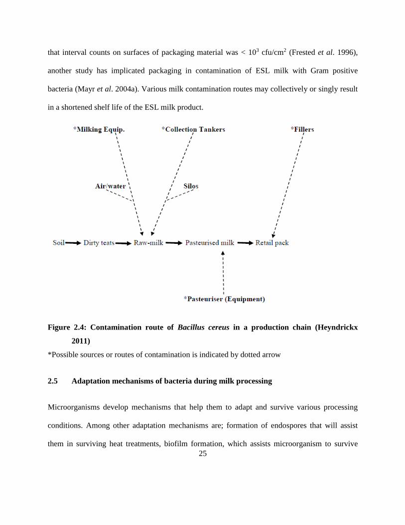
direct contact with the product during its entire shelf life. Although a previous study revealed
25
that interval counts on surfaces of packaging material was < 103 cfu/cm2 (Frested et al. 1996),
another study has implicated packaging in contamination of ESL milk with Gram positive
bacteria (Mayr et al. 2004a). Various milk contamination routes may collectively or singly result
in a shortened shelf life of the ESL milk product.
Figure 2.4: Contamination route of Bacillus cereus in a production chain (Heyndrickx
2011)
*Possible sources or routes of contamination is indicated by dotted arrow
2.5 Adaptation mechanisms of bacteria during milk processing
Microorganisms develop mechanisms that help them to adapt and survive various processing
conditions. Among other adaptation mechanisms are; formation of endospores that will assist
them in surviving heat treatments, biofilm formation, which assists microorganism to survive
26
cleaning and sanitisation and other organisms have managed to develop adaptation to cold stress
helping them to grow in milk during refrigeration (Drenkard 2003; Abel-Santos 2012).
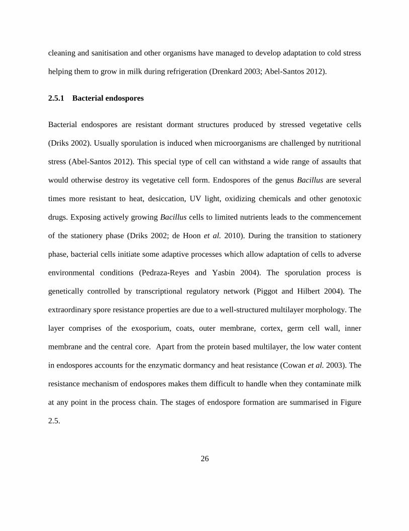
2.5.1 Bacterial endospores
Bacterial endospores are resistant dormant structures produced by stressed vegetative cells
(Driks 2002). Usually sporulation is induced when microorganisms are challenged by nutritional
stress (Abel-Santos 2012). This special type of cell can withstand a wide range of assaults that
would otherwise destroy its vegetative cell form. Endospores of the genus Bacillus are several
times more resistant to heat, desiccation, UV light, oxidizing chemicals and other genotoxic
drugs. Exposing actively growing Bacillus cells to limited nutrients leads to the commencement
of the stationery phase (Driks 2002; de Hoon et al. 2010). During the transition to stationery
phase, bacterial cells initiate some adaptive processes which allow adaptation of cells to adverse
environmental conditions (Pedraza-Reyes and Yasbin 2004). The sporulation process is
genetically controlled by transcriptional regulatory network (Piggot and Hilbert 2004). The
extraordinary spore resistance properties are due to a well-structured multilayer morphology. The
layer comprises of the exosporium, coats, outer membrane, cortex, germ cell wall, inner
membrane and the central core. Apart from the protein based multilayer, the low water content
in endospores accounts for the enzymatic dormancy and heat resistance (Cowan et al. 2003). The
resistance mechanism of endospores makes them difficult to handle when they contaminate milk
at any point in the process chain. The stages of endospore formation are summarised in Figure
2.5.
27
Figure 2.5: Stages that a bacterial cell goes through in a sporulation cycle
http://zf2t.dromibi.top/c/spore-formation/ (Available online. Accessed 27/03/2016)
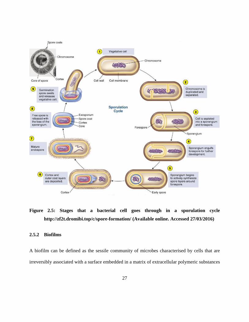
2.5.2 Biofilms
A biofilm can be defined as the sessile community of microbes characterised by cells that are
irreversibly associated with a surface embedded in a matrix of extracellular polymeric substances
28
and display an altered phenotype with respect to gene expression, protein production and growth
(Dhillon 2012). Microorganisms in a biofilm display some particular features that are not shared
with the same microorganisms in suspension form. Microorganisms in a biofilm can be up to a
1000 times more resistant to antimicrobials than their planktonic counterparts (Drenkard 2003).
Biofilm formation begins with attachment of a single cell to the surface. Studies have shown that
B. cereus among others spore-formers can attach to stainless steel (Khoza 2016). Adhesion of
bacterial cells to the conditioning layer then follows (Dhillon 2012). Irreversibly attached cells
utilise nutrients present in the conditioning film to multiply (Khoza 2016). Continuous
attachment of cells over time results in formation of a biofilm. Mixed species biofilms are more
common as they are more stable than single cells (Dhillon 2012). This allows microorganisms to
protect one another during the application of cleaning chemical agents. As the biofilm matures
the attached bacteria in order to survive, detach and disperse to colonise a new niche (Myszka
and Czaczyk 2011). It is at this detachment stage that the milk is contaminated by organisms
from the biofilm.
2.5.3 Cold shock response
Some microorganisms have developed mechanisms to grow well under cold temperatures and
amongst them are some members of Bacillus and Paenibacillus spp. The ability to produce cold
shock proteins (CSP) allows microorganisms to continue with metabolic processes at low
temperatures. Psychrotrophic strains are said to display a particular sequence in their cspA gene
that was proposed to be a psychrotolerant-associated signature (Francis et al. 1998). Another
psychrotolerant-associated signature was also proposed based on the rrs sequence, while a 16S
29
based signature has also been described (von Stetten et al. 1998). In a classification proposed by
Guinebretierre et al. (2008; 2010) it was reported that group 2 comprises of both mesophilic and
psychrotrophic strains with 50% of the stains being intermediate. It is important, however, to
note that this group does not have a cspA signature, owing their psychrotrophic nature to a
different signature.
When bacterial cells are subjected to a temperature downshift they elaborate an adaptive
response known as cold shock response (Requena 2012). The first notable aspect under cold
stress is that growth significantly slows down or stops, representing the lag phase and this
correspond to a cold acclimation phase. This is followed by a massive and transient synthesis of
CSP and a significant decrease of non-CSP synthesis including housekeeping proteins. The
amount of cold acclimation proteins (CAP) already present also increases moderately (Requena
2012). The second phase of cold adaptation begins with a decrease in synthesis of CSP and
CAPs, concomitantly to a non-CSP protein neo-synthesis CSPs and CAPs proteins are involved
in several processes such as DNA and RNA metabolism, cell metabolism, protein folding and
degradation among other functions (Phadtare 2004).
Compatible solute acquisition also plays a role in cold adaptation as was shown that glycine
betaine is an effective protectant of B. subtilis during cold adaptation (Hoffman and Bremer
2011). Membrane fluidity is also modified when temperature drops (Zhang and Rock 2008,
2009). Bacterial cell membrane switches from a liquid fluid crystalline to a rigid state under low
temperatures (Requena 2012). A study on B. subtilis also suggests a great need for DNA excision
repair during growth at low temperature (Budde et al. 2006). During adaptation to cold stress,
30
protein folding and misfolding is also observed (Requena 2012). Metabolic pathways
modification is also noticed during cold adaptation (Budde et al. 2006). While other studies have
shown that low temperature influences spore production in B. weihenstephanensis (Garcia et al.
2010), it has also been established that B. cereus spores can germinate at temperatures below 6
oC. It is this complex adaptive mechanism of Bacillus spp. that makes it an important organism
in extending the shelf life of milk.
2.6 Spore-forming bacteria associated with ESL milk
2.6.1 Paenibacillus spp.
Formerly known as Bacillus polyxma that was reclassified in 1993, Paenibacillus spp. emerged
from early phylogenetic dissection of Bacillus sensu lato based on the 16S rDNA gene sequences
(Ash et al. 1993). Similar to the Bacillus spp., Paenibacillus spp. are rod shaped Gram positive
microorganisms that form endospores. However, they regularly appear gram-negative under the
microscope (Bergey 2009). All Paenibacillus spp. produce endospores that are usually of greater
diameter than the mother cell and under suitable conditions some produce capsules.
Paenibacillus differs with the Bacillus spp. in the DNA encoding their 16S RNA (Ouyang 2008).
It was once believed that Paenibacillus was not pathogenic however, research has shown that P.
thiaminolyticus was the cause of a renal failure case in the USA (Ouyang 2008). Paenibacillus is
a thermoduric (Huck et al. 2007), aerobic endospore forming microorganism that is characterised
by a ubiquitous nature and to aid to their survival skills are the ability to secrete peptide
antibiotics, signal molecules as well as enzymes though variations exist (Gardener 2004). Some
members of the Paenibacillus such as P. polymyxa are able to fix nitrogen in the soil
31
(Yegorenkova 2008). In milk Paenibacillus and Bacillus are the predominant spore formers that
spoil milk after pasteurisation (Ranieri 2012). Bacillus represents the bacteria that dominate in
the early stages of shelf life in pasteurised milk, however, Paenibacillus increases with time at
refrigeration temperatures to contribute about 95% of the dominating microbes in the pasteurised
milk (Ranieri 2012). This clearly shows the ability of Paenibacillus to grow as a psychrotroph in
milk. Paenibacillus is able to degrade proteins (Ash et al. 1993), polyaromatic hydrocarbons
(Daane et al. 2002) and polysaccharides (Scheldeman et al. 2004). Paenibacillus usually
produces small translucent, light brown/white sometimes pink/yellowish colonies on agar plates
(Bergey 2009). Methods of isolation, characterisation and identification for Paenibacillus vary
but most are almost the same as those used for the Bacillus hence the PCR is used to differentiate
the two.
2.6.2 Bacillus spp.
These are aerobic endorespore forming gram-positive rods. The genus was created in 1872 by F.
Cohn who changed the name of Ehrenberg’s 1935 Vibrio subtilis to Bacillus subtilis (Harwood
1989). Representatives of this genus are widely distributed in the air, soil and water. Some
Bacillus strains are able to tolerate extreme conditions such as high and low pH as well as high
and low temperatures. In the soils, they have been isolated from extreme desserts as well as
Antarctic samples. Soils of low organic matter are dominated by B. subtilis, B. licheniformis and
B. cereus but the range increases with increase in fertility of the soil. Bacillus, contribute to 20%
of heterotrophic flora in the seawater (Harwood 1989). In fresh water most Bacillus bacteria
represent the microflora of the soil. Few species have been isolated in plant leaves. B. macerans,
32
is often isolated from the roots while cereals such as rice and pulses have a varied Bacillus flora
including B. cereus. Bacillus flora of food is related to the distribution of those bacteria in soil
water and plants. The ubiquitous distribution of Bacillus spp. makes them prominent milk
spoilage organisms. B. sporothermodurans has been identified as a cause of spoilage in UHT
milk (Tabit and Buys 2011). Lecithinase-positive strain of B. cereus is responsible for the broken
flavour or bitty cream condition (Jan·Tová et al. 2006), although others occur in raw milk and
pasteurised milk e.g. B. sphaecicus, B. megatarium and B. subtilis (Scheldeman et al. 2004).
2.6.2.1 B. pumilus
B. pumilus is widely distributed in clinical, veterinary, food ingredients, leather, paper samples
and has also been isolated from the interior of Sonoran desert basalt (Benardini et al. 2003),
while its spores usually reside in soils and some colonise the root area of some plants where it
has some antibacterial and antifungal activity. B. pumilus has also been reported to be the second
most predominant Bacillus species in space crafts (La Duc et al. 2003). In addition, it has been
isolated in both milk and milk processing environments (Schmidit et al. 2012; Aoudhi et al.
2014; Khoza 2016). Although Bacillus spp. spores are notoriously resistant to unfavorable
conditions such as low or no nutrient availability, extreme desiccation, H2O2, UV, gamma-
radiation, or chemical disinfection (Nicholson et al. 2000). B. pumilus has shown elevated
resistance to these factors when compared to the spores of other Bacillus members (Nicholson et
al. 2000; Kempf et al. 2005). B. pumilus has also been used at industrial level. Some purposes
of B. pumilus that are being researched are its involvement in bacterial hay preservation and the
use of B. pumilus plasmids in gene transfer systems. The proteases from B. pumilus are used in
33
various industries such as food, chemical, detergent, and leather industries (Pan et al. 2004).
Although B. pumilus is regarded as non-pathogenic, previous studies have shown toxigenic
potential in some of its strains through production of pumilacidins (Suominem et al. 2001; From
et al. 2007; Nieminem et al. 2007).
2.6.2.2 B. subtilis
Originally named Vibrio subtilis in 1835, this organism was renamed B. subtilis in 1872. B.
subtilis is a Gram positive, catalase-positive bacterium. Although B. subtilis has been isolated in
milk and dairy environment (Schmidit et al. 2012; Aoudhi et al. 2014), it is usually found in soil
and grass hence the name grass bacillus. B. subtilis is considered an obligate aerobe, but can also
function anaerobically in the presence of nitrates or glucose (Heyndrickx and Scheldeman 2002).
Along with enzymes, B. subtilis also produces a toxin called subtilisin (Ryan and Ray 2004).
Subtilisin can cause allergic reactions if there is repeated exposure in high
concentrations. Industrially B. subtilis can be used to produce proteases and amylase enzymes
(van Dijl and Hecker 2013). Previously it was widely used as a broad spectrum antibiotic,
however, this was lost after the ability to produce cheaper, large-scale antibiotics although it is
still used in Western Europe and the Middle East in alternative medicine. Other commercial
applications of B. subtilis include cleaning agents in detergents, de-haring and batting in the
leather industry, production of special Japanese and Korean food, starch modification, de-sizing
of textiles and other specialized chemicals. B. subtilis has become the model agent in laboratory
research because of its easy genetic manipulation (Earl et al. 2008).
34
2.6.2.3 B. cereus
B. cereus is a ubiquitous, Gram positive, spore forming, and motile aerobic bacteria with some
strains that have shown ability to grow well anaerobically. Its habitats ranges from milking
parlours to cattle pans, decaying organic matter, fresh and marine waters, vegetables and fomites,
and the intestinal tract of invertebrates among others (Jensen et al. 2003; Arnesen et al. 2008;
Bottone 2010). B. cereus contamination can be mediated by various substrates including raw
milk and processing equipment such as milk fillers (Khoza 2016) as its spores are highly
adhesive (Arnesen et al. 2007). Spores germinate when they come into contact with organic
matter or within an insect or animal host (Arnesen et al. 2008).
Although it is non-competitive, B. cereus has managed to adapt to different and diverse habitats
ranging from cold to hot environments and from refrigerated to dehydrated foods (Larsen and
Jogernsen 1997; Guinebretiere et al. 2008). B. cereus has previously been reported as mesophilic
bacteria, however, emerging research has shown a number of strains that are psychrotrophic
(Stenfors et al. 2001). B. cereus spores can germinate at 4-6 oC and they grow well at
temperatures below 10 oC (Larsen and Jogernsen 1997). The ability to adapt to various
conditions has raised the concern of B. cereus as it is said to account for 25% of shelf life related
problems in pasteurised milk (Francis et al. 1998). B. cereus is also known to cause sweet
curdling in milk due to its ability to hydrolyse casein and studies have also shown that some
strains are able to utilise lactose (Kotiranta et al. 2000) while others can hydrolyse lipids (De
Jonghe et al. 2010). In addition, B. cereus is also involved in gelatin hydrolysis, DNase,
lecithinase, protease, amylase as well as xylanase production (Molva et al. 2008).
35
2.6.2.3.1 B. cereus pathogenicity and outbreaks
Apart from causing spoilage problems in milk, B. cereus is an opportunistic human pathogen
(Bartoszewicz et al. 2008) that causes two principal types of food poisoning; emetic and
diarrheal (Hansen and Hendriksen 2001). The emetic poisoning is caused by a small cyclic heat
stable peptide which induces vomiting a few hours after ingestion while the diarrheal types are
attributed to enterotoxins that induce abdominal pain and diarrhoea after incubation for 8-16 h
and vegetative growth of the bacteria in the intestine (Hansen and Hendriksen 2001). Three main
toxins that have been commonly implicated in the diarrhoeal disease are cytotoxins haemolysin
BL (Hbl), nonhaemolytic enterotoxin (Nhe) and cytotoxin K (cytK) (Arnesen et al. 2008; Ehling-
Schulz et al. 2004; Lund et al. 2000b). B. cereus enterotoxins are heat-labile proteins and can be
inactivated by trypsin, pepsin and pronase as well as low pH (pH < 4) (Wang et al. 2014).
Haemolysin is a three component toxin consisting of the proteins B, L1 and L2 (Beecher and
Wong 1997) encoded by the genes hblA, hblD and hblC respectively (Ryan et al. 1997). Similar
to the Hbl is the Nhe complex that is also a three component toxin consisting of the proteins
NheA, NheB and NheC encoded by the nheABC operon (Granum et al. 1999). Both proteins of
Nhe and Hbl have shown homology that suggests that they originate from a similar gene.
Furthermore, they have all been termed to belong to the tripartite family of toxins (Arnesen et al.
2008). Despite the similarities that exist between these toxins, cooperation between them is very
limited (Lund and Granum 1997). Several studies have described the effects of B. cereus toxins
and are summarised in Table 2.1.
36
Apart from the tripartite family toxins another diarrhoeal toxin is cytK that falls under the β-
barrel pore-forming toxins (Lund et al. 2000b). CytK is a 34 kDa water-soluble protein with
dermonecrotic, cytotoxic and haemolytic activities, and shows similar cytotoxic potency towards
cell cultures as Hbl and Nhe (Lund et al. 2000b). Originally isolated from B. cereus strain NVH
391/98 that was responsible for a severe foodborne outbreak of diarrhoeal disease in a French
nursing home 1998 which claimed three elderly people and several suffering bloody diarrhoea
(Lund et al. 2000b), cytK virulence was later shown to be depended on expression level of the
gene not mere presence of the gene (Brillard and Lereclus 2004; Fagerland et al. 2007; Arnesen
et al. 2008).
The emetic syndrome that has a rapid onset of 0.5-6 h after consumption is caused by the toxin
cereulide, a cyclic dodecadepsipeptide with a molecular mass of 1.2 kDa (Arnesen et al. 2008).
The toxin is produced by a peptide synthetase encoded by the 24 kb cereulide synthetase (ces)
gene cluster (Ehling-Schulz et al. 2005) and it consists of a ring structure with three repeats of
four amino and/or oxy acids; [d-O-Leu-d-Ala-d-O-Val-d-Val]3 (Ehling-Schulz et al. 2005).
Although cereulide that is also said to be closely related to potassium ionophore valinomycin is
not antigenic, it is resistant to heat, pH and proteolysis (Arnesen et al. 2008). Its production
commences at the end of the logarithmic phase during vegetative growth of B. cereus and
reaches its highest level during the early stages of the stationary phase (Arnesen et al. 2008). Its
synthesis takes place between 12-22 oC, although a previous study reported two isolates
belonging to B. weihenstephanensis that showed ability to produce cereulide at 8 oC (Thorsen et
al. 2006). Cereulide inhibits mitochondrial activity by inhibition of fatty acid oxidation as it acts
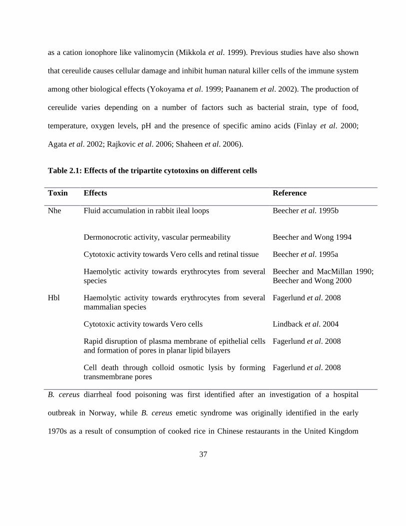
37
as a cation ionophore like valinomycin (Mikkola et al. 1999). Previous studies have also shown
that cereulide causes cellular damage and inhibit human natural killer cells of the immune system
among other biological effects (Yokoyama et al. 1999; Paananem et al. 2002). The production of
cereulide varies depending on a number of factors such as bacterial strain, type of food,
temperature, oxygen levels, pH and the presence of specific amino acids (Finlay et al. 2000;
Agata et al. 2002; Rajkovic et al. 2006; Shaheen et al. 2006).
Table 2.1: Effects of the tripartite cytotoxins on different cells
Toxin Effects Reference
Nhe Fluid accumulation in rabbit ileal loops Beecher et al. 1995b
Dermonocrotic activity, vascular permeability Beecher and Wong 1994
Cytotoxic activity towards Vero cells and retinal tissue Beecher et al. 1995a
Haemolytic activity towards erythrocytes from several
species
Beecher and MacMillan 1990;
Beecher and Wong 2000
Hbl Haemolytic activity towards erythrocytes from several
mammalian species
Fagerlund et al. 2008
Cytotoxic activity towards Vero cells Lindback et al. 2004
Rapid disruption of plasma membrane of epithelial cells
and formation of pores in planar lipid bilayers
Fagerlund et al. 2008
Cell death through colloid osmotic lysis by forming
transmembrane pores
Fagerlund et al. 2008
B. cereus diarrheal food poisoning was first identified after an investigation of a hospital
outbreak in Norway, while B. cereus emetic syndrome was originally identified in the early
1970s as a result of consumption of cooked rice in Chinese restaurants in the United Kingdom
38
(Wang et al. 2014). While CDC estimates that each year roughly 1 in 6 Americans (or 48 million
people) get sick, 128 000 are hospitalized, and 3 000 die of foodborne diseases. It is reported
that Bacillus spp. food borne outbreaks in Europe were 1.4% of the total outbreaks in 2005
(Arnesen et al. 2008). Naranjo et al. (2011), also reported of a B. cereus food poisoning case that
claimed a life in America while Al-Abri et al. (2011), reported of an outbreak implicating
hospital kitchen food affecting 58 people and Dierick et al. (2005), reported of 5 children from
the same family who were seriously ill in a B. cereus poisoning incident that claimed the life of
the youngest girl. In another incident in July 2014 Public Health England reported of B. cereus
outbreak implicated in intravenous liquid (Total Parenteral Nutrition, TPN). In this out break 23
cases were reported, 19 were confirmed while 3 children died and the whole batch of the product
recalled (https://www.gov.uk/government/news/bacillus-cereus-infections). In Argentina the
Health Ministry reported 5783 cases of food-borne disease between 2008 and 2013, of which 26
were positive for B. cereus. Despite the fact that B. cereus outbreaks are believed to be under
reported on official lists (Al-Abri et al. 2011), food poisoning reporting and record management
is generally poor in developing countries hence no information is available pertaining B. cereus
poisoning in Africa. Although no exact data is available on the effective dose of B. cereus,
several researchers have suggested doses between 105-108 cfu/ml (Arnesen et al. 2008; Naranjo
et al. 2011).
39
2.7 Advances in detection and identification of microorganisms in the food industry
2.7.1 Protein based methods
2.7.1.1 Matrix Assisted Laser Desorption / Ionization Time of Flight Mass Spectrometry
(MALDI-TOF-MS)
First introduced in 1985, matrix-assisted laser desorption ionization (MALDI), when it was
found that the amino acid alanine could be ionized more easily if it was mixed with the amino
acid tryptophan and irradiated with a pulsed 266 nm laser (Karas et al. 1985). The technique had
a breakthrough when biomolecules as large as the 34 472 Da protein carboxypeptidase-A was
able to be ionized in 1987. Since then improvements to the technique have been made and it has
since found use as a rapid method of microorganisms’ identification because it requires minimal
sample pretreatment (Fox 2006). The technique is based on three main steps. First, the sample is
mixed with a suitable matrix material and applied to a metal plate. This is followed by a
pulsed laser that irradiates the sample, triggering ablation and desorption of the sample and
matrix material.
Finally, the analyte molecules are ionized by being protonated or deprotonated in the hot plume
of ablated gases, and can then be accelerated into a mass spectrometer used to analyse them. The
mass spectra of whole cells provide a ‘fingerprint’ of biomolecular composition and thus
constitute a strain-specific spectral pattern (Wahl et al. 2002) that can be applied to differentiate
micro-organisms even down to the strain level (Liu et al. 2007). The technique has shown high
reproducibility in both intralaboratory and interlaboratory tests (Mellmann et al. 2008). It has
40
been used to identify microflora of water, vegetables, milk and milk processing equipment,
among others in the food industry (Muller 2014; Khoza 2016; du Plessis et al. 2015).
2.7.2 DNA based methods
2.7.2.1 End point PCR
Developed in 1983 by Kary Mullis, PCR has become an indispensable technique in modern
microbiology. PCR is based on using the ability of DNA polymerase to synthesize new strand of
DNA complementary to the offered template strand using a primer to which it can add the first
nucleotide (Bartlett and Stirling 2003). This requirement makes it possible to delineate a specific
region of template sequence that the researcher wants to amplify (Erlich 2015). At the end of the
PCR reaction, the specific sequence will be accumulated in billions of copies often referred to as
amplicons. Typically, agarose gel electrophoresis, followed by staining with fluorescent
ethidium bromide is used to detect the amplified DNA fragments (Innis et al. 2012). This method
is time consuming and not sensitive enough to measure the accumulated DNA copies accurately,
so can only give a qualitative result. It has been used in the detection of virulence genes in
pathogens (Aijuka et al. 2015) as well as identification of different organisms related to spoilage
in the food industry (Muller 2014).
2.7.2.2 rep-PCR
The term rep-PCR refers to the general methodology involving the use of oligonucleotide
primers based on short repetitive sequence elements dispersed throughout the bacterial genome.
Repetitive element sequence-based PCR (rep-PCR) is a typing method that enables the
41
generation of DNA fingerprinting that discriminates bacterial strains, (Spigaglia and
Mastrantonio 2003). It differentiates microbes by using primers complementary to interspersed
repetitive consensus sequences that enable amplification of diverse-sized DNA fragments
consisting of sequences between the repetitive elements (Woods et al. 1993; Versalovic et al.
1994; Olive and Bean 1999). Multiple amplicons of different sizes can be fractioned by
electrophoresis, and the resulting DNA fingerprint patterns, specific for individual bacterial
clones, can be compared. The initial discovery of repetitive extragenic palindromic (REP)
elements occurred in the genomes of E. coli and Salmonella spp. (Hiett and Seal 2009). The
family of REP elements is generally between 33 and 40 bp in length and has 500-1 000 copies
per genome. The method has been used for epidemiological studies and discrimination of
pathogens in food (Hiett and Seal 2009; Muller 2014).
2.7.2.3 16S sequencing
DNA sequencing is the process of determining the precise order of nucleotides within
a DNA molecule. The use of 16S rDNA gene sequences to study bacterial phylogeny and
taxonomy has been by far the most common housekeeping genetic marker used because of its
presence in almost all bacteria, often existing as a multigene family or operons (Patel 2001).
Furthermore, the function of the 16S rDNA gene over time has not changed, suggesting that
random sequence changes are a more accurate measure of time (evolution) and the 16S rDNA
gene (1 500 bp) is large enough for informatics purposes (Janda and Abott 2007). For bacterial
identification, 16S rDNA sequencing is particularly important in the case of bacteria with
unusual phenotypic profiles, rare bacteria, slow-growing bacteria, uncultivable bacteria and
42
culture-negative infections. Not only has it provided insights into aetiologies of infectious
disease, but it is also useful in identifying novel species (Wu et al. 2009).
2.7.3.4 Multilocus sequence typing (MLST)
Proposed in 1998 as universal and definitive method for characterising bacteria, using the human
pathogen Neisseria meningitidis as an example (Maiden 2006). MLST is a nucleotide sequence
based approach for the unambiguous characterisation of isolates of bacteria and other organisms
via the internet (http://www.mlst.net). MLST has been described as a generic typing method,
employed to date, principally but not solely, bacterial pathogens, that aims to be a robust and
portable method for the characterisation of bacterial isolates at the molecular level. It differs
from many other former and current approaches to isolate characterisation in that it is based
explicitly on the population genetic concepts that underpinned the earlier technique of multilocus
enzyme electrophoresis (Maiden 2006). It is based on concepts of multilocus enzyme
electrophoresis (MLEE) and has adapted them so that alleles at each locus are defined directly,
by nucleotide sequencing, rather than indirectly from the electrophoretic mobility of their gene
products (http://www.mlst.net). The procedure characterises isolates of microbial species using
the DNA sequences of internal fragments of multiple housekeeping genes on both strands using
an automated DNA sequencer. For each housekeeping gene, the different sequences present
within a bacterial species are assigned as distinct alleles and, for each isolate, the alleles at each
of the loci define the allelic profile or sequence type (Maiden et al. 1998). Each isolate of a
species is therefore unambiguously characterised by a series of seven integers which correspond
to the alleles at the seven housekeeping loci. In MLST the number of nucleotide differences
43
between alleles is ignored and sequences are given different allele numbers whether they differ at
a single nucleotide site or at many sites (Urwin and Maiden 2003). The aim of MLST is to
provide a portable, accurate, and highly discriminating typing system that can be used for most
bacteria and some other organisms (Maiden 2006). MLST has the additional aims of providing
an unified bacterial isolate characterisation approach that generates data that can also be an
electronic data repository, the quality of which is maintained by active curation of the submitted
data used for evolutionary and population studies of a wide range of bacteria, regardless of their
diversity, population structure, or evolution (Urwin and Maiden 2003). At the core of the MLST
concept is the provision of freely accessible, curated databases of nucleotide sequence data
(Chan et al. 2001). MLST has been used for a number of bacteria that includes B. cereus, S.
epidermidis, E. coli and S. typhi among others. Currently applications of MLST include
examination of the evolution of virulence, population and evolutionary analyses and isolate
characterisation and population structure analyses (Maiden 2006).
2.7.3.5 Whole genome sequencing (WGS)
Whole genome sequencing (WGS) is the process of determining the complete DNA sequence of
an organism's genome at a single time. This entails sequencing all of an
organism's chromosomal DNA as well as DNA contained in the mitochondria. WGS overcame
the obstacle faced by other typing methods of failing to sufficiently discriminate closely related
strains. WGS represents a relatively new and increasingly accessible means for tracking disease
outbreaks that has garnered success in multiple applied contexts. Using massively parallel (or
“next-generation”) DNA sequencing technologies, it is now possible to examine the complete or
44
nearly complete genomes of bacterial isolates (Salipantea et al. 2015). WGS has provided greater
resolution in studies where direct comparisons were made with PFGE (Pendleton et al. 2013;
Leekitcharoenphon et al. 2014). Theoretically WGS is said to be able to distinguish strains which
differ at only a single nucleotide. Furthermore, genomic differences distinguishing strains can be
precisely measured and are highly reproducible, allowing high-resolution inference of
phylogenomic relationships (Didelot et al. 2013). WGS can enable exploration of isolates'
virulence genes, antibiotic resistance mechanisms, and other medically relevant factors,
concordantly with molecular epidemiology investigation. WGS has successfully been used in
source tracking studies of S. aureus and E. coli among other bacteria (Rasko et al. 2011; Harris
et al. 2013). The use of WGS in the food safety and microbiology has bright prospects
considering the high resolution the technique offers. However, current limitation to its wide
application in the food industry is the cost price per sample.
45
CHAPTER THREE
HYPOTHESES AND OBJECTIVES
46
3.1 Hypothesis 1
Extended shelf life milk processing will reduce both the bacterial counts and diversity of spore
forming bacteria in ESL milk. Although Bacillus spp. will be isolated throughout the ESL milk
process, Paenibacillus spp. will dominate the spore-formers population during cold storage to the
end of ESL milk shelf life. Pasteurisation temperatures destroys spore forming bacteria in their
vegetative state (Tammine 2009), however, addition of a non-thermal process to HTST
pasteurisation has shown to be effective in reducing the spore-formers population (Sepulveda et
al. 2005; Schmidt et al. 2012; Caplan and Barbano 2013). Paenibacillus spp. are low in numbers
in raw milk and early in pasteurised milk, however, they reproduce during chilled temperatures
cold storage to outcompete most microorganisms (Ranieri et al. 2012); this could be because
Paenibacillus spp. spores use L-tryosine as one of its germinants (Alvarado et al. 2012). The
Bacillus cereus enzyme increase the amount of free tryosine in milk during protein degradation
(Jan·tová 2006). Paenibacillus spp. a microaerophile organism (Owayss 2007), also requires
small amounts of oxygen for its spores to germinate.
3.1.1 Objective
To identify and characterise the spore-forming bacteria population associated with ESL milk
during processing and chilled storage with the aim of validating the effectiveness of heat and
bactofugation based ESL milk process on the spore-formers.
47
3.2 Hypothesis 2
ESL milk product contamination by B. cereus will be from both filler nozzles and raw milk
among other sources. B. cereus, a ubiquitous spore-forming bacteria, has been identified as the
main spoilage organism in pasteurised milk, frequently isolated in the dairy products and
processing environment (Ranieri et al. 2012; Aouadhi et al. 2014). While raw milk has been
implicated as an important source of endospores in milk products (Miller et al. 2015), filling
equipment is a common source of psychrotrophic bacteria in packaged milk (Scott et al. 2007;
Burgess et al. 2010; Hill and Smythe 2012). Some microorganisms are able to attach to
processing equipment and these adhered bacteria can detach and contaminate the product as it
passes the processing surfaces (Malek et al. 2012).
3.2.1 Objective
To characterise B. cereus isolates obtained from ESL milk processing and during storage with
the aim of understanding the potential of ESL milk filler nozzles and raw milk as a sources of
contamination in ESL milk.
48
CHAPTER FOUR
Bacillus and Paenibacillus spp. associated with extended shelf life milk during processing
and storage
Redrafted from:
Mugadza, D.T. and Buys, E.M. 2017. Bacillus and Paenibacillus spp. associated with extended
shelf life milk during processing and storage. International Journal of Dairy Technology. (In
press)
49
4.1.1 Abstract
Characterisation of spore-formers associated with extended shelf life milk was done by analysing
bacteriological quality of milk samples collected at various processing stages and during storage.
Isolates were identified with MALDI-TOF-MS. Milk had spore counts < 2 log10 cfu/ml and 4
log10 cfu/ml during processing and storage respectively. Bacillus pumilus dominated the bacterial
population. Bacterial species were inoculated in sterile milk for a shelf life study and population
change observed over 42 days at 7 oC. Although ESL milk process was effective in the reduction
of bacterial counts and species diversity, the presence of Bacillus cereus shows a potential safety
problem in ESL milk.
4.1.2 Introduction
Extended shelf life (ESL) milk bridges the gap between high temperature short time (HTST)
pasteurised milk with a shelf life of around 10 days under refrigeration and ultra-heat treated
(UHT) milk with a shelf life of at least 3 months at ambient storage temperature. Over the past
years, various processing methods for the production of ESL milk with sensory characteristics of
pasteurised milk have been developed and implemented (Hoffman et al. 2006). In addition to
high heat treatment at 123-127 oC for 1-5 s (Mayr et al. 2004; Schmidt et al. 2012) which has
been reported to cause sensory quality problems in the final product (Schmidt et al. 2012),
another method is a combination of HTST pasteurisation and a non-thermal process such as
microfiltration (Hoffman et al. 2006) and bactofugation (Fox and McSweeney 1998).
Psychrotolerant endospore-forming spoilage bacteria have been identified as the greatest
biological barrier currently limiting shelf life extension of fluid milk and related products (Huck
50
et al. 2007; Huck et al. 2008). Various authors have reported the diversity of endospore-forming
bacteria in HTST pasteurised milk (Fromm and Boor 2004; Aouadhi et al. 2014). Among the
psychrotolerant endospore-forming bacteria that have been reported is Bacillus spp. which is not
only of spoilage concern but safety as well. Although regarded as a mesophile, various
researchers have reported the existence of psychrotolerant strains of Bacillus cereus (Stenfors
and Granum 2001). These strains have been reported to have the ability to germinate at
temperatures between 4-6 oC and grow well at temperatures below 10 oC (Langveld and Cuperus
1993; Larsen and Jørgensen 1997). While some authors have described B. cereus as the common
contaminant in HTST pasteurised milk, it has also been reported that Paenibacillus spp. is the
dominating endospore forming bacteria at the end of shelf life in ESL milk under refrigeration
temperatures (Larsen and Jorgensen 1997; Larsen and Jorgensen 1999; Ranieri et al. 2012). The
existence of Bacillus pumilus in milk has also been reported (Coorevits et al. 2008; De Jonghe et
al. 2010), however, no research focusing on B. pumilus in ESL milk has been done despite the
fact that some of its strains have shown the ability to produce toxins such as pumilacidins which
pose risk to human health (From et al. 2007; Nieminem et al. 2007; De Jonghe et al. 2010). The
objective of this study was to characterise the spore-forming population associated with ESL
milk spoilage during processing and chilled storage with the aim of validating the effectiveness
of heat and bactofugation based ESL milk process on the spore-formers.
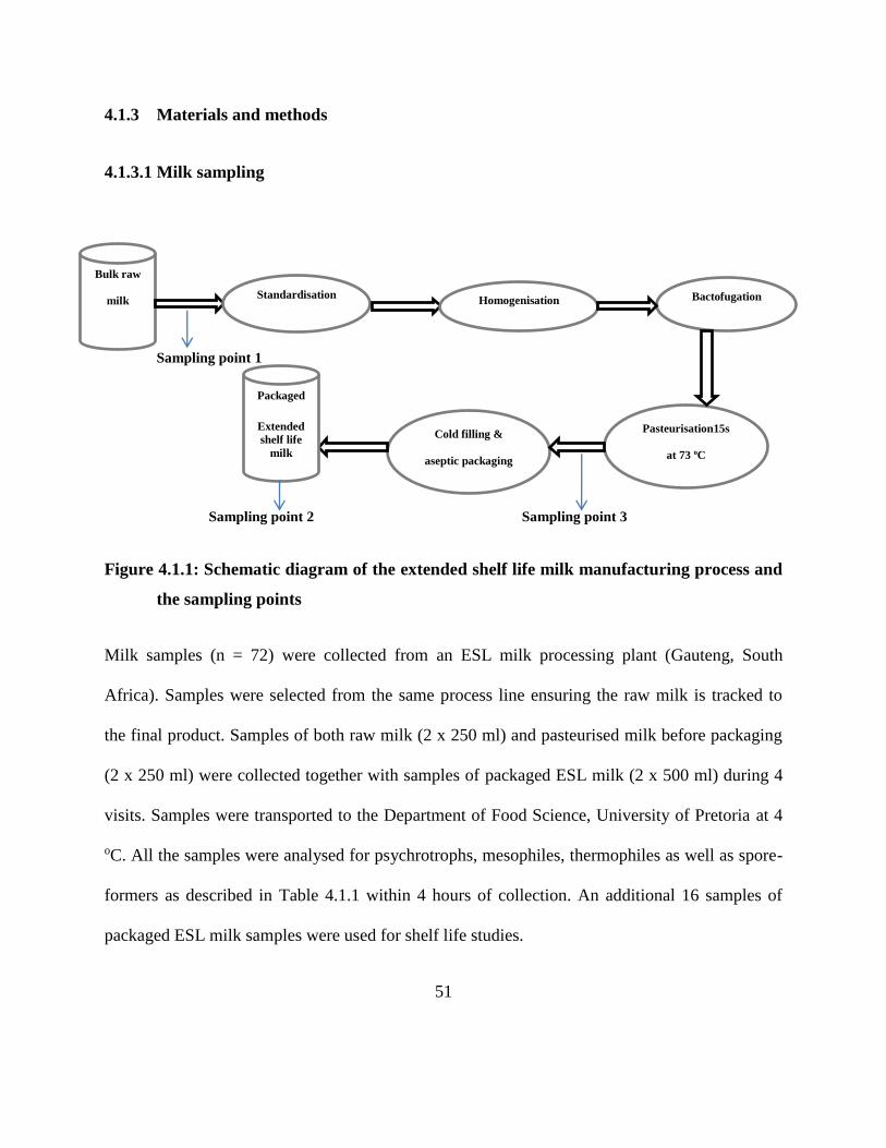
51
4.1.3 Materials and methods
4.1.3.1 Milk sampling
Sampling point 1
Sampling point 2 Sampling point 3
Figure 4.1.1: Schematic diagram of the extended shelf life milk manufacturing process and
the sampling points
Milk samples (n = 72) were collected from an ESL milk processing plant (Gauteng, South
Africa). Samples were selected from the same process line ensuring the raw milk is tracked to
the final product. Samples of both raw milk (2 x 250 ml) and pasteurised milk before packaging
(2 x 250 ml) were collected together with samples of packaged ESL milk (2 x 500 ml) during 4
visits. Samples were transported to the Department of Food Science, University of Pretoria at 4
oC. All the samples were analysed for psychrotrophs, mesophiles, thermophiles as well as spore-
formers as described in Table 4.1.1 within 4 hours of collection. An additional 16 samples of
packaged ESL milk samples were used for shelf life studies.
Bulk raw
milk Standardisation
Homogenisation Bactofugation
Pasteurisation15s
at 73 oC
73
Cold filling &
aseptic packaging
Packaged
Extended
shelf life
milk
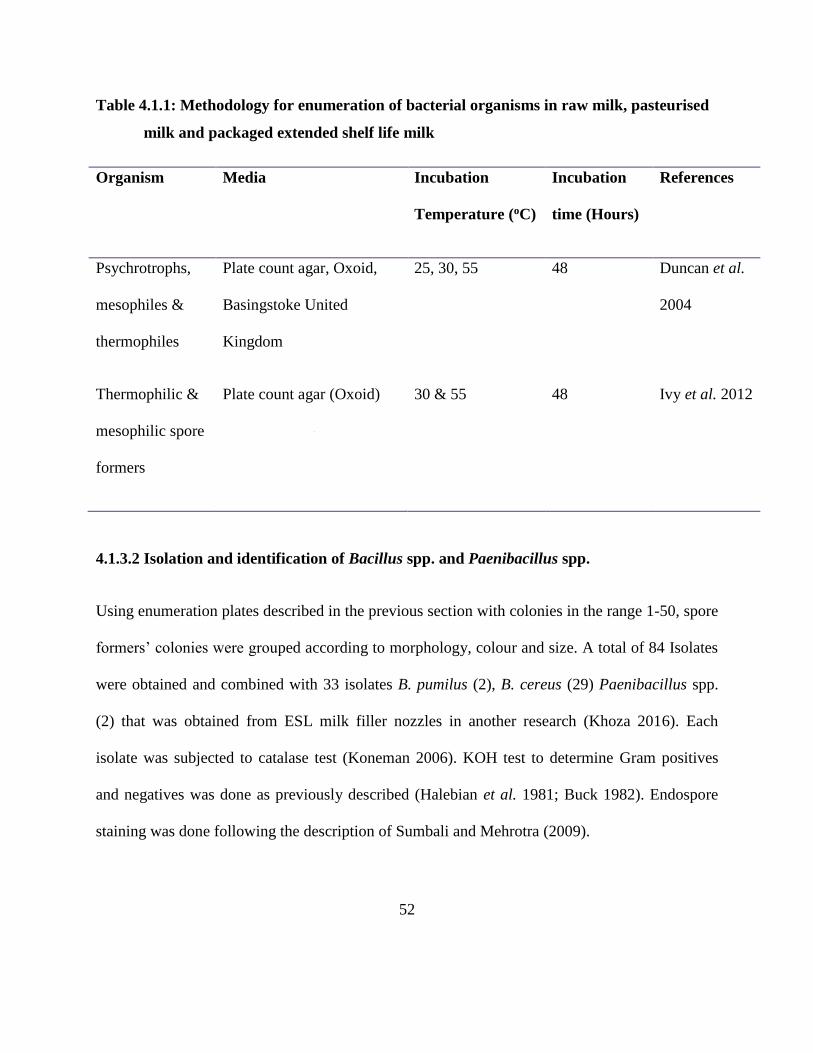
52
Table 4.1.1: Methodology for enumeration of bacterial organisms in raw milk, pasteurised
milk and packaged extended shelf life milk
Organism Media Incubation
Temperature (oC)
Incubation
time (Hours)
References
Psychrotrophs,
mesophiles &
thermophiles
Plate count agar, Oxoid,
Basingstoke United
Kingdom
25, 30, 55 48 Duncan et al.
2004
Thermophilic &
mesophilic spore
formers
Plate count agar (Oxoid) 30 & 55 48 Ivy et al. 2012
4.1.3.2 Isolation and identification of Bacillus spp. and Paenibacillus spp.
Using enumeration plates described in the previous section with colonies in the range 1-50, spore
formers’ colonies were grouped according to morphology, colour and size. A total of 84 Isolates
were obtained and combined with 33 isolates B. pumilus (2), B. cereus (29) Paenibacillus spp.
(2) that was obtained from ESL milk filler nozzles in another research (Khoza 2016). Each
isolate was subjected to catalase test (Koneman 2006). KOH test to determine Gram positives
and negatives was done as previously described (Halebian et al. 1981; Buck 1982). Endospore
staining was done following the description of Sumbali and Mehrotra (2009).
53
4.1.3.3 Determination of heamolysis, proteolytic and lipolytic enzyme activity
Ninety eight isolates from this study of presumptive B. pumilus, B. cereus, B. subtilis and
Paenibacillus spp. isolates were analysed for their enzymatic activity. The enzyme activity of
proteases, lipases and haemolysis were performed using skim milk agar (Oxoid Basingstoke,
UK), tributyrin agar (Oxoid) and Columbia + 5% sheep blood agar plates (Bio-Rad, Marnes-la-
Conquette, France) respectively following previously described procedures (Lucking et al.
2013). B. cereus ATCC 10876 was used as a positive control.
4.1.3.4 Shelf life studies
Eight samples of ESL milk were stored at 4 oC and the other 8 samples at 7 oC. Two samples of
the ESL milk from each storage temperature were analysed for total bacterial count as described
in Table 4.1.1 at days 7, 14, 18 and 21.
4.1.3.5 Growth profiles at 7 oC
One isolate that originated from milk stored at 7 oC was selected for each species (B. pumilus,
Bacillus subtilis, B. cereus and Paenibacillus spp.). Eighteen hour old cultures of these isolates
were standardised to 0.5 McFarland standards in saline solution. The 4 cultures were diluted in
buffered peptone water then each inoculated in 500 ml of sterile milk to achieve a concentration
range of 3 – 3.5 log10 cfu/ml. A cocktail of the 4 isolates was also prepared and inoculated in
commercial UHT milk in the same concentration as the single isolates. The milk was sealed
under sterile conditions, stored at 7 oC and population change observed at days 0, 14, 28 and 42
by analysing the total bacterial count as described in Table 1 coupled with Matrix-assisted laser
54
desorption/ionization Time of Flight Mass Spectrometry (MALDI-TOF-MS) identification. The
growth rate of each strain and the strain cocktail was calculated using DMFit in ComBase
(www.combase.cc)
4.1.3.6 Isolates identification by MALDI-TOF-MS
All isolates presumptively identified as Bacillus and Paenibacillus spp. by phenotypic
identification were further analysed using MALDI-TOF-MS. A total of 117 isolates were
streaked on plate count agar and incubated for 24 h at 30 oC. Samples for MALDI-TOF-MS
analysis were prepared using ethanol treatment followed by extraction with formic acid and
acetonitrile as described by Drevinek et al. (2012). They were identified using MALDI-TOF-MS
software (MALDI biotyper 3.0 Bruker Daltonics) after a mass spectra was obtained using a
MALDI-TOF-MS (Bruker Daltonics) following a procedure by Dybwad et al. (2012) and Fykse
et al. (2015). The peak lists generated were used directly to obtain matches against the reference
library (SARAMIS database). Using the spectra generated the relatedness of the isolates was
determined through creation of MSP dendrograms using Bruker Daltonics MALDI Biotyper 3.0
software (Bruker Daltonics) according to the manufacturer’s instructions.
4.1.3.7 Statistical analysis
ANOVA (p ≤ 0.05) with multiple comparisons (Tukey’s HSD test) was used to compare the log
counts for the different groups of organisms in raw milk, pasteurised milk and ESL milk.
Analysis was done using GraphPad Prism 7.0 (GraphPad Software, Inc., USA).
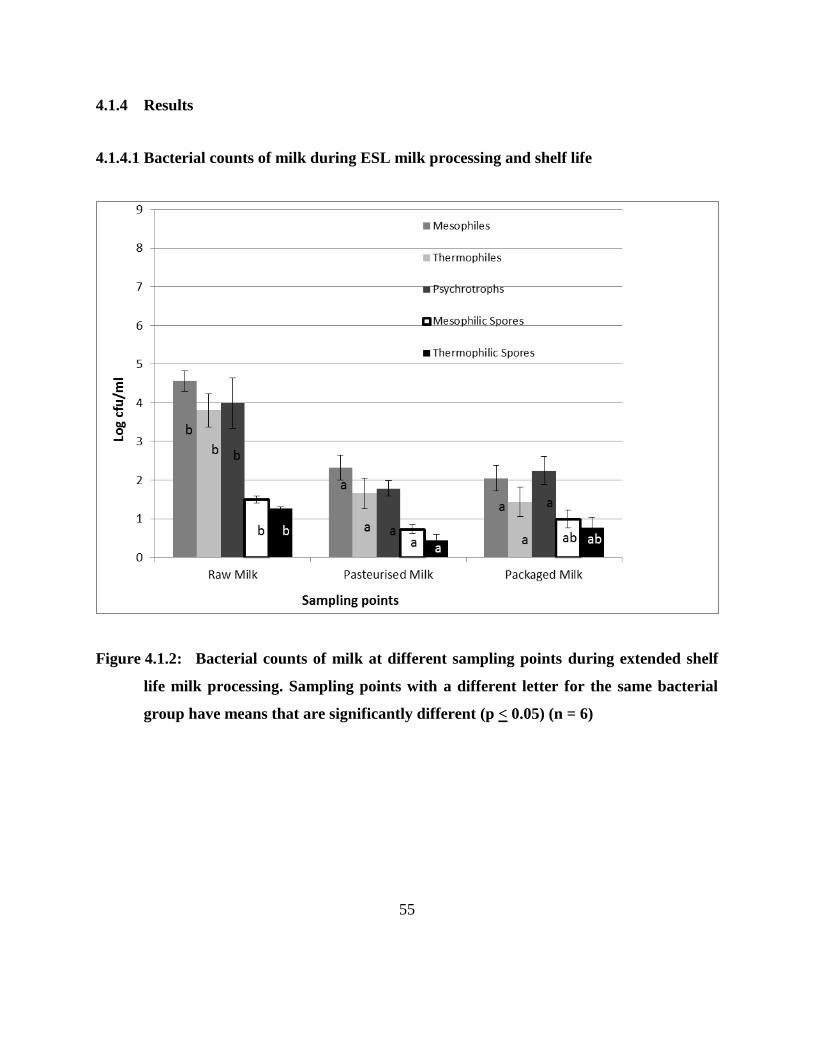
55
4.1.4 Results
4.1.4.1 Bacterial counts of milk during ESL milk processing and shelf life
Figure 4.1.2: Bacterial counts of milk at different sampling points during extended shelf
life milk processing. Sampling points with a different letter for the same bacterial
group have means that are significantly different (p < 0.05) (n = 6)
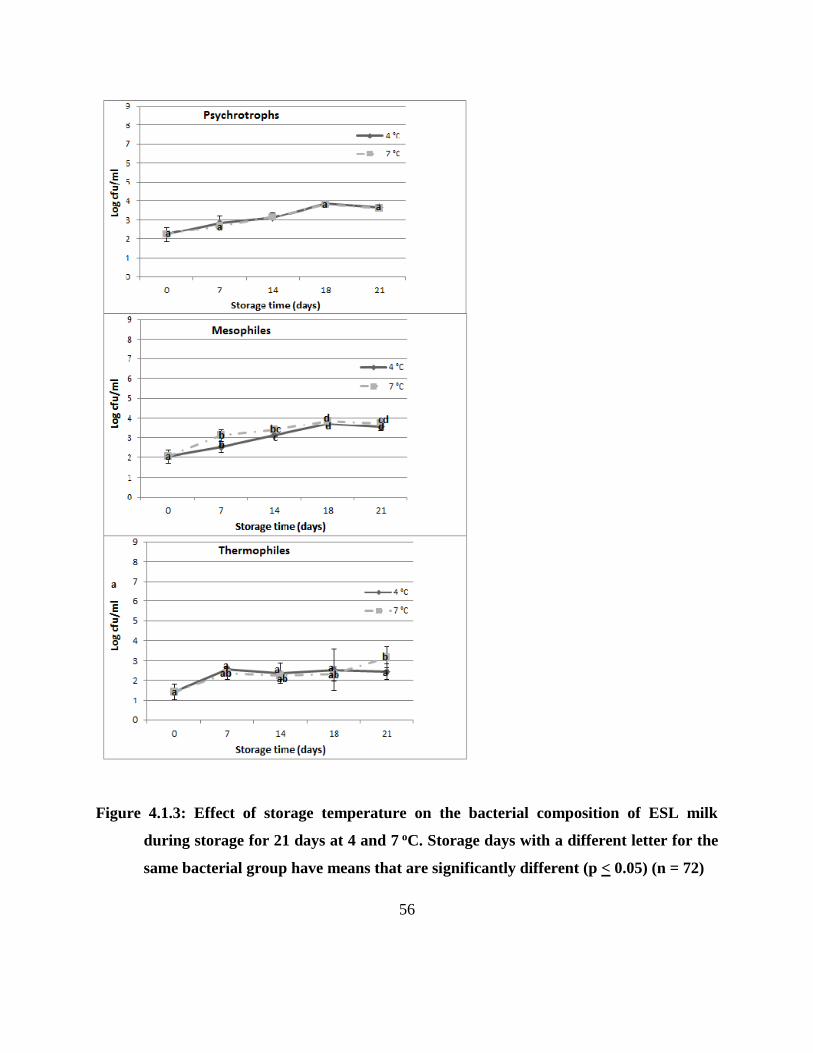
56
Figure 4.1.3: Effect of storage temperature on the bacterial composition of ESL milk
during storage for 21 days at 4 and 7 oC. Storage days with a different letter for the
same bacterial group have means that are significantly different (p < 0.05) (n = 72)
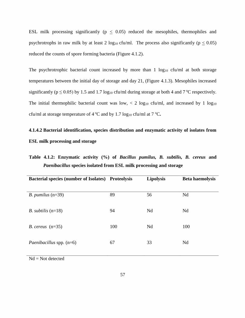
57
ESL milk processing significantly (p < 0.05) reduced the mesophiles, thermophiles and
psychrotrophs in raw milk by at least 2 log10 cfu/ml. The process also significantly (p < 0.05)
reduced the counts of spore forming bacteria (Figure 4.1.2).
The psychrotrophic bacterial count increased by more than 1 log10 cfu/ml at both storage
temperatures between the initial day of storage and day 21, (Figure 4.1.3). Mesophiles increased
significantly (p ≤ 0.05) by 1.5 and 1.7 log10 cfu/ml during storage at both 4 and 7 oC respectively.
The initial thermophilic bacterial count was low, < 2 log10 cfu/ml, and increased by 1 log10
cfu/ml at storage temperature of 4 oC and by 1.7 log10 cfu/ml at 7 oC.
4.1.4.2 Bacterial identification, species distribution and enzymatic activity of isolates from
ESL milk processing and storage
Table 4.1.2: Enzymatic activity (%) of Bacillus pumilus, B. subtilis, B. cereus and
Paenibacillus species isolated from ESL milk processing and storage
Bacterial species (number of Isolates) Proteolysis Lipolysis Beta haemolysis
B. pumilus (n=39) 89 56 Nd
B. subtilis (n=18) 94 Nd Nd
B. cereus (n=35) 100 Nd 100
Paenibacillus spp. (n=6) 67 33 Nd
Nd = Not detected
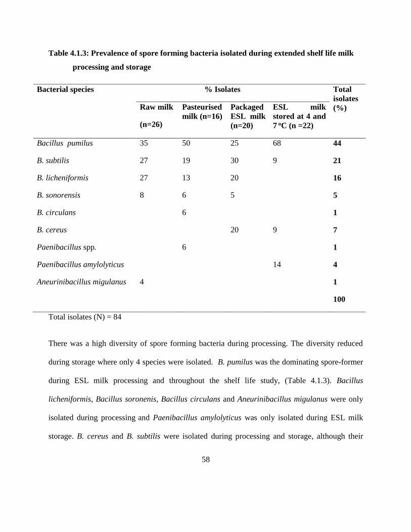
58
Table 4.1.3: Prevalence of spore forming bacteria isolated during extended shelf life milk
processing and storage
Bacterial species % Isolates Total
isolates
(%) Raw milk
(n=26)
Pasteurised
milk (n=16)
Packaged
ESL milk
(n=20)
ESL milk
stored at 4 and
7 oC (n =22)
Bacillus pumilus 35 50 25 68 44
B. subtilis 27 19 30 9 21
B. licheniformis 27 13 20 16
B. sonorensis 8 6 5 5
B. circulans 6 1
B. cereus 20 9 7
Paenibacillus spp. 6 1
Paenibacillus amylolyticus 14 4
Aneurinibacillus migulanus 4 1
100
Total isolates (N) = 84
There was a high diversity of spore forming bacteria during processing. The diversity reduced
during storage where only 4 species were isolated. B. pumilus was the dominating spore-former
during ESL milk processing and throughout the shelf life study, (Table 4.1.3). Bacillus
licheniformis, Bacillus soronenis, Bacillus circulans and Aneurinibacillus migulanus were only
isolated during processing and Paenibacillus amylolyticus was only isolated during ESL milk
storage. B. cereus and B. subtilis were isolated during processing and storage, although their
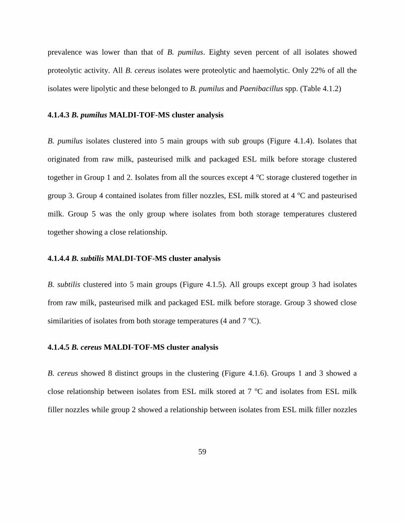
59
prevalence was lower than that of B. pumilus. Eighty seven percent of all isolates showed
proteolytic activity. All B. cereus isolates were proteolytic and haemolytic. Only 22% of all the
isolates were lipolytic and these belonged to B. pumilus and Paenibacillus spp. (Table 4.1.2)
4.1.4.3 B. pumilus MALDI-TOF-MS cluster analysis
B. pumilus isolates clustered into 5 main groups with sub groups (Figure 4.1.4). Isolates that
originated from raw milk, pasteurised milk and packaged ESL milk before storage clustered
together in Group 1 and 2. Isolates from all the sources except 4 oC storage clustered together in
group 3. Group 4 contained isolates from filler nozzles, ESL milk stored at 4 oC and pasteurised
milk. Group 5 was the only group where isolates from both storage temperatures clustered
together showing a close relationship.
4.1.4.4 B. subtilis MALDI-TOF-MS cluster analysis
B. subtilis clustered into 5 main groups (Figure 4.1.5). All groups except group 3 had isolates
from raw milk, pasteurised milk and packaged ESL milk before storage. Group 3 showed close
similarities of isolates from both storage temperatures (4 and 7 oC).
4.1.4.5 B. cereus MALDI-TOF-MS cluster analysis
B. cereus showed 8 distinct groups in the clustering (Figure 4.1.6). Groups 1 and 3 showed a
close relationship between isolates from ESL milk stored at 7 oC and isolates from ESL milk
filler nozzles while group 2 showed a relationship between isolates from ESL milk filler nozzles
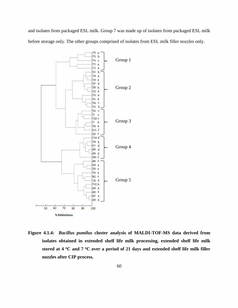
60
and isolates from packaged ESL milk. Group 7 was made up of isolates from packaged ESL milk
before storage only. The other groups comprised of isolates from ESL milk filler nozzles only.
Figure 4.1.4: Bacillus pumilus cluster analysis of MALDI-TOF-MS data derived from
isolates obtained in extended shelf life milk processing, extended shelf life milk
stored at 4 oC and 7 oC over a period of 21 days and extended shelf life milk filler
nozzles after CIP process.
Group 1
Group 2
Group 3
Group 4
Group 5
61
Isolate sources: a – Raw milk, b – Pasteurised milk, c – Packaged ESL milk on the day of
sampling, d- Stored ESL milk at 4 oC, e – Stored ESL milk at 7 oC and f – ESL milk filler nozzles
Figure 4.1.5: Bacillus subtilis cluster analysis of MALDI-TOF-MS data derived from
isolates obtained in extended shelf life milk processing, extended shelf life milk
stored at 4 oC and 7 oC over a period of 21 days and Extended Shelf Life milk filler
nozzles after CIP process.
Group 1
Group 2
Group 4
Group 5
Group 3
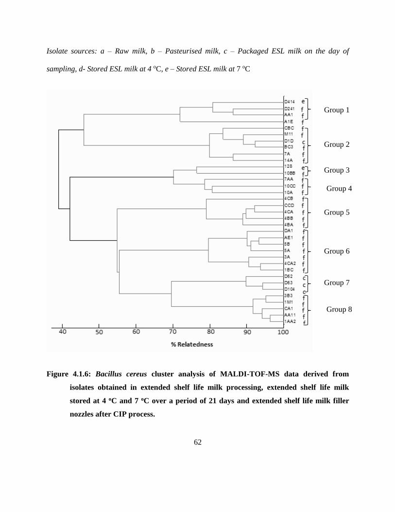
62
Isolate sources: a – Raw milk, b – Pasteurised milk, c – Packaged ESL milk on the day of
sampling, d- Stored ESL milk at 4 oC, e – Stored ESL milk at 7 oC
Figure 4.1.6: Bacillus cereus cluster analysis of MALDI-TOF-MS data derived from
isolates obtained in extended shelf life milk processing, extended shelf life milk
stored at 4 oC and 7 oC over a period of 21 days and extended shelf life milk filler
nozzles after CIP process.
Group 1
Group 8
Group 7
Group 6
Group 5
Group 3
Group 2
Group 4
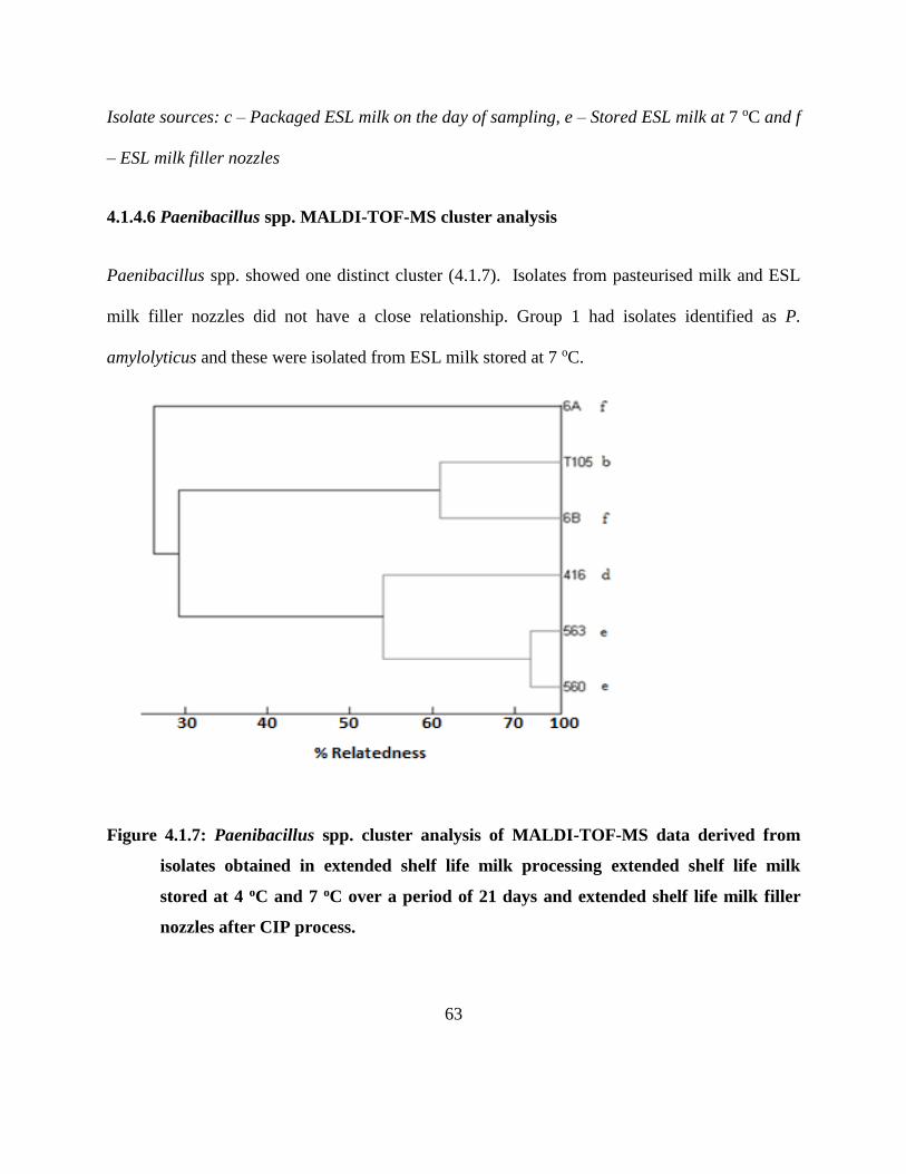
63
Isolate sources: c – Packaged ESL milk on the day of sampling, e – Stored ESL milk at 7 oC and f
– ESL milk filler nozzles
4.1.4.6 Paenibacillus spp. MALDI-TOF-MS cluster analysis
Paenibacillus spp. showed one distinct cluster (4.1.7). Isolates from pasteurised milk and ESL
milk filler nozzles did not have a close relationship. Group 1 had isolates identified as P.
amylolyticus and these were isolated from ESL milk stored at 7 oC.
Figure 4.1.7: Paenibacillus spp. cluster analysis of MALDI-TOF-MS data derived from
isolates obtained in extended shelf life milk processing extended shelf life milk
stored at 4 oC and 7 oC over a period of 21 days and extended shelf life milk filler
nozzles after CIP process.
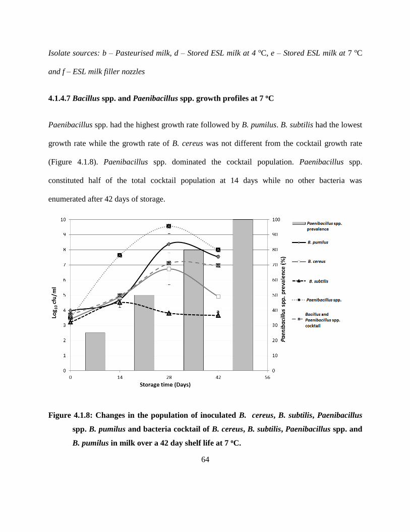
64
Isolate sources: b – Pasteurised milk, d – Stored ESL milk at 4 oC, e – Stored ESL milk at 7 oC
and f – ESL milk filler nozzles
4.1.4.7 Bacillus spp. and Paenibacillus spp. growth profiles at 7 oC
Paenibacillus spp. had the highest growth rate followed by B. pumilus. B. subtilis had the lowest
growth rate while the growth rate of B. cereus was not different from the cocktail growth rate
(Figure 4.1.8). Paenibacillus spp. dominated the cocktail population. Paenibacillus spp.
constituted half of the total cocktail population at 14 days while no other bacteria was
enumerated after 42 days of storage.
Figure 4.1.8: Changes in the population of inoculated B. cereus, B. subtilis, Paenibacillus
spp. B. pumilus and bacteria cocktail of B. cereus, B. subtilis, Paenibacillus spp. and
B. pumilus in milk over a 42 day shelf life at 7 oC.
65
Bacteria species with a different letter have growth rates that are significantly different at 7 oC (p
< 0.05). (Growth rates: B. subtilis 0.004±0.000, B. cereus 0.006±0.003; B. pumilus 0.011±0.000;
Paenibacillus spp. 0.006±0.001)
4.1.5 Discussion
Similar to other reported ESL milk processes (Fromm and Boor 2004; Sepulveda et al. 2005;
Schmidt et al. 2012; Caplan and Barbano 2013), the ESL milk process under investigation,
which involves bactofugation, reduced vegetative bacterial counts significantly. This reduction
was also facilitated by the raw milk which had low bacterial counts (< 5 log10 cfu/ml, Foodstuffs,
cosmetics and disinfectants Act 54 of 1972) (DAFF 1972). Despite the process significantly
reducing the bacterial count of spore-formers after bactofugation, there was an increase of the
spore formers in the packaged product, possibly due to post process contamination and growth of
survivors a trend that is similar to findings of Eneroth et al. (2001), who in their study concluded
contamination by filling machines. The dominance of mesophilic bacteria at 7 oC storage is
likely to have been a result of mesophilic bacteria that may exhibit psychrotrophic characteristics
due to the presence of the 16S psychrotrophic signature, (von Stetten et al. 1998). Stenfors and
Granum (2001) reported the presence of both mesophilic and psychrotrophic characteristics in
majority of B. cereus strains in their study while other studies have also shown that minimum
growth temperature for spore-forming Bacillus and Paenibacillus spp. range from 5-20 oC, and
maximum range from 35-55 oC (De Vos et al. 2009). The increase in thermophilic counts under
refrigeration (Figure 4.1.3) may be an indication that they are neither true thermophiles nor
thermotrophs. Thermophilic organisms do not grow at temperatures below 40 oC while
66
thermotrophs have a growth range of 20-60 oC (Deák and Farkas 2013). These organisms could
possibly have been mesophiles that contain psychrotrophic signatures (Stenfors and Granum
2001). However more elaborate studies are needed to verify this hypothesis especially in B.
pumilus that dominated this group. The bacterial counts obtained from the ESL milk product in
this study were lower than those previously reported for pasteurised milk stored at 4-7 oC
(Larsen and Jorgensen 1999; Fromm and Boor 2004; Petrus et al. 2010), by more than 1 log10
cfu/ml. The presence of the bactofugation step accounts for the difference in microbial load with
pasteurised milk from previous studies. However, the bacterial count observed in this study was
higher than that reported for microfiltered ESL milk (Schmidt et al. 2012). The difference in the
bacterial count of the two processes maybe due to a number of factors described in previous
researches that include raw milk quality, season of production, water and equipment among
others (Coorevits et al. 2008; De Jonghe et al. 2010; Eneroth et al. 2001).
B. pumilus isolates had the highest prevalence among the endospore forming bacteria,
contributing 45% of the total isolated endospore forming bacteria. This is contrary to most
reported results on endospore forming bacteria in heat treated milk. Aoudhi et al. (2014) reported
B. cereus as the dominating Bacillus spp. in pasteurised milk hence, concurs with Larsen and
Jørgensen (1999) and Ranieri et al. (2012) who also observed similar results. However, our
results showed similarity with those obtained from microfiltered ESL milk by Schmidit et al.
(2012) who observed 48% prevalence of B. pumilus. Apart from raw milk composition
(Coorevits et al. 2008) the dominance of B. pumilus can be attributed to its resistance to a
number of stresses including UV light exposure and presence of oxidisers such as H2O2 among
others as well as the ubiquitous nature of its endospore (Benardini et al. 2003; Link et al. 2004;
67
Dickinson et al. 2004). In line with available data (Ranieri and Boor 2009; Ranieri et al. 2012;
Schmidt et al. 2012), our results showed the presence of Paenibacillus spp. B. cereus as well as
B. licheniformis although the percentage prevalence we observed was lower than that reported.
B. licheniformis did not grow in the ESL milk product stored at both 4 and 7 oC because it is a
mesophile (Heyndrickx and Scheldeman 2002). The few B. cereus isolates obtained in our study
are in agreement with previous research findings, (Larsen and Jorgensen 1997; Larsen and
Jorgensen 1999). Apart from the fact that B. cereus is a poor competitor, good raw milk quality
also accounts for the low prevalence levels of B. cereus and Paenibacillus spp. also suggesting
that good hygienic processing conditions existed in this processing plant. When inoculated as
single organisms, Paenibacillus spp. had the highest growth rate. It dominated the cocktail
composition mainly because of its ability to be competitive against accompanying microbiota at
refrigerated conditions. Paenibacillus spp. requires L-tyrosine as one of its spore germinants
(Alvarado et al. 2012). It is likely that the proteolytic activity of B. cereus increases the amount
of free tyrosine in milk thereby aiding the growth of Paenibacillus spp. (Jan·tová et al. 2006).
Paenibacillus spp. is also a microaerophile (Owayss 2007) that requires small amounts of
oxygen for its spores to germinate thus helping more spores to germinate as milk ages.
Although there is no direct correlation between enzyme activity and milk spoilage, the fact that
more than 70% of isolates under study were proteolytic, shows a great potential of these
organisms to be the main causes of bacterial spoilage in ESL milk. These results show a similar
trend to previous studies (De Jonghe et al. 2010; Lucking et al. 2013). Our study observed low
lypolytic activity in B. cereus and Paenibacillus spp. a result that is similar to previous research
68
findings (Lucking et al. 2013). This implies that B. cereus and Paenibacillus spp. pose low risk
of ESL milk spoilage by lipid hydrolysis. However, contrary to previous milk studies (De Jonghe
et al. 2010; Lucking et al. 2013), none of the B. subtilis isolates under investigation were
lypolytic. With the highest lipolytic activity (62%), B. pumilus has potential to contribute to ESL
milk spoilage by lipolysis. All B. cereus isolates showed haemolytic activity similar to previous
results (Lucking et al. 2013). Despite being isolated in low numbers this shows that B. cereus
poses a potential safety risk to ESL milk consumers.
B. pumilus exhibited psychrotrophic characteristics as they managed to grow both at 4 and 7 oC
and dominate the microflora of ESL milk. In this research it was observed that B. pumilus
isolates that could grow at both 4 and 7 oC showed more than 75% similarity with isolates of B.
pumilus isolated from the filler nozzles after Cleaning In Place (CIP) at the same dairy plant.
This suggests post pasteurisation contamination of ESL milk by possible psychrotrophic B.
pumilus. A previous study on pasteurised milk has also demonstrated the ability of processing
equipment to contaminate the product, (Eneroth et al. 2001). Although the Paenibacillus spp.
isolates did not show close similarities, P. amylolyticus showed ability to grow at both 4 and 7 oC
possibly owing to its psychrotrophic nature (Ivy et al. 2012). The existence of most B. subtilis
isolates during processing compared to storage shows that the available strains could not exhibit
psychrotrophic properties as it is a well-known mesophile (Heyndrickx and Scheldeman 2002).
The presence of B. cereus in stored milk cements reports that some of its strains have
psychrotrophic properties, (Heyndrickx and Scheldeman 2002) and the similarity of isolates from
stored ESL milk and fillers is in line with the findings of Eneroth et al. (2001) suggesting post
69
pasteurisation contamination by the fillers. While the growth temperature of psychrotrophic
endospores of B. cereus have been reported to be in the range between 4-11 oC (Larsen and
Jørgensen 1999), it has also been shown that they germinate below their minimum growth
temperatures (Larsen and Jørgensen 1999).
4.1.6 Conclusions
The study indicated that bactofugation and pasteurisation based ESL milk process is effective in
reduction of both bacterial count and species diversity. While B. pumilus dominated the spore-
formers in ESL milk process, Paenibacillus spp. showed ability to outgrow other spore-formers
when the initial population is approximately the same. Although a few B. cereus isolates were
obtained, its presence is a potential safety problem in ESL milk. Paenibacillus spp. and Bacillus
spp. have great potential to cause spoilage of ESL milk through lipid and protein hydrolysis.
70
CHAPTER FIVE
Diversity of Bacillus cereus strains in extended shelf life milk
Redrafted from:
Mugadza, D.T. and Buys, E.M. 2017. Diversity of Bacillus cereus strains in extended shelf
life. International Dairy Journal, 73, 144-150.
71
5.1.1 Abstract
Characterisation of 49 Bacillus cereus strains obtained from extended shelf life (ESL) milk
processing and filler nozzles was done using (GTG)5 Rep PCR fingerprinting; determining the
presence of virulence genes; cytK, nheA, cer and hblA and discrimination of psychrotrophic and
mesophilic strains with 16S rDNA. Fourteen isolates were selected for 16S partial sequencing.
Fingerprinting and sequencing showed evidence of filler nozzles contaminating ESL milk
despite high heterogeneity existing between the isolates. While there is high prevalence of cer,
hblA and nheA; cytK was not widely distributed. There was 100% and 8% prevalence of
mesophilic and psychrotrophic signatures respectively. Despite the large diversity of the B.
cereus strains in this study, there is evidence that filler nozzles and raw milk are a source of
contamination of B. cereus in ESL milk.
5.1.2 Introduction
Bacillus cereus, a ubiquitous spore-forming bacteria has been identified as the main spoilage
organism in pasteurised milk (Aouadhi et al. 2014). Its presence in milk products does not only
cause spoilage concern, but safety too as it is known to produce a number of toxins (Hansen and
Hendriksen 2001; Bartoszewicz et al. 2008). Extended shelf life (ESL) milk is a product bridging
the gap between UHT and pasteurised milk that has gained popularity over the past few years.
Although previous studies have reported that B. cereus was not the dominating organism in ESL
milk (Schmidit et al. 2012; Mugadza and Buys 2014), its ability to grow at 7 oC (Mugadza and
Buys 2017a), makes it a significant organism in ESL milk.
72
While raw milk has been implicated as an important source of endospores in milk products
(Miller et al. 2015) other studies have shown that a different population of endospore forming
microorganisms exists in raw milk and milk products made from the same raw milk. This
population change has been attributed to a number of reasons that include post process
contamination among others (Scott et al. 2007; Burgess et al. 2010; Hill and Smythe 2012). Post
heat treatment contamination by processing equipment such as fillers has also proved to cause
bacterial population variations between raw milk and milk products (Scott et al. 2007; Burgess et
al. 2010; Hill and Smythe 2012). Other studies have shown that B. cereus isolates from the
environments (soil, food, the dairy production chain) have a higher degree of heterogeneity
compared to clinical isolates (Helgason et al. 2000; Ehling-Schulz et al. 2005). Banyko´ and
Vyletelova 2009 concluded that B. cereus does not originate exclusively in either the raw or
pasteurised milk but from post-pasteurisation contamination and propagation during the
manufacturing process.
A full understanding of the characteristics and relationship between B. cereus strains isolated in
the ESL milk processing environment becomes important in determining the source of product
contamination. The objective of this study was to characterise B. cereus isolates obtained from
ESL milk processing and during shelf life under refrigeration with the aim of determining cross
contamination routes within the ESL milk processing plant.
73
5.1.3 Materials and methods
5.1.3.1 Isolates and DNA extraction
Samples of raw milk, pasteurised milk before packaging and packaged ESL milk were collected
together with filler nozzles swabs during 3 visits at an ESL milk processing plant (Gauteng,
South Africa). Samples were selected from the same process line ensuring the raw milk is
tracked to the final product. Furthermore, some packaged ESL milk samples were stored at 4 and
7 oC over a 21 day shelf life period. B. cereus isolates were obtained from these samples as
described elsewhere (Khoza 2016; Mugadza and Buys 2017a). Forty nine B. cereus isolates that
were obtained from raw milk (2), pasteurised milk (4), filler nozzles (40) and ESL milk during
storage at 7 oC (3), were grown in nutrient agar at 30 °C together with a reference strain, B.
cereus ATCC 10876 until the late exponential phase which required an incubation period of 16-
24 h. DNA was extracted using the ZR Fungal/Bacterial DNA MiniPrep™ (Zymo Research,
California, USA) according to the manufacturer’s instructions for use in the PCR reactions.
5.1.3.2 (GTG)5 Rep PCR Fingerprinting
Fingerprinting PCR was carried out in 20 µL reaction tubes in a Bio-Rad T100™ Thermal
Cycler (Bio-Rad, Singapore, Singapore). The reaction mixture consisted of 10 µL KAPA
BIOSYSTEMS 2X KAPA Taq Ready Mix (Kapa Biosystems Cape Town, South Africa), 0.3 µL
GTGGTGGTGGTGGTG oligonucleotide primer (Versalovic et al. 1994), 2 µL DNA template,
7.3 µL PCR-grade water and 0.4 µL Dimethyl sulfoxide (DMSO). The PCR program was as
follows: 95 oC for 5 min, 95 oC for 1 min, annealing at 52 oC for 1 min, extension at 72 oC for 3
74
min. The program was repeated for 34 cycles and a final extension at 72 oC for 10 minutes. PCR
products were separated by Gel electrophoresis using a 1.5% Agarose gel (55 V for 4 h) and the
image viewed using a Bio-Rad Gel Doc™ EZ (Bio-Rad, California, USA). The (GTG)5
fingerprints were analysed using Gel-Compar II version 6.5 (Applied Maths NV, Sint-Martens-
Latem, Belgium). The similarity of digitised band patterns was calculated using Pearson's
correlation coefficient, and unweighted pair group method with arithmetic means. Complete
linkage algorithms were used to construct an average linkage dendrogram to show relationship of
isolates. Isolates were considered to be within a clonal cluster if relatedness was 70% and above
(Stackebrandt et al. 2002). However, due to the close similarities that existed between the
isolates sub clusters were further considered at 95%.
5.1.3.3 PCR to determine virulence genes and discriminate psychrotrophic from mesophilic
B. cereus
Pathogenicity of the B. cereus was determined by observing the presence of the following genes;
cytotoxin K (cytK), nonhemolytic enterotoxin A (nheA), emetic toxin cereulide (cer) and
enterotoxin hemolysin BL (hblA). Discrimination of psychrotrophic and mesophilic B. cereus
was done by targeting the 16S rDNA signatures. The primers used are described in Table 5.1.1.
All PCRs were carried out in 10µL reaction tubes in a Bio-Rad T100™ Thermal Cycler (Bio-
Rad Singapore, Singapore). The reaction mix consisted of 1 µL bacterial DNA template, 5 µL
KAPA BIOSYSTEMS 2X KAPA Taq Ready Mix (Cape Town, South Africa), 3.4 µL PCR-
grade water and 0.3 µL of each primer. The PCR conditions for cytK, cer, hblA and nheA are
described in a previous study (Swiecicka and Mahillon 2006), while conditions for mesophilic
75
and psychrotrophic B. cereus PCR are also previously described (von Stetten et al. 1998).
Correlation between isolates was also determined by assigning a score of 1 and 2 for the
presence and absence of a gene respectively and similarity calculated using principle component
analysis (PCA) and agglomerative hierarchical clustering (AHC) in XLSTAT an add-in
programme of Microsoft Excel.
5.1.3.4 B. cereus 16S rRNA Sequencing
Sixteen B. cereus isolates were randomly selected, ensuring representation of each sampling
point for 16S sequencing. Using the ZR Fungal/Bacterial DNA Kit™(Zymo Research,
California, USA), DNA was obtained from the cultures. The 16S target region was amplified
using DreamTaq™ DNA polymerase (Thermo Scientific™, Johannesburg, South Africa) and the
primers, (Amplicon size 1465 bp) shown in Table 5.1.2. PCR products were gel extracted
(Zymo Research, Zymoclean™ Gel DNA Recovery Kit, California, USA), and sequenced in the
forward and reverse directions on the ABI PRISM™ 3500xl Genetic Analyser. Purified
sequencing products (Zymo Research, ZR-96 DNA Sequencing Clean-up Kit™, California,
USA) were analysed using CLC Main Workbench 7 followed by a BLAST search (NCBI).
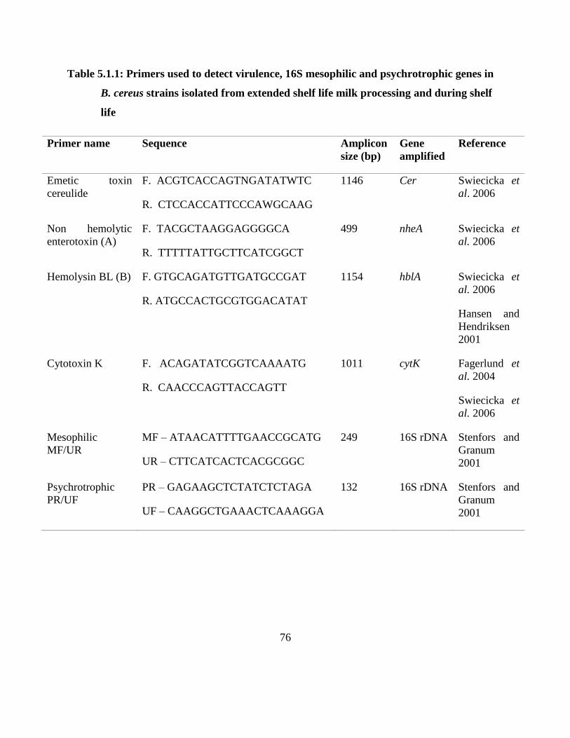
76
Table 5.1.1: Primers used to detect virulence, 16S mesophilic and psychrotrophic genes in
B. cereus strains isolated from extended shelf life milk processing and during shelf
life
Primer name Sequence Amplicon
size (bp)
Gene
amplified
Reference
Emetic toxin
cereulide
F. ACGTCACCAGTNGATATWTC
R. CTCCACCATTCCCAWGCAAG
1146 Cer Swiecicka et
al. 2006
Non hemolytic
enterotoxin (A)
F. TACGCTAAGGAGGGGCA
R. TTTTTATTGCTTCATCGGCT
499 nheA Swiecicka et
al. 2006
Hemolysin BL (B) F. GTGCAGATGTTGATGCCGAT
R. ATGCCACTGCGTGGACATAT
1154 hblA Swiecicka et
al. 2006
Hansen and
Hendriksen
2001
Cytotoxin K F. ACAGATATCGGTCAAAATG
R. CAACCCAGTTACCAGTT
1011 cytK Fagerlund et
al. 2004
Swiecicka et
al. 2006
Mesophilic
MF/UR
MF – ATAACATTTTGAACCGCATG
UR – CTTCATCACTCACGCGGC
249 16S rDNA Stenfors and
Granum
2001
Psychrotrophic
PR/UF
PR – GAGAAGCTCTATCTCTAGA
UF – CAAGGCTGAAACTCAAAGGA
132 16S rDNA Stenfors and
Granum
2001
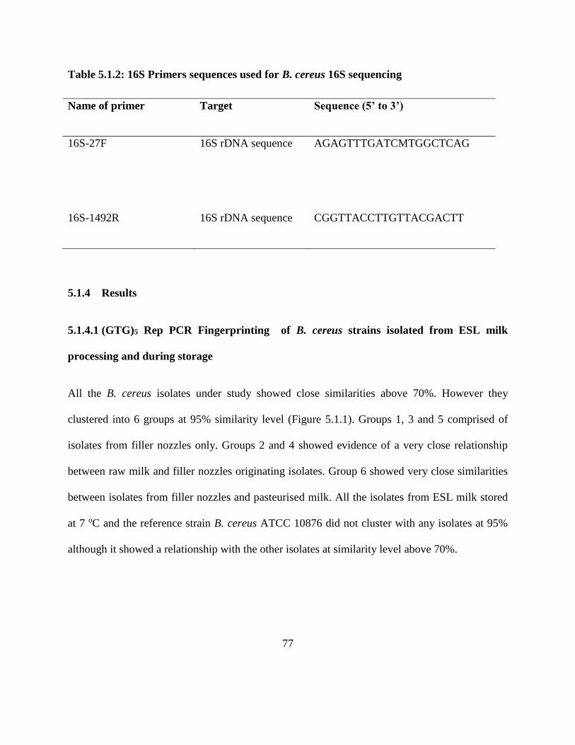
77
Table 5.1.2: 16S Primers sequences used for B. cereus 16S sequencing
Name of primer Target Sequence (5’ to 3’)
16S-27F
16S rDNA sequence AGAGTTTGATCMTGGCTCAG
16S-1492R 16S rDNA sequence CGGTTACCTTGTTACGACTT
5.1.4 Results
5.1.4.1 (GTG)5 Rep PCR Fingerprinting of B. cereus strains isolated from ESL milk
processing and during storage
All the B. cereus isolates under study showed close similarities above 70%. However they
clustered into 6 groups at 95% similarity level (Figure 5.1.1). Groups 1, 3 and 5 comprised of
isolates from filler nozzles only. Groups 2 and 4 showed evidence of a very close relationship
between raw milk and filler nozzles originating isolates. Group 6 showed very close similarities
between isolates from filler nozzles and pasteurised milk. All the isolates from ESL milk stored
at 7 oC and the reference strain B. cereus ATCC 10876 did not cluster with any isolates at 95%
although it showed a relationship with the other isolates at similarity level above 70%.
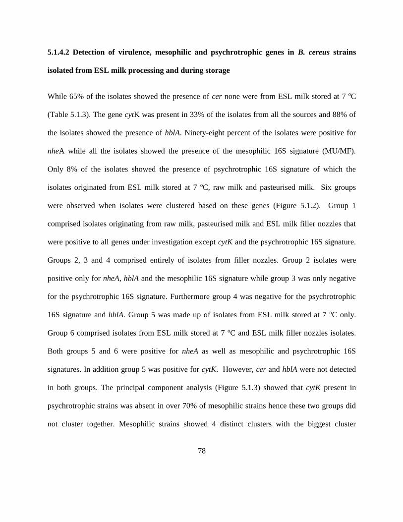
78
5.1.4.2 Detection of virulence, mesophilic and psychrotrophic genes in B. cereus strains
isolated from ESL milk processing and during storage
While 65% of the isolates showed the presence of cer none were from ESL milk stored at 7 oC
(Table 5.1.3). The gene cytK was present in 33% of the isolates from all the sources and 88% of
the isolates showed the presence of hblA. Ninety-eight percent of the isolates were positive for
nheA while all the isolates showed the presence of the mesophilic 16S signature (MU/MF).
Only 8% of the isolates showed the presence of psychrotrophic 16S signature of which the
isolates originated from ESL milk stored at 7 oC, raw milk and pasteurised milk. Six groups
were observed when isolates were clustered based on these genes (Figure 5.1.2). Group 1
comprised isolates originating from raw milk, pasteurised milk and ESL milk filler nozzles that
were positive to all genes under investigation except cytK and the psychrotrophic 16S signature.
Groups 2, 3 and 4 comprised entirely of isolates from filler nozzles. Group 2 isolates were
positive only for nheA, hblA and the mesophilic 16S signature while group 3 was only negative
for the psychrotrophic 16S signature. Furthermore group 4 was negative for the psychrotrophic
16S signature and hblA. Group 5 was made up of isolates from ESL milk stored at 7 oC only.
Group 6 comprised isolates from ESL milk stored at 7 oC and ESL milk filler nozzles isolates.
Both groups 5 and 6 were positive for nheA as well as mesophilic and psychrotrophic 16S
signatures. In addition group 5 was positive for cytK. However, cer and hblA were not detected
in both groups. The principal component analysis (Figure 5.1.3) showed that cytK present in
psychrotrophic strains was absent in over 70% of mesophilic strains hence these two groups did
not cluster together. Mesophilic strains showed 4 distinct clusters with the biggest cluster

79
showing the presence of hblA and cer genes. Cer and nheA dominated the second cluster while
hblA dominated the other cluster. The smallest cluster was more aligned to the presence of cytK
and nheA.
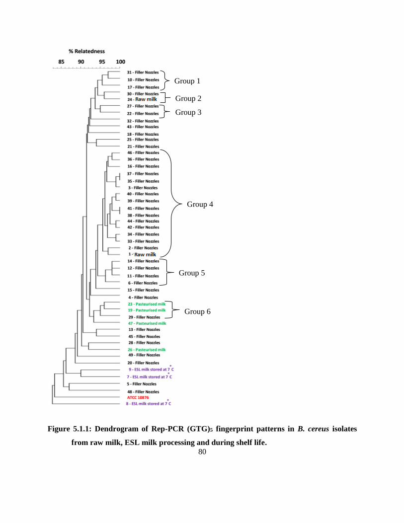
80
Figure 5.1.1: Dendrogram of Rep-PCR (GTG)5 fingerprint patterns in B. cereus isolates
from raw milk, ESL milk processing and during shelf life.
Group 1
Group 4
Group 5
Group 6
Group 2
Group 3

81
Table 5.1.3: Detection of virulence, mesophilic and psychrotrophic genes in B. cereus
strains isolated from raw milk, ESL milk processing and during shelf life
Isolate source
Isolate
(%)
Gene
Cer nheA hblA CytK MF/UR PR/UF
Raw Milk
Pasteurised Milk
Filler Nozzles
2
4
33
+
+
+
+
+
+
+
+
+
-
-
-
+
+
+
-
-
-
Pasteurised Milk
Filler Nozzles
2
20
+
+
+
+
+
+
+
+
+
+
-
-
Filler Nozzles 22 - + + - + -
Filler Nozzles 4 + + - + + -
Filler Nozzles
ESL Milk
2
2
-
-
+
+
-
-
-
-
+
+
+
+
ESL Milk 4 - + - + + +
Raw Milk 2 - + + + + -
Pasteurised Milk 2 - - + - + -
ATCC 10876 - + + + + + +
+ means positive, - means negative. * Total percentage is 99 % due to rounding off
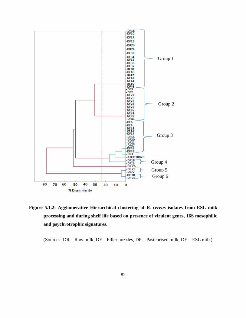
82
Figure 5.1.2: Agglomerative Hierarchical clustering of B. cereus isolates from ESL milk
processing and during shelf life based on presence of virulent genes, 16S mesophilic
and psychrotrophic signatures.
(Sources: DR – Raw milk, DF – Filler nozzles, DP – Pasteurised milk, DE – ESL milk)
Group 2
Group 1
Group 3
Group 4
Group 5
Group 6
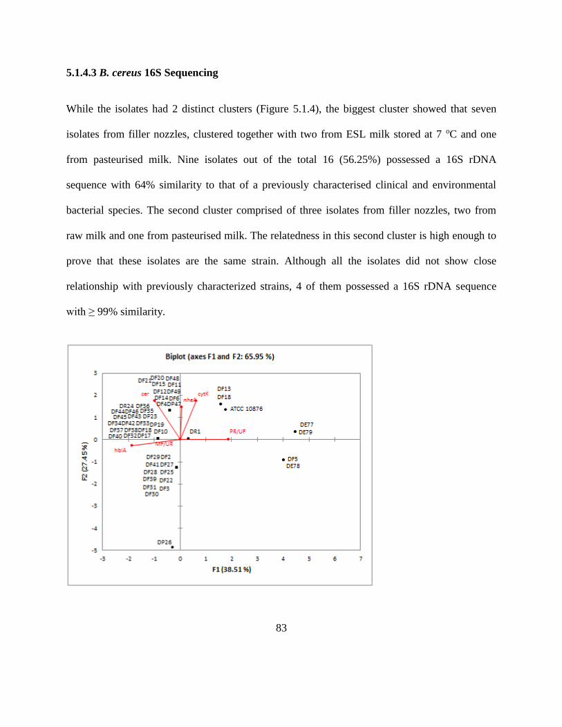
83
5.1.4.3 B. cereus 16S Sequencing
While the isolates had 2 distinct clusters (Figure 5.1.4), the biggest cluster showed that seven
isolates from filler nozzles, clustered together with two from ESL milk stored at 7 oC and one
from pasteurised milk. Nine isolates out of the total 16 (56.25%) possessed a 16S rDNA
sequence with 64% similarity to that of a previously characterised clinical and environmental
bacterial species. The second cluster comprised of three isolates from filler nozzles, two from
raw milk and one from pasteurised milk. The relatedness in this second cluster is high enough to
prove that these isolates are the same strain. Although all the isolates did not show close
relationship with previously characterized strains, 4 of them possessed a 16S rDNA sequence
with ≥ 99% similarity.
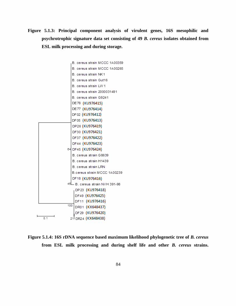
84
Figure 5.1.3: Principal component analysis of virulent genes, 16S mesophilic and
psychrotrophic signature data set consisting of 49 B. cereus isolates obtained from
ESL milk processing and during storage.
Figure 5.1.4: 16S rDNA sequence based maximum likelihood phylogenetic tree of B. cereus
from ESL milk processing and during shelf life and other B. cereus strains.
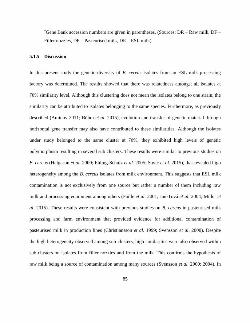
85
*Gene Bank accession numbers are given in parentheses. (Sources: DR – Raw milk, DF –
Filler nozzles, DP – Pasteurised milk, DE – ESL milk)
5.1.5 Discussion
In this present study the genetic diversity of B. cereus isolates from an ESL milk processing
factory was determined. The results showed that there was relatedness amongst all isolates at
70% similarity level. Although this clustering does not mean the isolates belong to one strain, the
similarity can be attributed to isolates belonging to the same species. Furthermore, as previously
described (Aminov 2011; Böhm et al. 2015), evolution and transfer of genetic material through
horizontal gene transfer may also have contributed to these similarities. Although the isolates
under study belonged to the same cluster at 70%, they exhibited high levels of genetic
polymorphism resulting in several sub clusters. These results were similar to previous studies on
B. cereus (Helgason et al. 2000; Ehling-Schulz et al. 2005; Savic et al. 2015), that revealed high
heterogeneity among the B. cereus isolates from milk environment. This suggests that ESL milk
contamination is not exclusively from one source but rather a number of them including raw
milk and processing equipment among others (Faille et al. 2001; Jan·Tová et al. 2004; Miller et
al. 2015). These results were consistent with previous studies on B. cereus in pasteurised milk
processing and farm environment that provided evidence for additional contamination of
pasteurised milk in production lines (Christiansson et al. 1999; Svensson et al. 2000). Despite
the high heterogeneity observed among sub-clusters, high similarities were also observed within
sub-clusters on isolates from filler nozzles and from the milk. This confirms the hypothesis of
raw milk being a source of contamination among many sources (Svensson et al. 2000; 2004). In
86
an earlier study, it was revealed that pasteurised milk and final products were contaminated by B.
cereus strains that germinated from spores in raw milk (Lin et al. 1998). The presence of the
similar isolates of B. cereus in raw milk, filler nozzles and commercially pasteurised milk
samples, further confirms the role of raw milk and filling machines as sources of B. cereus
contamination (Eneroth et al. 2001; Huck et al. 2007; Banyko´ and Vyletelova 2009).
B. cereus contaminants associated with food borne illness are reported to be mesophilic strains
(Larsen and Jørgensen 1997; Francis et al. 1998; Stenfors and Granum 2001; Guinebretire et al.
2008). This shows that the risk posed by psychrotrophic B. cereus in food is obviously low.
However, in the present trial psychrothrophic strains isolated from ESL milk contained some
virulence genes. This difference could indicate that virulence genes are closely associated with
mesophilic strains. Furthermore, the coexistence of mesophilic and psychrotophic signatures may
have resulted in the presence of virulence genes in psychrotrophic strains. However, scientific
evidence is needed to support this hypothesis.
The study also revealed that all B. cereus isolates linked to ESL milk contamination showed
mesophilic characteristics. This was in line with a previous study that demonstrated a clustering
of B. cereus strains from a dairy plant and suggested contamination caused by mesophilic
Bacillus strains. It was concluded that contamination occurred early in the production chain
probably in raw milk silos through biofilms (Svensson et al. 1999).
This study revealed that only 8% (4) isolates showed the presence of the 16S psychrotrophic
signature. Similarly, a previous study reported that most B. cereus strains obtained from the food
chain environment were mesophilic while a few exhibited psychrotrophic properties (Stenfors
87
and Granum 2001). Although previous studies have indicated that most food contaminants and
virulent B. cereus are mesophilic strains (Larsen and Jørgensen 1997; Francis et al. 1998;
Stenfors and Granum 2001; Guinebretire et al. 2008), this study revealed a contrary trend. This
may be a result of the emergence of psychrotrophic B. cereus strains resulting from increased
cold exposure to cold environments leading to increased adaptation and evolution (Guinebretire
et al. 2008). This indicates that the most problematic strains in ESL milk are not psychrotrophic
strains of B. cereus only but mesophilic that have psychrotrophic signatures or vice versa.
Contrary to a previous study which concluded that emetic toxin, cereulide producing B. cereus
are rare in the environment (Altayar and Sutherland 2005) our results showed 65% prevalence of
the emetic toxin producing B. cereus. However, in agreement with previous studies (Carlin et al.
2006; Hoton et al. 2009), all cereulide positive isolates were mesophilic. The increased presence
of the cer gene is probably due to adaptation as most cer-containing strains usually propagate at
low incidences in milk (Bartoszewicz et al. 2008). Furthermore, the fact that toxigenic
determinants spread horizontally among Bacillus species (Bartoszewicz et al. 2008) may have
contributed to increased cer prevalence. Although cereulide synthesis is known to occur at a
temperature range of 12-37 oC (Finlay et al. 2000; Häggblom et al. 2002), depending on strain
and media too (Apetroaie-Constantin et al. 2008) cold chain abuse of ESL milk product therefore
puts consumers at risk of this emetic toxin.
The distribution of enterotoxin gene hblA did not follow a definitive trend. Nevertheless, it has
been hypothesised that many food-borne strains of B. cereus are less prone to cause diarrhoea
(in’t Veld et al. 2001; Guinebretière and Broussolle 2002). The high prevalence of the
88
enterotoxin genes hblA and nheA in the total isolates and low prevalence of the genes in the final
ESL milk product could be result of failure of the hblA and nheA possessing strains to withstand
processing conditions and proliferate during cold storage (Hansen and Hendriksen 2001; Ehling-
Schulz et al. 2006b; Bartoszewicz et al. 2008). The presence of the hblA does not directly
translate to production of the toxin since other components are required to be present (in’t Veld
et al. 2001; Arnesen et al. 2008). However, the presence of this toxin determinant in ESL milk
shows that consumers of cold-stored ESL milk product may be at risk of the diarrhoeal causing
toxins.
Although cytK is regarded as highly toxic to humans (Hardy et al. Lund and Granum 2001;
Fagerlund et al. 2004), it is not widely distributed in B. cereus (Lund et al. 2000; Stenfors and
Granum 2001; Fagerlund et al. 2004; Swiecicka and Mahillon 2006). While the reason for low
frequency of cytK in food related B. cereus isolates is not clear, a possible explanation could be
the failure of cytK containing strains to propagate in the food matrix. However studies need to be
done to prove this hypothesis. Despite the fact that enterotoxins are heat labile and prone to
degradation by pH changes (Hansen and Hendriksen 2001) the presence of the cytK in
psychrotolerant isolates from the ESL milk poses a potential safety threat to consumers.
5.1.6 Conclusion
The simultaneous presence of psychrotrophic, mesophilic and at least 2 virulence genes in some
of the isolates shows large diversity of B. cereus strains in ESL milk process that does not follow
any specific pattern. This shows that contamination of ESL milk with B. cereus follows several

89
routes. This study provided evidence that filler nozzles and raw milk are sources of
contamination of ESL milk.
90
CHAPTER SIX
Source tracking Bacillus cereus in an extended shelf life milk processing plant using partial
sequencing of rpoB and multilocus sequence typing
91
6.1.1 Abstract
Characterisation of seven B. cereus strains obtained from extended shelf life (ESL) milk stored at
7 oC, pasteurised milk, raw milk and filler nozzles after CIP was performed using rpoB partial
sequencing and multilocus sequence typing. The objective of the study was to determine
relatedness within B. cereus isolates from several sampling points with the aim of source
tracking B. cereus in an ESL milk processing plant. The study revealed that some isolates from
pasteurised milk shared 100% similarity with isolates from raw milk and ESL milk using rpoB
sequencing. It also revealed very close similarity between isolates from filler nozzles and ESL
milk using MLST. This study showed that B. cereus contamination of ESL milk is through raw
milk and biofilms from filler nozzles. Furthermore, the study also proposes at least 3 routes of B.
cereus contamination in ESL milk. In addition, the study also showed that rpoB partial
sequencing and MLST can be used as tools for source tracking in ESL milk processing
6.1.2 Introduction
Despite advancement in preservation and processing technologies, Bacillus cereus remains a
shelf life and consumer safety challenge in the dairy industry (Huck et al. 2007a; Ranieri et al.
2012; Aouadhi et al. 2014; Mugadza and Buys 2017a). B. cereus is a ubiquitous spore-forming
bacterium (Ranieri et al. 2012; Aouadhi et al. 2014) that is reported to produce various toxins
responsible for diarrhoeal and emetic food poison (Hansen and Hendriksen 2001; Arnesen et al.
2008; Bartoszewicz et al. 2008). Studies conducted at an ESL milk processing plant revealed the
presence of psychrotrophic B. cereus in milk during processing as well as during storage and in
filler nozzles after CIP (Khoza 2016; Mugadza and Buys 2017a). A follow up study on these B.
92
cereus strains then showed close relatedness between these strains using repPCR and 16S partial
sequencing (Mugadza and Buys 2017b).
Furthermore, B. cereus endospores can germinate at refrigeration temperatures. In addition, some
strains can also grow, as well as produce toxins under these refrigeration conditions (Larsen and
Jørgensen 1997; Stenfors and Granum 2001; Thorsen et al. 2006). B. cereus has also been
reported to be able to attach to stainless steel (Eneroth et al. 1998, 2001; Khoza 2016) the main
material used in manufacturing processing equipment, while other studies have isolated B. cereus
on the processing equipment, suggesting the potential to form biofilms which can later be
responsible for post process contamination of processed milk (Eneroth et al. 1998, 2001).
While raw milk has been implicated as a source of B. cereus contamination in pasteurised milk
(Huck et al. 2007a,b), post processing contamination by processing plant equipment has also
been described (te Giffel et al. 1997; Svensson et al. 2000; Huck et al. 2007a,b). Identification of
points of entry for these bacteria may allow the development of effective strategies for reducing
or eliminating their presence in milk production systems (Huck et al. 2008).
While several methods have been suggested for source tracking (Fu and Li 2014), multilocus
sequence typing (MLST) has been described as a source tracking tool that determines exact
nucleotide differences for conserved loci (Cardazzo et al. 2008). MLST studies have previously
been used to examine the phylogeny of the B. cereus complex (Helgason et al. 2000, 2004;
Barker et al. 2005), identifying three distinct lineages that largely correspond to the species
distribution (Cardazzo et al. 2008). In addition to MLST, partial sequencing of protein coding
genes such as rpoB have also been successfully used to discriminate closely related species
93
difficult to distinguish with other methods that are based on the 16S rRNA (Adékambi et al.
2009; Jiménez et al. 2013).
The objective of this research was to source track B. cereus in an extended shelf life milk
processing plant using partial sequencing of rpoB and multilocus sequence typing with the aim
of determining route of B. cereus contamination in ESL milk.
6.1.3 Materials and methods
6.1.3.1 Bacteria strains and DNA preparation
Seven B. cereus isolates (BC7, BC8, BC5, BC29, BC23, BC26, BC24) obtained from ESL milk
stored at 7 oC, pasteurised milk, raw milk as well as filler nozzles after CIP and described in
previous studies (Khoza 2015; Mugadza and Buys 2017a), were selected for sequencing. The
isolates were selected based on previously described (GTG)5 fingerprints (Mugadza and Buys
2017b), ensuring representation of each sampling point. Description and characteristics of the
isolates is in Table 6.1.1. Bacterial cultures were grown on nutrient agar at 30 oC for period of
16-24 h. DNA was extracted from the B. cereus isolates using the ZR Fungal/Bacterial DNA
MiniPrep (California, USA) according to manufacturer’s instructions for use in the PCR
reactions.
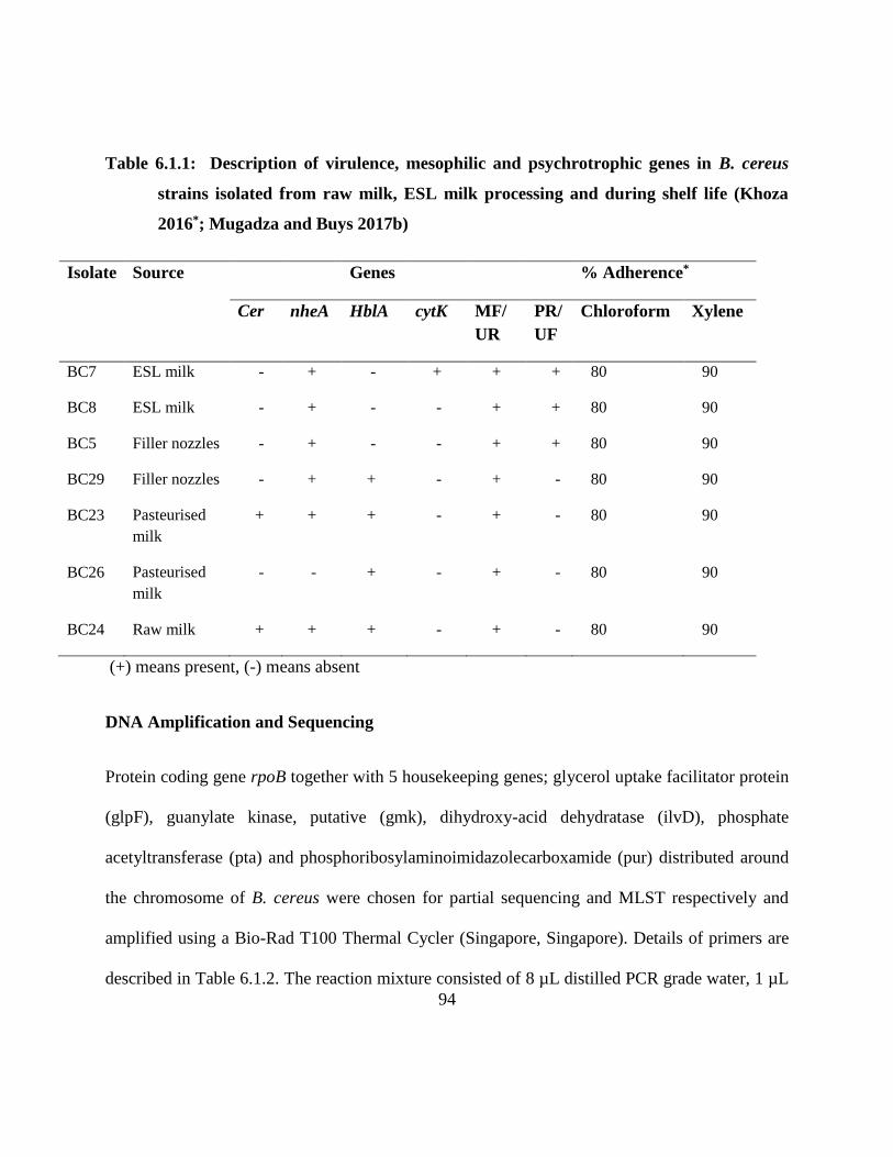
94
Table 6.1.1: Description of virulence, mesophilic and psychrotrophic genes in B. cereus
strains isolated from raw milk, ESL milk processing and during shelf life (Khoza
2016*; Mugadza and Buys 2017b)
Isolate Source Genes % Adherence*
Cer
nheA HblA cytK MF/
UR
PR/
UF
Chloroform Xylene
BC7 ESL milk - + - + + + 80 90
BC8 ESL milk - + - - + + 80 90
BC5 Filler nozzles - + - - + + 80 90
BC29 Filler nozzles - + + - + - 80 90
BC23 Pasteurised
milk
+ + + - + - 80 90
BC26 Pasteurised
milk
- - + - + - 80 90
BC24 Raw milk + + + - + - 80 90
(+) means present, (-) means absent
DNA Amplification and Sequencing
Protein coding gene rpoB together with 5 housekeeping genes; glycerol uptake facilitator protein
(glpF), guanylate kinase, putative (gmk), dihydroxy-acid dehydratase (ilvD), phosphate
acetyltransferase (pta) and phosphoribosylaminoimidazolecarboxamide (pur) distributed around
the chromosome of B. cereus were chosen for partial sequencing and MLST respectively and
amplified using a Bio-Rad T100 Thermal Cycler (Singapore, Singapore). Details of primers are
described in Table 6.1.2. The reaction mixture consisted of 8 µL distilled PCR grade water, 1 µL
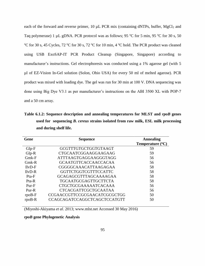
95
each of the forward and reverse primer, 10 µL PCR mix (containing dNTPs, buffer, MgCl2 and
Taq polymerase) 1 µL gDNA. PCR protocol was as follows; 95 oC for 5 min, 95 oC for 30 s, 50
oC for 30 s, 45 Cycles, 72 oC for 30 s, 72 oC for 10 min, 4 oC hold. The PCR product was cleaned
using USB ExoSAP-IT PCR Product Cleanup (Singapore, Singapore) according to
manufacturer’s instructions. Gel electrophoresis was conducted using a 1% agarose gel (with 5
µl of EZ-Vision In-Gel solution (Solon, Ohio USA) for every 50 ml of melted agarose). PCR
product was mixed with loading dye. The gel was run for 30 min at 100 V. DNA sequencing was
done using Big Dye V3.1 as per manufacturer’s instructions on the ABI 3500 XL with POP-7
and a 50 cm array.
Table 6.1.2: Sequence description and annealing temperatures for MLST and rpoB genes
used for sequencing B. cereus strains isolated from raw milk, ESL milk processing
and during shelf life.
Gene Sequence Annealing
Temperature (oC)
Glp-F GCGTTTGTGCTGGTGTAAGT 59
Glp-R CTGCAATCGGAAGGAAGAAG 59
Gmk-F ATTTAAGTGAGGAAGGGTAGG 56
Gmk-R GCAATGTTCACCAACCACAA 56
IlvD-F CGGGGCAAACATTAAGAGAA 58
IlvD-R GGTTCTGGTCGTTTCCATTC 58
Pta-F GCAGAGCGTTTAGCAAAAGAA 58
Pta-R TGCAATGCGAGTTGCTTCTA 58
Pur-F CTGCTGCGAAAAATCACAAA 56
Pur-R CTCACGATTCGCTGCAATAA 56
rpoB-F CCGAACCGTTCCGCGAACATCGCGCTGG 50
rpoB-R CCAGCAGATCCAGGCTCAGCTCCATGTT 50
(Miyoshi-Akiyama et al. 2013; www.mlst.net Accessed 30 May 2016)
rpoB gene Phylogenetic Analysis
96
Sequence analysis was performed at Inqaba Biotechnologies, Pretoria, South Africa and the
chromatograms from the ABI 3100 sequences were exported, visually examined and gene
sequences were analyzed using FinchTV version 1.4.0 (Geospiza). The rpoB gene sequences
were aligned using BIOEDIT version 7.2.1 (Hall 1999). Multiple sequence alignment was
performed using Clustal Omega (EMBL-EBI, Hinxton). A phylogenetic tree was inferred using
the neighbor-joining and maximum-likelihood algorithm in CLC Genomics workbench version
10.0.0 (Qiagen Bioinformatics, Aarhus). The strength of the internal branches of the
phylogenetic tree was statistically estimated by bootstrap analysis of 1000 bootstrap replications.
MLST Data analysis
The chromatograms from the ABI 3100 sequences were exported, visually examined and gene
sequences were edited using FinchTV version 1.4.0 (Geospiza). The sequences of the five
housekeeping genes (gmk, ilv, pur, pyc, tpi) were edited to the previous allele lengths (between
348 and 504 bp). The genes sequences were then assigned allele numbers based on the already
described alleles of B. cereus MLST database (http://www.pubmlst.org/cereus). Isolates were
assigned sequence type (ST) based on the combination of 5 alleles. The five gene fragments of
each of the 7 isolates were concatenated and downloaded from the MLST website. A
phylogenetic tree was derived using the neighbor joining method, with 1000 step bootstrap
analysis of the data using CLC Genomics Workbench version 10.0.0 (Qiagen Bioinformatics,
Aarhus).
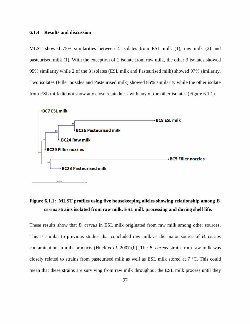
97
6.1.4 Results and discussion
MLST showed 75% similarities between 4 isolates from ESL milk (1), raw milk (2) and
pasteurised milk (1). With the exception of 1 isolate from raw milk, the other 3 isolates showed
95% similarity while 2 of the 3 isolates (ESL milk and Pasteurised milk) showed 97% similarity.
Two isolates (Filler nozzles and Pasteurised milk) showed 85% similarity while the other isolate
from ESL milk did not show any close relatedness with any of the other isolates (Figure 6.1.1).
Figure 6.1.1: MLST profiles using five housekeeping alleles showing relationship among B.
cereus strains isolated from raw milk, ESL milk processing and during shelf life.
These results show that B. cereus in ESL milk originated from raw milk among other sources.
This is similar to previous studies that concluded raw milk as the major source of B. cereus
contamination in milk products (Huck et al. 2007a,b). The B. cereus strain from raw milk was
closely related to strains from pasteurised milk as well as ESL milk stored at 7 oC. This could
mean that these strains are surviving from raw milk throughout the ESL milk process until they
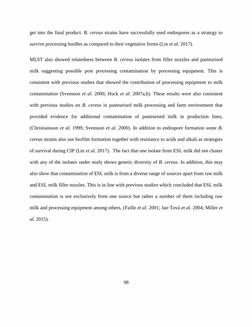
98
get into the final product. B. cereus strains have successfully used endospores as a strategy to
survive processing hurdles as compared to their vegetative forms (Lin et al. 2017).
MLST also showed relatedness between B. cereus isolates from filler nozzles and pasteurised
milk suggesting possible post processing contamination by processing equipment. This is
consistent with previous studies that showed the contribution of processing equipment to milk
contamination (Svensson et al. 2000; Huck et al. 2007a,b). These results were also consistent
with previous studies on B. cereus in pasteurised milk processing and farm environment that
provided evidence for additional contamination of pasteurised milk in production lines,
(Christiansson et al. 1999; Svensson et al. 2000). In addition to endospore formation some B.
cereus strains also use biofilm formation together with resistance to acids and alkali as strategies
of survival during CIP (Lin et al. 2017). The fact that one isolate from ESL milk did not cluster
with any of the isolates under study shows genetic diversity of B. cereus. In addition, this may
also show that contamination of ESL milk is from a diverse range of sources apart from raw milk
and ESL milk filler nozzles. This is in line with previous studies which concluded that ESL milk
contamination is not exclusively from one source but rather a number of them including raw
milk and processing equipment among others, (Faille et al. 2001; Jan·Tová et al. 2004; Miller et
al. 2015).
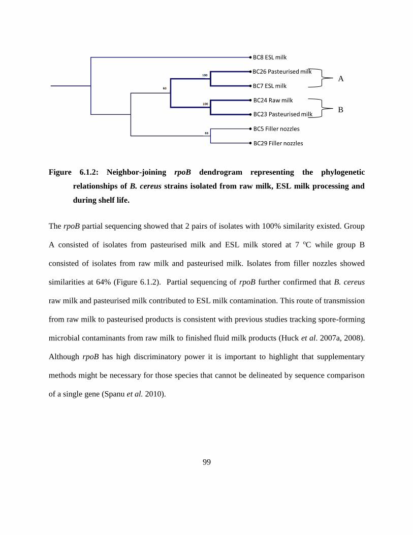
99
Figure 6.1.2: Neighbor-joining rpoB dendrogram representing the phylogenetic
relationships of B. cereus strains isolated from raw milk, ESL milk processing and
during shelf life.
The rpoB partial sequencing showed that 2 pairs of isolates with 100% similarity existed. Group
A consisted of isolates from pasteurised milk and ESL milk stored at 7 oC while group B
consisted of isolates from raw milk and pasteurised milk. Isolates from filler nozzles showed
similarities at 64% (Figure 6.1.2). Partial sequencing of rpoB further confirmed that B. cereus
raw milk and pasteurised milk contributed to ESL milk contamination. This route of transmission
from raw milk to pasteurised products is consistent with previous studies tracking spore-forming
microbial contaminants from raw milk to finished fluid milk products (Huck et al. 2007a, 2008).
Although rpoB has high discriminatory power it is important to highlight that supplementary
methods might be necessary for those species that cannot be delineated by sequence comparison
of a single gene (Spanu et al. 2010).
A
B
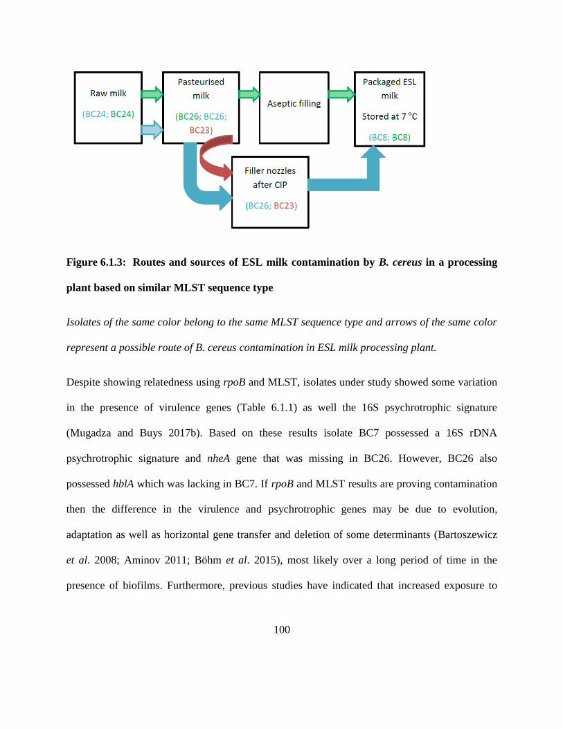
100
Figure 6.1.3: Routes and sources of ESL milk contamination by B. cereus in a processing
plant based on similar MLST sequence type
Isolates of the same color belong to the same MLST sequence type and arrows of the same color
represent a possible route of B. cereus contamination in ESL milk processing plant.
Despite showing relatedness using rpoB and MLST, isolates under study showed some variation
in the presence of virulence genes (Table 6.1.1) as well the 16S psychrotrophic signature
(Mugadza and Buys 2017b). Based on these results isolate BC7 possessed a 16S rDNA
psychrotrophic signature and nheA gene that was missing in BC26. However, BC26 also
possessed hblA which was lacking in BC7. If rpoB and MLST results are proving contamination
then the difference in the virulence and psychrotrophic genes may be due to evolution,
adaptation as well as horizontal gene transfer and deletion of some determinants (Bartoszewicz
et al. 2008; Aminov 2011; Böhm et al. 2015), most likely over a long period of time in the
presence of biofilms. Furthermore, previous studies have indicated that increased exposure to
101
cold environments leading to increased adaptation and evolution resulted in the emergence of
psychrotrophic B. cereus strains (Guinebretire et al. 2008).
6.1.6 Conclusions
B. cereus contamination of ESL milk maybe through raw milk and biofilms associated with filler
nozzles after CIP. The study also established three possible routes of B. cereus contamination in
ESL milk processing as shown in Figure 6.1.3. However, further work will be needed using more
isolates to get a more robust picture about these possible routes of B. cereus contamination in
ESL milk. In addition, the study has also shown that rpoB partial sequencing and MLST can be
used as a tool for source tracking in ESL milk processing.
102
CHAPTER SEVEN
GENERAL DISCUSSION
103
7.1 Methodological considerations
In this study spore-formers were isolated using a method previously described (Ivy et al. 2012)
in which samples are preheated at 80 oC for 10-12 min. This method is effective in reducing the
background flora and vegetative cells that may outcompete spore-formers thus reducing isolation
efficiency of spore-formers. Bacillus spp. is a non-competitive bacterium, hence they are
difficult to isolate in the presence of non-spore-formers such as Pseudomonas spp. The isolation
method has been successfully used in previous studies (Huck et al. 2007; Ranieri 2009; Ivy et al.
2012). While other scholars have used the filtration method (Svensson et al. 2003; Bartoszewicz
et al. 2008) to successfully harvest spores, we believe the method we used was the best for the
purpose of enumeration and further identification. The enumeration method was followed by
grouping colonies according to colour, morphology and size. Representative colonies were
purified by streaking on fresh nutrient agar. This technique has been used in previous studies
(Fromm and Boor 2004; Ranieri 2009) and has proved to be a cost effective way of successfully
screening isolates.
Purified isolates were confirmed as spore-forming bacteria by the Schaeffer-Fulton endospore
staining method previously described (Schaeffer and Fulton 1933; Hussey and Zayaitz 2012).
The method is based on the ability of a primary stain-malachite green to be forced into the
endospore by steaming the bacterial emulsion.
Malachite green is water soluble and has a low affinity for cellular material, so vegetative
cells may be decolourized with water (Vasanthakumari 2009). Safranin is then applied to
counterstain any cells which have been decolorized. At the end of the staining
104
process, vegetative cells will be pink, and endospores will be dark green. However, the only
challenge of this stain is that when staining Mycobacterium the bacteria may look positive for
endospores because some cells will stain green due to the its thick, wax coats (Hussey and
Zayaitz 2012).
Isolates were further identified as Gram positives using the KOH method described in previous
studies (Halebian et al. 1981; Buck 1982). This is a non-staining method that bases on the ability
of Gram negative bacterial cell wall to dissolve with 3% KOH and not in Gram positive cell
walls. Dissolved cell wall releases the intercellular material and the liberated cellular DNA
makes the mixture viscous or “stringy.” The positive string test indicates a Gram negative
organism. This method has been shown to be effective in previous studies (Halebian et al. 1981;
Buck 1982; Fromm and Boor 2004) and has time and cost advantages over the microscope based
Gram staining method.
To aid identification of the isolates, their pathogenicity was also examined. This was done by
determining haemolysis on 5% blood agar (Lucking et al. 2013). Blood agar, which is a mixture
of tryptic soy agar and sheep blood, allows differentiation of bacteria based on their ability to
haemolyse red blood cells. The haemolysin (extotoxin) radially diffuses outwards from the
colony causing complete or partial destruction of the red cells in the medium and complete
denaturation of haemoglobin within the cells to colourless products. Haemolysis method is semi-
quantitative and there are three major types of haemolysis namely beta haemolysis, alpha
haemolysis and gamma haemolysis (Sood 2006). Beta haemolysis is the complete destruction of
red blood cells and haemoglobin, and results in a clearing of the medium around the colonies.
105
The method has been used successfully for Bacillus spp. (Fromm and Boor 2004; Lucking et al.
2013).
Enzymes activity was also used in the identification process. Catalase test that determines the
ability to degrade H2O2 was done following procedures in previous studies (Koneman 2006).
Catalase is an enzyme, which is produced by microorganisms that live in oxygenated
environments to neutralize toxic forms of oxygen metabolites such as H2O2. The catalase
enzyme neutralises the bactericidal effects of H2O2 and protects them. Anaerobes generally lack
the catalase enzyme (Góth et al. 2004). Another enzyme that was used for identification as well
as to study the potential of the isolates to participate in ESL milk spoilage was lipase. Lipase is
an exoenzyme which allows the organisms that produce it to break down lipids into smaller
fragments of triglycerides, fatty acids and glycerol (Mobarak-Qamsari et al. 2011). The
tributyrin agar based method was used as previously described (Lucking et al. 2013). Despite
giving a true indication of the isolates with regards to lipase the method, however, cannot
quantify the amount and rate at which lipid hydrolysis can occur in a milk product. Several
methods have since been developed for quantification purposes (Thomson et al. 1999). However,
the method applied in this study was enough to fulfil the objective. Following lipase activity of
the isolates was determination of the protease activity in the isolates as described by Lucking et
al. (2013). The protease under study was casease an exoenzyme that is produced by some
bacteria in order to degrade casein. Casein is a large protein that is responsible for the white
color of milk. This test is conducted on milk agar which is a complex media containing casien,
peptone and beef extract. If an organism can produce casein, then there will be a zone of clearing
106
around the bacterial growth. Similar to the lipase activity test, the casease activity test is
qualitative.
MALDI-TOF-MS was used to characterise and identify the genus and species of the isolates
(Drevinek et al. 2012). MALDI-TOF-MS has been introduced in bacterial taxonomy (Van Baar
2000; Fenselau and Demirev 2001; Lay and Liyanage 2006) and successfully applied to a
number of taxa (Vargha et al. 2006; Barbuddhe et al. 2008; Grosse-Herrenthey et al. 2008). It
has the ability to measure peptides and other compounds in the presence of salts and to analyze
complex peptide mixtures, making it an ideal method for measuring non-purified extracts and
intact bacterial cells (De Bruyne et al. 2011). MALDI-TOF-MS relies on proteomic profiling of
highly conserved proteins generated from direct ionization of a colony of intact organisms or
bacterial protein extract, and correlates this spectral signature to a database of spectra collected
from reference strains (Kaleta et al. 2011). Different experimental factors, including sample
preparation, cell lysis method, matrix solutions and organic solvents, affect the quality and
reproducibility of bacterial MALDI-TOF-MS fingerprints (Ruelle et al. 2004; Vargha et al.
2006; Jaskolla et al. 2011). Also, differences in instrumental performance, mass range, and mass
resolution have profound effects on the obtained spectra (Wunschel et al. 2005; De Bruyne et al.
2011). Although MALDI-TOF-MS has proved to be an effective identification tool that has been
used to distinguish different strains of Escherichia coli (Holland et al. 1999) and also to identify
rare bacterial species implicated in human infectious diseases (Seng et al. 2013), for very
accurate results it must be accompanied by traditional identification methods such as
biochemical tests and API system among other as other studies have noted disparities of the
107
system on Staphylococcus aureus (Risch et al. 2010). The system has the potential to establish a
complementary tool for phenotypic analysis of microbial taxa, especially for physiologically
inert species such as Porphyromonas, Acinetobacter, Kingella, Moraxella and those such as
Bacillus species that are difficult to separate by rDNA analysis due to their evolutionary
relatedness (Shah et al. 2002; Keys et al. 2004). A further advantage of the system is that only a
loopful of cells is needed for MALDI-TOF-MS analysis and the profile is generated in minutes
with minimal consumables, cost and operational manipulation (Keys et al. 2004). The power of
MALDI-TOF-MS is also inseparable to its database hence it is important to create a microbial
database and standardise analysis protocols and pattern matching systems (Keys et al. 2004).
Pathogenicity of B. cereus was done by determining the presence of virulence genes. While B.
cereus has been linked to several toxins (Arnesen et al. 2008), in this study we focused on cytK,
cer, hblA and nheA that are described in a previous study (Swiecicka and Mahillon 2006). Cer is
the emetic toxin responsible for vomiting (Arnesen et al. 2008). Various methods have been used
to detect cer including; monkey feeding (Melling et al. 1976), cell culture assays (Hughes et al.
1988; Sakurai et al. 1994; Finlay et al. 1999), boar sperm biological assay (Hoornstra et al. 2003;
Anderson et al. 2004) and rat liver utilisation (Kawamura-Sato et al. 2005). These methods
however, do not specifically detect cer (Arnesen et al. 2008), hence the introduction of the
laborious and costly HPLC-MS (Häggblom et al. 2002). Since the genes encoding the production
of cer are restricted to emetic toxin producing strains (Ehling-Shulz et al. 2005; 2006) PCR
method has become relevant and ideal for identifying potential harmful strains. The PCR method
108
used in this study has been used with success in previous studies (Ehling-Shulz et al. 2004; 2005;
Swiecicka and Mahillon 2006; Fricker et al. 2007)
The three toxins that have mainly been implicated with the diarrheal disease are the pore-forming
cytotoxin haemolysin BL (Hbl), non-haemolytic enterotoxin (Nhe) and cytotoxin K (cytK) (Lund
et al. 2000; Arnesen et al. 2008). Although antibodies based detection kits have been developed
for detection of the three-component toxins Nhe and Hbl (Buchanan and Schultz 1994; Day et al.
1994) none of the methods will confirm an active toxin as only one of the three components of
the toxin is targeted. Currently there is no antibody based assay for the cytK (Arnesen et al.
2008). For non-specific detection and characterisation of B. cereus enterotoxins different
laboratory animal and tissue assays have been developed (Buchanan and Schultz 1994; Hardy et
al. 2001; Fletcher and Logan 1999; From et al. 2005). Although the detection of an encoding
gene does not translate to expression or production of the toxin PCR is still widely used to give
an indication of potential hazard in food. Several PCR assays for enterotoxins have been
developed and have been useful in identifying potential food safety threat (Hansen and
Hendriksen 2001; Guinebretière et al. 2002; Ehling-Shulz et al. 2005). In this study we chose
similar PCR assay that were previously described (Hansen and Hendriksen 2001; Fagerlund et
al. 2004; Swiecicka and Mahillon 2006) because it is the best method available to accurately
indicate potential food safety threat of B. cereus regarding enterotoxins.
To discriminate between psychrotrophic and mesophilic B. cereus, a PCR assay that targets the
16S signature (von Stetten et al. 1998) was used. Studies have indicated that ribosomes (Condon
et al. 1995; Graumann et al. 1996) and major cold shock proteins play a role in cold adaptation
109
of bacteria. In addition traditional microbiology relied on growth at 7 oC for detecting
psychrotolerant strains (von von Stetten et al. 1998) which may take up to 14 days to be detected
(Meer et al. 1991). PCR based assays have been developed targeting the cold shock protein cspA
(Francis et al. 1998) and the 16S signature (von Stetten et al. 1998). Although the presence of a
psychrotrophic 16S signature does not necessarily translate to phenotypic expression, it however
gives an indication of what potential the strain has when subjected to favourable environmental
conditions.
In this study rep-PCR was done using (GTG)5 primer because it is an easy to perform method
basing on primers complementary to certain repetitive sequences dispersed in bacterial genomes
(Lupski and Weinstock 1992). In addition PCR amplification of repetitive bacterial DNA
elements (rep-PCR) has been recognized as a simple PCR-based technique with a high
discriminatory power, low cost, suitable for a high-throughput of strains, and considered to be a
reliable tool for classifying and typing a wide range of Gram-negative and several Gram-positive
bacteria (Olive and Bean 1999; Gevers et al. 2001; Švec et al. 2008). Apart from the labour
intensive techniques of discrimination such as 16S sequencing, randomly amplified polymorphic
DNA (RAPD) fingerprinting is by far the most used PCR-based genomic technique for
identification of bacteria (Khaled et al. 1997; Gevers et al. 2001). However, RAPD primers are
not directed against a particular genetic locus, the resulting band patterns often exhibit poor
reproducibility (Olive and Bean 1999) hence (GTG)5 becomes a better method. It is frequently
used in bacterial taxonomy and has been successfully applied for reliable and fast identification
of different bacterial groups such as Lactobacilli (Gevers et al. 2001), Staphylococci (Wieser and
110
Busse 2000), Mycobacteria (Cangelosi et al. 2004), Streptomycetes (Lanoot et al. 2004) and S.
mutans (Švec et al. 2008). Although the homogeneity of B. licheniformis in previous studies
basing on (GTG)5 raised questions on the discriminatory power of (GTG)5-PCR (De Clerck and
De Vos 2003), another study showed that it is a high discriminatory tool in B. cereus
(Samapundo et al. 2011).
16S rDNA partial sequencing was used to confirm the identity of B. cereus as well as determine
the relatedness among isolates from filler nozzles and ESL milk processing. Although it has been
emphasised that 16S rDNA data may not be useful in defining species and strains especially in B.
cereus group (Fox et al. 1992), it is still, in most instances, a powerful tool for determining to
which species a strain probably belongs once the relevant species are represented in the 16S
rDNA sequence data base. This molecular approach was used as it has been extensively used
with success for bacterial phylogeny (Woese et al. 1990), leading to the establishment of large
public domain databases (Maidack et al. 1996; Drancourt et al. 2000) and its application to
bacterial identification, including that of environmental and clinical uncultured microorganisms
(Relman et al. 1992; Strous et al. 1999), unique or unusual isolates and collections of
phenotypically identified isolates (Tang et al. 1998, 2000; Drancourt et al. 2000). 16S rDNA
based molecular identification could achieve identification, for reasons including its universal
distribution among bacteria and the presence of species-specific variable regions. In a previous
study the overall performance of 16S rDNA sequence analysis proved excellent, since it was able
to resolve almost 90% of identifications, when applied to a large collection of phenotypically
unidentifiable bacterial isolates (Drancourt et al. 2000). Although the downside of this technique
111
is failure to discriminate between species of the B. cereus group, it was useful in confirming the
identity as well as determining the relatedness of the isolates under study. A previous study
found the 16S rDNAs of B. anthracis, B. cereus, B. mycoides, and B. thuringiensis to have
almost complete sequence identity and only two differences were found in the 23S rDNAs of B.
anthracis and B. cereus (Ash et al. 1991).
Sequencing the protein coding gene rpoB was done as a way of confirming the identity of B.
cereus isolates as well as source tracking the organism. While 16S rDNA gene sequencing has
been used as a framework for modern bacterial classification, it often shows limited variation for
members of closely related taxa (Fox et al. 1992; Janda and Abbort 2007). The differences that
do exist among B. anthracis, B. cereus, and B. thuringiensis are due largely to the presence of
plasmids (McDowell and Mann 1991; Ombui et al. 1996; Yang et al. 2003). However, plasmids
may be lost, making it difficult to differentiate rapidly among species (Hurtle et al. 2004). On the
other hand, protein-coding genes exhibit much higher genetic variation, which can be used for
classification and identification of closely related taxa (Mollet et al. 1997; Kim et al. 1999;
Yamamoto et al. 1999; Chun and Bae 2000). The gene for the RNA polymerase beta subunit,
rpoB, is suggested to be used as an alternative to 16S rDNA in species identification as it
provides a much more efficient tool for molecular subtype analysis (Rantsiou et al. 2004; Huck
et al. 2008; Ki et al. 2009). The rpoB gene has been reported to provide improved phylogenetic
resolution over the 16S rDNA gene. This is essential when studying subspecies diversity in
which analysis of the rpoB single copy gene could be used to complement the information
gathered from the 16S rDNA gene (Adékambi et al. 2009). Several studies have indicated that
112
rpoB gene can be successfully used for identification at the species or subspecies level of
bacteria (Adékambi et al. 2003; Khamis et al. 2009). This was therefore one of the best methods
to use in this research.
To assist in source tracking, B. cereus was analysed using the multilocus sequencing technique
that focuses on sequencing housekeeping genes (Urwin and Maiden 2003). It is based on
concepts of multilocus enzyme electrophoresis (MLEE) and has adapted them so that alleles at
each locus are defined directly, by nucleotide sequencing, rather than indirectly from the
electrophoretic mobility of their gene products (http://www.mlst.net). The aim of MLST is to
provide a portable, accurate, and highly discriminating typing system that can be used for most
bacteria and some other organisms (Maiden 2006). Most MLST schemes are based on
housekeeping genes, which are subject to purifying selection and slow evolution, and the
variation within these genes is nearly neutral (Aanensen and Spratt 2005). Although there are
normally fewer polymorphic sites in individual housekeeping genes compared with hyper
variable genes, using the combined sequences of multiple housekeeping genes has been shown to
provide high discriminatory power while retaining signatures of longer-term evolutionary
relationships or clonal stability (Urwin and Maiden 2003; Pérez-Losada et al. 2013).
Furthermore, analyses of multiple loci can buffer against potentially skewed evolutionary
pictures obtained by single-locus analyses (Enright and Spratt 1999). Current applications of
MLST include examination of the evolution of virulence, population and evolutionary analyses
and isolate characterization and population structure analyses (Maiden 2006). Bacterial
subtyping methods not only improve our ability to detect and track pathogen outbreaks, but also
113
provide tools to track sources of pathogens contamination throughout the food system. It also
provides an opportunity to better understand the population genetics, epidemiology, and ecology
of bacteria (Enright and Spratt 1999; Pérez-Losada et al. 2013). These features of MLST make it
relevant to this section of the study where the objective was to source track B. cereus in an ESL
milk processing plant. MLST has been used for a number of bacteria that includes B. cereus,
(Helgason et al. 2004), Staphylococcus spp. (Thomas et al. 2007; Enright et al. 2000), E. coli
(Gordon et al. 2008), Streptococcus spp. (Ahmad et al. 2009) and Salmonella spp. (Kotetishvili
et al. 2002) among others.
7.2 Isolation of spore-formers in ESL milk and characterisation of B. cereus from ESL
milk processing and during storage
The ESL milk process under study reduced vegetative bacterial counts significantly. This was
also aided by the fact that the raw milk used also showed satisfactory microbial quality according
to most international dairy standards (Marth and Steele 2001). Previous studies indicate that
pasteurisation temperatures are sufficient to completely destroy vegetative bacteria if raw milk
has good microbial quality (Ranieri and Boor 2009; Tammine 2009). Apart from reducing the
vegetative population the process also reduced the spore-former population before packaging.
The reduction of spore-forming bacteria is a combination of the pasteurisation effect on their
vegetative form as well as bactofugation. Bactofugation has proven to be effective in reducing
spore-formers and is believed to reduce 90-99% of bacterial cells and clostridal spores which
cause late blowing of Swiss cheese (Fox and McSweeney 1998; Faccia et al. 2013). When
isolates were identified by MALDI-TOF-MS it showed that apart from reducing the counts, the
114
process also reduced the diversity of organisms. This can be attributed to the inability of other
spore-formers to continue with normal metabolic processes under cold storage. Previous studies
have indicated that the ability to produce cold shock proteins (CSPs) enables organisms to
protect themselves from cold conditions and hence continue with normal metabolic processes
(Francis et al. 1998). Another study has also indicated that the presence of the psychrotrophic
16S signature plays a similar role too (von Stetten et al. 1998).
Although B. pumilus has not been reported as a psychrotroph, its dominance in the ESL milk
process as well as during storage can be attributed to its resistance to a number of stresses
including UV light exposure and presence of oxidisers such as H2O2 among others as well as the
ubiquitous nature of its endospore (Benardini et al. 2003; Link et al. 2004; Dickinson et al.
2004). The dominance of B. pumilus even in ESL milk stored at 4 oC suggests possible presence
of psychrotrophic features within the species. Previous studies have indicated the presence of
mesophilic and psychrotrophic 16S signatures, a property which may allow mesophilic
organisms to adapt well at low temperatures. Although no horizontal gene transfer has been
reported in that regard it however, cannot be ruled out as a possibility. The presence of
Paenibacillus spp. is in line with previous studies that have described it as a psychrotrophic
microaerophile that dominate the spore-former population at the end of shelf life (Owayss 2007;
Ranieri et al. 2012). Paenibacillus spp. had the highest growth rate compared to its Bacillus spp.
competitors when inoculated as single organisms in sterile milk and it also dominated the
cocktail composition probably because of its ability to be competitive against accompanying
microbiota at refrigerated conditions. Paenibacillus spp. is also reported to require L-tryosine as
one of its spore germinants (Alvarado et al. 2012), while the proteolytic activity of B. cereus
115
increases the amount of free tryosine in milk (Jan·tová 2006), thus aiding the growth of
Paenibacillus spp.
Although the presence of both proteolytic and lipolytic enzymes does not directly translate to the
ability to spoil milk, it is still an indication of the potential to cause spoilage, considering that
more than 70% of isolates under study were proteolytic. Bacillus spp. and Paenibacillus spp.
have been implicated as the major hurdles to shelf life extension of milk because of their ability
to participate in hydrolysis of milk components such as lipids and proteins at refrigeration
temperatures, (Boor et al. 1998; De Jonghe et al. 2010). B. pumilus has greatest potential to
cause spoilage because it also had the highest percentage of isolates that were positive for
proteolytic activity apart from dominating the spore-forming bacteria population in the whole
ESL milk processes and during cold storage. While all B. cereus strains exhibited mesophilic
characteristics by showing the presence of the 16S mesophilic signature the psychrotrophic
signature was not widely distributed. This shows that B. cereus spoilage and possibly poisoning
will greatly depend on storage practices exercised on the ESL milk.
Although the B. cereus population was low in this study, it remains a relevant bacterium in ESL
milk as it showed the ability to hydrolyse casein as well as lipids. Furthermore, it was the only
spore-former to be positive for haemolysis, exhibiting β haemolysis on 5% sheep blood agar.
This shows the organism has the ability to produce toxins that target the red blood cells.
However, a previous study has shown that the production of toxins is not only dependent on the
strain but also the substrate and temperature among others (Apetroaie-Constantin et al. 2008).
PCR results revealed that 3 toxin production genes were present in > 65% of the B. cereus under
study; these results confirmed the previously described potential of B. cereus to cause
116
gastrointestinal diseases (Hansen and Hendriksen 2001; Guinebretière and Broussolle 2002;
Ehling-Schulz et al. 2006b). Our results concurred with previous studies that reported that all
cereulide positive isolates were mesophilic although a recent study has shown that 2 isolates
belonging to the psychrotolerant species B. weihenstephanensis were able to produce cereulide at
8 oC, (Thorsen et al. 2006). Contrary to previous reports which hypothesise that many food-
borne strains of B. cereus are less prone to cause diarrhoea (Guinebretière and Broussolle 2002),
our study showed high prevalence of the enterotoxin genes hblA and nheA showing that the ESL
milk product consumers maybe at potential risk of the diarrheal causing toxins. Although cytK is
not widley distributed in B. cereus despite being regarded as highly toxic to humans (Hardy et al.
2001; Fagerlund et al. 2004), it still remains important as this was present in psychrotophic
strains too.
The increase in the spore-former population after packaging can be attributed to post-process
contamination. While raw milk has always been implicated as the major source of contamination
in modern dairy processing (Miller et al. 2015), research has also shown that processing
equipment also contribute (Eneroth et al. 2001). MALDI-TOF-MS dendrograms showed high
similarities within species (B. pumilus, B. subtilis, B. cereus and Paenibacillus spp.) that
suggested ESL milk contamination from both raw milk and ESL milk filler nozzles. Further
analysis on B. cereus using rep-PCR fingerprinting, 16S rDNA and rpoB partial sequencing as
well as MLST confirmed the role of biofilms from ESL milk filler nozzles on contamination of
ESL milk. This was in line with previous studies that suggested that milk contamination is not
exclusively from one source but rather a number of them including raw milk and processing
equipment among others, (Faille et al. 2001; Jan·Tová et al. 2004; Miller et al. 2015).
117
7.3 Future research
The study determined the dominating spore forming bacteria in ESL milk processing and during
storage. Although B. pumilus showed the highest prevalence throughout the process and during
storage followed by B. subtilis no further characterisation was done on the organisms to ascertain
their contribution to ESL milk spoilage. It is therefore necessary to characterise B. pumilus and
B. subtilis both at the genetic and phenotypic levels. Characterisation techniques to be used
should also have potential to source-track the organisms as well as determine their
psychrotrophic characteristics because some of the isolates were enumerated in milk stored at 4
and 7 oC. MALDI-TOF-MS revealed a close relationship between isolates from ESL milk
process, during storage and ESL milk filler nozzles. The potential of B. pumilus and B. subtilis to
attach and form biofilms on milking equipment that subsequently contaminate ESL milk must be
investigated.
The presence of potentially psychrotrophic B. cereus shows that ESL milk can be a potential
threat to consumers. It is therefore necessary to conduct a quantitative risk analysis of B. cereus
in ESL milk so as to determine the level of risk posed by the product to consumers. Although
source tracking of B. cereus was conducted within the ESL milk processing plant, it is necessary
to use all the B. cereus obtained from this plant to have a better understanding of the
contamination sources in this ESL milk processing plant. Paenibacillus spp. did not dominate the
organisms isolated in the ESL milk process under study; nevertheless, it showed the highest
growth rate and also outnumbered other major spore-formers when inoculated in sterile milk as a
cocktail of microorganisms in approximately equal quantities. It is therefore ideal to study the
118
mechanism that allows it to outnumber other spore-formers as milk ages as well as determining
its ability to participate in biofilm formation and subsequently spoil milk.
The spore-formers isolated in this ESL milk process under study do not exist in isolation but as a
community of microorganisms. While filler nozzles have been shown to participate in
contamination and most studies have focussed on attachment of single organisms on processing
equipment, it is also important to study the attachment and possible biofilm formation of these
spore-formers as a community as well as their release rate at any particular time.
119
CHAPTER EIGHT
CONCLUSIONS AND RECOMMENDATIONS
120
This study showed that apart from reducing the spore-forming population the ESL milk process
reduces the diversity of spore-forming species. It also showed that B. pumilus is the dominating
spore-former under study as well as during storage at 4 and 7 oC. While Paenibacillus spp. was
isolated in very low quantities both in ESL processing and during storage it was also revealed
that it dominates the spore-formers as milk ages during storage if the initial concentration is
equal to other spore-formers.
B. pumilus, B. subtilis, B. cereus and Paenibacillus spp. strains in this study possessed at least
one hydrolytic enzyme showing potential to cause spoilage of ESL milk. Although isolated in
low levels, B. cereus is a safety threat as all isolates showed ability to participate in haemolysis
and also contained at least one virulence gene. Although all B. cereus isolates were mesophilic,
the presence of a psychrotrophic signature in some isolates further increases the safety threat of
B. cereus in ESL milk
Despite MALDI-TOF-MS analysis showing great diversity within each spore-forming species,
very close relatedness existed within each species especially in B. pumilus as well as B. cereus.
A further repPCR based analysis on B. cereus isolates showed great diversity, as well as a very
close relationship among isolates. The relatedness among isolates suggests that both raw milk
and filler nozzles are contaminating ESL milk product with B. cereus.
Partial sequencing of rpoB and 16S rDNA as well as MLST further confirmed that raw milk and
filler nozzles are contaminating ESL milk. The study also established three possible routes of B.
cereus contamination in ESL milk. Furthermore, the study showed that MLST and rpoB can be
used for source tracking spore-formers in ESL milk processing.
121
CHAPTER NINE
REFERENCES
122
Aanensen, D.M. and Spratt, B.G. 2005. The multilocus sequence typing network:
mlst.net. Nucleic Acids Research, 33(suppl 2), W728-W733.
Abel-Santos, E. 2012. Bacterial spores: Current research and applications. Caister Academic
Press. Norfolk, UK. p 9-13.
Adékambi, T., Colson, P. and Drancourt, M. 2003. rpoB-based identification of non-pigmented
and late-pigmenting rapidly growing mycobacteria. Journal of Clinical
Microbiology, 41(12), 5699-5708.
Adékambi, T., Drancourt, M. and Raoult, D. 2009. The rpoB gene as a tool for clinical
microbiologists. Trends in Microbiology, 17(1), 37-45.
Agata, N., Mori, M., Ohta, M., Suwan, S., Ohtani, I. and Isobe, M. 1994. A novel
dodecadepsipeptide, cereulide, isolated from Bacillus cereus causes vacuole formation in
HEp-2 cells. FEMS Microbiology Letters, 121(1), 31-34.
Agata, N., Ohta, M. and Yokoyama, K. 2002. Production of Bacillus cereus emetic toxin
(cereulide) in various foods. International Journal of Food Microbiology, 73(1), 23-27.
Ahmad, Y., Gertz, R.E., LI, Z., Sakota, V., Broyles, L.N., Van beneden, C., Facklam, R.,
Shewmaker, P.L., Reingold, A., Farley, M.M. and Beall, B.W. 2009. Genetic
relationships deduced from emm and multilocus sequence typing of invasive
Streptococcus dysgalactiae subsp. equisimilis and S. canis recovered from isolates
collected in the United States. Journal of Clinical Microbiology, 47(7), 2046-2054.
123
Aijuka, M., Charimba, G., Hugo, C.J. and Buys, E.M. 2015. Characterization of bacterial
pathogens in rural and urban irrigation water. Journal of Water and Health, 13(1), 103-
117.
Al-Abri, S.S., Al-Jardani, A.K., Al-Hosni, M.S., Kurup, P.J., Al-Busaidi, S. and Beeching, N.J.
2011. A hospital acquired outbreak of Bacillus cereus gastroenteritis, Oman. Journal of
Infection and Public Health, 4(4), 180-186.
Altayar, M. and Sutherland, A.D. (2006). Bacillus cereus is common in the environment but
emetic toxin producing isolates are rare. Journal of Applied Microbiology, 100(1), 7-14.
Alvarado, I., Phui, A., Elekonich, M.M. and Abel-Santos, E. 2012. Requirements for in vitro
germination of Paenibacillus larvae spores. Journal of Bacteriolology, 195(5), 1005-
1011.
Aminov, R.I. 2011. Horizontal gene exchange in environmental microbiota. Frontiers in
Microbiology, 2, 158.
Andersson, M.A., Jääskeläinen, E.L., Shaheen, R., Pirhonen, T., Wijnands, L.M. and Salkinoja-
Salonen, M.S. 2004. Sperm bioassay for rapid detection of cereulide-producing Bacillus
cereus in food and related environments. International Journal of Food
Microbiology, 94(2), 175-183.
124
Ansari, I. A. and Datta, A. K. 2003. An overview of sterilization methods for packaging
materials used in aseptic packaging systems. Institution of Chemical Engineers, 81, 57-
65.
Aouadhi, C., Maaroufi, A. and Mejri, S. 2014. Incidence and characterisation of aerobic
spore‐forming bacteria originating from dairy milk in Tunisia. International Journal of
Dairy Technology, 67, 95-102.
Apetroaie-Constantin, C., Shaheen, R., Andrup, L., Smidt, L., Rita, H. and Salkinoja-Salonen,
M. 2008. Environment driven cereulide production by emetic strains of Bacillus
cereus. International Journal of Food Microbiology,127(1), 60-67.
Arnesen, L.P.S., Fagerlund, A. and Granum, P.E. 2008. From soil to gut: Bacillus cereus and its
food poisoning toxins. FEMS Microbiology Reviews, 32(4), 579-606.
Arnesen, L.P.S., O'sullivan, K. and Granum, P.E. 2007. Food poisoning potential of Bacillus
cereus strains from Norwegian dairies. International Journal of Food
Microbiology, 116(2), 292-296.
Ash, C., Farrow, J.A., Dorsch, M., Stackebrandt, E. and Collins, M.D. 1991. Comparative
analysis of Bacillus anthracis, Bacillus cereus, and related species on the basis of reverse
transcriptase sequencing of 16S rRNA. International Journal of Systematic and
Evolutionary Microbiology, 41(3), 343-346.
125
Ash, C., Priest, F.G. and Collins, M.D. 1993. Molecular identification of rRNA group 3 bacilli
using a PCR probe test. Antonie van Leeuwenhoek, 64(3-4), 253-260.
Banykó, J. and Vyletělová, M. 2009. Determining the source of Bacillus cereus and Bacillus
licheniformis isolated from raw milk, pasteurized milk and yoghurt. Letters in Applied
Microbiology, 48(3), 318-323.
Barbuddhe, S.B., Maier, T., Schwarz, G., Kostrzewa, M., Hof, H., Domann, E., Chakraborty, T.
and Hain, T. 2008. Rapid identification and typing of Listeria species by matrix-assisted
laser desorption ionization-time of flight mass spectrometry. Applied and Environmental
Microbiology, 74(17), 5402-5407.
Barker, M., Thakker, B. and Priest, F.G. 2005. Multilocus sequence typing reveals that Bacillus
cereus strains isolated from clinical infections have distinct phylogenetic origins. FEMS
Microbiology Letters, 245, 179-184.
Bartlett, J.M. and Stirling, D. 2003. PCR protocols (Vol. 226). Humana Press. Totowa.
Bartoszewicz, M., Hansen, B.M. and Swiecicka, I. 2008. The members of the Bacillus cereus
group are commonly present contaminants of fresh and heat-treated milk. Food
Microbiology, 25(4), 588-596.
Beecher, D.J. and Macmillan, J.D. 1990. A novel bicomponent hemolysin from Bacillus
cereus. Infection and Immunity, 58(7), 2220-2227.
126
Beecher, D.J. and Wong, A.C. 1994. Improved purification and characterization of hemolysin
BL, a hemolytic dermonecrotic vascular permeability factor from Bacillus
cereus. Infection and Immunity, 62(3), 980-986.
Beecher, D.J. and Wong, A.C. 1997. Tripartite Hemolysin BL from Bacillus cereus hemolytic
analysis of component interactions and a model for its characteristic paradoxical zone
phenomenon. Journal of Biological Chemistry, 272(1), 233-239.
Beecher, D.J. and Wong, A.C. 2000. Tripartite haemolysin BL: isolation and characterization of
two distinct homologous sets of components from a single Bacillus cereus
isolate. Microbiology, 146(6), 1371-1380.
Beecher, D.J., Pulido, J.S., Barney, N.P. and Wong, A.C. 1995a. Extracellular virulence factors
in Bacillus cereus endophthalmitis: methods and implication of involvement of
hemolysin BL. Infection and Immunity, 63(2), 632-639.
Beecher, D.J., Schoeni, J.L. and Wong, A.C. 1995b. Enterotoxic activity of hemolysin BL from
Bacillus cereus. Infection and Immunity, 63(11), 4423-4428.
Benardini, J.N., Sawyer, J., Venkateswaran, K. and Nicholson, W.L. 2003. Spore UV and
acceleration resistance of endolithic Bacillus pumilus and Bacillus subtilis isolates
obtained from Sonoran desert basalt: implications for lithopanspermia. Astrobiology, 3,
709-717.
Bergey, D.H. 2009. Bergey’s Manual of Systematic Bacteriology 2nd Ed Vol 3, Springer
127
Böhm, M.E., Huptas, C., Krey, V.M. and Scherer, S. 2015. Massive horizontal gene transfer,
strictly vertical inheritance and ancient duplications differentially shape the evolution of
Bacillus cereus enterotoxin operons hbl, cytK and nhe. BMC Evolutionary
Biology, 15(1), 246.
Bottone, E.J. 2010. Bacillus cereus, a volatile human pathogen. Clinical Microbiology
Reviews, 23(2), 382-398.
Brillard, J. and Lereclus, D. 2004. Comparison of cytotoxin cytK promoters from Bacillus cereus
strain ATCC 14579 and from a B. cereus food-poisoning strain. Microbiology, 150(8),
2699-2705.
Britz, T.J. and Robinson, R.K. 2008. Advanced dairy science and technology. Blackwell
Publishing. UK. p 13-14.
Buchanan, R.L. and Schultz, F.J. 1994. Comparison of the Tecra VIA kit, Oxoid BCET‐RPLA
kit and CHO cell culture assay for the detection of Bacillus cereus diarrhoeal
enterotoxin. Letters in Applied Microbiology, 19(5), 353-356.
Buck, J.D. 1982. Nonstaining (KOH) method for determination of gram reactions of marine
bacteria. Applied and Environmental Microbiology, 44, 992-993.
Buckenhüskes. 2014. DLG-Expert report - ESL milk . http://www.dlg.org/fileadmin/downloads
/food/Expertenwissen/Ernaehrung/e_2014_4_Expertenwissen_ESL.pdf. Accessed 03
December 15.
128
Budde, I., Steil, L., Scharf, C., Völker, U. and Bremer, E. 2006. Adaptation of Bacillus subtilis to
growth at low temperature: a combined transcriptomic and proteomic
appraisal. Microbiology, 152(3), 831-853.
Burgess, S.A., Lindsay, D. and Flint, S.H. 2010. Thermophilic bacilli and their importance in
dairy processing. International Journal of Food Microbiology, 144(2), 215-225.
Burton, H. 1988. Ultra High Temperature Processing of Milk and Milk
Products. Elsevier Applied Science. Barking, UK
Cangelosi, G.A., Freeman, R.J., Lewis, K.N., Livingston-Rosanoff, D., Shah, K.S., Milan, S.J.
and Goldberg, S.V. 2004. Evaluation of a high-throughput repetitive-sequence-based
PCR system for DNA fingerprinting of Mycobacterium tuberculosis and Mycobacterium
avium complex strains. Journal of Clinical Microbiology, 42(6), 2685-2693.
Caplan, Z. and Barbano, D.M. 2013. Shelf life of pasteurized microfiltered milk containing 2%
fat. Journal of Dairy Science, 96, 8035-8046.
Cardazzo, B., E. Negrisolo, L. Carraro, L. Alberghini, T. Patarnello, and V. Giaccone. 2008.
Multiple-locus sequence typing and analysis of toxin genes in Bacillus cereus food-borne
isolates. Applied and Environmental Microbiology, 74(3), 850-860.
Carlin, F., Fricker, M., Pielaat, A., Heisterkamp, S., Shaheen, R., Salonen, M.S., Svensson, B.,
Nguyen-the, C. and Ehling-Schulz, M. 2006. Emetic toxin-producing strains of Bacillus
129
cereus show distinct characteristics within the Bacillus cereus group. International
Journal of Food Microbiology, 109(1), 132-138.
Chambers, J.V. 2005. The microbiology of raw milk. Dairy microbiology handbook: the
microbiology of milk and milk products. John Wiley and Sons. Hoboken, NJ, USA. p 39-
90.
Chan, M.S., Maiden, M.C. and Spratt, B.G. 2001. Database-driven multi locus sequence typing
(MLST) of bacterial pathogens. Bioinformatics, 17, 1077-83
Christiansson, A., Bertilsson, J. and Svensson, B. 1999. Bacillus cereus spores in raw milk:
factors affecting the contamination of milk during the grazing period. Journal of Dairy
Science, 82(2), 305-314.
Chun, J. and Bae, K.S. 2000. Phylogenetic analysis of Bacillus subtilis and related taxa based on
partial gyrA gene sequences. Antonie van Leeuwenhoek, 78(2), 123-127.
Condon, C., Liveris, D., Squires, C., Schwartz, I. and Squires, C.L. 1995. rRNA operon
multiplicity in Escherichia coli and the physiological implications of rrn
inactivation. Journal of Bacteriology, 177(14), 4152-4156.
Coorevits, A., De Jonghe, V., Vandroemme, J., Reekmans, R., Heyrman, J., Messens, W. and
Heyndrickx, M. 2008. Comparative analysis of the diversity of aerobic spore-forming
bacteria in raw milk from organic and conventional dairy farms. Systematic and Applied
Microbiology, 31, 126-140.
130
Cowan, A.E., Koppel, D.E., Setlow, B. and Setlow, P. 2003. A soluble protein is immobile in
dormant spores of Bacillus subtilis but is mobile in germinated spores: implications for
spore dormancy. Proceedings of the National Academy of Sciences, 100(7), 4209-4214.
Daane, L.L., Harjono, I, Barns S.M, Launen L.A, Palleroni, N.J. and Haggblom, M.M. 2002.
PAH-degradation by Paenibacillus spp. and description of Paenibacillus
naphthalenovorans sp. nov., a naphthalene-degrading bacterium from the rhizosphere of
salt marsh plants. International Journal of Systematic and Evolutionary Microbiology,
52, 131-139.
Day, T.L., Tatani, S.R., Notermans, S. and Bennett, R.W. 1994. A comparison of ELISA and
RPLA for detection of Bacillus cereus diarrhoeal enterotoxin. Journal of Applied
Bacteriology, 77(1), 9-13.
De Bruyne, K., Slabbinck, B., Waegeman, W., Vauterin, P., De Baets, B. and Vandamme, P.
2011. Bacterial species identification from MALDI-TOF mass spectra through data
analysis and machine learning. Systematic and Applied Microbiology, 34(1), 20-29.
De Clerck, E. and De Vos, P. 2004. Genotypic diversity among Bacillus licheniformis strains
from various sources. FEMS Microbiology Letters, 231(1), 91-98.
De Hoon, M.J., Eichenberger, P. and Vitkup, D. 2010. Hierarchical evolution of the bacterial
sporulation network. Current Biology, 20(17), R735-R745.
131
De Jonghe, V., Coorevits, A., De Block, J., Van Coillie, E., Grijspeerdt, K., Herman, L. and
Heyndrickx, M, 2010. Toxinogenic and spoilage potential of aerobic spore-formers
isolated from raw milk. International Journal of Food Microbiology, 136, 318-325.
De Vos, P., Garrity, G.M., Jones, D., Krieg, N.R., Ludwig, W., Rainey, F.A., Schleifer, K. and
Whitman, W.B. 2009. Bergey’s Manual of Systematic Bacteriology Vol. 3. The
Firmicutes. Springer. New York. p 21.
Deák, T. and Farkas, J. 2013. Microbiology of thermally preserved foods: canning and novel
physical methods. DEStech Publications. Pennsylvania. p 274.
Department of Agriculture Forestry and Fisheries (DAFF). 1972. Foodstuffs, cosmetics and
disinfectants Act 54 of 1972. Republic of South Africa.
Dhillon, M. 2012. ‘The effect of silver ion-implantation of stainless steel on bacterial adhesion
and biofilm formation.’ MSc. dissertation, Department of Microbiology, Massey
University. p 33-55.
Dickinson, D.N., La Duc, M.T., Satomi, M., Winefordner, J.D., Powell, D.H. and
Venkateswaran, K. 2004. MALDI-TOF-MS compared with other polyphasic taxonomy
approaches for the identification and classification of Bacillus pumilus spores. Journal of
Microbiological Methods, 58, 1-12.
Didelot, X., Bowden. R., Wilson, D.J., Peto, T.E., Crook, D.W. 2012. Transforming clinical
microbiology with bacterial genome sequencing. Nature Review Genetics, 13, 601–612.
132
Dierick, K., Van Coillie, E., Swiecicka, I., Meyfroidt, G., Devlieger, H., Meulemans, A.,
Hoedemaekers, G., Fourie, L., Heyndrickx, M. and Mahillon, J. 2005. Fatal family
outbreak of Bacillus cereus-associated food poisoning. Journal of Clinical
Microbiology, 43(8), 4277-4279.
Drancourt, M., Bollet, C., Carlioz, A., Martelin, R., Gayral, J.P. and Raoult, D. 2000. 16S
ribosomal DNA sequence analysis of a large collection of environmental and clinical
unidentifiable bacterial isolates. Journal of Clinical Microbiology, 38(10), 3623-3630.
Drenkard, E. 2003. Antimicrobial resistance of Pseudomonas aeruginosa biofilms. Microbes and
Infection, 5(13), 1213-1219.
Drevinek, M., Dresler, J., Klimentova, J., Pisa, L. and Hubalek, M. 2012. Evaluation of sample
preparation methods for MALDI‐TOF MS identification of highly dangerous
bacteria. Letters in Applied Microbiology, 55, 40-46.
Driks, A. 2002. Overview: development in bacteria: spore formation in Bacillus subtilis. Cellular
and Molecular Life Sciences, 59(3), 389-391.
Du Plessis, E.M., Duvenage, F. and Korsten, L. 2015. Determining the potential link between
irrigation water quality and the microbiological quality of onions by phenotypic and
genotypic characterization of Escherichia coli isolates. Journal of Food
Protection, 78(4), 643-651.
133
Duncan, S.E., Yaun, B.R., Sumner, S.S. and Bruhn, S.E. 2004. Microbiological methods for
dairy products. Standard Methods for the Examination of Dairy Products, 17, 261-263.
Dybwad, M., Granum, P.E., Bruheim, P. and Blatny, J.M. 2012. Characterization of airborne
bacteria at an underground subway station. Applied and Environmental
Microbiology, 78(6), 1917-1929.
Earl, A.M., Losick, R. and Kolter, R. 2008. Ecology and genomics of Bacillus subtilis. Trends in
Microbiology, 16(6), 269-275.
Ehling‐Schulz, M., Fricker, M. and Scherer, S. 2004. Bacillus cereus, the causative agent of an
emetic type of food‐borne illness. Molecular Nutrition and Food Research, 48(7), 479-
487.
Ehling-Schulz, M., Fricker, M. and Scherer, S. 2004. Identification of emetic toxin producing
Bacillus cereus strains by a novel molecular assay. FEMS Microbiology Letters, 232(2),
189-195.
Ehling-Schulz, M., Fricker, M., Grallert, H., Rieck, P., Wagner, M., and Scherer, S. 2006a.
Cereulide synthetase gene cluster from emetic Bacillus cereus: structure and location on a
mega virulence plasmid related to Bacillus anthracis toxin plasmid pXO1. BMC
Microbiology, 6(1), 20.
134
Ehling-Schulz, M., Guinebretiere, M.H., Monthán, A., Berge, O., Fricker, M. and Svensson, B.
2006b. Toxin gene profiling of enterotoxic and emetic Bacillus cereus. FEMS
Microbiology Letters, 260(2), 232-240.
Ehling-Schulz, M., Svensson, B., Guinebretiere, M.H., Lindbäck, T., Andersson, M., Schulz, A.,
Fricker, M., Christiansson, A., Granum, P.E., Märtlbauer, E. and Salkinoja-Salonen, M.
2005. Emetic toxin formation of Bacillus cereus is restricted to a single evolutionary
lineage of closely related strains. Microbiology, 151(1), 183-197.
Eneroth, A., Christiansson, A., Brendehaug, J. and Molin, G. 1998. Critical contamination sites
in the production line of pasteurised milk, with reference to the psychrotrophic spoilage
flora. International Dairy Journal, 8, 829-834.
Eneroth, A., Ahrné, S. and Molin, G. 2000. Contamination routes of Gram-negative spoilage
bacteria in the production of pasteurised milk, evaluated by randomly amplified
polymorphic DNA (RAPD). International Dairy Journal, 10(5), 325-331.
Eneroth, A., Svensson, B., Molin, G. and Christiansson, A. 2001. Contamination of pasteurized
milk by Bacillus cereus in the filling machine. Journal of Dairy Research, 68, 189-196.
Enright, M.C. and Spratt, B.G. 1999. Multilocus sequence typing. Trends in Microbiology, 7(12),
482-487.
135
Enright, M.C., Day, N.P., Davies, C.E., Peacock, S.J. and Spratt, B.G. 2000. Multilocus
sequence typing for characterization of methicillin-resistant and methicillin-susceptible
clones of Staphylococcus aureus. Journal of Clinical Microbiology, 38(3), 1008-1015.
Erlich, H. 2015. PCR technology: principles and applications for DNA amplification. Springer.
Faccia, M., Mastromatteo, M., Conte, A. and Del Nobile, M.A. 2013. Influence of the Milk
Bactofugation and natural whey culture on the microbiological and physico-chemical
characteristics of mozzarella cheese. Journal of Food Processing and Technology, 4, 218.
Fagerlund, A., Brillard, J., Fürst, R., Guinebretière, M.H. and Granum, P.E. 2007. Toxin
production in a rare and genetically remote cluster of strains of the Bacillus cereus
group. BMC Microbiology, 7(1), 43.
Fagerlund, A., Lindbäck, T., Storset, A.K., Granum, P.E. and Hardy, S.P. 2008. Bacillus cereus
Nhe is a pore-forming toxin with structural and functional properties similar to the ClyA
(HlyE, SheA) family of haemolysins, able to induce osmotic lysis in
epithelia. Microbiology, 154(3), 693-704.
Fagerlund, A., Ween, O., Lund, T., Hardy, S.P. and Granum, P.E. 2004. Genetic and functional
analysis of the cytK family of genes in Bacillus cereus. Microbiology, 150(8), 2689-
2697.
136
Faille, C., Fontaine, F. and Bénézech, T. 2001. Potential occurrence of adhering living Bacillus
spores in milk product processing lines. Journal of Applied Microbiology, 90(6), 892-
900.
Faille, C., Jullien, C., Fontaine, F., Bellon-Fontaine, M.N., Slomianny, C and Benezech, T. 2002.
Adhesion of Bacillus spores and Escherichia coli cells to inert surfaces: role of surface
hydrophobicity. Canadian Journal of Microbiology, 48(8), 728-738.
FAO. 2016. Dairy production and products: Milk preservation.
http://www.fao.org/agriculture/dairy-gateway/milk-processing/milk-
preservation/en/#.WKwK29J97IU. Accessed 20 April 2016.
FAO. 2016. Dairy production and products. http://www.fao.org/agriculture/dairy-gateway/milk-
production/en/#.VwYTUfl97Dc. Accessed 03 April 2016.
FAO/WHO. 2000. CODEX committee on milk and milk products 4th session: Heat treatment
definitions. Wellington, New Zealand.
Fenselau, C. and Demirev, P.A. 2001. Characterization of intact microorganisms by MALDI
mass spectrometry. Mass Spectrometry Reviews, 20(4), 157-171.
Finlay, W.J.J., Logan, N.A. and Sutherland, A.D. 1999. Semiautomated metabolic staining assay
for Bacillus cereus emetic toxin. Applied and Environmental Microbiology, 65(4), 1811-
1812.
137
Finlay, W.J.J., Logan, N.A. and Sutherland, A.D. 2000. Bacillus cereus produces most emetic
toxin at lower temperatures. Letters in Applied Microbiology, 31(5), 385-389.
Fitzgerald, L. E. 2012. ‘The application of microfiltration as a partial sterilisation technique for
the reduction of psychrotrophic spore forming bacteria from viscous dairy feeds’, PhD
thesis, Department of Chemical Engineering, University of Bath. p 16.
Fletcher, P. and Logan, N.A. 1999. Improved cytotoxicity assay for Bacillus cereus diarrhoeal
enterotoxin. Letters in Applied Microbiology, 28(5), 394-400.
Fondation De Technologie Laitière Du Québec. 1985. Dairy science and technology: Principles
and applications. Presses Université Laval. Canada. p 171.
Fox, A. 2006. Mass spectrometry for species or strain identification after culture or without
culture: past, present, and future. Journal of Clinical Microbiology, 44(8), 2677-2680.
Fox, G.E., Wisotzkey, J.D. and Jurtshuk JR, P. 1992. How close is close: 16S rRNA sequence
identity may not be sufficient to guarantee species identity. International Journal of
Systematic and Evolutionary Microbiology, 42(1), 166-170.
Fox, P.F. and Mcsweeney, P.L.H. 1998. Dairy Chemistry and Biochemistry. Plenum Publishers.
New York. p 113.
Fox, P.F., Uniacke-Lowe, T., Mcsweeney, P.L.H. and O’Mahony, J.A. 2015. Chemistry and
biochemistry of cheese. In Dairy Chemistry and Biochemistry. Springer International
Publishing. p 499-546
138
Francis, K.P., Mayr, R., von Stetten, F., Stewart, G.S. and Scherer, S. 1998. Discrimination of
psychrotrophic and mesophilic strains of the Bacillus cereus group by PCR targeting of
major cold shock protein genes. Applied and Environmental Microbiology, 64(9), 3525-
3529.
Fredsted, L.B. 1996. APV Unit Systems the innovative steps ahead. Scandinavian Dairy
Information.
Fricker, M., Messelhäußer, U., Busch, U., Scherer, S. and Ehling-Schulz, M. 2007. Diagnostic
real-time PCR assays for the detection of emetic Bacillus cereus strains in foods and
recent food-borne outbreaks. Applied and Environmental Microbiology, 73(6), 1892-
1898.
From, C., Hormazabal, V. and Granum, P.E. 2007. Food poisoning associated with pumilacidin-
producing Bacillus pumilus in rice. International Journal of Food Microbiology, 115(3),
319-324.
From, C., Pukall, R., Schumann, P., Hormazábal, V. and Granum, P.E. 2005. Toxin-producing
ability among Bacillus spp. outside the Bacillus cereus group. Applied and
Environmental Microbiology, 71(3), 1178-1183.
Fromm, H.I. and Boor, K.J. 2004. Characterization of pasteurized fluid milk shelf‐life attributes.
Journal of Food Science, 69(8), M207-M214.
139
Fu, L.L. and Li, J.R. 2014. Microbial source tracking: a tool for identifying sources of microbial
contamination in the food chain. Critical Reviews in Food Science and Nutrition, 54(6),
699-707.
Fykse, E.M., Tjärnhage, T., Humppi, T., Eggen, V.S., Ingebretsen, A., Skogan, G. and Blatny,
J.M. 2015. Identification of airborne bacteria by 16S rDNA sequencing, MALDI-TOF
MS and the MIDI microbial identification system. Aerobiologia, 31(3), 1-11.
Garcia, D., Van der Voort, M. and Abee, T. 2010. Comparative analysis of Bacillus
weihenstephanensis KBAB4 spores obtained at different temperatures. International
Journal of Food Microbiology, 140(2), 146-153.
Gardener, M.B.B. 2004. Ecology of Bacillus and Paenibacillus spp. in agricultural systems.
Phytopathology, 94, 1252 -1258.
Gevers, D., Huys, G. and Swings, J. 2001. Applicability of rep-PCR fingerprinting for
identification of Lactobacillus species. FEMS Microbiology Letters, 205(1), 31-36.
Gordon, D.M., Clermont, O., Tolley, H. and Denamur, E. 2008. Assigning Escherichia coli
strains to phylogenetic groups: multi‐locus sequence typing versus the PCR triplex
method. Environmental Microbiology, 10(10), 2484-2496.
Góth, L., Rass, P. and Páy, A. 2004. Catalase enzyme mutations and their association with
diseases. Molecular Diagnosis, 8(3), 141-149.
140
Gould, G.W. 2006. History of science–spores. Journal of Applied Microbiology, 101(3), 507-
513.
Grabowski, N.T., Ahlfeld, B., Brix, A., Hagemann, A., Von Münchhausen, C. and Klein, G.
(2013). Similarities and differences among fluid milk products: traditionally produced,
extended shelf life and ultrahigh-temperature processed. Food Science and Technology
International, 19(3), 235-241.
Gram, L., Ravn, L., Rasch, M., Bruhn, J.B., Christensen, A.B. and Givskov, M. 2002. Food
spoilage—interactions between food spoilage bacteria. International Journal of Food
Microbiology, 78(1), 79-97.
Granum, P.E., O'Sullivan, K. and Lund, T. 1999. The sequence of the non-haemolytic
enterotoxin operon from Bacillus cereus. FEMS Microbiology Letters, 177(2), 225-229.
Graumann, P. and Marahiel, M.A. 1996. Some like it cold: response of microorganisms to cold
shock. Archives of Microbiology, 166(5), 293-300.
Grosse-Herrenthey, A., Maier, T., Gessler, F., Schaumann, R., Böhnel, H., Kostrzewa, M. and
Krüger, M. 2008. Challenging the problem of clostridial identification with matrix-
assisted laser desorption and ionization-time-of-flight mass spectrometry (MALDI-TOF
MS). Anaerobe, 14(4), 242-249.
Guinebretière, M.H. and Broussolle, V. 2002. Enterotoxigenic profiles of food-poisoning and
food-borne Bacillus cereus strains. Journal of Clinical Microbiology, 40(8), 3053-3056.
141
Guinebretière, M.H., Thompson, F.L., Sorokin, A., Normand, P., Dawyndt, P., Ehling‐Schulz,
M., Svensson, B., Sanchis, V., Nguyen‐The, C., Heyndrickx, M. and De Vos, P. 2008.
Ecological diversification in the Bacillus cereus group. Environmental
Microbiology, 10(4), 851-865.
Guinebretière, M.H., Velge, P., Couvert, O., Carlin, F. and Debuyser, M.L. 2010. Ability of
Bacillus cereus group strains to cause food poisoning varies according to phylogenetic
affiliation (groups I to VII) rather than species affiliation. Journal of Clinical
Microbiology, 48(9), 3388-3391.
Häggblom, M.M., Apetroaie, C., Andersson, M.A. and Salkinoja-Salonen, M.S. 2002.
Quantitative analysis of cereulide, the emetic toxin of Bacillus cereus, produced under
various conditions. Applied and Environmental Microbiology, 68(5), 2479-2483.
Halebian, S., Harris, B., Finegold, S.M. and Rolfe, R.D. 1981. Rapid method that aids in
distinguishing Gram-positive from Gram-negative anaerobic bacteria. Journal of Clinical
Microbiology, 13(3), 444-448.
Hansen, B.M. and Hendriksen, N.B. 2001. Detection of Enterotoxic Bacillus cereus and Bacillus
thuringiensis Strains by PCR Analysis. Applied and Environmental Microbiology, 67(1),
185-189.
Hardy, S.P., Lund, T. and Granum, P.E. 2001. CytK toxin of Bacillus cereus forms pores in
planar lipid bilayers and is cytotoxic to intestinal epithelia. FEMS Microbiology Letters,
197, 47-51.
142
Harris, S.R., Cartwright, E.J., Torok, M.E., Holden, M.T., Brown, N.M., Ogilvy-Stuart, AL.,
Ellington, M.J., Quail, M.A., Bentley, S.D., Parkhill, J., Peacock, S.J. 2013. Whole-
genome sequencing for analysis of an outbreak of meticillin-resistant Staphylococcus
aureus: a descriptive study. The Lancet Infectious Diseases, 13, 130–136.
Harwood C.R. 1989. Bacillus. Plenum press. New York p 29-38.
Hayes, W., White, C.H. and Drake, M.A. 2002. Sensory aroma characteristics of milk spoilage
by Pseudomonas species. Journal of Food Science, 67(1), 448-454.
Helgason, E., Caugant, H.A., Olsen, I. and Kolstø, A.B. 2000. Genetic structure of population of
Bacillus cereus and Bacillus thuringiensis isolates associated with periodontitis and other
human infections. Journal of Clinical Microbiology, 38, 1615-1622.
Helgason, E., Økstad, O.A., Caugant, D.A., Johansen, H.A., Fouet, A., Mock, M., Hegna, I. and
Kolstø, A.B. 2000. Bacillus anthracis, Bacillus cereus, and Bacillus thuringiensis—one
species on the basis of genetic evidence. Applied and Environmental
Microbiology, 66(6), 2627-2630.
Helgason, E., Tourasse, N.J., Meisal, R., Caugant, D.A. and Kolstø, A.B. 2004. Multilocus
sequence typing scheme for bacteria of the Bacillus cereus group. Applied and
Environmental Microbiology, 70(1), 191-201.
Hemme, T., Otte, J., Echeverri Perico, R., Paarlberg, R.L., Walker, I., Pino, H.N., Horton, D.E.,
Polanía Vorenberg, J., Toro Calderón, J., López Balmaceda, C. and Rodríguez Ruales, A.
143
2010. Status and prospects for smallholder milk production. A global perspective (No.
FAO 636.234 S797). FAO, Roma
Heyndrickx, M. and Scheldeman, P. 2002. Bacilli associated with spoilage in dairy products and
other food in Applications and Systematics of Bacillus and Relatives. Blackwell
publishing. p 64-82.
Heyndrickx, M. 2011. The importance of endospore-forming bacteria originating from soil for
contamination of industrial food processing. Applied and Environmental Soil Science.
Hiett, K.L. and Seal, B.S. 2009. Use of repetitive element palindromic PCR (rep-PCR) for the
epidemiologic discrimination of foodborne pathogens. Molecular Epidemiology of
Microorganisms: Methods and Protocols, 49-58.
Hill, B.M. and Smythe, B.W. 2012. Endospores of thermophilic bacteria in ingredient milk
powders and their significance to the manufacture of sterilized milk products: an
industrial perspective. Food Reviews International, 28(3), 299-312.
Hoffmann, W., Kiesner, C., Clawin‐Rädecker, I., Martin, D., Einhoff, K., Lorenzen, P.C. and
Teufel, P., 2006. Processing of extended shelf life milk using
microfiltration. International Journal of Dairy Technology, 59(4), 229-235.
Hoffmann, T. and Bremer, E. 2011. Protection of Bacillus subtilis against cold stress via
compatible-solute acquisition. Journal of Bacteriology, 193(7), 1552-1562.
144
Hoffmann, W., Martin, D., Molkentin, J., Walte, H.G. and Devrese, M. 2011. A survey of the
quality of extended shelf life (ESL) milk in relation to HTST and UHT
milk. International Journal of Dairy Technology, 64(2), 166-178.
Holland, R.D., Duffy, C.R., Rafii, F., Sutherland, J.B., Heinze, T.M., Holder, C.L., Voorhees,
K.J. and Lay JR, J.O. 1999. Identification of bacterial proteins observed in MALDI TOF
mass spectra from whole cells. Analytical Chemistry, 71(15), 3226-3230.
Hoornstra, D., Andersson, M.A., Mikkola, R. and Salkinoja-Salonen, M.S. 2003. A new method
for in vitro detection of microbially produced mitochondrial toxins. Toxicology in
vitro, 17(5), 745-751.
Hoton, F.M., Fornelos, N., N'guessan, E., Hu, X., Swiecicka, I., Dierick, K., Jääskeläinen, E.,
Salkinoja‐Salonen, M. and Mahillon, J. 2009. Family portrait of Bacillus cereus and
Bacillus weihenstephanensis cereulide‐producing strains. Environmental Microbiology
Reports, 1(3), 177-183.
http://www.drgailbarnes.com/2013/01/extended-shelf-life-future-for-chilled.html. Accessed 10
March 2016.
http://www.mlst.net. Accessed 30 May 2016.
http://www.mlst.net. Accessed 10 January 2016.
http://zf2t.dromibi.top/c/spore-formation/. Accessed 27 March 2016.
145
https://www.gov.uk/government/news/bacillus-cereus-infections. Accessed 30 July 2015.
https://www.uoguelph.ca/foodscience/book-page/thermal-destruction-microorganisms. Accessed
13 March 2016.
Huck, J.R., Sonnen, M. and Boor, K.J. 2008. Tracking heat-resistant, cold-thriving fluid milk
spoilage bacteria from farm to packaged product. Journal of Dairy Science, 91(3), 1218-
1228.
Huck, J. R., B. H. Hammond, S. C. Murphy, N. H. Woodcock, and K.J. Boor. 2007a. Tracking
spore-forming bacterial contaminants in fluid milk processing systems. Journal of Dairy
Science, 90, 4872-4883.
Huck, J. R., Woodcock, N.H., Ralyea, R.D. and Boor. K.J. 2007b. Molecular subtyping and
characterization of psychrotolerant endospore-forming bacteria in two New York State
fluid milk processing systems. Journal of Food Protection, 70, 2354-2364.
Hughes, S., Bartholomew, B., Hardy, J.C. and Kramer, J.M. 1988. Potential application of a
HEp-2 cell assay in the investigation of Bacillus cereus emetic-syndrome food
poisoning. FEMS Microbiology Letters, 52(1-2), 7-11.
Hussey, M.A. and Zayaitz, A. 2012. Endospore stain protocol. American Society for
Microbiology. Washington DC
Innis, M.A., Gelfand, D.H., Sninsky, J.J. and White, T.J. 2012. PCR protocols: a guide to
methods and applications. Academic Press. London, UK. p 3-7.
146
in't Veld, P. H., Ritmeester, W.S., DelfgouvanAsch, E.H.M., Dufrenne, J.B., Wernars, K., Smit,
E., and van Leusden, F.M. 2001. Detection of genes encoding for enterotoxins and
determination of the production of enterotoxins by HBL blood plates and immunoassays
of psychrotrophic strains of Bacillus cereus isolated from pasteurised milk. International
Journal of Food Microbiology, 64, 63-70.
International Commission on Microbiological Specifications for Foods (ICMSF). 1998.
Application of the hazard analysis critical control point (HACCP) system to ensure
microbiological safety and quality. Microorganisms in food Vol. 4. Blackwell Scientific
Publications. Oxford.
Ivy, R.A., Ranieri, M.L., Martin, N.H., den Bakker, H.C., Xavier, B.M., Wiedmann, M. and
Boor, K.J. 2012. Identification and characterization of psychrotolerant sporeformers
associated with fluid milk production and processing. Applied and Environmental
Microbiology, 78(6), 1853-1864.
Jan·Tová, B., Draâková, M. and Vorlová, L. 2006. Effect of Bacillus cereus Enzymes on the
Milk Quality following Ultra High Temperature Processing. Acta Veterinaria
Scandinavica, 75, 601-609.
Janda, J.M. and Abbott, S.L. 2007. 16S rRNA gene sequencing for bacterial identification in the
diagnostic laboratory: pluses, perils, and pitfalls. Journal of Clinical Microbiology, 45(9),
2761-2764.
147
Jaskolla, T.W., Karas, M., Roth, U., Steinert, K., Menzel, C. and Reihs, K. 2009. Comparison
between vacuum sublimed matrices and conventional dried droplet preparation in
MALDI-TOF mass spectrometry. Journal of the American Society for Mass
Spectrometry, 20(6), 1104-1114.
Jensen, G.B., Hansen, B.M., Eilenberg, J. and Mahillon, J. 2003. The hidden lifestyles of
Bacillus cereus and relatives. Environmental Microbiology, 5(8), 631-640.
Jiménez, G., Urdiain, M., Cifuentes, A., López-López, A., Blanch, A.R., Tamames, J., Kämpfer,
P., Kolstø, A.B., Ramón, D., Martínez, J.F. and Codoner, F.M. 2013. Description of
Bacillus toyonensis sp. nov., a novel species of the Bacillus cereus group, and pairwise
genome comparisons of the species of the group by means of ANI
calculations. Systematic and Applied Microbiology, 36(6), 383-391.
Kaleta, E.J., Clark, A.E., Cherkaoui, A., Wysocki, V.H., Ingram, E.L., Schrenzel, J. and Wolk,
D.M. 2011. Comparative analysis of PCR–electrospray ionization/mass spectrometry
(MS) and MALDI-TOF/MS for the identification of bacteria and yeast from positive
blood culture bottles. Clinical Chemistry, 57(7), 1057-1067.
Karas, M., Bachmann, D. and Hillenkamp, F. 1985. Influence of the wavelength in high-
irradiance ultraviolet laser desorption mass spectrometry of organic molecules. Analytical
Chemistry, 57(14), 2935-2939.
Kawamura‐Sato, K., Hirama, Y., Agata, N., Ito, H., Torii, K., Takeno, A., Hasegawa, T.,
Shimomura, Y. and Ohta, M. 2005. Quantitative analysis of cereulide, an emetic toxin of
148
Bacillus cereus, by using rat liver mitochondria. Microbiology and Immunology, 49(1),
25-30.
Kempf, M.J., Chen, F., Kern, R. and Venkateswaran, K. 2005. Recurrent isolation of hydrogen
peroxide-resistant spores of Bacillus pumilus from a spacecraft assembly
facility. Astrobiology, 5(3), 391-405.
Keys, C.J., Dare, D.J., Sutton, H., Wells, G., Lunt, M., Mckenna, T., Mcdowall, M. and Shah,
H.N. 2004. Compilation of a MALDI-TOF mass spectral database for the rapid screening
and characterisation of bacteria implicated in human infectious diseases. Infection,
Genetics and Evolution, 4(3), 221-242.
Khaled, A.D., Neilan, B.A., Henriksson, A. and Conway, P.L. 1997. Identification and
phylogenetic analysis of Lactobacillus using multiplex RAPD-PCR. FEMS Microbiology
Letters, 153(1), 191-197.
Khamis, A., Raoult, D. and La Scola, B., 2005. Comparison between rpoB and 16S rRNA gene
sequencing for molecular identification of 168 clinical isolates of
Corynebacterium. Journal of Clinical Microbiology, 43(4), 1934-1936.
Khoza, S. 2016. ‘Effect of extended shelf life milk processing on the bacterial composition
associated with the nozzles of filling machines,’ MSc dissertation, Department of Food
Science, University of Pretoria. p40-45.
149
Kim, B.J., Lee, S.H., Lyu, M.A., Kim, S.J., Bai, G.H., Kim, S.J., Chae, G.T., Kim, E.C., Cha,
C.Y. and Kook, Y.H. 1999. Identification of mycobacterial species by comparative
sequence analysis of the RNA polymerase gene (rpoB). Journal of Clinical
Microbiology, 37(6), 1714-1720.
Knips, V. 2005. Developing countries and the global dairy sector part I global overview. FAO,
Pro-Poor Livestock Policy Inniciative.
Koneman, E.W. 2006. Koneman's color atlas and textbook of diagnostic microbiology,
Lippincott Williams and Wilkins. Philadelphia. p 38.
Kotetishvili, M., Stine, O.C., Kreger, A., Morris JR, J.G. and Sulakvelidze, A. 2002. Multilocus
sequence typing for characterization of clinical and environmental Salmonella
strains. Journal of Clinical Microbiology, 40(5), 1626-1635.
Kotiranta., A., Lounatmaa, K and Haapasalo, M. (2000). Epidemiology and pathogenesis of
Bacillus cereus infections. Microbes and Infection, 2(2), 189-198.
La Duc, M.T., Nicholson, W., Kern, R. and Venkateswaran, K. 2003. Microbial characterization
of the Mars Odyssey spacecraft and its encapsulation facility. Environmental
Microbiology, 5(10), 977-985.
Lacto Data. 2015. Statistics http://www. milksa.co.za/sites/default/files/BIPLAC022%20
Lactodata%20May%202015%20Draft%202.pdf. Accessed 05 January 16.
150
Lacto Data. 2016. http://www.milksa.co.za/sites/default/files/LAC003%20March%202017.pdf.
Accessed 20 August 2017.
Langeveld, L.P.M. and Cuperus, F. 1993. Aspects regarding the application of a predictive
model for growth of Bacillus cereus in pasteurized milk. Bulletin of the International
Dairy Federation, 287, 6–10.
Lanoot, B., Vancanneyt, M., Dawyndt, P., Cnockaert, M., Zhang, J., Huang, Y., Liu, Z. and
Swings, J. 2004. BOX-PCR fingerprinting as a powerful tool to reveal synonymous
names in the genus Streptomyces. Emended descriptions are proposed for the species
Streptomyces cinereorectus, S. fradiae, S. tricolor, S. colombiensis, S. filamentosus, S.
vinaceus and S. phaeopurpureus. Systematic and Applied Microbiology, 27(1), 84-92.
Larsen, H.D. and Jørgensen, K. 1997. The occurrence of Bacillus cereus in Danish pasteurized
milk. International Journal of Food Microbiology, 34(2), 179-186.
Larsen, H.D. and Jørgensen, K. 1999. Growth of Bacillus cereus in pasteurized milk
products. International Journal of Food Microbiology, 46(2), 173-176.
Lay, J.O. Jr. and Liyanage, R. 2006. MALDI-TOF Mass Spectrometry of intact bacteria. In:
Wilkins, C.L., Lay, J.O., Jr. (Eds.), Identification of Microorganisms by Mass
Spectrometry, John Wiley and Sons. p 352.
151
Leekitcharoenphon, P., Nielsen, E.M., Kaas, R.S., Lund, O., Aarestrup, F.M. 2014. Evaluation of
whole genome sequencing for outbreak detection of Salmonella enterica. PLoS One, 9,
e87991
Lewis, M.J. 2006. Thermal processing. Food processing handbook 2. p 33.
Lin, S., Schraft, H., Odumeru, J.A. and Griffiths, M.W. 1998. Identification of contamination
sources of Bacillus cereus in pasteurized milk. International Journal of Food
Microbiology, 43(3), 159-171.
Lindbäck, T., Fagerlund, A., Rødland, M.S. and Granum, P.E. 2004. Characterization of the
Bacillus cereus Nhe enterotoxin. Microbiology, 150(12), 3959-3967.
Link, L., Sawyer, J., Venkateswaran, K. and Nicholson, W. 2004. Extreme spore UV resistance
of Bacillus pumilus isolates obtained from an ultraclean spacecraft assembly facility.
Microbial Ecology, 47(2), 159-163.
Liu, H., Du, Z., Wang, J. and Yang, R. 2007. Universal sample preparation method for
characterization of bacteria by matrix-assisted laser desorption ionization-time of flight
mass spectrometry. Applied and Environmental Microbiology, 73(6), 1899-1907.
Lorenzen, P.C., Clawin‐Rädecker, I.N.G.R.I.D., Einhoff, K., Hammer, P., Hartmann, R.,
Hoffmann, W., Martin, D., Molkentin, J., Walte, H.G. and Devrese, M. 2011. A survey of
the quality of extended shelf life (ESL) milk in relation to HTST and UHT
milk. International Journal of Dairy Technology, 64(2), 166-178.
152
Lucking, G., Stoeckel, M., Atamer, Z., Hinrichs, J. and Ehling-Schulz, M. 2013. Characterization
of aerobic spore-forming bacteria associated with industrial dairy processing
environments and product spoilage. International Journal of Food Microbiology. 166(2),
270-279.
Lund, B., Baird-Parker, T.C. and Gould W.G. 2000a. The Microbiological Safety and Quality of
Food. Vol 2. Aspen Publishers. p 526.
Lund, B.M., Gould, G.W. and Rampling, A.M. 2002. Pasteurization of milk and the heat
resistance of Mycobacterium avium subsp. paratuberculosis: a critical review of the
data. International Journal of Food Microbiology, 77(1), 135-145.
Lund, T., De Buyser, M.L. and Granum, P.E. 2000b. A new cytotoxin from Bacillus cereus that
may cause necrotic enteritis. Molecular Microbiology, 38(2), 254-261.
Lupski, J.R. and Weinstock, G.M. 1992. Short, interspersed repetitive DNA sequences in
prokaryotic genomes. Journal of Bacteriology, 174(14), 4525.
Maidak, B.L., Olsen, G.J., Larsen, N., Overbeek, R., Mccaughey, M.J. and Woese, C.R. 1996.
The ribosomal database project (RDP). Nucleic Acids Research, 24(1), 82-85.
Maiden, M.C. 2006. Multilocus sequence typing of bacteria. Annual Review of Microbiology, 60,
561-588.
Maiden, M.C., Bygraves, J.A., Feil, E., Morelli, G., Russell, J.E., Urwin, R., Zhang, Q., Zhou, J.,
Zurth, K., Caugant, D.A. and Feavers, I.M. 1998. Multilocus sequence typing: a portable
153
approach to the identification of clones within populations of pathogenic
microorganisms. Proceedings of the National Academy of Sciences, 95(6), 3140-3145.
Malek, F., Mousa-Boudjemaa, B., Khouani-Yousfi, F., Kalai, A. and Kihel, M. 2012. Microflora
of biofilm on Algerian dairy processing lines: An approach to improve microbial quality
of pasteurized milk. African Journal of Microbiology Research, 17, 3836-3844.
Marth, E.H. and Steele, J. 2001. Applied Dairy Microbiology. CRC Press.
Mayr, R., Gutser, K., Busse, M. and Seiler, H. 2004a. Gram positive non-sporeforming
recontaminants are frequent spoilage organisms of German retail ESL (extended shelf
life) milk. Milchwissenschaft, 59, 262-266.
Mayr, R., Gutser, K., Busse, M. and Seiler, H. 2004b. Indigenous aerobic sporeformers in high
heat treated (127 C, 5 s) German ESL (Extended Shelf Life)
milk. Milchwissenschaft, 59(3-4), 143-146.
Mcdowell, D. G. and Mann, N.H. 1991. Characterization and sequence analysis of a small
plasmid from Bacillus thuringiensis var. kurstaki strain HD1-DIPEL. Plasmid, 25, 113-
120.
Mcsweeney, P.L. 2007. Cheese problems solved. Elsevier. p 26.
Meer, R.R., Baker, J., Bodyfelt, F.W. and Griffiths, M.W. 1991. Psychrotrophic Bacillus spp. in
fluid milk products: a review. Journal of Food Protection, 54(12), 969-979.
154
Melling, J., Capel, B.J., Turnbull, P.C. and Gilbert, R.J. 1976. Identification of a novel
enterotoxigenic activity associated with Bacillus cereus. Journal of Clinical
Pathology, 29(10), 938-940.
Mellmann, A., Cloud, J., Maier, T., Keckevoet, U., Ramminger, I., Iwen, P., Dunn, J., Hall, G.,
Wilson, D., Lasala, P. and Kostrzewa, M. 2008. Evaluation of matrix-assisted laser
desorption ionization-time-of-flight mass spectrometry in comparison to 16S rRNA gene
sequencing for species identification of nonfermenting bacteria. Journal of Clinical
Microbiology, 46(6), 1946-1954.
Mikkola, R., Saris, N.E.L., Grigoriev, P.A., Andersson, M.A. and Salkinoja‐Salonen, M.S. 1999.
Ionophoretic properties and mitochondrial effects of cereulide. European Journal of
Biochemistry, 263(1), 112-117.
Miller, R., Kent, D.J., Boor, K.J., Martin, N.H. and Wiedmann, M. 2015. Different management
practices are associated with mesophilic and thermophilic spore levels in bulk tank raw
milk. Journal of Dairy Science, 98, 4338-4351.
Miyoshi-Akiyama, T., Hayakawa, K., Ohmagari, N., Shimojima, M., and Kirikae. T. 2013.
Multilocus sequence typing (MLST) for characterization of Enterobacter cloacae. PLoS
One, 8(6), e66358.
Mobarak-Qamsari, E., Kasra-Kermanshahi, R. and Moosavi-Nejad, Z. 2011. Isolation and
identification of a novel, lipase-producing bacterium, Pseudomnas aeruginosa
KM110. Iranian Journal of Microbiology, 3(2), 92.
155
Mollet, C., Drancourt, M. and Raoult, D. 1997. rpoB sequence analysis as a novel basis for
bacterial identification. Molecular Microbiology, 26(5), 1005-1011.
Molva, C., Sudagidan, M. and Okuklu, B. 2009. Extracellular enzyme production and
enterotoxigenic gene profiles of Bacillus cereus and Bacillus thuringiensis strains
isolated from cheese in Turkey. Food Control, 20(9), 829-834.
Moravek, M., Dietrich, R., Buerk, C., Broussolle, V., Guinebretière, M.H., Granum, P.E. and
Märtlbauer, E. 2006. Determination of the toxic potential of Bacillus cereus isolates by
quantitative enterotoxin analyses. FEMS Microbiology Letters, 257(2), 293-298.
Mugadza, D.T. and Buys, E.M. 2014. Bacillus spp. and Paenibacillus spp. associated with
extend shelf life (ESL) Milk. 24th International ICFMH FOOD MICRO conference.
Nantes France.
Mugadza, D.T. and Buys, E.M. 2017a. Bacillus and Paenibacillus spp. associated with Extended
shelf life milk during processing and storage. International Journal of Dairy Technology.
(In press).
Mugadza, D.T. and E.M. Buys. 2017b. Diversity of Bacillus cereus strains in extended shelf life.
International Dairy Journal, 73, 144-150.
Muller, M. 2014. ‘The occurrence of microbial hazards and succession of Enterobacteriaceae on
fresh-cut fruit during minimal processing and storage,’ Msc. dissertation, Department of
Food Science, University of Pretoria.
156
Myszka, K. and Czaczyk, K. 2011. Bacterial biofilms on food contact surfaces-a review. Polish
Journal of Food and Nutrition Sciences, 61(3), 173-180.
Naranjo, M., Denayer, S., Botteldoorn, N., Delbrassinne, L., Veys, J., Waegenaere, J., Sirtaine,
N., Driesen, R.B., Sipido, K.R., Mahillon, J. and Dierick, K. 2011. Sudden death of a
young adult associated with Bacillus cereus food poisoning. Journal of Clinical
Microbiology, 49(12), 4379-4381.
Nicholson, W.L., Munakata, N., Horneck, G., Melosh, H.J. and Setlow, P. 2000. Resistance of
Bacillus endospores to extreme terrestrial and extraterrestrial
environments. Microbiology and Molecular Biology Reviews, 64(3), 548-572.
Nieminen, T., Rintaluoma, N., Andersson, M., Taimisto, A.M., Ali-Vehmas, T., Seppälä, A. and
Salkinoja-Salonen, M. 2007, Toxinogenic Bacillus pumilus and Bacillus licheniformis
from mastitic milk. Veterinary Microbiology, 124(3), 329-339.
Ntuli, V., Njage, P.M.K and Buys, E.M. 2016. Characterization of Escherichia coli and other
Enterobacteriaceae in producer-distributor bulk milk. Journal of Dairy Science, 9, 9534–
9549.
Olive, D.M. and Bean, P. 1999. Principles and applications of methods for DNA-based typing of
microbial organisms. Journal of Clinical Microbiology, 37(6), 1661-1669.
157
Ombui, J.N., Mathenge, J.M., Kimotho, A.M., Macharia, J.K. and Nduhiu, G. 1996. Frequency
of antimicrobial resistance and plasmid profiles of Bacillus cereus strains isolated from
milk. East African Medical Journal, 73(6), 380-384.
Ouyang, J., Pei, Z., Lutwick, L., Dalai, S., Yang, L., Cassai, N., Sandhu, K. and Pincus, M.R.
2008. Case report: Paenibacillus thiaminolyticus: A new cause of human infection,
inducing bacteremia in a patient on hemodialysis. Annals of Clinical and Laboratory
Science, 38, 393-400.
Owayss, A.A. 2007. Preliminary Investigation on American Foulbrood Disease: 1. Recording the
Infection in the Apiaries at Fayoum Governorate. Annals of Agricultural Science
Moshtohor Journal, 45(2), 903-910.
Özer, B.H. and Akdemir-Evrendilek, G. 2014. Dairy Microbiology and Biochemistry: Recent
Developments. CRC Press.
Paananen, A., Mikkola, R., Sareneva, T., Matikainen, S., Hess, M., Andersson, M., Julkunen, I.,
Salkinoja‐Salonen, M.S. and Timonen, T., 2002. Inhibition of human natural killer cell
activity by cereulide, an emetic toxin from Bacillus cereus. Clinical and Experimental
Immunology, 129(3), 420-428.
Pan, J., Huang, Q. and Zhang, Y. 2004. Gene cloning and expression of an alkaline serine
protease with dehairing function from Bacillus pumilus. Current Microbiology, 49(3),
165-169.
158
Patel, J.B. 2001. 16S rRNA gene sequencing for bacterial pathogen identification in the clinical
laboratory. Molecular Diagnosis, 6(4), 313-321.
Pedraza-Reyes, M. and Yasbin, R.E. 2004. Contribution of the mismatch DNA repair system to
the generation of stationary-phase-induced mutants of Bacillus subtilis. Journal of
Bacteriology, 186(19), 6485-6491.
Pendleton, S., Hanning, I., Biswas, D., Ricke, S.C. 2013. Evaluation of whole-genome
sequencing as a genotyping tool for Campylobacter jejuni in comparison with pulsed-
field gel electrophoresis and flaA typing. Poultry Science, 92, 573–580.
Pérez-Losada, M., Cabezas, P., Castro-Nallar, E. and Crandall, K.A. 2013. Pathogen typing in
the genomics era: MLST and the future of molecular epidemiology. Infection, Genetics
and Evolution, 16, 38-53.
Petrus, R.R., Loiola, C.G. and Oliveira, C.A.F. 2010. Microbiological shelf life of pasteurized
milk in bottle and pouch. Journal of Food Science, 75(1), M36-M40.
Phadtare, S. 2004. Recent developments in bacterial cold-shock response. Current Issues in
Molecular Biology, 6(2), 125-136.
Piggot, P.J. and Hilbert, D.W. 2004. Sporulation of Bacillus subtilis. Current Opinion in
Microbiology, 7(6), 579-586.
159
Priest, F.G., Barker, M., Baillie, L.W., Holmes, E.C. and Maiden, M.C. 2004. Population
structure and evolution of the Bacillus cereus group. Journal of Bacteriology, 186(23),
7959-7970.
Rajkovic, A., Uyttendaele, M., Ombregt, S.A., Jaaskelainen, E., Salkinoja-Salonen, M. and
Debevere, J. 2006. Influence of type of food on the kinetics and overall production of
Bacillus cereus emetic toxin. Journal of Food Protection, 69(4), 847-852.
Ranieri, M.L. and Boor, K.J. 2009. Short communication: bacterial ecology of high-temperature,
short-time pasteurized milk processed in the United States. Journal of Dairy Science,
92(10), 4833-4840.
Ranieri, M.L., Ivy, R.A., Mitchell, W.R., Call, E., Masiello, S.N., Wiedmann, M. and Boor, K.J.
2012. Real-time PCR detection of Paenibacillus spp. in raw milk to predict shelf life
performance of pasteurized fluid milk products. Applied and Environmental
Microbiology, 78(16), 5855-5863.
Ranieri, M.L. 2009. Bacterial ecology of high temperature short time pasteurized fluid milk”
PhD thesis, Cornell University. p 26
Rasko, D.A., Webster, D.R., Sahl, J.W., Bashir, A., Boisen, N., Scheutz, F., Paxinos, E.E., Sebra,
R., Chin, C.S., Iliopoulos, D., Klammer, A., Peluso, P., Lee, L., Kislyuk, A.O., Bullard,
J., Kasarskis, A., Wang, S., Eid, J., Rank, D., Redman, J.C., Steyert, S.R., Frimodt-
Moller, J., Struve, C., Petersen, A.M., Krogfelt, K.A., Nataro, J.P., Schadt, E.E., Waldor,
160
M.K. 2011. Origins of the E. coli strain causing an outbreak of hemolytic-uremic
syndrome in Germany. The New England Journal of Medicine, 365, 709–717
Relman, D.A., Schmidt, T.M., Macdermott, R.P. and Falkow, S. 1992. Identification of the
uncultured bacillus of Whipple's disease. New England Journal of Medicine, 327(5), 293-
301.
Requena, J.M. 2012. Stress response in microbiology. Horizon Scientific Press.
Risch, M., Radjenovic, D., Han, J.N., Wydler, M., Nydegger, U. and Risch, L. 2010. Comparison
of MALDI TOF with conventional identification of clinically relevant bacteria. Swiss
Medical Weekly, 140, w13095.
Ruelle, V., Moualij, B.E., Zorzi, W., Ledent, P. and Pauw, E.D. 2004. Rapid identification of
environmental bacterial strains by matrix‐assisted laser desorption/ionization
time‐of‐flight mass spectrometry. Rapid Communications in Mass Spectrometry, 18(18),
2013-2019.
Ryan, K.J. and Ray, C.G. 2004. Medical microbiology. McGraw Hill. p 370.
Ryan, P.A., Macmillan, J.D. and Zilinskas, B.A. 1997. Molecular cloning and characterization of
the genes encoding the L1 and L2 components of hemolysin BL from Bacillus
cereus. Journal of Bacteriology, 179(8), 2551-2556.
Ryser, E.T., Marth, E. and Steele, J. 1998. Applied Dairy Microbiology. Marcel Dekker. New
York.
161
Rysstad, G. and Kolstad, J. 2006. Extended shelf life milk—advances in
technology. International Journal of Dairy Technology, 59(2), 85-96.
Sakurai, N., Koike, K.A., Irie, Y. and Hayashi, H. 1994. The Rice Culture Filtrate of Bacillus
cereus Isolated from Emetic‐Type Food Poisoning Causes Mitochondrial Swelling in a
HEp‐2 Cell. Microbiology and Immunology, 38(5), 337-343.
Salipantea, S.J., SenGuptaa, D.J., Cummings, L.A., Landa, T.A., Hoogestraata, D.R. and
Cooksona, B.T. 2015. Application of Whole-Genome Sequencing for Bacterial Strain
Typing in Molecular Epidemiology. Journal of Clinical Microbiology, 53(4), 1072-1079.
Samapundo, S., Heyndrickx, M., Xhaferi, R. and Devlieghere, F. 2011. Incidence, diversity and
toxin gene characteristics of Bacillus cereus group strains isolated from food products
marketed in Belgium. International Journal of Food Microbiology, 150(1), 34-41.
Savic, D., Josic, D., Ristanovic, E., Pivic, R., Stanojkovic-Sebic, A. and Lepsanovic, Z. 2015.
Detection of toxin genes and RAPD analysis of Bacillus cereus isolates from different
soil types. Genetika, 47(2), 627-38.
Schaeffer, A.B. and Fulton, M.D. 1933. A simplified method of staining endospores. Science,
77(1990), 194-194.
Scheldeman, P., Goossens, K., Rodriguez-Diaz M., Pil, A., Goris, J., Herman, L., De Vos, P. and
Heyndrickx, M. 2004. Paenibacillus lactis sp. nov., isolated from raw and heat treated
milk. International Journal of Systematic and Evolutionary Microbiology, 54, 885-891.
162
Schmidt, V.S., Kaufmann, V., Kulozik, U., Scherer, S. and Wenning, M. 2012. Microbial
biodiversity, quality and shelf life of microfiltered and pasteurized extended shelf life
(ESL) milk from Germany, Austria and Switzerland. International Journal of Food
Microbiology, 154(1), 1-9.
Scott, S. A., Brooks, J.D., Rakonjac, J., Walker, K.M.R. and Flint, S.H. 2007. The formation of
thermophilic spores during the manufacture of whole milk powder. International Journal
of Dairy Technology, 60, 109-117.
Seng, P., Abat, C., Rolain, J.M., Colson, P., Lagier, J.C., Gouriet, F., Fournier, P.E., Drancourt,
M., La Scola, B. and Raoult, D. 2013. Identification of rare pathogenic bacteria in a
clinical microbiology laboratory: impact of MALDI-TOF mass spectrometry. Journal of
Clinical Microbiology, JCM-00492.
Sepulveda, D.R., Góngora-Nieto, M.M., Guerrero, J.A. and Barbosa-Cánovas, G.V. 2005.
Production of extended-shelf life milk by processing pasteurized milk with pulsed
electric fields. Journal of Food Engineering, 67(1), 81-86.
Shah, H.N., Keys, C.J., Schmid, O. and Gharbia, S.E. 2002. Matrix-assisted laser
desorption/ionization time-of-flight mass spectrometry and proteomics: a new era in
anaerobic microbiology. Clinical Infectious Diseases, 35(Supplement 1), S58-S64.
Simmonds, P., Mossel. B. L., Intaraphan, T. and Deeth, H. C. 2003. Heat resistance of Bacillus
spores when adhere to stainless steel and its relationship to spore hydrophobicity. Journal
of Food Protection, 66, 2070-2075.
163
Smit, G. 2003. Dairy Processing: Improving Quality. Woodhead Publishing. England. p 82
Sood, S. 2006. Microbiology for Nurses. Elsevier. India. p 157
Spigaglia, P. and Mastrantonio, P. 2003. Evaluation of repetitive element sequence-based PCR
as a molecular typing method for Clostridium difficile. Journal of Clinical
Microbiology, 41(6), 2454-2457.
Staal, P. 1986. IDF and the development of milk pasteurisation. International Dairy Federation
Bulletin No. 200/1986, Monograph on Pasteurized Milk, 4-8.
Stackebrandt, E., Frederiksen, W., Garrity, G. M., Grimont, P. A., Kämpfer, P., Maiden, M. C.,
Nesme, X., Rosselló-Mora, R., Swings, J. and Trüper, H. G. 2002. Report of the ad hoc
committee for the re-evaluation of the species definition in bacteriology. International
Journal of Systematic and Evolutionary Microbiology, 52, 1043-1047.
Stenfors, L.P. and Granum, P.E. 2001. Psychrotolerant species from the Bacillus cereus group
are not necessarily Bacillus weihenstephanensis. FEMS Microbiology Letters, 197(2),
223-228.
Strous, M., Fuerst, J.A., Kramer, E.H., Logemann, S., Muyzer, G., Van de Pas-Schoonen, K.T.,
Webb, R., Kuenen, J.G. and Jetten, M.S. 1999. Missing lithotroph identified as new
planctomycete. Nature, 400(6743), 446-449.
Sumbali, G. and Mehrotra, R. 2009. Growth, culture and maintenance of microorganisms.
Principles of microbiology. Tata McGraw Hill. New Delhi. p 113-115.
164
Suominen, I., Andersson, M.A., Andersson, M.C., Hallaksela, A.M., Kämpfer, P., Rainey, F.A.
and Salkinoja-Salonen, M. 2001. Toxic Bacillus pumilus from indoor air, recycled paper
pulp, Norway spruce, food poisoning outbreaks and clinical samples. Systematic and
Applied Microbiology, 24(2), 267-276.
Švec, P., Nováková, D., Žáčková, L., Kukletová, M. and Sedláček, I. 2008. Evaluation of (GTG)
5-PCR for rapid identification of Streptococcus mutans. Antonie van Leeuwenhoek,
94(4), 573-579.
Svensson, B., Ekelund, K., Ogura, H. and Christiansson, A. 2004. Characterisation of Bacillus
cereus isolated from milk silo tanks at eight different dairy plants. International Dairy
Journal, 14(1), 17-27.
Svensson, B., Eneroth, A., Brendehaug, J. and Christiansson, A. 1999. Investigation of Bacillus
cereus contamination sites in a dairy plant with RAPD-PCR. International Dairy
Journal, 9(12), 903-912.
Svensson, B., Eneroth, A., Brendehaug, J., Molin, G. and Christiansson, A. 2000. Involvement of
a pasteurizer in the contamination of milk by Bacillus cereus in a commercial dairy
plant. Journal of Dairy Research, 67(03), 455-460.
Swiecicka, I. and Mahillon, J. 2006. Diversity of commensal Bacillus cereus sensu lato isolated
from the common sow bug (Porcellio scaber, Isopoda). FEMS Microbiology Ecology,
56(1), 132-140.
165
Tabit, F.T. and Buys, E.M. 2011. Incidence and survival of Bacillus sporothermodurans during
processing of UHT milk. British Food Journal. 113, 505-518.
Tabit, F.T. 2010. ‘Incidence and characterisation of Bacillus sporothermodurans isolated from
UHT milk’, PhD Thesis, Department of Food Science, University of Pretoria.
Tammine, Y.A. 2009. Milk Processing and Quality Management. Blackwell Publishing.
Tang, Y.W., Ellis, N.M., Hopkins, M.K., Smith, D.H., Dodge, D.E. and Persing, D.H. 1998.
Comparison of phenotypic and genotypic techniques for identification of unusual aerobic
pathogenic gram-negative bacilli. Journal of Clinical Microbiology, 36(12), 3674-3679.
Tang, Y.W., Von Graevenitz, A., Waddington, M.G., Hopkins, M.K., Smith, D.H., LI, H.,
Kolbert, C.P., Montgomery, S.O. and Persing, D.H. 2000. Identification of coryneform
bacterial isolates by ribosomal DNA sequence analysis. Journal of Clinical
Microbiology, 38(4), 1676-1678.
te Giffel, M. C., Beumer, R.R., Langeveld, L.P.M. and Rombouts. F.M. 1997. The role of heat
exchangers in the contamination of milk with Bacillus cereus in dairy processing plants.
International Journal of Dairy Technology, 50, 43-47.
Thomas, J.C., Vargas, M.R., Miragaia, M., Peacock, S.J., Archer, G.L. and Enright, M.C. 2007.
Improved multilocus sequence typing scheme for Staphylococcus epidermidis. Journal of
Clinical Microbiology, 45(2), 616-619.
166
Thomson, C.A., Delaquis, P.J. and Mazza, G. 1999. Detection and measurement of microbial
lipase activity: a review. Critical Reviews in Food Science and Nutrition, 39(2), 165-187.
Thorsen, L., Hansen, B.M., Nielsen, K.F., Hendriksen, N.B., Phipps, R.K. and Budde, B.B.
2006. Characterization of emetic Bacillus weihenstephanensis, a new cereulide-producing
bacterium. Applied and Environmental Microbiology, 72(7), 5118-5121.
Touch, V. and Deeth, H.C. 2009. Microbiology of raw and market milks. Milk processing and
quality management. Wiley and Sons. UK. p 48.
Urwin, R. and Maiden, M.C. 2003. Multi-locus sequence typing: a tool for global
epidemiology. Trends in Microbiology, 11(10), 479-487.
Van Baar, B.L. 2000. Characterisation of bacteria by matrix-assisted laser desorption/ionisation
and electrospray mass spectrometry. FEMS Microbiology Reviews, 24(2), 193-219.
Van Dijl, J. and Hecker, M. 2013. Bacillus subtilis: from soil bacterium to super-secreting cell
factory. Microbial Cell Factories, 12(1), 3.
Vargha, M., Takáts, Z., Konopka, A. and Nakatsu, C.H. 2006. Optimization of MALDI-TOF MS
for strain level differentiation of Arthrobacter isolates. Journal of Microbiological
Methods, 66(3), 399-409.
Varnam, A. and Sutherland, J.P. 2001. Milk and milk products: technology, chemistry and
microbiology Vol. 1. Springer Science and Business Media.
167
Vasanthakumari, R. 2009. Practical Microbiology. BI Publications.
Versalovic, J., Schneider, M., De Bruijn, F. J. and Lupski, J. R. 1994. Genomic fingerprinting of
bacteria using repetitive sequence-based polymerase chain reaction. Methods in
Molecular and Cellular Biology, 5, 25-40.
Vidali, M. 2001. Bioremediation. An overview. Pure and Applied Chemistry, 73(7), 1163-1172.
von Stetten, F., Francis, K.P., Lechner, S., Neuhaus, K. and Scherer, S. 1998. Rapid
discrimination of psychrotolerant and mesophilic strains of the Bacillus cereus group by
PCR targeting of 16S rDNA. Journal of Microbiological Methods, 34(2), 99-106.
Wahl, K.L., Wunschel, S.C., Jarman, K.H., Valentine, N.B., Petersen, C.E., Kingsley, M.T.,
Zartolas, K.A. and Saenz, A.J. 2002. Analysis of microbial mixtures by matrix-assisted
laser desorption/ionization time-of-flight mass spectrometry. Analytical
Chemistry, 74(24), 6191-6199.
Walstra, P., Walstra, P., Wouters, J.T. and Geurts, T.J. 2005. Dairy science and technology. CRC
Press.
Wang, J., Ding, T. and Oh, D.H. 2014. Effect of temperatures on the growth, toxin production,
and heat resistance of Bacillus cereus in cooked rice. Foodborne Pathogens and
Disease, 11(2), 133-137.
168
Wieser, M. and Busse, H.J. 2000. Rapid identification of Staphylococcus
epidermidis. International Journal of Systematic and Evolutionary Microbiology, 50(3),
1087-1093.
Woese, C.R., Kandler, O. and Wheelis, M.L. 1990. Towards a natural system of organisms:
proposal for the domains Archaea, Bacteria, and Eucarya. Proceedings of the National
Academy of Sciences, 87(12), 4576-4579.
Wolf, I.V., Bergamini, C.V., Perotti, M.C. and Hynes, E.R. 2013. Sensory and flavor
characteristics of milk. Milk and Dairy Products in Human Nutrition: Production,
Composition and Health, 310-337.
Woods, C.R., Versalovic, J., Koeuth, T. and Lupski, J.R. 1993. Whole-cell repetitive element
sequence-based polymerase chain reaction allows rapid assessment of clonal
relationships of bacterial isolates. Journal of Clinical Microbiology, 31(7), 1927-1931.
Wu, D., Hugenholtz, P., Mavromatis, K., Pukall, R., Dalin, E., Ivanova, N.N., Kunin, V.,
Goodwin, L., Wu, M., Tindall, B.J. and Hooper, S.D. 2009. A phylogeny-driven genomic
encyclopaedia of Bacteria and Archaea. Nature, 462(7276), 1056-1060.
Wunschel, S.C., Jarman, K.H., Petersen, C.E., Valentine, N.B., Wahl, K.L., Schauki, D.,
Jackman, J., Nelson, C.P. and White, E. 2005. Bacterial analysis by MALDI-TOF mass
spectrometry: an inter-laboratory comparison. Journal of the American Society for Mass
Spectrometry, 16(4), 456-462.
169
www.combase.cc
Yamamoto, S., Bouvet, P.J. and Harayama, S. 1999. Phylogenetic structures of the genus
Acinetobacter based on gyrB sequences: comparison with the grouping by DNA-DNA
hybridization. International Journal of Systematic and Evolutionary Microbiology, 49(1),
87-95.
Yang, C.Y., Pang, J.C., Kao, S.S. and Tsen, H.Y. 2003. Enterotoxigenicity and cytotoxicity of
Bacillus thuringiensis strains and development of a process for Cry1Ac
production. Journal of Agricultural and Food Chemistry, 51(1), 100-105.
Yegorenkova, I.V., Tregubova, K.V., Matora, L.Y., Burygin, G.L and Ignatov, V.V. (2008).
Composition and immunochemical characteristics of exopolysaccharides from the
rhizobacterium Paenibacillus polymyxa 1465. Microbiology, 77, 553-558.
Yokoyama, K., Ito, M., Agata, N., Isobe, M., Shibayama, K., Horii, T. and Ohta, M. 1999.
Pathological effect of synthetic cereulide, an emetic toxin of Bacillus cereus, is reversible
in mice. FEMS Immunology and Medical Microbiology, 24(1), 115-120.
Zhang, Y.M. and Rock, C.O. 2008. Membrane lipid homeostasis in bacteria. Nature Reviews
Microbiology, 6(3), 222-233.
Zhang, Y.M. and Rock, C.O. 2009. Transcriptional regulation in bacterial membrane lipid
synthesis. Journal of Lipid Research, 50(Supplement), 115-119.
170
CHAPTER TEN
PUBLICATIONS AND AWARDS
171
10.1 Peer reviewed journal articles
Mugadza, D.T. and Buys, E.M. 2017. Bacillus and Paenibacillus spp. associated with extended
shelf life milk during processing and storage. International Journal of Dairy Technology,
Mugadza, D.T. and E.M. Buys. 2017. Diversity of Bacillus cereus strains in extended shelf life
milk. International Dairy Journal, 73, 144-150
10.2 Popular publications
Mugadza, D.T. and Buys, E.M. 2014. Bacteria Diversity in ESL Milk. Dairy mail, September
2014, p 113
Mugadza, D.T and Buys, E.M. 2017. It’s time to B. cereus. Dairy mail, August 2017, p 117
10.3 Conference presentations
10.3.1 Oral presentations
Mugadza, D.T. and Buys, E.M. 2017. Source tracking of Bacillus cereus in an extended shelf life
(ESL) milk processing factory. South African Society of Dairy Technology (SASDT) 48th
Annual AGM and Symposium. Kievits Kroon, Pretoria, South Africa. 8-11 May 2017.
Mugadza, D.T. and Buys, E.M. 2015. A Risk Assessment of Bacillus cereus extended shelf life
milk, 21st SAAFoST International Congress and Exhibition. Durban, South Africa. 7-9
September 2015.
172
Mugadza, D.T. and Buys, E.M. 2015. Spoilage potential of Bacillus spp. & Paenibacillus spp. in
extended shelf life milk, South African Society of Dairy Technology (SASDT) 48th Annual
AGM and Symposium. Cape St Francis, South Africa. 23-24 April 2015.
Mugadza, D.T. and Buys, E.M. 2014. Spore-formers and non-spore-formers associated with
extended shelf life milk, ASSAf Annual South African Young Scientists Conference. Pretoria,
South Africa. 14-16 October 2014.
Mugadza, D.T. and Buys, E.M. 2014. Spore-formers and non-spore formers in extended shelf
life milk, South African Society of Dairy Technology (SASDT) 47th Annual AGM and
Symposium. Cape town, South Africa. 23-24 April 2014.
10.3.2 Poster presentations
Mugadza, D.T. and Buys, E.M. 2016. A Quantitative risk Assessment of Bacillus cereus in
extended shelf life milk, 25th International ICFMH conference - FOOD MICRO 2016. Dublin,
Ireland. 19-22 July 2016.
Mugadza, D.T. and Buys, E.M. 2015. Bacillus and Paenibacillus spp. associated with extended
shelf life milk, International Dairy Federation (IDF) World Dairy Summit. Vilnius, Lithuania.
20-24 September 2015.
Mugadza, D.T. and Buys, E.M. 2015. Characterisation of Bacillus cereus in extended shelf life
milk, International Association for Food Protection Annual Meeting, Portland, Oregon. 25-28
July 2015.
173
Mugadza, D.T. and Buys, E.M. 2014. ‘Bacillus spp. and Paenibacillus spp. associated with
extend shelf life (ESL) milk, 24th International ICFMH conference - FOOD MICRO 2014.
Nantes, France. 1-4 September 2014.
Mugadza, D.T. and Buys, E.M. 2013. Characterisation of Bacillus spp. and Paenibacillus spp. in
extended shelf life milk, South African Association for Food Science and Technology
(SAAFoST) 20th Biennial International Congress. Pretoria, South Africa. 7-9 October 2013.
10.4 Awards
3rd Best presenter, South African Society of Dairy Technology (SASDT) student night
presentations. University of Pretoria 25 August 2016
International Association for Food Protection student travel scholarship. International
Association for Food Protection Annual meeting 2015. Portland, Oregon, USA
Best presenter, South African Society of Dairy Technology (SASDT) student night
presentations. 14 August 2014. University of Pretoria
ICFMH Food microbiologist travel grant – Food Micro conference 2014. Nantes, France.
Related Documents