FINAL ACCEPTED VERSION: H-00689-2006.R1 Role of oxidative stress in PKC- up-regulation and cardioprotection induced by chronic intermittent hypoxia František Kolá, 1,4 Jana Ježková, 2,4 Patricie Balková, 2 Jií Beh, 2 Jan Necká, 1,4 František Novák, 2 Olga Nováková, 2,4 Helena Tomášová, 3,4 Martina Srbová, 3,4 Bohuslav Ošádal, 1,4 Jií Wilhelm, 3,4 Jan Herget, 3,4 1 Institute of Physiology, Academy of Sciences of the Czech Republic, 2 Departments of Animal Physiology and Biochemistry, Faculty of Science, 3 2nd Faculty of Medicine, Charles University, and 4 Centre for Cardiovascular Research, Prague, Czech Republic Running head: Cardioprotection by chronic hypoxia. Address for correspondence: F. Kolar, Institute of Physiology, Academy of Sciences of the Czech Republic, Videnska 1083, 142 20 Prague 4, Czech Republic (E-mail: [email protected]) Page 1 of 30 Copyright Information Articles in PresS. Am J Physiol Heart Circ Physiol (August 25, 2006). doi:10.1152/ajpheart.00689.2006 Copyright © 2006 by the American Physiological Society.

Welcome message from author
This document is posted to help you gain knowledge. Please leave a comment to let me know what you think about it! Share it to your friends and learn new things together.
Transcript
FINAL ACCEPTED VERSION: H-00689-2006.R1
Role of oxidative stress in PKC-δ up-regulation and cardioprotection induced
by chronic intermittent hypoxia
František Kolář,1,4 Jana Ježková,2,4 Patricie Balková,2 Jiří Břeh,2 Jan Neckář,1,4 František
Novák,2 Olga Nováková,2,4 Helena Tomášová,3,4 Martina Srbová,3,4 Bohuslav Ošťádal,1,4
Jiří Wilhelm,3,4 Jan Herget,3,4
1 Institute of Physiology, Academy of Sciences of the Czech Republic, 2Departments of Animal
Physiology and Biochemistry, Faculty of Science, 32nd Faculty of Medicine, Charles University,
and 4Centre for Cardiovascular Research, Prague, Czech Republic
Running head: Cardioprotection by chronic hypoxia.
Address for correspondence:
F. Kolar, Institute of Physiology, Academy of Sciences of the Czech Republic, Videnska 1083,
142 20 Prague 4, Czech Republic (E-mail: [email protected])
Page 1 of 30
Copyright Information
Articles in PresS. Am J Physiol Heart Circ Physiol (August 25, 2006). doi:10.1152/ajpheart.00689.2006
Copyright © 2006 by the American Physiological Society.
FINAL ACCEPTED VERSION: H-00689-2006.R1
Abstract
The aim was to determine whether increased oxidative stress during the adaptation to chronic
intermittent hypoxia (CIH) plays a role in the induction of improved cardiac ischemic tolerance.
Adult male Wistar rats were exposed to CIH in a hypobaric chamber (7000 m, 8 h/day, 5
days/wk, 24-30 exposures). Half of the animals received antioxidant N-acetylcysteine (NAC;
100 mg/kg) daily before the exposure; the remaining rats received saline. Controls were kept
under normoxia and treated in a corresponding manner. One day after the last exposure (and/or
NAC injection), anesthetized animals were subject to 20-min coronary artery occlusion and 3-h
reperfusion for infarct size determination. In parallel subgroups, biochemical analyses of the left
ventricular myocardium were performed. Adaptation to CIH reduced infarct size from 56.7±4.5
% of the area at risk in the normoxic controls to 27.7±4.9 %. NAC treatment decreased the
infarct size in the controls to 42.0±3.4 % but it abolished protection provided by CIH (to
41.1±4.9 %). CIH decreased the reduced-to-oxidized glutathione ratio and increased the relative
amount of protein kinase C (PKC) isoform-δ in the particulate fraction; NAC prevented these
effects. The expression of PKC-ε was decreased by CIH and not affected by NAC. Activities of
superoxide dismutase, catalase and glutathione peroxidase were affected by neither CIH nor
NAC treatment. It is concluded that oxidative stress associated with CIH plays a role in the
development of increased cardiac ischemic tolerance. The infarct size-limiting mechanism of
CIH seems to involve the PKC-δ-dependent pathway but apparently not the increased capacity of
major antioxidant enzymes.
Keywords:
ischemia-reperfusion, oxygen radicals; chronic hypoxia; infarct size; protein kinase C.
Page 2 of 30
Copyright Information
FINAL ACCEPTED VERSION: H-00689-2006.R1
INTRODUCTION
Adaptation of rats to chronic intermittent hypoxia (CIH) increases cardiac tolerance to
acute ischemia/reperfusion injury, as evidenced by reduced myocardial infarction, improved
recovery of contractile function and limitation of ventricular arrhythmias (24, 25). Although the
mechanism of this long-lasting protective phenomenon is not precisely understood, it has been
shown that a key role is played by mitochondrial ATP-sensitive potassium channels (mitoKATP)
(3, 31, 48) and protein kinase C (PKC) (13, 28). Emerging evidence based on studies of
preconditioning suggests that the link between PKC activation and mitoKATP opening are
reactive oxygen species (ROS) formed during the trigger phase in various forms of protection (6,
26, 35, 47) although their source and the sequence of signaling events is a matter of debate. CIH
is also associated with oxidative stress (7, 8), and increased ROS generation may be implicated
in the induction of its cardioprotective mechanism. We have shown previously that in rats
exposed simultaneously to hypoxia and hypercapnia, which is known to reduce oxidative stress
(27), the infarct size-limiting effect of hypoxic adaptation was blunted (30). Increased ROS
production also appears to be involved in adverse effects of chronic hypoxia such as the
structural remodeling of pulmonary vessels and resulting pulmonary hypertension because
hypercapnia or antioxidants attenuated these pathological manifestations (22, 25, 32).
It is well known that ROS-induced acute preconditioning is mediated by activation of
PKC (4, 43). Therefore, the present study was designed to verify the hypothesis that oxidative
stress acting during the long-term adaptation to hypoxia contributes to the development of
increased cardiac ischemic tolerance in a PKC-dependent manner. Effects of chronic treatment
with the antioxidant N-acetylcysteine (NAC) on myocardial abundance and sub-cellular
distribution of PKC isoforms-δ and -ε, and the size of myocardial infarction induced by acute
Page 3 of 30
Copyright Information
FINAL ACCEPTED VERSION: H-00689-2006.R1
coronary artery occlusion were compared in rats adapted to CIH and in normoxic controls.
Moreover, activities of major antioxidant enzymes in the myocardium were determined.
MATERIALS AND METHODS
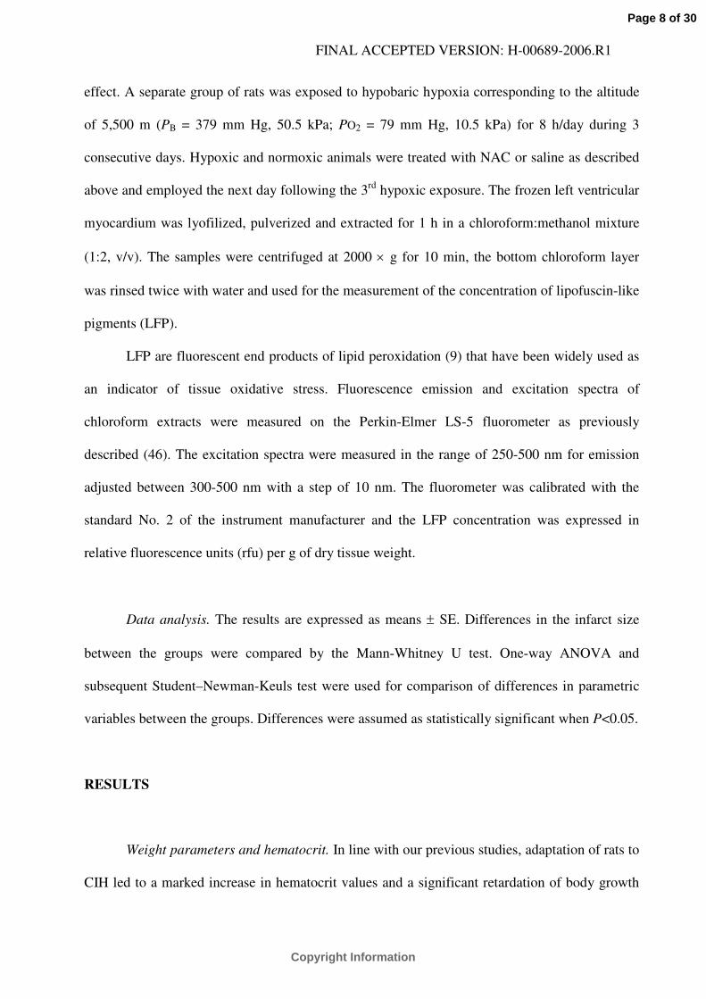
Animals. Adult male Wistar rats (250 - 280 g body weight) were exposed to intermittent
hypobaric hypoxia corresponding to the altitude of 7000 m for 8 h/day, 5 days a week (Fig. 1).
Barometric pressure (PB) was lowered stepwise, so that the level equivalent to an altitude of
7000 m (PB = 308 mm Hg, 41 kPa; PO2 = 65 mm Hg, 8.6 kPa) was reached after 13 exposures.
The total number of exposures was 24-30 to allow for successive processing of animals in
physiological experiments; no appreciable changes of hypoxia-induced responses occurred
within this interval. A subgroup of the animals received NAC by subcutaneous injections in a
dose of 100 mg/kg daily before the hypoxic exposure; the remaining rats received the same
volume (2 ml/kg) of saline. The control group of animals was kept for the same period of time at
PB and PO2 equivalent to an altitude of 200 m (PB = 742 mm Hg, 99 kPa; PO2 = 155 mm Hg,
20.7 kPa); a subgroup was treated with NAC or saline in a corresponding manner. All animals
had free access to water and a standard laboratory diet. The study was conducted in accordance
with the Guide for the Care and Use of Laboratory Animals published by the US National
Institutes of Health (NIH Publication No. 85-23, revised 1996).
All animals were employed on the next day following the last hypoxic exposure and/or
NAC injection. Hematocrit was measured in the tail blood. The animals assigned to biochemical
analyses were sacrificed by de-capitation, their hearts were rapidly excised, washed in cold (0
°C) saline and dissected into the right (RV) and left (LV) free ventricular walls and the septum.
All parts were weighed and the left ventricles were frozen in liquid nitrogen and stored at –80 °C
until use. All of the chemicals were purchased from Sigma, unless otherwise indicated.
Page 4 of 30
Copyright Information
FINAL ACCEPTED VERSION: H-00689-2006.R1
Infarct size determination. Animals were subjected to myocardial ischemia/reperfusion as
described previously (29). Anesthetized (sodium pentobarbital, 60 mg/kg i.p., Sanofi) rats were
ventilated (Columbus Instruments) with room air at 68 strokes/min (tidal volume of 1.2 ml/100 g
body weight). Blood pressure in the left carotid artery was measured (Gould P23Gb) and
subsequently analyzed by our custom-designed software. The rectal temperature was maintained
between 36.5 and 37.5 °C by a heated table throughout the experiment.
Left thoracotomy was performed and a polyester suture 6/0 (Ethibond - Ethicon) was
placed around the left anterior descending coronary artery about 1-2 mm distal to its origin. After
10-min stabilization, regional myocardial ischemia was induced by the tightening of the suture
threaded through a polyethylene tube. After a 20-min occlusion period, the ligature was released
and reperfusion of previously ischemic tissue continued for 3 h. Then the hearts were excised
and washed with saline through the aorta. The infarct area and the area at risk were delineated by
perfusion with 2,3,5-triphenyltetrazolium chloride and potassium permanganate (after coronary
artery occlusion), respectively. The hearts were cut into slices 1 mm thick and fixed in
formaldehyde solution. The size of the infarct area (IA), the size of the area at risk (AR) and the
size of the LV were determined by computerized planimetry. The IA was normalized to the AR
(IA/AR) and the AR was normalized to the LV (AR/LV).
Tissue fractionation and Western blot analysis of PKC isoforms. Frozen LV myocardium
was pulverized to a fine powder at the temperature of liquid nitrogen, followed by Potter-
Elvehjem homogenization in 8 volumes of ice-cold buffer composed of (in mmol/l): 12.5 Tris-
HCl (pH 7.4), 250 sucrose, 2.5 EGTA, 1 EDTA, 100 NaF, 5 DTT, 0.3 phenylmethylsulfonyl
fluoride, 0.2 leupeptin, and 0.02 aprotinin. The homogenate was centrifuged at 100,000 × g for
90 min. The resulting pellet represented the particulate fraction; the supernatant was the
Page 5 of 30
Copyright Information
FINAL ACCEPTED VERSION: H-00689-2006.R1
cytosolic fraction. The homogenate and pellet of the particulate fraction were re-suspended in
homogenization buffer containing 1 % Triton X-100 held on ice for 90 min and then centrifuged
at 100,000 × g for further 90 min. The resulting detergent-treated supernatants were used for
immunoblot analyses. Triton X-100 was added to the cytosolic fraction to reach the final
concentration of 1 %. Protein content was determined according to Lowry’s assay modified by
Peterson (37).
Detergent-treated extracts of sub-cellular fractions were subjected to SDS-PAGE
electrophoresis on 8 % bis-acrylamide polyacrylamide gel at 20 mA/gel for 90 min on a Mini-
Protean II apparatus (Bio-Rad). After electrophoresis, the resolved proteins were transferred to a
nitro-cellulose membrane (Amersham Int.). Equal protein transfer efficiency was verified by
staining with Ponceau S. After blocking with 5 % dry low-fat milk in Tris-buffered saline with
Tween 20 (TTBS), the membranes were immunoblotted using the ECL detection system
(Amersham Int.) as previously described (28). Samples from all experimental groups compared
were run on the same gel and quantified on the same membrane. To ensure the specificity of
PKC-δ and PKC-ε immunoreactive proteins, pre-stained MW protein standards (Fluka),
recombinant human PKC-δ and PKC-ε standards (Sigma), rat brain extract and the respective
blocking immunizing peptides (Sigma) were used.
Measurement of antioxidant enzyme activities. Myocardium was pulverized and
homogenized as described above. The homogenate was clarified by centrifugation at 5000 × g
for 10 min. Catalase (CAT) activity was measured by the method of Aebi (1). The rate of
hydrogen peroxide decomposition was monitored spectrophotometrically at 240 nm in 50 mM
phosphate buffer (pH 7.0) containing 10 mM hydrogen peroxide at 28 ˚C.
Glutathione peroxidase (GPX) activity was determined by the indirect procedure
described by Paglia and Valentine (34). GSSG was produced by GPX reaction and immediately
Page 6 of 30
Copyright Information
FINAL ACCEPTED VERSION: H-00689-2006.R1
reduced by NADPH in the presence of glutathione reductase. The rate of NADPH consumption
was recorded at 340 nm as a measure of GSSG formation. The reaction was conducted in 1 M
Tris-HCl buffer containing 5 mM Na2EDTA, 2 mM NADPH, 20mM GSH, 10 U/ml glutathione
reductase and started by the addition of t-butyl hydroperoxide. Consumption of NADPH was
calculated using mM extinction coefficient for NADPH of 6.22.
Total superoxide dismutase (SOD) activity was determined by the modified nitroblue
tetrazolium method (14). Xanthine−xanthine oxidase reaction was utilized to generate a
superoxide flux. Nitroblue tetrazolium reduction by superoxide anion to blue formazan was
measured spectrophotometrically at 540 nm (28 ˚C). Chloroform-ethanol extracts of
homogenates were then used to determine SOD activity. The assay contained the following
reagents: 0.1 mM phosphate buffer (pH 7.8), 4 g/l bovine serum albumin, 2 mg/ml nitroblue
tetrazolium, and 1 mM xanthine. Manganese SOD (Mn SOD) activity was quantified in the
presence of 5 mM NaCN, the selective inhibitor of copper-zinc SOD (41).
Measurement of glutathione concentration. Myocardium was homogenized in 1 % picric
acid using a glass-Teflon device and the homogenate was centrifuged at 10,000 × g for 10 min.
Concentration of total glutathione in supernatant was determined spectrophotometrically at 412
nM using glutathione reductase-coupled enzymic assay at 30 ˚C (16). The assay contained the
following reagents: 0.2 mmol/l NADPH, 100 mmol/l phosphate, 5 mmol/l EDTA, 0.6 mmol/l
5,5-dithio-bis(2-nitrobenzoic acid), and 1 kU/l glutathione reductase. Oxidized glutathione
(glutathione disulfide, GSSG) was measured by masking the reduced glutathione (GSH) with 2-
vinylpyridine. The ratio of GSH/GSSG was taken as a measure of tissue oxidative stress.
Measurement of lipofuscin-like pigments. Additional experiments were performed to
verify that CIH is associated with increased oxidative stress and NAC treatment prevents this
Page 7 of 30
Copyright Information
FINAL ACCEPTED VERSION: H-00689-2006.R1
effect. A separate group of rats was exposed to hypobaric hypoxia corresponding to the altitude
of 5,500 m (PB = 379 mm Hg, 50.5 kPa; PO2 = 79 mm Hg, 10.5 kPa) for 8 h/day during 3
consecutive days. Hypoxic and normoxic animals were treated with NAC or saline as described
above and employed the next day following the 3rd hypoxic exposure. The frozen left ventricular
myocardium was lyofilized, pulverized and extracted for 1 h in a chloroform:methanol mixture
(1:2, v/v). The samples were centrifuged at 2000 × g for 10 min, the bottom chloroform layer
was rinsed twice with water and used for the measurement of the concentration of lipofuscin-like
pigments (LFP).
LFP are fluorescent end products of lipid peroxidation (9) that have been widely used as
an indicator of tissue oxidative stress. Fluorescence emission and excitation spectra of
chloroform extracts were measured on the Perkin-Elmer LS-5 fluorometer as previously
described (46). The excitation spectra were measured in the range of 250-500 nm for emission
adjusted between 300-500 nm with a step of 10 nm. The fluorometer was calibrated with the
standard No. 2 of the instrument manufacturer and the LFP concentration was expressed in
relative fluorescence units (rfu) per g of dry tissue weight.
Data analysis. The results are expressed as means ± SE. Differences in the infarct size
between the groups were compared by the Mann-Whitney U test. One-way ANOVA and
subsequent Student–Newman-Keuls test were used for comparison of differences in parametric
variables between the groups. Differences were assumed as statistically significant when P<0.05.
RESULTS
Weight parameters and hematocrit. In line with our previous studies, adaptation of rats to
CIH led to a marked increase in hematocrit values and a significant retardation of body growth
Page 8 of 30
Copyright Information
FINAL ACCEPTED VERSION: H-00689-2006.R1
compared with age-matched normoxic animals. Treatment with NAC during the adaptation
period slightly decreased the body weight in both normoxic and chronically hypoxic groups and
did not affect the level of the hematocrit. The heart weight of chronically hypoxic rats increased
due to hypertrophy of both ventricles. The RV weight normalized to body weight increased to
177 % and that of the LV to 128 % of the respective normoxic values. NAC-treatment had no
effect on heart weight parameters (Table 1).
Myocardial infarct size. The normalized area at risk (AR/LV) did not significantly differ
between the groups (Table 1). CIH decreased the infarct size from 56.7 ± 4.5 % of the AR in the
normoxic control group to 27.7 ± 4.9 %. NAC treatment decreased IA/AR in the normoxic
animals to 42.0 ± 3.4 % but it abolished protection induced by CIH (to 41.1 ± 4.9 %). The
IA/AR did not differ between the NAC-treated groups (Fig. 2).
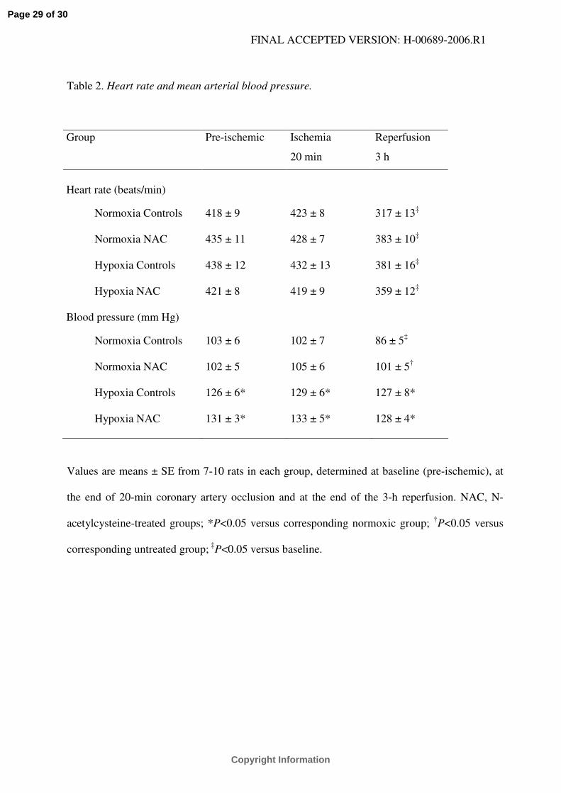
CIH increased mean arterial blood pressure (MAP) compared with normoxic groups, and
the higher level of MAP persisted in the course of ischemia and reperfusion. In normoxic
controls, MAP significantly decreased at the end of reperfusion compared with baseline values.
This decrease was prevented by both NAC treatment and adaptation to hypoxia. Heart rate did
not differ between the groups and it significantly decreased at the end of reperfusion in all
groups (Table 2).
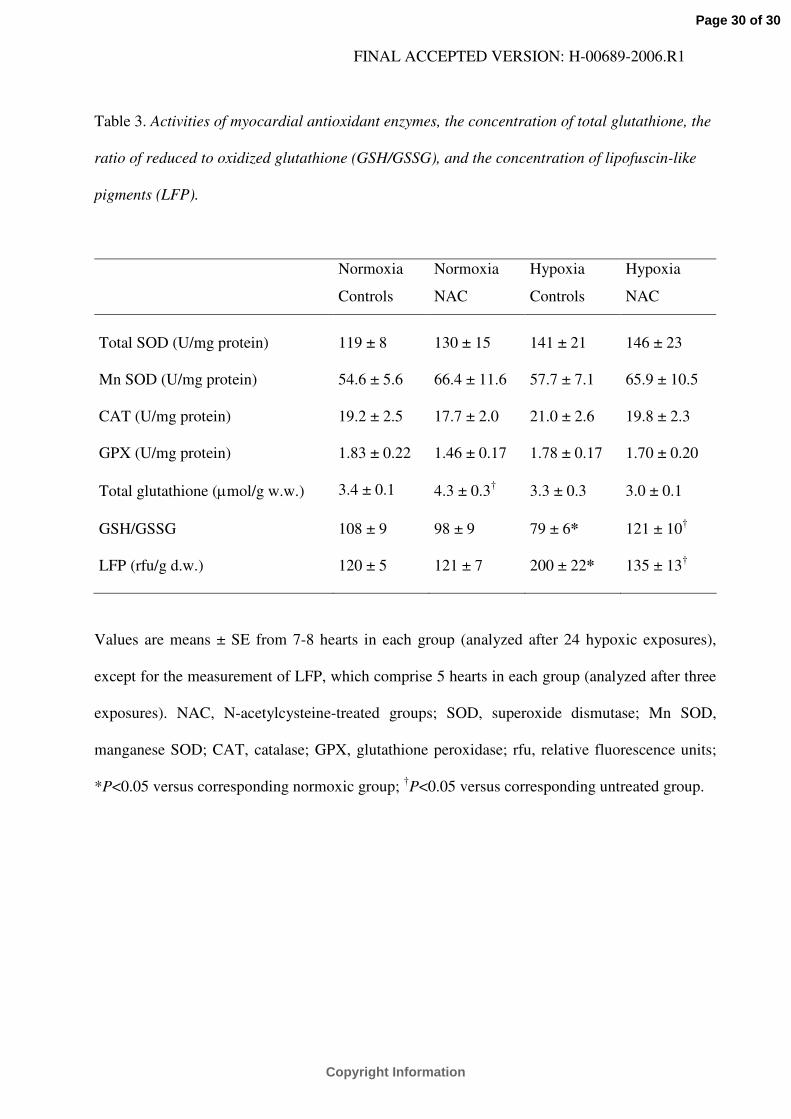
Myocardial glutathione, LFP, and antioxidant enzyme activities. Table 3 summarizes
myocardial activities of total SOD, Mn SOD, CAT and GPX. Neither CIH nor NAC treatment
induced an appreciable effect on these enzymes.
CIH did not change the myocardial concentration of total glutathione but it significantly
increased the proportion of GSSG and decreased the GSH/GSSG ratio. NAC treatment prevented
Page 9 of 30
Copyright Information
FINAL ACCEPTED VERSION: H-00689-2006.R1
the effect of CIH on GSH/GSSG ratio and it increased the concentration of total glutathione
without affecting the ratio in normoxic hearts (Table 3).
Myocardial concentration of LFP significantly increased already after 3 daily exposures
to hypoxia of 5,500 m and NAC treatment prevented this effect (Table 3).
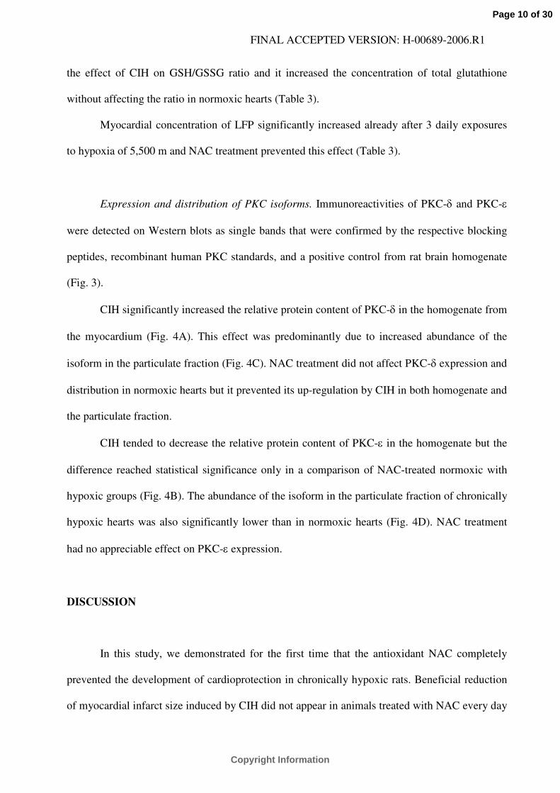
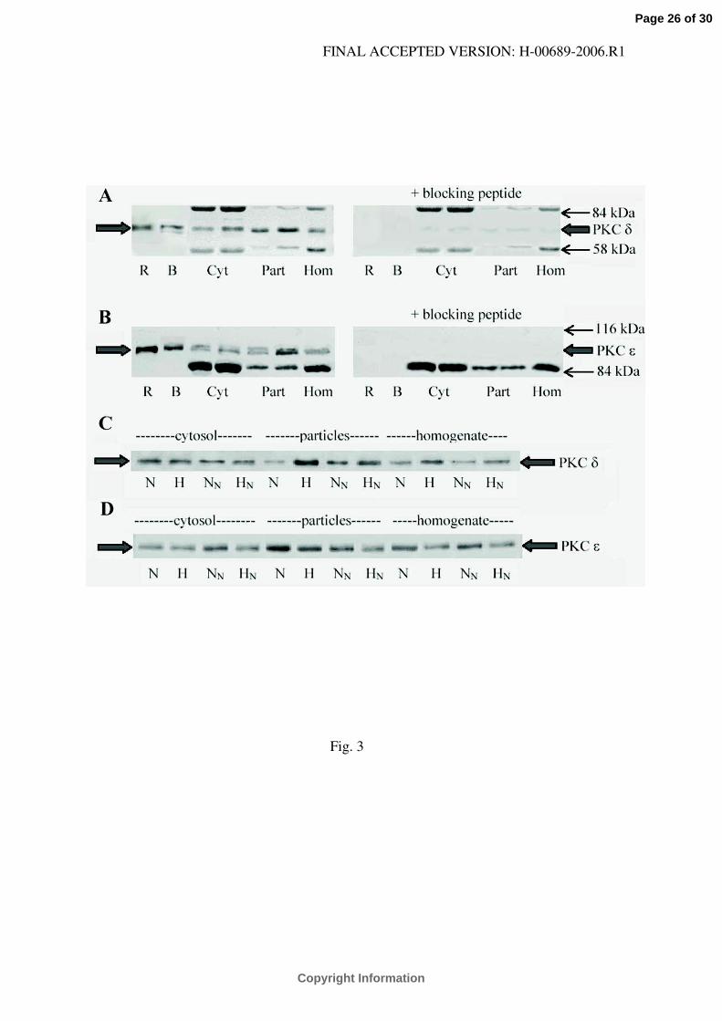
Expression and distribution of PKC isoforms. Immunoreactivities of PKC-δ and PKC-ε
were detected on Western blots as single bands that were confirmed by the respective blocking
peptides, recombinant human PKC standards, and a positive control from rat brain homogenate
(Fig. 3).
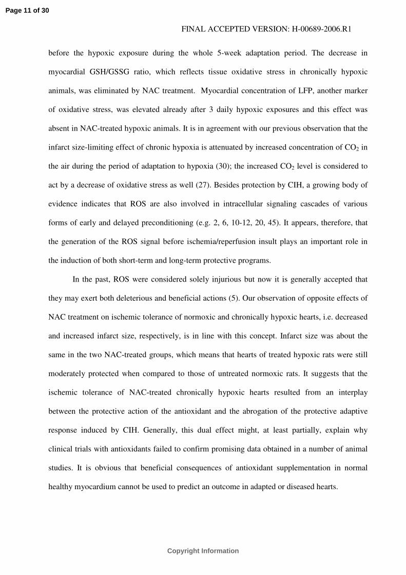
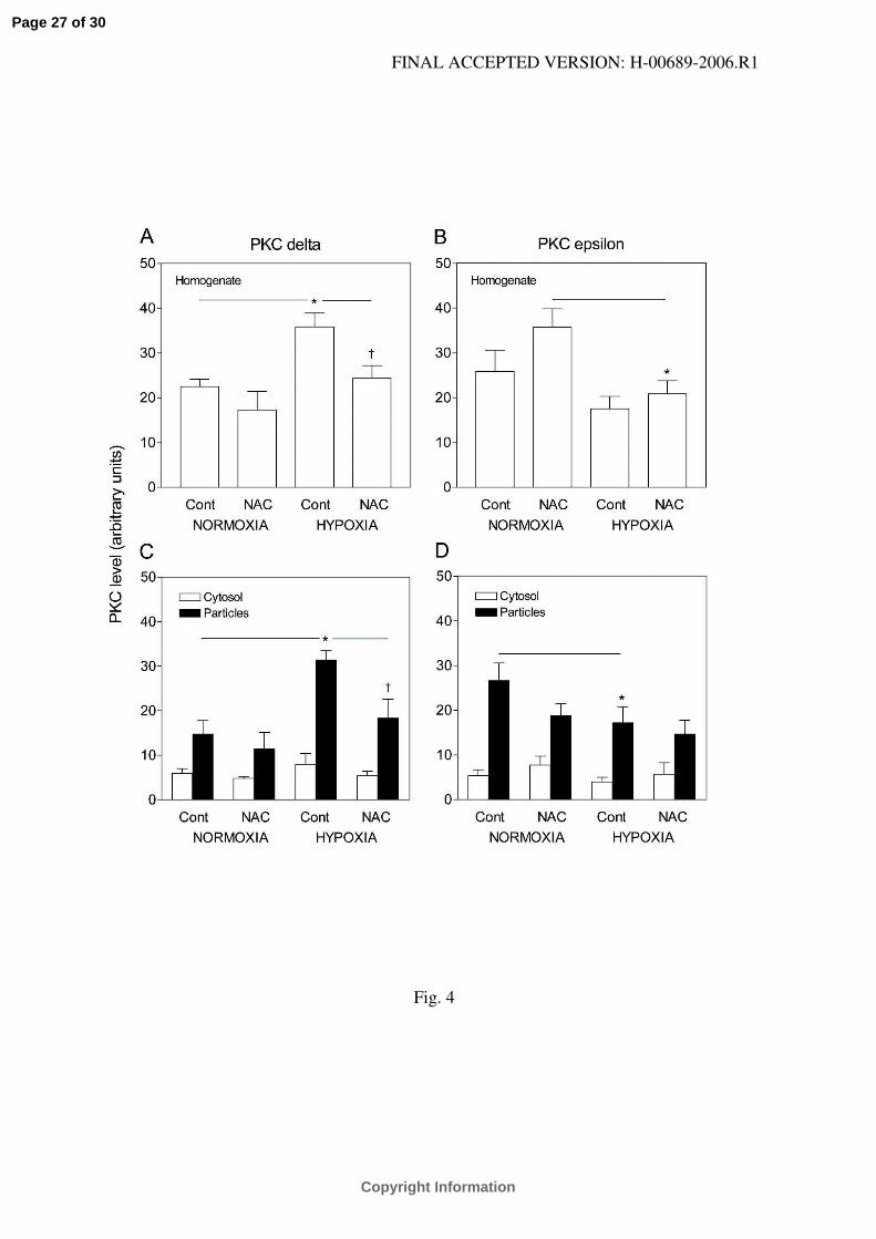
CIH significantly increased the relative protein content of PKC-δ in the homogenate from
the myocardium (Fig. 4A). This effect was predominantly due to increased abundance of the
isoform in the particulate fraction (Fig. 4C). NAC treatment did not affect PKC-δ expression and
distribution in normoxic hearts but it prevented its up-regulation by CIH in both homogenate and
the particulate fraction.
CIH tended to decrease the relative protein content of PKC-ε in the homogenate but the
difference reached statistical significance only in a comparison of NAC-treated normoxic with
hypoxic groups (Fig. 4B). The abundance of the isoform in the particulate fraction of chronically
hypoxic hearts was also significantly lower than in normoxic hearts (Fig. 4D). NAC treatment
had no appreciable effect on PKC-ε expression.
DISCUSSION
In this study, we demonstrated for the first time that the antioxidant NAC completely
prevented the development of cardioprotection in chronically hypoxic rats. Beneficial reduction
of myocardial infarct size induced by CIH did not appear in animals treated with NAC every day
Page 10 of 30
Copyright Information
FINAL ACCEPTED VERSION: H-00689-2006.R1
before the hypoxic exposure during the whole 5-week adaptation period. The decrease in
myocardial GSH/GSSG ratio, which reflects tissue oxidative stress in chronically hypoxic
animals, was eliminated by NAC treatment. Myocardial concentration of LFP, another marker
of oxidative stress, was elevated already after 3 daily hypoxic exposures and this effect was
absent in NAC-treated hypoxic animals. It is in agreement with our previous observation that the
infarct size-limiting effect of chronic hypoxia is attenuated by increased concentration of CO2 in
the air during the period of adaptation to hypoxia (30); the increased CO2 level is considered to
act by a decrease of oxidative stress as well (27). Besides protection by CIH, a growing body of
evidence indicates that ROS are also involved in intracellular signaling cascades of various
forms of early and delayed preconditioning (e.g. 2, 6, 10-12, 20, 45). It appears, therefore, that
the generation of the ROS signal before ischemia/reperfusion insult plays an important role in
the induction of both short-term and long-term protective programs.
In the past, ROS were considered solely injurious but now it is generally accepted that
they may exert both deleterious and beneficial actions (5). Our observation of opposite effects of
NAC treatment on ischemic tolerance of normoxic and chronically hypoxic hearts, i.e. decreased
and increased infarct size, respectively, is in line with this concept. Infarct size was about the
same in the two NAC-treated groups, which means that hearts of treated hypoxic rats were still
moderately protected when compared to those of untreated normoxic rats. It suggests that the
ischemic tolerance of NAC-treated chronically hypoxic hearts resulted from an interplay
between the protective action of the antioxidant and the abrogation of the protective adaptive
response induced by CIH. Generally, this dual effect might, at least partially, explain why
clinical trials with antioxidants failed to confirm promising data obtained in a number of animal
studies. It is obvious that beneficial consequences of antioxidant supplementation in normal
healthy myocardium cannot be used to predict an outcome in adapted or diseased hearts.
Page 11 of 30
Copyright Information
FINAL ACCEPTED VERSION: H-00689-2006.R1
Cardioprotective properties of NAC have been demonstrated in several experimental
studies using various in vivo or in vitro models (15, 23, 40). NAC is a sulfhydryl-containing
compound that exerts its complex antioxidant effect both through direct interaction with ROS
and as a precursor of L-cysteine and glutathione. In this study, we assessed cardiac ischemic
tolerance on the next day following the last administration of NAC. It seems unlikely that direct
scavenging activity of NAC itself was responsible for its protective effect at this time as the
elimination half-life of total plasma NAC is about 2 h in rats (18). NAC is rapidly converted to
L-cysteine, which is also cardioprotective as a ROS scavenger (39) or can enter in the synthesis
of glutathione, a central component of the cellular antioxidant defense system. The potential role
of glutathione in the infarct size-limiting effect of NAC treatment in normoxic animals cannot be
excluded as its total myocardial concentration was increased although the GSH/GSSG ratio
remained unchanged in our study. In addition, NAC increases nitric oxide availability by
scavenging ROS and stimulating endothelial nitric oxide synthase activity and protein expression
in the heart (36) that might contribute to the protective effect of a prolonged treatment.
Adaptation to CIH led to a marked up-regulation of PKC-δ that was significant in both
myocardial homogenate and particulate fraction. This observation confirms the results of our
recent study which demonstrated that CIH-induced increase in the relative protein content of
PKC-δ was most prominent in mitochondrial and nuclear fractions (28). We also showed that the
PKC-δ isoform-selective inhibitor rottlerin, administered before the acute ischemia/reperfusion
insult, attenuated the infarct size-limiting effect of CIH, suggesting that this isoform is involved
in the cardioprotective mechanism. The novel finding of the present study is that the preventive
treatment of chronically hypoxic rats with NAC eliminated the up-regulation of PKC-δ. This
observation suggests that the induction of this isoform during the adaptation period is dependent
on oxidative stress. The absence of both protection and PKC-δ up-regulation in NAC-treated
Page 12 of 30
Copyright Information
FINAL ACCEPTED VERSION: H-00689-2006.R1
hypoxic animals further supports our previous conclusion regarding the role of this isoform in
increased ischemic tolerance of chronically hypoxic hearts (28).
PKC-δ may be both protective and detrimental as reported by a growing number of
studies. It appears that protective effects of PKC-δ are manifested when the enzyme is activated
well before ischemia/reperfusion insult (19). This condition was satisfied in our experiments.
Consequences of PKC-δ activation also depend on its localization to various sub-cellular
compartments that is controlled by phosphorylation at multiple sites (42). For example,
phosphorylation of PKC-δ at serine-643 is associated with its translocation to mitochondria and
activation of mitoKATP channels following a protective stimulus (44). Opening of mitoKATP
channels is considered to play a crucial role in various forms of myocardial protection including
that afforded by CIH (3, 31, 48). The sequence of signaling events linking ROS, PKC-δ and
mitoKATP channels in the protective mechanism of CIH remains to be elucidated. Nevertheless,
our data are compatible with the view that ROS generation precedes PKC-δ activation and
mitoKATP opening as recently demonstrated in cardioprotection induced by the volatile anesthetic
sevoflurane (6). Obviously, the infarct size-limiting pathways induced by CIH may also involve
other redox-sensitive steps (24) that were not addressed in the present study.
Unlike PKC-δ, the abundance of PKC-ε, the key enzyme isoform involved in the
mechanism of preconditioning, was rather decreased in the myocardium of chronically hypoxic
rats and NAC treatment did not exert any appreciable effect. More detailed analysis performed in
our recent study did not reveal any significant change in PKC-ε abundance and sub-cellular
distribution due to CIH (28). Taken together, these data suggest that this isoform does not seem
to play a major role in the increased cardiac ischemic tolerance in our model of severe CIH. In
contrast, cardioprotection afforded by permanent chronic hypoxia in neonatal rabbits appears to
involve PKC-ε activation and translocation (38) suggesting that the role of PKC isoforms differs
in species- and/or age-dependent manner. However, we cannot exclude that, apart from PKC-δ,
Page 13 of 30
Copyright Information
FINAL ACCEPTED VERSION: H-00689-2006.R1
other PKC isoform(s) contribute to protection in our experimental model. It should be noted that
a moderately increased expression of PKC isoforms-α, -δ and -ε was observed in the myocardial
particulate fraction isolated from rats adapted to much less severe hypoxia (13).
It has been well documented that ROS can induce myocardial antioxidant enzymes. In
particular, the expression and activity of Mn SOD, a key enzyme that converts superoxide to
hydrogen peroxide in mitochondria, increase under various conditions associated with oxidative
stress. It has been demonstrated to play a role in delayed preconditioning elicited by ischemia,
heat stress, inflammatory cytokines or exercise training (e.g. 17, 20, 21); a close correlation
exists between the increase in Mn SOD activity and the reduction of infarct size under these
conditions (21). Increased activities of Mn SOD and CAT were also observed in hearts of rats
exposed to CIH just after birth for 60 days (49). However, the present study failed to detect any
effect of long-term adaptation of adult rats to CIH and/or NAC treatment on total myocardial
activities of Mn SOD and other major antioxidant enzymes. We cannot exclude that CIH had a
stimulatory effect during the first exposures, which disappeared later on when the animals
became fully adapted. Nevertheless, the increased ischemic tolerance of adult chronically
hypoxic hearts seems unlikely to be mediated by the increased capacity of enzymic antioxidant
defense.
In conclusion, oxidative stress acting during adaptation of rats to CIH, play an important
role in the induction of endogenous cardioprotective mechanism, which involves the up-
regulation of PKC-δ but not PKC-ε. Moreover, our data point to a potentially adverse effect of
antioxidant supplementation under conditions, which alone evoke ROS-dependent adaptive
responses. This might be considered as one of the reasons why clinical data are rather weak and
do not justify use of antioxidants for the prevention and treatment of cardiovascular diseases.
Page 14 of 30
Copyright Information
FINAL ACCEPTED VERSION: H-00689-2006.R1
GRANTS
This work was supported by the Grant Agency of the Czech Republic grant 305/04/0465,
Grant Agency of the Charles University grant 153/2005/B-Bio/PrF, and AVOZ 50110509.
Page 15 of 30
Copyright Information
FINAL ACCEPTED VERSION: H-00689-2006.R1
REFERENCES
1. Aebi H. Catalase in vitro. Methods Enzymol 105: 121-126, 1984.
2. Arnaud C, Joyeux M, Garrel C, Godin-Ribuot D, Demenge P, and Ribuot C. Free-
radical production triggered by hyperthermia contributes to heat stress-induced
cardioprotection in isolated rat hearts. Br J Pharmacol 135: 1776-1782, 2002.
3. Asemu G, Papousek F, Ostadal B, and Kolar F. Adaptation to high altitude hypoxia
protects the rats hearts against ischemia-induce arrhythmias. Involvement of
mitochondrial KATP channels. J Mol Cell Cardiol 31: 1821-1831, 1999.
4. Baines CP, Goto M, and Downey JM. Oxygen radicals released during ischemic
preconditioning contribute to cardioprotection in the rabbit myocardium. J Mol Cell
Cardiol 29: 207-216, 1997.
5. Becker LB. New concepts in reactive oxygen species and cardiovascular reperfusion
physiology. Cardiovasc Res 61: 461-470, 2004.
6. Bouwman RA, Musters RJP, van Beek-Harmsen BJ, de Lange JJ, and Boer C.
Reactive oxygen species precede protein kinase C- δ activation independent of adenosine
triphosphate-sensitive mitochondrial channel opening in sevoflurane-induced
cardioprotection. Anesthesiology 100: 506-517, 2004.
7. Chang SW, Selzner TJ, Weil JV, and Voelkel NF. Hypoxia increases plasma
glutathione disulfide in rats. Lung 167: 269-272, 1989.
8. Chen L, Einbinder E, Zhang Q, Hasday J, Balke CW, and Scharf SM. Oxidative
stress and left ventricular function with chronic intermittent hypoxia in rats. Am J Respir
Crit Care Med 172: 915-920, 2005.
9. Chio KS, Reiss V, Fletcher B, and Tappel AL. Peroxidation of subcellular organelles:
formation of lipofuscin-like pigments. Science 166: 1535-1536, 1969.
Page 16 of 30
Copyright Information
FINAL ACCEPTED VERSION: H-00689-2006.R1
10. Cohen MV, Yang XM, Liu GS, Heusch G, and Downey JM. Acetylcholine,
bradykinin, opioids, and phenylephrine, but not adenosine, trigger preconditioning by
generating free radicals and opening mitochondrial KATP channels. Circ Res 89: 273-278,
2001.
11. Das DK, Engelman RM, Maulik N. Oxygen free radical signaling in ischemic
preconditioning. Ann N Y Acad Sci 874: 49-65, 1999.
12. Das S, Engelman RM, Maulik N, Das DK. Angiotensin preconditioning of the heart:
evidence for redox signaling. Cell Biochem Biophys 44: 103-110, 2006.
13. Ding HL, Zhu HF, Dong JW, Zhu WZ, and Zhou ZN. Intermittent hypoxia protects
the rat heart against ischemia/reperfusion injury by activating protein kinase C. Life Sci
75: 2587-2603, 2004.
14. Elstner EF, Youngman RJ, and Oswald WF. Superoxide dismutase. In: Methods of
Enzymatic Analysis, vol. III, edited by Bergmeyer HU. Weinheim: Verlag Chemie, 1983,
p. 293-302.
15. Fiordaliso F, Bianchi R, Staszewsky L, Cuccovillo I, Doni M, Laragione T, Salio M,
Savino C, Melucci S, Santangelo F, Scaniziani E, Masson S, Ghezzi P and Latini R.
Antioxidant treatment attenuates hyperglycemia-induced cardiomyocyte death in rats. J
Mol Cell Cardiol 37: 959-968, 2004.
16. Griffith OW. Determination of glutathione and glutathione disulfide using glutathione
reductase and 2-vinylpyridine. Anal Biochem 106: 207-212, 1980.
17. Hamilton KL, Powers SK, Sugiura T, Kim S, Lennon S, Tumer N, and Mehta JL.
Short-term exercise training can improve myocardial tolerance to I/R without elevation in
heat shock proteins. Am J Physiol Heart Circ Physiol 281: H1346-H1352, 2001.
Page 17 of 30
Copyright Information
FINAL ACCEPTED VERSION: H-00689-2006.R1
18. Harada D, Naito S, Hiraoka I, and Otagiri M. In vivo kinetic analysis of covalent
binding between N-acetyl-L-cysteine and plasma protein through the formation of mixed
disulfide in rats. Pharm Res 19: 615-620, 2002.
19. Hirotani S and Sadoshima J. Preconditioning effects of PKCδ. J Mol Cell Cardiol 39:
719-721, 2005.
20. Hoshida S, Yamashita N, Otsu K, and Hori M. Repeated physiological stresses
provide persistent cardioprotection against ischemia-reperfusion injury in rats. J Am Coll
Cardiol 40 :826-831, 2002.
21. Hoshida S, Yamashita N, Otsu K, and Hori M. The importance of manganese
superoxide dismutase in delayed preconditioning: Involvement of reactive oxygen
species and cytokines. Cardiovasc Res 55: 495-505, 2002.
22. Hoshikawa Y, Ono S, Suzuki S, Tanita T, Chida M, Song C, Noda M, Tabata T,
Voelkel NF, and Fujimura S. Generation of oxidative stress contributes to the
development of pulmonary hypertension induced by hypoxia. J Appl Physiol 90: 1299-
1306, 2001.
23. Inserte J, Taimor G, Hofstaetter B, Garcia-Dorado D, and Piper HM. Influence of
simulated ischemia on apoptosis induction by oxidative stress in adult cardiomyocytes of
rats. Am J Physiol Heart Circ Physiol 278: H94-H99, 2000.
24. Kolar F and Ostadal B. Molecular mechanisms of cardiac protection by adaptation to
chronic hypoxia. Physiol Res 53(Suppl 1): S3-S13, 2004.
25. Lachmanova V, Hnilickova O, Povysilova V, Hampl V, and Herget J. N-
acetylcysteine inhibits hypoxic pulmonary hypertension most effectively in the initial
phase of chronic hypoxia. Life Sci 77: 175-182, 2005.
26. Ludwig LM, Weihrauch D, Kersten JR, Pagel PS, and Warltier DC. Protein kinase C
translocation and Src protein tyrosine kinase activation mediate isoflurane-induced
Page 18 of 30
Copyright Information
FINAL ACCEPTED VERSION: H-00689-2006.R1
preconditioning in vivo: potential downstream targets of mitochondrial adenosine
triphosphate-sensitive potassium channels and reactive oxygen species. Anesthesiology
100: 532-539, 2004.
27. Lymar SV and Hurst JK. Carbon dioxide: physiological catalyst for peroxynitrite-
mediated cellular damage or cellular protectant? Chem Res Toxicol 9: 845-850, 1996.
28. Neckar J, Markova I, Novak F, Novakova O, Szarszoi O, Ostadal B, and Kolar F.
Increased expression and altered subcellular distribution of PKC-δ in chronically hypoxic
rat myocardium: involvement in cardioprotection. Am J Physiol Heart Circ Physiol 288:
H1566-H1572, 2005.
29. Neckar J, Papousek F, Novakova O, Ostadal B, and Kolar F. Cardioprotective effects
of chronic hypoxia and preconditioning are not additive. Basic Res Cardiol 97: 161-167,
2002.
30. Neckar J, Szarszoi O, Herget J, Ostadal B, and Kolar F. Cardioprotective effect of
chronic hypoxia is blunted by concomitant hypercapnia. Physiol Res 52: 171-175, 2003.
31. Neckar J, Szarszoi O, Koten L, Papousek F, Ostadal B, Grover GJ, and Kolar F.
Effects of mitochondrial KATP modulators on cardioprotection induced by chronic high
altitude hypoxia in rats. Cardiovasc Res 55: 567-575, 2002.
32. Ooi H, Cadogan E, Sweeney M, Howell K, O´Regan RG, and McLoughlin P.
Chronic hypercapnia inhibits hypoxic pulmonary vascular remodeling. Am J Physiol
Heart Circ Physiol 278: H331-H338, 2000.
33. Ostadal B, Ostadalova I, Kolar F, Pelouch V, and Dhalla NS. Cardiac adaptation to
chronic hypoxia. In: Advances in Organ Biology, vol. 6, edited by Bittar EE and Das DK.
London: JAI Press, 1998, p. 43-60.
Page 19 of 30
Copyright Information
FINAL ACCEPTED VERSION: H-00689-2006.R1
34. Paglia DE and Valentine WN. Studies on the quantitative and qualitative
characterization of erythrocyte glutathione peroxidase. J Lab Clin Med 70: 158-169,
1967.
35. Pain T, Yang X-M, Critz SD, Yue Y, Nakano A, Liu GS, Heusch G, Cohen MV, and
Downey JM. Opening of mitochondrial KATP channels triggers the preconditioned state
by generating free radicals. Circ Res 87: 460-466, 2000.
36. Pechanova O, Zicha J, Kojsova S, Dobesova Z, Jendekova L, and Kunes J. Effect of
chronic N-acetylcysteine treatment on the development of spontaneous hypertension.
Clin Sci (Lond) 110: 235-242, 2006.
37. Peterson GL. A simplification of the protein assay method of Lowry et al. which is more
generally applicable. Anal Biochem 83: 346-356, 1977.
38. Rafiee P, Shi Y, Kong X, Pritchard KA, Tweddell JS, Litwin SB, Mussato K,
Jaquiss RD, Su J, and Baker JE. Activation of protein kinases in chronically hypoxic
infant human and rabbit hearts: role in cardioprotection. Circulation 106: 239-245, 2002.
39. Shackebaei D, King N, Shukla B, and Suleiman M-S. Mechanisms underlying the
cardioprotective effect of L-cysteine. Mol Cell Biochem 277: 27-31, 2005.
40. Sochman J, Kolc J, Vrana M, and Fabian J. Cardioprotective effects of N-
acetylcysteine: the reduction of the extent of infarction and occurrence of reperfusion
arrhythmias in the dog. Int J Cardiol 28: 191-196, 1990.
41. Spanier AM, Weglicki WB, Stiers DL, and Misra HP. Superoxide dismutase: tissue,
cellular, and subcellular distribution in adult canine heart. Am J Physiol 249: C379-C384,
1985.
42. Steinberg SF. Distinctive activation mechanisms and functions for protein kinase Cδ.
Biochem J 384: 449-459, 2004.
Page 20 of 30
Copyright Information
FINAL ACCEPTED VERSION: H-00689-2006.R1
43. Tritto I, D’Andrea D, Eramo N, Scognamiglio A, De Simone C, Violante A, Esposito
A, Chiariello M, and Ambrosio G. Oxygen radicals can induce preconditioning in
rabbit hearts. Circ Res 80: 743-748, 1997.
44. Uecker M, da Silva R, Grampp T, Pasch T, Schaub MC, and Zaugg M.
Translocation of protein kinase C isoforms to subcellular targets in ischemic and
anesthetic preconditioning. Anesthesiology 99: 138-147, 2003.
45. Vanden Hoek TL, Becker LB, Shao Z, Li C, and Schumacker PT. Reactive oxygen
species released from mitochondria during brief hypoxia induce preconditioning in
cardiomyocytes. J Biol Chem 273: 18092-18098, 1998.
46. Wilhelm J and Herget J. Hypoxia induces free radical damage to rat erythrocytes and
spleen: analysis of the fluorescent end-products of lipid peroxidation. Int J Biochem Cell
Biol 31: 671-681, 1999.
47. Zhang HY, McPherson BC, Liu H, Baman TS, Rock P, and Yao Z. H2O2 opens
mitochondrial KATP channels and inhibits GABA receptors via protein kinase C-ε in
cardiomyocytes. Am J Physiol Heart Circ Physiol 282: H1395-H1403, 2002.
48. Zhu HF, Dong JW, Zhu WZ, Ding HL, and Zhou ZN. ATP-dependent potassium
channels involved in the cardiac protection induced by intermittent hypoxia against
ischemia/reperfusion injury. Life Sci 73: 1275-1287, 2003.
49. Zhu WZ, Dong JW, Ding HL, Yang HT, and Zhou ZN. Postnatal development in
intermittent hypoxia enhances resistance to myocardial ischemia/reperfusion in male rats.
Eur J Appl Physiol 91: 716-722, 2004.
Page 21 of 30
Copyright Information
FINAL ACCEPTED VERSION: H-00689-2006.R1
FIGURE LEGENDS
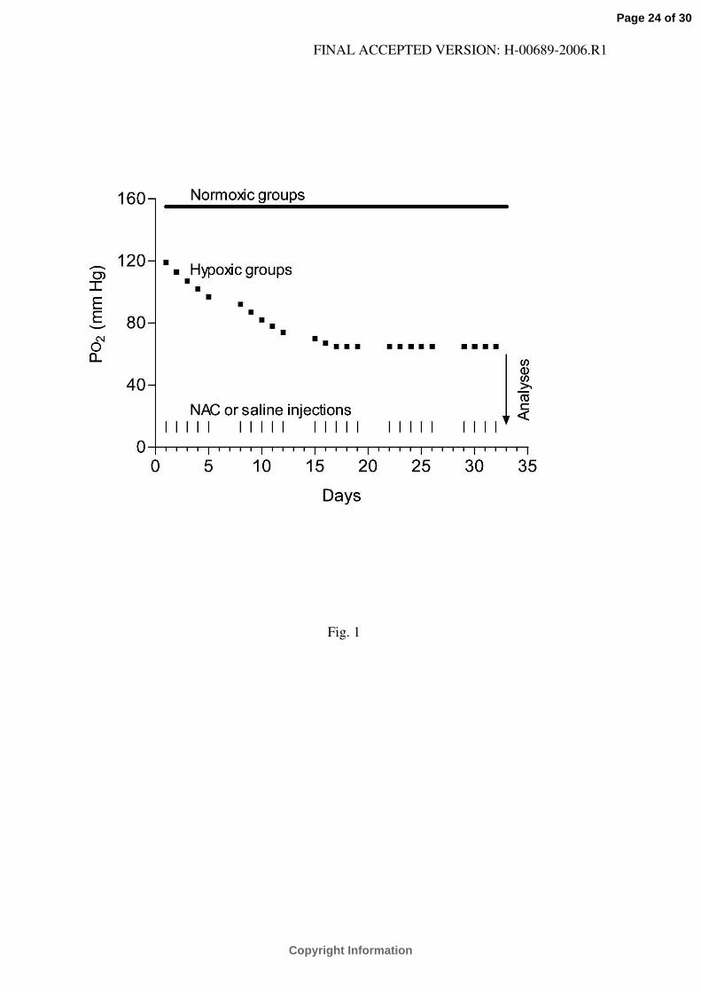
Fig. 1.
Model of chronic intermittent hypoxia and the experimental protocol. Hypoxic animals were
subject to hypobaric hypoxia starting at PO2 = 119 mm Hg (equivalent to an altitude of 2,400 m)
and decreasing stepwise up to PO2 = 65 mm Hg (equivalent to an altitude of 7000 m) during the
first 13 exposures; this level of hypoxia was maintained for additional 11-17 exposures. Full
squares indicate daily exposures lasting 8 h. For the remaining period of each day and for 2 days
after each 5-day series of hypoxic exposures, the animals were kept at normoxia (PO2 = 155 mm
Hg, equivalent to an altitude of 200 m). Vertical lines at the bottom of the graph indicate N-
acetylcysteine (NAC) or saline injections given before each hypoxic exposure. Normoxic
animals were kept at PO2 = 155 mm Hg during the whole experiment (indicated by a continuous
line) and treated with NAC or saline in a corresponding manner. All animals were employed on
the next day following the last hypoxic exposure and NAC or saline injection.
Fig. 2.
Myocardial infarct size expressed as a percentage of the area at risk (IA/AR) in control (Cont)
and N-acetylcysteine-treated (NAC) rats adapted to chronic hypoxia and in normoxic animals.
Values are means ± SE from 7–10 hearts in each group. *P<0.05 versus corresponding normoxic
group, †P<0.05 versus corresponding untreated group.
Fig. 3.
Western blot analysis of PKC isoforms in myocardial homogenate (Hom) and in cytosolic (Cyt)
and particulate (Part) fractions. A and B, representative Western blots of PKC-δ and PKC-ε,
respectively, in fractions from the control normoxic heart in the absence (left part) and presence
Page 22 of 30
Copyright Information
FINAL ACCEPTED VERSION: H-00689-2006.R1
(right part) of the respective blocking peptides. C and D, representative Western blots comparing
the expression of PKC-δ and PKC-ε, respectively, in fractions from normoxic (N), normoxic N-
acetylcysteine-treated (NN), chronically hypoxic (H) and chronically hypoxic N-acetylcysteine-
treated (HN) rats. Total amounts of protein loaded were as follows: PKC-δ - cytosol 16 µg,
particles 5 µg, homogenate 10 µg; PKC-ε - cytosol 18 µg, particles 8 µg, homogenate 14 µg.
For details see Materials and methods. Numbers on the right indicate the positions of pre-stained
molecular mass standards in kDa. R, recombinant human PKC-δ or PKC-ε standards; B, extract
from rat brain homogenate.
Fig. 4.
The expression of PKC-δ (A, C) and PKC-ε (B, D) in homogenate (A, B), and their distributions
between cytosolic and particulate fractions (C, D) from the myocardium of control (Cont) and N-
acetylcysteine-treated (NAC) rats adapted to chronic hypoxia and of normoxic animals. Values
are expressed as arbitrary units: the sum of densitometric volumes (related to 1 µg of protein)
measured in all groups on the same blot equals 100. Values are means ± SE from 5 (PKC-δ) or 6
(PKC-ε) hearts in each group. *P<0.05 versus corresponding normoxic group, †P<0.05 versus
corresponding untreated group.
Page 23 of 30
Copyright Information
FINAL ACCEPTED VERSION: H-00689-2006.R1
Fig. 1
Page 24 of 30
Copyright Information
FINAL ACCEPTED VERSION: H-00689-2006.R1
Fig. 2
Page 25 of 30
Copyright Information
FINAL ACCEPTED VERSION: H-00689-2006.R1
Fig. 3
Page 26 of 30
Copyright Information
FINAL ACCEPTED VERSION: H-00689-2006.R1
Fig. 4
Page 27 of 30
Copyright Information
FINAL ACCEPTED VERSION: H-00689-2006.R1
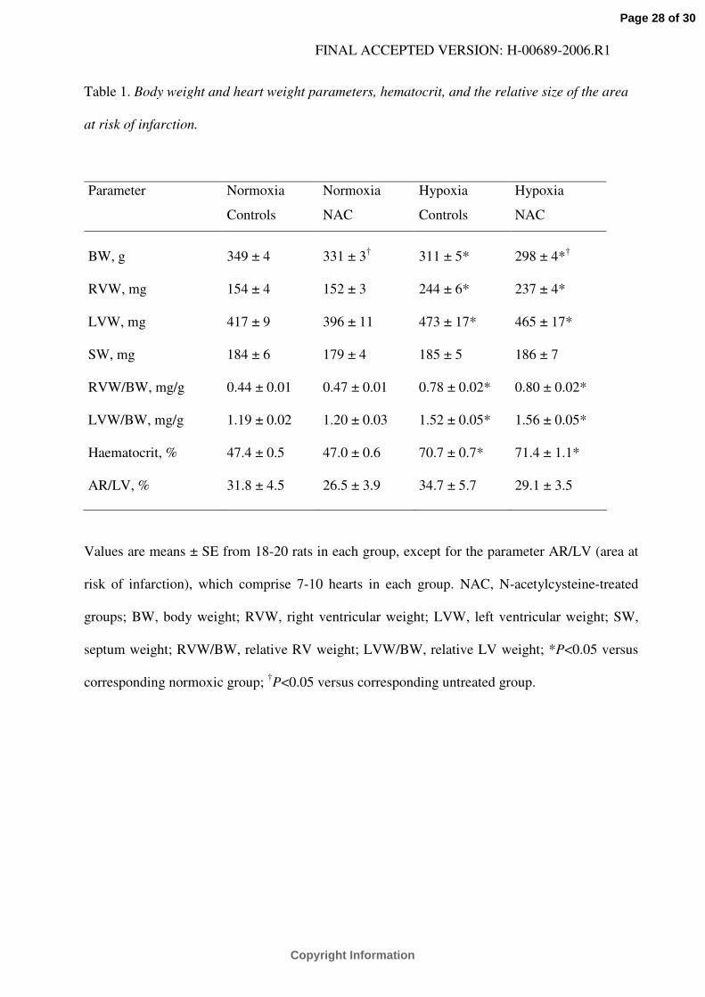
Table 1. Body weight and heart weight parameters, hematocrit, and the relative size of the area
at risk of infarction.
Parameter Normoxia
Controls
Normoxia
NAC
Hypoxia
Controls
Hypoxia
NAC
BW, g 349 ± 4 331 ± 3† 311 ± 5* 298 ± 4*†
RVW, mg 154 ± 4 152 ± 3 244 ± 6* 237 ± 4*
LVW, mg 417 ± 9 396 ± 11 473 ± 17* 465 ± 17*
SW, mg 184 ± 6 179 ± 4 185 ± 5 186 ± 7
RVW/BW, mg/g 0.44 ± 0.01 0.47 ± 0.01 0.78 ± 0.02* 0.80 ± 0.02*
LVW/BW, mg/g 1.19 ± 0.02 1.20 ± 0.03 1.52 ± 0.05* 1.56 ± 0.05*
Haematocrit, % 47.4 ± 0.5 47.0 ± 0.6 70.7 ± 0.7* 71.4 ± 1.1*
AR/LV, % 31.8 ± 4.5 26.5 ± 3.9 34.7 ± 5.7 29.1 ± 3.5
Values are means ± SE from 18-20 rats in each group, except for the parameter AR/LV (area at
risk of infarction), which comprise 7-10 hearts in each group. NAC, N-acetylcysteine-treated
groups; BW, body weight; RVW, right ventricular weight; LVW, left ventricular weight; SW,
septum weight; RVW/BW, relative RV weight; LVW/BW, relative LV weight; *P<0.05 versus
corresponding normoxic group; †P<0.05 versus corresponding untreated group.
Page 28 of 30
Copyright Information
FINAL ACCEPTED VERSION: H-00689-2006.R1
Table 2. Heart rate and mean arterial blood pressure.
Values are means ± SE from 7-10 rats in each group, determined at baseline (pre-ischemic), at
the end of 20-min coronary artery occlusion and at the end of the 3-h reperfusion. NAC, N-
acetylcysteine-treated groups; *P<0.05 versus corresponding normoxic group; †P<0.05 versus
corresponding untreated group; ‡P<0.05 versus baseline.
Group Pre-ischemic Ischemia Reperfusion
20 min 3 h
Heart rate (beats/min)
Normoxia Controls 418 ± 9 423 ± 8 317 ± 13‡
Normoxia NAC 435 ± 11 428 ± 7 383 ± 10‡
Hypoxia Controls 438 ± 12 432 ± 13 381 ± 16‡
Hypoxia NAC 421 ± 8 419 ± 9 359 ± 12‡
Blood pressure (mm Hg)
Normoxia Controls 103 ± 6 102 ± 7 86 ± 5‡
Normoxia NAC 102 ± 5 105 ± 6 101 ± 5†
Hypoxia Controls 126 ± 6* 129 ± 6* 127 ± 8*
Hypoxia NAC 131 ± 3* 133 ± 5* 128 ± 4*
Page 29 of 30
Copyright Information
FINAL ACCEPTED VERSION: H-00689-2006.R1
Table 3. Activities of myocardial antioxidant enzymes, the concentration of total glutathione, the
ratio of reduced to oxidized glutathione (GSH/GSSG), and the concentration of lipofuscin-like
pigments (LFP).
Normoxia
Controls
Normoxia
NAC
Hypoxia
Controls
Hypoxia
NAC
Total SOD (U/mg protein) 119 ± 8 130 ± 15 141 ± 21 146 ± 23
Mn SOD (U/mg protein) 54.6 ± 5.6 66.4 ± 11.6 57.7 ± 7.1 65.9 ± 10.5
CAT (U/mg protein) 19.2 ± 2.5 17.7 ± 2.0 21.0 ± 2.6 19.8 ± 2.3
GPX (U/mg protein) 1.83 ± 0.22 1.46 ± 0.17 1.78 ± 0.17 1.70 ± 0.20
Total glutathione (µmol/g w.w.) 3.4 ± 0.1 4.3 ± 0.3† 3.3 ± 0.3 3.0 ± 0.1
GSH/GSSG 108 ± 9 98 ± 9 79 ± 6* 121 ± 10†
LFP (rfu/g d.w.) 120 ± 5 121 ± 7 200 ± 22* 135 ± 13†
Values are means ± SE from 7-8 hearts in each group (analyzed after 24 hypoxic exposures),
except for the measurement of LFP, which comprise 5 hearts in each group (analyzed after three
exposures). NAC, N-acetylcysteine-treated groups; SOD, superoxide dismutase; Mn SOD,
manganese SOD; CAT, catalase; GPX, glutathione peroxidase; rfu, relative fluorescence units;
*P<0.05 versus corresponding normoxic group; †P<0.05 versus corresponding untreated group.
Page 30 of 30
Copyright Information
Related Documents