Opposing cardioprotective actions and parallel hypertrophic effects of dPKC and «PKC Leon Chen*, Harvey Hahn , Guangyu Wu , Che-Hong Chen*, Tamar Liron*, Deborah Schechtman*, Gabriele Cavallaro ‡ , Lucia Banci ‡ , Yiru Guo § , Roberto Bolli § , Gerald W. Dorn II , and Daria Mochly-Rosen* ¶ *Division of Chemical Biology, Department of Molecular Pharmacology, Stanford University School of Medicine, Stanford, CA 94305; ² Department of Medicine, University of Cincinnati, Cincinnati, OH 45967; ‡ Centro Risonanze Magnetiche, University of Florence, 50019 Florence, Italy; and § Departments of Medicine, Physiology, and Biophysics, University of Louisville, Louisville, KY 40292 Communicated by Daniel E. Koshland, Jr., University of California, Berkeley, CA, July 17, 2001 (received for review April 5, 2001) Conflicting roles for protein kinase C (PKC) isozymes in cardiac disease have been reported. Here, dPKC-selective activator and inhibitor peptides were designed rationally, based on molecular modeling and structural homology analyses. Together with previ- ously identified activator and inhibitor peptides of «PKC, dPKC peptides were used to identify cardiac functions of these isozymes. In isolated cardiomyocytes, perfused hearts, and transgenic mice, dPKC and «PKC had opposing actions on protection from ischemia- induced damage. Specifically, activation of «PKC caused cardiopro- tection whereas activation of dPKC increased damage induced by ischemia in vitro and in vivo. In contrast, dPKC and «PKC caused identical nonpathological cardiac hypertrophy; activation of either isozyme caused nonpathological hypertrophy of the heart. These results demonstrate that two related PKC isozymes have both parallel and opposing effects in the heart, indicating the danger in the use of therapeutics with nonselective isozyme inhibitors and activators. Moreover, reduction in cardiac damage caused by isch- emia by perfusion of selective regulator peptides of PKC through the coronary arteries constitutes a major step toward developing a therapeutic agent for acute cardiac ischemia. T here are conflicting reports on the role of protein kinase C (PKC) in mediating myocardial protection from ischemia (1). Moreover, changes in the expression of some PKC isozymes in cardiac hypertrophy and heart failure (2–4) could reflect either their active role or a consequence of these diseases. Current approaches include the use of isozyme nonselective tools and overexpression of individual PKC isozymes, which prevent a meaningful interpretation of the data. Each of the six PKC isozymes in cardiac myocytes translocates to different subcellular sites upon activation (5–7). We proposed a mechanism for isozyme-specific translocation involving binding of activated PKC to isozyme-specific anchoring proteins termed RACKs [receptors for activated C kinase (7)]. Binding of a specific PKC isozyme to its RACK occurs after phospholipid-induced allosteric conformational changes that activate the enzyme and expose its RACK-binding domain. This leads to PKC translocation and binding to isozyme-specific RACKs at different subcellular sites, which is thought to determine the function of each isozyme (5, 6). Based on this hypothesis, we developed several peptide trans- location inhibitors and activators, which when introduced into cells, cause selective regulation of translocation and function of the corresponding PKC isozyme (8). Translocation inhibitor peptides correspond to specific RACK-binding sites in the C2yV1 domain (9–12) of each isozyme and act as isozyme-selective competitors of PKC- RACK binding and function (10, 12). Peptide activators, on the other hand, are derived from a pseudoRACK (cRACK) se- quence in each PKC isozyme that is similar to a sequence in its corresponding RACK (8, 10, 13). These cRACK or RACK-like PKC sequences are thought to engage in intramolecular inter- actions with the RACK-binding site in PKC, thus stabilizing PKC in its inactive ‘‘closed’’ conformation (8, 10, 13). Therefore, interference with this intramolecular interaction and destabili- zation of the inactive enzyme should enhance PKC translocation and binding to its RACK, thus stabilizing the activated state of that PKC isozyme. Using this rationale, we identified isozyme- selective activator peptides for bPKC and «PKC (10, 13). Relevant to this study, these activator peptides were used to show that «PKC confers cardiac protection from transient ischemic insult (13) and normal postnatal cardiac development (14). In this study, we used a rational approach to identify dPKC inhibitor and activator peptides and used them to determine the role of dPKC in cardiac functions. Materials and Methods Peptide Synthesis. We synthesized dV1–1, amino acids 8–17 [SFN- SYELGSL]; cdRACK, amino acids 74–81 [MRAAEDPM]; and c«RACK, amino acids 85–92 [HDAPIGYD] peptides at Stanford’s Protein and Nucleic Acid facility and conjugated them either to Antennapedia, amino acids 43–58 [RQIKIWFQRRMKKWK] (15) or Tat, amino acids 47–57 [YGRKKRRQRRR] (16) via a cysteine- cysteine bond at their N termini. «V1 Structure Prediction. We used the sequence alignment of the C2yV1 (17) and then modeled «V1 according to dV1 (Protein Data Bank accession no. 1BDY), the C2 domain in phospho- lipase d1 (Protein Data Bank accession no. 2ISD), and 115–132 of the carboxypeptidase (Protein Data Bank accession no. 1LBU), which has sequence homology to a long gap in the alignment corresponding to residues 16–30 of «V1. Isolation of Adult Rat Cardiac Myocytes and Simulated Ischemia. Cardiac myocytes from 12-week-old male Wistar rats were isolated as described (13), treated with peptide conjugates in the presence or absence of phorbol 12-myristate 13-acetate (PMA), and subjected to Western blot analysis (13, 18) or simulated ischemia (13, 18). To simulate ischemia, we kept cardiac myocyte pellets for either 90 or 180 min in sealed test tubes in a small volume of buffer, saturated in N 2 and devoid of oxygen (,0.5%), glucose, and nutrients (19). We assessed damage to cardiac myocytes by trypan blue dye exclusion assay (19, 20). The staining correlates with increased myocyte rounding, propidium iodide staining, membrane blebbing, and leak of cytosolic en- zymes into the cell medium (18). Simulated Ischemia in Intact Rat Hearts. We simulated ischemia for 45 min in isolated hearts from 12- to 20-week-old rats by using Langendorff apparatus (18) and determined damage by creatine kinase (CK; used also as a diagnostic marker for cardiac damage in patients) activity (Sigma) in perfusate collected during 30 min Abbreviations: PKC, protein kinase C; RACK, receptor for activated C kinase E; cRACK, pseudoRACK; PMA, phorbol 12-myristate 13-acetate; CK, creatine kinase. ¶ To whom reprint requests should be addressed. E-mail: [email protected]. The publication costs of this article were defrayed in part by page charge payment. This article must therefore be hereby marked “advertisement” in accordance with 18 U.S.C. §1734 solely to indicate this fact. www.pnas.orgycgiydoiy10.1073ypnas.191369098 PNAS Early Edition u 1 of 6 BIOCHEMISTRY

Welcome message from author
This document is posted to help you gain knowledge. Please leave a comment to let me know what you think about it! Share it to your friends and learn new things together.
Transcript

Opposing cardioprotective actions and parallelhypertrophic effects of dPKC and «PKCLeon Chen*, Harvey Hahn†, Guangyu Wu†, Che-Hong Chen*, Tamar Liron*, Deborah Schechtman*, Gabriele Cavallaro‡,Lucia Banci‡, Yiru Guo§, Roberto Bolli§, Gerald W. Dorn II†, and Daria Mochly-Rosen*¶
*Division of Chemical Biology, Department of Molecular Pharmacology, Stanford University School of Medicine, Stanford, CA 94305; †Department ofMedicine, University of Cincinnati, Cincinnati, OH 45967; ‡Centro Risonanze Magnetiche, University of Florence, 50019 Florence, Italy;and §Departments of Medicine, Physiology, and Biophysics, University of Louisville, Louisville, KY 40292
Communicated by Daniel E. Koshland, Jr., University of California, Berkeley, CA, July 17, 2001 (received for review April 5, 2001)
Conflicting roles for protein kinase C (PKC) isozymes in cardiacdisease have been reported. Here, dPKC-selective activator andinhibitor peptides were designed rationally, based on molecularmodeling and structural homology analyses. Together with previ-ously identified activator and inhibitor peptides of «PKC, dPKCpeptides were used to identify cardiac functions of these isozymes.In isolated cardiomyocytes, perfused hearts, and transgenic mice,dPKC and «PKC had opposing actions on protection from ischemia-induced damage. Specifically, activation of «PKC caused cardiopro-tection whereas activation of dPKC increased damage induced byischemia in vitro and in vivo. In contrast, dPKC and «PKC causedidentical nonpathological cardiac hypertrophy; activation of eitherisozyme caused nonpathological hypertrophy of the heart. Theseresults demonstrate that two related PKC isozymes have bothparallel and opposing effects in the heart, indicating the danger inthe use of therapeutics with nonselective isozyme inhibitors andactivators. Moreover, reduction in cardiac damage caused by isch-emia by perfusion of selective regulator peptides of PKC throughthe coronary arteries constitutes a major step toward developinga therapeutic agent for acute cardiac ischemia.
There are conflicting reports on the role of protein kinase C(PKC) in mediating myocardial protection from ischemia
(1). Moreover, changes in the expression of some PKC isozymesin cardiac hypertrophy and heart failure (2–4) could reflecteither their active role or a consequence of these diseases.Current approaches include the use of isozyme nonselectivetools and overexpression of individual PKC isozymes, whichprevent a meaningful interpretation of the data.
Each of the six PKC isozymes in cardiac myocytes translocates todifferent subcellular sites upon activation (5–7). We proposed amechanism for isozyme-specific translocation involving binding ofactivated PKC to isozyme-specific anchoring proteins termedRACKs [receptors for activated C kinase (7)]. Binding of a specificPKC isozyme to its RACK occurs after phospholipid-inducedallosteric conformational changes that activate the enzyme andexpose its RACK-binding domain. This leads to PKC translocationand binding to isozyme-specific RACKs at different subcellularsites, which is thought to determine the function of each isozyme (5,6). Based on this hypothesis, we developed several peptide trans-location inhibitors and activators, which when introduced into cells,cause selective regulation of translocation and function of thecorresponding PKC isozyme (8).
Translocation inhibitor peptides correspond to specificRACK-binding sites in the C2yV1 domain (9–12) of eachisozyme and act as isozyme-selective competitors of PKC-RACK binding and function (10, 12). Peptide activators, on theother hand, are derived from a pseudoRACK (cRACK) se-quence in each PKC isozyme that is similar to a sequence in itscorresponding RACK (8, 10, 13). These cRACK or RACK-likePKC sequences are thought to engage in intramolecular inter-actions with the RACK-binding site in PKC, thus stabilizingPKC in its inactive ‘‘closed’’ conformation (8, 10, 13). Therefore,interference with this intramolecular interaction and destabili-
zation of the inactive enzyme should enhance PKC translocationand binding to its RACK, thus stabilizing the activated state ofthat PKC isozyme. Using this rationale, we identified isozyme-selective activator peptides for bPKC and «PKC (10, 13).Relevant to this study, these activator peptides were used to showthat «PKC confers cardiac protection from transient ischemicinsult (13) and normal postnatal cardiac development (14). Inthis study, we used a rational approach to identify dPKC inhibitorand activator peptides and used them to determine the role ofdPKC in cardiac functions.
Materials and MethodsPeptide Synthesis. We synthesized dV1–1, amino acids 8–17 [SFN-SYELGSL]; cdRACK, amino acids 74–81 [MRAAEDPM]; andc«RACK, amino acids 85–92 [HDAPIGYD] peptides at Stanford’sProtein and Nucleic Acid facility and conjugated them either toAntennapedia, amino acids 43–58 [RQIKIWFQRRMKKWK] (15)or Tat, amino acids 47–57 [YGRKKRRQRRR] (16) via a cysteine-cysteine bond at their N termini.
«V1 Structure Prediction. We used the sequence alignment of theC2yV1 (17) and then modeled «V1 according to dV1 (ProteinData Bank accession no. 1BDY), the C2 domain in phospho-lipase d1 (Protein Data Bank accession no. 2ISD), and 115–132of the carboxypeptidase (Protein Data Bank accession no.1LBU), which has sequence homology to a long gap in thealignment corresponding to residues 16–30 of «V1.
Isolation of Adult Rat Cardiac Myocytes and Simulated Ischemia.Cardiac myocytes from 12-week-old male Wistar rats wereisolated as described (13), treated with peptide conjugates in thepresence or absence of phorbol 12-myristate 13-acetate (PMA),and subjected to Western blot analysis (13, 18) or simulatedischemia (13, 18). To simulate ischemia, we kept cardiac myocytepellets for either 90 or 180 min in sealed test tubes in a smallvolume of buffer, saturated in N2 and devoid of oxygen (,0.5%),glucose, and nutrients (19). We assessed damage to cardiacmyocytes by trypan blue dye exclusion assay (19, 20). Thestaining correlates with increased myocyte rounding, propidiumiodide staining, membrane blebbing, and leak of cytosolic en-zymes into the cell medium (18).
Simulated Ischemia in Intact Rat Hearts. We simulated ischemia for45 min in isolated hearts from 12- to 20-week-old rats by usingLangendorff apparatus (18) and determined damage by creatinekinase (CK; used also as a diagnostic marker for cardiac damagein patients) activity (Sigma) in perfusate collected during 30 min
Abbreviations: PKC, protein kinase C; RACK, receptor for activated C kinase E; cRACK,pseudoRACK; PMA, phorbol 12-myristate 13-acetate; CK, creatine kinase.
¶To whom reprint requests should be addressed. E-mail: [email protected].
The publication costs of this article were defrayed in part by page charge payment. Thisarticle must therefore be hereby marked “advertisement” in accordance with 18 U.S.C.§1734 solely to indicate this fact.
www.pnas.orgycgiydoiy10.1073ypnas.191369098 PNAS Early Edition u 1 of 6
BIO
CHEM
ISTR
Y

of reperfusion. Peptide delivery conjugates were added toperfusion buffer 20 min before simulated ischemia. Assays werealways performed within 24 h of the experiment. There was nochange in CK activity over this period.
Hemodynamic Measurements of Transgenic Mice. Hemodynamicparameters were monitored in 12-week-old transgenic mice andtheir littermates. Mice were anesthetized with avertin i.p., andhearts were rapidly removed and cannulated via the aorta forretrograde perfusion with Kreb–Henseleit buffer on a Langen-dorff apparatus. Left ventricular pressure and real-time deriv-ative (dPydt) was monitored via a catheter placed in the ven-tricular apex. These parameters were measured for 20 min untilequilibration. Simulated ischemia was induced by interruption ofbuffer perfusion for 35 min. Hemodynamic measurements weretaken every 20 sec throughout 30-min reperfusion (13).
Dry Heart Weights and Fractional Shortening. Left ventricular frac-tional shortening was measured in transgenic mice and theirlittermates as described (13, 21). Hearts from 12-week-oldtransgenic and nontransgenic animals were desiccated, and dryweight was determined to demonstrate hypertrophy in trans-genic mice.
Dot Blot Analysis of mRNA. We determined cardiac gene expres-sion from transgenic mice and their littermates by RNA dot
Northern blot analysis of total ventricular RNA (3 mgydot) byusing 32P-labeled oligonucleotide probes as described (21).
Simulated Ischemia in Vivo. Using open chest coronary occlusionand a nontraumatic balloon inflation, we induced ischemia at 37°C(22, 23) for 30 min. To identify the infarcted myocardium, 24 h afterocclusion, we perfused the hearts on Langendorff apparatus witha 1% solution of 2,3,5-triphenyltetrazolium chloride (22, 23). Re-gion at risk was determined by tying the coronary artery at the siteof the previous occlusion and perfusing hearts with a 5% solutionof phthalo blue dye (Heucotech, Fairless Hill, PA). Using comput-erized video planimetry of transverse slices, we calculated infarctsize as a percentage of the region at risk (22, 23).
Hemodynamic Measurements, Dry Heart Weights, and Dot Blot Anal-ysis of mRNA. We measured left ventricular fractional shortening,changes in cardiac weight, and gene expression as described (13, 21).
ResultsRational Design of dPKC Selective Inhibitor and Activator Peptides. Inthe absence of any information about the dRACK sequence, weused a rational design to identify dPKC-selective activator andinhibitor peptides. We had previously observed that the V1 domainof dPKC contains the RACK-binding site (12). We then consideredthat of the three other novel PKC isozymes («, u, and h), dPKC is
Fig. 1. Rational design of dPKC translocation inhibitor and activator. (A) Alignment of the primary sequence of rat dPKC and mouse uPKC V1 domains (ProteinData Bank accession nos. KIRTCD and NP_032885, respectively); shadowed boxes indicate identity. Location of b-strands and the a-helix based on dV1 structureanalysis (26) are indicated below the sequence; sequences most different between the two isozymes are marked above with a color bracket. (B) The secondarystructure of dV1 (26) (Lower) and a modeled secondary structure of «V1 (G.C., D.M.-R., and L.B., unpublished work; Upper) are schematically depicted, accordingto ref. 32. Numbering of b-strands in dV1 and «V1 domains are marked as in A for dV1. The sequence corresponding to dV1–1, amino acids 8–17 [SFNSYELGSL],dV1–2, amino acids 35–45 [ALTTDRGKTLV], and cdRACK, amino acids 74–81 [MRAAEDPM], are marked as in A, in red, yellow, and green, respectively. Thesequence corresponding to the «PKC-selective inhibitor peptide, «V1–2, amino acids 14–21 [EAVSLKPT] (12), and activator peptide, c«RACK, amino acids 85–92[HDAPIGYD] (13), are marked in red and green, respectively (Upper). (C) Crystal structure of the dV1 domain (Protein Data Bank ID no. 1BDY; ref. 26) is depictedwith areas marked in colors corresponding to those in A and B. (D) Western blot analysis of cytosolic and particulate fractions from adult rat cardiac myocyteswas carried out as described (13) to demonstrate isozyme-selective effects on dPKC translocation. Cells were treated with PMA in the presence and absence ofdV1–1. (Left) Autoradiogram of soluble (S) and particulate (P) fractions probed with anti-dPKC (Upper) and the same blot probed with anti-«PKC antibodies(Lower). (Right) Mean 6 SEM of data from three experiments; translocation is expressed as the amount of each isozyme in the particulate fraction over theamount of that isozyme in nontreated cells. *, P , 0.05; NS, not significant; n 5 3. (E) Same as in D, except cells were treated with PMA or cdRACK and translocationof dPKC and aPKC is shown. **, P , 0.01. (F) Same as D, except cells were treated with dV1–1 in the presence and absence of cdRACK. **, P , 0.01.
2 of 6 u www.pnas.orgycgiydoiy10.1073ypnas.191369098 Chen et al.
most similar to uPKC (52% amino acid identity in the first variableor V1 domain) (24, 25). Because each PKC isozyme should interactwith a different RACK, we predicted that the sequences that are theleast similar between dPKC and uPKC were likely to mediateRACK binding. Based on this assumption, we identified threeregions in the V1 domain of dPKC with only '10% identity touPKC (colored bars in Fig. 1A).
To determine which of the peptides corresponding to thesesequences was most likely to be a dPKC inhibitor or activator, wecompared the tertiary structure of dV1 (previously solved in ref.26) to the structure of «V1 that we modeled (Fig. 1B; (G.C.,D.M.-R., and L.B., unpublished work). Both the dV1 domain andthe modeled «V1 domain are composed of two b-sheets (Fig. 1Cand schematically presented in Fig. 1B). We selected the first(dV1–1) and the third (cdRACK) unique dV1 sequences aspotential dPKC-selective inhibitor and activator peptides, be-cause their positions in the structure overlap that of «V1–2, the«PKC-selective inhibitor peptide (12), and c«RACK (13), the«PKC-selective activator peptide, respectively (red and green inFig. 1 B and C).
To determine whether the dPKC peptides had the anticipatedactivities, we delivered the peptides conjugated to Antennapediacarrier peptide into isolated adult rat cardiac myocytes (13). Aspredicted, dV1–1 inhibited PMA-induced dPKC translocation, butnot the translocation of «PKC (Fig. 1D) or aPKC (not shown).cdRACK had an opposite effect to that of dV1–1; i.e., it selectivelyinduced dPKC translocation in cardiac myocytes, without affectingthe translocation of aPKC (Fig. 1E) or «PKC (not shown). Fur-
thermore, basal partitioning of dPKC in the particulate fraction wasinhibited by dV1–1, and cdRACK reversed this dV1–1 effect (Fig.1F). Similar to our data on the «PKC activator and inhibitorpeptides (13, 27), scrambled peptides cross-linked to Antennapedia,or Antennapedia peptide alone had no effect on dPKC translocation(not shown). In addition, dV1–1 and cdRACK also caused the sameselective effect on hormone-induced translocation of dPKC asdetermined by immunofluorescence and Western blot analysis (notshown). Furthermore, peptides applied to dPKC or «PKC in vitrohad no effect on enzymatic activity (not shown), demonstratingtheir specific effects on translocation. Therefore, dV1–1 is a selec-tive translocation inhibitor and cdRACK is a selective translocationactivator of dPKC.
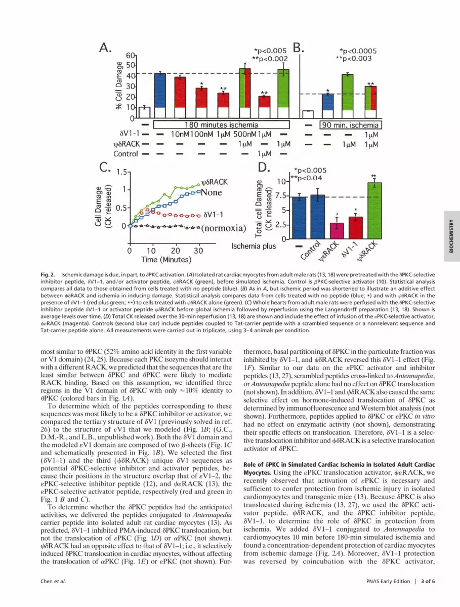
Role of dPKC in Simulated Cardiac Ischemia in Isolated Adult CardiacMyocytes. Using the «PKC translocation activator, c«RACK, werecently observed that activation of «PKC is necessary andsufficient to confer protection from ischemic injury in isolatedcardiomyocytes and transgenic mice (13). Because dPKC is alsotranslocated during ischemia (13, 27), we used the dPKC acti-vator peptide, cdRACK, and the dPKC inhibitor peptide,dV1–1, to determine the role of dPKC in protection fromischemia. We added dV1–1 conjugated to Antennapedia tocardiomyocytes 10 min before 180-min simulated ischemia andfound a concentration-dependent protection of cardiac myocytesfrom ischemic damage (Fig. 2A). Moreover, dV1–1 protectionwas reversed by coincubation with the dPKC activator,
Fig. 2. Ischemic damage is due, in part, to dPKC activation. (A) Isolated rat cardiac myocytes from adult male rats (13, 18) were pretreated with the dPKC-selectiveinhibitor peptide, dV1–1, andyor activator peptide, cdRACK (green), before simulated ischemia. Control is bPKC-selective activator (10). Statistical analysiscompares all data to those obtained from cells treated with no peptide (blue). (B) As in A, but ischemic period was shortened to illustrate an additive effectbetween cdRACK and ischemia in inducing damage. Statistical analysis compares data from cells treated with no peptide (blue; *) and with cdRACK in thepresence of dV1–1 (red plus green; **) to cells treated with cdRACK alone (green). (C) Whole hearts from adult male rats were perfused with the dPKC-selectiveinhibitor peptide dV1–1 or activator peptide cdRACK before global ischemia followed by reperfusion using the Langendorff preparation (13, 18). Shown isaverage levels over time. (D) Total CK released over the 30-min reperfusion (13, 18) are shown and include the effect of infusion of the «PKC-selective activator,c«RACK (magenta). Controls (second blue bar) include peptides coupled to Tat-carrier peptide with a scrambled sequence or a nonrelevant sequence andTat-carrier peptide alone. All measurements were carried out in triplicate, using 3–4 animals per condition.
Chen et al. PNAS Early Edition u 3 of 6
BIO
CHEM
ISTR
Y

cdRACK, but not with bPKC-selective translocation activators(28) or other control peptides (Fig. 2 A and not shown).
In addition to the opposing protective effect induced bydV1–1, cdRACK caused a slight increase in myocyte damageafter an ischemic insult (Fig. 2 A). When we reduced the time ofischemia from 180 to 90 min, the cdRACK-induced increase incell damage became significant and was partially reversed bycotreatment with the dPKC inhibitor, dV1–1 (Fig. 2B). There-fore, cell damage induced by simulated ischemia is caused, atleast in part, by activation of dPKC.
Role of dPKC and «PKC in Simulated Ischemia in Intact Heart. Werecently found that conjugation of PKC inhibitor or activatorpeptides to a Tat-derived peptide [Tat 47–57 (16)] enables theirdelivery through the coronary arteries into intact heart (34).Therefore, to determine the role of dPKC and «PKC on cardiacdamage caused by 45 min of no-flow myocardial ischemia, weperfused Tat-conjugated peptide regulators of these isozymesinto isolated rat hearts. As predicted from our previous cellularand transgenic studies (13), acute activation of «PKC by infusionof c«RACK through the coronary arteries protected the heartfrom ischemia by more than 60% (Fig. 2 C and D). In contrast,activation of dPKC with cdRACK increased cardiac damage by'30% (Fig. 2 C and D). Moreover, acute inhibition of dPKC byinfusion of dV1–1 protected isolated hearts from ischemicdamage as shown by decreased release of CK (Fig. 2 C and D).Scrambled peptides cross-linked to Tat-derived peptide, pep-tides not linked to Tat-derived carrier peptide, or Tat-derivedpeptide alone had no effect on the response of the hearts toischemic insult (Fig. 2D). Together, these data indicate that inthe intact heart as well as in cardiac myocytes, these two closelyrelated PKC isozymes have opposing effects; inhibition of dPKCor activation of «PKC both conferred at least 50% protectionagainst ischemic damage (Fig. 2D).
Our data demonstrating that dPKC activity increases damageby cardiac ischemia may be in contrast to the study of Kihara andcollaborators (29), using a compound that alters the subcellularlocation of dPKC (JTV). They suggested that dPKC protectsisolated rat cardiomyocytes from ischemic damage. However,the drug, which was initially designed to affect annexin, aphospholipid-binding protein, may in fact sequester dPKC andthus inhibit its normal activity.
Role of dPKC and «PKC in Simulated Ischemia in Transgenic Mice. Todetermine the consequences of sustained activation of dPKC byexpression of cdRACK, we created transgenic mice in which thea-myosin heavy chain promoter drove expression of cdRACK,as described for c«RACK (13). Multiple independent lines ofcdRACK mice exhibited identical phenotypes, including in-creased association of dPKC with the subcellular particulate
Fig. 3. cdRACK transgenic mice exhibit increased damage by cardiac isch-emia. (A) Western blot and analysis of PKC distribution in cytosolic andparticulate fractions from cdRACK mice (cdR, Upper; green bars) and c«RACKtransgenic mice (c«R, Lower; magenta bars) and their nontransgenic litter-mates (NTG; white bars) was carried out as described (13) to demonstrateisozyme-selective translocation of PKC. (Right) Histogram shows mean 6 SEMof data from eight mice for each group. (B) Hemodynamic parameters weremonitored in hearts from transgenic cdRACK transgenic mice (green symbols)and their nontransgenic littermates (white symbols) after global ischemia.Left ventricular pressure and real time derivative was monitored via a catheterplaced in the ventricular apex (13). Hemodynamic measurements were re-corded every 20 sec throughout reperfusion. Data are mean 6 SEM of 12cdRACK and 11 nontransgenic mice. (C) Fractions of perfusate from mice usedin B were collected throughout reperfusion and CK activity was assessed todetermine cell damage. For comparison, data from c«RACK mice are alsoincluded. Data are mean 6 SEM of six cdRACK mice (green bar), six nontrans-genic mice (white bar), and seven c«RACK mice (magenta bar). (D) Infarct size
as a percent of the region of risk in mice with sustained dPKC activation(cdRACK mice; green bar) and nontransgenic littermates (white bar) wasdetermined in vivo after coronary occlusion followed by 24 h of reperfusionas described (mean 6 SEM) (22, 23). The area at risk was not significantlydifferent between the nontransgenic and cdRACK mice (36 6 3% and 41 6 5%of left ventricle for nontransgenic and cdRACK mice, respectively). Data arefrom eight 30- to 34-week-old transgenic females and five nontransgenicfemale littermates. So far only three males were available for analysis andtherefore they are not included in the analysis; in all of the other studies, equalnumbers of male and female transgenic and nontransgenic mice were used.(E) Example of infarcts in a cdRACK transgenic mouse (Right) and a littermate(Left) subjected to coronary occlusion and 24 h of reperfusion in vivo. Theportion of the left ventricle supplied by the occluded coronary artery (regionof risk) was identified by the absence of Phthalo blue dye, which was perfusedonly through the nonoccluded vascular bed (22, 23). The infarcted area wasidentified by perfusion with 2,3,5-triphenyltetrazolium chloride, which stainsviable tissue bright red, whereas infarcted tissue is light yellow (22, 23).
4 of 6 u www.pnas.orgycgiydoiy10.1073ypnas.191369098 Chen et al.

fraction (Fig. 3A Upper). We found no changes in overall dPKClevels or in the levels and subcellular association of other PKCisozymes (Fig. 3A and not shown.) For comparison, parallelexperiments were carried out by using c«RACK transgenicmouse hearts (Fig. 3A Lower). We next determined functionalrecovery and cardiomyocyte damage in cdRACK and c«RACKmouse hearts after transient (35 min) no-flow global ischemia.Consistent with the results obtained when peptides were acutelyintroduced into isolated cardiomyocytes (Fig. 2 A and B) orperfused rat hearts (Fig. 2 C and D), the cdRACK mouse heartsshowed significantly impaired recovery of systolic (contraction,1dPydt) and diastolic (relaxation, 2dPydt) functions from isch-emia (Fig. 3B; P , 0.05 and Table 1). In contrast, we previouslyfound that c«RACK mouse hearts had significantly improvedrecovery of cardiac functions after ischemia (13). Furthermore,a significant decrease in cellular damage (CK release) dur-ing reperfusion was observed in c«RACK-expressing mice (Fig.3C, magenta), but not in cdRACK-expressing mice (Fig. 3C,green bar).
Role of dPKC in Simulated Ischemia in Vivo. Next, we used aphysiologically relevant mouse model of acute ischemic injuryinduced by a 30-min in vivo coronary occlusion (22, 23) andmeasured infarct size 24 h after reperfusion. Although the areaat risk for infarction was similar in the two groups, cdRACKmice exhibited over a 2-fold increase in infarct size as comparedwith nontransgenic littermates (Fig. 3D). The marked in vivoincrease in myocyte death in the transgenic mice with sustaineddPKC activation (cdRACK) is illustrated in Fig. 3E, where amuch larger light yellow area (infarcted area) is seen in thecdRACK heart than in the nontransgenic heart. Therefore,activation of dPKC exacerbates damage during ischemia in threedifferent models: isolated myocytes, intact hearts ex vivo, andintact hearts in vivo, which is in contrast to the protective role of«PKC in ischemia (here and ref. 13).
Role of dPKC and «PKC in Cardiac Hypertrophy. We previouslyobserved that older transgenic mouse hearts with sustained«PKC activation (c«RACK mice) developed increased myocar-dial mass (hypertrophy) with normal contractile function,whereas mice with sustained inhibition of «PKC activation
developed lethal dilated cardiomyopathy (21). Given the oppos-ing effects of dPKC and «PKC on cardiac injury in response tosimulated ischemia shown here, we anticipated that transgenicmice with sustained activation of dPKC (cdRACK mice) wouldexhibit an effect on myocardial hypertrophy opposite to that ofc«RACK. Unexpectedly, the cdRACK mice also developed anincrease in cardiac mass similar to age-matched c«RACK mice(Fig. 4A). Note, however, that these mice had normal basal andb-adrenergic-stimulated contractile function (Fig. 4B and Table2; ref. 21) and a normal histological appearance (ref. 21 for«PKC, and data not shown for dPKC). Furthermore, bothtransgenic lines of mice developed identical perturbations ofgene expression; a selective increase in expression of the b-myosin heavy chain gene, a known marker for cardiac hyper-trophy in mice (Fig. 4C) (30, 31). Therefore, the molecular andfunctional characteristics of cardiac hypertrophy in the cdRACKand the c«RACK mice appear identical, indicating a parallelrole for dPKC and «PKC in regulation of cardiac hypertrophy.
Table 2. Basal and b-adrenergic-stimulated contractile function in cdRACK transgenic mice
Measurement Nontransgenic cdRACK*
Fractional shortening 56 1 3% 55 1 3%Baseline 1dPydt 6,315 6 474 mmHgysec 8,386 6 373 mmHgysecPeak 1dPyDt 19,020 6 387 mmHgysec 18,257 6 476 mmHgysecBaseline 2dPydt 27,773 6 306 mmHgysec 29,049 6 621 mmHgysecPeak 2dPyDt 210,858 6 461 mmHgysec 211,751 6 995 mmHgysec
Echocardiogram measurements were taken as described (13, 21). In vivo catheterization measurements shownfor baseline and b-adrenergic-stimulated contractile function (dobutamine, 1 ngyg per min for 3 min; dose wascontinuously doubled up to 32 ngyg per min). Data are mean 6 SEM of nine cdRACK and nine nontransgenic miceof 12 weeks of age. *, P 5 not significant for all measurements.
Table 1. Left ventricular developed pressure of cdRACKtransgenic mice
MeasurementNontransgenic,
mmHgyseccdRACK,*mmHgysec
1dPydt 3,652 6 167 2,582 6 3732dPydt 22,248 6 140 21,572 6 169
Positive developed pressure over time (1dPydt) and negative developedpressure over time (2dPydt) were recorded throughout reperfusion (13). Datawere taken 7 min after reperfusion and are mean 6 SEM of 12 cdRACK and 11nontransgenic mice of 12 weeks of age. *, P , 0.05.
Fig. 4. cdRACK transgenic mice exhibit hypertrophy similar to c«RACK mice.(A) Dry heart weights of nontransgenic and cdRACK-expressing transgenicmice were measured as in ref. 13, demonstrating hypertrophy in hearts ofcdRACK and c«RACK mice. Data are mean 6 SEM from 10 cdRACK mice (greenbar), five nontransgenic mice (white bar), and three c«RACK mice (magentabar). (B) Left ventricular fractional shortening was measured in the nontrans-genic and transgenic mice as described (13, 21). Data are mean 6 SEMobtained from nine cdRACK mice (green bar), 10 nontransgenic mice (whitebar), and four c«RACK mice (magenta bar). (C) Dot blot analysis of mRNA fromhearts of transgenic and nontransgenic mice showing increased expression ofbMHC, a marker for hypertrophy (33). GADPH, glyceraldehyde-3-phosphatedehydrogenase; aMHC, a-myosin heavy chain; bMHC, b-myosin heavy chain;ANF, atrial natriuritic factor; SERCA, sarcoplasmic reticular ATPase; PLB, phos-pholamban; aSK actin, a skeletal actin.
Chen et al. PNAS Early Edition u 5 of 6
BIO
CHEM
ISTR
Y
DiscussionWe have used selective inhibitor and activator peptides of dPKCand «PKC translocation to show opposing roles in response toischemia and similar roles in cardiac hypertrophy for these twoisozymes. We presented data demonstrating the role of theseisozymes in cardiac protection from ischemia by using twospecies (rat and mouse), three model systems for simulatedischemia (isolated myocytes, intact heart, and in vivo), threemethods of peptide delivery (transgene and acute peptide de-livery using Antennapedia- or Tat-carrier peptides), and at leastfour methods of damage assessment (CK release, dye exclusion,functional recovery of contraction, and infarct size). Therefore,multiple independent methods used to assess the role of dPKCand «PKC yielded the same conclusion: inhibiting dPKC oractivating «PKC reduce damage from simulated ischemia. Sim-ilarly, using multiple methods we found that activation andtranslocation of dPKC and «PKC isozymes in transgenic micecause cardiac hypertrophy.
These data raise four important points. First, it is possible toidentify highly efficacious activator and inhibitor peptides ofdPKC translocation, using a structure-based rational design.[These peptides were active at intracellular concentrations of'25–50 nM, about 5–10% of the applied concentration (8).] Toour knowledge, there are no examples of rationally designedactivators of signaling enzymes that act intracellularly except forour own. The development of these selective regulators of dPKCrelied not only on the assumption that PKC-RACK interactionscould be regulated as a means to selectively modify dPKCactivity, but was achieved without any knowledge of the dPKCbinding protein, dRACK. This method provides an alternative tomutational mapping of protein–protein interactions or random
peptide library searches requiring the presence of both proteinsand should be generally applicable to research on other protein–protein interactions.
Second, our findings that PKC isozymes can exert an opposingeffect on one function and the same effect on another functionin a single cell can explain contradictory findings when isozymenonselective drugs are used. We suggest that previous conflictingreports on the role of PKC in ischemic preconditioning (1), forexample, may reflect such use of drugs that do not distinguishbetween dPKC and «PKC.
Third, our observation that dPKC and «PKC have opposingeffects on ischemic injury was particularly unexpected, becausein rats and mice both isozymes are activated by simulatedischemia as well as by stimuli that lead to cardioprotection fromischemia (18, 27). These opposing forces (‘‘yin yang’’) illustratethe need for selective therapeutic agents to treat ischemic heartdisease.
Finally, we show that these biologically active peptides can beeffectively delivered into organs via the blood vessels by usingTat-conjugated peptides. Moreover, this mode of peptide deliv-ery also should be useful for the study of other diseases in intactanimals. Because delivery is immediate, it is superior to genedelivery, as adaptations to the changes induced by a signaltransduction modulator are less likely to occur. Because wefound a greater than 50% reduction in cardiac damage caused byischemia by coronary perfusion of dPKC translocation inhibitorpeptide or «PKC activator peptide, we believe that these pep-tides may be useful therapeutic agents for acute cardiac ischemia.
This work was supported, in part, by National Institutes of Health GrantsHL52141 (to D.M.-R.), HL43151 and HL55757 (to R.B.), and HL52318(to G.W.D.). D.S. is supported by the Ares Serano Foundation.
1. Brooks, G. & Hearse, D. J. (1996) Circ. Res. 79, 627–630.2. Gu, X. & Bishop, S. P. (1994) Circ. Res. 75, 926–931.3. Bowling, N., Walsh, R. A., Song, G., Estridge, T., Sandusky, G. E., Fouts, R. L.,
Mintze, K., Pickard, T., Roden, R., Bristow, M. R., et al. (1999) Circulation 99,384–391.
4. Jalili, T., Takeishi, Y., Song, G., Ball, N. A., Howles, G. & Walsh, R. A. (1999)Am. J. Physiol. 277, H2298–H2304.
5. Mochly-Rosen, D., Henrich, C. J., Cheever, L., Khaner, H. & Simpson, P. C.(1990) Mol. Biol. Cell 1, 693–706.
6. Disatnik, M.-H., Buraggi, G. & Mochly-Rosen, D. (1994) Exp. Cell Res. 210,287–297.
7. Mochly-Rosen, D. (1995) Science 268, 247–251.8. Souroujon, M. & Mochly-Rosen, D. (1998) Nat. Biotechnol. 16, 919–924.9. Mochly-Rosen, D., Miller, K. G., Scheller, R. H., Khaner, H., Lopez, J. &
Smith, B. L. (1992) Biochemistry 31, 8120–8124.10. Ron, D., Luo, J. & Mochly-Rosen, D. (1995) J. Biol. Chem. 270, 24180–24187.11. Sossin, W. S. & Schwartz, J. H. (1993) Trends Biochem. Sci. 18, 207–208.12. Johnson, J. A., Gray, M. O., Chen, C.-H. & Mochly-Rosen, D. (1996) J. Biol.
Chem. 271, 24962–24966.13. Dorn, G. W., II, Souroujon, M. C., Liron, T., Chen, C. H., Gray, M. O., Zhou,
H. Z., Csukai, M., Wu, G., Lorenz, J. N. & Mochly-Rosen, D. (1999) Proc. Natl.Acad. Sci. USA 96, 12798–12803.
14. Ping, P., Zhang, J., Pierce, W. M. & Bolli, R. (2001) Circ. Res. 88, 59–62.15. Derossi, D., Joliot, A. H., Chassaing, G. & Prochiantz, A. (1994) J. Biol. Chem.
269, 10444–10450.16. Schwarze, S. R., Ho, A., Vocero-Akbani, A. & Dowdy, S. F. (1999) Science 285,
1569–1572.17. Nalefski, E. A. & Falke, J. J. (1996) Protein Sci. 5, 2375–2390.18. Chen, C. H., Gray, M. O. & Mochly-Rosen, D. (1999) Proc. Natl. Acad. Sci.
USA 96, 12784–12789.19. Armstrong, S., Downey, J. M. & Ganote, C. E. (1994) Cardiovasc. Res. 28, 72–77.
20. Armstrong, S. C., Kao, R., Gao, W., Shivell, L. C., Downey, J. M., Honkanen,R. E. & Ganote, C. E. (1997) J. Mol. Cell. Cardiol. 29, 3009–3024.
21. Mochly-Rosen, D., Wu, G., Hahn, H., Osinska, H., Liron, T., Lorenz, J. N.,Yatani, A., Robbins, J. & Dorn, G. W., II (2000) Circ. Res. 86, 1173–1179.
22. Guo, Y., Wu, W. J., Qiu, Y., Tang, X. L., Yang, Z. & Bolli, R. (1998) Am. J.Physiol. 275, H1375–H1387.
23. Guo, Y., Jones, W. K., Xuan, Y. T., Tang, X. L., Bao, W., Wu, W. J., Han, H.,Laubach, V. E., Ping, P., Yang, Z., et al. (1999) Proc. Natl. Acad. Sci. USA 96,11507–11512.
24. Osada, S.-I., Mizuno, K., Saido, T. C., Suzuki, K., Kuroki, T. & Ohno, S. (1992)Mol. Cell. Biol. 12, 3930–3938.
25. Baier, G., Telford, D., Giampa, L., Coggeshall, K. M. & Baier-Bitterlich, G.(1993) J. Biol. Chem. 268, 4997–5004.
26. Pappa, H., Murray-Rust, J., Dekker, L. V., Parker, P. J. & McDonald, N. Q.(1998) Structure (London) 6, 885–894.
27. Gray, M. O., Karliner, J. S. & Mochly-Rosen, D. (1997) J. Biol. Chem. 272,30945–30951.
28. Ron, D. & Mochly-Rosen, D. (1995) Proc. Natl. Acad. Sci. USA 92, 492–496.29. Inagaki, K., Kihara, Y., Hayashida, W., Izumi, T., Iwanaga, Y., Yoneda, T.,
Takeuchi, Y., Suyama, K., Muso, E. & Sasayama, S. (2000) Circulation 101,797–809.
30. Sakata, Y., Hoit, B. D., Liggett, S. B., Walsh, R. A. & Dorn, G. W., II (1998)Circulation 97, 1488–1495.
31. Adams, J. W., Sakata, Y., Davis, M. G., Sah, V. P., Wang, Y., Liggett, S. B.,Chien, K. R., Brown, J. H. & Dorn, G. W., II (1998) Proc. Natl. Acad. Sci. USA95, 10140–10145.
32. Rizo, J. & Sudhof, T. C. (1998) J. Biol. Chem. 273, 15879–15882.33. Dorn, G. W., II, Tepe, N. M., Lorenz, J. N., Koch, W. J. & Liggett, S. B. (1999)
Proc. Natl. Acad. Sci. USA 96, 6400–6405.34. Chen, L., Wright, L., Chen, C. H., Oliver, S., Wender, P. & Mochly-Rosen,
D. (2001) Chem. Biol., in press.
6 of 6 u www.pnas.orgycgiydoiy10.1073ypnas.191369098 Chen et al.
Related Documents