1 Rh Disease: Prevention and Management Gregory L Goyert, MD Division Head, MFM, WHS’s Henry Ford Health System Management of Rh disease Isoimmunization and prevention Overview Prophylaxis Background Management details Isoimmunization Etiology Management details ACOG Practice Bulletin # 181; August 2017 Management of Rh disease Great obstetric success story First successful in utero therapy Uncommon encounter in practice Other Rh antigens (C, c, E, e) Primary duty is to prevent isoimmunization Isoimmunization management evolving 1 2 3

Welcome message from author
This document is posted to help you gain knowledge. Please leave a comment to let me know what you think about it! Share it to your friends and learn new things together.
Transcript
1
Rh Disease: Prevention and Management
Gregory L Goyert, MD
Division Head, MFM, WHS’s
Henry Ford Health System
Management of Rh diseaseIsoimmunization and prevention
Overview
Prophylaxis Background
Management details
Isoimmunization Etiology
Management details
ACOG Practice Bulletin # 181; August 2017
Management of Rh disease Great obstetric success story
First successful in utero therapy
Uncommon encounter in practice
Other Rh antigens (C, c, E, e)
Primary duty is to prevent isoimmunization
Isoimmunization management evolving
1
2
3
2
Management of Rh diseaseGenetics
Fisher and Race 1946 Proposed 3 genes for 3 rhesus antigen groups
D, C/c, E/e
1991, rhesus locus localized to short arm chromosome #1 1p34-1p36
Only 2 genes identified: RhD and RhCE
RhD encodes D; absent in Rh negative
RhC/c and E/e inherited linked manner to RhD
ACOG Practice Bulletin # 181; August 2017
Management of Rh diseaseGenetics
D antigen 7-10,000 mw Appears early: 38-day embryo Physiologic function unclear “D” antigen critical Three RhD antigen twists
Weak D’s Reduced number D antigens expressed
Partial D’s ‘Missing’ portions of D antigen
When exposed to RhD+ rbc’s, patients can form anti-D antibodies to their missing or variant D epitopes
RhD pseudogene
ACOG Practice Bulletin # 181; August 2017
Management of Rh DiseaseGenetics
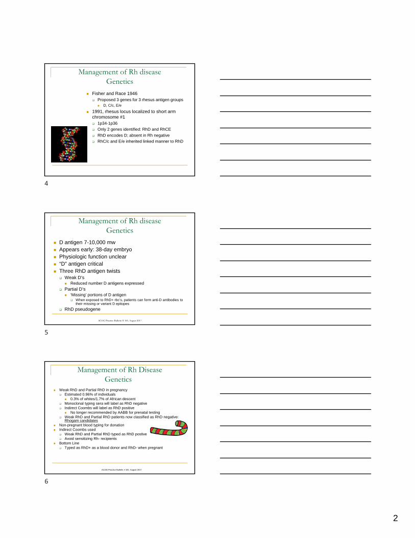
Weak RhD and Partial RhD in pregnancy Estimated 0.96% of individuals
0.3% of whites/1.7% of African descent Monoclonal typing sera will label as RhD negative Indirect Coombs will label as RhD positive
No longer recommended by AABB for prenatal testing Weak RhD and Partial RhD patients now classified as RhD negative:
Rhogam candidates Non-pregnant blood typing for donation Indirect Coombs used
Weak RhD and Partial RhD typed as RhD postive Avoid sensitizing Rh- recipients
Bottom Line Typed as RhD+ as a blood donor and RhD- when pregnant
4
5
6
3
Obstet Gynecol 2012; 120:1428-38
RhoGam GuidelinesD Variants
Management of Rh diseaseGenetics
RhD pseudogene 69% S. African blacks; 21% African Americans
Serologically RhD negative But, entire RhD gene present on chromosome
Amniotic PCR testing would yield false + Fetus RhD negative phenotype (serology)
Fetus RhD positive by genotype
Risk of unnecessary intervention Submit maternal blood with AF for fetal RhD typing to exclude
presence of RhD pseudogene
ACOG Practice Bulletin #4 Reaffirmed 2016
Management of Rh diseasePrevention
Rh negative incidence 16%
Concept of passive antibody to prevent active isoimmunization
Antibody mediated immune suppression
First applied to Rh disease in early 1960’s
Half-life of RhoGAM approximately 24 days
ACOG Practice Bulletin # 181; August 2017
7
8
9
4
Management of Rh diseasePrevention
First large postpartum trial 1968
Yielded 10 fold decrease (1.8% v. 16%)
72 hour ‘window’ due to prisoner trials
Protection demonstrated at 13 days
Some recommend administration out to 28 days
Antepartum administration effective
Yielded another 10 fold decrease (0.1% v. 1.8%)
300 ug RhoGAM “covers” 15cc fetal RBC’s or 30cc fetal whole blood
ACOG Practice Bulletin # 181; August 2017
Management of Rh diseasePrevention
Indications for RhoGAM Spontaneous/voluntary abortion
Threatened abortion Ectopic CVS/amniocentesis/cordocentesis 28 weeks/postpartum Antepartum hemorrhage External cephalic version Trauma Hydatidiform mole IUFD ? Following postpartum tubal ligation
TL failure with future pregnancies Avoid cross-matching issues with future transfusions
ACOG Practice Bulletin # 181; August 2017
NEJM 1999;340:1228-33
Management of Rh diseasePrevention
RhoGAM mechanism of action Likely central inhibition Rh IgG-D antigen complexes may stimulate “immune suppressor
substance” that blunts immunologic response Antigen blocking/deviation mechanism less likely Derived from male donors who undergo repeated injections of
RhD positive rbc’s No reported cases viral infection
Scattered Hep C exposures prior to 1995
10
11
12
5
Management of Rh diseasePrevention
Routine testing first prenatal visit ABO typing Rh status determination Antibody screen
Repeat antibody screen at 28 weeks’ Low risk of isoimmunization before 28 weeks Administer 300 ug RhoGAM 50 ug dose not often employed clinically
ACOG Practice Bulletin # 181; August 2017
ACOG Practice Bulletin # 181; August 2017
Management of Rh diseasePrevention
Routine testing at time of delivery Maternal antibody screen
Neonatal typing
Routine testing for excessive fetal-maternal hemorrhage for Rh negative patients Rosette test as screen
Kleihauer-Betke if rosette screen positive
Percentage of fetal cells multiplied by factor of 50 to estimate fetal-maternal hemorrhage
ACOG Practice Bulletin # 181; August 2017
Details of Screening for Fetomaternal Hemorrhage
Rosette test Qualitative; identifies Rh+ cells in Rh- patient
Exogenous anti-D antibodies are mixed with maternal blood and adhere to any Rh D+ fetal red cells
Rh D+ “indicator” red cells then added; form rosettes around coated fetal red cells
Clusters or rosettes easily identified under microscopy
Not appropriate when antenatal fetomaternal hemorrhage suspected; quantitative test should be pursued
13
14
15
6
ACOG Practice Bulletin # 181; August 2017
Details of Screening for Fetomaternal Hemorrhage
Kleihauer-Betke test; 1957 Semi-quantitative
Based on fetal blood having hemoglobin F
Smear of maternal blood obtained Dried; immersed in fixative; incubated in acid solution;
stained with erythrosine B
Hemoglobin F- containing red cells (fetal) appear cherry red; adult red cells appear as uncolored ghost cells
Fetal cells counted; expressed as % of adult cells
ACOG Practice Bulletin # 181; August 2017
Details of Screening for Fetomaternal Hemorrhage
Kleihauer-Betke
Labor intensive; 10,000 cells must be counted
Turnaround time variable; technician dependent
May underestimate amount of hemorrhage
Cells fail to stain; decreasing Hbg F concentration
Overestimate if maternal blood contains hemoglobin F
Increases in pregnancy; peaks mid-gestation
SC anemia; beta thalassemia; hereditary persistence
ACOG Practice Bulletin # 181; August 2017
Details of Screening for Fetomaternal Hemorrhage
Flow cytometry as an alternative
Quantifies fetal cells by measuring fluorescence intensity of monoclonal antibodies to Hbg F
Fluorescence intensity fetal Hbg F-containing cells greater than adult Hgb F-containing cells
Also measures red cell size/distinguish from adult
More objective; improved precision and accuracy
Coefficient of variation 10% (FC) v. 153% (KB)
Less labor intensive; 60 minutes to perform
Currently, only used in 4% of US labs for screening
16
17
18
7
Obstet Gynecol 2012;120:1428-38
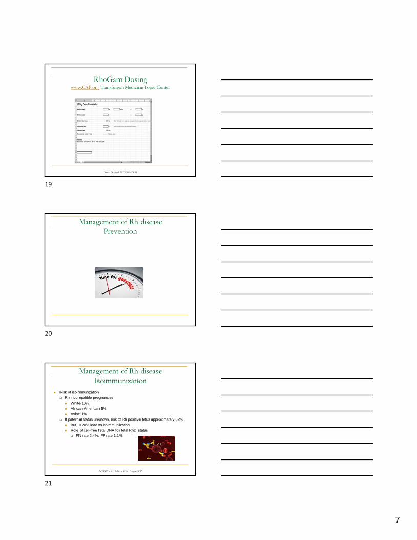
RhoGam Dosingwww.CAP.org Transfusion Medicine Topic Center
Management of Rh diseasePrevention
ACOG Practice Bulletin # 181; August 2017
Management of Rh diseaseIsoimmunization
Risk of isoimmunization
Rh incompatible pregnancies
White 10%
African-American 5%
Asian 1%
If paternal status unknown, risk of Rh positive fetus approximately 62%
But, < 20% lead to isoimmunization
Role of cell-free fetal DNA for fetal RhD status
FN rate 2.4%; FP rate 1.1%
19
20
21
8
Management of Rh diseaseIsoimmunization
Risk of isoimmunization
If fetus ABO compatible; 16%
If fetus ABO incompatible; 1.5-2%
Most protective
Maternal type “O”
Paternal type “A”, “B”, or “AB”
First trimester Spontaneous abortion; 2%
Second trimester vtp 4-5%
ACOG Practice Bulletin # 181; August 2017
Management of Rh diseaseIsoimmunization
Requirements for isoimmunization
Rh positive fetus
Rh negative mother
Maternal immunocompetence
Fetal-maternal hemorrhage
First sensitized pregnancy usually results in minimal fetal/neonatal disease
Subsequent gestations associated with worsening degrees of fetal anemia
In general, these principles apply to other antigens
Kell, Kidd, Duffy
ACOG Practice Bulletin #75 Reaffirmed 2016
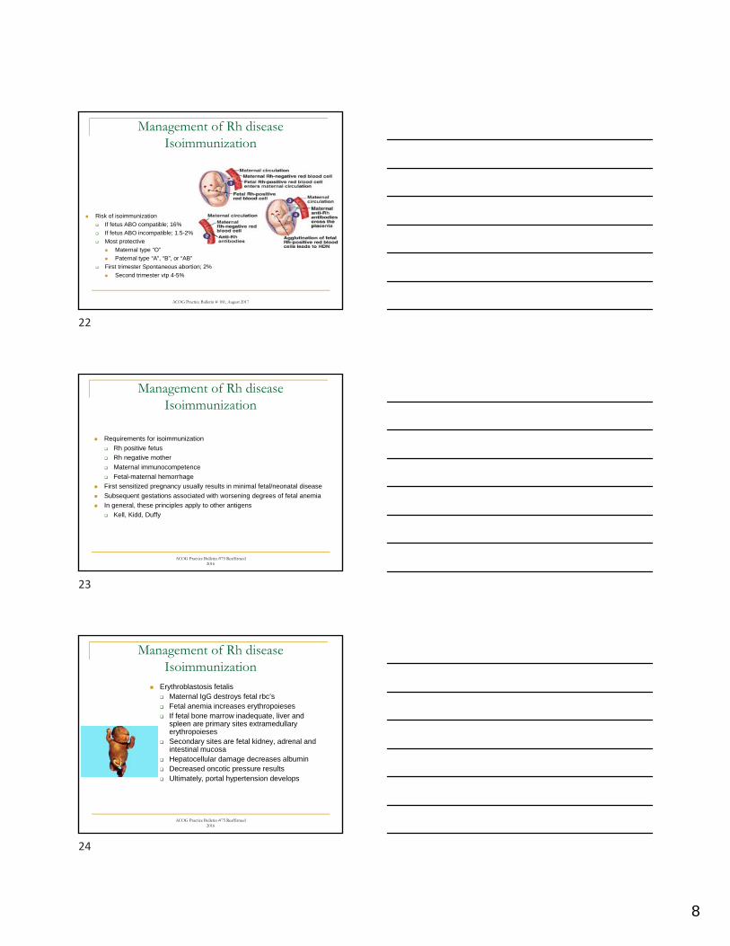
Management of Rh diseaseIsoimmunization
Erythroblastosis fetalis Maternal IgG destroys fetal rbc’s Fetal anemia increases erythropoieses If fetal bone marrow inadequate, liver and
spleen are primary sites extramedullary erythropoieses
Secondary sites are fetal kidney, adrenal and intestinal mucosa
Hepatocellular damage decreases albumin Decreased oncotic pressure results Ultimately, portal hypertension develops
ACOG Practice Bulletin #75 Reaffirmed 2016
22
23
24
9
Obstet Gynecol 2012;120:1132-39
Management of Rh diseaseIsoimmunization
Prior obstetric history important Fetal demise Neonatal transfusion
Evaluate paternal antigen status/zygosity Historically, linkage analysis used Quantitative PCR better tool Cell-free fetal DNA for fetal RhD detection evolving
Standard in many European centers Reverse transcriptase PCR amplify specific RhD exons If RhD positive, fetus at risk for anemia If RhD negative, must confirm fetal DNA via SNPs analysis
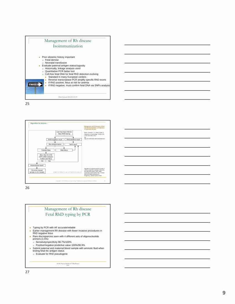
Algorithm for determ...
Algorithm for determining the results of cell-free fetal DNA testing to determine the fetal RHD status. SNP, single-nucleotide polymorphism.Fig. 1. Moise. Red Cell Alloimmunization in Pregnancy. Obstet Gynecol 2012.
Copyright © 2013 Obstetrics & Gynecology. Published by Lippincott Williams & Wilkins. 26
Management and Prevention of Red Cell Alloimmunization in Pregnancy: A Systematic Review
Moise, Kenneth J. Jr; Argoti, Pedro S.
Obstetrics & Gynecology. 120(5):1132-1139, November 2012.
doi: http://10.1097/AOG.0b013e31826d7dc1
Management of Rh diseaseFetal RhD typing by PCR
Typing by PCR with AF accurate/reliable Earlier management Rh disease with fewer invasive procedures in
RhD negative fetus Rare discrepancies seen with 4 different sets of oligonucleotide
primers (1.5%) Sensitivity/specificity 98.7%/100% Positive/negative predictive value 100%/96.9%
Submit paternal and maternal blood sample with amniotic fluid when testing fetal rbc antigen status Evaluate for RhD pseudogene
ACOG Practice Bulletin #75 Reaffirmed 2016
25
26
27
10
Management of Rh diseaseIsoimmunization
“Critical” titer of 1:16
Varies from 1:8-1:32
At critical titer, additional testing required
First affected pregnancy only
Titers less reliable for Kell isoimmunization
Evolution of surveillance tools
Amniocentesis
Essentially historical
Cordocentesis
For IUIVT
MCA PSV Doppler interrogation
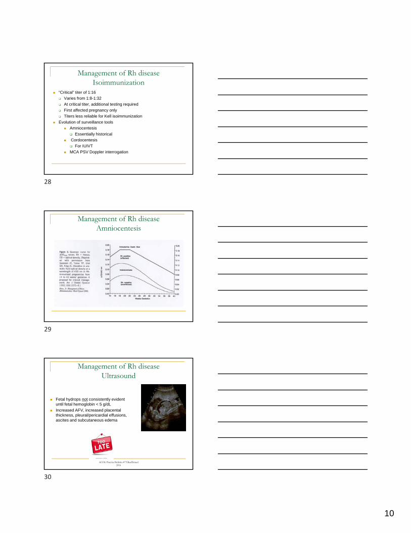
Management of Rh diseaseAmniocentesis
Management of Rh diseaseUltrasound
Fetal hydrops not consistently evident until fetal hemoglobin < 5 g/dL
Increased AFV, increased placental thickness, pleural/pericardial effusions, ascites and subcutaneous edema
ACOG Practice Bulletin #75 Reaffirmed 2016
28
29
30
11
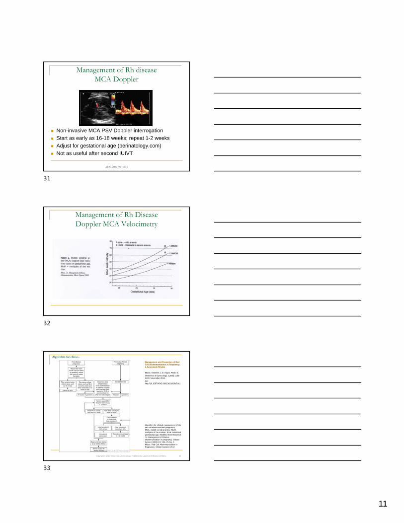
AJOG 2006;195:1550-6
Management of Rh disease MCA Doppler
Non-invasive MCA PSV Doppler interrogation
Start as early as 16-18 weeks; repeat 1-2 weeks
Adjust for gestational age (perinatology.com)
Not as useful after second IUIVT
Management of Rh DiseaseDoppler MCA Velocimetry
Algorithm for clinic...
Algorithm for clinical management of the red cell alloimmunized pregnancy. MCA, middle cerebral artery; MoM, multiples of the median; EGA, estimated gestational age. Modified from Moise KJ Jr. Management of Rhesus alloimmunization in pregnancy. Obstet Gynecol 2008;112:164–76.Fig. 2. Moise. Red Cell Alloimmunization in Pregnancy. Obstet Gynecol 2012.
Copyright © 2013 Obstetrics & Gynecology. Published by Lippincott Williams & Wilkins. 33
Management and Prevention of Red Cell Alloimmunization in Pregnancy: A Systematic Review
Moise, Kenneth J. Jr; Argoti, Pedro S.
Obstetrics & Gynecology. 120(5):1132-1139, November 2012.
doi: http://10.1097/AOG.0b013e31826d7dc1
31
32
33
12
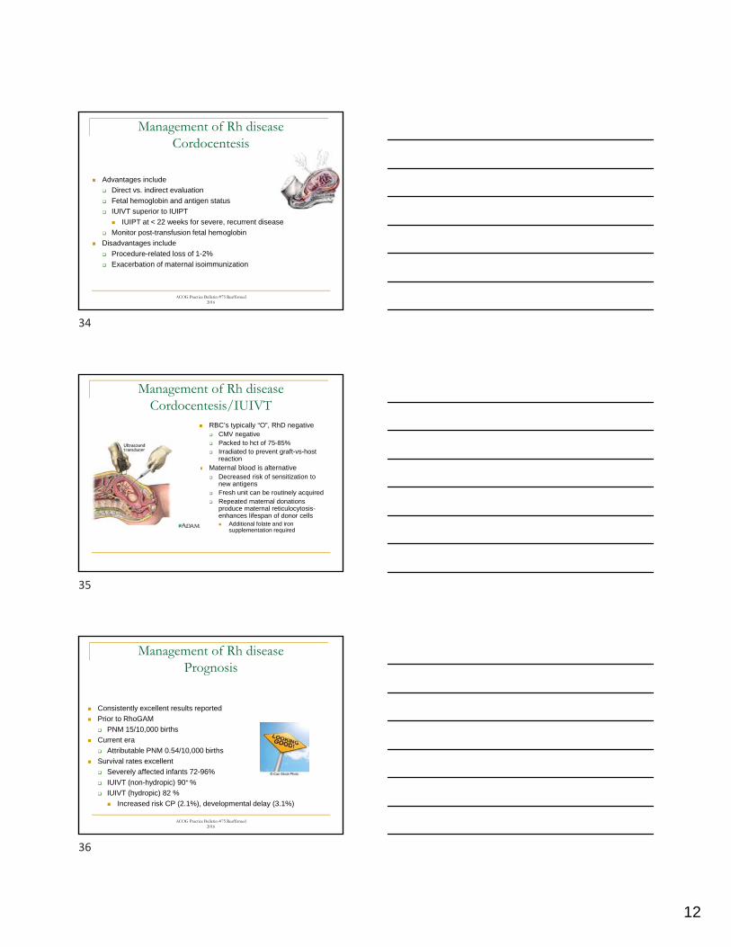
Management of Rh diseaseCordocentesis
Advantages include
Direct vs. indirect evaluation
Fetal hemoglobin and antigen status
IUIVT superior to IUIPT
IUIPT at < 22 weeks for severe, recurrent disease
Monitor post-transfusion fetal hemoglobin
Disadvantages include
Procedure-related loss of 1-2%
Exacerbation of maternal isoimmunization
ACOG Practice Bulletin #75 Reaffirmed 2016
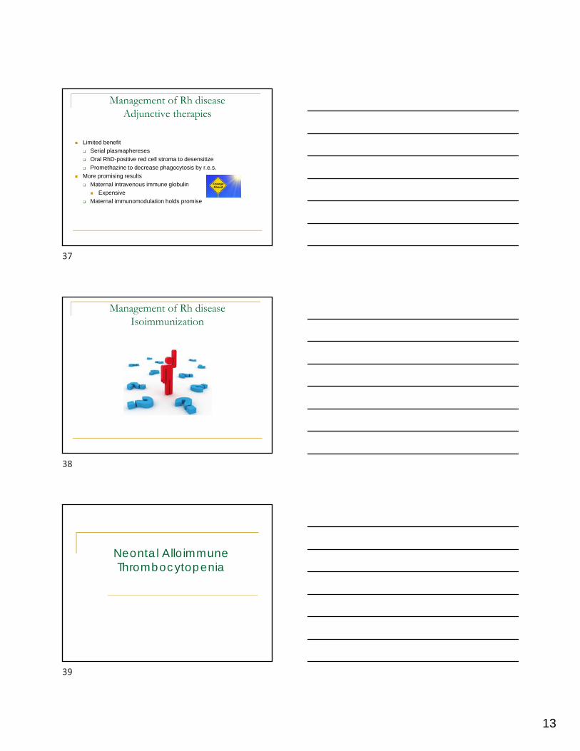
Management of Rh diseaseCordocentesis/IUIVT
RBC’s typically “O”, RhD negative CMV negative Packed to hct of 75-85% Irradiated to prevent graft-vs-host
reaction Maternal blood is alternative
Decreased risk of sensitization to new antigens
Fresh unit can be routinely acquired Repeated maternal donations
produce maternal reticulocytosis-enhances lifespan of donor cells Additional folate and iron
supplementation required
ACOG Practice Bulletin #75 Reaffirmed 2016
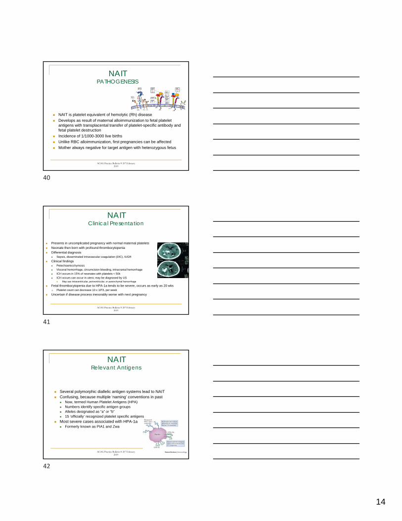
Management of Rh diseasePrognosis
Consistently excellent results reported
Prior to RhoGAM
PNM 15/10,000 births
Current era
Attributable PNM 0.54/10,000 births
Survival rates excellent
Severely affected infants 72-96%
IUIVT (non-hydropic) 90+ %
IUIVT (hydropic) 82 %
Increased risk CP (2.1%), developmental delay (3.1%)
34
35
36
13
Management of Rh diseaseAdjunctive therapies
Limited benefit
Serial plasmaphereses
Oral RhD-positive red cell stroma to desensitize
Promethazine to decrease phagocytosis by r.e.s.
More promising results
Maternal intravenous immune globulin
Expensive
Maternal immunomodulation holds promise
Management of Rh diseaseIsoimmunization
Neontal Alloimmune Thrombocytopenia
37
38
39
14
NAITPATHOGENESIS
NAIT is platelet equivalent of hemolytic (Rh) disease
Develops as result of maternal alloimmunization to fetal platelet antigens with transplacental transfer of platelet-specific antibody and fetal platelet destruction
Incidence of 1/1000-3000 live births
Unlike RBC alloimmunization, first pregnancies can be affected
Mother always negative for target antigen with heterozygous fetus
ACOG Practice Bulletin # 207 February 2019
NAIT Clinical Presentation
Presents in uncomplicated pregnancy with normal maternal platelets
Neonate then born with profound thrombocytopenia
Differential diagnosis Sepsis, disseminated intravascular coagulation (DIC), IUGR
Clinical findings Petechiae/ecchymosis
Visceral hemorrhage, circumcision bleeding, intracranial hemorrhage
ICH occurs in 15% of neonates with platelets < 50k
ICH occurs can occur in utero; may be diagnosed by US May see intraventricular, periventricular, or parenchymal hemorrhage
Fetal thrombocytopenia due to HPA-1a tends to be severe, occurs as early as 20 wks Platelet count can decrease 10 x 109/L per week
Uncertain if disease process inexorably worse with next pregnancy
ACOG Practice Bulletin # 207 February 2019
NAIT Relevant Antigens
Several polymorphic diallelic antigen systems lead to NAIT Confusing, because multiple ‘naming’ conventions in past
Now, termed Human Platelet Antigens (HPA) Numbers identify specific antigen groups Alleles designated as “a” or “b” 15 ‘officially’ recognized platelet specific antigens
Most severe cases associated with HPA-1a Formerly known as PIA1 and Zwa
ACOG Practice Bulletin # 207 February 2019
40
41
42
15
NAITEvaluation
BloodCenter of Wisconsin most reliable lab for consultation
Determination of maternal/paternal HPA type and zygosity
Confirmation of maternal antiplatelet antibodies with specificity for paternal (or fetal-neonatal) platelets and incompatible antigen
Fetal platelet antigen typing from amniocytes or cff DNA (if paternal heterozygote)
No reliable indirect method to assess fetal platelet count
Maternal antiplatelet antibody titers correlate poorly with disease severity
Prior siblings’ clinical course does not reliably predict
Umbilical blood sampling only accurate means to assess fetal platelets
Associated with 10+% rate of emergency CS
ACOG Practice Bulletin # 207 February 2019
NAITManagement
Risk assessment
Standard: presence of HPA antibody but no prior intracranial bleed
High: HPA antibody with intracranial bleed in prior child > 28 weeks
Very high: HPA antibody with intracranial bleed/IUFD prior child < 28 weeks
Start treatment between 16-20 weeks depending upon risk
IVIG 1 gm/kg maternal bw administered weekly
May add prednisone 1 mg/kg starting at 24 weeks based on risk
Platelet transfusions (IUIVT) only for rescue therapy
Allogenieic or washed maternal platelets
Fetal platelet sampling only after 32 weeks if TOL being considered
Route of delivery counseling important
Recommend CS at 38 weeks/prior to labor for high risk groups
ACOG Practice Bulletin # 207 February 2019
Non-Immune Hydrops
Gregory L Goyert, MD
Division Head, MFM, WHS
Henry Ford Health System
43
44
45
16
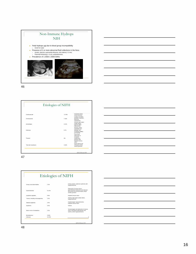
Non-Immune HydropsNIH
Fetal Hydrops not due to blood group incompatibility Greek for water
Presence of 2 or more abnormal fluid collections in the fetus Ascites, pleural or pericardial effusions, skin edema (> 5 mm)
Placental thickening (> 4 cm), polyhydramnios
Prevalence of 1:2500-1:3500 births
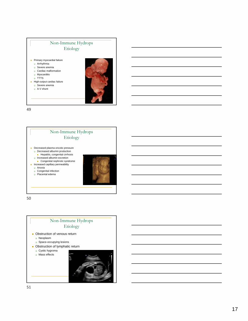
Etiologies of NIFH
Cardiovascular 17-35%Increased central venous pressure
Chromosomal 7-16%
Cardiac anomalies, lymphatic dysplasia, abnormal myelopoiesis
Hematologic 4-12%
Anemia, high output cardiac failure; hypoxia (alpha thalassemia)
Infectious 5-7%
Anemia, anoxia, endothelial cell damage, and increased capillary permeability
Thoracic 6%
Vena cavalobstruction or increased intrathoracicpressure with impaired venous return
Twin-twin transfusion 3-10%Hypervolemia and increased central venous pressure
AJOG February 2015
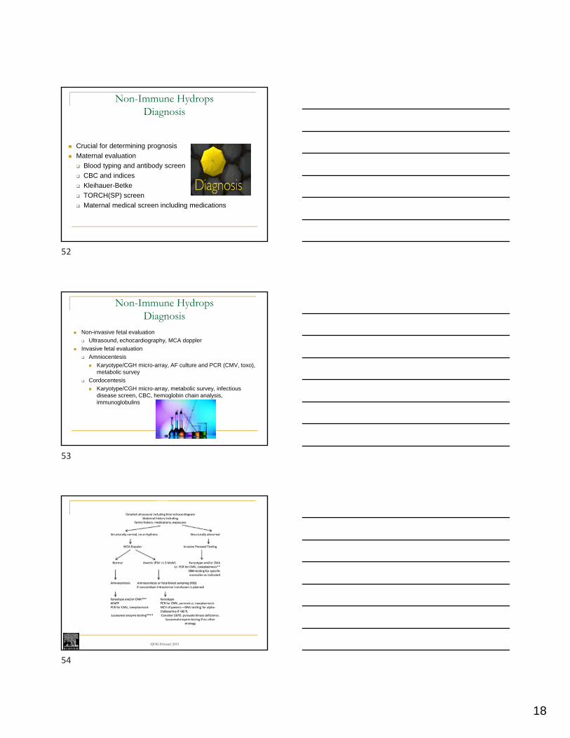
Etiologies of NIFH
Urinary tract abnormalities 2-3%Urinary ascites; nephrotic syndrome with hypoproteinemia
Gastrointestinal 0.5-4%
Obstruction of venous return; gastrointestinal obstruction and infarction with protein loss and decreased colloid osmotic pressure
Lymphatic dysplasia 5-6% Impaired venous return
Tumors, including chorioangiomas 2-3%Anemia, high output cardiac failure, hypoproteinemia
Skeletal dysplasias 3-4%Hepatomegaly, hypoproteinemia, impaired venous return
Syndromic 3-4% Various
Inborn errors of metabolism 1-2%Visceromegaly and obstruction of venous return, decreased erythropoiesis and anemia, and/or hypoproteinemia
Miscellaneous 3-15%
Unknown 15-25%
AJOG February 2015
46
47
48
17
Non-Immune HydropsEtiology
Primary myocardial failure
Arrhythmia
Severe anemia
Cardiac malformation
Myocarditis
TTTS
High-output cardiac failure
Severe anemia
A-V shunt
Non-Immune HydropsEtiology
Decreased plasma oncotic pressure Decreased albumin production
Hepatitis, congenital cirrhosis Increased albumin excretion
Congenital nephrotic syndrome Increased capillary permeability
Anoxia Congenital infection Placental edema
Non-Immune HydropsEtiology
Obstruction of venous return Neoplasm
Space-occupying lesions
Obstruction of lymphatic return Cystic hygroma
Mass effects
49
50
51
18
Non-Immune HydropsDiagnosis
Crucial for determining prognosis
Maternal evaluation
Blood typing and antibody screen
CBC and indices
Kleihauer-Betke
TORCH(SP) screen
Maternal medical screen including medications
Non-Immune HydropsDiagnosis
Non-invasive fetal evaluation
Ultrasound, echocardiography, MCA doppler
Invasive fetal evaluation
Amniocentesis
Karyotype/CGH micro-array, AF culture and PCR (CMV, toxo), metabolic survey
Cordocentesis
Karyotype/CGH micro-array, metabolic survey, infectious disease screen, CBC, hemoglobin chain analysis, immunoglobulins
Figure 2
American Journal of Obstetrics & Gynecology 2015 212, 127-139DOI: (10.1016/j.ajog.2014.12.018) Copyright © 2015 Elsevier I
AJOG February 2015
52
53
54
19
Non-Immune HydropsPrognosis
Prognosis generally poor
95 % mortality if structural anomaly present
Therapy dependent upon etiology
Transfusion
Anti-Arrhythmic medications
Best prognosis groups (70% survival)
Tachyarrhythmias
Hematologic disorders
Hydro/chylo-thorax groups
Therapy for Selected Etiologies of NIFH
Etiology Therapy Recommendation
Cardiac tachyarrhythmia, supraventricular tachycardia, atrial flutter, or atrial fibrillation
Maternal transplacental administration of antiarrhythmic medication(s)
Treatment with antiarrhythmic medication unless gestational age is close to term or there is maternal or obstetrical contraindication to therapy
Fetal anemia secondary to parvovirus infection or fetomaternal hemorrhage
Fetal blood sampling followed by intrauterine transfusion
Fetal intrauterine transfusion if anemia is confirmed, unless pregnancy is at an advanced gestational age and risks associated with delivery are considered to be less than those associated with procedure
Fetal hydrothorax, chylothorax, or large pleural effusion associated with bronchopulmonary sequestration
Fetal needle drainage of effusion or placement of thoracoamniotic shunt; if gestational age is advanced, needle drainage prior to delivery in selected cases
Consider drainage of large unilateral pleural effusion(s) resulting in NIHF, or, if gestational age is advanced, consideration of needle drainage prior to delivery
Fetal CPAM
Macrocystic type: fetal needle drainage of effusion or placement of thoracoamniotic shunt; microcystic type: maternal administration of corticosteroids, betamethasone 12.5 mg IM q24 h × 2 doses or dexamethasone 6.25 mg IM q12 h × 4 doses
Consider drainage of large macrocystic CPAM that has resulted in NIHF; if large microcystic CPAM has resulted in NIHF, we suggest that management options include maternal corticosteroid administration
TTTS or TAPSLaser ablation of placental anastomoses or selective termination
Consideration of fetoscopic laser photocoagulation of placental anastomoses for TTTS or TAPS that has resulted in NIHF <26 wk
Twin-reversed arterial perfusion sequence Percutaneous radiofrequency ablationReferral for consideration of percutaneous radiofrequency ablation that has resulted in NIHF
AJOG February 2015
Non-Immune Hydrops
55
56
57
Related Documents