Rh Isoimmunization Professor Hassan A Nasrat irman of the Department of Obstetrics and Gynecolog Faculty of Medicine King Abdul-Aziz University

Welcome message from author
This document is posted to help you gain knowledge. Please leave a comment to let me know what you think about it! Share it to your friends and learn new things together.
Transcript
Rh Isoimmunization
Professor Hassan A NasratChairman of the Department of Obstetrics and Gynecology
Faculty of Medicine King Abdul-Aziz University

ISO: is a prefix means similar, equal or uniform.
Isoimmunization: is the process of immunizing a species with antigen derived from the same subject.
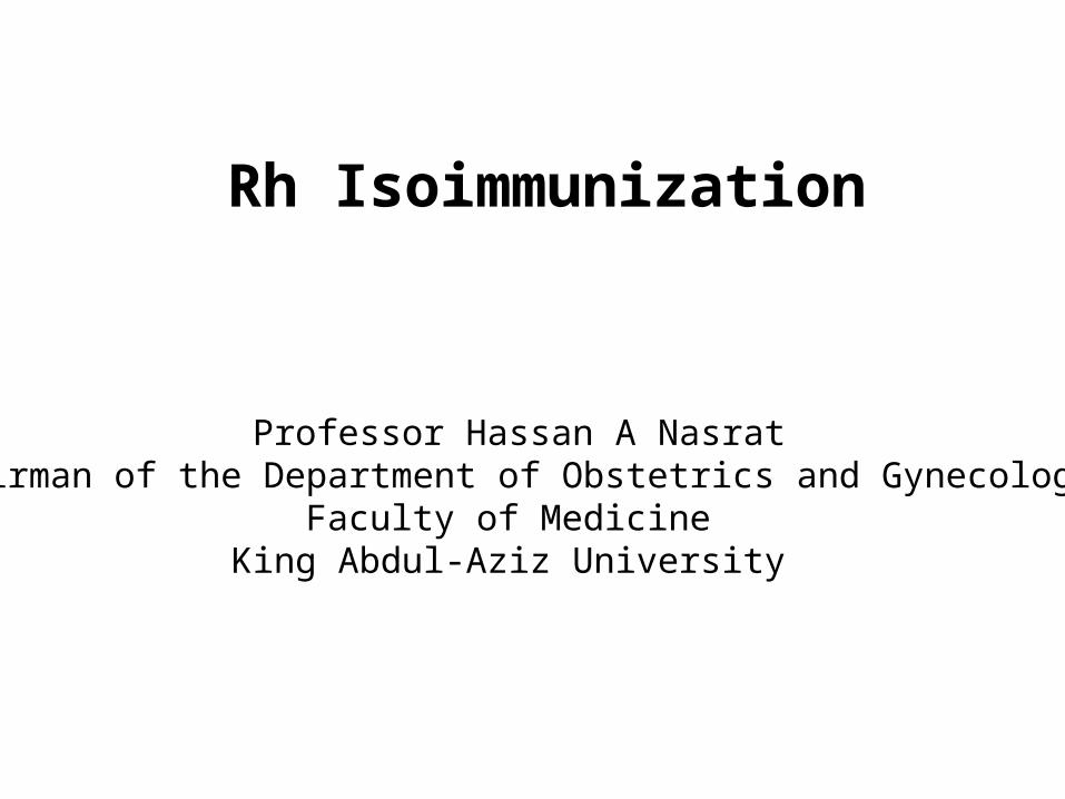
The Antibodies Arise In The Mother As The Direct Result Of A Blood Group Incompatibility Between The Mother And Fetus
e.g. When An RhD Negative Mother Carries An RhD Positive Fetus.
In The Fetus: Erythroblastosis FetalisIn The Newborn: HDN.
Alloimmune Hemolytic Disease Of The Fetus / Newborn:
Definition: The Red Cells Of The Fetus Or Newborn Are Destroyed By
Maternally Derived Alloantibodies

Antibodies That May Be Detected During Pregnancy:
Innocuous Antibodies:
Most Of These Antibody Are IgM Therefore Cannot Cross The Placental Barrier E.G. Those Directed Against Such Specificities As A, P(1), Le(a), M, I, IH And Sd(a).
Antibodies Capable Of Causing Significant Hemolytic Transfusion Reactions:
IgG antibodies, Their Corresponding Antigens Are Not Well Developed At Birth E.g. Lu (b), Yt (a), And VEL —
Antibodies That Are Responsible For HDN : Anti-c, Anti-d, Anti-e, And Anti-k (Kell)

The RH Antigen – Biochemical and Genetic Aspects
Mechanism of Development of Maternal Rh Isoimmunization
Natural History of Maternal isoimmunization /HD of the Newborn
Diagnosis of Rh isoimmunization

The Rh Antigen- Biochemical Aspects:
The Rh Antigen Is A Complex Lipoprotein.
It Has A Molecular Weight Of Approximately 30,000.
It Is Distributed Throughout The Erythrocyte Membrane In
A Nonrandom Fashion.
The Surface Antigens Can Not Be Seen By Routine
Microscopy, But Can Be Identified By Specific Antisera
Function of the Rh antigen:
Its Precise Function Is Unknown. Rh Null Erythrocytes Have Increased Osmotic Fragility And Abnormal Shapes.
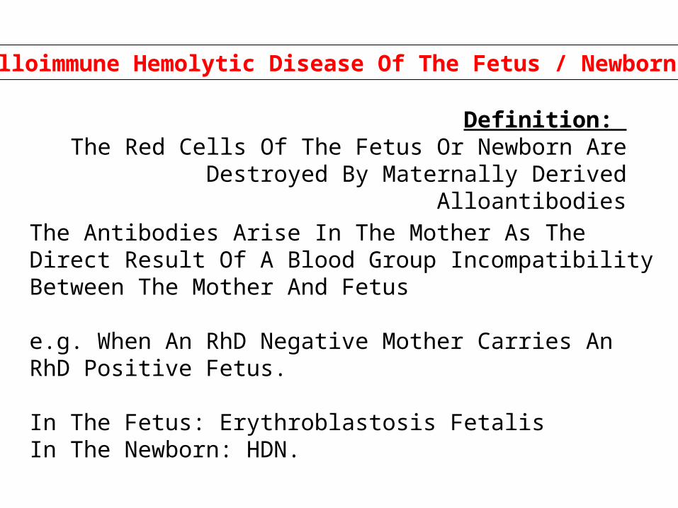
The RH Antigen- Genetic Aspect
The Rh gene complex is located on the distal end of the short arm of chromosome one.
A given Rh antigen complex is determined by a specific gene sequence inherited in a Mendelian fashion from the
parents. one haploid from the mother and one from the father.
Three genetic loci, determine the Rh antigen (i.e. Rh blood group).
Each chromosome will be either D positive or D negative (there is no "d" antigen), C or c positive, and E or e positive.

Grades Of “Positively” Due To Variation In The Degree Genetic Expression Of The D Antigen.
Incomplete Expression May Result In A Weakly Positive Patient e.g. Du Variant Of Weakly Rh Positive Patient
(They May Even Be Determined As Rh Negative).
A Mother With Du Rh Blood Group (Although Genetically Positive) May Become Sensitized From A D-positive Fetus
Or The Other Way Around May Take Place.
Genetic Expression (Rh Surface Protein Antigenicity):

Incomplete Expression Of The D Antigen Result In A Weakly Positive Patient e.g. Du Variant Of Weakly Rh Positive Patient.
Genetic Expression (Rh Surface Protein Antigenicity):
Du VariantFrank D Positive

Factors Affect The Expression Of The Rh Antigen
The Number Of Specific Rh-antigen Sites: - The Gene Dose, - The Relative Position Of The Alleles, - The Presence Or Absence Of Regulator Genes.
Interaction Of Other Components Of The Rh Blood Group. Erythrocytes Of Individuals Of Genotype Cde/cde Express Less D Antigen Than Do The Erythrocytes Of Individuals Of Genotype cDE/cde.
The Exposure Of The D Antigen On The Surface Of The Red Cell Membrane.

DcE
eCd
eCd/EcD
PhenotypeGenotype
D positive
Antigenicity of the Rh surface protein:
genetic expression of the D allele.
Number of specific Rh antigen sites.
Interaction of components of the Rh gene complex.
Exposure of the D antigen on the surface of the red cell

The Mechanism of Development of the Rh Immune Response:
Fetal RBC with Rh +ve antigen
Maternal circulation of an Rh –ve mother
(Primary immune response)
The Rh +ve antigen will be cleared by macrophages; processed and transferred to plasma stem cell precursors (Develop an almost
permanent immunologic memory)
With subsequent exposure the plasma cell line proliferate to produce humeral antibodies
(Secondary immune response).

The Primary Response:
Is a slow response (6 weeks to 6 months). IgM antibodies a molecular weight of 900,000 that does not cross the placenta.
The Secondary Response:
Is a Rapid response IgG antibodies a molecular weight of 160,000 that cross the placenta.
Exposure to maternal antigen in utero “the grandmother theory”:
This theory explains the development of fetal isoimmunization in a primigravida, who has no history of exposure to incompatible Rh blood. If a fetus is Rh negative and the mother is Rh positive, the may be exposed to the maternal Rh antigen through maternal-fetal transplacental bleed. In such cases the fetus immune system develop a permanent template (memory) for the Rh-positive antigen. When the fetus becomes a mother herself and exposed to a new load of D antigen from her fetus (hence the grandmother connection) the immune memory is recalled and a secondary immune response occur.
Without treatment: less than 20% of Rh D incompatible pregnancies actually lead to maternal isoimmunization
25-30% of the offspring will have some degree of hemolytic anemia and hyperbilirubinemia.
20-25% will be hydropic and often will die either in utero or in the neonatal period.
Cases of hemolysis in the newborn that do not result in fetal hydrops still can lead to kernicterus.
Natural History of Rh Isoimmunization And HD Fetus and Newborn

The Risk of development of Fetal Rh-disease is affected by:
The Husband Phenotype And Genotype (40 % Of Rh Positive Men Are Homozygous And 60% Are Heterozygous).
The Antigen Load And Frequency Of Exposure.
ABO Incompatibility
Less than 20% of Rh D incompatible pregnancies actually lead to maternal alloimmunization
The Amount Of Fetal Cells In Maternal Blood
Why Not All the Fetuses of Isoimmunized Women Develop the Same Degree of Disease?
The Non-responders:
ABO Incompatibility:
Antigenic Expression Of The Rh Antigen:
Classes Of IgG Family

Diagnosis of Rh isoimmunization
The diagnose is Based on the presence of anti-Rh (D) antibody in maternal
serum.
The Enzymatic Method The Antibody Titer In Saline, In Albumin The Indirect Coombs Tests.
Methods of Detecting Anti D Antibodies in Maternal Serum:
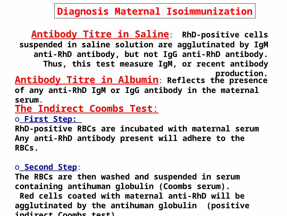
Antibody Titre in Saline: RhD-positive cells suspended in saline solution are agglutinated by IgM anti-RhD antibody, but not
IgG anti-RhD antibody. Thus, this test measure IgM, or recent antibody production.
Antibody Titre in Albumin: Reflects the presence of any anti-RhD IgM or IgG antibody in the maternal serum.
The Indirect Coombs Test: o First Step: RhD-positive RBCs are incubated with maternal serumAny anti-RhD antibody present will adhere to the RBCs.
o Second Step:The RBCs are then washed and suspended in serum containing antihuman globulin (Coombs serum). Red cells coated with maternal anti-RhD will be agglutinated by the antihuman globulin (positive indirect Coombs test).
Diagnosis Maternal Isoimmunization

Is Done After Birth To Detect The Presence Of Maternal Antibody On The Neonate's RBCs.
The Infant's RBCs Are Placed In Coombs Serum.If The Cells Are Agglutinated This Indicate The Presence Of Maternal Antibody
The Direct Coombs Test

Interpretation of Maternal Anti-D Titer
Antibody Titer Is A Screening Test.
A Positive Anti-d Titer Means That The Fetus Is At Risk For Hemolytic Disease, Not That It Has Occurred Or Will
Develop.
Variation In Titer Results Between Laboratories And Intra Laboratory Is Common.
A Truly Stable Titer Should Not Vary By More Than One Dilution When Repeated In A Given Laboratory.

Pathogenesis of The HD of the Fetus and Newborn
Fetal Rhesus Determination
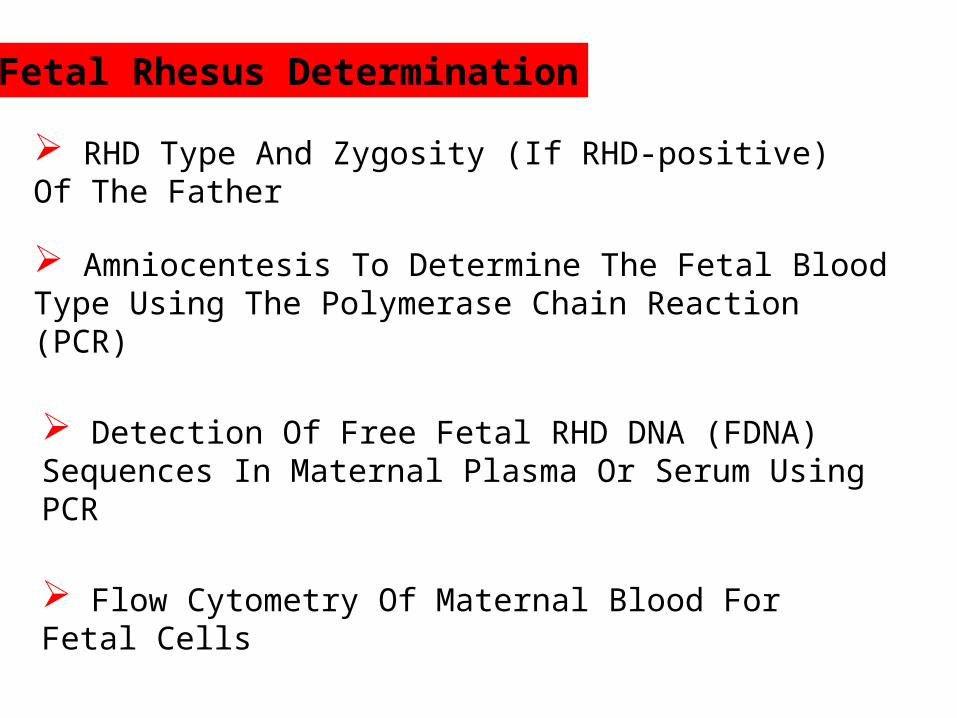
RHD Type And Zygosity (If RHD-positive) Of The Father
Amniocentesis To Determine The Fetal Blood Type Using The Polymerase Chain Reaction (PCR)
Detection Of Free Fetal RHD DNA (FDNA) Sequences In Maternal Plasma Or Serum Using PCR
Flow Cytometry Of Maternal Blood For Fetal Cells

Pathogenesis of Fetal Hemolytic Disease

Methods of Diagnosis and Evaluation of Fetal Rh Isoimmunization
Measurements Of Antibodies in Maternal Serum
Determination of Fetal Rh Blood Group
Ultrasonography
Amniocentesis
Fetal Blood Sampling

To Establish The Correct Gestational Age.
In Guiding Invasive Procedures And Monitoring Fetal Growth And Well-being.
Ultrasonographic Parameters To Determine Fetal Anemia: o Placental Thickness.o Umbilical Vein Diametero Hepatic Size.o Splenic Size.o Polyhydramnios. o Fetal Hydrops (e.g. Ascites, Pleural Effusions, Skin Edema).
Ultrasonography:

Anemic Fetus Preserves Oxygen Delivery To The Brain By Increasing Cerebral Flow Of Its Already
Low Viscosity Blood.
Doppler Velocimetry Of The Fetal Middle Cerebral Artery (MCA)
To Predict The Timing Of A Second Intrauterine Fetal Transfusion.
For Predicting Fetal Anemia

Previous Seriously Affected Fetus Or Infant (e.g. Intrauterine Fetal Transfusion, Early Delivery, Fetal Hydrops, Neonatal Exchange Transfusion).
A Critical Anti-D Titer:I.E. A Titer Associated With A Significant Risk For Fetal Hydrops. Anti-D Titer Value Between 8 And 32
Invasive Techniques ( Amniocentesis and Fetal Blood Sampling):
Indications:

Amniocentesis
Normally Bilirubin In Amniotic Fluid Decreases With Advanced Gestation.
It Derives From Fetal Pulmonary And Tracheal Effluents.
Its Level Rises in Correlation With Fetal Hemolysis.
Determination Of Amniotic Fluid Bilirubin:
By The Analysis Of The Change In Optical Density Of Amniotic Fluid At 450 nm On The Spectral Absorption Curve
(delta OD450)
Procedures Are Undertaken At 10-15 Days Intervals Until Delivery Data Are Plotted On A Normative Curve Based Upon
Gestational Age.

Extended Liley graph.

Queenan curve (Deviation in amniotic fluid optical density at a wavelength of 450 nm in Rh-immunized pregnancies from 14 to 40 weeks' gestation)
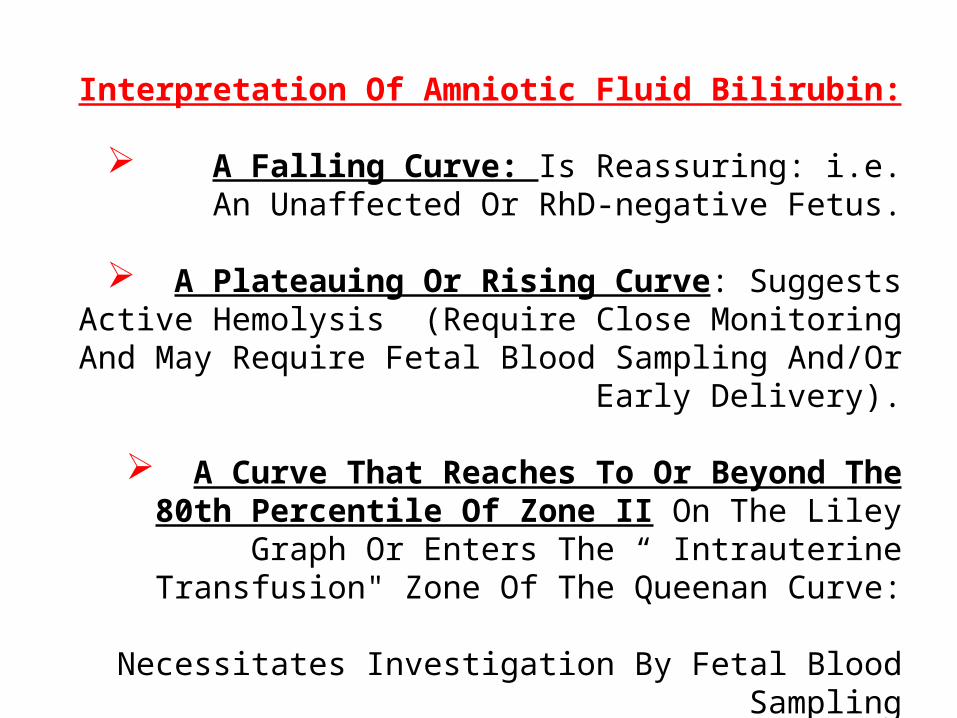
Interpretation Of Amniotic Fluid Bilirubin:
A Falling Curve: Is Reassuring: i.e. An Unaffected Or RhD-negative Fetus.
A Plateauing Or Rising Curve: Suggests Active Hemolysis (Require Close Monitoring And May Require
Fetal Blood Sampling And/Or Early Delivery).
A Curve That Reaches To Or Beyond The 80th Percentile Of Zone II On The Liley Graph Or Enters The “
Intrauterine Transfusion" Zone Of The Queenan Curve:
Necessitates Investigation By Fetal Blood Sampling
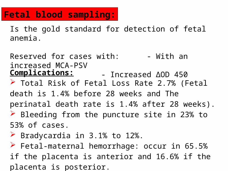
Is the gold standard for detection of fetal anemia.
Reserved for cases with: - With an increased MCA-PSV- Increased ΔOD 450
Complications: Total Risk of Fetal Loss Rate 2.7% (Fetal death is 1.4% before 28 weeks and The perinatal death rate is 1.4% after 28 weeks). Bleeding from the puncture site in 23% to 53% of cases. Bradycardia in 3.1% to 12%. Fetal-maternal hemorrhage: occur in 65.5% if the placenta is anterior and 16.6% if the placenta is posterior. Infection and abruptio placentae are rare complications
Fetal blood sampling:
Most polyclonal RhIg comes from male volunteers who are intentionally exposed to RhD-positive red blood cells.
Potential Problems: infectious risksolve supply problems.ethical issues
MONOCLONAL ANTI-D
anti-D monoclonal antibody: Although monoclonal anti-D is promising, it cannot be recommended at this time as a replacement for polyclonal RhIg.


Complications of Fetal-Neonatal Anemia:
Fetal Hydrops And Stillbirth
Hepatosplenomegaly
Neonatal Jaundice
Compilations Of Neonatal Kernicterus (Lethargy,
Hypertonicity, Hearing Loss, Cerebral Palsy And
Learning Disability)
Neonatal Anemia

Alloimmune Hemolytic Disease Of The Newborn (HDN):
Causes Of Fetal Neonatal Anemia:
o Abnormal Placental Separation (Abruptio Placentae) Or Placenta Previao Traumatic Tear Of The Umbilical Cord o Occult Blood Loss In Utero As A Result Of Fetomaternal Hemorrhage. o A Chronic Twin-to-twin Transfusion In Identical Twins
Blood Loss:
Infections:
Anemia Due To Congenital Spherocytosis
Nonspherocytic Hemolytic Anemias
Hemoglobinopathies:

The RH Antigen

Diagnostic algorithm for neonatal anemia. *Note that the direct antiglobulin (Coombs) test can be negative or weakly positive despite the presence of ABO incompatibility and hemolysis. (Adapted from Blanchette VS, Zipursky A. Assessment of anemia in newborn infants. Clin Perinatol 1984;11(2):489–510; with permission.)
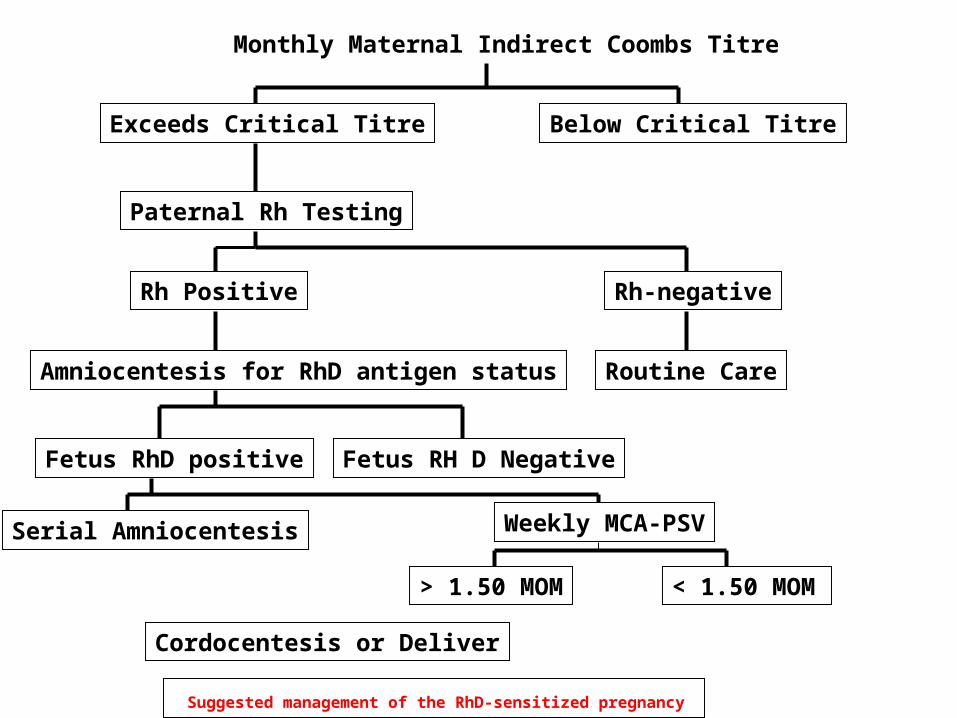
Suggested management of the RhD-sensitized pregnancy
Monthly Maternal Indirect Coombs Titre
Below Critical TitreExceeds Critical Titre
Paternal Rh Testing
Rh Positive Rh-negative
Amniocentesis for RhD antigen status Routine Care
Fetus RhD positive Fetus RH D Negative
Serial Amniocentesis Weekly MCA-PSV
< 1.50 MOM
Cordocentesis or Deliver
> 1.50 MOM

Suggested management of the RhD-sensitized pregnancy
Monthly Maternal Indirect Coombs Titre
Below Critical TitreExceeds Critical Titre
Paternal Rh Testing
Rh Positive Rh-negative
Amniocentesis for RhD antigen status Routine Care
Fetus RhD positive Fetus RH D Negative
Serial Amniocentesis Weeklyl MCA-PSV
< 1.50 MOM >1.5 MOMCordocentesis or Deliver

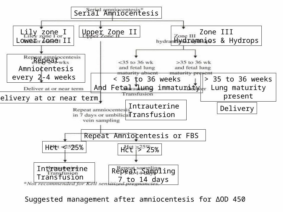
Suggested management after amniocentesis for ΔOD 450
Serial Amniocentesis
Lily zone ILower Zone II
Upper Zone II Zone IIIHydramnios & Hydrops
Repeat Amniocentesis every
2-4 weeks
Delivery at or near term
Repeat Amniocentesis in 7 days or FBS
Hct < 25% Hct > 25%
Intrauterine Transfusion
Repeat Sampling7 to 14 days
< 35 to 36 weeksAnd Fetal lung
immaturity
> 35 to 36 weeks Lung
maturity present
Intrauterine Transfusion
Delivery
Suggested management after amniocentesis for ΔOD 450
Serial Amniocentesis
Lily zone ILower Zone II
Upper Zone II Zone IIIHydramnios & Hydrops
Repeat Amniocentesis every 2-4 weeks
Delivery at or near term
Repeat Amniocentesis or FBS
Hct < 25% Hct > 25%
Intrauterine Transfusion
Repeat Sampling7 to 14 days
< 35 to 36 weeksAnd Fetal lung immaturity
> 35 to 36 weeks Lung maturity present
Intrauterine Transfusion
Delivery

Average regression lines for healthy fetuses (dotted line), mildly anemic fetuses (thin line), and severely anemic fetuses (thick line). (From Detti L, Mari G, Akiyama M, Cosmi E, Moise Jr KJ, Stefor T, et al. Longitudinal assessment of the middle cerebral artery peak systolic velocity in healthy fetuses and in fetuses at risk for anemia. Am J Obstet Gynecol 2002;187:938; with permission.)
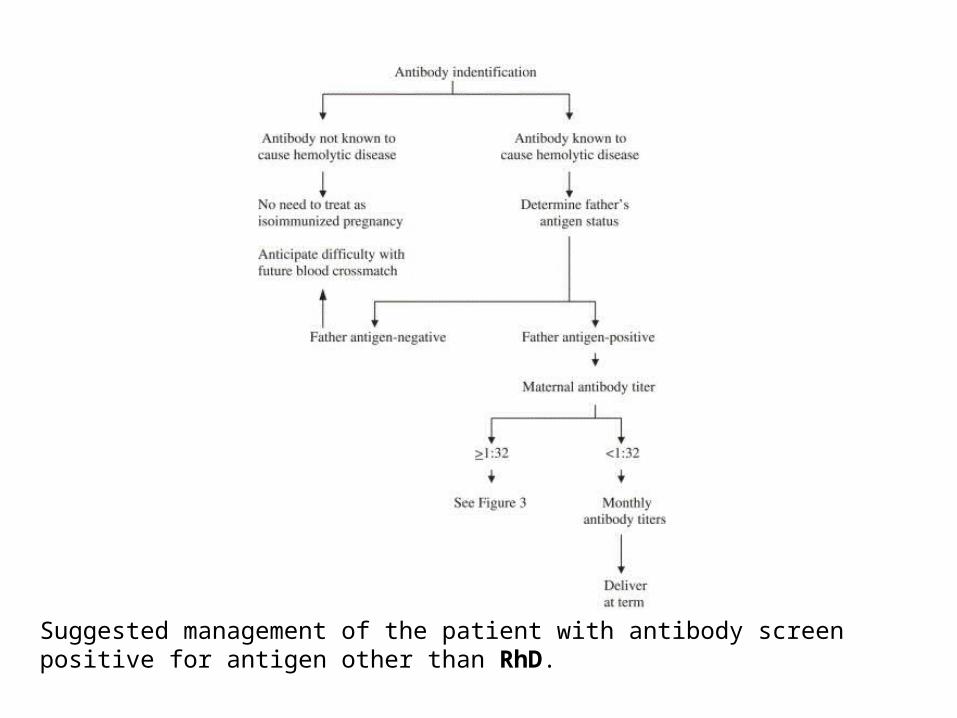
Suggested management of the patient with antibody screen positive for antigen other than RhD.

Incidence Of Maternal Alloimmunization
The overall incidence of maternal alloimmunization to clinically significant RBC antigens has been estimated to be 25 per 10,000 live births

RhD D negativity primarily occurs among Caucasians; the average incidence is 15 percent in this group. Examples of the blood group distribution in various populations are illustrated below: Basques — 30 to 35 percent
Finland — 10 to 12 percent
American blacks — 8 percent
Indo-Eurasians — 2 percent
Native Americans and Inuit Eskimos — 1 to 2 percent.
Changes since introduction of Anti-D

• Chronic transplacental hemorrhage.• Failure to administer Rh immune globulin when indicated.• or non-detection of a large fetal bleed at delivery
PATHOGENESIS

As an example, in a study of 110 pregnant mothers with 111 at-risk fetuses, and maternal serum titers of 1:16 or greater, antibodies to D, K, E, and c were present in 84, 18, 8, and 3 fetuses, respectively

The nature of the Rh antigen complex is determined by a specific gene sequence inherited in a Mendelian fashion from the parents, one haploid from the mother and one from the father. In 1974 the location of the Rh gene complex was pinpointed on the distal end of the short arm of chromosome one. Three genetic loci, each with two possible alleles determined the Rh antigen (i.e. Rh blood group).
The amount of fetal cells in maternal blood:
the Kleihauer-Braun-Betke test

The severity of fetal anemia is influenced:Primarily by antibody concentration,Additional factors that are not fully understood. These include the subclass and glycosylation of maternal antibodies.The structure, site density, maturational development and tissue distribution of blood group antigens.The efficiency of transplacental IgG transport.The functional maturity of the fetal spleen.Polymorphisms which affect Fc receptor function; and the presence of HLA-related inhibitory antibodies [13].

DIAGNOSIS
Blood and Rh(D) typing and an antibody screen should always be performed at the first prenatal visit
Below the critical titer there is a risk of mild to moderate, but not severe, fetal or neonatal hemolytic anemia. Fetal assessment with invasive techniques (eg, amniocentesis, fetal blood sampling) is required when a critical titer is present or if the patient has had a prior significantly affected pregnancy (eg, intrauterine fetal transfusion, early delivery, fetal hydrops, neonatal exchange transfusion). The purpose of these invasive tests is to determine whether severe fetal anemia is present.

Ultrasonography
A variety of ultrasonographic parameters have been used to determine whether fetal anemia is present. These parameters include: placental thickness; umbilical vein diameter; hepatic size; splenic size; and polyhydramnios
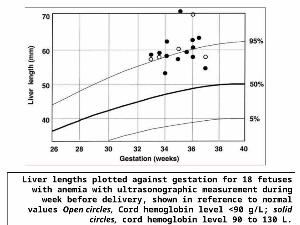
Liver lengths plotted against gestation for 18 fetuses with anemia with ultrasonographic measurement during week before delivery, shown in
reference to normal values Open circles, Cord hemoglobin level <90 g/L; solid circles, cord hemoglobin level 90 to 130 L.

Liver length measurements made within 48 hours of fetal blood sampling in all fetuses with anemia at first fetal blood sampling, shown in reference to normal values.

Ultrasound image of amniocentesis at 16 weeks of gestation

Ultrasound image of transabdominal chorionic villus sampling.

Diagram of cordocentesis procedure


Doppler velocimetry — Doppler assessment of the fetal middle cerebral artery (MCA)
Amniocentesis — Amniocentesis is performed when the critical titer is reached or if there has been a previous seriously affected fetus or infant.
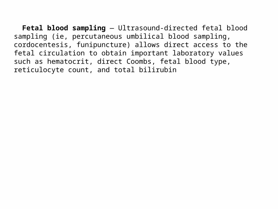
Fetal blood sampling — Ultrasound-directed fetal blood sampling (ie, percutaneous umbilical blood sampling, cordocentesis, funipuncture) allows direct access to the fetal circulation to obtain important laboratory values such as hematocrit, direct Coombs, fetal blood type, reticulocyte count, and total bilirubin

Multiple antibodies Some women develop antibodies to more than one red blood cell antigen.

Ultrasound image of cordocentesis with the needle tip located in a free loop of cord.

Ultrasound-guided transabdominal fetocentesis
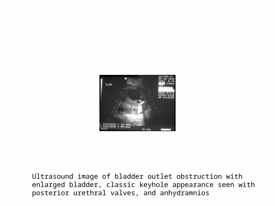
Ultrasound image of bladder outlet obstruction with enlarged bladder, classic keyhole appearance seen with posterior urethral valves, and anhydramnios

Double pig-tailed Rocket catheter and trocar used for vesicoamniotic shunting.


Related Documents