Reversal of West Nile virus-induced blood-brain barrier disruption and tight junction proteins degradation by matrix metalloproteinases inhibitor Saguna Verma a , Mukesh Kumar a , Ulziijargal Gurjav a,b , Stephanie Lum a , and Vivek R. Nerurkar a,b,* a Retrovirology Research Laboratory, Department of Tropical Medicine, Medical Microbiology and Pharmacology, John A. Burns School of Medicine, University of Hawai’i at Manoa, Honolulu, HI 96813 b Molecular Biosciences and Bioengineering Graduate Program, University of Hawai’i at Manoa, Honolulu, HI 96813 Abstract Though compromised blood-brain barrier (BBB) is a pathological hallmark of WNV- associated neurological sequelae, underlying mechanisms are unclear. We characterized the expression of matrix metalloproteinases (MMP) in WNV-infected human brain-microvascular endothelial cells (HBMVE) and -cortical astrocytes (HBCA), components of BBB and their role in BBB disruption. Expression of multiple MMPs was significantly induced in WNV-infected HBCA cells. Naïve HBMVE cells incubated with the supernatant from WNV-infected HBCA cells demonstrated loss of tight junction proteins, which was rescued in the presence of MMP inhibitor, GM6001. Further, supernatant from WNV-infected HBCA cells compromised the in-vitro BBB models integrity. Our data suggests astrocytes as one of the sources of MMP in the brain, which mediates BBB disruption allowing unrestricted entry of immune cells into the brain, thereby contributing to WNV-neuropathogenesis. Because of the unavailability of WNV antivirals and vaccines, use of MMP inhibitors as an adjunct therapy to ameliorate WNV disease progression is warranted. Keywords MMP; TIMP; tight junction proteins; ZO-1; Claudin-1; meningoencephalitis; BBB; astrocytes; human brain microvascular endothelial cells; GM6001 Introduction Although immune surveillance in the brain is a normal physiological response, increased traversing of activated immune cells and pathogens across the blood-brain barrier (BBB) leads to pathophysiological changes in neuroinflammatory diseases such as multiple sclerosis, cerebral ischemia (Persidsky et al., 2006; Petty and Lo, 2002), bacterial meningitis © 2009 Elsevier Inc. All rights reserved. * Corresponding author: Vivek R. Nerurkar, Ph.D., 651 Ilalo Street, BSB 325AA, Honolulu, HI 96813, Phone: (808) 692-1668, Fax: (808) 692-1980; [email protected]. Publisher's Disclaimer: This is a PDF file of an unedited manuscript that has been accepted for publication. As a service to our customers we are providing this early version of the manuscript. The manuscript will undergo copyediting, typesetting, and review of the resulting proof before it is published in its final citable form. Please note that during the production process errors may be discovered which could affect the content, and all legal disclaimers that apply to the journal pertain. NIH Public Access Author Manuscript Virology. Author manuscript; available in PMC 2011 May 25. Published in final edited form as: Virology. 2010 February 5; 397(1): 130–138. doi:10.1016/j.virol.2009.10.036. NIH-PA Author Manuscript NIH-PA Author Manuscript NIH-PA Author Manuscript

Welcome message from author
This document is posted to help you gain knowledge. Please leave a comment to let me know what you think about it! Share it to your friends and learn new things together.
Transcript

Reversal of West Nile virus-induced blood-brain barrierdisruption and tight junction proteins degradation by matrixmetalloproteinases inhibitor
Saguna Vermaa, Mukesh Kumara, Ulziijargal Gurjava,b, Stephanie Luma, and Vivek R.Nerurkara,b,*
a Retrovirology Research Laboratory, Department of Tropical Medicine, Medical Microbiology andPharmacology, John A. Burns School of Medicine, University of Hawai’i at Manoa, Honolulu, HI96813b Molecular Biosciences and Bioengineering Graduate Program, University of Hawai’i at Manoa,Honolulu, HI 96813
AbstractThough compromised blood-brain barrier (BBB) is a pathological hallmark of WNV- associatedneurological sequelae, underlying mechanisms are unclear. We characterized the expression ofmatrix metalloproteinases (MMP) in WNV-infected human brain-microvascular endothelial cells(HBMVE) and -cortical astrocytes (HBCA), components of BBB and their role in BBB disruption.Expression of multiple MMPs was significantly induced in WNV-infected HBCA cells. NaïveHBMVE cells incubated with the supernatant from WNV-infected HBCA cells demonstrated lossof tight junction proteins, which was rescued in the presence of MMP inhibitor, GM6001. Further,supernatant from WNV-infected HBCA cells compromised the in-vitro BBB models integrity.Our data suggests astrocytes as one of the sources of MMP in the brain, which mediates BBBdisruption allowing unrestricted entry of immune cells into the brain, thereby contributing toWNV-neuropathogenesis. Because of the unavailability of WNV antivirals and vaccines, use ofMMP inhibitors as an adjunct therapy to ameliorate WNV disease progression is warranted.
KeywordsMMP; TIMP; tight junction proteins; ZO-1; Claudin-1; meningoencephalitis; BBB; astrocytes;human brain microvascular endothelial cells; GM6001
IntroductionAlthough immune surveillance in the brain is a normal physiological response, increasedtraversing of activated immune cells and pathogens across the blood-brain barrier (BBB)leads to pathophysiological changes in neuroinflammatory diseases such as multiplesclerosis, cerebral ischemia (Persidsky et al., 2006; Petty and Lo, 2002), bacterial meningitis
© 2009 Elsevier Inc. All rights reserved.*Corresponding author: Vivek R. Nerurkar, Ph.D., 651 Ilalo Street, BSB 325AA, Honolulu, HI 96813, Phone: (808) 692-1668, Fax:(808) 692-1980; [email protected]'s Disclaimer: This is a PDF file of an unedited manuscript that has been accepted for publication. As a service to ourcustomers we are providing this early version of the manuscript. The manuscript will undergo copyediting, typesetting, and review ofthe resulting proof before it is published in its final citable form. Please note that during the production process errors may bediscovered which could affect the content, and all legal disclaimers that apply to the journal pertain.
NIH Public AccessAuthor ManuscriptVirology. Author manuscript; available in PMC 2011 May 25.
Published in final edited form as:Virology. 2010 February 5; 397(1): 130–138. doi:10.1016/j.virol.2009.10.036.
NIH
-PA Author Manuscript
NIH
-PA Author Manuscript
NIH
-PA Author Manuscript
(BM) and infections with viruses such as HIV (Kanmogne et al., 2007), human T cellleukemia virus (Afonso et al., 2007) and West Nile virus (WNV) (Arjona et al., 2007a;Wang et al., 2004). Matrix metalloproteinases (MMP), a large family of endopeptidases,play a major role in leukocyte migration, modulating cytokine activity and disruption of theBBB (Gearing et al., 1995; Johnatty et al., 1997; Leppert et al., 2001; Mun-Bryce et al.,2002; Yong et al., 2007). Excess MMP production and activation is the keypathophysiological hallmark of several neurodegenerative diseases (Cunningham, Wetzel,and Rosenberg, 2005; Hu et al., 2007; Rosenberg, 1995; Rosenberg, 2002). Physiologically,MMP activity is tightly regulated at the level of gene transcription, pro-enzyme activationand by the action of tissue inhibitors of MMP (TIMP). Though the basal levels of MMPexpression in the brain are low, under pathological conditions, MMP are synthesized bymost of the resident CNS cells such as endothelial cells, astrocytes, microglia and neurons aswell as the infiltrating immune cells (Hummel et al., 2001; Mandal et al., 2003; Rosenberg,2002).
WNV, an enveloped, single-stranded positive-sense, neurotropic flavivirus, is an importanthuman pathogen that targets neurons to cause potentially lethal encephalitis (Diamond et al.,2003; Hayes and Gubler, 2006). Neurological complications such as inflammation, failure ofthe BBB, and neuronal death contribute to the mortality and morbidity associated withWNV-induced meningitis. Increased production of pro-inflammatory mediators facilitateWNV neuroinvasion by compromising the BBB integrity and are associated with high virustiters in the brain and increased mortality in WNV mouse models (Arjona et al., 2007b;Wang et al., 2008; Wang et al., 2004). Recent studies using MMP-9 knock out mouse modelprovides important evidence for the role of WNV-induced MMP-9 in opening of the BBB(Wang et al., 2008). The BBB primarily consists of microvascular endothelial cells andperivascular astrocytes, separated by the basement membrane (Persidsky et al., 2006). Thetight junction proteins (TJP) such as zona occludens (ZO), claudins and occludin, the mainstructural basis of BBB integrity, play a key role in the physiology of the BBB (Persidsky etal., 2006).
Though neurons are the prime target of WNV infection, we and others have previouslycharacterized WNV infection in the BBB cells, human brain microvascular endothelial(HBMVE) cells and brain astrocytes (Cheeran et al., 2005; van Marle et al., 2007; Verma etal., 2009). Recent report documenting the presence of WNV antigen in the astrocytes ofWNV-infected human brain tissue further suggests the role of these cells in WNVneuropathogenesis (van Marle et al., 2007). However, our understanding of the cellularmechanisms associated with WNV-induced BBB disruption, specifically the contribution ofBBB-associated cells is limited. Therefore, the aim of this study was to examine the effect ofWNV infection on the expression profile of MMP family genes in the BBB cells,characterize their role in disrupting the tight junctions of the BBB and assess the ability ofMMP inhibitor GM6001 in reversing the disruption of an in-vitro BBB model.
ResultsHuman brain cortical astrocytes cells are susceptible to WNV infection
Brain endothelial cells and astrocytes are the two main components of the BBB. In thisreport we demonstrate that the WNV titer in infected human brain cortical astrocytes(HBCA) cells peaks (log 7–8 PFU/mL) at day 3 after infection, followed by a sharp decline(Fig. 1A). Similar to our previous study, we further confirmed that WNV can efficientlyreplicate in HBMVE cells (Fig. 1A) (Verma et al., 2009). The specificity of WNV infectionin HBCA cells was further characterized by immunostaining of HBCA cells with astrocytespecific marker, glial fibrillary acidic protein (GFAP), and WNV antigen (Fig 1B). Almost100% cells were found to be GFAP positive, thereby confirming the purity of these primary
Verma et al. Page 2
Virology. Author manuscript; available in PMC 2011 May 25.
NIH
-PA Author Manuscript
NIH
-PA Author Manuscript
NIH
-PA Author Manuscript
HBCA cells (Fig. 1B, i). Robust staining of WNV antigen was detected in the cytoplasm ofHBCA cells at day 2 after infection (Fig. 1B, ii). Infected HBCA cells stained with onlysecondary antibody against both, anti-GFAP (data not shown) and WNV antigen (Fig. 1B,iii) did not show any immunostaining.
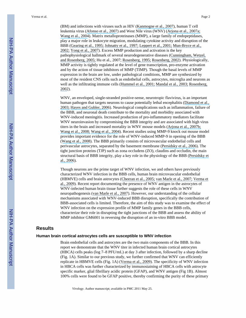
WNV induces mRNA expression of MMP family genes in HBCA cellsThe global response of HBMVE and HBCA cells infected with WNV at multiplicity ofinfection-5 (MOI-5) was determined at days 1 and 3 after infection by cDNA microarrayanalysis. Though WNV infection was robust and comparable in both the cell types, WNVinfection did not alter the expression profile of MMP and TIMP genes in HBMVE cells atdays 1 and 3 after infection (data not shown). Whereas WNV infection did not induce anyMMP family genes in HBCA cells at day 1 after infection, a significant increase in theexpression of MMP-1 (34-fold) and -3 (26-fold) genes was observed in these cells at day 3after infection. Increase in the expression of MMP and TIMP in HBCA cells was furthervalidated by qRT-PCR at different time points after infection. In concurrence with themicroarray data, as seen in Figure 2A, MMP-1 and -3 genes expression increased at day 2,and was significantly up-regulated, 20- to 40-fold, at days 3 and 4 after infection whichcoincided with the peak in the WNV titers. In addition, MMP-9 expression demonstratedsignificant increase (9- to 30-fold) at day 3 and 4 after infection. A 2- to 3-fold decrease inTIMP-2 and -3 transcripts were observed only at days 3 and 4 after infection (Fig. 2B).Infection of HBCA cells with UV-WNV did not induce any change in the expression profileof these genes (data not shown), further indicating that these alterations were a result ofWNV replication, rather than just virus entry into the cells.
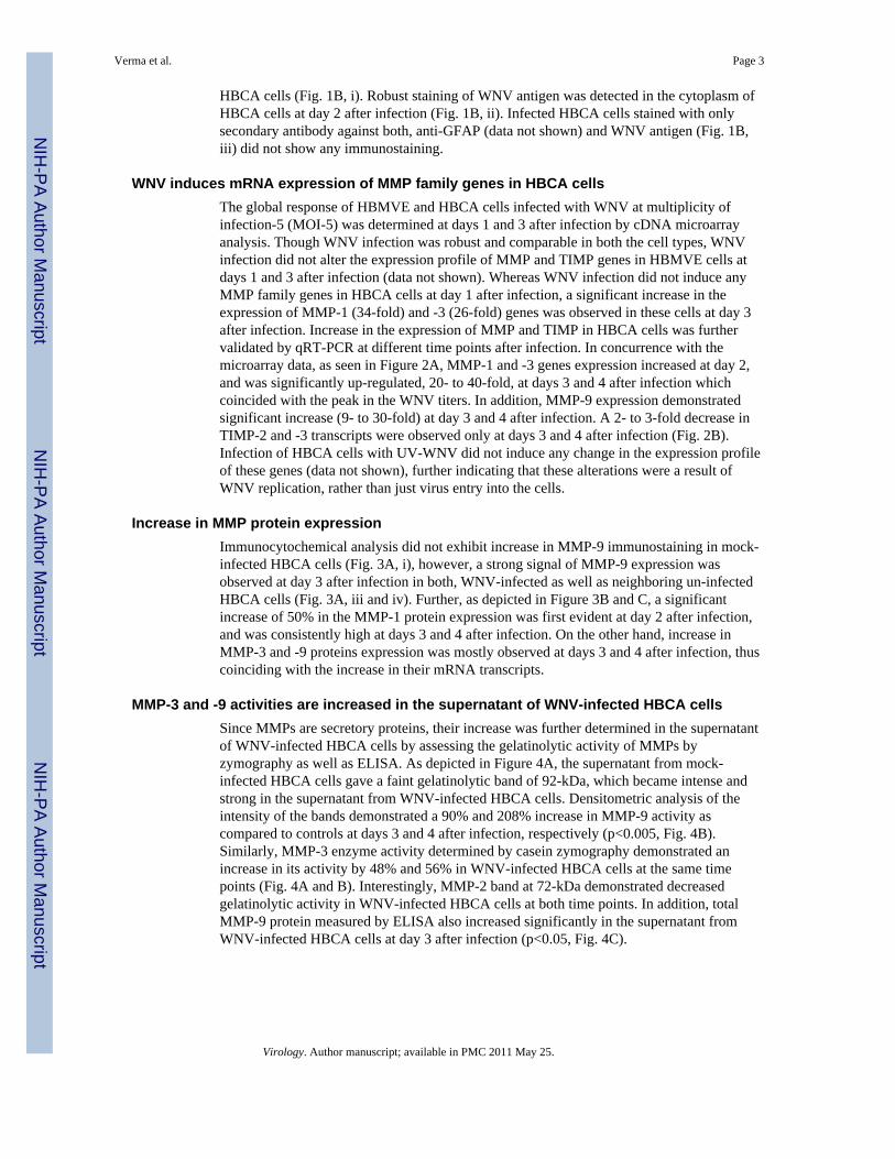
Increase in MMP protein expressionImmunocytochemical analysis did not exhibit increase in MMP-9 immunostaining in mock-infected HBCA cells (Fig. 3A, i), however, a strong signal of MMP-9 expression wasobserved at day 3 after infection in both, WNV-infected as well as neighboring un-infectedHBCA cells (Fig. 3A, iii and iv). Further, as depicted in Figure 3B and C, a significantincrease of 50% in the MMP-1 protein expression was first evident at day 2 after infection,and was consistently high at days 3 and 4 after infection. On the other hand, increase inMMP-3 and -9 proteins expression was mostly observed at days 3 and 4 after infection, thuscoinciding with the increase in their mRNA transcripts.
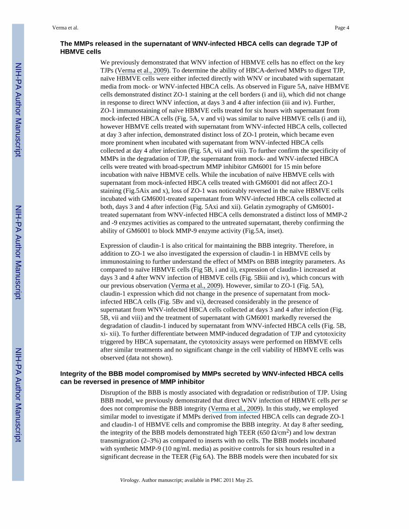
MMP-3 and -9 activities are increased in the supernatant of WNV-infected HBCA cellsSince MMPs are secretory proteins, their increase was further determined in the supernatantof WNV-infected HBCA cells by assessing the gelatinolytic activity of MMPs byzymography as well as ELISA. As depicted in Figure 4A, the supernatant from mock-infected HBCA cells gave a faint gelatinolytic band of 92-kDa, which became intense andstrong in the supernatant from WNV-infected HBCA cells. Densitometric analysis of theintensity of the bands demonstrated a 90% and 208% increase in MMP-9 activity ascompared to controls at days 3 and 4 after infection, respectively (p<0.005, Fig. 4B).Similarly, MMP-3 enzyme activity determined by casein zymography demonstrated anincrease in its activity by 48% and 56% in WNV-infected HBCA cells at the same timepoints (Fig. 4A and B). Interestingly, MMP-2 band at 72-kDa demonstrated decreasedgelatinolytic activity in WNV-infected HBCA cells at both time points. In addition, totalMMP-9 protein measured by ELISA also increased significantly in the supernatant fromWNV-infected HBCA cells at day 3 after infection (p<0.05, Fig. 4C).
Verma et al. Page 3
Virology. Author manuscript; available in PMC 2011 May 25.
NIH
-PA Author Manuscript
NIH
-PA Author Manuscript
NIH
-PA Author Manuscript
The MMPs released in the supernatant of WNV-infected HBCA cells can degrade TJP ofHBMVE cells
We previously demonstrated that WNV infection of HBMVE cells has no effect on the keyTJPs (Verma et al., 2009). To determine the ability of HBCA-derived MMPs to digest TJP,naïve HBMVE cells were either infected directly with WNV or incubated with supernatantmedia from mock- or WNV-infected HBCA cells. As observed in Figure 5A, naïve HBMVEcells demonstrated distinct ZO-1 staining at the cell borders (i and ii), which did not changein response to direct WNV infection, at days 3 and 4 after infection (iii and iv). Further,ZO-1 immunostaining of naïve HBMVE cells treated for six hours with supernatant frommock-infected HBCA cells (Fig. 5A, v and vi) was similar to naïve HBMVE cells (i and ii),however HBMVE cells treated with supernatant from WNV-infected HBCA cells, collectedat day 3 after infection, demonstrated distinct loss of ZO-1 protein, which became evenmore prominent when incubated with supernatant from WNV-infected HBCA cellscollected at day 4 after infection (Fig. 5A, vii and viii). To further confirm the specificity ofMMPs in the degradation of TJP, the supernatant from mock- and WNV-infected HBCAcells were treated with broad-spectrum MMP inhibitor GM6001 for 15 min beforeincubation with naïve HBMVE cells. While the incubation of naïve HBMVE cells withsupernatant from mock-infected HBCA cells treated with GM6001 did not affect ZO-1staining (Fig.5Aix and x), loss of ZO-1 was noticeably reversed in the naïve HBMVE cellsincubated with GM6001-treated supernatant from WNV-infected HBCA cells collected atboth, days 3 and 4 after infection (Fig. 5Axi and xii). Gelatin zymography of GM6001-treated supernatant from WNV-infected HBCA cells demonstrated a distinct loss of MMP-2and -9 enzymes activities as compared to the untreated supernatant, thereby confirming theability of GM6001 to block MMP-9 enzyme activity (Fig.5A, inset).
Expression of claudin-1 is also critical for maintaining the BBB integrity. Therefore, inaddition to ZO-1 we also investigated the experssion of claudin-1 in HBMVE cells byimmunostaining to further understand the effect of MMPs on BBB integrity parameters. Ascompared to naïve HBMVE cells (Fig 5B, i and ii), expression of claudin-1 increased atdays 3 and 4 after WNV infection of HBMVE cells (Fig. 5Biii and iv), which concurs withour previous observation (Verma et al., 2009). However, similar to ZO-1 (Fig. 5A),claudin-1 expression which did not change in the presence of supernatant from mock-infected HBCA cells (Fig. 5Bv and vi), decreased considerably in the presence ofsupernatant from WNV-infected HBCA cells collected at days 3 and 4 after infection (Fig.5B, vii and viii) and the treatment of supernatant with GM6001 markedly reversed thedegradation of claudin-1 induced by supernatant from WNV-infected HBCA cells (Fig. 5B,xi- xii). To further differentiate between MMP-induced degradation of TJP and cytotoxicitytriggered by HBCA supernatant, the cytotoxicity assays were performed on HBMVE cellsafter similar treatments and no significant change in the cell viability of HBMVE cells wasobserved (data not shown).
Integrity of the BBB model compromised by MMPs secreted by WNV-infected HBCA cellscan be reversed in presence of MMP inhibitor
Disruption of the BBB is mostly associated with degradation or redistribution of TJP. UsingBBB model, we previously demonstrated that direct WNV infection of HBMVE cells per sedoes not compromise the BBB integrity (Verma et al., 2009). In this study, we employedsimilar model to investigate if MMPs derived from infected HBCA cells can degrade ZO-1and claudin-1 of HBMVE cells and compromise the BBB integrity. At day 8 after seeding,the integrity of the BBB models demonstrated high TEER (650 Ω/cm2) and low dextrantransmigration (2–3%) as compared to inserts with no cells. The BBB models incubatedwith synthetic MMP-9 (10 ng/mL media) as positive controls for six hours resulted in asignificant decrease in the TEER (Fig 6A). The BBB models were then incubated for six
Verma et al. Page 4
Virology. Author manuscript; available in PMC 2011 May 25.
NIH
-PA Author Manuscript
NIH
-PA Author Manuscript
NIH
-PA Author Manuscript
hours with cell supernatants from HBCA cells collected at day 4 after infection. While theTEER of the BBB models incubated with the supernatant from mock-infected HBCA cellsdid not change as compared to control models (only HBMVE cell media), it decreasedsignificantly, (~400 Ω/cm2, p<0.005), in the BBB models incubated with supernatant fromWNV-infected HBCA cells (Fig. 6A). Treatment of WNV-infected HBCA cells supernatantwith GM6001 prior to the incubation restored the TEER values (p<0.05). Similarly, therewas no significant change in the FITC-dextran transmigrated across the BBB modelsincubated with supernatant from mock-infected HBCA cells as compared to control BBBmodels with only CSC media (Fig. 6B). However, BBB models incubated with supernatantfrom WNV-infected HBCA cells demonstrated a significant increase in the percent FITC-dextran transmigration (190%, p<0.005) as compared to controls, and the treatment ofsupernatant with GM6001 significantly inhibited FITC-dextran transmigration from 190% to140% (p<0.05).
DiscussionThe tight junctions of the BBB play a critical role in preserving the integrity of the BBB andrestricting the entry of immune cells as well as pathogens into the CNS. Increasedpermeability of the BBB is shown to be associated with increased WNV titers in the brain(Diamond and Klein, 2004). This study characterizing the acute MMP gene and proteinchanges as a result of WNV infection of BBB cells highlights that astrocytes are one of thepotential source of multiple MMPs, which degrade key TJPs of HBMVE cells andcompromise the integrity of the BBB model thereby contributing to WNV-associatedneuropathogenesis.
Recently, non-neuronal brain cells such as astrocytes, microglia and brain endothelial cellshave also been shown to be susceptible to WNV infection (Cheeran et al., 2005; van Marleet al., 2007). Presence of WNV antigen has been recently demonstrated in the astrocytes ofWNV-infected human brain tissue (van Marle et al., 2007). In our study, the WNV infectionkinetics seen in HBCA cells is similar to the results observed in the previous studies ofWNV infection in astrocytes (Cheeran et al., 2005; van Marle et al., 2007). IncreasedMMP-9 activity has been recently reported in the CSF of WNV-infected patients as well asin mice brain (Wang et al., 2008). However, the question remains, what is the specific cellsource of MMPs in the brain and their precise role in modulating BBB tight junctionproperties. In other neuroinflammatory diseases such as MS, cerebral ischemia, Alzheimer’sdisease and virus infections such as HIV, human herpesvirus-8 (HHV-8), and HTLV,increase in MMP-9 has been reported in brain endothelial cells (Chaudhuri et al., 2008;Conant et al., 1999; Cunningham, Wetzel, and Rosenberg, 2005; Ottino et al., 2004; Qian etal., 2007; Rosenberg, 1995; Rosenberg and Yang, 2007; Sellner et al., 2006; Shigemori etal., 2006; Sporer et al., 2000). As compared to these disease scenarios, our data demonstrateno alterations in the expression of MMPs or TIMPs in WNV-infected HBMVE cells andneuronal cells (data not shown). This observation, in context with our previous dataindicating either no degradation of any TJP (Verma et al., 2009) or no significant inductionof inflammatory cytokines (data not shown) in WNV-infected HBMVE cells suggest thatspecific host defense response of HBMVE cells to WNV infection most likely is protectivein-vivo and might provide a barrier to the WNV-CNS entry, which fails in-vivo due to theneuroinflammatory mediators secreted by other cells.
Activation of glial cells is a key pathogenic feature of WNV-associated meningoencephalitis(Cheeran et al., 2005; Diniz et al., 2006; van Marle et al., 2007). Recent data by Marle et al.provide direct evidence for the role of neurotoxic mediators released by WNV-infectedastrocytes in causing neuronal cell death (van Marle et al., 2007). Both, in-vitro and in-vivo,resident astrocytes have been demonstrated to respond to neuropathological injuries and
Verma et al. Page 5
Virology. Author manuscript; available in PMC 2011 May 25.
NIH
-PA Author Manuscript
NIH
-PA Author Manuscript
NIH
-PA Author Manuscript
undergo functional changes including increased production of MMPs (Anthony et al., 1997;Chaudhuri et al., 2008; Leppert et al., 2001; Rosenberg, 2002). In this report, the peakincrease in both mRNA expression as well as enzyme activities detected between days 2 to 4after infection when the virus titers were also high strongly suggest that MMP production istriggered by virus replication, not merely by virus entry. The activities of MMPs are tightlycontrolled at the level of mRNA expression, zymogen activation and an imbalance in theratio between TIMP and MMP which determines the extent of the degradation of the extracellular matrix (Cunningham, Wetzel, and Rosenberg, 2005). Our data demonstratingelevated levels of MMP-1, -3 and -9 along with reduced level of TIMP-2 in WNV-infectedHBCA cells indicate a clear imbalance in their ratios.
Another observation in our data was that WNV induced multiple MMPs in HBCA cells.Even though the exact role of individual MMPs is unclear, it is likely that MMP-1 and -3also contribute to the BBB injury either by directly digesting TJP or by activating pro-MMP-9 to active MMP-9. Several novel roles of MMP-1 and -3 have been describedrecently such as triggering neuroinflammation (Mun-Bryce et al., 2002; Suzuki et al., 2007),apoptosis (Kim et al., 2005) and direct activation of pro-MMP-9 (Conant et al., 2002). Basedon our data demonstrating increase in MMP-3 and decrease MMP-2 activity, we speculatethat the activation of MMP-9 in WNV-infected astrocytes is via MMP-3.
The kinetics of TJP expression during WNV infection has not been characterized. In thisreport, we for the first time demonstrate that degradation of TJP of HBMVE cells ismediated by MMPs derived from WNV-infected astrocytes. In addition, we have used aBBB model to demonstrate that the loss of TJP by MMPs directly results in decreased TEERand increased FITC-dextran transmigration representing loss of membrane integrity. In-vitroBBB models are routinely used to asses the effect of virus infections or transmigration ofvirus-infected immune cells on the BBB permeability (Kanmogne et al., 2007; Mahajan etal., 2008; Persidsky et al., 1997). Such degradation of TJP by MMPs released by infectedmacrophages and glial cells has been shown in infections with other neurotropic virusessuch as HIV (Johnston et al., 2001; Sporer et al., 2000). Similarly, decreased degradation ofcollagen IV, an important component of the basement membrane in WNV-infectedMMP-9−/− mice as compared to wild type mice was observed by Wang et al. (Wang et al.,2008). Based on these observations, we believe that in vivo, these WNV-induced MMPsderived from astrocytes contribute to BBB disruption by degrading components of bothbasement membranes as well as tight junctions ultimately resulting in infiltration of virusand immune cells into the CNS.
TNF-α is another well known mediator of BBB disruption and responds significantly toWNV infection (Cheng, King, and Kesson, 2004; Wang et al., 2004). In HBCA cells too,WNV infection significantly induces TNF-α from days 1 to 4 after infection (data notshown). However, our data suggesting the role of HBCA-derived MMPs in directlydisrupting the tight junctions is further strengthened by the fact that GM6001 can efficientlyblock the TJP degradation and BBB disruption. GM6001 is a broad-spectrum MMPinhibitor and is routinely used in-vitro as well as in-vivo models to understand theirpathogenic roles (Chen et al., 2006; Zozulya et al., 2007). Treatment of mice with GM6001(Ilomastat) has shown to reduce/delay BBB disruption and neuroinflammation resulting inimproved disease outcome of several neurodegenerative disease models such as cerebralischemia and infection with HIV and Semliki forest virus (Amantea et al., 2007; Johnston etal., 2001; Keogh et al., 2003). In addition, LPS-induced disruption of the BBB is reported tobe reversed significantly in the presence of MMP inhibitors including GM6001 (Mun-Bryceand Rosenberg, 1998; Rosenberg, Estrada, and Mobashery, 2007). Due to the ability ofMMP inhibitors to improve disease symptoms, specific synthetic MMP inhibitors have beenused or proposed as short term supporting therapies for acute inflammatory diseases such as
Verma et al. Page 6
Virology. Author manuscript; available in PMC 2011 May 25.
NIH
-PA Author Manuscript
NIH
-PA Author Manuscript
NIH
-PA Author Manuscript

MS, BM and vascular inflammatory diseases (Leppert et al., 2001; Mandal et al., 2003;Yong et al., 2007).
In summary, our results emphasize that complex neuroinflammatory responses associatedwith WNV infection in the CNS are mediated by astrocytes as one of the key players, andinvolves up-regulation of several MMPs. Though WNV-induced TJP degradation remains tobe validated in-vivo mouse model, we conclude that MMPs-induced perturbations in the TJPexpression might be an important pathway in WNV-induced BBB injury in-vivo, resultingin complex downstream pathological events such as the unrestricted entry of infected and/oractivated inflammatory and immune cells as ‘Trojan horse’ into the CNS. The ability ofGM6001 to rescue the BBB permeability is encouraging and the therapeutic use of MMPinhibitors to block WNV-associated BBB injury, inflammation and CNS-infiltration ofinfected or activated immune cells in-vivo is warranted.
Materials and methodsCells, virus and plaque assay
Primary HBCA cells were obtained from ACBRI (Kirkland, WA) and propagated in theastrocyte media (ATCC) while HBMVE cells were grown as described previously(Chapagain et al., 2007). Only early passage primary HBCA and HBMVE cells (passage 6to 9) were used for all experiments. WNV strain NY99 was used for all infectionexperiments at MOI-5 as described previously (Verma et al., 2009; Verma et al., 2008).Production of infectious virus in the supernatant was determined by plaque assay (Verma etal., 2008).
qRT-PCR analysiscDNA prepared from RNA extracted from WNV-, UV-WNV-, and mock-infected HBCAand HBMVE cells were used for qRT-PCR as described previously (Verma et al., 2009;Verma et al., 2008; Verma et al., 2006). Primer sequences and annealing temperatures usedfor amplification of MMPs and TIMP are described in Table 1.
Western immunoblot analysisTotal 40–50 μg of cellular protein extracted from mock- and infected-HBCA cells at days 2to 4 after infection was used for western blotting (Verma et al., 2009; Verma et al., 2008).The membranes were incubated with antibodies against rabbit polyclonal anti-MMP-1 (1mg/mL, Chemicon), mouse polyclonal anti-MMP-3 (1:1000, Calbiochem) and rabbitpolyclonal anti-MMP-9 (1:1000, Abcam) followed by HRP-conjugated secondaryantibodies, and were developed using ECL detection kit and analyzed by Bio-Rad Quantityone Program.
MMP-3 and -9 enzymes activity analysis by zymography and ELISAAt days 3 and 4 after infection, the supernatant medium from HBCA cells was collected forzymography and ELISA assays. For zymography, the supernatant was concentrated 10-foldusing Microcon centrifugal filter units (Millipore), electrophoresed on 10% SDS-polyacrylamide gels containing 1 mg/mL gelatin or casein as a protease substrate and thegelatinolytic bands were determined as described previously (Chua et al., 2003). ELISA fortotal MMP-9 in the supernatant from mock- and WNV-infected HBCA cells at day 3 afterinfection was performed using Quantikine human MMP-9 ELISA kit according to themanufacturers’ instructions (R&D systems), and a standard curve was generated in the rangeof 0 to 20 ng/mL. The plates were read using a Victor 3 microtiter reader equipped withWorkout 1.5 software (Perkin Elmer).
Verma et al. Page 7
Virology. Author manuscript; available in PMC 2011 May 25.
NIH
-PA Author Manuscript
NIH
-PA Author Manuscript
NIH
-PA Author Manuscript

Treatment of HBMVE cells with supernatant of HBCA cells and cytotoxicity assaysHBMVE cells grown on coverslips were incubated for six hours with supernatant fromaforementioned mock- and infected HBCA cells. Briefly 250 μL of the supernatant fromHBCA cells was mixed with 250 μL of the CSC media of HBMVE cells and applied tonaïve HBMVE cells. HBCA media collected on days 3 and 4 after infection were treatedwith broad-spectrum MMP inhibitor GM6001 (25 μM/mL, Calbiochem) for 15 min at roomtemperature before applying to naïve HBMVE cells. Following incubation, the cells werewashed twice with 1X PBS and fixed with 4% PFA. Similar treatments were performed onHBMVE cells grown in 96-well plate and cell viability was assayed as described previously(Verma et al., 2008).
Immunocytochemical analysisFixed HBCA cells were immunostained using monoclonal anti-WNV envelope (1:800),polyclonal anti- GFAP, (1:100, Chemicon) or anti-MMP-9 (1:100) antibodies followed bysecondary antibodies conjugated with Alexa Fluor 546 (GFAP and WNV) or FITC(MMP-9). HBMVE cells with or without treatment with supernatant from HBCA cells wereimmunostained with monoclonal mouse anti-ZO-1 or polyclonal rabbit anti-claudin-1(Verma et al., 2009).
Development of BBB model and WNV infectionAn in-vitro monolayer BBB model comprised of HBMVE cells was developed on aBioCoat® Cell Environment™ Human Fibronectin PET (polyethylene terephthalate) insertwith 3.0 μm pore size in a 24-well plate (BD Bioscience, Bedford, MA) as describedpreviously (Verma et al., 2009). The integrity of the in-vitro BBB model was determinedevery day, as described previously (Verma et al., 2009). The FITC-dextran (4-KDa MW)transmigration across the inserts was calculated as percentage of the total amount added inthe upper well (10 μg/100 μL) and the TEER was measured using EVOMX and wasexpressed as Ω/cm2. At day 8 after seeding, upper and lower chambers of the BBB modelswere incubated with 400 μL of the supernatant from HBCA cells collected at day 4 afterinfection in the presence or absence of GM6001 (25 nM/mL). After six hours of incubation,the models were washed twice with 1X PBS and the BBB integrity was assayed by TEERand FITC-dextran transmigration assay. For positive control, the inserts were treated withsynthetic MMP-2/9 enzyme (10 μg/membrane).
Statistical analysisData are reported as mean ± standard error of mean of at least three independentexperiments. Unpaired student t-test using GraphPad prism 5.0 (GraphPad software) wasused to compare the values of arbitrary units of densitometry scans and permeability assays.
AcknowledgmentsThis work was partially supported by grants from the Hawaii Community Foundation (20050405), ResearchCenters in Minority Institutions Program (G12RR003061) and Centers of Biomedical Research Excellence(P20RR018727), National Center for Research Resources, National Institutes of Health. We thank Dr. Duane J.Gubler for the generous gift of the WNV strain NY99 and the monoclonal human anti-WNV envelope antibody.We also thank Dr. Kathy Conant for valuable discussions related to MMPs.
ReferencesAfonso PV, Ozden S, Prevost MC, Schmitt C, Seilhean D, Weksler B, Couraud PO, Gessain A,
Romero IA, Ceccaldi PE. Human blood-brain barrier disruption by retroviral-infected lymphocytes:role of myosin light chain kinase in endothelial tight-junction disorganization. J Immunol. 2007;179(4):2576–83. [PubMed: 17675520]
Verma et al. Page 8
Virology. Author manuscript; available in PMC 2011 May 25.
NIH
-PA Author Manuscript
NIH
-PA Author Manuscript
NIH
-PA Author Manuscript

Amantea D, Russo R, Gliozzi M, Fratto V, Berliocchi L, Bagetta G, Bernardi G, Corasaniti MT. Earlyupregulation of matrix metalloproteinases following reperfusion triggers neuroinflammatorymediators in brain ischemia in rat. Int Rev Neurobiol. 2007; 82:149–69. [PubMed: 17678960]
Anthony DC, Ferguson B, Matyzak MK, Miller KM, Esiri MM, Perry VH. Differential matrixmetalloproteinase expression in cases of multiple sclerosis and stroke. Neuropathol Appl Neurobiol.1997; 23(5):406–15. [PubMed: 9364466]
Arjona A, Foellmer HG, Town T, Leng L, McDonald C, Wang T, Wong SJ, Montgomery RR, FikrigE, Bucala R. Abrogation of macrophage migration inhibitory factor decreases West Nile viruslethality by limiting viral neuroinvasion. J Clin Invest. 2007a; 117(10):3059–66. [PubMed:17909632]
Arjona A, Ledizet M, Anthony K, Bonafe N, Modis Y, Town T, Fikrig E. West Nile Virus EnvelopeProtein Inhibits dsRNA-Induced Innate Immune Responses. J Immunol. 2007b; 179(12):8403–9.[PubMed: 18056386]
Chapagain ML, Verma S, Mercier F, Yanagihara R, Nerurkar VR. Polyomavirus JC infects humanbrain microvascular endothelial cells independent of serotonin receptor 2A. Virology. 2007; 364(1):55–63. [PubMed: 17399760]
Chaudhuri A, Yang B, Gendelman HE, Persidsky Y, Kanmogne GD. STAT1 signaling modulatesHIV-1-induced inflammatory responses and leukocyte transmigration across the blood-brain barrier.Blood. 2008; 111(4):2062–72. [PubMed: 18003888]
Cheeran MC, Hu S, Sheng WS, Rashid A, Peterson PK, Lokensgard JR. Differential responses ofhuman brain cells to West Nile virus infection. J Neurovirol. 2005; 11(6):512–24. [PubMed:16338745]
Chen KM, Liu JY, Lai SC, Hsu LS, Lee HH. Association of plasminogen activators and matrixmetalloproteinase-9 proteolytic cascade with blood-CNS barrier damage of angiostrongyliasis. Int JExp Pathol. 2006; 87(2):113–9. [PubMed: 16623755]
Cheng Y, King NJ, Kesson AM. The role of tumor necrosis factor in modulating responses of murineembryo fibroblasts by flavivirus, West Nile. Virology. 2004; 329(2):361–70. [PubMed: 15518815]
Chua PK, Melish ME, Yu Q, Yanagihara R, Yamamoto KS, Nerurkar VR. Elevated levels of matrixmetalloproteinase 9 and tissue inhibitor of metalloproteinase 1 during the acute phase of Kawasakidisease. Clin Diagn Lab Immunol. 2003; 10(2):308–14. [PubMed: 12626459]
Conant K, Haughey N, Nath A, St Hillaire C, Gary DS, Pardo CA, Wahl LM, Bilak M, Milward E,Mattson MP. Matrix metalloproteinase-1 activates a pertussis toxin-sensitive signaling pathwaythat stimulates the release of matrix metalloproteinase-9. J Neurochem. 2002; 82(4):885–93.[PubMed: 12358794]
Conant K, McArthur JC, Griffin DE, Sjulson L, Wahl LM, Irani DN. Cerebrospinal fluid levels ofMMP-2, 7, and 9 are elevated in association with human immunodeficiency virus dementia. AnnNeurol. 1999; 46(3):391–8. [PubMed: 10482270]
Cunningham LA, Wetzel M, Rosenberg GA. Multiple roles for MMPs and TIMPs in cerebralischemia. Glia. 2005; 50(4):329–39. [PubMed: 15846802]
Diamond MS, Klein RS. West Nile virus: crossing the blood-brain barrier. Nat Med. 2004; 10(12):1294–5. [PubMed: 15580248]
Diamond MS, Shrestha B, Mehlhop E, Sitati E, Engle M. Innate and adaptive immune responsesdetermine protection against disseminated infection by West Nile encephalitis virus. ViralImmunol. 2003; 16(3):259–78. [PubMed: 14583143]
Diniz JA, Da Rosa AP, Guzman H, Xu F, Xiao SY, Popov VL, Vasconcelos PF, Tesh RB. West Nilevirus infection of primary mouse neuronal and neuroglial cells: the role of astrocytes in chronicinfection. Am J Trop Med Hyg. 2006; 75(4):691–6. [PubMed: 17038696]
Gearing AJ, Beckett P, Christodoulou M, Churchill M, Clements JM, Crimmin M, Davidson AH,Drummond AH, Galloway WA, Gilbert R, et al. Matrix metalloproteinases and processing of pro-TNF-alpha. J Leukoc Biol. 1995; 57(5):774–7. [PubMed: 7759957]
Hayes EB, Gubler DJ. West Nile virus: epidemiology and clinical features of an emerging epidemic inthe United States. Annu Rev Med. 2006; 57:181–94. [PubMed: 16409144]
Verma et al. Page 9
Virology. Author manuscript; available in PMC 2011 May 25.
NIH
-PA Author Manuscript
NIH
-PA Author Manuscript
NIH
-PA Author Manuscript

Hu J, Van den Steen PE, Sang QX, Opdenakker G. Matrix metalloproteinase inhibitors as therapy forinflammatory and vascular diseases. Nat Rev Drug Discov. 2007; 6(6):480–98. [PubMed:17541420]
Hummel V, Kallmann BA, Wagner S, Fuller T, Bayas A, Tonn JC, Benveniste EN, Toyka KV,Rieckmann P. Production of MMPs in human cerebral endothelial cells and their role in sheddingadhesion molecules. J Neuropathol Exp Neurol. 2001; 60(4):320–7. [PubMed: 11305867]
Johnatty RN, Taub DD, Reeder SP, Turcovski-Corrales SM, Cottam DW, Stephenson TJ, Rees RC.Cytokine and chemokine regulation of proMMP-9 and TIMP-1 production by human peripheralblood lymphocytes. J Immunol. 1997; 158(5):2327–33. [PubMed: 9036981]
Johnston JB, Zhang K, Silva C, Shalinsky DR, Conant K, Ni W, Corbett D, Yong VW, Power C.HIV-1 Tat neurotoxicity is prevented by matrix metalloproteinase inhibitors. Ann Neurol. 2001;49(2):230–41. [PubMed: 11220743]
Kanmogne GD, Schall K, Leibhart J, Knipe B, Gendelman HE, Persidsky Y. HIV-1 gp120compromises blood-brain barrier integrity and enhances monocyte migration across blood-brainbarrier: implication for viral neuropathogenesis. J Cereb Blood Flow Metab. 2007; 27(1):123–34.[PubMed: 16685256]
Keogh B, Sheahan BJ, Atkins GJ, Mills KH. Inhibition of matrix metalloproteinases amelioratesblood-brain barrier disruption and neuropathological lesions caused by avirulent Semliki Forestvirus infection. Vet Immunol Immunopathol. 2003; 94(3–4):185–90. [PubMed: 12909414]
Kim YS, Kim SS, Cho JJ, Choi DH, Hwang O, Shin DH, Chun HS, Beal MF, Joh TH. Matrixmetalloproteinase-3: a novel signaling proteinase from apoptotic neuronal cells that activatesmicroglia. J Neurosci. 2005; 25 (14):3701–11. [PubMed: 15814801]
Leppert D, Lindberg RL, Kappos L, Leib SL. Matrix metalloproteinases: multifunctional effectors ofinflammation in multiple sclerosis and bacterial meningitis. Brain Res Brain Res Rev. 2001; 36(2–3):249–57. [PubMed: 11690622]
Mahajan SD, Aalinkeel R, Sykes DE, Reynolds JL, Bindukumar B, Adal A, Qi M, Toh J, Xu G,Prasad PN, Schwartz SA. Methamphetamine alters blood brain barrier permeability via themodulation of tight junction expression: Implication for HIV-1 neuropathogenesis in the context ofdrug abuse. Brain Res. 2008; 1203:133–48. [PubMed: 18329007]
Mandal M, Mandal A, Das S, Chakraborti T, Sajal C. Clinical implications of matrixmetalloproteinases. Mol Cell Biochem. 2003; 252(1–2):305–29. [PubMed: 14577606]
Mun-Bryce S, Lukes A, Wallace J, Lukes-Marx M, Rosenberg GA. Stromelysin-1 and gelatinase Aare upregulated before TNF-alpha in LPS-stimulated neuroinflammation. Brain Res. 2002; 933(1):42–9. [PubMed: 11929634]
Mun-Bryce S, Rosenberg GA. Gelatinase B modulates selective opening of the blood-brain barrierduring inflammation. Am J Physiol. 1998; 274(5 Pt 2):R1203–11. [PubMed: 9644031]
Ottino P, Finley J, Rojo E, Ottlecz A, Lambrou GN, Bazan HE, Bazan NG. Hypoxia activates matrixmetalloproteinase expression and the VEGF system in monkey choroid-retinal endothelial cells:Involvement of cytosolic phospholipase A2 activity. Mol Vis. 2004; 10:341–50. [PubMed:15162095]
Persidsky Y, Ramirez SH, Haorah J, Kanmogne GD. Blood-brain barrier: structural components andfunction under physiologic and pathologic conditions. J Neuroimmune Pharmacol. 2006; 1(3):223–36. [PubMed: 18040800]
Persidsky Y, Stins M, Way D, Witte MH, Weinand M, Kim KS, Bock P, Gendelman HE, Fiala M. Amodel for monocyte migration through the blood-brain barrier during HIV-1 encephalitis. JImmunol. 1997; 158(7):3499–510. [PubMed: 9120312]
Petty MA, Lo EH. Junctional complexes of the blood-brain barrier: permeability changes inneuroinflammation. Prog Neurobiol. 2002; 68(5):311–23. [PubMed: 12531232]
Qian LW, Xie J, Ye F, Gao SJ. Kaposi’s sarcoma-associated herpesvirus infection promotes invasionof primary human umbilical vein endothelial cells by inducing matrix metalloproteinases. J Virol.2007; 81(13):7001–10. [PubMed: 17442715]
Rosenberg GA. Matrix metalloproteinases in brain injury. J Neurotrauma. 1995; 12(5):833–42.[PubMed: 8594211]
Verma et al. Page 10
Virology. Author manuscript; available in PMC 2011 May 25.
NIH
-PA Author Manuscript
NIH
-PA Author Manuscript
NIH
-PA Author Manuscript

Rosenberg GA. Matrix metalloproteinases in neuroinflammation. Glia. 2002; 39(3):279–91. [PubMed:12203394]
Rosenberg GA, Estrada EY, Mobashery S. Effect of synthetic matrix metalloproteinase inhibitors onlipopolysaccharide-induced blood-brain barrier opening in rodents: Differences in response basedon strains and solvents. Brain Res. 2007; 1133(1):186–92. [PubMed: 17184743]
Rosenberg GA, Yang Y. Vasogenic edema due to tight junction disruption by matrixmetalloproteinases in cerebral ischemia. Neurosurg Focus. 2007; 22 (5):E4. [PubMed: 17613235]
Sellner J, Simon F, Meyding-Lamade U, Leib SL. Herpes-simplex virus encephalitis is characterizedby an early MMP-9 increase and collagen type IV degradation. Brain Res. 2006; 1125(1):155–62.[PubMed: 17109833]
Shigemori Y, Katayama Y, Mori T, Maeda T, Kawamata T. Matrix metalloproteinase-9 is associatedwith blood-brain barrier opening and brain edema formation after cortical contusion in rats. ActaNeurochir Suppl. 2006; 96:130–3. [PubMed: 16671440]
Sporer B, Koedel U, Paul R, Kohleisen B, Erfle V, Fontana A, Pfister HW. Human immunodeficiencyvirus type-1 Nef protein induces blood-brain barrier disruption in the rat: role of matrixmetalloproteinase-9. J Neuroimmunol. 2000; 102(2):125–30. [PubMed: 10636480]
Suzuki Y, Nagai N, Umemura K, Collen D, Lijnen HR. Stromelysin-1 (MMP-3) is critical forintracranial bleeding after t-PA treatment of stroke in mice. J Thromb Haemost. 2007; 5(8):1732–9. [PubMed: 17596135]
van Marle G, Antony J, Ostermann H, Dunham C, Hunt T, Halliday W, Maingat F, Urbanowski MD,Hobman T, Peeling J, Power C. West Nile virus-induced neuroinflammation: glial infection andcapsid protein-mediated neurovirulence. J Virol. 2007; 81(20):10933–49. [PubMed: 17670819]
Verma S, Lo Y, Chapagain M, Lum S, Kumar M, Gurjav U, Luo H, Nakatsuka A, Nerurkar VR. WestNile virus infection modulates human brain microvascular endothelial cells tight junction proteinsand cell adhesion molecules: Transmigration across the in vitro blood-brain barrier. Virology.2009; 385:425–433. [PubMed: 19135695]
Verma S, Molina Y, Lo YY, Cropp B, Nakano C, Yanagihara R, Nerurkar VR. In vitro effects ofselenium deficiency on West Nile virus replication and cytopathogenicity. Virol J. 2008; 5:66.[PubMed: 18513435]
Verma S, Ziegler K, Ananthula P, Co JK, Frisque RJ, Yanagihara R, Nerurkar VR. JC virus inducesaltered patterns of cellular gene expression: interferon-inducible genes as major transcriptionaltargets. Virology. 2006; 345(2):457–67. [PubMed: 16297951]
Wang P, Dai J, Bai F, Kong KF, Wong SJ, Montgomery RR, Madri JA, Fikrig E. Matrixmetalloproteinase 9 facilitates west nile virus entry into the brain. J Virol. 2008; 82(18):8978–85.[PubMed: 18632868]
Wang T, Town T, Alexopoulou L, Anderson JF, Fikrig E, Flavell RA. Toll-like receptor 3 mediatesWest Nile virus entry into the brain causing lethal encephalitis. Nat Med. 2004; 10(12):1366–73.[PubMed: 15558055]
Yong VW, Zabad RK, Agrawal S, Goncalves Dasilva A, Metz LM. Elevation of matrixmetalloproteinases (MMPs) in multiple sclerosis and impact of immunomodulators. J Neurol Sci.2007; 259(1–2):79–84. [PubMed: 17382965]
Zozulya AL, Reinke E, Baiu DC, Karman J, Sandor M, Fabry Z. Dendritic cell transmigration throughbrain microvessel endothelium is regulated by MIP-1alpha chemokine and matrixmetalloproteinases. J Immunol. 2007; 178(1):520–9. [PubMed: 17182592]
Verma et al. Page 11
Virology. Author manuscript; available in PMC 2011 May 25.
NIH
-PA Author Manuscript
NIH
-PA Author Manuscript
NIH
-PA Author Manuscript

Fig. 1.WNV replication kinetics in HBCA cells. (A) WNV titers in infected HBCA and HBMVEcells supernatants collected at various time points after infection were determined by plaqueassay using Vero cells. Viral titers, expressed as plaque forming units (PFU)/mL ofsupernatant represent mean ± SD of data obtained from at least three independentexperiments. (B) HBCA cells grown and fixed on coverslips at day 2 after infection werestained with anti-GFAP antibody or anti-WNV envelope antibody, and counterstained withDAPI (blue). (i) mock-infected HBCA cells demonstrate GFAP staining in the cytoplasm,(ii) WNV-infected HBCA cells demonstrate robust virus staining in the cytoplasm, and (iii)WNV-infected HBCA cells stained with secondary antibody, negative control, did not showany immunostaining.
Verma et al. Page 12
Virology. Author manuscript; available in PMC 2011 May 25.
NIH
-PA Author Manuscript
NIH
-PA Author Manuscript
NIH
-PA Author Manuscript

Fig. 2.WNV differentially modulates expression of MMP family genes in HBCA cells. qRT-PCRwas conducted on cDNA templates from WNV- and mock-infected HBCA cells harvestedfrom days 1 to 4 after infection to determine the fold-change of (A) MMP-1, -2, -3 and -9and (B) TIMP-1, -2 and -3 genes expression. Change in the levels of each gene was firstnormalized to the GAPDH gene and then the fold-change in infected cells as compared tocorresponding control cells was calculated. Data represents mean of at least fourindependent experiments conducted in duplicate.
Verma et al. Page 13
Virology. Author manuscript; available in PMC 2011 May 25.
NIH
-PA Author Manuscript
NIH
-PA Author Manuscript
NIH
-PA Author Manuscript
Fig. 3.WNV induces protein expression of MMPs in HBCA cells. (A) Confluent primary HBCAcells were infected with WNV at MOI-5 and MMP-9 expression was determined usingimmunofluorescence. (i) Mock-infected HBCA cells demonstrated very faint and diffusedMMP-9 immunoreactivity (green), and (ii) Mock-infected cells incubated without MMP-9antibody did not show any staining. (iii and iv) marked increase in MMP-9 staining (green)was observed at day 3 in WNV-infected HBCA cells. MMP-9 increase was evident in both,WNV-infected (red) as well as neighboring un-infected HBCA cells. The images shown arerepresentative results of three independent experiments. Scale bar represents 10 μm at 20×magnification. (B) Western blot analysis of MMP-1, -3 and -9. Cellular proteins extractedfrom WNV- and mock-infected HBCA cells were separated on PAGE, transferred ontoPVDF membranes and immunoblotted with antibodies specific to MMP-1, -3 and -9. Themembranes were stripped and re-probed with anti-β-actin and the bands were detected bythe chemiluminescence method. (C) Quantitative analysis of western blots represented aspercentage increase in MMPs compared to respective control HBCA cells at thecorresponding time point. Mean comparisons were based upon extrapolated CI for three datapoints. *p<0.05, compared to corresponding controls. C, mock- and I, WNV-infected HBCAcells.
Verma et al. Page 14
Virology. Author manuscript; available in PMC 2011 May 25.
NIH
-PA Author Manuscript
NIH
-PA Author Manuscript
NIH
-PA Author Manuscript
Fig. 4.WNV infection induces enzymatic activities of MMP-3 and -9 in HBCA cells. (A) Theconditioned media from WNV- and mock-infected HBCA cells were analyzed for MMP-2/9 and -3 enzymes activities by gelatin and casein zymography, respectively. (B) Thedensitometric analysis demonstrates significant increase in MMP-3 and -9 activities ininfected HBCA cells. (C) Total MMP-9 levels were measured by ELISA in the supernatantfrom mock- and WNV-infected HBCA cells at day 3 after infection. Mean comparisonswere based upon extrapolated CI for three data points. Values represent mean ± S.D.*p<0.005, **p<0.05 compared to corresponding control cells.
Verma et al. Page 15
Virology. Author manuscript; available in PMC 2011 May 25.
NIH
-PA Author Manuscript
NIH
-PA Author Manuscript
NIH
-PA Author Manuscript
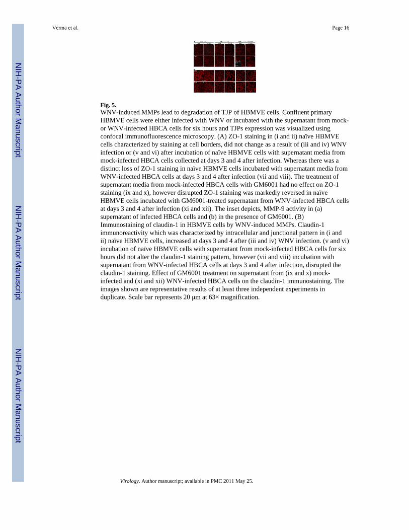
Fig. 5.WNV-induced MMPs lead to degradation of TJP of HBMVE cells. Confluent primaryHBMVE cells were either infected with WNV or incubated with the supernatant from mock-or WNV-infected HBCA cells for six hours and TJPs expression was visualized usingconfocal immunofluorescence microscopy. (A) ZO-1 staining in (i and ii) naïve HBMVEcells characterized by staining at cell borders, did not change as a result of (iii and iv) WNVinfection or (v and vi) after incubation of naïve HBMVE cells with supernatant media frommock-infected HBCA cells collected at days 3 and 4 after infection. Whereas there was adistinct loss of ZO-1 staining in naïve HBMVE cells incubated with supernatant media fromWNV-infected HBCA cells at days 3 and 4 after infection (vii and viii). The treatment ofsupernatant media from mock-infected HBCA cells with GM6001 had no effect on ZO-1staining (ix and x), however disrupted ZO-1 staining was markedly reversed in naïveHBMVE cells incubated with GM6001-treated supernatant from WNV-infected HBCA cellsat days 3 and 4 after infection (xi and xii). The inset depicts, MMP-9 activity in (a)supernatant of infected HBCA cells and (b) in the presence of GM6001. (B)Immunostaining of claudin-1 in HBMVE cells by WNV-induced MMPs. Claudin-1immunoreactivity which was characterized by intracellular and junctional pattern in (i andii) naïve HBMVE cells, increased at days 3 and 4 after (iii and iv) WNV infection. (v and vi)incubation of naïve HBMVE cells with supernatant from mock-infected HBCA cells for sixhours did not alter the claudin-1 staining pattern, however (vii and viii) incubation withsupernatant from WNV-infected HBCA cells at days 3 and 4 after infection, disrupted theclaudin-1 staining. Effect of GM6001 treatment on supernatant from (ix and x) mock-infected and (xi and xii) WNV-infected HBCA cells on the claudin-1 immunostaining. Theimages shown are representative results of at least three independent experiments induplicate. Scale bar represents 20 μm at 63× magnification.
Verma et al. Page 16
Virology. Author manuscript; available in PMC 2011 May 25.
NIH
-PA Author Manuscript
NIH
-PA Author Manuscript
NIH
-PA Author Manuscript

Fig. 6.Supernatant from WNV-infected HBCA cells augment the paracellular permeability of theBBB model. At day 8 post seeding, the BBB models were incubated with supernatant frommock- and WNV-infected HBCA cells or synthetic MMP-9 enzyme as positive control forsix hours and (A) The integrity of the BBB models was determined by measuring the TEERbefore and after the incubation. TEER values, presented as Ω/cm2, demonstrated asignificant decrease only in the BBB model incubated with either supernatant from WNV-infected HBCA cells or synthetic MMP-9 enzyme. Presence of GM6001 rescued the BBBpermeability. (B) FITC-dextran permeability assay of the BBB models. After six hours ofincubation, the percentage of FITC-dextran that crossed the BBB models as compared tocontrol BBB models did not vary significantly in BBB models incubated with supernatantfrom mock-infected HBCA cells. BBB models incubated with supernatant from WNV-infected HBCA cells demonstrated significant increase in the FITC-dextran transmigrationand this increase was reversed in presence of GM6001. The data is representative of at leastthree independent experiments in duplicate. *p<0.005 percent change as compared to controlinserts and **p<0.05 percent change of GM6001-treated supernatants as compared tocorresponding BBB models without GM6001 treatment.
Verma et al. Page 17
Virology. Author manuscript; available in PMC 2011 May 25.
NIH
-PA Author Manuscript
NIH
-PA Author Manuscript
NIH
-PA Author Manuscript

NIH
-PA Author Manuscript
NIH
-PA Author Manuscript
NIH
-PA Author Manuscript
Verma et al. Page 18
Table 1
Primer sequences used for qRT-PCR
Gene
Primer Sequence (5′-3′)
Amplicon
GenBank No. (bp) Tm (°C)
MMP1
NM_002421
Forward ATGAAGCAGCCCAGATGTG 98 55
Reverse TCAATCCTGTAGGTCAGATGTG
MMP2
NM_004530
Forward TGCTGAAGGACACACTAAAGAAG 147 55
Reverse CTTGCGAGGGAAGAAGTTGTAG
MMP3
NM_002422
Forward TCCTGGCATCCCGAAGTG 146 57
Reverse AGCCTGGAGAATGTGAGTGG
MMP9
NM_004994
Forward GCCACTTCCCCTTCATCTTCG 168 56
Reverse ATTGCCGTCCTGGGTGTAGAG
TIMP-1
M12670
Forward TGTTGGCTGTGAGGAATGCA 84 55
Reverse GGTCCGTCCACAAGCAATG
TIMP-2
NM_003255
Forward CGTTGGAGGAAAGAAGGAATATC 86 55
Reverse GCACGATGAAGTCACAGAGG
TIMP-3
U67195
Forward GCGTCTATGATGGCAAGATG 148 55
Reverse AAGCAAGGCAGGTAGTAGC
Virology. Author manuscript; available in PMC 2011 May 25.
Related Documents