28 The Role of Matrix Metalloproteinases and Tissue Inhibitors of Metalloproteinases in the Progression of Astrocytomas Fábio Motta et al. * Federal University of Piauí, Parnaíba Brazil 1. Introduction Neoplasms of the central nervous system (CNS) constitute a group of tumors that are heterogeneous in epidemiology, biological behavior, genetic alterations, histological type, tumor location, pattern of spread, clinical presentation, natural history, age of occurrence, and prognosis (Behin et al., 2003; Louis, 2006; Stroher et al., 2000). According to the 2010 Surveillance Epidemiology and End Results [SEER] data, there were approximately 22,020 deaths from cancers of the brain and nervous system in the U.S. As reported by Binder et al., (2003) and Wrensch et al., (2002), some 7 to 16 cases of primary CNS tumors per 100,000 person-years are diagnosed in adults; approximately 50% of these cases are metastatic tumors, according to Hill et al. 1999. CNS tumors are the third leading cause of cancer deaths in middle-aged adults, the second most common cause of death in children, and the most common solid tumor in children (Giles, 1995; Heuer et al., 2007). The incidence of CNS tumors in children and adolescents is approximately 2.7 cases per 100,000 patients per year. This figure accounts for 16% of the neoplasms within this group, making CNS tumors the second most frequent type of cancer (after leukemia) in young patients (20 years old and younger). The mortality rate observed with CNS tumors is one of the highest among childhood cancers. Diagnosis and treatment improvements, however, has contributed to the survival of these patients (Potter et al., 1998). Epidemiological studies have shown a slight increase in the incidence of CNS tumors during the 1980s and 1990s in children and the elderly (Fisher et al. 2007; Swensen & Bushouse 1998). The apparent increase in the number of cases of CNS cancers is directly linked to the implementation of high-resolution neuroimaging tools, such as Magnetic Resonance Imaging (MRI) and Computed Tomography (CT), which have impacted the clinical diagnosis of neurological diseases (Cristensen et al., 2003; Fisher et al., 2007). Another * Renata Canalle 1 , France Yoshioka 1 , Giovanny Pinto 1 , Lúcia Harada 2 , Carlotti Jurnior 3 , Elvis Valera 3 , Carlos Scrideli 3 and Luiz Tone 3 1 Federal University of Piauí, Parnaíba, Brazil 2 Federal University of Pará, Belém, Brazil 3 University of São Paulo, Ribeirão Preto, Brazil

Welcome message from author
This document is posted to help you gain knowledge. Please leave a comment to let me know what you think about it! Share it to your friends and learn new things together.
Transcript
28
The Role of Matrix Metalloproteinases and Tissue Inhibitors of Metalloproteinases in the
Progression of Astrocytomas Fábio Motta et al.*
Federal University of Piauí, Parnaíba Brazil
1. Introduction Neoplasms of the central nervous system (CNS) constitute a group of tumors that are heterogeneous in epidemiology, biological behavior, genetic alterations, histological type, tumor location, pattern of spread, clinical presentation, natural history, age of occurrence, and prognosis (Behin et al., 2003; Louis, 2006; Stroher et al., 2000). According to the 2010 Surveillance Epidemiology and End Results [SEER] data, there were approximately 22,020 deaths from cancers of the brain and nervous system in the U.S. As reported by Binder et al., (2003) and Wrensch et al., (2002), some 7 to 16 cases of primary CNS tumors per 100,000 person-years are diagnosed in adults; approximately 50% of these cases are metastatic tumors, according to Hill et al. 1999. CNS tumors are the third leading cause of cancer deaths in middle-aged adults, the second most common cause of death in children, and the most common solid tumor in children (Giles, 1995; Heuer et al., 2007). The incidence of CNS tumors in children and adolescents is approximately 2.7 cases per 100,000 patients per year. This figure accounts for 16% of the neoplasms within this group, making CNS tumors the second most frequent type of cancer (after leukemia) in young patients (20 years old and younger). The mortality rate observed with CNS tumors is one of the highest among childhood cancers. Diagnosis and treatment improvements, however, has contributed to the survival of these patients (Potter et al., 1998). Epidemiological studies have shown a slight increase in the incidence of CNS tumors during the 1980s and 1990s in children and the elderly (Fisher et al. 2007; Swensen & Bushouse 1998). The apparent increase in the number of cases of CNS cancers is directly linked to the implementation of high-resolution neuroimaging tools, such as Magnetic Resonance Imaging (MRI) and Computed Tomography (CT), which have impacted the clinical diagnosis of neurological diseases (Cristensen et al., 2003; Fisher et al., 2007). Another
* Renata Canalle1, France Yoshioka1, Giovanny Pinto1, Lúcia Harada2, Carlotti Jurnior3, Elvis Valera3, Carlos Scrideli3 and Luiz Tone3
1Federal University of Piauí, Parnaíba, Brazil 2Federal University of Pará, Belém, Brazil 3University of São Paulo, Ribeirão Preto, Brazil

Molecular Targets of CNS Tumors 594
important cause for the apparent increase in the incidence of brain tumors is the recent inclusion of low-grade neoplasms, which were not previously counted in the population-based registries (Linet et al., 1999). Brain tumors have different incidence rates in different populations, with the highest rates in developed countries. Some factors have influenced the increased incidence in these countries, such as the increased accessibility of medical care and the implementation of cutting-edge technologies in health.However, cultural influences and ethnic or geographical differences cannot be disregarded as risk factors (Davis, 2007; Ohgaki & Kleihues, 2005). The incidence of brain tumors in Japan, for example, is less than half that of the northern European countries. In the U.S., neuroepithelial tumors are found more frequently in Caucasians than in blacks, Hispanics and Asians (SEER, 2010; Wrensch et al., 2002). The etiologic factors of CNS tumors are not well established, and several studies have sought to elucidate the risk factors for these cancers. Some of the possible factors are shown in Table 1. Among the numerous associations that have been studied, the ones that definitely cause primary CNS tumors are the hereditary syndromes (tuberous sclerosis and neurofibromatosis Types 1 and 2) and ionizing radiation used for therapeutic purposes (Davis, 2007; Guney et al. 2001; Skalr, 2002)
Hereditary syndromes** Family history of brain tumors Constitutive polymorphisms Lymphocyte mutagen sensitivity Prior cancers Infectious agents or immunological response Allergies Head trauma Epilepsy, seizures, or convulsions Drugs and medications Diet and vitamins Tobacco smoke exposure Alcohol Hair dyes and sprays Traffic-related air pollution Occupational and industrial exposures Ionizing radiation** Cellular telephones Other radio frequency exposures Power frequency electromagnetic fields
Table 1. Factors that have been studied in relation to the risk of primary tumors of the neuroepithelial tissue or meninges (adapted from Wrensh et al., 2002). **These factors have been shown to cause primary brain tumors.

The Role of Matrix Metalloproteinases and Tissue Inhibitors of Metalloproteinases in the Progression of Astrocytomas 595
The anatomical and pathological classifications of clinical brain tumors are complex and controversial, especially with regard to mixed tumors. As a result, the histological classification of brain tumors has been subject to constant review from the first attempt at histological classification proposed by Bailey & Cushing until today (Louis et al. 2007; Wrensh et al., 2002). The histological classification of brain tumors currently adopted by the World Health Organization (WHO) divides these entities according to the predominant cell type in the lesions. This classification extends the concept of the degree of malignancy by incorporating data from phenotypic and molecular changes observed in these tumors. The WHO classification organizes brain tumors into neuroepithelial tumors of peripheral nerves, tumors of the meninges, lymphomas and hematological malignancies, germ cell tumors and metastatic tumors of the sellar region, with a total of over 100 distinct histological entities (Louis et al. 2007; Kleihues et al., 2002). CNS tumors that originate from neuroepithelial tissue, generically called gliomas, are the CNS neoplasms that are most frequently encountered in children and adults. The subtypes have different biological behaviors and are distinguished by their histological characteristics, such as nuclear atypia, mitotic index, blood microvessel formation and necrosis. Such peculiarities are also used to assess the degree of malignancy according to the standards adopted by the WHO (Louis, 2006; Kleihues et al., 2002). Table 2 shows the WHO classification for neuroepithelial tumors in simplified form.
Neuroepithelial tumors
Astrocytic tumors
Pilocytic astrocytoma 9421/1*
Diffuse astrocytoma 9420/3
Anaplastic astrocytoma 9401/3
Glioblastoma Gliomatosis cerebri
9440/3 9381/3
Oligodendroglial tumors
Oligodendroglioma 9450/3
Anaplastic oligodendroglioma 9382/3
Ependymal tumors
Ependymoma 9391/3
Anaplastic ependymoma 9392/3
Myxopapillary ependymoma 9394/1

Molecular Targets of CNS Tumors 596
Neuroepithelial tumors
Subependymoma
9383/1
Choroid plexus tumors
Choroid plexus papilloma Atypical choroid plexus papilloma Choroid plexus carcinoma
9390/0 9390/1 9390/3
Mixed gliomas of uncertain origin
Astroblastoma
9430/3
Angiocentric glioma 9431/1
Chordoid gliomas of the third ventricle 9444/1
Neuronal and mixed neuronal-glial tumors
Ganglioglioma Anaplastic ganglioglioma
9505/1 9505/3
Central neurocytoma 9506/1
Embryonal tumors
Medulloblastoma 9470/3
Medulloepithelioma
9501/3
Atypical teratoid/rhabdoid tumor 9508/3
Table 2. WHO classification for neuroepithelial tumors. * International Classification of Diseases - Oncology (ICD-O) (adapted from Louis et al., 2007).
1.1 Astrocytic tumors The astrocytic tumors are derived from astrocytes, the principal glial support cells. These tumors are the most common primary brain tumors. They present extensive heterogeneity, differing in the location, age, extent, invasive potential, clinical course and tendency to progress (Khatua et al., 2003; Wang et al., 2003). Classically, astrocytomas are divided according to the degree of malignancy: pilocytic astrocytoma (Grade I), fibrillar diffuse astrocytoma (Grade II), anaplastic astrocytoma (Grade III) and glioblastoma multiforme (Grade IV). The pilocytic astrocytomas are rare neoplasms, with an estimated incidence of less than 1 case per 100.000 person-years. They are predominantly pediatric tumors and are usually located in the cerebral hemispheres, with no infiltrative and circumscribed lesions. They are
The Role of Matrix Metalloproteinases and Tissue Inhibitors of Metalloproteinases in the Progression of Astrocytomas 597
slow-growing and rarely lead to malignant transformation. Thus, these diffuse astrocytoma tumors are considered to be low-grade malignant neoplasms (Evren-Keles, et al., 2004). Although these neoplasms display benign behavior when located in the cerebral hemispheres, the same cannot be said when the lesions occur in the visual pathways or hypothalamus. Histologically, pilocytic astrocytomas are presented as tumors of low to moderate cellularity. They consist of astrocytes with regular, uniform nuclei and little atypia. They are characterized by the presence of numerous Rosenthal fibers, which are protein inclusions within their astrocytic prolongations (Strother et al., 2000). Surgery is the recommended treatment for these lesions and is curative in most cases. Chemotherapy has been used in specific cases, especially in children, with tumors of the optic tracts who experience progression after an initial period of observation and potential deterioration of vision (Packer et al., 1997). Diffuse or fibrillary astrocytomas (Grade II) are less frequent in children, and commonly affecting young adults. Histologically, these tumors have increased cellularity, often do not exhibit microcysts or Rosenthal fibers, tend to undergo anaplastic transformation more frequently and they are more infiltrative (Behin et al., 2003). Anaplastic astrocytomas (Grade III) are infiltrative, have a high proliferative potential and affect mainly adults. This type of tumor is associated with a shorter survival time than the two previously mentioned; presenting the three- or four-year survival in 70% of the cases. Surgery remains the best treatment option and this should target as wide an area as possible (Behin et al., 2003). Grade IV astrocytoma, more commonly called glioblastoma multiforme, is most frequent in adults and is the most aggressive tumor of this lineage; it corresponds to approximately 50% of the malignant tumors of the CNS. It is clinically aggressive, highly invasive and capable of invading extra-axially. This neoplasm often appears in adulthood, after 40 years of age, which does not mean it is exclusive to this age group (Binder et al., 2003). The histological features of glioblastoma multiforme are hypercellularity, cellular atypia and anaplasia, frequent nuclear mitotic figures, necrosis and endothelial proliferation. (Behin et al., 2003; Strother et al., 2000). The optimal treatment is surgery, but many patients with neurological problems, the very elderly (over 80 years), and those at high risk of complications from anesthesia should be carefully evaluated; in many cases, biopsy is most suitable. Local radiotherapy is also indicated, even in cases where there is no indication for surgery. Glioblastomas may arise from the transformation of low-grade or anaplastic tumors (in which case they are called secondary glioblastomas), or they can be primary (in which case they are called primary glioblastomas) (Kleihues et al., 2000). Although this classification is widely accepted in clinical practice, the designation of primary or secondary glioblastomas is more conceptual than diagnostic and has no pratical impact on treatment. The molecular analysis of these tumors has led to a classification based on the genetic alterations commonly found in them, which can distinguish primary from secondary glioblastomas (Figure 1) (Kleihues & Ohgaki, 2007).
2. The Extracellular Matrix (ECM) The ECM consists of a complex network of proteins and proteoglycans, secreted by the connective tissue, whose purpose is to support the tissues that make up an organism. This

Molecular Targets of CNS Tumors 598
network of proteins has a decisive influence on various biological activities (Giancotti et al. 1999; Matrisian, 1990). The degradation of the ECM is a physiological process that is strictly controlled by the dynamic equilibrium between the activity of matrix metalloproteinases (MMPs) and tissue inhibitors of metalloproteinases (TIMPs). The cell–ECM interactions send signals promoting cell differentiation, migration and mobility that are necessary for cell homeostasis (Chang & Werb, 2001). MMPs degrade the ECM proteins and pericellular microenvironment, activating growth factors and adhesion molecules. These events are essential for a variety of physiological processes, such as embryonic development, remodeling, tissue repair and angiogenesis (Chakraborti et al. 2003; Löffek et al. 2010; Matrisian, 1990). The disordered destruction of the ECM plays a crucial role in the development of various pathologies, such as liver fibrosis, aneurysms, periodontosis, rheumatoid arthritis, multiple sclerosis, cystic fibrosis and tumor formation. The development of tumors is highly dependent on the microenvironment because the production and/or uncontrolled activation of proteolytic enzymes involved in the degradation and remodeling of the ECM are the determining factors in tumor development (Overall et al., 2002; Pollet et al. 2004).
Fig. 1. The genetic pathways involved in the development of primary and secondary glioblastomas (adapted from Ohgaki & Kleihues, 2007).
The Role of Matrix Metalloproteinases and Tissue Inhibitors of Metalloproteinases in the Progression of Astrocytomas 599
2.1 Matrix metalloproteinases The metalloproteinases are divided according to their biochemical characteristics as follows: MMPs; a disintegrin and metalloproteinases (ADAMs); and ADAMs with thrombospondin domains (ADAMS-TS) (Malemud, 2006; Werb, 1997). MMPs are zinc-dependent (Zn+2) peptidases with highly conserved substrate specificity that are capable of degrading proteoglycans, laminin, fibronectin, gelatin and fibrillar collagen (Types I, II, III, V and XI) on the basal membrane and in the ECM (Curran & Murray, 2000, Yong et al., 1998). There are more than 20 known members of this family of proteases. Mammalian MMPs are classified into soluble (secreted) and membrane (MT-MMPs) (Amalinei et al. 2007; Pei et al. 2000; Seiki, 1999). Soluble MMPs are synthesized as pro-enzymes, while the MT-MMPs are activated in the intracellular space and expressed on the cell surface as active enzymes. These MMPs are divided into four subclasses according to their structural and functional characteristics: collagenases, gelatinases, stromelysins, and matrilysins (Nabeshima et al., 2002; Nagase & Woessner, 1999; Visse & Nagase, 2003). The degradation of ECM is a physiological process that is strictly controlled by the dynamic equilibrium between the activity of matrix metalloproteinases (MMPs) and tissue inhibitors of metalloproteinases (TIMPs). The cell–ECM interactions send signals that promote cell differentiation, migration and mobility, which are necessary for cell homeostasis (Chang & Werb, 2001). MMPs degrade ECM proteins and the pericellular microenvironment, activating growth factors and adhesion molecules. These events are essential for a variety of physiological processes, such as embryonic development, remodeling, tissue repair and angiogenesis (Chakraborti et al. 2003; Löffek et al. 2010; Matrisian, 1990). The general structure of MMPs consists of three domains that are highly conserved among this family but still distinct: a pro-N-terminal domain, a catalytic domain, and a C-terminal domain that contains the conserved hemopexin (HEXGHXXGXXH) motif (the matrilysins subfamilies do not have this motif). The histidine residues (H) contained in the hemopexin domain are involved in binding to Zn+2, which is required for its proteolytic activity. The pro-domain of MMPs also contains a highly conserved PRCGVDP motif that bonds covalently to Zn+2, keeping the protein in its non-activated zymogen form. Gelatinases have a binding site for fibronectin in the catalytic domain that confers specificity to the substrate (Figure 2) (Nakada et al. 1999; Pollet et al. 2004; Vihinen & Kalahari, 2002). The collagenase subfamily includes MMP-1, MMP-8, MMP-13 and MMP-18, which exhibit biochemical characteristics that allow them to cleave interstitial collagen I, II and III in a site-specific N-terminal domain. Interstitial collagen I is one of the most abundant substances in the human body, so collagenases play an important role in the process of remodeling and degrading the ECM (Curran & Murray, 2000; Visse & Nagase, 2003). The gelatinases include gelatinase A (MMP-2) and gelatinase B (MMP-9). These enzymes are specialized in degrading Type IV collagen, the main constituent of the basement membrane of cerebral blood vessels. As previously mentioned, these enzymes have three repeats of a fibronectin domain in their catalytic domains, which allows the gelatinases to bind to laminins and gelatin (Yong et al., 1998). MMP-2, is a 72-kDa gelatinase, encoded on human chromosome 16q13 and constitutively expressed in variety of cell types. This enzyme has the ability to degrade collagen V, VII, XI and fibronectin. MMP-2 also mediates proliferation, adhesion and cell migration. This enzyme is secreted in its inactive form and gains catalytic activity by interacting with the complex formed by membrane metalloproteinase 1 (MT1-MMP, or MMP-14) and TIMP-2 (Chintala et al., 1999).

Molecular Targets of CNS Tumors 600
Fig. 2. The general structure of MMPs and their subfamilies (Vihinen & Kalahari, 2002).
MMP-9 is a 90–110-kDa protein encoded in the region 20q11.2-13.1. This enzyme is also secreted as a zymogen that can be stored in an active or latent form in the cytosol. The expression of MMP-9 is not constitutive and may be influenced by many factors, such as cell growth, cytokine signaling, cell–cell adhesion and cell–ECM interactions. The mechanism of protease activation thus involves a complex web of interactions with other MMPs and TIMPs (Amalinei et al. 2007; Chintala et al. 1999).

The Role of Matrix Metalloproteinases and Tissue Inhibitors of Metalloproteinases in the Progression of Astrocytomas 601
The stromelysins group is composed of the following proteases: MMP-3 (stromelysin-1), MMP-10 (stromelysin-2); and MMP-11 (stromelysis-3). These proteases have similar substrates, but MMP-3 has higher proteolytic activity than the other stromelysins. MMP-3 is expressed in several cell types and acts on the following ECM components: collagen, gelatin, elastin, fibronectin, and vitronectin. In addition to digesting the ECM components, MMP-3 participates in the activation of pro-MMP-1 and pro-MMP-9 (Egeblad & Werb, 2002). The membrane-MMP subfamily consists of six MT-MMPs (MT1-MMP through MT6-MMP) and two glycosylphosphatidylinositol (GPI)-anchored MMPs (MMP-17 and MMP-25). One of the most prominent members of this subfamily is MT1-MMP (MMP-14), whose substrates are collagen Types I, II and III, gelatin, fibronectin and laminin (Koshikawa et al., 2000). MT1-MMP is considered a multifunctional molecule. It is directly involved in cell growth, inflammation, the formation of new vessels, and the processes of cell adhesion and migration. Consequently, it plays a role in the formation of metastases (Shiomi & Okada, 2003; Yana & Seiki, 2002). Its interactions with MT1-MMP cause TIMP-2 to play a crucial role in the activation of pro-MMP-2. The catalytic domain of MT1-MMP binds to the N-terminal domain of TIMP-2, causing the C-terminal region of TIMP-2 to become capable of connecting to the hemopexin domain of pro-MMP-2. The location of this protease in the membrane plays a role in pericellular proteolysis because this molecule activates pro-MMP-2, enabling it to use exogenous substrates to mediate the processes of invasion and metastasis. (Sternlicht & Werb, 2001; Sounni et al., 2003). The expressions of MMPs are under the control of a sophisticated genetic mechanism. Under basal circumstances, levels of MMPs are relatively low; however, the expression of MMPs is increased by the processes of ECM remodeling that result from physiological or pathological conditions. The expression of some subfamilies of MMPs is tissue-specific. The regulation of the expressions of MMPs occurs by three main routes: transcriptional regulation of MMPs; activation of pro-enzymes, and inhibition by the action of TIMPs (Nagase et al., 2006; Yong et al. 1998).
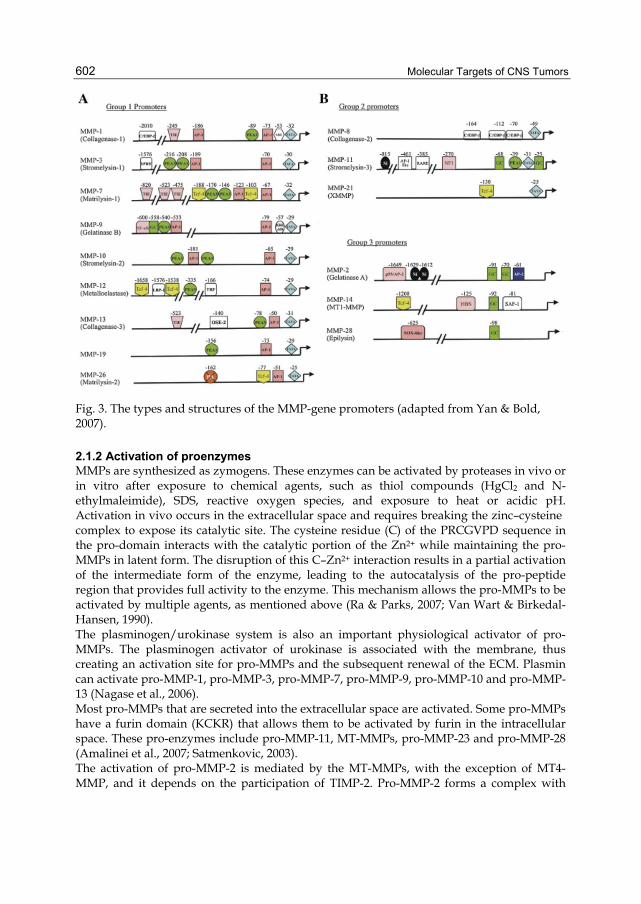
2.1.1 Transcriptional regulation of MMP genes The transcription factors that regulate MMP gene expression include activating protein-1 (AP-1), polyoma enhancer activator 3 (PEA3), SP1, β-catenin/Tcf-4 and NF-κB, these factors seem to have synergistic effects on MMP gene regulation. Based on the composition of their regulatory elements, the promoters of these genes can be divided into Classes I, II and III (Yan & Bold, 2007). The Class I promoters contain a TATA box at position -30 (relative to the transcription start site) and an AP1-binding site at position -70. Many of these promoters also contain a binding site for PEA3 that is adjacent to the AP1 site. The Class II promoters also have a TATA box but without the proximal AP1 site. The regulation of these promoters is relatively simple and it is distinct from the regulation of the Class I promoters (Benbow & Brinckerhoff, 1997; Chakraborti et al., 2003). The third and final group of promoters does not have a TATA box, meaning that transcription begins at multiple sites. However, the SP1 family of transcription factors, which bind close to the GC box, . controls the regulation of this group. The FOS and JUN proteins actively participate in the transcription regulatory mechanism by forming a heterodimer that binds to the AP1 sites and stimulates the transcription of MMPs in response to inflammatory cytokines (Matrisian, 1990). The structural differences between the MMP promoters are shown in Figure 3.
Molecular Targets of CNS Tumors 602
Fig. 3. The types and structures of the MMP-gene promoters (adapted from Yan & Bold, 2007).
2.1.2 Activation of proenzymes MMPs are synthesized as zymogens. These enzymes can be activated by proteases in vivo or in vitro after exposure to chemical agents, such as thiol compounds (HgCl2 and N-ethylmaleimide), SDS, reactive oxygen species, and exposure to heat or acidic pH. Activation in vivo occurs in the extracellular space and requires breaking the zinc–cysteine complex to expose its catalytic site. The cysteine residue (C) of the PRCGVPD sequence in the pro-domain interacts with the catalytic portion of the Zn2+ while maintaining the pro-MMPs in latent form. The disruption of this C–Zn2+ interaction results in a partial activation of the intermediate form of the enzyme, leading to the autocatalysis of the pro-peptide region that provides full activity to the enzyme. This mechanism allows the pro-MMPs to be activated by multiple agents, as mentioned above (Ra & Parks, 2007; Van Wart & Birkedal-Hansen, 1990). The plasminogen/urokinase system is also an important physiological activator of pro-MMPs. The plasminogen activator of urokinase is associated with the membrane, thus creating an activation site for pro-MMPs and the subsequent renewal of the ECM. Plasmin can activate pro-MMP-1, pro-MMP-3, pro-MMP-7, pro-MMP-9, pro-MMP-10 and pro-MMP-13 (Nagase et al., 2006). Most pro-MMPs that are secreted into the extracellular space are activated. Some pro-MMPs have a furin domain (KCKR) that allows them to be activated by furin in the intracellular space. These pro-enzymes include pro-MMP-11, MT-MMPs, pro-MMP-23 and pro-MMP-28 (Amalinei et al., 2007; Satmenkovic, 2003). The activation of pro-MMP-2 is mediated by the MT-MMPs, with the exception of MT4-MMP, and it depends on the participation of TIMP-2. Pro-MMP-2 forms a complex with

The Role of Matrix Metalloproteinases and Tissue Inhibitors of Metalloproteinases in the Progression of Astrocytomas 603
TIMP-2 through its C-terminal domain, allowing the N-terminal domain of TIMP-2 to bind to MT1-MMP on the cell surface. The complex containing MT1-MMP, TIMP-2, and pro-MMP-2 is then presented to an adjacent free MT1-MMP, which activates the pro-MMP-2. The hemopexin domain seems to facilitate this activation process (Jo et al., 2000).
2.2 Tissue inhibitors of metalloproteinases The TIMPs constitute the third regulatory level of MMP expression. These proteins are major physiological inhibitors of MMPs. The TIMPs are low-molecular-weight, 21–30 kDa, molecules that have approximately 12 cysteine residues that are involved in disulfide bridges, six of which are responsible for the formation of two domains that are essential for the interaction of these molecules with MMPs. The C-terminal domain is responsible for the interaction with and activation of pro-MMPs and for the subcellular localization of these proteins. The N-terminal domain acts as an independent unit that inhibits the activity of MMPs (Baker et al., 2002; Brew et al. 2000). Blocking the proteolytic activity of both soluble and membrane-bound MMPs is accomplished by TIMPs through the formation of monocovalent complexes at a 1:1 ratio between the catalytic site of MMPs and the N-terminus of TIMPs. The amino groups and carbonyl groups of the cysteine residues in the N-terminus of TIMPs act together to chelate the Zn2+ in the catalytic domain of MMPs, making Zn2+ unavailable to activate the MMPs (Nagase et al., 2006; Visse & Nagase, 2003). Four TIMPs, designated as TIMP-1 through TIMP-4, have been characterized in humans. They are resistant to heat denaturation and proteolytic degradation. These proteins are expressed in a variety of cell types and fluids (Gómez et al., 1997). Although all TIMPs are capable of inhibiting all the MMPs that have been studied, they differ in many aspects, such as solubility, the complementary nature of the pro-enzymes and the regulation of expression. TIMP-1, TIMP-2 and TIMP-4 are present in the soluble form, while TIMP-3 is connected to the ECM (Lamfers et al., 2005). The expression of TIMP-1 is inducible. This molecule has weak inhibitory action against MMP-19 and MT-MMPs (MT-MMP1, MT-MMP-2, MT-MMP-3 and MT-MMP-5) and even inhibits ADAM -10. The expression of TIMP-2 is constitutive, and it has strong inhibitory action against the MT-MMPs. TIMP-3 may also inhibit other members of the ADAM family, such as ADAM-17, ADAMTS-4 and ADAMTS-5, and the inhibitory effects of this molecule on MMP-9 are greater than those observed for TIMP-1 and TIMP-2. TIMP-4 expression is inducible, and it appears to inhibit the activities of MMPs constitutively. Table 3 presents some of the molecular characteristics and biological properties of TIMPs. The TIMPs have also the ability to form complexes with pro-MMPs, thus regulating their activation. TIMP-1 preferentially forms complexes with pro-MMP-9, and TIMP-2 binds preferentially to pro-MMP-2 to facilitate its activation (Baker et al., 2002). TIMP-3 binds to both pro-MMP-2 and pro-MMP-9 (Bigg et al., 1997), and TIMP-4 can bind to the C-terminal domain of pro-MMP-2 (Butler et al., 1999). The inhibition of MMPs is not unique to TIMPs. Molecules such as α-macroglobulin, general protease inhibitors, β-amyloid precursors linked to the cell membrane, and the membrane protein RECK play the same. RECK is a GPI-type, membrane-anchored protein that decreases the catalytic activity of MMP-2, MMP-9 and MT1-MMP and can overcome the angiogenic activity in neoplastic processes, leading to the death of tumor cells (Oh et al., 2001). A scheme for the interaction between the activation and inhibition of MMPs by TIMPs is presented in Figure 4.

Molecular Targets of CNS Tumors 604
TIMP-1 TIMP-2 TIMP-3 TIMP-4 Chromosomal location
Xp11.23-11.4 17q23-17q25 22q12.1-22q13.2
3p25
mRNA (kb) 0,9 3,5 5,0 1,4 Molecular weight (kDa)
28,5 21 22/27 22
Protein localization Soluble Soluble ECM Soluble Associated pro-MMPs pro-MMP9 pro-MMP2 pro-MMP2/9 pro-MMP2 Gene Expression Induced Constitutive Induced Induced Tumorigenic effects Inhibition Inhibition Inhibition Inhibition Metastases Stimulates ____ ____ Stimulates
Table 3. The molecular and biological characteristics of TIMPs (adapted from Baker et al., 2002).
Fig. 4. MMPs are a class of proteases secreted by tumor cells. RECK is a membrane-anchored inhibitor of MMPs. It inhibits MMP-2, MMP-9 and MT1-MMP. Soluble, secreted MMP inhibitors, TIMPs, have also been identified. They appear to be less active at inhibiting MMPs and are perhaps even essential for MMP maturation. The inhibition of MMPs by RECK inhibits invasion of tissues, metastasis and tumor angiogenesis and is essential for normal development. (www.biocarta.com).
The Role of Matrix Metalloproteinases and Tissue Inhibitors of Metalloproteinases in the Progression of Astrocytomas 605
The main function of TIMPs is to act as natural receptor inhibitors of metalloproteinases. However, some studies have attributed other functions to TIMPs. Some of these functions are related to inhibiting MMPs and others are independent of it, such as promoting cell proliferation, anti-apoptosis activity and anti-angiogenesis (Chang & Werb, 2001; Chirco et al., 2006). The induction of cell proliferation by TIMPs was first described for TIMP-1 and TIMP-2; they enhance the effect of erythropoietin in the proliferation and differentiation of erythroid progenitors. This property was observed not only in this cell type but also in keratinocytes, chondrocytes, epithelial cells and cancer cells (Hayakawa et al. 1992; Murat et al., 1993). In addition to promoting cell proliferation, the TIMPs may also modulate apoptosis in various ways, including opposing effects. TIMP-1 and TIMP-2 are involved in resistance to apoptosis in normal and cancer cells. The effect of TIMP-3 is different because it promotes apoptosis in smooth muscle cells, inducing the shedding of the ECM from transformed cells (Baker et al., 1998). The effects of TIMP-4 in modulating apoptosis are contradictory; fibroblast-transformed heart tissue appear to be induced to apoptosis by TIMP-4, and the protein seems to have an inhibiting effect on apoptosis when hyperexpressed in breast-tumor cell lines (Baker et al., 2002). Angiogenesis is described as the biological process of new capillary formation from existing vessels, which occurs in normal and pathological events. The TIMPs have anti-angiogenic activity, as was first demonstrated for TIMP-1 and TIMP-2. When hyperexpressed, they reduce angiogenic activity in pancreatic adenocarcinomas (Bloomston et al., 2002). TIMP-3 also has anti-angiogenesis and anti-invasion activities. For example, it reduces the invasiveness of the HT1080 cell line derived from fibrosarcoma and the expression of MMP-9, a molecule with known angiogenic activity (Lamfers et al., 2005). These observations corroborate the notion that the imbalance between the expressions of MMPs and TIMPs has a decisive role in the initiation and development of neoplastic processes.
2.3 The role of MMPs in progression of astrocytic tumors Since the discovery of MMPs in 1962 by Gross and LaPierre, these enzymes have attracted great interest among researchers for their involvement in various physiological and pathological events. However, only in the 1980s, MMPs were discovered to be involved in the development and progression of tumors, a function that is widely studied to this day (Bourboulia & Stetler-Stevenson, 2010; Matrisian et al., 1986). The invasive properties of CNS tumors are of great clinical importance because they contribute to the aggressive behavior of these tumors (Rao, 2003). For reasons still not well clarified yet , most primary neuroepithelial tumors do not evolve into metastatic disease. However, these tumors invade surrounding tissues, infiltrate their cells into normal tissue and (more rarely) spread along the neuro-axis into the cerebrospinal fluid. The mechanism of astrocytic-tumor invasion is a complex process in which tumor cells separate from the parental tumor and rejoin the ECM. This process is associated with an increase in cell motility and with the ability of tumor cells to hydrolyze proteins, carbohydrates and proteoglycans in the ECM. These cells perform this task through the excessive production of proteases that modify the ECM, thus creating access to the surrounding tissues and promoting migration to other parts of the brain (Levicar et al., 2003).

Molecular Targets of CNS Tumors 606
MMPs are among the proteases that can degrade the ECM in brain tissue. The integrity of the ECM depends on a dynamic balance between MMP and TIMP activities. The loss of this balance can lead to the creation of a permissive environment for the initiation and development of a neoplastic process in brain tissue. MMPs, TIMPs and cell adhesion molecules are involved in the development of CNS malignancies (Forsyth et al. 1999; Mohana et al. 1995; Nakada et al. 2001; Rooprai et al., 1998). Changes in the expression of these proteins appear to be directly linked to the invasiveness of tumor cells. Classically, MMPs are considered to be responsible for promoting tumor growth, while TIMPs are thought to block the invasive ability of tumors (Amalinei et al. 2010; Deryugina & Quinley, 2006). High-grade astrocytomas and medulloblastomas are neuroepithelial tumors of different cellular origins and are highly invasive. The degradation of the ECM is a prerequisite for invasion by these tumors. Among the MMPs that are commonly associated with the progression of astrocytic tumors are the gelatinases and the membrane MMPs, especially MT1-MMP1. Gelatinases A (MMP-2) and B (MMP-9) are undoubtedly the most well-studied MMPs in CNS tumors, and they have been identified as important factors in the development of high-grade gliomas (Munaut et al. 2003; Pagenstecher et al. 2001; Zhao, et al., 2007). This observation is directly linked to the invasive nature of these tumors because the expression levels of these gelatinases are generally low in normal brain tissue (Van Meter et al., 2001). A study by Jäälinojä et al. (2000) demonstrated that the high expression of MMP-2 is a characteristic of highly malignant tumors and is associated with poor patient survival, similar data were also observed by Kunishio et al. (2003), who used immunohistochemistry analysis to show that MMP-2 and MMP-9 are not associated with increased aggressiveness in astrocytic tumors. Komatsu et al. (2004) studied the expression of MMP-2 and MMP-9 in human gliomas by using two different techniques, RQ-PCR and immunohistochemistry, the results revealed no correlation between the expression of MMP-2 protein and the degree of malignancy. However, quantitative analysis of the mRNA showed that tumors of the highest grade, such as glioblastomas, express much higher levels of the MMP-2 and MMP-9 genes than do low-grade tumors. This study clearly demonstrated that methodological differences may explain some of the conflicting results found in the literature. Increased levels of MMP-2 and MMP-9 and their relationships with astrocytic tumors of greater aggressiveness and invasiveness have also been reported by a number of authors, such as Forsyth et al. (1999), Lampert et al. (1998), Munaut et al. (2003), Pagenstecher et al. (2001) and Wang et al. (2003). Wild-Bode et al. (2001) reported that the overexpression of MMP-2 and MMP-9 is the best predictor of cell invasion by high-grade gliomas. Another important result that highlights the prognostic significance of elevated MMP-9 expression was discovered by Hormigo et al. (2006), who found elevated MMP-9 protein in the serum of patients suffering from high-grade astrocytomas, which was not observed in low-grade tumors or in non-neoplastic tissue. These authors also suggested that MMP-9 can be used as a biomarker in the management of glioblastoma patients. The analysis of MMP-9 expression conducted by our group revealed that this gene is expressed more highly in astrocytic tumors than in non-tumor tissue. The expression is much greater in tumors with the highest degree of malignancy, especially glioblastomas, than in low-grade astrocytomas. Our study also revealed that pilocytic astrocytomas arising in the optic tracts, hypothalamus and pituitary gland, which appear to exhibit more
The Role of Matrix Metalloproteinases and Tissue Inhibitors of Metalloproteinases in the Progression of Astrocytomas 607
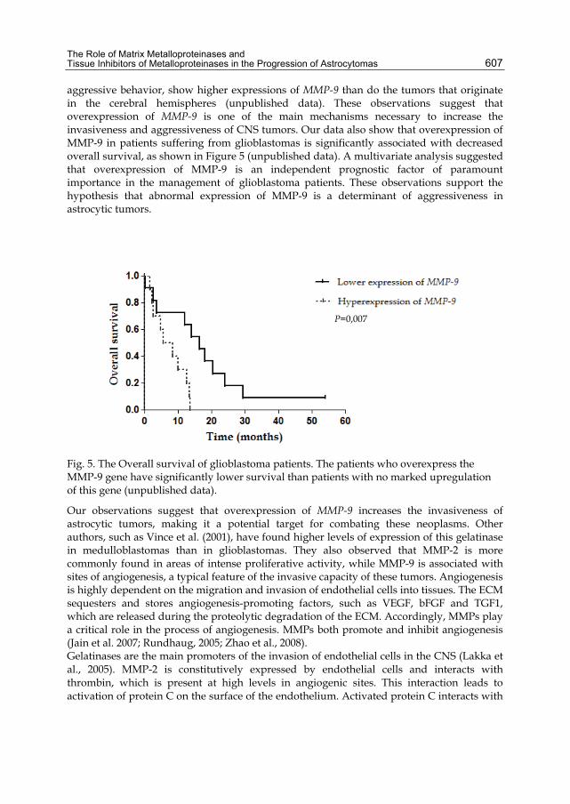
aggressive behavior, show higher expressions of MMP-9 than do the tumors that originate in the cerebral hemispheres (unpublished data). These observations suggest that overexpression of MMP-9 is one of the main mechanisms necessary to increase the invasiveness and aggressiveness of CNS tumors. Our data also show that overexpression of MMP-9 in patients suffering from glioblastomas is significantly associated with decreased overall survival, as shown in Figure 5 (unpublished data). A multivariate analysis suggested that overexpression of MMP-9 is an independent prognostic factor of paramount importance in the management of glioblastoma patients. These observations support the hypothesis that abnormal expression of MMP-9 is a determinant of aggressiveness in astrocytic tumors.
Fig. 5. The Overall survival of glioblastoma patients. The patients who overexpress the MMP-9 gene have significantly lower survival than patients with no marked upregulation of this gene (unpublished data).
Our observations suggest that overexpression of MMP-9 increases the invasiveness of astrocytic tumors, making it a potential target for combating these neoplasms. Other authors, such as Vince et al. (2001), have found higher levels of expression of this gelatinase in medulloblastomas than in glioblastomas. They also observed that MMP-2 is more commonly found in areas of intense proliferative activity, while MMP-9 is associated with sites of angiogenesis, a typical feature of the invasive capacity of these tumors. Angiogenesis is highly dependent on the migration and invasion of endothelial cells into tissues. The ECM sequesters and stores angiogenesis-promoting factors, such as VEGF, bFGF and TGF1, which are released during the proteolytic degradation of the ECM. Accordingly, MMPs play a critical role in the process of angiogenesis. MMPs both promote and inhibit angiogenesis (Jain et al. 2007; Rundhaug, 2005; Zhao et al., 2008). Gelatinases are the main promoters of the invasion of endothelial cells in the CNS (Lakka et al., 2005). MMP-2 is constitutively expressed by endothelial cells and interacts with thrombin, which is present at high levels in angiogenic sites. This interaction leads to activation of protein C on the surface of the endothelium. Activated protein C interacts with
P=0,007

Molecular Targets of CNS Tumors 608
latent MMP-2 to increase its protease activity, thus allowing the endothelial cells to degrade the basement membrane. MMP-9 is then activated by intracellular vesicles secreted from endothelial cells to enhance the degradation of basement membrane. When the basement membrane reaches the interstice, the abundant collagen Type I contributes to the prolonged activation of MMP-2, thus consolidating the process of angiogenesis (Nguyen et al., 2001). Glioblastomas are astrocytic tumors with increased invasive behavior, this property is closely related to their high vascularization. In vivo and in vitro, MMP-2 and MMP-9 are hyperexpressed in endothelial cells of glioblastomas (Vince et al., 2001; Zhao et al., 2010). The direct involvement of these proteins in angiogenesis may explain our observation of increased expression of MMP-2 and MMP-9 mRNA in highly malignant tumors (unpublished data). Similar results were found by Munaut et al. (2003), who also observed that overexpression of MMP-2 and MMP-9 is correlated with overexpression of VEGF in glioblastomas, a key mediator of angiogenesis. Elevated expression of MMP-2 and MMP-9 and its clinical and biological implications for other cancers have been reported in several studies. Cho et al. (2007) reported that overexpression of MMP-9 is associated with poor prognosis in colorectal tumors. Similar observations were obtained by Kawata et al. (2007) for renal tumors and Iniesta et al. (2007) for non–small cell lung cancer. Like the gelatinases, MT1-MMP is considered to be a promoter of tumor cell invasion and a facilitator of metastasis. MT1-MMP promotes the acquisition of invasive capacity in both normal and neoplastic cells through its ability to degrade pericellular collagen. It also facilitates angiogenesis by activating endothelial cells. This activity has been documented both in vivo and in vitro (Martin & Matrisian, 2007). The pro-invasive activity of MT1-MMP has been associated by some authors with invasion and progression in other tumors, such as neck tumors by Zhai et al. (2005) and astrocytic tumors by Forsyth et al. (1999); Nakada et al. (1999, 2001) and Kong et al. (2007). Among the studies that have found a direct relationship between the increased expression of MT1-MMP and the evolution of gliomas, that of Nuttall et al. (2003) stands out because it found a greater than 21-fold increase in the level of MT1-MMP mRNA in glioblastomas relative to non-tumor brain tissue. The overexpression of MT1-MMP was also reported in a glioblastoma cell line (U251), the most invasive of the strains tested by the authors. This increase in the level of MT1-MMP was accompanied by the overexpression of MMP-2 and MMP-9 and of genes related to angiogenesis, such as VEGF. Munaut et al. (2003) also reported that strong expression of VEGF is accompanied by overexpression of MMP-2, MMP-9 and MT1-MMP and that these proteins act synergistically to create a permissive environment for glioblastoma cell invasion. Pagenstecher et al. (1999) also observed that the levels of MMP expression in pilocytic astrocytomas are comparable to those observed in anaplastic astrocytomas and glioblastomas, suggesting that this molecule is a major player in the progression of astrocytic tumors. Arroyo et al. (2007) and Basile et al. (2007) demonstrated that MT1-MMP plays a crucial role in angiogenesis and that this protein is usually hyperexpressed at sites of neoangiogenesis. We believe that our observations are related to the level of vascularization of these tumors, and high-grade pilocytic astrocytomas have a significant level of vascularization. The overexpression of MT1-MMP and its relationship with the aggressive behavior of tumors have also been described in soft tissue cancers Roebuck et al. (2005) and more recently by Sroka et al. (2008) in prostate tumors.

The Role of Matrix Metalloproteinases and Tissue Inhibitors of Metalloproteinases in the Progression of Astrocytomas 609
2.4 The role of TIMPs in the progression of astrocytic tumors. A high level of TIMP expression may occur in two distinct contexts: (1) as part of the defense mechanisms of the host tissue against the invasion; and (2) as promoter, either directly or indirectly, of cell growth in the tumor mass that helps the tumor progress and invade adjacent structures (Brand, 2002). The latter property directly promotes the evolution of these lesions to higher-malignancy tumors. TIMP-1 is involved in a series of biological and pathological processes, including growth-promoting activity, inhibition of angiogenesis and changes in cell morphology. In the CNS, this molecule seems to possess the neuroprotective effect of maintaining the blood–brain barrier by its inherent inhibitory action on MMPs. TIMP-1 also has the distinguishing characteristic of not being able to inhibit MT1-MMP, in contrast to other TIMPs (Duffy et al., 2008). TIMP-1 is also involved in the development of neuroepithelial tumors (Gardner & Ghorpade, 2003; Lampert et al. 1998; Kakagawa et al. 1994; Pagentecher et al. 1999). Groft et al. (2001) showed that overexpression of TIMP-1 is associated with malignancy in gliomas and that TIMP-1 is detected mainly near the sites of blood vessel formation. These observations could be a result of interactions with pro-MMP-9 and the angiogenesis-promoting ability of this MMP. Huang et al (2005) and Pagentecher et al. (1999) found higher levels of TIMP-1 expression in pilocytic astrocytomas and glioblastomas. This result seems contradictory, but it may act to offset the simultaneous increases in the expression of MMP-9 and TIMP-1 because TIMP-1 preferentially inhibits MMP-9. The results of Pagentecher et al. (1999) also showed that this overexpression is linked to sites of neoangiogenesis. Overexpression of TIMP-1 appears to be an important prognostic factor for patients with breast cancer, since those patients with greater expression of this gene have higher survival rates (Nakopoulou et al., 2003). However, we found no similar reports for astrocytic tumors. TIMP-2 preferentially inhibits MMP-2 and activates pro-MMP-2 by forming complexes with MT1-MMP. Béliveau et al. (1999), Huang et al. (2005) and Nakada et al. (2001) suggests that there is an inverse correlation between the expression of TIMP-2 and the degree of malignancy in CNS tumors. This result may be related to the antagonistic effects of these genes in the classical biological behavior of astrocytic tumors, in which MMPs act as promoters of invasion and TIMPs as blockers of tumor growth. Nuttal et al. (2003) has also detected high levels of TIMP-2 mRNA and protein expression in non-neoplastic brain-tissue samples. The in vitro protective effects of TIMP-2 were also described by Papi et al. (2007) in experiments using cells derived from gliomas. Moreover, Saxena et al. (1999) found a positive correlation between the expressions of TIMP-2 and MMP-2 and the degree of malignancy of CNS tumors, in which the highest levels of expression of these genes are primarily found in recurrent tumors. The increased expression of TIMP-2 in these tumors may be related to defense against invasion or, more precisely, to the attempt to block the proteolytic activity of MMP-2, which facilitates tumor invasion. Other associations between the expression of TIMP-2 and survival in patients affected by CNS tumors have not been found. Curran et al. (2004) observed that the loss of TIMP-2 expression contributes significantly to the low survival rate of patients suffering from colorectal tumors. The loss of TIMP-3 expression is related to cancer development, and normal brain tissue has high levels of TIMP-3. This physiological inhibitor of MMPs antagonizes the activity of most metalloproteinases, thereby leading to the inhibition of tumor growth, angiogenesis, invasion and metastasis formation. The overexpression of this gene in tumor cells seems to promote apoptosis and suppress early tumor growth (Hoe et al., 2007).

Molecular Targets of CNS Tumors 610
A study using microarray analysis that was conducted by Bozinov et al. (2008) showed that TIMP-3 is one of the genes preferentially expressed in less-malignant CNS tumors. Huang et al. (2003) also observed that non-neoplastic tissue has low expression of TIMP-3. Furthermore, Lampert et al. (1998) and Pangestecher et al. (1999) found no variation in the gene expression levels between gliomas and suggested that this observation is consistent with constitutive expression. The protective effect of TIMPs against invasion by glioma cells was demonstrated by Lamfers et al. (2005), who used an adenovirus to transfer the gene for TIMP-3 into cultured cells, which resulted in increased expression of the gene and a reduction in the invasive capacity of the cells. The main cause of the decreased expression of TIMP-3 is the hypermethylation of its promoter region. Gonzalez-Gomez et al. (2003a, 2003b) have shown that the promoter region of this gene is hypermethylated in glioblastomas, especially in secondary glioblastomas, and in medulloblastomas. Nakamura et al. (2005) have also found that the loss of heterozygosity in region 22q12.3, the region containing the TIMP- gene, is a frequent event in glioblastomas. These authors also suggested that these molecular alterations may be the primary cause of decreased expression of TIMP-3 in high-grade astrocytic tumors. Nakamura et al. (2005) have shown that hypermethylation of TIMP-3, and the consequent reduced expression of the gene, is an important factor in the poor survival of patients affected by highly malignant tumors. Correlations between decreased expression of this gene and low survival rates have also been observed in colorectal cancer (Hilska et al., 2007), non–small cell lung cancer (Mino et al., 2007) and bladder carcinomas (Hoque et al., 2008). TIMP-4 has been studied less than the other TIMPs. It was discovered by Greene et al. (1996), who identified an expressed sequence tag (EST) with that was closely related to that of other TIMPs. This EST was identified and cloned in cDNA libraries from the human heart. They identified low levels of this transcript in a variety of tissues and organs, such as kidney and placenta, and did not observe expression of the gene in tissues derived from brain, lung, thymus or liver. These authors suggested that the contribution of TIMP-4 to maintaining the homeostasis of the ECM is tissue-specific. Bigg et al. (1997) also showed that TIMP-4 has a strong affinity for pro-MMP-2 and that, together with TIMP-2, it participates in the activation of pro-gelatinase A. The literature addressing the involvement of TIMP-4 in the development and progression of CNS tumors is sparse. Groft et al. (2001) have identified an inverse relationship between the expression of TIMP-4 and malignant CNS tumors. Microarray data obtained by Rorive et al. (2006) also show that TIMP-4 is preferentially expressed in pilocytic astrocytomas relative to normal brain tissue, Grade II astrocytomas and high-grade astrocytomas (Grades III and IV).The elevated expression of TIMP-4 in samples with low invasive potential compared to more aggressive tumors is not unique to CNS tumors. Similar findings have also been observed in studies of cells derived from breast tumors (Wang et al., 1997), prostate tumors (Lee et al., 2006) and rectal tumors (Hilska et al., 2007), in which the authors also observed that patients with higher expressions of TIMP-4 showed increased survival. However, our data revealed no relationship between the expression of TIMP-4 and patient survival. These findings suggest that the supposed protection afforded by the overexpression of TIMP-4 is directly related to the ability of this molecule to promote the apoptosis of tumor cells (Guo et al., 2004). The inherent properties of TIMPs can be exploited to achieve therapeutic benefits in various diseases. The role of the MMP/TIMP balance in blocking or even reversing tumor progression has led to the development of synthetic inhibitors of MMPs (ISMPs). Clinical trials evaluating the ISMPs, like batimastat and marimastat for cancer treatment have shown
The Role of Matrix Metalloproteinases and Tissue Inhibitors of Metalloproteinases in the Progression of Astrocytomas 611
encouraging results, although they are less effective in advanced cancers and do have some side effects (Baker et al., 2002). However, the results observed in animal models have shown that the use of Chantix prevented tumor growth and vascularization of pre-malignant tumors (Bergers et al. 1999; Mucha et al., 2007). TIMP gene therapy has also emerged as an important therapy for certain cancers, such as prostate (Deng et al., 2008) and hepatocellular carcinoma (Xia et al., 2006).
3. Conclusion Recent advances in genetics and molecular biology have provided a great leap in understanding the molecular events responsible for the development and progression of brain tumors. The assessment of the level of expression of genes involved in the creation and maintenance of tumors of the nervous system can provide accurate information as which genes are involved in the development of a specific type of CNS tumor and to determine the molecular pathways that may become targets for therapy. Thereby, improving the prognosis of patients affected by this neoplasia, and their chances of cure. In this scenario the imbalance in the expression of MMPs and TIMPs seems to be an important clue to the progression, and therefore the aggressiveness, of astrocytic tumors. The invasive nature of these tumors is a major constraint on the efficacy of available therapies, especially for highly malignant astrocytomas. Therefore, a complete understanding of the interaction between the molecules that promote and block the migration routes of tumor cells will contribute significantly to developing new therapies to combat the progression of CNS tumors.
4. References Amalinei, C., Caruntu, I.D. & Balan, R.A. (2007). Biology of metalloproteinases. Rom J
Morphol Embryol, Vol.48, No.4, pp. 323-334 Amalinei, C., Caruntu, I.D., Giusca, S.E. & Balan, R.A. (2010). Matrix metalloproteinases
involvement in pathologic conditions. Rom J Morphol Embryol, Vol.51, No.2, pp. 215-228
Arroyo, A.G., Genis, L., Gonzalo, P., Matias-Roman, S., Pollan, A. & Galvez, B.G. (2007). Matrix metalloproteinases: new routes to the use of MT1-MMP as a therapeutic target in angiogenesis-related disease. Curr Pharm Des, Vol.13, No.17, pp. 1787-1802
Baker, A.H., Zaltsman, A.B., George, S.J. & Newby, A.C. (1998). Divergent effects of tissue inhibitor of metalloproteinase-1, -2, or -3 overexpression on rat vascular smooth muscle cell invasion, proliferation, and death in vitro. TIMP-3 promotes apoptosis. J Clin Invest, Vol.101, No.6, pp. 1478-1487
Baker, A.H., Edwards, D.R. & Murphy, G. (2002). Metalloproteinase inhibitors: biological actions and therapeutic opportunities. J Cell Sci, Vol.115, No.Pt 19, pp. 3719-3727
Basile, J.R., Holmbeck, K., Bugge, T.H. & Gutkind, J.S. (2007). MT1-MMP controls tumor-induced angiogenesis through the release of semaphorin 4D. J Biol Chem, Vol.282, No.9, pp. 6899-6905
Behin, A., Hoang-Xuan, K., Carpentier, A.F. & Delattre, J.Y. (2003). Primary brain tumours in adults. Lancet, Vol.361, No.9354, pp. 323-331
Beliveau, R., Delbecchi, L., Beaulieu, E., Mousseau, N., Kachra, Z., Berthelet, F., Moumdjian, R. & Del Maestro, R. (1999). Expression of matrix metalloproteinases and their inhibitors in human brain tumors. Ann N Y Acad Sci, Vol.886, pp. 236-239

Molecular Targets of CNS Tumors 612
Benbow, U. & Brinckerhoff, C.E. (1997). The AP-1 site and MMP gene regulation: what is all the fuss about? Matrix Biol, Vol.15, No.8-9, pp. 519-526
Bergers, G., Javaherian, K., Lo, K.M., Folkman, J. & Hanahan, D. (1999). Effects of angiogenesis inhibitors on multistage carcinogenesis in mice. Science, Vol.284, No.5415, pp. 808-812
Bigg, H.F., Shi, Y.E., Liu, Y.E., Steffensen, B. & Overall, C.M. (1997). Specific, high affinity binding of tissue inhibitor of metalloproteinases-4 (TIMP-4) to the COOH-terminal hemopexin-like domain of human gelatinase A. TIMP-4 binds progelatinase A and the COOH-terminal domain in a similar manner to TIMP-2. J Biol Chem, Vol.272, No.24, pp. 15496-15500
Binder Dk.(2003). Aggressive glial Neoplasm. In: Textbook of neurological surgery – principles and practice. Batjer AZ\HH, Loftus CM. pp. (1270-1280), Lippincott Williams and Wilkins, ISBN 0781712718, Philadelphia
Bloomston, M., Shafii, A., Zervos, E.E. & Rosemurgy, A.S. (2002). TIMP-1 overexpression in pancreatic cancer attenuates tumor growth, decreases implantation and metastasis, and inhibits angiogenesis. J Surg Res, Vol.102, No.1, pp. 39-44
Bourboulia, D. & Stetler-Stevenson, W.G. Matrix metalloproteinases (MMPs) and tissue inhibitors of metalloproteinases (TIMPs): Positive and negative regulators in tumor cell adhesion. Semin Cancer Biol, Vol.20, No.3, pp. 161-168
Bozinov, O., Kohler, S., Samans, B., Benes, L., Miller, D., Ritter, M., Sure, U. & Bertalanffy, H. (2008). Candidate genes for the progression of malignant gliomas identified by microarray analysis. Neurosurg Rev, Vol.31, No.1, pp. 83-89; discussion 89-90
Brand, K. (2002). Cancer gene therapy with tissue inhibitors of metalloproteinases (TIMPs). Curr Gene Ther, Vol.2, No.2, pp. 255-271
Brew, K., Dinakarpandian, D. & Nagase, H. (2000). Tissue inhibitors of metalloproteinases: evolution, structure and function. Biochim Biophys Acta, Vol.1477, No.1-2, pp. 267-283
Butler, G.S., Apte, S.S., Willenbrock, F. & Murphy, G. (1999). Human tissue inhibitor of metalloproteinases 3 interacts with both the N- and C-terminal domains of gelatinases A and B. Regulation by polyanions. J Biol Chem, Vol.274, No.16, pp. 10846-10851
Chakraborti, S., Mandal, M., Das, S., Mandal, A. & Chakraborti, T. (2003). Regulation of matrix metalloproteinases: an overview. Mol Cell Biochem, Vol.253, No.1-2, pp. 269-285
Chang, C. & Werb, Z. (2001). The many faces of metalloproteases: cell growth, invasion, angiogenesis and metastasis. Trends Cell Biol, Vol.11, No.11, pp. S37-43
Chintala, S.K., Tonn, J.C. & Rao, J.S. (1999). Matrix metalloproteinases and their biological function in human gliomas. Int J Dev Neurosci, Vol.17, No.5-6, pp. 495-502
Chirco, R., Liu, X.W., Jung, K.K. & Kim, H.R. (2006). Novel functions of TIMPs in cell signaling. Cancer Metastasis Rev, Vol.25, No.1, pp. 99-113
Cho, Y.B., Lee, W.Y., Song, S.Y., Shin, H.J., Yun, S.H. & Chun, H.K. (2007). Matrix metalloproteinase-9 activity is associated with poor prognosis in T3-T4 node-negative colorectal cancer. Hum Pathol, Vol.38, No.11, pp. 1603-1610
Christensen, H.C., Kosteljanetz, M. & Johansen, C. (2003). Incidences of gliomas and meningiomas in Denmark, 1943 to 1997. Neurosurgery, Vol.52, No.6, pp. 1327-1333; discussion 1333-1324

The Role of Matrix Metalloproteinases and Tissue Inhibitors of Metalloproteinases in the Progression of Astrocytomas 613
Curran, S. & Murray, G.I. (2000). Matrix metalloproteinases: molecular aspects of their roles in tumour invasion and metastasis. Eur J Cancer, Vol.36, No.13 Spec No, pp. 1621-1630
Curran, S., Dundas, S.R., Buxton, J., Leeman, M.F., Ramsay, R. & Murray, G.I. (2004). Matrix metalloproteinase/tissue inhibitors of matrix metalloproteinase phenotype identifies poor prognosis colorectal cancers. Clin Cancer Res, Vol.10, No.24, pp. 8229-8234
Davis, F.S. (2007). Epidemiology of brain tumors. Expert Rev Anticancer Ther, Vol.7, No.12 Suppl, pp. S3-6
Deng, X., He, G., Levine, A., Cao, Y. & Mullins, C. (2008). Adenovirus-mediated expression of TIMP-1 and TIMP-2 in bone inhibits osteolytic degradation by human prostate cancer. Int J Cancer, Vol.122, No.1, pp. 209-218
Deryugina, E.I. & Quigley, J.P. (2006). Matrix metalloproteinases and tumor metastasis. Cancer Metastasis Rev, Vol.25, No.1, pp. 9-34
Duffy, M.J., McGowan, P.M. & Gallagher, W.M. (2008). Cancer invasion and metastasis: changing views. J Pathol, Vol.214, No.3, pp. 283-293
Egeblad, M. & Werb, Z. (2002). New functions for the matrix metalloproteinases in cancer progression. Nat Rev Cancer, Vol.2, No.3, pp. 161-174
Evren-Keles G.(2004). Supratentorial gliomas. In: Pediatric CNS tumors. Gupta N, Banerjee AB, Haas-Kogan D, pp. 7-26. Springer-Verlag, ISBN 978354087973,Berlin
Fisher, J.L., Schwartzbaum, J.A., Wrensch, M. & Wiemels, J.L. (2007). Epidemiology of brain tumors. Neurol Clin, Vol.25, No.4, pp. 867-890, vii
Forsyth, P.A., Wong, H., Laing, T.D., Rewcastle, N.B., Morris, D.G., Muzik, H., Leco, K.J., Johnston, R.N., Brasher, P.M., Sutherland, G. & Edwards, D.R. (1999). Gelatinase-A (MMP-2), gelatinase-B (MMP-9) and membrane type matrix metalloproteinase-1 (MT1-MMP) are involved in different aspects of the pathophysiology of malignant gliomas. Br J Cancer, Vol.79, No.11-12, pp. 1828-1835
Gardner, J. & Ghorpade, A. (2003). Tissue inhibitor of metalloproteinase (TIMP)-1: the TIMPed balance of matrix metalloproteinases in the central nervous system. J Neurosci Res, Vol.74, No.6, pp. 801-806
Giancotti, F.G. & Ruoslahti, E. (1999). Integrin signaling. Science, Vol.285, No.5430, pp. 1028-1032
Giles, G.G. (1995). The epidemiology of central nervous system tumours;trends and risk factors. J Clin Neurosci, Vol.2, No.3, pp. 191-205
Gomez, D.E., Alonso, D.F., Yoshiji, H. & Thorgeirsson, U.P. (1997). Tissue inhibitors of metalloproteinases: structure, regulation and biological functions. Eur J Cell Biol, Vol.74, No.2, pp. 111-122
Gonzalez-Gomez, P., Bello, M.J., Arjona, D., Lomas, J., Alonso, M.E., De Campos, J.M., Vaquero, J., Isla, A., Gutierrez, M. & Rey, J.A. (2003). Promoter hypermethylation of multiple genes in astrocytic gliomas. Int J Oncol, Vol.22, No.3, pp. 601-608
Gonzalez-Gomez, P., Bello, M.J., Lomas, J., Arjona, D., Alonso, M.E., Aminoso, C., De Campos, J.M., Vaquero, J., Sarasa, J.L., Casartelli, C. & Rey, J.A. (2003). Epigenetic changes in pilocytic astrocytomas and medulloblastomas. Int J Mol Med, Vol.11, No.5, pp. 655-660
Molecular Targets of CNS Tumors 614
Greene, J., Wang, M., Liu, Y.E., Raymond, L.A., Rosen, C. & Shi, Y.E. (1996). Molecular cloning and characterization of human tissue inhibitor of metalloproteinase 4. J Biol Chem, Vol.271, No.48, pp. 30375-30380
Groft, L.L., Muzik, H., Rewcastle, N.B., Johnston, R.N., Knauper, V., Lafleur, M.A., Forsyth, P.A. & Edwards, D.R. (2001). Differential expression and localization of TIMP-1 and TIMP-4 in human gliomas. Br J Cancer, Vol.85, No.1, pp. 55-63
Guo, P., Imanishi, Y., Cackowski, F.C., Jarzynka, M.J., Tao, H.Q., Nishikawa, R., Hirose, T., Hu, B. & Cheng, S.Y. (2005). Up-regulation of angiopoietin-2, matrix metalloprotease-2, membrane type 1 metalloprotease, and laminin 5 gamma 2 correlates with the invasiveness of human glioma. Am J Pathol, Vol.166, No.3, pp. 877-890
Hayakawa, T., Yamashita, K., Tanzawa, K., Uchijima, E. & Iwata, K. (1992). Growth-promoting activity of tissue inhibitor of metalloproteinases-1 (TIMP-1) for a wide range of cells. A possible new growth factor in serum. FEBS Lett, Vol.298, No.1, pp. 29-32
Heuer, G.G., Jackson, E.M., Magge, S.N. & Storm, P.B. (2007). Surgical management of pediatric brain tumors. Expert Rev Anticancer Ther, Vol.7, No.12 Suppl, pp. S61-68
Hill, J.R., Kuriyama, N., Kuriyama, H. & Israel, M.A. (1999). Molecular genetics of brain tumors. Arch Neurol, Vol.56, No.4, pp. 439-441
Hilska, M., Roberts, P.J., Collan, Y.U., Laine, V.J., Kossi, J., Hirsimaki, P., Rahkonen, O. & Laato, M. (2007). Prognostic significance of matrix metalloproteinases-1, -2, -7 and -13 and tissue inhibitors of metalloproteinases-1, -2, -3 and -4 in colorectal cancer. Int J Cancer, Vol.121, No.4, pp. 714-723
Hoe, H.S., Cooper, M.J., Burns, M.P., Lewis, P.A., van der Brug, M., Chakraborty, G., Cartagena, C.M., Pak, D.T., Cookson, M.R. & Rebeck, G.W. (2007). The metalloprotease inhibitor TIMP-3 regulates amyloid precursor protein and apolipoprotein E receptor proteolysis. J Neurosci, Vol.27, No.40, pp. 10895-10905
Hoque, M.O., Begum, S., Brait, M., Jeronimo, C., Zahurak, M., Ostrow, K.L., Rosenbaum, E., Trock, B., Westra, W.H., Schoenberg, M., Goodman, S.N. & Sidransky, D. (2008). Tissue inhibitor of metalloproteinases-3 promoter methylation is an independent prognostic factor for bladder cancer. J Urol, Vol.179, No.2, pp. 743-747
Hormigo, A., Gu, B., Karimi, S., Riedel, E., Panageas, K.S., Edgar, M.A., Tanwar, M.K., Rao, J.S., Fleisher, M., DeAngelis, L.M. & Holland, E.C. (2006). YKL-40 and matrix metalloproteinase-9 as potential serum biomarkers for patients with high-grade gliomas. Clin Cancer Res, Vol.12, No.19, pp. 5698-5704
Huang, H., Hara, A., Homma, T., Yonekawa, Y. & Ohgaki, H. (2005). Altered expression of immune defense genes in pilocytic astrocytomas. J Neuropathol Exp Neurol, Vol.64, No.10, pp. 891-901
Iniesta, P., Moran, A., De Juan, C., Gomez, A., Hernando, F., Garcia-Aranda, C., Frias, C., Diaz-Lopez, A., Rodriguez-Jimenez, F.J., Balibrea, J.L. & Benito, M. (2007). Biological and clinical significance of MMP-2, MMP-9, TIMP-1 and TIMP-2 in non-small cell lung cancer. Oncol Rep, Vol.17, No.1, pp. 217-223
Jaalinoja, J., Herva, R., Korpela, M., Hoyhtya, M. & Turpeenniemi-Hujanen, T. (2000). Matrix metalloproteinase 2 (MMP-2) immunoreactive protein is associated with poor grade and survival in brain neoplasms. J Neurooncol, Vol.46, No.1, pp. 81-90

The Role of Matrix Metalloproteinases and Tissue Inhibitors of Metalloproteinases in the Progression of Astrocytomas 615
Jain, R.K., di Tomaso, E., Duda, D.G., Loeffler, J.S., Sorensen, A.G. & Batchelor, T.T. (2007). Angiogenesis in brain tumours. Nat Rev Neurosci, Vol.8, No.8, pp. 610-622
Jo, Y., Yeon, J., Kim, H.J. & Lee, S.T. (2000). Analysis of tissue inhibitor of metalloproteinases-2 effect on pro-matrix metalloproteinase-2 activation by membrane-type 1 matrix metalloproteinase using baculovirus/insect-cell expression system. Biochem J, Vol.345 Pt 3, pp. 511-519
Kawata, N., Nagane, Y., Hirakata, H., Ichinose, T., Okada, Y., Yamaguchi, K. & Takahashi, S. (2007). Significant relationship of matrix metalloproteinase 9 with nuclear grade and prognostic impact of tissue inhibitor of metalloproteinase 2 for incidental clear cell renal cell carcinoma. Urology, Vol.69, No.6, pp. 1049-1053
Khatua, S., Peterson, K.M., Brown, K.M., Lawlor, C., Santi, M.R., LaFleur, B., Dressman, D., Stephan, D.A. & MacDonald, T.J. (2003). Overexpression of the EGFR/FKBP12/HIF-2alpha pathway identified in childhood astrocytomas by angiogenesis gene profiling. Cancer Res, Vol.63, No.8, pp. 1865-1870
Kleihues, P., Louis, D.N., Scheithauer, B.W., Rorke, L.B., Reifenberger, G., Burger, P.C. & Cavenee, W.K. (2002). The WHO classification of tumors of the nervous system. J Neuropathol Exp Neurol, Vol.61, No.3, pp. 215-225; discussion 226-219
Komatsu, K., Nakanishi, Y., Nemoto, N., Hori, T., Sawada, T. & Kobayashi, M. (2004). Expression and quantitative analysis of matrix metalloproteinase-2 and -9 in human gliomas. Brain Tumor Pathol, Vol.21, No.3, pp. 105-112
Kong, L., Li, Q., Wang, L., Liu, Z. & Sun, T. (2007). The value and correlation between PRL-3 expression and matrix metalloproteinase activity and expression in human gliomas. Neuropathology, Vol.27, No.6, pp. 516-521
Koshikawa, N., Giannelli, G., Cirulli, V., Miyazaki, K. & Quaranta, V. (2000). Role of cell surface metalloprotease MT1-MMP in epithelial cell migration over laminin-5. J Cell Biol, Vol.148, No.3, pp. 615-624
Kunishio, K., Okada, M., Matsumoto, Y. & Nagao, S. (2003). Matrix metalloproteinase-2 and -9 expression in astrocytic tumors. Brain Tumor Pathol, Vol.20, No.2, pp. 39-45
Lakka, S.S., Gondi, C.S. & Rao, J.S. (2005). Proteases and glioma angiogenesis. Brain Pathol, Vol.15, No.4, pp. 327-341
Lamfers, M.L., Gianni, D., Tung, C.H., Idema, S., Schagen, F.H., Carette, J.E., Quax, P.H., Van Beusechem, V.W., Vandertop, W.P., Dirven, C.M., Chiocca, E.A. & Gerritsen, W.R. (2005). Tissue inhibitor of metalloproteinase-3 expression from an oncolytic adenovirus inhibits matrix metalloproteinase activity in vivo without affecting antitumor efficacy in malignant glioma. Cancer Res, Vol.65, No.20, pp. 9398-9405
Lampert, K., Machein, U., Machein, M.R., Conca, W., Peter, H.H. & Volk, B. (1998). Expression of matrix metalloproteinases and their tissue inhibitors in human brain tumors. Am J Pathol, Vol.153, No.2, pp. 429-437
Lee, S., Desai, K.K., Iczkowski, K.A., Newcomer, R.G., Wu, K.J., Zhao, Y.G., Tan, W.W., Roycik, M.D. & Sang, Q.X. (2006). Coordinated peak expression of MMP-26 and TIMP-4 in preinvasive human prostate tumor. Cell Res, Vol.16, No.9, pp. 750-758
Levicar, N., Nuttall, R.K. & Lah, T.T. (2003). Proteases in brain tumour progression. Acta Neurochir (Wien), Vol.145, No.9, pp. 825-838
Linet, M.S., Ries, L.A., Smith, M.A., Tarone, R.E. & Devesa, S.S. (1999). Cancer surveillance series: recent trends in childhood cancer incidence and mortality in the United States. J Natl Cancer Inst, Vol.91, No.12, pp. 1051-1058

Molecular Targets of CNS Tumors 616
Loffek, S., Schilling, O. & Franzke, C.W. Biological role of matrix metalloproteinases: a critical balance. Eur Respir J, pp.
Louis, D.N. (2006). Molecular pathology of malignant gliomas. Annu Rev Pathol, Vol.1, pp. 97-117
Louis, D.N., Ohgaki, H., Wiestler, O.D., Cavenee, W.K., Burger, P.C., Jouvet, A., Scheithauer, B.W. & Kleihues, P. (2007). The 2007 WHO classification of tumours of the central nervous system. Acta Neuropathol, Vol.114, No.2, pp. 97-109
Malemud, C.J. (2006). Matrix metalloproteinases (MMPs) in health and disease: an overview. Front Biosci, Vol.11, pp. 1696-1701
Martin, M.D. & Matrisian, L.M. (2007). The other side of MMPs: protective roles in tumor progression. Cancer Metastasis Rev, Vol.26, No.3-4, pp. 717-724
Matrisian, L.M., Bowden, G.T., Krieg, P., Furstenberger, G., Briand, J.P., Leroy, P. & Breathnach, R. (1986). The mRNA coding for the secreted protease transin is expressed more abundantly in malignant than in benign tumors. Proc Natl Acad Sci U S A, Vol.83, No.24, pp. 9413-9417
Matrisian, L.M. (1990). Metalloproteinases and their inhibitors in matrix remodeling. Trends Genet, Vol.6, No.4, pp. 121-125
Mino, N., Takenaka, K., Sonobe, M., Miyahara, R., Yanagihara, K., Otake, Y., Wada, H. & Tanaka, F. (2007). Expression of tissue inhibitor of metalloproteinase-3 (TIMP-3) and its prognostic significance in resected non-small cell lung cancer. J Surg Oncol, Vol.95, No.3, pp. 250-257
Mohanam, S., Wang, S.W., Rayford, A., Yamamoto, M., Sawaya, R., Nakajima, M., Liotta, L.A., Nicolson, G.L., Stetler-Stevenson, W.G. & Rao, J.S. (1995). Expression of tissue inhibitors of metalloproteinases: negative regulators of human glioblastoma invasion in vivo. Clin Exp Metastasis, Vol.13, No.1, pp. 57-62
Mucha, S.A., Melen-Mucha, G., Godlewski, A. & Stepien, H. (2007). Inhibition of estrogen-induced pituitary tumor growth and angiogenesis in Fischer 344 rats by the matrix metalloproteinase inhibitor batimastat. Virchows Arch, Vol.450, No.3, pp. 335-341
Munaut, C., Noel, A., Hougrand, O., Foidart, J.M., Boniver, J. & Deprez, M. (2003). Vascular endothelial growth factor expression correlates with matrix metalloproteinases MT1-MMP, MMP-2 and MMP-9 in human glioblastomas. Int J Cancer, Vol.106, No.6, pp. 848-855
Murate, T., Yamashita, K., Ohashi, H., Kagami, Y., Tsushita, K., Kinoshita, T., Hotta, T., Saito, H., Yoshida, S., Mori, K.J. & et al. (1993). Erythroid potentiating activity of tissue inhibitor of metalloproteinases on the differentiation of erythropoietin-responsive mouse erythroleukemia cell line, ELM-I-1-3, is closely related to its cell growth potentiating activity. Exp Hematol, Vol.21, No.1, pp. 169-176
Nabeshima, K., Inoue, T., Shimao, Y. & Sameshima, T. (2002). Matrix metalloproteinases in tumor invasion: role for cell migration. Pathol Int, Vol.52, No.4, pp. 255-264
Nagase, H. & Woessner, J.F., Jr. (1999). Matrix metalloproteinases. J Biol Chem, Vol.274, No.31, pp. 21491-21494
Nagase, H., Visse, R. & Murphy, G. (2006). Structure and function of matrix metalloproteinases and TIMPs. Cardiovasc Res, Vol.69, No.3, pp. 562-573
Nakada, M., Nakamura, H., Ikeda, E., Fujimoto, N., Yamashita, J., Sato, H., Seiki, M. & Okada, Y. (1999). Expression and tissue localization of membrane-type 1, 2, and 3

The Role of Matrix Metalloproteinases and Tissue Inhibitors of Metalloproteinases in the Progression of Astrocytomas 617
matrix metalloproteinases in human astrocytic tumors. Am J Pathol, Vol.154, No.2, pp. 417-428
Nakada, M., Kita, D., Futami, K., Yamashita, J., Fujimoto, N., Sato, H. & Okada, Y. (2001). Roles of membrane type 1 matrix metalloproteinase and tissue inhibitor of metalloproteinases 2 in invasion and dissemination of human malignant glioma. J Neurosurg, Vol.94, No.3, pp. 464-473
Nakagawa, T., Kubota, T., Kabuto, M., Sato, K., Kawano, H., Hayakawa, T. & Okada, Y. (1994). Production of matrix metalloproteinases and tissue inhibitor of metalloproteinases-1 by human brain tumors. J Neurosurg, Vol.81, No.1, pp. 69-77
Nakamura, M., Ishida, E., Shimada, K., Kishi, M., Nakase, H., Sakaki, T. & Konishi, N. (2005). Frequent LOH on 22q12.3 and TIMP-3 inactivation occur in the progression to secondary glioblastomas. Lab Invest, Vol.85, No.2, pp. 165-175
Nakopoulou, L., Giannopoulou, I., Lazaris, A., Alexandrou, P., Tsirmpa, I., Markaki, S., Panayotopoulou, E. & Keramopoulos, A. (2003). The favorable prognostic impact of tissue inhibitor of matrix metalloproteinases-1 protein overexpression in breast cancer cells. Apmis, Vol.111, No.11, pp. 1027-1036
Nguyen, M., Arkell, J. & Jackson, C.J. (2001). Human endothelial gelatinases and angiogenesis. Int J Biochem Cell Biol, Vol.33, No.10, pp. 960-970
Nuttall, R.K., Pennington, C.J., Taplin, J., Wheal, A., Yong, V.W., Forsyth, P.A. & Edwards, D.R. (2003). Elevated membrane-type matrix metalloproteinases in gliomas revealed by profiling proteases and inhibitors in human cancer cells. Mol Cancer Res, Vol.1, No.5, pp. 333-345
Oh, J., Takahashi, R., Kondo, S., Mizoguchi, A., Adachi, E., Sasahara, R.M., Nishimura, S., Imamura, Y., Kitayama, H., Alexander, D.B., Ide, C., Horan, T.P., Arakawa, T., Yoshida, H., Nishikawa, S., Itoh, Y., Seiki, M., Itohara, S., Takahashi, C. & Noda, M. (2001). The membrane-anchored MMP inhibitor RECK is a key regulator of extracellular matrix integrity and angiogenesis. Cell, Vol.107, No.6, pp. 789-800
Ohgaki, H. & Kleihues, P. (2005). Epidemiology and etiology of gliomas. Acta Neuropathol, Vol.109, No.1, pp. 93-108
Ohgaki, H. & Kleihues, P. (2007). Genetic pathways to primary and secondary glioblastoma. Am J Pathol, Vol.170, No.5, pp. 1445-1453
Overall, C.M. & Lopez-Otin, C. (2002). Strategies for MMP inhibition in cancer: innovations for the post-trial era. Nat Rev Cancer, Vol.2, No.9, pp. 657-672
Packer, R.J., Ater, J., Allen, J., Phillips, P., Geyer, R., Nicholson, H.S., Jakacki, R., Kurczynski, E., Needle, M., Finlay, J., Reaman, G. & Boyett, J.M. (1997). Carboplatin and vincristine chemotherapy for children with newly diagnosed progressive low-grade gliomas. J Neurosurg, Vol.86, No.5, pp. 747-754
Pagenstecher, A., Wussler, E.M., Opdenakker, G., Volk, B. & Campbell, I.L. (2001). Distinct expression patterns and levels of enzymatic activity of matrix metalloproteinases and their inhibitors in primary brain tumors. J Neuropathol Exp Neurol, Vol.60, No.6, pp. 598-612
Papi, A., Bartolini, G., Ammar, K., Guerra, F., Ferreri, A.M., Rocchi, P. & Orlandi, M. (2007). Inhibitory effects of retinoic acid and IIF on growth, migration and invasiveness in the U87MG human glioblastoma cell line. Oncol Rep, Vol.18, No.4, pp. 1015-1021
Pei, D., Kang, T. & Qi, H. (2000). Cysteine array matrix metalloproteinase (CA-MMP)/MMP-23 is a type II transmembrane matrix metalloproteinase regulated by a single

Molecular Targets of CNS Tumors 618
cleavage for both secretion and activation. J Biol Chem, Vol.275, No.43, pp. 33988-33997
Polette, M., Nawrocki-Raby, B., Gilles, C., Clavel, C. & Birembaut, P. (2004). Tumour invasion and matrix metalloproteinases. Crit Rev Oncol Hematol, Vol.49, No.3, pp. 179-186
Potter, R. (1998). Tumours of central nervous system, in: A Cancer in children – clinical management, Voûte PA, Kalifa C, Barret, pp. (172-192), Oxford University Press, ISBN 0198529325,USA
Ra, H.J. & Parks, W.C. (2007). Control of matrix metalloproteinase catalytic activity. Matrix Biol, Vol.26, No.8, pp. 587-596
Rao, J.S. (2003). Molecular mechanisms of glioma invasiveness: the role of proteases. Nat Rev Cancer, Vol.3, No.7, pp. 489-501
Roebuck, M.M., Helliwell, T.R., Chaudhry, I.H., Kalogrianitis, S., Carter, S., Kemp, G.J., Ritchie, D.A., Jane, M.J. & Frostick, S.P. (2005). Matrix metalloproteinase expression is related to angiogenesis and histologic grade in spindle cell soft tissue neoplasms of the extremities. Am J Clin Pathol, Vol.123, No.3, pp. 405-414
Rorive, S., Maris, C., Debeir, O., Sandras, F., Vidaud, M., Bieche, I., Salmon, I. & Decaestecker, C. (2006). Exploring the distinctive biological characteristics of pilocytic and low-grade diffuse astrocytomas using microarray gene expression profiles. J Neuropathol Exp Neurol, Vol.65, No.8, pp. 794-807
Rundhaug, J.E. (2005). Matrix metalloproteinases and angiogenesis. J Cell Mol Med, Vol.9, No.2, pp. 267-285
Saxena A, Robertson , J., Kufta, C. (1995). Increased expression of gelatinase A and TIMP-2 in primary human glioblastomas. Int J Oncol Vol.7, pp. 469–473
Seiki, M. (1999). Membrane-type matrix metalloproteinases. Apmis, Vol.107, No.1, pp. 137-143
Shiomi, T. & Okada, Y. (2003). MT1-MMP and MMP-7 in invasion and metastasis of human cancers. Cancer Metastasis Rev, Vol.22, No.2-3, pp. 145-152
Sklar, C.A. (2002). Childhood brain tumors. J Pediatr Endocrinol Metab, Vol.15 Suppl 2, pp. 669-673
Sounni, N.E., Janssen, M., Foidart, J.M. & Noel, A. (2003). Membrane type-1 matrix metalloproteinase and TIMP-2 in tumor angiogenesis. Matrix Biol, Vol.22, No.1, pp. 55-61
Sroka, I.C., McDaniel, K., Nagle, R.B. & Bowden, G.T. (2008). Differential localization of MT1-MMP in human prostate cancer tissue: role of IGF-1R in MT1-MMP expression. Prostate, Vol.68, No.5, pp. 463-476
Stamenkovic, I. (2003). Extracellular matrix remodelling: the role of matrix metalloproteinases. J Pathol, Vol.200, No.4, pp. 448-464
Sternlicht, M.D. & Werb, Z. (2001). How matrix metalloproteinases regulate cell behavior. Annu Rev Cell Dev Biol, Vol.17, pp. 463-516
Swensen, A.R. & Bushhouse, S.A. (1998). Childhood cancer incidence and trends in Minnesota, 1988-1994. Minn Med, Vol.81, No.12, pp. 27-32
Van Wart, H.E. & Birkedal-Hansen, H. (1990). The cysteine switch: a principle of regulation of metalloproteinase activity with potential applicability to the entire matrix metalloproteinase gene family. Proc Natl Acad Sci U S A, Vol.87, No.14, pp. 5578-5582

The Role of Matrix Metalloproteinases and Tissue Inhibitors of Metalloproteinases in the Progression of Astrocytomas 619
VanMeter, T.E., Rooprai, H.K., Kibble, M.M., Fillmore, H.L., Broaddus, W.C. & Pilkington, G.J. (2001). The role of matrix metalloproteinase genes in glioma invasion: co-dependent and interactive proteolysis. J Neurooncol, Vol.53, No.2, pp. 213-235
Vihinen, P. & Kahari, V.M. (2002). Matrix metalloproteinases in cancer: prognostic markers and therapeutic targets. Int J Cancer, Vol.99, No.2, pp. 157-166
Vince, G.H., Herbold, C., Klein, R., Kuhl, J., Pietsch, T., Franz, S., Roosen, K. & Tonn, J.C. (2001). Medulloblastoma displays distinct regional matrix metalloprotease expression. J Neurooncol, Vol.53, No.2, pp. 99-106
Visse, R. & Nagase, H. (2003). Matrix metalloproteinases and tissue inhibitors of metalloproteinases: structure, function, and biochemistry. Circ Res, Vol.92, No.8, pp. 827-839
Wang, M., Liu, Y.E., Greene, J., Sheng, S., Fuchs, A., Rosen, E.M. & Shi, Y.E. (1997). Inhibition of tumor growth and metastasis of human breast cancer cells transfected with tissue inhibitor of metalloproteinase 4. Oncogene, Vol.14, No.23, pp. 2767-2774
Wang, M., Wang, T., Liu, S., Yoshida, D. & Teramoto, A. (2003). The expression of matrix metalloproteinase-2 and -9 in human gliomas of different pathological grades. Brain Tumor Pathol, Vol.20, No.2, pp. 65-72
Werb, Z. (1997). ECM and cell surface proteolysis: regulating cellular ecology. Cell, Vol.91, No.4, pp. 439-442
Wild-Bode, C., Weller, M. & Wick, W. (2001). Molecular determinants of glioma cell migration and invasion. J Neurosurg, Vol.94, No.6, pp. 978-984
Wrensch, M., Minn, Y., Chew, T., Bondy, M. & Berger, M.S. (2002). Epidemiology of primary brain tumors: current concepts and review of the literature. Neuro Oncol, Vol.4, No.4, pp. 278-299
Xia, D., Yan, L.N., Xie, J.G., Tong, Y., Yan, M.L., Wang, X.P., Zhang, M.M. & Zhao, L.Y. (2006). Overexpression of TIMP-1 mediated by recombinant adenovirus in hepatocellular carcinoma cells inhibits proliferation and invasion in vitro. Hepatobiliary Pancreat Dis Int, Vol.5, No.3, pp. 409-415
Yan, C. & Boyd, D.D. (2007). Regulation of matrix metalloproteinase gene expression. J Cell Physiol, Vol.211, No.1, pp. 19-26
Yana, I. & Seiki, M. (2002). MT-MMPs play pivotal roles in cancer dissemination. Clin Exp Metastasis, Vol.19, No.3, pp. 209-215
Yong, V.W., Krekoski, C.A., Forsyth, P.A., Bell, R. & Edwards, D.R. (1998). Matrix metalloproteinases and diseases of the CNS. Trends Neurosci, Vol.21, No.2, pp. 75-80
Zhai, Y., Hotary, K.B., Nan, B., Bosch, F.X., Munoz, N., Weiss, S.J. & Cho, K.R. (2005). Expression of membrane type 1 matrix metalloproteinase is associated with cervical carcinoma progression and invasion. Cancer Res, Vol.65, No.15, pp. 6543-6550
Zhao, J.X., Yang, L.P., Wang, Y.F., Qin, L.P., Liu, D.Q., Bai, C.X., Nan, X., Shi, S.S. & Pei, X.J. (2007). Gelatinolytic activity of matrix metalloproteinase-2 and matrix metalloproteinase-9 in rat brain after implantation of 9L rat glioma cells. Eur J Neurol, Vol.14, No.5, pp. 510-516

Molecular Targets of CNS Tumors 620
Zhao, Y., Xiao, A., diPierro, C.G., Carpenter, J.E., Abdel-Fattah, R., Redpath, G.T., Lopes, M.B. & Hussaini, I.M. (2010). An extensive invasive intracranial human glioblastoma xenograft model: role of high level matrix metalloproteinase 9. Am J Pathol, Vol.176, No.6, pp. 3032-3049
Related Documents


