Resting state functional connectivity of the basal nucleus of Meynert in humans: In comparison to the ventral striatum and the effects of age Chiang-shan R. Li a,b,c, ⁎, Jaime S. Ide a,d , Sheng Zhang a , Sien Hu a , Herta H. Chao e,f , Laszlo Zaborszky g a Department of Psychiatry, Yale University, New Haven, CT 06519, USA b Department of Neurobiology, Yale University, New Haven, CT 06520, USA c Interdepartmental Neuroscience Program, Yale University, New Haven, CT 06520, USA d Department of Science and Technology, Federal University of Sao Paulo, Sao Jose dos Campos, SP 12231, Brazil e Department of Internal Medicine, Yale University New Haven, CT 06519, USA f Medical Service, VA Connecticut Health Care System, West Haven, CT 06516, USA g Center for Molecular and Behavioral Neuroscience, Rutgers, NJ 07102, USA abstract article info Article history: Accepted 6 April 2014 Available online 13 April 2014 Keywords: Functional connectivity fMRI Resting state Nucleus of Meynert Ventral striatum Basal forebrain The basal nucleus of Meynert (BNM) provides the primary cholinergic inputs to the cerebral cortex. Loss of neurons in the BNM is linked to cognitive deficits in Alzheimer's disease and other degenerative conditions. Nu- merous animal studies described cholinergic and non-cholinergic neuronal responses in the BNM; however, work in humans has been hampered by the difficulty of defining the BNM anatomically. Here, on the basis of a previous study that delineated the BNM of post-mortem human brains in a standard stereotaxic space, we sought to examine functional connectivity of the BNM, as compared to the nucleus accumbens (or ventral striatum, VS), in a large resting state functional magnetic resonance imaging data set. The BNM and VS shared but also showed a distinct pattern of cortical and subcortical connectivity. Compared to the VS, the BNM showed stronger positive connectivity with the putamen, pallidum, thalamus, amygdala and midbrain, as well as the anterior cingulate cor- tex, supplementary motor area and pre-supplementary motor area, a network of brain regions that respond to salient stimuli and orchestrate motor behavior. In contrast, compared to the BNM, the VS showed stronger pos- itive connectivity with the ventral caudate and medial orbitofrontal cortex, areas implicated in reward processing and motivated behavior. Furthermore, the BNM and VS each showed extensive negative connectivity with visual and lateral prefrontal cortices. Together, the distinct cerebral functional connectivities support the role of the BNM in arousal, saliency responses and cognitive motor control and the VS in reward related behavior. Consid- ering the importance of BNM in age-related cognitive decline, we explored the effects of age on BNM and VS con- nectivities. BNM connectivity to the visual and somatomotor cortices decreases while connectivity to subcortical structures including the midbrain, thalamus, and pallidum increases with age. These findings of age-related changes of cerebral functional connectivity of the BNM may facilitate research of the neural bases of cognitive decline in health and illness. © 2014 Elsevier Inc. All rights reserved. Introduction A prominent feature of the basal forebrain is the collection of large cortically projecting neurons (basal nucleus of Meynert/BNM or magnocellular basal forebrain cell groups) that serve as the primary source of cholinergic input to the entire cortical mantle (Mufson et al., 2003; Raghanti et al., 2011; Selden et al., 1998; Wenk, 1997; Zaborszky et al., 2013, in press). In Alzheimer's and related neurodegen- erative diseases, there is a severe loss of cholinergic neurons, and the decrement in cholinergic inputs to the cerebral cortex may underlie cognitive deficits that characterize these age-related conditions (Garibotto et al., 2013). Furthermore, decades of clinical trials showed that medications enhancing cholinergic signals have efficacy in improv- ing cognitive functions in some individuals with Alzheimer's diseases (Doody et al., 2012; Wallace and Bertrand, 2013). Despite its broad involvement in attention, memory, and other cognitive capacities, how the BNM functionally integrates with the rest of the brain is not well understood. This gap of knowledge arises at least in part from the anatomical complexity of the basal forebrain. The BNM is interdigitated with several anatomical systems in the basal forebrain, including the ventral striopallidal system (ventral pallidum and nucleus accumbens), and cell groups underneath the globus pallidus that bridge the centromedial amygdala to the bed nucleus of the stria terminalis (so-called ‘extended amygdala’; Heimer et al., 1991). This complexity has hampered precise anatomical delinea- tion of the BNM. Recently, with cytoarchitectonics of postmortem NeuroImage 97 (2014) 321–332 ⁎ Corresponding author at: Connecticut Mental Health Center S112, 34 Park Street, New Haven CT 06519, USA. Fax: +1 203 974 7076. E-mail address: [email protected] (C.R. Li). http://dx.doi.org/10.1016/j.neuroimage.2014.04.019 1053-8119/© 2014 Elsevier Inc. All rights reserved. Contents lists available at ScienceDirect NeuroImage journal homepage: www.elsevier.com/locate/ynimg

Welcome message from author
This document is posted to help you gain knowledge. Please leave a comment to let me know what you think about it! Share it to your friends and learn new things together.
Transcript
NeuroImage 97 (2014) 321–332
Contents lists available at ScienceDirect
NeuroImage
j ourna l homepage: www.e lsev ie r .com/ locate /yn img
Resting state functional connectivity of the basal nucleus of Meynert inhumans: In comparison to the ventral striatum and the effects of age
Chiang-shan R. Li a,b,c,⁎, Jaime S. Ide a,d, Sheng Zhang a, Sien Hu a, Herta H. Chao e,f, Laszlo Zaborszky g
a Department of Psychiatry, Yale University, New Haven, CT 06519, USAb Department of Neurobiology, Yale University, New Haven, CT 06520, USAc Interdepartmental Neuroscience Program, Yale University, New Haven, CT 06520, USAd Department of Science and Technology, Federal University of Sao Paulo, Sao Jose dos Campos, SP 12231, Brazile Department of Internal Medicine, Yale University New Haven, CT 06519, USAf Medical Service, VA Connecticut Health Care System, West Haven, CT 06516, USAg Center for Molecular and Behavioral Neuroscience, Rutgers, NJ 07102, USA
⁎ Corresponding author at: ConnecticutMental Health CHaven CT 06519, USA. Fax: +1 203 974 7076.
E-mail address: [email protected] (C.R. Li).
http://dx.doi.org/10.1016/j.neuroimage.2014.04.0191053-8119/© 2014 Elsevier Inc. All rights reserved.
a b s t r a c t
a r t i c l e i n f oArticle history:Accepted 6 April 2014Available online 13 April 2014
Keywords:Functional connectivityfMRIResting stateNucleus of MeynertVentral striatumBasal forebrain
The basal nucleus of Meynert (BNM) provides the primary cholinergic inputs to the cerebral cortex. Loss ofneurons in the BNM is linked to cognitive deficits in Alzheimer's disease and other degenerative conditions. Nu-merous animal studies described cholinergic and non-cholinergic neuronal responses in the BNM; however,work in humans has been hampered by the difficulty of defining the BNM anatomically. Here, on the basis of aprevious study that delineated the BNMof post-mortemhumanbrains in a standard stereotaxic space,we soughtto examine functional connectivity of the BNM, as compared to the nucleus accumbens (or ventral striatum, VS),in a large resting state functionalmagnetic resonance imaging data set. The BNMandVS shared but also showed adistinct pattern of cortical and subcortical connectivity. Compared to the VS, the BNM showed stronger positiveconnectivitywith the putamen, pallidum, thalamus, amygdala andmidbrain, aswell as the anterior cingulate cor-tex, supplementary motor area and pre-supplementary motor area, a network of brain regions that respond tosalient stimuli and orchestrate motor behavior. In contrast, compared to the BNM, the VS showed stronger pos-itive connectivitywith the ventral caudate andmedial orbitofrontal cortex, areas implicated in rewardprocessingandmotivated behavior. Furthermore, the BNMand VS each showed extensive negative connectivity with visualand lateral prefrontal cortices. Together, the distinct cerebral functional connectivities support the role of theBNM in arousal, saliency responses and cognitive motor control and the VS in reward related behavior. Consid-ering the importance of BNM in age-related cognitive decline, we explored the effects of age on BNMand VS con-nectivities. BNM connectivity to the visual and somatomotor cortices decreases while connectivity to subcorticalstructures including the midbrain, thalamus, and pallidum increases with age. These findings of age-relatedchanges of cerebral functional connectivity of the BNM may facilitate research of the neural bases of cognitivedecline in health and illness.
© 2014 Elsevier Inc. All rights reserved.
Introduction
A prominent feature of the basal forebrain is the collection of largecortically projecting neurons (basal nucleus of Meynert/BNM ormagnocellular basal forebrain cell groups) that serve as the primarysource of cholinergic input to the entire cortical mantle (Mufson et al.,2003; Raghanti et al., 2011; Selden et al., 1998; Wenk, 1997;Zaborszky et al., 2013, in press). In Alzheimer's and related neurodegen-erative diseases, there is a severe loss of cholinergic neurons, and thedecrement in cholinergic inputs to the cerebral cortex may underliecognitive deficits that characterize these age-related conditions
enter S112, 34 Park Street, New
(Garibotto et al., 2013). Furthermore, decades of clinical trials showedthat medications enhancing cholinergic signals have efficacy in improv-ing cognitive functions in some individuals with Alzheimer's diseases(Doody et al., 2012; Wallace and Bertrand, 2013). Despite its broadinvolvement in attention, memory, and other cognitive capacities,how the BNM functionally integrates with the rest of the brain is notwell understood. This gap of knowledge arises at least in part from theanatomical complexity of the basal forebrain.
The BNM is interdigitated with several anatomical systems in thebasal forebrain, including the ventral striopallidal system (ventralpallidum and nucleus accumbens), and cell groups underneath theglobus pallidus that bridge the centromedial amygdala to the bednucleus of the stria terminalis (so-called ‘extended amygdala’; Heimeret al., 1991). This complexity has hampered precise anatomical delinea-tion of the BNM. Recently, with cytoarchitectonics of postmortem
Table 1Demographic information and imaging parameters of the resting-state functional MRIdata obtained from the image repository for the 1000 Functional Connectomes Projectand our laboratory.
Dataset Subjects Ages (years) Time points TR (s) Slice acquisitionorder
Beijing_Zang 31 M/66 F 18–26 225 2 Interleavedascending
Leiden_2180 10 M/0 F 20–27 215 2.18 Sequentialdescending
Leiden_2200 11 M/8 F 18–28 215 2.2 Sequentialdescending
Newark 9 M/9 F 21–39 135 2 Interleavedascending
Our own 48 M/33 F 19–53 295 2 Interleavedascending
Note: M, males; F, females; and TR: repetition time.
322 C.R. Li et al. / NeuroImage 97 (2014) 321–332
human brains, we presented stereotaxic probabilistic maps of the basalforebrain areas containing the magnocellular cell groups (Zaborszkyet al., 2008). On histological sections in ten postmortem brains, theindividual compartments of the magnocellular cell groups of the basalforebrain were delineated, 3D reconstructed, andwarped to a referencespace in Montreal Neurological Institute (MNI) coordinates. The super-position of the cytoarchitectonic maps in the MNI brain showsintersubject variability of the various nuclei and their stereotaxic posi-tion relative to other brain structures. Both the right and left BNMshowed significantly smaller volumes with increasing age (Zaborszkyet al., 2008). This probabilisticmapwould provide a valuable tool for re-search of the functions of the BNM in humans that has heretofore beendifficult.
Numerous studies have suggested connectivity analysis of restingstate fMRI data as a useful alternative to characterizing functional archi-tecture of a brain region. Specifically, low frequency blood oxygenationlevel dependent (BOLD) signal fluctuations reflect connectivitybetween functionally related brain regions (Biswal et al., 1995; Fairet al., 2007; Fox and Raichle, 2007). For instance, based on the findingsthat regions with similar functionality tend to be correlated in theirspontaneous BOLD activity, investigators described functionalsubdivisions of many brain structures including the thalamus (Zhanget al., 2008, 2010), basal ganglia (Barnes et al., 2010), medial superiorfrontal cortex (Kim et al., 2010; Zhang et al., 2012), anterior cingulatecortex (Margulies et al., 2007), orbitofrontal cortex (Kahnt et al.,2012), cerebellum (O'Reilly et al., 2010), and precuneus (Cauda et al.,2010a,b; Margulies et al., 2009; Zhang and Li, 2012a).
The current study aimed to employ the probabilisticmap (Zaborszkyet al., 2008) and characterize whole brain functional connectivity of theBNM, as a step to understanding the systems-level functions of thisbasal forebrain structure in humans. In particular, we compared thefunctional connectivities of BNM and nucleus accumbens, a ventralstriatal structure that is in proximity of the cholinergic space, in thehope of delineating the distinct roles of these two anatomically adjacentstructures. In addition to their spatial proximity, both BNM and VS havebeen implicated in goal-directed behavior (Da Cunha et al., 2012; Graceet al., 2007; Pauli and O'Reilly, 2008; Sarter et al., 2006). In particular,evidence is accruing that the cholinergic and dopaminergic systemsmay interact to mediate these motivated behaviors (Lester et al.,2010;Mark et al., 2011; Threlfell and Cragg, 2011). For instance, nicotin-ic receptor blockade or desensitization alters neuronal bursting anddopamine outflow from the VS (Rice and Cragg, 2004). Depletion ofcholinergic signals in the ventral striatum resulted in deficits in sensori-motor gating and other cognitive functions (Laplante et al., 2012). Thus,examining the shared anddistinct circuitswould also further our under-standing of the interacting roles of BNM and VS.
Additional goalswere to explore the effects of age and gender on thefunctional connectivities of the BNM and VS. Because the BNM is impli-cated in age-related cognitive changes, examining age-dependent pat-terns of BNM connectivity will facilitate research of the neural bases ofmild cognitive impairment, Alzheimer's disease and other memorydisorders.
Materials and methods
Resting state data
Resting-state fMRI scans were pooled from three data sets(Leiden_2180/Leiden_2200, Newark, and Beijing_Zang, n = 144),downloadable from the 1000 Functional Connectomes Project (Biswalet al., 2010), and our own data (n = 81). In selecting the data, wetried to include as many subjects as possible in order to have more sta-ble findings in the current study, as in our earlier work (Zhang and Li,2014; Zhang et al., 2012). Thus, we used only datasets acquired underconditions identical to our own datasets (e.g., similar TR, all under 3 T,all eye closed). Individual subjects' images were viewed one by one to
ensure that thewhole brainwas covered. A total of 225healthy subjects'resting state data (18–53 years of age; 109 men; one scan per partici-pant; duration: 4.5–10 min) were analyzed. Table 1 summarizes thesedata sets.
Imaging data preprocessing
Brain imaging data were preprocessed using Statistical ParametricMapping (SPM 8, Wellcome Department of Imaging Neuroscience,University College London, U.K.). Images of each individual subjectwere first realigned (motion corrected) and corrected for slice timing.Individual structural image was normalized to an MNI (Montreal Neu-rological Institute) EPI (echo-planar imaging) template with affine reg-istration followed by nonlinear transformation (Ashburner and Friston,1999, 2005). The normalization parameters determined for the struc-ture volume were then applied to the corresponding functional imagevolumes for each subject. Finally, the images were smoothed with aGaussian kernel of 8 mm at Full Width at Half Maximum.
Additional preprocessing was applied to reduce spurious BOLD var-iances that were unlikely to reflect neuronal activity (Fair et al., 2007;Fox and Raichle, 2007; Fox et al., 2005; Rombouts et al., 2003). Thesources of spurious variance were removed through linear regressionby including the signal from the ventricular system, white matter, andwhole brain, in addition to the six parameters obtained by rigid bodyheadmotion correction. First-order derivatives of the whole brain, ven-tricular and white matter signals were also included in the regression.
Cordes and colleagues suggested that BOLD fluctuations below afrequency of 0.1 Hz contribute to regionally specific BOLD correlations(Cordes et al., 2001). Thus, we applied a temporal band-pass filter(0.009 Hz b f b 0.08 Hz) to the time course in order to obtain low-frequency fluctuations, as in previous studies (Fair et al., 2007; Foxand Raichle, 2007; Fox et al., 2005; Lowe et al., 1998).
Head motion
As extensively investigated in VanDijk et al., 2012,microheadmotion(N0.1 mm) is an important source of spurious correlations in restingstate functional connectivity analysis. Therefore, we applied a “scrub-bing”method proposed by Power et al. (2012) and successfully appliedin previous studies (Power et al., 2012; Smyser et al., 2010; Tomasi andVolkow, 2014) to remove time points affected by head motions. Briefly,for every time point t, we computed the framewise displacement givenby FD(t) = |Δdx(t)| + |Δdy(t)| + |Δdz(t)| + r|α(t)| + r|β(t)| + r|γ(t)|,where (dx, dy, dz) and (α, β, γ) are the translational and rotational move-ments, respectively, and r (=50 mm) is a constant that approximatesthe mean distance between center of MNI space and the cortex andtransform rotations into displacements (Power et al., 2012). The secondhead movement metric was the root mean square variance (DVARS) of
323C.R. Li et al. / NeuroImage 97 (2014) 321–332
the differences in % BOLD intensity I(t) between consecutive time points
across brain voxels, computed as follows: DVARS tð Þ ¼ffiffiffiffiffiffiffiffiffiffiffiffiffiffiffiffiffiffiffiffiffiffiffiffiffiffiffiffiffiffiffiI tð Þ−I t−1ð Þj j2
q,
where the brackets indicate the mean across brain voxels. Finally, tocompute each subject's correlation map, we removed every time pointthat exceeded the headmotion limit FD(t) N 0.5 mm or DVARS(t) N 0.5%(Power et al., 2012; Tomasi and Volkow, 2014). On average, 1% of thetime pointswere removed across subjects, and 2 (out of 225) individualswere excluded from further analyses because of excessive headmovements.
Seed regions: basal nucleus of Meynert and ventral striatum
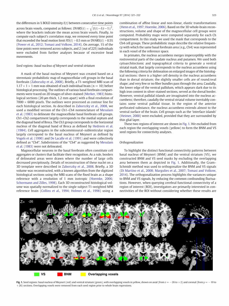
A mask of the basal nucleus of Meynert was created based on astereotaxic probabilistic map of magnocellular cell groups in the basalforebrain (Zaborszky et al., 2008). Briefly, a T1-weighted MRI scan of1.17 × 1 × 1mmwas obtained of each individual brain (n= 10) beforehistological processing. The outlines of various basal forebrain compart-ments were traced on 2D images of silver-stained (Merker, 1983) histo-logical sections (20 μm thick, 1.2 mm apart) with a final resolution of7000 × 6000 pixels. The outlines were processed as contour line foreach histological section. As described in Zaborszky et al., 2008, weused a modified version of the Ch1–Ch4 nomenclature of Mesulamet al. (1983) to delineate the magnocellular basal forebrain cell groups.Ch1–Ch2 compartment largely corresponds to the medial septum andthe diagonal band of Broca. The Ch3 group corresponds to the horizontalnucleus of the diagonal band of Broca as defined by Hedreen et al.(1984). Cell aggregates in the subcommissural–sublenticular regionlargely correspond to the basal nucleus of Meynert as defined byVogels et al. (1990) and De Lacalle et al. (1991) and were collectivelydefined as “Ch4”. Subdivisions of the “Ch4” as suggested by Mesulamet al. (1983) were not delineated.
Magnocellular neurons in the basal forebrain often constitute cellaggregates or clusters that facilitate their recognition. As a rule, bordersof delineated areas were drawn where the number of large cellsdecreased precipitously. Details of reconstruction of these nuclei on a3D template were described in Zaborszky et al., 2008. Briefly, a 3Dvolume was reconstructed, with a known algorithm from the digitizedhistological sections using the MRI scans of the fixed brain as a shapereference with a resolution of 1 mm isotropic (Hoemke, 2006;Schormann and Zilles, 1998). Each 3D reconstructed histological vol-ume was spatially normalized to the single subject T1-weighted MNIreference brain (Collins et al., 1994; Holmes et al., 1998) using a
-26 -22 -18 -14 -10Z=
-10 -4 2 8Y=
VS BNM Overlapping voxels
Fig. 1. Seed regions: basal nucleus ofMeynert (red) and ventral striatum (green), with overlapp+26) sections. Overlapping voxels were removed from each seed region prior to whole brain
combination of affine linear and non-linear, elastic transformations(Henn et al., 1997; Hoemke, 2006). Based on the 3Dwhole-brain recon-structions, volume and shape of the magnocellular cell groups werecomputed. Probability maps were computed separately for each Chcompartment. In this study we used the mask that corresponds to theCh4 cell groups. These probabilistic maps describe the relative frequen-cywithwhich the same basal forebrain area (e.g., Ch4)was representedin each voxel of the reference space.
In primates, the nucleus accumbens merges imperceptibly with therostroventral parts of the caudate nucleus and putamen. We used bothcytoarchitectonic and topographical criteria to generate a ventralstriatal mask, that largely corresponds to the nucleus accumbens usingthe following criteria for delineation on enlarged images of the histolog-ical sections: there is a higher cell density in the nucleus accumbensthan in dorsal striatum; the slightly smaller cells are of round/ovalshape; and very few or no fiber bundles pass through the area. Caudally,the lower edge of the ventral pallidum, which appears dark due to itshigh iron content in silver-stained sections, served as the dorsal border.However, ventral pallidal islands are invaginated into the nucleus ac-cumbens; therefore, caudally the ventral striatal volume inevitably con-tains some ventral pallidal tissue. In the region of the anteriorperforated substance, the nucleus accumbens extends almost to theventral surface of the brain. Cell groups such as the ‘interface islands’(Heimer, 2000) were excluded, provided that they are surrounded bythin glial layer.
These two regions of interest are shown in Fig. 1. We excluded fromeach region the overlapping voxels (yellow) to form the BNM and VSseed regions for connectivity analyses.
Orthogonalization
To highlight the distinct functional connectivity patterns betweenbasal nucleus of Meynert (BNM) and the ventral striatum (VS), weconstructed BNM and VS seed masks by excluding the overlappingarea between them as depicted in Fig. 1. Additionally, the Gram–
Schmidt method was used to orthogonalize the BNM and VS signals(Di Martino et al., 2008; Margulies et al., 2007; Tomasi and Volkow,2014). The orthogonalization process highlights the variances uniqueto BNM and VS signals, by reducing the common confounding fluctua-tions. However, when querying cerebral functional connectivity of aregion of interest (ROI), investigators are primarily interested in con-nectivities of the ROI without considering whether these results are
-6 -2 2
14 20 26
ing voxels in yellow, shown on axial (from z=−26 to+2) and coronal (fromy=−10 toregressions.
324 C.R. Li et al. / NeuroImage 97 (2014) 321–332
independent of those of other brain regions. Therefore, we presentedthe results obtained with the original time series in the main text andthose with the orthogonalized time series in the Supplementary data.In practice, orthogonalization is expected to have little impact whencomparing the BNM and VS correlation maps, since common variationsare eliminated in the statistical test.
Seed region-based linear correlation and random effects analysis
The BOLD time courses were averaged spatially over each of the twoseeds. For individual subjects, we computed the correlation coefficientbetween the averaged time course of each seed region and the timecourses of all other brain voxels. To assess and compare the restingstate “correlograms,” we converted these image maps, which were notnormally distributed, to z score maps by Fisher's z transform (Berry andMielke, 2000; Jenkins and Watts, 1968): z = 0.5 loge[(1 + r)/ (1− r)].The Z maps were used in group random effect analyses. We performedone-sample t test each on the Z maps of BNM and VS and paired-sample t test comparing the two Z maps.
Age dependent changes and gender differences in BNM and VS connectivity
We performed a simple regression of the Z maps against age, eachfor the BNM and VS, to identify age-related changes of functionalconnectivity in the two structures. To examine gender differences, wecompared men and women with age as a covariate in an analysis ofvariance, each for the BNM and VS. All results were reported for acorrected threshold.
Results
Whole brain functional connectivity of the nucleus of Meynert and ventralstriatum
For each seed region, we performed one sample t-test of the Zmaps (positively and negatively correlated regions) across thegroup (n = 223). Regions functionally connected with nucleus ofMeynert (BNM) and ventral striatum (VS) are presented in Figs. 2and 3, respectively.
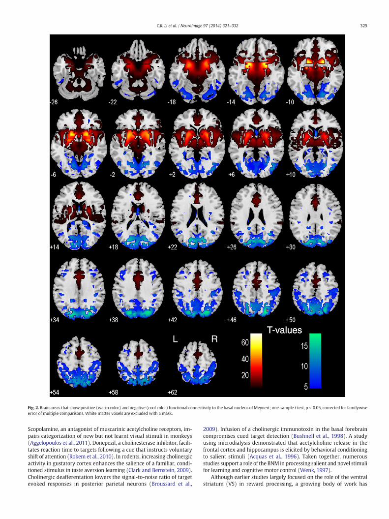
The BNM showed positive connectivity with the medial prefrontalcortex, includingdorsal/rostral/perigenual/subgenual anterior cingulatecortex (ACC), supplementary motor area (SMA) as well as pre-SMA,medial orbitofrontal cortex, inferior temporal pole, hippocampus,amygdala, insula, thalamus, midbrain, and basal ganglia. The BNMshowed negative connectivity with the cuneus, parahippocampal gyri,precuneus, somatosensory cortex, posterior parietal cortices, middleand lateral frontal cortices, including the dorsolateral prefrontal cortexand frontal eye field, and cerebellum (Fig. 2).
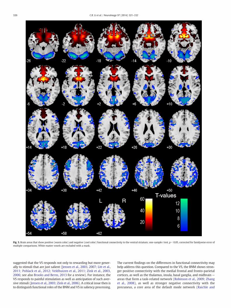
The VS showed positive connectivity with the medial orbitofrontalcortex, rectus gyrus, subcallosal gyrus, inferior temporal pole, amygdala,hippocampus, perigenual/subgenual ACC, and ventral part of the cau-date head. TheVS showed negative connectivitywith the visual cortices,posterior parietal cortex, temporoparietal junction, superior temporalgyri, precentral cortex, lateral frontal cortices including the dorsolateralprefrontal cortex, inferior frontal cortices, posterior thalamus/pulvinar,parahippocampal gyrus, and cerebellum (Fig. 3). These results areconsistent with previous findings of resting state VS connectivity withthe medial orbitofrontal cortex and ventral caudate (Baliki et al., 2013;Cauda et al., 2011; Di Martino et al., 2008; Gopinath et al., 2011),amygdala and midbrain (Cauda et al., 2011), and hippocampus (Caudaet al., 2011; Kahn and Shohamy, 2013).
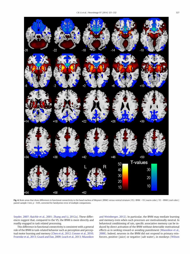
We performed a paired t-test to compare functional connectivity ofthe BNM and VS (Fig. 4). Compared to the VS, the BNM showed greaterconnectivity with the medial frontal cortex, including rostral/dorsalACC, SMA, pre-SMA, dorsolateral prefrontal cortex, inferior frontal cor-tex, lateral orbitofrontal cortex, inferior parietal lobule, temporo-
parietal junction, insula, thalamus, dorsal caudate, midbrain, and cere-bellum. Compared to the BNM, VS showed greater connectivity withthe medial orbitofrontal cortex, ventral caudate, subcallosal/rectusgyri, olfactory sulcus, cuneus, precuneus, posterior cingulate cortex,parahippocampal gyrus, middle/inferior occipital cortex, andpostcentral gyri. We summarize these results in Tables 2 and 3,where we further distinguish whether a contrast arises from differ-ences in positive or negative connectivity.
The effects of age and gender on BNM and VS connectivity
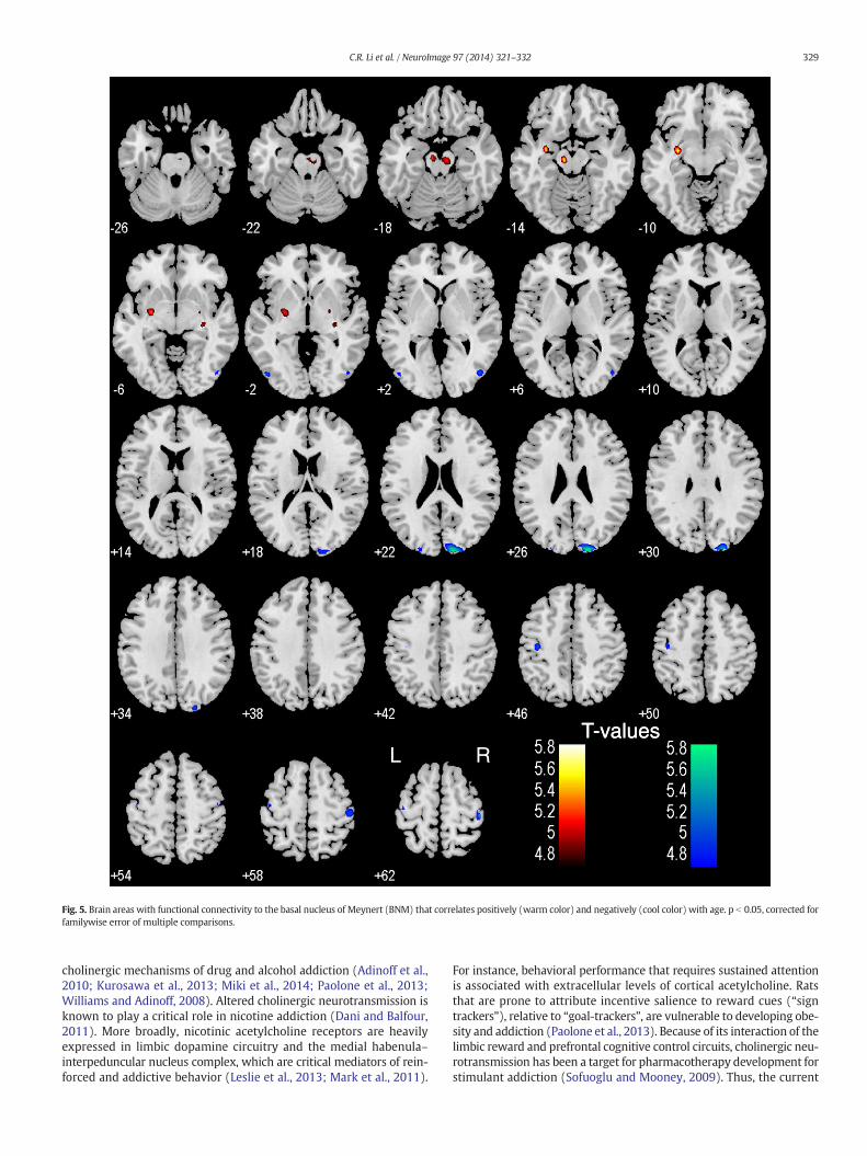
In a simple regression, BNM connectivity to the midbrain in the areaof the ventral tegmental area and substantia nigra (MNI coordinates[−9,−16,−14], Z = 5.41; and [6, −19, −20], Z = 5.08, 1215 mm3),left pallidum and parahippocampal gyrus ([−30, −7, −11], Z = 5.62,1026 mm3), and right pallidum ([27, −7 −5], Z = 5.04, 189 mm3)showed a positive correlationwith age (Fig. 5). In contrast, BNM connec-tivity to the bilateral motor cortices ([−33, −37, 70], Z = 5.63; and[−45, −16, 61], Z = 4.85, 2187 mm3; and [45, −25, 64], Z = 5.44,1161 mm3) and right visual cortex ([15, −94, 25], Z = 5.59,2133 mm3) showed a negative correlation with age. At thesame, corrected threshold, VS connectivities did not show any agedependence.
In a covariance analysiswith age as a covariate, we examined genderdifferences.Men andwomen did not differ in BNMor VS connectivity atthe same, corrected threshold.
Discussion
Functional connectivity of the nucleus of Meynert and ventral striatum
Compared to the VS, the BNM showed stronger positive connectivitywith the putamen, pallidum, thalamus, amygdala and midbrain, cere-bellum, as well as the anterior cingulate cortex, supplementary motorarea and pre-supplementary motor area, a network of brain regionsthat respond to salient stimuli and orchestrate goal-directed behavior.In contrast, the VS showed stronger positive connectivity with the ven-tral caudate andmedial orbitofrontal cortex, areas implicated in rewardprocessing, compared to the BNM. Together, the distinct cerebral func-tional connectivities support the role of the BNM in arousal and cogni-tive motor control and the VS in reward related behavior.
The BNM shows connectivity with a selective set of cortical and sub-cortical structures, consistent with anatomical studies in rodents andnon-human primates (Chandler and Waterhouse, 2012; Mesulamet al., 1984; Zaborszky et al., in press). In particular, studies in monkeysshowed that the density of cholinergic fibers and choline acetyltransfer-ase activities are particularly rich in agranular areas of the insula, caudalorbitofrontal cortex (OFC), temporal pole, and parahippocampal region(Mesulam et al., 1984, 1986), a patternmirrored by the current findingsof regions positively connected to the BNM (Fig. 2). The VS receives ex-tensive projections from the OFC (Haber et al., 2006), amygdala, anddysgranular part of the insula that process taste, olfaction and other vis-ceral information (Fudge et al., 2005; Nakano et al., 1999), in accordwith VS responses to primary reinforcers.
Functional implications for the basal nucleus of Meynert and ventralstriatum
The BNM, alongwith the pulvinar and amygdala, responds to salientstimuli (Morris et al., 1997). Manipulations of cholinergic signaling alterneural responses to salient stimuli in a variety of behavioral paradigms,including odd-ball tasks (Klinkenberg et al., 2013). For instance, duringauditory conditioning in humans, augmenting cholinergic signalingwith physostigmine (an inhibitor of acetylcholinesterase) enhancesprocessing of behaviorally irrelevant stimuli and attenuates differentialconditioning-related cortical activations (Thiel et al., 2002).
T valuesRL
T-values
Fig. 2. Brain areas that show positive (warm color) and negative (cool color) functional connectivity to the basal nucleus of Meynert; one-sample t test, p b 0.05, corrected for familywiseerror of multiple comparisons. White matter voxels are excluded with a mask.
325C.R. Li et al. / NeuroImage 97 (2014) 321–332
Scopolamine, an antagonist of muscarinic acetylcholine receptors, im-pairs categorization of new but not learnt visual stimuli in monkeys(Aggelopoulos et al., 2011). Donepezil, a cholinesterase inhibitor, facili-tates reaction time to targets following a cue that instructs voluntaryshift of attention (Rokem et al., 2010). In rodents, increasing cholinergicactivity in gustatory cortex enhances the salience of a familiar, condi-tioned stimulus in taste aversion learning (Clark and Bernstein, 2009).Cholinergic deafferentation lowers the signal-to-noise ratio of targetevoked responses in posterior parietal neurons (Broussard et al.,
2009). Infusion of a cholinergic immunotoxin in the basal forebraincompromises cued target detection (Bushnell et al., 1998). A studyusing microdialysis demonstrated that acetylcholine release in thefrontal cortex and hippocampus is elicited by behavioral conditioningto salient stimuli (Acquas et al., 1996). Taken together, numerousstudies support a role of the BNM in processing salient and novel stimulifor learning and cognitive motor control (Wenk, 1997).
Although earlier studies largely focused on the role of the ventralstriatum (VS) in reward processing, a growing body of work has
T valuesRL
T-values
Fig. 3. Brain areas that show positive (warm color) and negative (cool color) functional connectivity to the ventral striatum; one-sample t test, p b 0.05, corrected for familywise error ofmultiple comparisons. White matter voxels are excluded with a mask.
326 C.R. Li et al. / NeuroImage 97 (2014) 321–332
suggested that the VS responds not only to rewarding but more gener-ally to stimuli that are just salient (Jensen et al., 2003, 2007; Litt et al.,2011; Pohlack et al., 2012; Veldhuizen et al., 2011; Zink et al., 2003,2006; see also Brooks and Berns, 2013 for a review). For instance, theVS responds to painful stimulation as well as anticipation of such aver-sive stimuli (Jensen et al., 2003; Zink et al., 2006). A critical issue then isto distinguish functional roles of the BNM and VS in saliency processing.
The current findings on the differences in functional connectivity mayhelp address this question. Compared to the VS, the BNM shows stron-ger positive connectivity with the medial frontal and fronto-parietalcortices, as well as the thalamus, insula, basal ganglia, and midbrain —
areas that form a task-related network (Robinson et al., 2009; Zhanget al., 2008), as well as stronger negative connectivity with theprecuneus, a core area of the default mode network (Raichle and
T valuesRL
T-values
Fig. 4. Brain areas that show differences in functional connectivity to the basal nucleus of Meynert (BNM) versus ventral striatum (VS): BNM N VS (warm color); VS N BNM (cool color);paired-sample t test, p b 0.05, corrected for familywise error of multiple comparisons.
327C.R. Li et al. / NeuroImage 97 (2014) 321–332
Snyder, 2007; Raichle et al., 2001; Zhang and Li, 2012a). These differ-ences suggest that, compared to the VS, the BNM is more directly andreadily engaged in task related processing.
This difference in functional connectivity is consistentwith a generalrole of the BNM in task-related behavior such as perception and percep-tual motor learning andmemory (Chen et al., 2012; Conner et al., 2010;Froemke et al., 2013;Goard andDan, 2009; Leach et al., 2013;Miasnikov
and Weinberger, 2012). In particular, the BNM may mediate learningand memory even when such processes are motivationally neutral. Inbehavioral conditioning of rats, specific associative memory can be in-duced by direct activation of the BNM without detectable motivationaleffects as in seeking reward or avoiding punishment (Miasnikov et al.,2008). Indeed, neurons in the BNM did not respond to primary rein-forcers, positive (juice) or negative (salt water), in monkeys (Wilson
Table 2Regions showing greater connectivity with basal nucleus of Meynert (BNM) as compared to ventral striatum (VS); paired t-test, n = 223.
Volume(mm3)
Peak voxel(Z)
MNI coordinate Side Identified brain region Connectivity
x y z BNM VS
365472 inf 24 −1 −8 R Putamen/Pallidum/Thalamus/Midbrain/Cerebellum/Amygdala/Insula/TP/lOFC ++ +/~/−inf −24 −4 −8 L Putamen/Pallidum/Thalamus/Midbrain/Cerebellum/Amygdala/Insula/TP/lOFC ++ +/~/−inf 9 11 34 R ACC/SMA/pre-SMA + ~/−inf −9 14 37 L ACC/SMA/pre-SMA + ~/−
4563 7.48 63 −31 28 R Temporo-parietal junction/Supramarginal G +/~ −/−−7.33 −66 −28 25 L Temporo-parietal junction/Supramarginal G +/~ −/−−
1026 6.60 63 −46 1 R Middle temporal G/Inferior frontal G/DLPFC + −729 6.05 −60 −55 1 L Middle temporal G/Inferior frontal G/DLPFC + −
Note: inf: infinity; R: right; L: left; TP: temporal pole; lOFC: lateral orbitofrontal cortex; G: gyrus; ACC: anterior cingulate cortex; SMA: supplementary motor area; DLPFC: dorsolateralprefrontal cortex; +/− indicates positive and negative connectivity (with ++/−− for stronger connectivity), and ~ indicates no significant connectivity at one-sample t test, p b 0.05,FWE corrected.
328 C.R. Li et al. / NeuroImage 97 (2014) 321–332
and Rolls, 1990). While the BNM responds to visual or acoustic stimulithat signal reinforcers, it does not respond to the reinforcers themselves(Wilson andMa, 2004). Other work suggests that the BNMneurons en-code saliency irrespective of valence of the stimuli (Lin and Nicolelis,2008). These results are concordant with a learning-related ormemory-promoting role for the BNM that places it “downstream” ofmotivational systems (Miasnikov et al., 2008), consistent with the ana-tomical finding that, the cholinergic neurons in basal forebrain receiveinput from the nucleus accumbens (Zaborszky and Cullinan, 1992).Through its extensive cortical projections, the BNMmaymediate atten-tional effort in a circuitry that integrates brain systems involved inmodulation of input functions, incentive processing and performancemonitoring (Sarter et al., 2006).
On the other hand, one is cautioned not to over-interpret the differ-ences of BNM and VS in saliency- and reward-related processing. For in-stance, reward is intrinsically salient, and cerebral structures such as themedial orbitofrontal cortex, with positive connectivity to both the BNMand VS, is involved in processing reward and emotionally salient stimuli(Hardin et al., 2009; Kühn and Gallinat, 2012; Liu et al., 2011; Rothkirchet al., 2012). Likewise, as discussed earlier, although VS is predominantlyimplicated in reward processing, there is accumulating evidence that VSresponds to salient, non-rewarding stimuli (Esslinger et al., 2013; Jensenet al., 2007; Pohlack et al., 2012; Spicer et al., 2007; Zink et al., 2003,2006). Future researchmay take advantage of the current findings in fur-ther delineating the respective roles of BNM and VS in motivatedbehavior.
Age-related connectivity of the nucleus of Meynert
Previous imaging studies suggest that aging is related in a complicat-ed way to cerebral functional connectivities, with different brain areasincreasing and decreasingwith age in connectivity to regions of interest(Bernard et al., 2013; Campbell et al., 2013; Hafkemeijer et al., 2013;Hoffstaedter et al., 2014; Liu et al., 2012; Roski et al., 2013; Taniwakiet al., 2007; Ystad et al., 2010; see also Ferreira and Busatto, 2013 for areview). Here, we observed that age is associated with increased BNMconnectivity to subcortical structures including the midbrain and
Table 3Regions showing greater connectivity with ventral striatum (VS) as compared to the basal nuc
Volumemm3
Peak voxelZ
MNI coordinate Side
x y z
326160 inf 12 17 −11 Rinf −9 14 −14 Linf −18 −49 22 Linf 18 −46 25 R
1512 6.47 42 −28 64 R864 5.91 −39 −31 67 L
Note: inf: infinity; R: right; L: left; mOFC: medial orbitofrontal cortex; G: gyrus; S: sulcus; p++/−− indicating stronger connectivity), and ~ indicates no significant connectivity at
pallidum and decreased connectivity to somatomotor and visual corti-ces. Although statistically highly significant, these results are notstraightforward to interpret without a functional concomitant. Giventhat the current BOLD time series were obtained during resting state,one is tempted to speculate that these changes are not related to specificcontextual or task conditions. Future studies are needed to examine therelationship between this contrasting pattern of increased and de-creased connectivity to subcortical and cortical structures and cognitivemotor performance (Hu et al., 2012, 2013) and evaluate whether and towhat extent these changes may reflect a compensatory process.
In over 1000 adolescents and young adults, Tomasi and Volkowcharacterized age-related increases in the functional connectivity ofthe ventral tegmental area (VTA) with limbic regions and with the de-fault mode network and decreases in the connectivity of the substantianigra (SN) with motor and medial temporal cortices (Tomasi andVolkow, 2014). In terms of mean strength and statistical significanceof connectivity, the VTA demonstrated bilateral positive connectivitywith the nucleus accumbens, hippocampus, parahippocampus, globuspallidus, caudate, cerebellar vermis, and anterior insula, as well as theangular gyrus, inferior frontal cortex, and anterior cingulate cortex,and negative connectivity with the inferior occipital cortex. The SNdemonstrated bilateral positive connectivity with the globus pallidus,subthalamic nucleus, thalamus, and cerebellar vermis, as well as theBroca's and Wernickes's areas, anterior cingulate cortex, and supple-mentary motor area, and negative connectivity with orbitofrontal, tem-poral, and occipital cortices, ventral precuneus, and angular gyrus. Thus,there is much overlap between subcortical and cortical connectivitiesbetween the VTA/SN and BNM/VS, suggesting the need in future studiesto investigate early developmental changes in functional connectivity ofthe BNM/VS and their relevance to clinical conditions that manifestearly during development.
Potential clinical implications
In addition to implications for our understanding of the cognitivedeficits in Alzheimer's disease and other forms of age-related cognitivedecline, the current findings may also facilitate research of the
leus of Meynert (BNM); paired t-test, n = 223.
Identified brain region Connectivity
BNM VS
ventral Caudate/mOFC/Subcallosal G/Rectus G/Olfactory S ~/− ++ventral Caudate/mOFC/Subcallosal G/Rectus G/Olfactory S ~/− ++pCG/Cuneus/Precuneus/Parahippocampal G −− ~/−pCG/Cuneus/Precuneus/Parahippocampal G −− ~/−Postcentral G − ~Postcentral G − ~
CG: posterior cingulate gyrus; +/− indicates positive and negative connectivity (withone-sample t test, p b 0.05 FWE corrected.
T-valuesRL
T-values
Fig. 5. Brain areas with functional connectivity to the basal nucleus of Meynert (BNM) that correlates positively (warm color) and negatively (cool color) with age. p b 0.05, corrected forfamilywise error of multiple comparisons.
329C.R. Li et al. / NeuroImage 97 (2014) 321–332
cholinergic mechanisms of drug and alcohol addiction (Adinoff et al.,2010; Kurosawa et al., 2013; Miki et al., 2014; Paolone et al., 2013;Williams and Adinoff, 2008). Altered cholinergic neurotransmission isknown to play a critical role in nicotine addiction (Dani and Balfour,2011). More broadly, nicotinic acetylcholine receptors are heavilyexpressed in limbic dopamine circuitry and the medial habenula–interpeduncular nucleus complex, which are critical mediators of rein-forced and addictive behavior (Leslie et al., 2013; Mark et al., 2011).
For instance, behavioral performance that requires sustained attentionis associated with extracellular levels of cortical acetylcholine. Ratsthat are prone to attribute incentive salience to reward cues (“signtrackers”), relative to “goal-trackers”, are vulnerable to developing obe-sity and addiction (Paolone et al., 2013). Because of its interaction of thelimbic reward and prefrontal cognitive control circuits, cholinergic neu-rotransmission has been a target for pharmacotherapy development forstimulant addiction (Sofuoglu and Mooney, 2009). Thus, the current
330 C.R. Li et al. / NeuroImage 97 (2014) 321–332
findings of the systems level functional connectivity of the BNM willhelp future studies to pursue the etiology and treatment of addictionon these related fronts.
Furthermore, the VS and its cerebral connectivity has been implicat-ed in various neuropsychiatric disorders such as autism (Delmonteet al., 2013; DiMartino et al., 2011) or psychological processes of clinicalinterest such as consciousness (Cauda et al., 2009a) and pain (Balikiet al., 2013; Cauda et al., 2009b, 2010a,b). For instance, with differentialfunctional and structural connectivities, the shell and core regions of theVS may each mediate prediction of monetary reward and cessation ofpain (Baliki et al., 2013). Functional connectivity of the VS and hippo-campusmay allow integration ofmotivational significance andmemoryto guide behavior (Kahn and Shohamy, 2013), a process of relevance todepression and psychomotor retardation. Notably, animal studies havealso implicated the BNM in pain (Zhang et al., 2010) and anesthesia(Laalou et al., 2008); thus, the current findings may shed light on futurestudies to directly contrast the roles of VS and BNM in these neural pro-cesses of clinical significance.
A methodological consideration
Negative functional connectivity has been observed and reportedsince the very beginning of the resting state fMRI study (Biswal et al.,1995). Negative functional connectivity, also called anti-correlation,represents negative cross-correlation in spontaneous BOLD signal be-tween two brain regions. It was suggested that the global signal regres-sion, a common step of data preprocessing in seed region basedfunctional connectivity analyses, is a likely cause of anti-correlated func-tional networks (Murphy et al., 2009;Weissenbacher et al., 2009). How-ever, recent investigations demonstrated that the negative correlationsare not an artifact but have biological origins (Chai et al., 2012; Chenet al., 2011; Fox et al., 2009). For instance, negative functional connec-tivity is associated predominantly with long range connections and cor-relates with the shortest path length in the human brain network (Chenet al., 2011; Scholvinck et al., 2010; Schwarz and McGonigle, 2011). In-deed, the negative correlations between brain regions with presumablyopposing functional roles have been consistently observed in differentstudies (Chen et al., 2011; Fox et al., 2005; Fransson, 2005; Greiciuset al., 2003; Kelly et al., 2008; Uddin et al., 2009), including thoseusing independent component analysis, which does not involve globalsignal regression (Cole et al., 2010; Zhang and Li, 2012b; Zuo et al.,2010). Furthermore, the existence of the negative functional connectiv-ity was also suggested by computational simulations of cerebral net-work activities in both monkeys and humans (Deco et al., 2009;Honey et al., 2007; Izhikevich and Edelman, 2008) and supported by si-multaneous recording of unit activity and local field potential from task-positive and task-negative (default mode) networks in cats (Popa et al.,2009). Together, these earlier studies suggest functional significance ofnegative functional connectivity.
Acknowledgments
This study was supported by NIH grants R01DA023248,K02DA026990, R21AA018004, and R01AA021449 to C-S.R.L. andNINDS grant 23945 to LZ. The NIH had no further role in the study de-sign; in the collection, analysis and interpretation of data; in thewritingof the report; or in the decision to submit the paper for publication. J.S.I.was supported by Young Researcher Award (Sao Paulo Research Foun-dation FAPESP 2011/08573-4). We thank investigators of the 1000Functional Connectomes Project and those who shared the data set formaking this study possible.
Appendix A. Supplementary data
Supplementary data to this article can be found online at http://dx.doi.org/10.1016/j.neuroimage.2014.04.019.
References
Acquas, E., Wilson, C., Fibiger, H.C., 1996. Conditioned and unconditioned stimuli increasefrontal cortical and hippocampal acetylcholine release: effects of novelty, habituation,and fear. J. Neurosci. 16 (9), 3089–3096 (May 1).
Adinoff, B., Devous Sr., M.D., Williams, M.J., Best, S.E., Harris, T.S., Minhajuddin, A.,Zielinski, T., Cullum, M., 2010. Altered neural cholinergic receptor systems incocaine-addicted subjects. Neuropsychopharmacology. 35 (7), 1485–1499 (Jun).
Aggelopoulos, N.C., Liebe, S., Logothetis, N.K., Rainer, G., 2011. Cholinergic control of visualcategorization in macaques. Front. Behav. Neurosci. 5, 73 (Nov 15).
Ashburner, J., Friston, K.J., 1999. Nonlinear spatial normalization using basis functions.Hum. Brain Mapp. 7, 254–266.
Ashburner, J., Friston, K.J., 2005. Unified segmentation. Neuroimage 26, 839–851.Baliki, M.N., Mansour, A., Baria, A.T., Huang, L., Berger, S.E., Fields, H.L., Apkarian, A.V.,
2013. Parceling human accumbens into putative core and shell dissociates encodingof values for reward and pain. J. Neurosci. 33 (41), 16383–16393 (Oct 9).
Barnes, K.A., Cohen, A.L., Power, J.D., Nelson, S.M., Dosenbach, Y.B., Miezin, F.M., Petersen,S.E., Schlaggar, B.L., 2010. Identifying basal ganglia divisions in individuals usingresting-state functional connectivity MRI. Front. Syst. Neurosci. 4, 18.
Bernard, J.A., Peltier, S.J., Wiggins, J.L., Jaeggi, S.M., Buschkuehl, M., Fling, B.W., Kwak, Y.,Jonides, J., Monk, C.S., Seidler, R.D., 2013. Disrupted cortico-cerebellar connectivityin older adults. Neuroimage. 83, 103–119 (Dec).
Berry, K.J., Mielke Jr., P.W., 2000. A Monte Carlo investigation of the Fisher Z transforma-tion for normal and nonnormal distributions. Psychol. Rep. 87, 1101–1114.
Biswal, B., Yetkin, F.Z., Haughton, V.M., Hyde, J.S., 1995. Functional connectivity in themotor cortex of resting human brain using echo-planar MRI. Magn. Reson. Med. 34,537–541.
Biswal, B.B., Mennes, M., Zuo, X.N., Gohel, S., Kelly, C., Smith, S.M., Beckmann, C.F.,Adelstein, J.S., Buckner, R.L., Colcombe, S., Dogonowski, A.M., Ernst, M., Fair, D.,Hampson, M., Hoptman, M.J., Hyde, J.S., Kiviniemi, V.J., Kotter, R., Li, S.J., Lin, C.P.,Lowe, M.J., Mackay, C., Madden, D.J., Madsen, K.H., Margulies, D.S., Mayberg, H.S.,McMahon, K., Monk, C.S., Mostofsky, S.H., Nagel, B.J., Pekar, J.J., Peltier, S.J., Petersen,S.E., Riedl, V., Rombouts, S.A., Rypma, B., Schlaggar, B.L., Schmidt, S., Seidler, R.D.,Siegle, G.J., Sorg, C., Teng, G.J., Veijola, J., Villringer, A., Walter, M., Wang, L., Weng,X.C., Whitfield-Gabrieli, S., Williamson, P., Windischberger, C., Zang, Y.F., Zhang, H.Y.,Castellanos, F.X., Milham, M.P., 2010. Toward discovery science of human brain func-tion. Proc. Natl. Acad. Sci. U. S. A. 107, 4734–4739.
Brooks, A.M., Berns, G.S., 2013. Aversive stimuli and loss in the mesocorticolimbic dopa-mine system. Trends Cogn. Sci. 17 (6), 281–286 (Jun).
Broussard, J.I., Karelina, K., Sarter, M., Givens, B., 2009. Cholinergic optimization of cue-evoked parietal activity during challenged attentional performance. Eur. J. Neurosci.29 (8), 1711–1722 (Apr).
Bushnell, P.J., Chiba, A.A., Oshiro, W.M., 1998. Effects of unilateral removal of basal fore-brain cholinergic neurons on cued target detection in rats. Behav. Brain Res. 90 (1),57–71 (Jan).
Campbell, K.L., Grigg, O., Saverino, C., Churchill, N., Grady, C.L., 2013. Age differences in theintrinsic functional connectivity of default network subsystems. Front. AgingNeurosci. 5, 73 (Nov 14).
Cauda, F., Micon, B.M., Sacco, K., Duca, S., D'Agata, F., Geminiani, G., Canavero, S., 2009a.Disrupted intrinsic functional connectivity in the vegetative state. J. Neurol.Neurosurg. Psychiatry 80 (4), 429–431 (Apr).
Cauda, F., Sacco, K., Duca, S., Cocito, D., D'Agata, F., Geminiani, G.C., Canavero, S., 2009b. Al-tered resting state in diabetic neuropathic pain. PLoS One 4 (2), e4542.
Cauda, F., D'Agata, F., Sacco, K., Duca, S., Cocito, D., Paolasso, I., Isoardo, G., Geminiani, G.,2010a. Altered resting state attentional networks in diabetic neuropathic pain. J.Neurol. Neurosurg. Psychiatry 81 (7), 806–811 (Jul).
Cauda, F., Geminiani, G., D'Agata, F., Sacco, K., Duca, S., Bagshaw, A.P., Cavanna, A.E., 2010b.Functional connectivity of the posteromedial cortex. PLoS One 5 (9) (Sep 30).
Cauda, F., Cavanna, A.E., D'agata, F., Sacco, K., Duca, S., Geminiani, G.C., 2011. Functionalconnectivity and coactivation of the nucleus accumbens: a combined functional con-nectivity and structure-based meta-analysis. J. Cogn. Neurosci. 23 (10), 2864–2877(Oct).
Chai, X.J., Castanon, A.N., Ongur, D., Whitfield-Gabrieli, S., 2012. Anticorrelations in restingstate networks without global signal regression. Neuroimage 59, 1420–1428.
Chandler, D., Waterhouse, B.D., 2012. Evidence for broad versus segregated projectionsfrom cholinergic and noradrenergic nuclei to functionally and anatomically discretesubregions of prefrontal cortex. Front. Behav. Neurosci. 6, 20 (May 21).
Chen, G., Chen, G., Xie, C., Li, S.J., 2011. Negative functional connectivity and its depen-dence on the shortest path length of positive network in the resting-state humanbrain. Brain Connect. 1, 195–206.
Chen, N., Sugihara, H., Sharma, J., Perea, G., Petravicz, J., Le, C., Sur, M., 2012. Nucleusbasalis-enabled stimulus-specific plasticity in the visual cortex is mediated by astro-cytes. Proc. Natl. Acad. Sci. U. S. A. 109 (41), E2832–E2841 (Oct 9).
Clark, E.W., Bernstein, I.L., 2009. Boosting cholinergic activity in gustatory cortex enhancesthe salience of a familiar conditioned stimulus in taste aversion learning. Behav.Neurosci. 123 (4), 764–771 (Aug).
Cole, D.M., Beckmann, C.F., Long, C.J., Matthews, P.M., Durcan, M.J., Beaver, J.D., 2010. Nic-otine replacement in abstinent smokers improves cognitive withdrawal symptomswith modulation of resting brain network dynamics. Neuroimage 52, 590–599.
Collins, D.L., Neelin, P., Peters, T.M., Evans, A.C., 1994. Automatic 3D intersubject registra-tion of MR volumetric data in standardized Talairach space J. Comput. Assist. Tomogr.18, 192–205.
Conner, J.M., Kulczycki, M., Tuszynski, M.H., 2010. Unique contributions of distinct cholin-ergic projections to motor cortical plasticity and learning. Cereb. Cortex 20 (11),2739–2748 (Nov).
331C.R. Li et al. / NeuroImage 97 (2014) 321–332
Cordes, D., Haughton, V.M., Arfanakis, K., Carew, J.D., Turski, P.A., Moritz, C.H., Quigley,M.A., Meyerand, M.E., 2001. Frequencies contributing to functional connectivity inthe cerebral cortex in “resting-state” data. AJNR Am. J. Neuroradiol. 22, 1326–1333.
Da Cunha, C., Gomez-A, A., Blaha, C.D., 2012. The role of the basal ganglia inmotivated be-havior. Rev. Neurosci. 23 (5–6), 747–767.
Dani, J.A., Balfour, D.J., 2011. Historical and current perspective on tobacco use and nico-tine addiction. Trends Neurosci. 34 (7), 383–392 (Jul).
De Lacalle, S., Iraizoz, J., Ma Gonzalo, L., 1991. Differential changes in cell size and numberin topographic subdivisions of human basal nucleus in normal aging. Neuroscience43, 445–456.
Deco, G., Jirsa, V., McIntosh, A.R., Sporns, O., Kotter, R., 2009. Key role of coupling,delay, and noise in resting brain fluctuations. Proc. Natl. Acad. Sci. U. S. A. 106,10302–10307.
Delmonte, S., Gallagher, L., O'Hanlon, E., McGrath, J., Balsters, J.H., 2013. Functional andstructural connectivity of frontostriatal circuitry in Autism Spectrum Disorder.Front. Hum. Neurosci. 7, 430 (Aug 6).
Di Martino, A., Scheres, A., Margulies, D.S., Kelly, A.M., Uddin, L.Q., Shehzad, Z., Biswal, B.,Walters, J.R., Castellanos, F.X., Milham, M.P., 2008. Cereb. Cortex 18 (12), 2735–2747(Dec).
Di Martino, A., Kelly, C., Grzadzinski, R., Zuo, X.N., Mennes, M., Mairena, M.A., Lord, C.,Castellanos, F.X., Milham, M.P., 2011. Aberrant striatal functional connectivity in chil-dren with Autism. Biol. Psychiatry 69 (9), 847–856 (May).
Doody, R.S., Cummings, J.L., Farlow, M.R., 2012. Reviewing the role of donepezil in thetreatment of Alzheimer's disease. Curr. Alzheimer Res. 9 (7), 773–781 (Sep).
Esslinger, C., Braun, U., Schirmbeck, F., Santos, A., Meyer-Lindenberg, A., Zink,M., Kirsch, P.,2013. Activation of midbrain and ventral striatal regions implicates salience process-ing during a modified beads task. PLoS One 8 (3), e58536.
Fair, D.A., Schlaggar, B.L., Cohen, A.L., Miezin, F.M., Dosenbach, N.U., Wenger, K.K., Fox,M.D., Snyder, A.Z., Raichle, M.E., Petersen, S.E., 2007. A method for using blockedand event-related fMRI data to study “resting state” functional connectivity.Neuroimage 35, 396–405.
Ferreira, L.K., Busatto, G.F., 2013. Resting-state functional connectivity in normal brainaging. Neurosci. Biobehav. Rev. 37 (3), 384–400 (Mar).
Fox, M.D., Raichle, M.E., 2007. Spontaneous fluctuations in brain activity observed withfunctional magnetic resonance imaging. Nat. Rev. Neurosci. 8, 700–711.
Fox, M.D., Snyder, A.Z., Vincent, J.L., Corbetta, M., Van Essen, D.C., Raichle, M.E., 2005. Thehuman brain is intrinsically organized into dynamic, anticorrelated functional net-works. Proc. Natl. Acad. Sci. U. S. A. 102, 9673–9678.
Fox, M.D., Zhang, D., Snyder, A.Z., Raichle, M.E., 2009. The global signal and observedanticorrelated resting state brain networks. J. Neurophysiol. 101, 3270–3283.
Fransson, P., 2005. Spontaneous low-frequency BOLD signal fluctuations: an fMRI investi-gation of the resting-state default mode of brain function hypothesis. Hum. BrainMapp. 26, 15–29.
Froemke, R.C., Carcea, I., Barker, A.J., Yuan, K., Seybold, B.A., Martins, A.R., Zaika, N.,Bernstein, H., Wachs, M., Levis, P.A., Polley, D.B., Merzenich, M.M., Schreiner, C.E.,2013. Long-term modification of cortical synapses improves sensory perception.Nat. Neurosci. 16 (1), 79–88 (Jan).
Fudge, J.L., Breitbart, M.A., Danish, M., Pannoni, V., 2005. Insular and gustatory inputs tothe caudal ventral striatum in primates. J. Comp. Neurol. 490 (2), 101–118 (Sep 19).
Garibotto, V., Tettamanti, M., Marcone, A., Florea, I., Panzacchi, A., Moresco, R., Virta, J.R.,Rinne, J., Cappa, S.F., Perani, D., 2013. Cholinergic activity correlates with reserveproxies in Alzheimer's disease. Neurobiol. Aging 34 (11), 2694.e13–2694.e18 (Nov).
Goard, M., Dan, Y., 2009. Basal forebrain activation enhances cortical coding of naturalscenes. Nat. Neurosci. 12 (11), 1444–1449 (Nov).
Gopinath, K1., Ringe, W., Goyal, A., Carter, K., Dinse, H.R., Haley, R., Briggs, R., 2011. Striatalfunctional connectivity networks are modulated by fMRI resting state conditions.Neuroimage 54 (1), 380–388 (Jan 1).
Grace, A.A., Floresco, S.B., Goto, Y., Lodge, D.J., 2007. Regulation offiring of dopaminergic neu-rons and control of goal-directed behaviors. Trends Neurosci. 30 (5), 220–227 (May).
Greicius, M.D., Krasnow, B., Reiss, A.L., Menon, V., 2003. Functional connectivity in theresting brain: a network analysis of the default mode hypothesis. Proc. Natl. Acad.Sci. U. S. A. 100, 253–258.
Haber, S.N., Kim, K.S., Mailly, P., Calzavara, R., 2006. Reward-related cortical inputs definea large striatal region in primates that interface with associative cortical connections,providing a substrate for incentive-based learning. J. Neurosci. 26 (32), 8368–8376(Aug 9).
Hafkemeijer, A., Altmann-Schneider, I., Oleksik, A.M., van de Wiel, L., Middelkoop, H.A.,van Buchem, M.A., van der Grond, J., Rombouts, S.A., 2013. Increased functionalconnectivity and brain atrophy in elderly with subjective memory complaints.Brain Connect. 3 (4), 353–362.
Hardin, M.G., Pine, D.S., Ernst, M., 2009. The influence of context valence in the neuralcoding of monetary outcomes. Neuroimage 48 (1), 249–257 (Oct 15).
Hedreen, J.C., Struble, R.G., Whitehouse, P.J., Price, D.L., 1984. Topography of themagnocellular basal forebrain system in human brain. J. Neuropathol. Exp. Neurol. 43,1–21.
Heimer, L., 2000. Basal forebrain in the context of schizophrenia. Brain Res. Rev. 31,205–235.
Heimer, L., de Olmos, J., Alheid, G.F., Zaborszky, L., 1991. “Perestroika” in the basal fore-brain; opening the border between neurology and psychiatry. Prog. Brain Res. 87,109–165.
Henn, S., Schormannn, T., Engler, K., Zilles, K., 1997. Elastische Anpassung in der digitalenBildverarbeitung auf mehreren Auflosungstufen mit Hilfe von Mehrgittervehrfahren.In: Paulus, E., Wahl, F.M. (Eds.), Mustererkennung. Springer, Berlin, pp. 392–399.
Hoffstaedter, F., Grefkes, C., Roski, C., Caspers, S., Zilles, K., Eickhoff, S.B., 2014. Age-relateddecrease of functional connectivity additional to graymatter atrophy in a network formovement initiation. Brain Struct. Funct. (Jan 8. [Epub ahead of print]).
Hoemke, L., 2006. A multigrid method for anisotropic PDEs in elastic image registration.In: Falgout, R.D. (Ed.), Numerical Linear Algebra with Applications. Wiley InterSience,pp. 215–229.
Holmes, C.J., Hoge, R., Collins, L., Woods, R., Toga, A.W., Evans, A.C., 1998. Enhancement ofMR images using registration for signal averaging J. Comput. Assist. Tomogr. 22,324–333.
Honey, C.J., Kotter, R., Breakspear, M., Sporns, O., 2007. Network structure of cerebral cor-tex shapes functional connectivity onmultiple time scales. Proc. Natl. Acad. Sci. U. S. A.104, 10240–10245.
Hu, S., Chao, H.H., Winkler, A.D., Li, C.S., 2012. The effects of age on cerebral activations:internally versus externally driven processes. Front. Aging Neurosci. 4, 4 (Apr 24).
Hu, S., Chao, H.H., Zhang, S., Ide, J.S., Li, C.S., 2013. Changes in cerebral morphometry andamplitude of low-frequency fluctuations of BOLD signals during healthy aging: corre-lation with inhibitory control. Brain Struct. Funct. (Apr 4. [Epub ahead of print]).
Izhikevich, E.M., Edelman, G.M., 2008. Large-scale model of mammalian thalamocorticalsystems. Proc. Natl. Acad. Sci. U. S. A. 105, 3593–3598.
Jenkins, G.M., Watts, D.G., 1968. Spectral analysis and its applications. Holden-Day, SanFrancisco.
Jensen, J., McIntosh, A.R., Crawley, A.P., Mikulis, D.J., Remington, G., Kapur, S., 2003. Directactivation of the ventral striatum in anticipation of aversive stimuli. Neuron 40 (6),1251–1257 (Dec 18).
Jensen, J., Smith, A.J., Willeit, M., Crawley, A.P., Mikulis, D.J., Vitcu, I., Kapur, S., 2007. Sep-arate brain regions code for salience vs. valence during reward prediction in humans.Hum. Brain Mapp. 28 (4), 294–302 (Apr).
Kahn, I., Shohamy, D., 2013. Intrinsic connectivity between the hippocampus, nucleus ac-cumbens, and ventral tegmental area in humans. Hippocampus 23 (3), 187–192(Mar).
Kahnt, T., Chang, L.J., Park, S.Q., Heinzle, J., Haynes, J.D., 2012. Connectivity-basedparcellation of the human orbitofrontal cortex. J. Neurosci. 32, 6240–6250.
Kelly, A.M., Uddin, L.Q., Biswal, B.B., Castellanos, F.X., Milham, M.P., 2008. Competition be-tween functional brain networks mediates behavioral variability. Neuroimage 39,527–537.
Kim, J.H., Lee, J.M., Jo, H.J., Kim, S.H., Lee, J.H., Kim, S.T., Seo, S.W., Cox, R.W., Na, D.L., Kim, S.I.,Saad, Z.S., 2010. Defining functional SMA and pre-SMA subregions in human MFCusing resting state fMRI: functional connectivity-based parcellation method.Neuroimage 49, 2375–2386.
Klinkenberg, I., Blokland, A., Riedel, W.J., Sambeth, A., 2013. Cholinergic modulation of au-ditory processing, sensory gating and novelty detection in human participants. Psy-chopharmacology (Berl) 225 (4), 903–921 (Feb).
Kühn, S., Gallinat, J., 2012. The neural correlates of subjective pleasantness. Neuroimage61 (1), 289–294 (May 15).
Kurosawa, R., Taoka, N., Shinohara, F., Minami, M., Kaneda, K., 2013. Cocaine exposure en-hances excitatory synaptic drive to cholinergic neurons in the laterodorsal tegmentalnucleus. Eur. J. Neurosci. (Jul 3. Epub ahead of print]).
Laalou, F.Z., de Vasconcelos, A.P., Oberling, P., Jeltsch, H., Cassel, J.C., Pain, L., 2008. Involve-ment of the basal cholinergic forebrain in the mediation of general (propofol) anes-thesia. Anesthesiology 108 (5), 888–896 (May).
Laplante, F., Zhang, Z.W., Huppé-Gourgues, F., Dufresne, M.M., Vaucher, E., Sullivan, R.M.,2012. Cholinergic depletion innucleus accumbens impairsmesocortical dopamine ac-tivation and cognitive function in rats. Neuropharmacology 63 (6), 1075–1084 (Nov).
Leach, N.D., Nodal, F.R., Cordery, P.M., King, A.J., Bajo, V.M., 2013. Cortical cholinergic inputis required for normal auditory perception and experience-dependent plasticity inadult ferrets. J. Neurosci. 33 (15), 6659–6671 (Apr 10).
Leslie, F.M., Mojica, C.Y., Reynaga, D.D., 2013. Nicotinic receptors in addiction pathways.Mol. Pharmacol. 83 (4), 753–758 (Apr).
Lester, D.B., Rogers, T.D., Blaha, C.D., 2010. Acetylcholine-dopamine interactions in thepathophysiology and treatment of CNS disorders. CNS Neurosci. Ther. 16 (3),137–162 (Jun).
Lin, S.C., Nicolelis, M.A., 2008. Neuronal ensemble bursting in the basal forebrain encodessalience irrespective of valence. Neuron 59 (1), 138–149 (Jul 10).
Litt, A., Plassmann, H., Shiv, B., Rangel, A., 2011. Dissociating valuation and saliency signalsduring decision-making. Cereb. Cortex 21 (1), 95–102 (Jan).
Liu, X., Hairston, J., Schrier, M., Fan, J., 2011. Common and distinct networks underlyingreward valence and processing stages: a meta-analysis of functional neuroimagingstudies. Neurosci. Biobehav. Rev. 35 (5), 1219–1236 (Apr).
Liu, Z., Bai, L., Dai, R., Zhong, C., Wang, H., You, Y., Wei, W., Tian, J., 2012. Exploring the ef-fective connectivity of resting state networks in mild cognitive impairment: an fMRIstudy combining ICA and multivariate Granger causality analysis. Conf. Proc. IEEEEng. Med. Biol. Soc. 2012, 5454–5457.
Lowe, M.J., Mock, B.J., Sorenson, J.A., 1998. Functional connectivity in single andmultisliceechoplanar imaging using resting-state fluctuations. Neuroimage 7, 119–132.
Margulies, D.S., Kelly, A.M., Uddin, L.Q., Biswal, B.B., Castellanos, F.X., Milham, M.P., 2007.Mapping the functional connectivity of anterior cingulate cortex. Neuroimage 37,579–588.
Margulies, D.S., Vincent, J.L., Kelly, C., Lohmann, G., Uddin, L.Q., Biswal, B.B., Villringer, A.,Castellanos, F.X., Milham, M.P., Petrides, M., 2009. Precuneus shares intrinsicfunctional architecture in humans and monkeys. Proc. Natl. Acad. Sci. U. S. A. 106,20069–20074.
Mark, G.P., Shabani, S., Dobbs, L.K., Hansen, S.T., 2011. Cholinergic modulation ofmesolimbic dopamine function and reward. Physiol. Behav. 104 (1), 76–81 (Jul 25).
Merker, B., 1983. Silver staining of cell bodies by means of physical development. J.Neurosci. Methods 9, 235–241.
Mesulam, M.-M., Mufson, E.J., Levey, A.I., Wainer, B.H., 1983. Cholinergic innervation ofcortex by the basal forebrain: cytochemistry and cortical connections of the septalarea, diagonal band nuclei, nucleus basalis (substantia innominata), and hypothala-mus in the Rhesus monkey J. Comp. Neurol. 214, 170–197.
332 C.R. Li et al. / NeuroImage 97 (2014) 321–332
Mesulam, M.M., Rosen, A.D., Mufson, E.J., 1984. Regional variations in cortical cholinergicinnervation: chemoarchitectonics of acetylcholinesterase-containing fibers in themacaque brain. Brain Res. 311 (2), 245–258 (Oct 8).
Mesulam, M.M., Volicer, L., Marquis, J.K., Mufson, E.J., Green, C.R., 1986. Systematicregional differences in the cholinergic innervation of the primate cerebral cortex:distribution of enzyme activities and some behavioral implications. Ann. Neurol. 19,144–151.
Miasnikov, A.A., Weinberger, N.M., 2012. Detection of an inhibitory cortical gradient un-derlying peak shift in learning: a neural basis for a false memory. Neurobiol. Learn.Mem. 98 (4), 368–379 (Nov).
Miasnikov, A.A., Chen, J.C., Gross, N., Poytress, B.S., Weinberger, N.M., 2008. Motivationallyneutral stimulation of the nucleus basalis induces specific behavioral memory.Neurobiol. Learn. Mem. 90 (1), 125–137 (Jul).
Miki, T., Kusaka, T., Yokoyama, T., Ohta, K.I., Suzuki, S., Warita, K., Jamal, M., Wang, Z.Y.,Ueki, M., Liu, J.Q., Yakura, T., Tamai, M., Sumitani, K., Hosomi, N., Takeuchi, Y., 2014.Short-term ethanol exposure causes imbalanced neurotrophic factor allocation inthe basal forebrain cholinergic system: a novel insight into understanding the initialprocesses of alcohol addiction. J. Neural. Transm. 121, 201–210.
Morris, J.S., Friston, K.J., Dolan, R.J., 1997. Neural responses to salient visual stimuli. Proc.Biol. Sci. 264 (1382), 769–775 (May 22).
Mufson, E.J., Ginsberg, S.D., Ikonomovic, M.D., DeKosky, S.T., 2003. Human cholinergicbasal forebrain: chemoanatomy and neurologic dysfunction. J. Chem. Neuroanat. 26(2003), 233–242.
Murphy, K., Birn, R.M., Handwerker, D.A., Jones, T.B., Bandettini, P.A., 2009. The impact ofglobal signal regression on resting state correlations: are anti-correlated networks in-troduced? Neuroimage 44, 893–905.
Nakano, K., Kayahara, T., Chiba, T., 1999. Afferent connections to the ventral striatum fromthe medial prefrontal cortex (area 25) and the thalamic nuclei in the macaque mon-key. Ann. N. Y. Acad. Sci. 877, 667–670 (Jun 29).
O'Reilly, J.X., Beckmann, C.F., Tomassini, V., Ramnani, N., Johansen-Berg, H., 2010. Distinctand overlapping functional zones in the cerebellum defined by resting state function-al connectivity. Cereb. Cortex 20, 953–965.
Paolone, G., Angelakos, C.C., Meyer, P.J., Robinson, T.E., Sarter, M., 2013. Cholinergic con-trol over attention in rats prone to attribute incentive salience to reward cues. J.Neurosci. 33 (19), 8321–8335 (May 8).
Pauli, W.M., O'Reilly, R.C., 2008. Attentional control of associative learning—a possible roleof the central cholinergic system. Brain Res. 1202, 43–53 (Apr 2).
Pohlack, S.T., Nees, F., Ruttorf, M., Schad, L.R., Flor, H., 2012. Activation of the ventral stri-atum during aversive contextual conditioning in humans. Biol. Psychol. 91 (1), 74–80(Sep).
Popa, D., Popescu, A.T., Pare, D., 2009. Contrasting activity profile of two distributed corti-cal networks as a function of attentional demands. J. Neurosci. 29, 1191–1201.
Power, J.D., Barnes, K.A., Snyder, A.Z., Schlaggar, B.L., Petersen, S.E., 2012. Spurious but sys-tematic correlations in functional connectivity MRI networks arise from subject mo-tion. Neuroimage 59, 2142–2154.
Raghanti, M.A., Simic, G., Watson, S., Stimpson, C.D., Hof, P.R., Sherwood, C.C., 2011. Com-parative analysis of the nucleus basalis of Meynert among primates. Neuroscience184, 1–15 (Jun 16).
Raichle, M.E., Snyder, A.Z., 2007. A default mode of brain function: a brief history of anevolving idea. Neuroimage. 37 (4), 1083–1090 (Oct 1).
Raichle, M.E., MacLeod, A.M., Snyder, A.Z., Powers, W.J., Gusnard, D.A., Shulman, G.L.,2001. A default mode of brain function. Proc. Natl. Acad. Sci. U. S. A. 98 (2),676–682 (Jan 16).
Rice, M.E., Cragg, S.J., 2004. Nicotine amplifies reward-related dopamine signals in stria-tum. Nat. Neurosci. 7, 583–584.
Robinson, S., Basso, G., Soldati, N., Sailer, U., Jovicich, J., Bruzzone, L., Kryspin-Exner, I.,Bauer, H., Moser, E., 2009. A resting state network in the motor control circuit ofthe basal ganglia. BMC Neurosci. 10, 137.
Rokem, A., Landau, A.N., Garg, D., Prinzmetal, W., Silver, M.A., 2010. Cholinergic enhance-ment increases the effects of voluntary attention but does not affect involuntary at-tention. Neuropsychopharmacology 35 (13), 2538–2544 (Dec).
Rombouts, S.A., Stam, C.J., Kuijer, J.P., Scheltens, P., Barkhof, F., 2003. Identifying confoundsto increase specificity during a “no task condition”. Evidence for hippocampal con-nectivity using fMRI. Neuroimage 20, 1236–1245.
Roski, C., Caspers, S., Langner, R., Laird, A.R., Fox, P.T., Zilles, K., Amunts, K., Eickhoff, S.B.,2013. Adult age-dependent differences in resting-state connectivity within and be-tween visual-attention and sensorimotor networks. Front. Aging Neurosci. 5, 67(Oct 29).
Rothkirch, M., Schmack, K., Schlagenhauf, F., Sterzer, P., 2012. Implicit motivational valueand salience are processed in distinct areas of orbitofrontal cortex. Neuroimage 62(3), 1717–1725 (Sep).
Sarter, M., Gehring, W.J., Kozak, R., 2006. More attention must be paid: the neurobiologyof attentional effort. Brain Res. Rev. 51 (2), 145–160 (Aug).
Scholvinck, M.L., Maier, A., Ye, F.Q., Duyn, J.H., Leopold, D.A., 2010. Neural basis of globalresting-state fMRI activity. Proc. Natl. Acad. Sci. U. S. A. 107, 10238–10243.
Schormann, T., Zilles, K., 1998. Three-dimensional linear and nonlinear transformations:An integration of light microscopical and MRI data Hum. Brain Map. 6, 339–347.
Schwarz, A.J., McGonigle, J., 2011. Negative edges and soft thresholding in complexnetwork analysis of resting state functional connectivity data. Neuroimage 55,1132–1146.
Selden, N.R., Gitelman, D.R., Salamon-Murayama, N., Parrish, T.B., Mesulam, M.M., 1998.Trajectories of cholinergic pathways within the cerebral hemispheres of the humanbrain. Brain 121 (Pt 12), 2249–2257.
Smyser, C.D., Inder, T.E., Shimony, J.S., Hill, J.E., Degnan, A.J., Snyder, A.Z., Neil, J.J., 2010.Longitudinal analysis of neural network development in preterm infants. Cereb.Cortex 20, 2852–2862.
Sofuoglu,M., Mooney, M., 2009. Cholinergic functioning in stimulant addiction: implicationsfor medications development. CNS Drugs 23 (11), 939–952 (Nov).
Spicer, J., Galvan, A., Hare, T.A., Voss, H., Glover, G., Casey, B., 2007. Sensitivity of the nu-cleus accumbens to violations in expectation of reward. Neuroimage 34 (1),455–461 (Jan 1).
Taniwaki, T., Okayama, A., Yoshiura, T., Togao, O., Nakamura, Y., Yamasaki, T., Ogata, K.,Shigeto, H., Ohyagi, Y., Kira, J., Tobimatsu, S., 2007. Age-related alterations of the func-tional interactions within the basal ganglia and cerebellar motor loops in vivo.Neuroimage 36 (4), 126 (Jul 15).
Thiel, C.M., Bentley, P., Dolan, R.J., 2002. Effects of cholinergic enhancement onconditioning-related responses in human auditory cortex. Eur. J. Neurosci. 16 (11),2199–2206 (Dec).
Threlfell, S., Cragg, S.J., 2011. Dopamine signaling in dorsal versus ventral striatum: thedynamic role of cholinergic interneurons. Front. Syst. Neurosci. 5, 11 (Mar 3).
Tomasi, D., Volkow, N.D., 2014. Functional connectivity of substantia nigra and ventraltegmental area: maturation during adolescence and effects of ADHD. Cereb. Cortex24, 935–944.
Uddin, L.Q., Kelly, A.M., Biswal, B.B., Xavier Castellanos, F., Milham, M.P., 2009. Functionalconnectivity of default mode network components: correlation, anticorrelation, andcausality. Hum. Brain Mapp. 30, 625–637.
Van Dijk, K.R., Sabuncu, M.R., Buckner, R.L., 2012. The influence of head motion on intrin-sic functional connectivity MRI. Neuroimage 59, 431–438.
Veldhuizen, M.G., Douglas, D., Aschenbrenner, K., Gitelman, D.R., Small, D.M., 2011. Theanterior insular cortex represents breaches of taste identity expectation. J. Neurosci.31 (41), 14735–14744 (Oct 12).
Vogels, O.J.M., Broere, C.A.J., Ter Laak, H.J., Ten Donkelaar, H.J., Nieuwenhuys, R.,Schulte, B.P.M., 1990. Cell loss and shrinkage in the nucleus basalis Meynert com-plex in Alzheimer's disease. Neurobiol. Aging 11, 3–13.
Wallace, T.L., Bertrand, D., 2013. Importance of the nicotinic acetylcholine receptor systemin the prefrontal cortex. Biochem. Pharmacol. 85 (12), 1713–1720 (Jun 15).
Weissenbacher, A., Kasess, C., Gerstl, F., Lanzenberger, R., Moser, E., Windischberger,C., 2009. Correlations and anticorrelations in resting-state functional connectiv-ity MRI: a quantitative comparison of preprocessing strategies. Neuroimage 47,1408–1416.
Wenk, G.L., 1997. The nucleus basalis magnocellularis cholinergic system: one hundredyears of progress. Neurobiol. Learn. Mem. 67 (2), 85–95 (Mar).
Williams, M.J., Adinoff, B., 2008. The role of acetylcholine in cocaine addiction.Neuropsychopharmacology 33 (8), 1779–1797 (Jul).
Wilson, F.A., Ma, Y.Y., 2004. Reinforcement-related neurons in the primate basal forebrainrespond to the learned significance of task events rather than to the hedonic attri-butes of reward. Cogn. Brain Res. 19 (1), 74–81.
Wilson, F.A., Rolls, E.T., 1990. Neuronal responses related to reinforcement in the primatebasal forebrain. Brain Res. 509 (2), 213–231.
Ystad, M., Eichele, T., Lundervold, A.J., Lundervold, A., 2010. Subcortical functional connec-tivity and verbal episodic memory in healthy elderly—a resting state fMRI study.Neuroimage 52 (1), 379–388 (Aug 1).
Zaborszky, L., Cullinan, W.E., 1992. Projections from the nucleus accumbens to cholinergicneurons of the ventral pallidum: a correlated light and electron microscopic doubleimmunolabeling study in rat. Brain Res. 570, 92–101.
Zaborszky, L., Hoemke, L., Mohlberg, H., Schleicher, A., Amunts, K., Zilles, K., 2008. Stereo-taxic probabilistic maps of the magnocellular cell groups in human basal forebrain.Neuroimage 42, 1127–1141.
Zaborszky, L., Van den Pol, A., Gyengesi, E., 2012. The basal forebrain cholinergic projec-tion system in mice, In: Watson, C., Paxinos, G., Puelles, L. (Eds.), The Mouse NervousSystem, 1st ed. Elsevier, Amsterdam, pp. 684–718.
Zaborszky, L., Csordas, A., Mosca, K., Kim, J., Gielow, M.R., Vadasz, C., Nadasdy, Z., 2014.Neurons in the basal forebrain project to the cortex in a complex topographic organi-zation that reflects corticocortical connectivity patterns: an experimental study basedon retrograde tracing and 3D reconstruction. Cereb. Cortex (in press, 2013 Aug 19.[Epub ahead of print]).
Zhang, S., Li, C.S., 2012a. Functional connectivity mapping of the human precuneus byresting state fMRI. Neuroimage 59, 3548–3562.
Zhang, S., Li, C.S., 2012b. Functional networks for cognitive control in a stop signal task:independent component analysis. Hum. Brain Mapp. 33, 89–104.
Zhang, S., Li, C.-S., 2014. Functional clustering of the human inferior parietal lobule bywhole-brain connectivity mapping of resting-state functional magnetic resonanceimaging signals. Brain Connect. 4 (1), 53–69.
Zhang, D., Snyder, A.Z., Fox, M.D., Sansbury, M.W., Shimony, J.S., Raichle, M.E., 2008. In-trinsic functional relations between human cerebral cortex and thalamus. J.Neurophysiol. 100, 1740–1748.
Zhang, D., Snyder, A.Z., Shimony, J.S., Fox, M.D., Raichle, M.E., 2010. Noninvasive functionaland structural connectivity mapping of the human thalamocortical system. Cereb.Cortex 20, 1187–1194.
Zhang, S., Ide, J.S., Li, C.S., 2012. Resting-state functional connectivity of the medial supe-rior frontal cortex. Cereb. Cortex 22, 99–111.
Zink, C.F., Pagnoni, G., Martin, M.E., Dhamala, M., Berns, G.S., 2003. Human striatal re-sponse to salient nonrewarding stimuli. J. Neurosci. 23 (22), 8092–8097 (Sep 3).
Zink, C.F., Pagnoni, G., Chappelow, J., Martin-Skurski, M., Berns, G.S., 2006. Human striatalactivation reflects degree of stimulus saliency. Neuroimage 29 (3), 977–983 (Feb 1).
Zuo, X.N., Kelly, C., Adelstein, J.S., Klein, D.F., Castellanos, F.X., Milham, M.P., 2010. Reliableintrinsic connectivity networks: test–retest evaluation using ICA and dual regressionapproach. Neuroimage 49, 2163–2177.
Related Documents


![Defects of Tyrosine Hydroxylase-Immunoreactive Neurons in ... · ventral thalamus [zona incerta (Zi)], hypothalamus (paraventricu- lar nucleus), olfactory bulb, and basal telencephalon](https://static.cupdf.com/doc/110x72/5e030e51d9e2ea2f20418d21/defects-of-tyrosine-hydroxylase-immunoreactive-neurons-in-ventral-thalamus-zona.jpg)


![Index [rd.springer.com]978-3-642-54195-7/1.pdf · Index accessory basal nucleus of amygdala (nucleus basalis acces-soriusamygdalae) 62,70,81,82,104,105,115,117,137,149 adhaesio interthalamica](https://static.cupdf.com/doc/110x72/5c96f2bd09d3f26b0a8d36ee/index-rd-978-3-642-54195-71pdf-index-accessory-basal-nucleus-of-amygdala.jpg)