Research Article Preservation of Preloaded DMEK Lenticules in Dextran and Non-Dextran-Based Organ Culture Medium Mohit Parekh, Alessandro Ruzza, Stefano Ferrari, and Diego Ponzin International Center for Ocular Physiopathology, Fondazione Banca degli Occhi del Veneto Onlus, Venice, Italy Correspondence should be addressed to Mohit Parekh; [email protected] Received 2 July 2016; Revised 8 September 2016; Accepted 19 October 2016 Academic Editor: Edward Manche Copyright © 2016 Mohit Parekh et al. is is an open access article distributed under the Creative Commons Attribution License, which permits unrestricted use, distribution, and reproduction in any medium, provided the original work is properly cited. Purpose. To determine the optimum preservation conditions for preloading DMEK lenticules using organ culture system. Methods. 8.5mm DMEK lenticules were stripped and preserved with endothelium flap-in for 4 days at RT in an IOL cartridge that was blocked with rubber stoppers from each end. In C1, tissues were collected from tissue culture medium (TCM) and preserved in TCM. In C2, tissues were collected from transport medium (TCM + 6% dextran T500) (TM) and preserved in TM. In C3, tissues were collected from TCM and preserved in TM. Mortality, glucose uptake, histological staining, tight junctions and cell apoptosis were studied post-preservation. Results. Mortality in C1, C2, and C3 were 49.40%, 8.53%, and 27.74%, with 40.7%, 13%, and 41.8% uncovered areas. Glucose uptake (mg/mL) was 0.32, 0.43, and 0.56 in C1, C2, and C3. PAS staining showed presence of DM and endothelium in C2 but not in C1 and with fewer cells in C3. ZO-1 was expressed in all the conditions. Polymorphism was higher in C1 and C3. Mild apoptosis was observed in C3. Conclusions. Dextran may play an important role in preserving the endothelial cells before and aſter stripping for trifolded (endothelium-in) preloaded DMEK lenticules. 1. Introduction Descemet’s Membrane Endothelial Keratoplasty (DMEK) is a type of corneal surgery, which allows the transplantation of Descemet’s Membrane (DM) and endothelium [1–4]. DMEK has its own advantages as compared to penetrating keratoplasty (PK) in terms of better optical quality, early visual rehabilitation, and less postoperative astigmatism with a much protected eye. As it does not involve excision of the entire cornea (optic zone) from the patient’s eye like PK, it is considered a safer surgery. Various techniques have been identified for the preparation of this highly fragile tissue [5– 12]. We at the Veneto Eye Bank Foundation have recently started providing preloaded tissues for DSAEK and UT- DSAEK surgeries, a step further to precut tissues [13, 14]. is reduces the time and efforts in surgical theatre, increases efficiency of the DSAEK surgery, and allows validated tissue to be used. Eye bank prepared DMEK tissues are usually prestripped, rolled, or prebubbled and shipped to the oper- ating room [8, 12]. In our institute, these tissues are stripped and currently preserved in transport medium [TM] (tissue culture medium + 6% dextran T500) which is a deswelling medium required for transportation. As the tissue is only comprised of DM and endothelium, the requirement of dex- tran is not justified for preserving DMEK lenticule. However, due to the properties of dextran, which may be useful for keeping the cells adherent to the extracellular matrix, its evaluation therefore becomes necessary. Tissue culture medium (TCM) is the most commonly used corneal storage media in Europe while hypothermic- based preservation method is pursued in America and most of the world. As the tissue preservation is important to keep the endothelium viable, it becomes necessary to investigate the optimum condition to preload a DMEK lenticule, which is the next advancement in the field of endothelial keratoplasty [15]. Preloading is likely to reduce the undesired effects that are seen while shipping the tissues as free floating or prestripped and allows transplanting a validated tissue. us, the aim of this paper is to study the optimum preservation conditions (medium with and without dextran) and to evalu- ate the possibility of preserving the DMEK lenticules flapped Hindawi Publishing Corporation Journal of Ophthalmology Volume 2016, Article ID 5830835, 7 pages http://dx.doi.org/10.1155/2016/5830835

Welcome message from author
This document is posted to help you gain knowledge. Please leave a comment to let me know what you think about it! Share it to your friends and learn new things together.
Transcript
Research ArticlePreservation of Preloaded DMEK Lenticules in Dextran andNon-Dextran-Based Organ Culture Medium
Mohit Parekh, Alessandro Ruzza, Stefano Ferrari, and Diego Ponzin
International Center for Ocular Physiopathology, Fondazione Banca degli Occhi del Veneto Onlus, Venice, Italy
Correspondence should be addressed to Mohit Parekh; [email protected]
Received 2 July 2016; Revised 8 September 2016; Accepted 19 October 2016
Academic Editor: Edward Manche
Copyright © 2016 Mohit Parekh et al. This is an open access article distributed under the Creative Commons Attribution License,which permits unrestricted use, distribution, and reproduction in any medium, provided the original work is properly cited.
Purpose. To determine the optimum preservation conditions for preloading DMEK lenticules using organ culture system.Methods.8.5mm DMEK lenticules were stripped and preserved with endothelium flap-in for 4 days at RT in an IOL cartridge that wasblocked with rubber stoppers from each end. In C1, tissues were collected from tissue culture medium (TCM) and preserved inTCM. In C2, tissues were collected from transport medium (TCM + 6% dextran T500) (TM) and preserved in TM. In C3, tissueswere collected from TCM and preserved in TM. Mortality, glucose uptake, histological staining, tight junctions and cell apoptosiswere studied post-preservation. Results.Mortality in C1, C2, and C3 were 49.40%, 8.53%, and 27.74%, with 40.7%, 13%, and 41.8%uncovered areas. Glucose uptake (mg/mL) was 0.32, 0.43, and 0.56 in C1, C2, and C3. PAS staining showed presence of DM andendothelium in C2 but not in C1 and with fewer cells in C3. ZO-1 was expressed in all the conditions. Polymorphism was higherin C1 and C3. Mild apoptosis was observed in C3. Conclusions. Dextran may play an important role in preserving the endothelialcells before and after stripping for trifolded (endothelium-in) preloaded DMEK lenticules.
1. Introduction
Descemet’s Membrane Endothelial Keratoplasty (DMEK) isa type of corneal surgery, which allows the transplantationof Descemet’s Membrane (DM) and endothelium [1–4].DMEK has its own advantages as compared to penetratingkeratoplasty (PK) in terms of better optical quality, earlyvisual rehabilitation, and less postoperative astigmatism witha much protected eye. As it does not involve excision of theentire cornea (optic zone) from the patient’s eye like PK, itis considered a safer surgery. Various techniques have beenidentified for the preparation of this highly fragile tissue [5–12].
We at the Veneto Eye Bank Foundation have recentlystarted providing preloaded tissues for DSAEK and UT-DSAEK surgeries, a step further to precut tissues [13, 14].This reduces the time and efforts in surgical theatre, increasesefficiency of the DSAEK surgery, and allows validated tissueto be used. Eye bank prepared DMEK tissues are usuallyprestripped, rolled, or prebubbled and shipped to the oper-ating room [8, 12]. In our institute, these tissues are stripped
and currently preserved in transport medium [TM] (tissueculture medium + 6% dextran T500) which is a deswellingmedium required for transportation. As the tissue is onlycomprised of DM and endothelium, the requirement of dex-tran is not justified for preserving DMEK lenticule. However,due to the properties of dextran, which may be useful forkeeping the cells adherent to the extracellular matrix, itsevaluation therefore becomes necessary.
Tissue culture medium (TCM) is the most commonlyused corneal storage media in Europe while hypothermic-based preservation method is pursued in America and mostof the world. As the tissue preservation is important to keepthe endothelium viable, it becomes necessary to investigatethe optimumcondition to preload aDMEK lenticule, which isthe next advancement in the field of endothelial keratoplasty[15]. Preloading is likely to reduce the undesired effectsthat are seen while shipping the tissues as free floating orprestripped and allows transplanting a validated tissue.Thus,the aim of this paper is to study the optimum preservationconditions (mediumwith and without dextran) and to evalu-ate the possibility of preserving the DMEK lenticules flapped
Hindawi Publishing CorporationJournal of OphthalmologyVolume 2016, Article ID 5830835, 7 pageshttp://dx.doi.org/10.1155/2016/5830835
2 Journal of Ophthalmology
Table 1: Different conditions of preservation media to storepreloaded DMEK lenticules.
C1 C2 C3
Tissues collected from TCM TM TCMStripping Stripping Stripping
Preserved in TCM TM TMAnalysis Analysis Analysis
C1, C2, and C3: condition 1, condition 2, and condition 3.TCM: tissue culture medium.TM: transport medium.
(trifolded) in a closed chamber, that is, to preload and providea ready-to-use tissue to the surgeons for transplantation withminimal manipulations.
2. Materials and Methods
2.1. Ethical Statement. Thirty human donor corneal tissueswere collected from the Veneto Eye Bank Foundation,(Venice, Italy) with a written consent from the donor’s nextof kin to be used for research.
2.2. Media Constituents. TCM was composed of 2% new-born calf serum with MEM-Earle as a base medium alongwith 25mM Hepes buffer, 26mM sodium bicarbonate,1mMpyruvate, 2mM glutamine, 250 ng/mL amphotericin B,100 IU/mL penicillin G, and 100mg/mL streptomycin. TMwas composed of TCM incorporated with 6% dextran T500.TCM and TM were prepared in house (FBOV, Mestre, Italy)with full regulatory compliance.
2.3. Preevaluation. All the corneas were preserved in TCMbefore the study. However, to load the tissues and studythe effect of the preservation medium on the tissues, tencorneas were further preserved in TM. The endothelial cellswere evaluated using a hypotonic sucrose solution and theviability was checked using trypan blue staining for 30 sec-onds followed by washing the cells with phosphate-bufferedsaline (PBS). Corneal thickness from the TCM and TMgroups was recorded before peeling using Optical CoherenceTomography (OCT SS-1000, Tomey, Nagoya, Japan).
2.4. Preservation Conditions. The tissues were collected andpreserved in different conditions, as described in Table 1.
2.5. Stripping and Loading. The procedure was carried out asdescribed in our previously published article [15]. In brief, thecorneas [𝑛 = 30] were mounted on a vacuum punch base andsecured. The corneas were gently tapped on the endothelialside and a 9.5mm superficial cut was created using a Moriapunch (Moria, Antony, France).The endotheliumwas stainedusing trypan blue for 20 seconds to determine the cut area.After removing the periphery, the central DMEK lenticulewas excised and placed back on the tissue with endotheliumfacing the air.The prestripped membrane was punched againto excise an 8.5mm (Moria, Antony, France) lenticule. The
peripheral remnants were removed. The lenticule was folded(trifold)with endothelium, in position using an acute forceps.The lenticule was then gently moved in the preservationchamber of a 2.2 intraocular lens (IOL) cartridge (Viscoject,Wolfhalden, Switzerland) and was further pulled inside thefunnel of the cartridge using 25Gmicroincision forceps fromthe funnel end. The funnel was filled with the preservationmedium as listed in Table 1 before the lenticule was insertedand was later filled completely. The funnel and the backentrance were closed using rubber stoppers and the entiresystem was preserved in the media as listed in Table 1. Thelenticules were preserved for 4 days at room temperature.Thestopper was removed and the tissues were released out fromthe funnel pore using the microincision forceps and analyzedas described below.
2.6. Endothelial Cell Evaluation. All the lenticules [𝑛 =30] were stained after storage using trypan blue for 20seconds and washed with phosphate-buffered saline (PBS).The lenticules were exposed to sucrose solution in a petriplate. The endothelial cell density (ECD) and mortality werecounted using a 10 × 10mm reticule (grid) inserted in theeyepiece of an inverted microscope (Primovert; Zeiss, Milan,Italy) at 100x magnification by masked observers.
2.7. Glucose Uptake of the Preserved Lenticules to Determinethe Metabolic Activity. Glucose uptake was determined fromthe preservation media of all the samples [𝑛 = 30] after 4days of preservation (poststorage) in an IOL cartridge. Thishelped to check the metabolic activity of the endothelial cellswhen preserved in vitro. Quantitative analysis was performedusing D-Glucose HK kit (Megazyme International IrelandLtd, Bray Business Park, Bray, Co., Wicklow, Ireland) afterpreservation.
2.8. Histological Analysis to Determine the Presence ofEndothelial Cells onDM, Collagen Fibrils, or Stromal Residues.The lenticules were opened up after storage before processingthem for histological analysis. The presence of Descemet’sMembrane, collagenfibers, and endotheliumwas investigated[𝑛 = 9; 𝑛 = 3 from each condition].The tissue was fixed in 4%PFA overnight followed by washing it with sucrose solutionat 7.5%, 15%, and 30% for 15 minutes each. Final washingwas carried out with PBS and the tissues were embedded inOptimal Cutting Temperature (OCT) for microtome cutting.Periodic Acid-Schiff (PAS) staining was performed on all thesamples and sections were viewed at 10x magnifications inorder to check the variability and reproducibility in tissueselection and preservation performances along with stromalinterference, if any.
2.9. Antibody Staining to Determine the Polymorphism,Expression of Tight Junctional Proteins, and Cell ApoptosisStudy, for Cellular Integrity after Preservation
2.9.1. Tissue Fixation and Preparation for Cell Apoptosis andImmunostaining. The tissues (𝑛 = 21; 𝑛 = 7 from eachcondition) were opened up after storage before checking cell
Journal of Ophthalmology 3
apoptosis and immunostaining. The preserved tissues werefixed in 4% paraformaldehyde (PFA) at 4∘C overnight.
2.9.2. Immunostaining with Zonula Occludens-1 (ZO-1).Twelve tissues [𝑛 = 4 from each condition], previouslytreated as described above, were permeabilized with 0.5%Triton X-500 in PBS for 30 minutes. After blocking with2% goat serum, the tissues were incubated overnight at4∘C with a primary antibody (Zonula Occludens-1 [ZO-1], 1 : 500 dilution). The samples were incubated with goatanti-mouse fluorescein isothiocyanate- (FITC-) conjugatedsecondary antibody in 20% goat serum for 3 hours at roomtemperature. Mounting medium containing 4,6-diamidino-2-phenylindole (DAPI) was used to stain the nuclei. Aftereach step, the cells were washed 3 times with 10x PBS.Cells were examined with an LSM 510 Meta Laser ScanningMicroscope (Zeiss,Milan, Italy). Examinationwas performedunder the ultraviolet light or by excitation at 488 nm or547 nm, and subsequent detection of the fluorescence wasobtained.
2.9.3. Cell Apoptosis Using Terminal Deoxynucleotidyl Trans-ferase Deoxyuridine Triphosphate Nick-End Labeling Assay.Cell apoptosis was performed as described in the manu-facturer’s protocol for TACS 2 terminal deoxynucleotidyltransferase (TdT) diaminobenzidine (DAB) in situ apoptosisdetection kit (Cat# 4810-30-K; Trevigen, Maryland, USA).One separate positive sample was induced with apoptosisusing TACS nuclease and nine samples [𝑛 = 3 from eachof the Cs] were viewed at 100x magnifications of an invertedmicroscope. The images were analyzed using ZEN (Zeiss,Milan, Italy) software.
2.10. Statistical Analysis. Student’s 𝑡-test was employed tocheck statistical significance between the different groups.𝑝 < 0.05 was deemed statistically significant.
3. Results
3.1. Donor Characteristics and Preevaluation [n = 30]. Theaverage age of the donor was 67.1 (±6.20) years withmale : female donor ratio of 24 : 6. Average postmortem timewas 14.6 (±6.45) hours. All the corneas were previouslypreserved in TCMwith an average preservation time of 14.27(±6.09) days. The tissues were further preserved in TM for3.4 (±2.72) days before stripping. The average endothelialcell density recorded was 2203.33 (±335.52) cells/mm2 withinitial mortality of 0.2 (±0.54)%. Successful peeling wasobserved in 76% of cases when the tissues were collectedfrom TCM whereas 100% tissues were peeled successfullywhen the tissues were collected from TM. Average strippingtime for C1 and C3 was 25 minutes and that for C2 was19 minutes. Average loading time was 5 minutes for allthe tissues. The tissues collected from TCM had a higherthickness of 963.5 (±77.02)𝜇m as compared to TM, whichwas 570.3 (±49.47) 𝜇m thick before stripping. The tissuesfrom TM showed thickness similar to in vivo corneas as theywere deswelled.
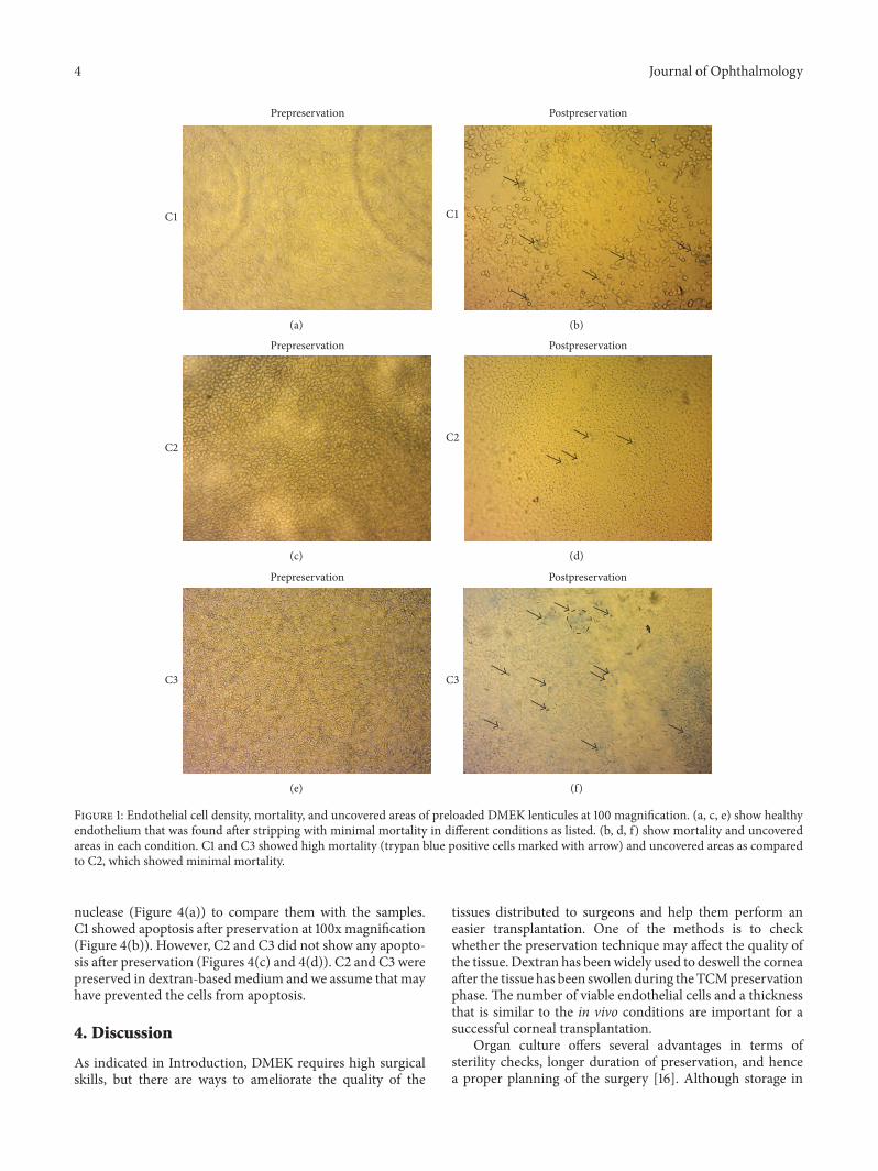
3.2. Lower Endothelial Cell Loss in Dextran-Based Media [n= 30]. ECD (cells/mm2) after preservation in C1, C2, andC3 was found to be 1130 (±944.05), 1950 (±108.01), and 1970(±512.18), and the mortality (%) was 25.94 (±44.23), 3.8(±7.74), and 21.8 (±36.41), respectively. It was also observedthat the cells detached from the lenticule during preser-vation and therefore the uncovered areas (%) determinedfor C1, C2, and C3 were 40.7 (±47.96), 13.0 (±18.55), and41.8 (±37.77). Loading to postpreservation in C1 showedstatistically significant endothelial cell loss (𝑝 = 0.0051);however, C2 (𝑝 = 0.1092) and C3 (𝑝 = 0.0819) did not showstatistical significance, although the average ECL observed inC3 was high. Figure 1 shows how the lenticules obtained fromcorneas preserved inC1, C2, andC3, respectively, appear afterstripping (Figures 1(a), 1(c), and 1(e)) and after preservation(Figures 1(b), 1(d), and 1(f)).
3.3. Glucose Was Utilized in All the Media [n = 30]. Averageglucose uptake by the endothelial cells in the preserva-tion chamber for 4 days at room temperature was 0.32(±0.18)mg/mL from C1, 0.43 (±0.27)mg/mL from C2, and0.56 (±0.18) mg/mL fromC3, which is one-third to half of thetotal amount of glucose present in themedia.These results aresimilar to those showed in the preloaded DSAEK lenticules[13].However, therewas no statistical difference seen betweeneither group (𝑝 > 0.05).
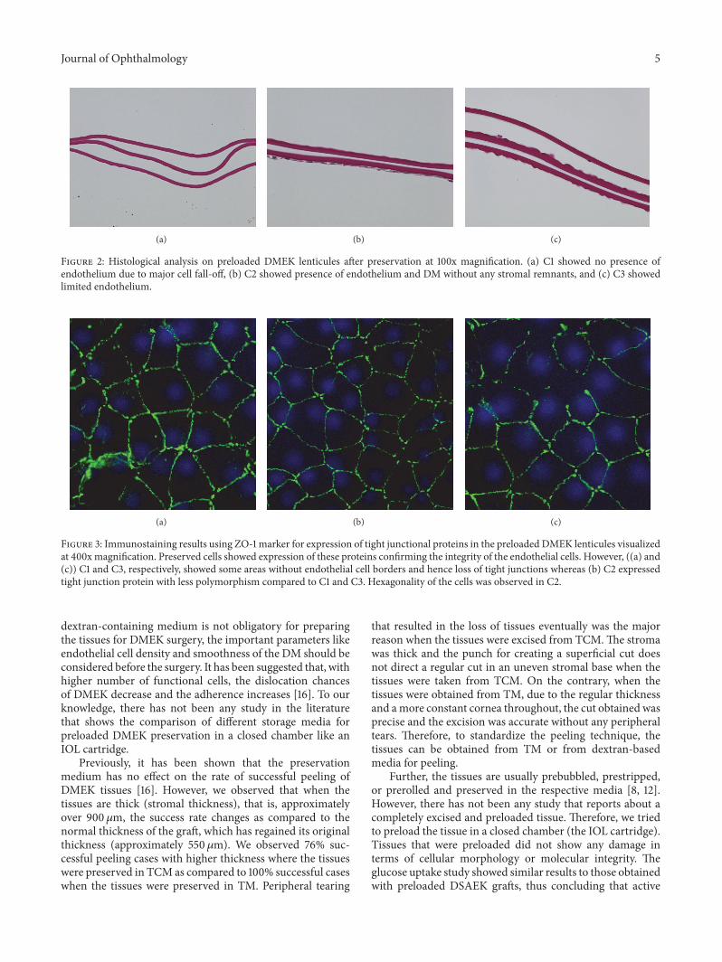
3.4. Presence of DM and Endothelial Sheet on the ExcisedDMEK Tissues from Dextran-Based Media [n = 9]. Theanalyzed tissues (100x magnification using PAS staining)showed no presence of endothelial cells, but only the DM(Figure 2(a)) in C1.Thismay be a reason due to high “fall-off”rate of the cells from C1. As observed in Figure 1, the cells donot maintain a hexagonal shape and have turned themselvesinto circular shape (stressed) along with high mortalityas seen using trypan blue staining. Descemet’s Membraneand endothelium (found in one of the tissues) without anycollagen fibrils or attached stromal residues in C2 are seenin Figure 2(b). However, lesser endothelial cells were foundin lenticules from C3 (Figure 2(c)). This phenomenon can bedue to the presence of dextran in the preservation mediumafter loading in the cartridge. Preservation of endothelial cellswith DM was found in C2.
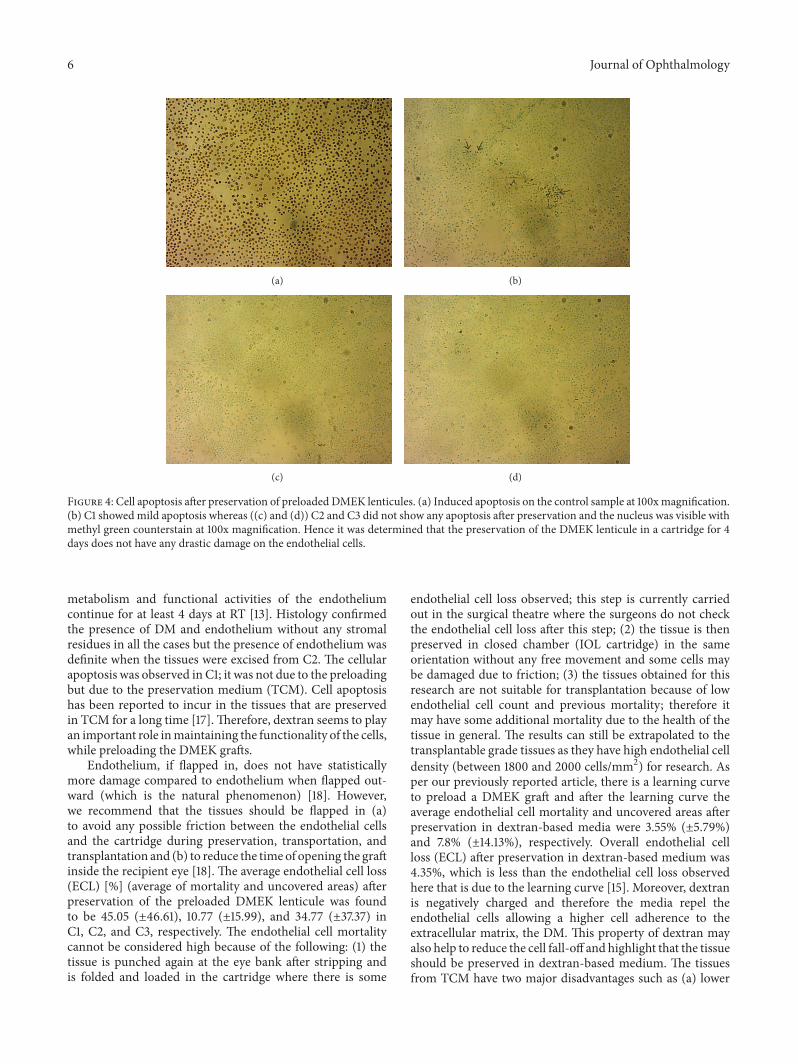
3.5. Expression of Tight Junctional Proteins and Polymorphismon the Endothelial Cells after Preservation [n = 12]. Theendothelial cells showed expression of tight junction protein(ZO-1) after preservation as seen in Figure 3 at 400x mag-nification. C1 and C3 (Figures 3(a) and 3(c), resp.) showedloss of hexagonality; most of the cells were polygonal tocircular. Tight junctional protein was not consistent in C3but was intense in C1 at many areas. C2 (Figure 3(b)) showedendothelial cell mosaic withmore hexagonal shaped cells andthe conservation of tight junctional proteins in the preloadedDMEK lenticules.
3.6. No Cell Apoptosis from Dextran-Based Media [n =9]. Control cells were induced with apoptosis using TACS
4 Journal of Ophthalmology
Prepreservation
C1
(a)
Postpreservation
C1
(b)
Prepreservation
C2
(c)
Postpreservation
C2
(d)
Prepreservation
C3
(e)
Postpreservation
C3
(f)
Figure 1: Endothelial cell density, mortality, and uncovered areas of preloaded DMEK lenticules at 100 magnification. (a, c, e) show healthyendothelium that was found after stripping with minimal mortality in different conditions as listed. (b, d, f) show mortality and uncoveredareas in each condition. C1 and C3 showed high mortality (trypan blue positive cells marked with arrow) and uncovered areas as comparedto C2, which showed minimal mortality.
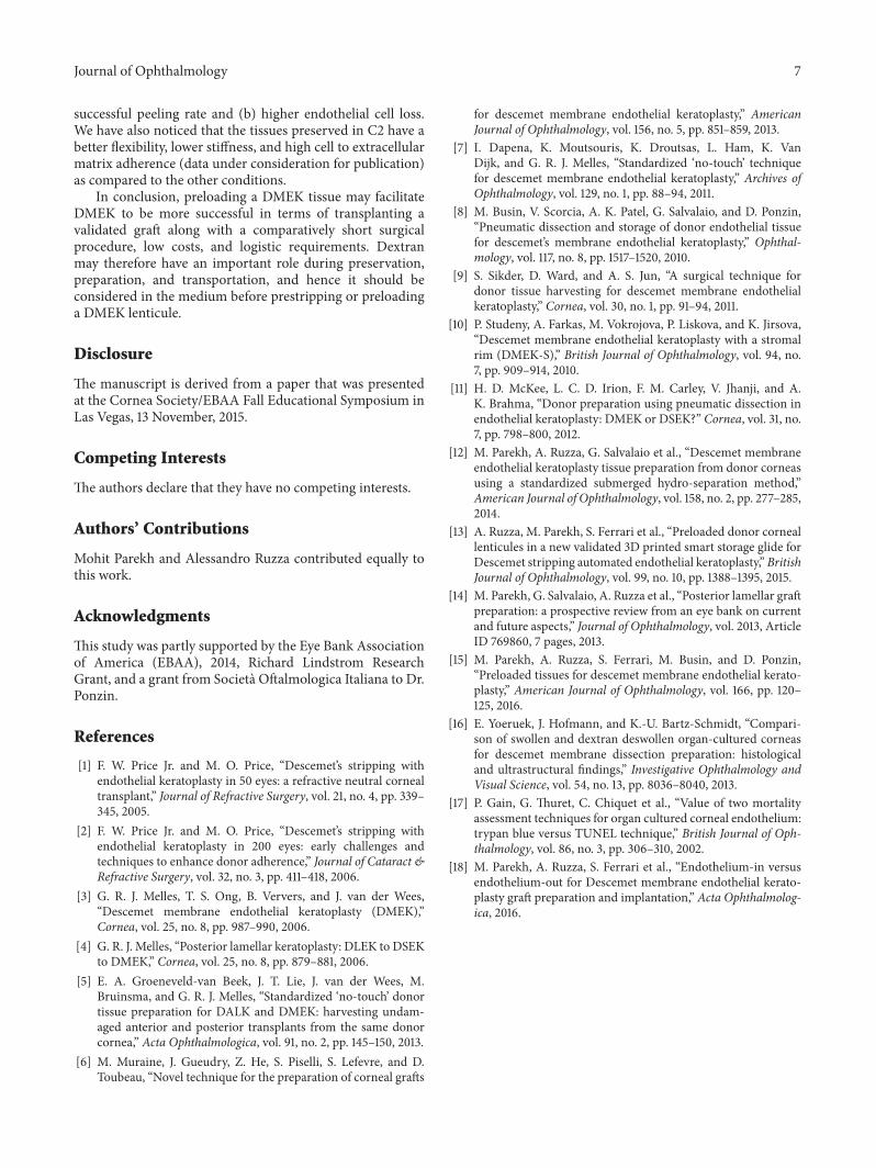
nuclease (Figure 4(a)) to compare them with the samples.C1 showed apoptosis after preservation at 100x magnification(Figure 4(b)). However, C2 and C3 did not show any apopto-sis after preservation (Figures 4(c) and 4(d)). C2 and C3 werepreserved in dextran-basedmedium and we assume that mayhave prevented the cells from apoptosis.
4. Discussion
As indicated in Introduction, DMEK requires high surgicalskills, but there are ways to ameliorate the quality of the
tissues distributed to surgeons and help them perform aneasier transplantation. One of the methods is to checkwhether the preservation technique may affect the quality ofthe tissue. Dextran has beenwidely used to deswell the corneaafter the tissue has been swollen during theTCMpreservationphase. The number of viable endothelial cells and a thicknessthat is similar to the in vivo conditions are important for asuccessful corneal transplantation.
Organ culture offers several advantages in terms ofsterility checks, longer duration of preservation, and hencea proper planning of the surgery [16]. Although storage in
Journal of Ophthalmology 5
(a) (b) (c)
Figure 2: Histological analysis on preloaded DMEK lenticules after preservation at 100x magnification. (a) C1 showed no presence ofendothelium due to major cell fall-off, (b) C2 showed presence of endothelium and DM without any stromal remnants, and (c) C3 showedlimited endothelium.
(a) (b) (c)
Figure 3: Immunostaining results using ZO-1 marker for expression of tight junctional proteins in the preloaded DMEK lenticules visualizedat 400xmagnification. Preserved cells showed expression of these proteins confirming the integrity of the endothelial cells. However, ((a) and(c)) C1 and C3, respectively, showed some areas without endothelial cell borders and hence loss of tight junctions whereas (b) C2 expressedtight junction protein with less polymorphism compared to C1 and C3. Hexagonality of the cells was observed in C2.
dextran-containing medium is not obligatory for preparingthe tissues for DMEK surgery, the important parameters likeendothelial cell density and smoothness of the DM should beconsidered before the surgery. It has been suggested that, withhigher number of functional cells, the dislocation chancesof DMEK decrease and the adherence increases [16]. To ourknowledge, there has not been any study in the literaturethat shows the comparison of different storage media forpreloaded DMEK preservation in a closed chamber like anIOL cartridge.
Previously, it has been shown that the preservationmedium has no effect on the rate of successful peeling ofDMEK tissues [16]. However, we observed that when thetissues are thick (stromal thickness), that is, approximatelyover 900𝜇m, the success rate changes as compared to thenormal thickness of the graft, which has regained its originalthickness (approximately 550 𝜇m). We observed 76% suc-cessful peeling cases with higher thickness where the tissueswere preserved in TCM as compared to 100% successful caseswhen the tissues were preserved in TM. Peripheral tearing
that resulted in the loss of tissues eventually was the majorreason when the tissues were excised from TCM.The stromawas thick and the punch for creating a superficial cut doesnot direct a regular cut in an uneven stromal base when thetissues were taken from TCM. On the contrary, when thetissues were obtained from TM, due to the regular thicknessand amore constant cornea throughout, the cut obtained wasprecise and the excision was accurate without any peripheraltears. Therefore, to standardize the peeling technique, thetissues can be obtained from TM or from dextran-basedmedia for peeling.
Further, the tissues are usually prebubbled, prestripped,or prerolled and preserved in the respective media [8, 12].However, there has not been any study that reports about acompletely excised and preloaded tissue. Therefore, we triedto preload the tissue in a closed chamber (the IOL cartridge).Tissues that were preloaded did not show any damage interms of cellular morphology or molecular integrity. Theglucose uptake study showed similar results to those obtainedwith preloaded DSAEK grafts, thus concluding that active
6 Journal of Ophthalmology
(a) (b)
(c) (d)
Figure 4: Cell apoptosis after preservation of preloaded DMEK lenticules. (a) Induced apoptosis on the control sample at 100xmagnification.(b) C1 showed mild apoptosis whereas ((c) and (d)) C2 and C3 did not show any apoptosis after preservation and the nucleus was visible withmethyl green counterstain at 100x magnification. Hence it was determined that the preservation of the DMEK lenticule in a cartridge for 4days does not have any drastic damage on the endothelial cells.
metabolism and functional activities of the endotheliumcontinue for at least 4 days at RT [13]. Histology confirmedthe presence of DM and endothelium without any stromalresidues in all the cases but the presence of endothelium wasdefinite when the tissues were excised from C2. The cellularapoptosis was observed in C1; it was not due to the preloadingbut due to the preservation medium (TCM). Cell apoptosishas been reported to incur in the tissues that are preservedin TCM for a long time [17].Therefore, dextran seems to playan important role inmaintaining the functionality of the cells,while preloading the DMEK grafts.
Endothelium, if flapped in, does not have statisticallymore damage compared to endothelium when flapped out-ward (which is the natural phenomenon) [18]. However,we recommend that the tissues should be flapped in (a)to avoid any possible friction between the endothelial cellsand the cartridge during preservation, transportation, andtransplantation and (b) to reduce the time of opening the graftinside the recipient eye [18]. The average endothelial cell loss(ECL) [%] (average of mortality and uncovered areas) afterpreservation of the preloaded DMEK lenticule was foundto be 45.05 (±46.61), 10.77 (±15.99), and 34.77 (±37.37) inC1, C2, and C3, respectively. The endothelial cell mortalitycannot be considered high because of the following: (1) thetissue is punched again at the eye bank after stripping andis folded and loaded in the cartridge where there is some
endothelial cell loss observed; this step is currently carriedout in the surgical theatre where the surgeons do not checkthe endothelial cell loss after this step; (2) the tissue is thenpreserved in closed chamber (IOL cartridge) in the sameorientation without any free movement and some cells maybe damaged due to friction; (3) the tissues obtained for thisresearch are not suitable for transplantation because of lowendothelial cell count and previous mortality; therefore itmay have some additional mortality due to the health of thetissue in general. The results can still be extrapolated to thetransplantable grade tissues as they have high endothelial celldensity (between 1800 and 2000 cells/mm2) for research. Asper our previously reported article, there is a learning curveto preload a DMEK graft and after the learning curve theaverage endothelial cell mortality and uncovered areas afterpreservation in dextran-based media were 3.55% (±5.79%)and 7.8% (±14.13%), respectively. Overall endothelial cellloss (ECL) after preservation in dextran-based medium was4.35%, which is less than the endothelial cell loss observedhere that is due to the learning curve [15]. Moreover, dextranis negatively charged and therefore the media repel theendothelial cells allowing a higher cell adherence to theextracellular matrix, the DM. This property of dextran mayalso help to reduce the cell fall-off and highlight that the tissueshould be preserved in dextran-based medium. The tissuesfrom TCM have two major disadvantages such as (a) lower
Journal of Ophthalmology 7
successful peeling rate and (b) higher endothelial cell loss.We have also noticed that the tissues preserved in C2 have abetter flexibility, lower stiffness, and high cell to extracellularmatrix adherence (data under consideration for publication)as compared to the other conditions.
In conclusion, preloading a DMEK tissue may facilitateDMEK to be more successful in terms of transplanting avalidated graft along with a comparatively short surgicalprocedure, low costs, and logistic requirements. Dextranmay therefore have an important role during preservation,preparation, and transportation, and hence it should beconsidered in the medium before prestripping or preloadinga DMEK lenticule.
Disclosure
The manuscript is derived from a paper that was presentedat the Cornea Society/EBAA Fall Educational Symposium inLas Vegas, 13 November, 2015.
Competing Interests
The authors declare that they have no competing interests.
Authors’ Contributions
Mohit Parekh and Alessandro Ruzza contributed equally tothis work.
Acknowledgments
This study was partly supported by the Eye Bank Associationof America (EBAA), 2014, Richard Lindstrom ResearchGrant, and a grant from Societa Oftalmologica Italiana to Dr.Ponzin.
References
[1] F. W. Price Jr. and M. O. Price, “Descemet’s stripping withendothelial keratoplasty in 50 eyes: a refractive neutral cornealtransplant,” Journal of Refractive Surgery, vol. 21, no. 4, pp. 339–345, 2005.
[2] F. W. Price Jr. and M. O. Price, “Descemet’s stripping withendothelial keratoplasty in 200 eyes: early challenges andtechniques to enhance donor adherence,” Journal of Cataract &Refractive Surgery, vol. 32, no. 3, pp. 411–418, 2006.
[3] G. R. J. Melles, T. S. Ong, B. Ververs, and J. van der Wees,“Descemet membrane endothelial keratoplasty (DMEK),”Cornea, vol. 25, no. 8, pp. 987–990, 2006.
[4] G. R. J. Melles, “Posterior lamellar keratoplasty: DLEK to DSEKto DMEK,” Cornea, vol. 25, no. 8, pp. 879–881, 2006.
[5] E. A. Groeneveld-van Beek, J. T. Lie, J. van der Wees, M.Bruinsma, and G. R. J. Melles, “Standardized ‘no-touch’ donortissue preparation for DALK and DMEK: harvesting undam-aged anterior and posterior transplants from the same donorcornea,” Acta Ophthalmologica, vol. 91, no. 2, pp. 145–150, 2013.
[6] M. Muraine, J. Gueudry, Z. He, S. Piselli, S. Lefevre, and D.Toubeau, “Novel technique for the preparation of corneal grafts
for descemet membrane endothelial keratoplasty,” AmericanJournal of Ophthalmology, vol. 156, no. 5, pp. 851–859, 2013.
[7] I. Dapena, K. Moutsouris, K. Droutsas, L. Ham, K. VanDijk, and G. R. J. Melles, “Standardized ‘no-touch’ techniquefor descemet membrane endothelial keratoplasty,” Archives ofOphthalmology, vol. 129, no. 1, pp. 88–94, 2011.
[8] M. Busin, V. Scorcia, A. K. Patel, G. Salvalaio, and D. Ponzin,“Pneumatic dissection and storage of donor endothelial tissuefor descemet’s membrane endothelial keratoplasty,” Ophthal-mology, vol. 117, no. 8, pp. 1517–1520, 2010.
[9] S. Sikder, D. Ward, and A. S. Jun, “A surgical technique fordonor tissue harvesting for descemet membrane endothelialkeratoplasty,” Cornea, vol. 30, no. 1, pp. 91–94, 2011.
[10] P. Studeny, A. Farkas, M. Vokrojova, P. Liskova, and K. Jirsova,“Descemet membrane endothelial keratoplasty with a stromalrim (DMEK-S),” British Journal of Ophthalmology, vol. 94, no.7, pp. 909–914, 2010.
[11] H. D. McKee, L. C. D. Irion, F. M. Carley, V. Jhanji, and A.K. Brahma, “Donor preparation using pneumatic dissection inendothelial keratoplasty: DMEK or DSEK?” Cornea, vol. 31, no.7, pp. 798–800, 2012.
[12] M. Parekh, A. Ruzza, G. Salvalaio et al., “Descemet membraneendothelial keratoplasty tissue preparation from donor corneasusing a standardized submerged hydro-separation method,”American Journal of Ophthalmology, vol. 158, no. 2, pp. 277–285,2014.
[13] A. Ruzza, M. Parekh, S. Ferrari et al., “Preloaded donor corneallenticules in a new validated 3D printed smart storage glide forDescemet stripping automated endothelial keratoplasty,”BritishJournal of Ophthalmology, vol. 99, no. 10, pp. 1388–1395, 2015.
[14] M. Parekh, G. Salvalaio, A. Ruzza et al., “Posterior lamellar graftpreparation: a prospective review from an eye bank on currentand future aspects,” Journal of Ophthalmology, vol. 2013, ArticleID 769860, 7 pages, 2013.
[15] M. Parekh, A. Ruzza, S. Ferrari, M. Busin, and D. Ponzin,“Preloaded tissues for descemet membrane endothelial kerato-plasty,” American Journal of Ophthalmology, vol. 166, pp. 120–125, 2016.
[16] E. Yoeruek, J. Hofmann, and K.-U. Bartz-Schmidt, “Compari-son of swollen and dextran deswollen organ-cultured corneasfor descemet membrane dissection preparation: histologicaland ultrastructural findings,” Investigative Ophthalmology andVisual Science, vol. 54, no. 13, pp. 8036–8040, 2013.
[17] P. Gain, G. Thuret, C. Chiquet et al., “Value of two mortalityassessment techniques for organ cultured corneal endothelium:trypan blue versus TUNEL technique,” British Journal of Oph-thalmology, vol. 86, no. 3, pp. 306–310, 2002.
[18] M. Parekh, A. Ruzza, S. Ferrari et al., “Endothelium-in versusendothelium-out for Descemet membrane endothelial kerato-plasty graft preparation and implantation,” Acta Ophthalmolog-ica, 2016.

Submit your manuscripts athttp://www.hindawi.com
Stem CellsInternational
Hindawi Publishing Corporationhttp://www.hindawi.com Volume 2014
Hindawi Publishing Corporationhttp://www.hindawi.com Volume 2014
MEDIATORSINFLAMMATION
of
Hindawi Publishing Corporationhttp://www.hindawi.com Volume 2014
Behavioural Neurology
EndocrinologyInternational Journal of
Hindawi Publishing Corporationhttp://www.hindawi.com Volume 2014
Hindawi Publishing Corporationhttp://www.hindawi.com Volume 2014
Disease Markers
Hindawi Publishing Corporationhttp://www.hindawi.com Volume 2014
BioMed Research International
OncologyJournal of
Hindawi Publishing Corporationhttp://www.hindawi.com Volume 2014
Hindawi Publishing Corporationhttp://www.hindawi.com Volume 2014
Oxidative Medicine and Cellular Longevity
Hindawi Publishing Corporationhttp://www.hindawi.com Volume 2014
PPAR Research
The Scientific World JournalHindawi Publishing Corporation http://www.hindawi.com Volume 2014
Immunology ResearchHindawi Publishing Corporationhttp://www.hindawi.com Volume 2014
Journal of
ObesityJournal of
Hindawi Publishing Corporationhttp://www.hindawi.com Volume 2014
Hindawi Publishing Corporationhttp://www.hindawi.com Volume 2014
Computational and Mathematical Methods in Medicine
OphthalmologyJournal of
Hindawi Publishing Corporationhttp://www.hindawi.com Volume 2014
Diabetes ResearchJournal of
Hindawi Publishing Corporationhttp://www.hindawi.com Volume 2014
Hindawi Publishing Corporationhttp://www.hindawi.com Volume 2014
Research and TreatmentAIDS
Hindawi Publishing Corporationhttp://www.hindawi.com Volume 2014
Gastroenterology Research and Practice
Hindawi Publishing Corporationhttp://www.hindawi.com Volume 2014
Parkinson’s Disease
Evidence-Based Complementary and Alternative Medicine
Volume 2014Hindawi Publishing Corporationhttp://www.hindawi.com
Related Documents