D escemet Membrane Endothelial Keratoplasty INNOVATION MADE BY GEUDER ® P R E P A R A T I O N THE INNOVATIVE SYSTEM FOR TREATING ENDOTHELIAL CORNEAL DISEASES I M P L A N T A T I O N DMEK

Welcome message from author
This document is posted to help you gain knowledge. Please leave a comment to let me know what you think about it! Share it to your friends and learn new things together.
Transcript
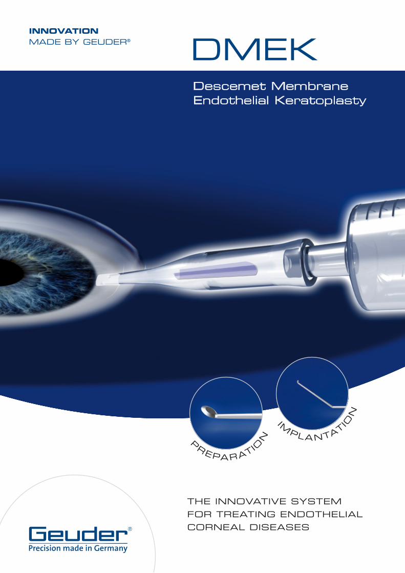
Descemet Membrane Endothelial Keratoplasty
INNOVATIONMADE BY GEUDER®
PREPARATIO
N
THE INNOVATIVE SYSTEMFOR TREATING ENDOTHELIALCORNEAL DISEASES
IMPLANTATIO
N
DMEK
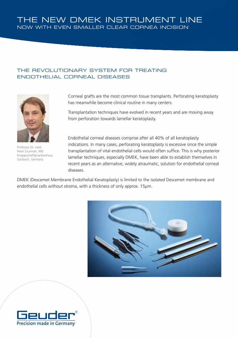
THE NEW DMEK INSTRUMENT LINENOW WITH EVEN SMALLER CLEAR CORNEA INCISION
THE REVOLUTIONARY SYSTEM FOR TREATING ENDOTHELIAL CORNEAL DISEASES
Corneal grafts are the most common tissue transplants. Perforating keratoplasty
has meanwhile become clinical routine in many centers.
Transplantation techniques have evolved in recent years and are moving away
from perforation towards lamellar keratoplasty.
Endothelial corneal diseases comprise after all 40% of all keratoplasty
indications. In many cases, perforating keratoplasty is excessive since the simple
transplantation of vital endothelial cells would often suffice. This is why posterior
lamellar techniques, especially DMEK, have been able to establish themselves in
recent years as an alternative, widely atraumatic, solution for endothelial corneal
diseases.
DMEK (Descemet Membrane Endothelial Keratoplasty) is limited to the isolated Descemet membrane and
endothelial cells without stroma, with a thickness of only approx. 15μm.
Professor Dr. med. Peter Szurman, MDKnappschaftskrankenhaus Sulzbach, Germany
Literature:1. Melles GR, Ong TS, Ververs B, van der Wees J (2006) Descemet membrane endothelial keratoplasty (DMEK). Cornea 2006; 25: 987-902. Cursiefen C, Kruse FE (2009) Descemet‘s stripping automated endothelial keratoplasty (DSAEK). Ophthalmologe. 106: 939-9523. Szurman P, Yörük E (2010) DMEK-Step by Step. ESCRS, Paris4. Yörük E, Szurman P (2010) DMEK Keratoplastik. Augenspiegel 11/20105. Yörük E, Szurman P (2011) Autologous Descemet´s Membrane Endothelial Keratoplasty. Cornea; in press
CLINICAL ADVANTAGES OF DMEK
Transplanting extremely thin lamella promotes considerably faster visual recovery than other lamellar
keratoplasty techniques. DMEK prevents interface problems, causes neither postoperative astigmatism nor
myopia and substitutes more endothelial cells (up to 9.5 mm graft size). As a result, visual acuity improves in
many cases by 0.8 or better after only one week. Due to these very good results, DMEK might become the
gold standard in the therapy of endothelial corneal diseases.
INSTRUMENTS FOR STANDARDIZATION OF DMEK
The success of this elegant technique is largely dependent on the number of vital endothelial cells and
quality of the fragile graft and the gentle manipulation thereof. It is important that the fragile endothelial
cells are not touched or stressed mechanically during preparation or implantation. In order to achieve
reproducible results a standardized technique and specific instruments, which ensure a touch-free surgical
procedure, are necessary. The new Liquid Bubble technique, which was developed in Sulzbach, Germany,
uses a liquid in order to gently separate the Descemet membrane from the stroma beneath. It is another step
towards the standardisation of DMEK.
On the following pages we introduce a new surgical set which will give experienced surgeons the
opportunity to perform DMEK in the clinical routine. This set allows a touch-free preparation of a Descemet
lamella and its subsequent transplantation.
Prof. Dr. med. Peter Szurman, MD
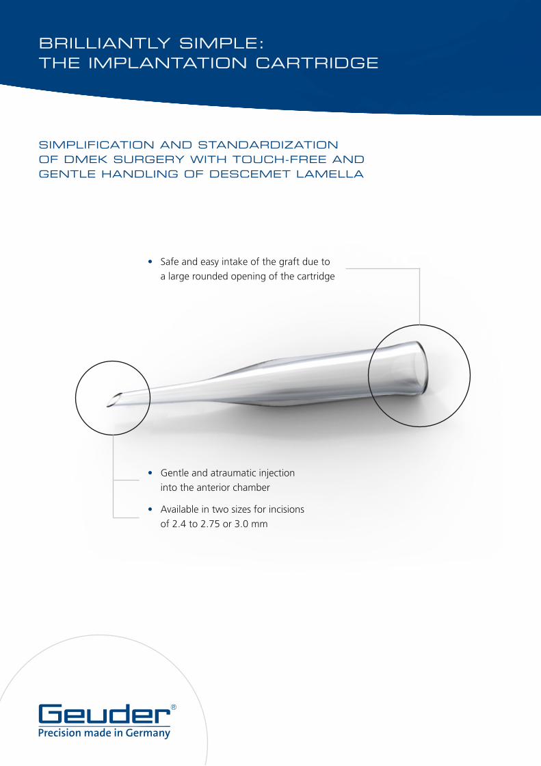
• Gentle and atraumatic injection
into the anterior chamber
• Available in two sizes for incisions
of 2.4 to 2.75 or 3.0 mm
• Safe and easy intake of the graft due to
a large rounded opening of the cartridge
BRILLIANTLY SIMPLE:THE IMPLANTATION CARTRIDGE
SIMPLIFICATION AND STANDARDIZATION OF DMEK SURGERY WITH TOUCH-FREE AND GENTLE HANDLING OF DESCEMET LAMELLA
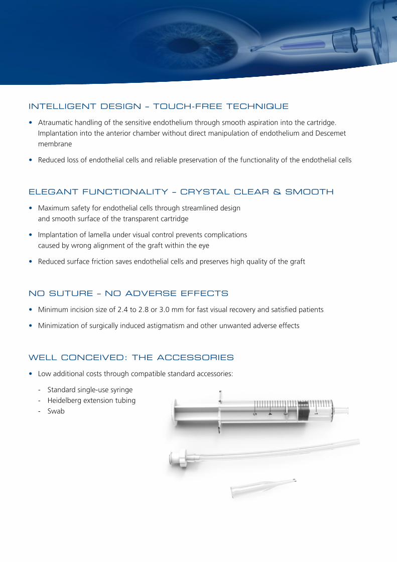
INTELLIGENT DESIGN – TOUCH-FREE TECHNIQUE
• Atraumatic handling of the sensitive endothelium through smooth aspiration into the cartridge.
Implantation into the anterior chamber without direct manipulation of endothelium and Descemet
membrane
• Reduced loss of endothelial cells and reliable preservation of the functionality of the endothelial cells
ELEGANT FUNCTIONALITY – CRYSTAL CLEAR & SMOOTH
• Maximum safety for endothelial cells through streamlined design
and smooth surface of the transparent cartridge
• Implantation of lamella under visual control prevents complications
caused by wrong alignment of the graft within the eye
• Reduced surface friction saves endothelial cells and preserves high quality of the graft
NO SUTURE – NO ADVERSE EFFECTS
• Minimum incision size of 2.4 to 2.8 or 3.0 mm for fast visual recovery and satisfied patients
• Minimization of surgically induced astigmatism and other unwanted adverse effects
WELL CONCEIVED: THE ACCESSORIES
• Low additional costs through compatible standard accessories:
- Standard single-use syringe
- Heidelberg extension tubing
- Swab
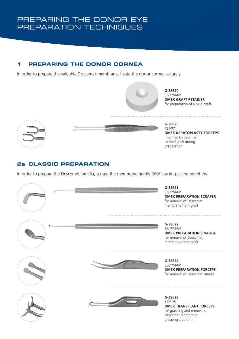
G-38623REMKYDMEK KERATOPLASTY FORCEPSmodified by Szurmanto hold graft duringpreparation
G-38620SZURMANDMEK GRAFT RETAINERfor preparation of DMEK graft
PREPARING THE DONOR EYEPREPARATION TECHNIQUES
1 PREPARING THE DONOR CORNEA
In order to prepare the valuable Descemet membrane, fixate the donor cornea securely.
2a CLASSIC PREPARATION
In order to prepare the Descemet lamella, scrape the membrane gently 360° starting at the periphery.
G-38624SZURMANDMEK PREPARATION FORCEPSfor removal of Descemet lamella
G-38622SZURMANDMEK PREPARATION SPATULAfor removal of Descemet membrane from graft
G-38621SZURMANDMEK PREPARATION SCRAPERfor removal of Descemet membrane from graft
G-38628YÖRÜKDMEK TRANSPLANT FORCEPSfor grasping and removal of Descemet membranegrasping area 6 mm
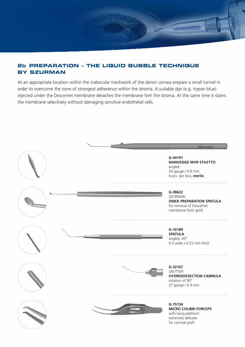
2b PREPARATION – THE LIQUID BUBBLE TECHNIQUE BY SZURMAN
At an appropriate location within the trabecular meshwork of the donor cornea prepare a small tunnel in
order to overcome the zone of strongest adherence within the stroma. A suitable dye (e.g. trypan blue)
injected under the Descemet membrane detaches the membrane fom the stroma. At the same time it stains
the membrane selectively without damaging sensitive endothelial cells.
G-38622SZURMANDMEK PREPARATION SPATULAfor removal of Descemet membrane from graft
G-32167SAUTTERHYDRODISSECTION CANNULArotation of 90°27 gauge / 0.4 mm
G-15134MICRO COLIBRI FORCEPSwith tying platform extremely delicatefor corneal graft
G-34191NANOEDGE MVR STILETTOangled20 gauge / 0.9 mm6 pcs. per box, sterile
G-16189SPATULAangled, 45°0.5 wide x 0.25 mm thick
PREPARING THE DONOR EYEPREPARATION TECHNIQUES
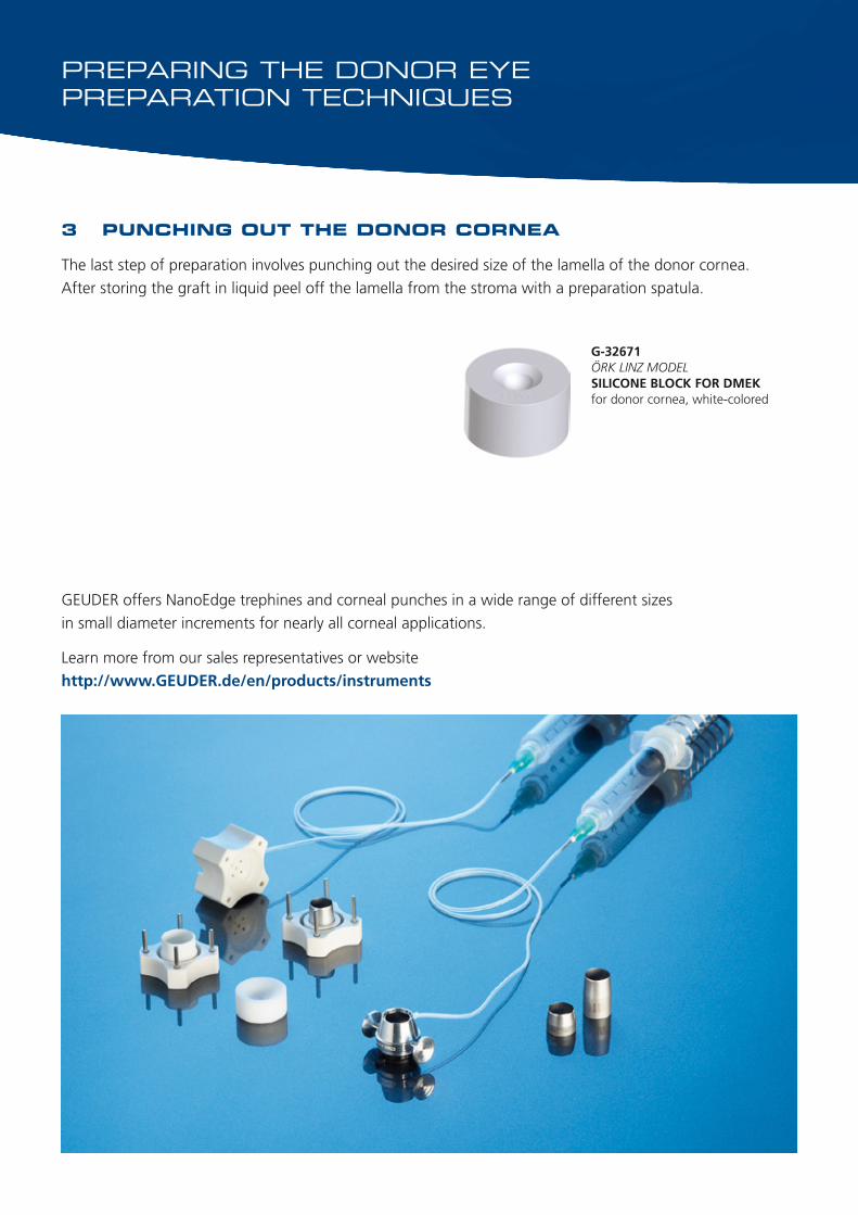
G-32671ÖRK LINZ MODELSILICONE BLOCK FOR DMEKfor donor cornea, white-colored
3 PUNCHING OUT THE DONOR CORNEA
The last step of preparation involves punching out the desired size of the lamella of the donor cornea.
After storing the graft in liquid peel off the lamella from the stroma with a preparation spatula.
GEUDER offers NanoEdge trephines and corneal punches in a wide range of different sizes
in small diameter increments for nearly all corneal applications.
Learn more from our sales representatives or website
http://www.GEUDER.de/en/products/instruments
PREPARING THE PATIENT’S EYE
G-32948HATTENBACHHYBRID SCISSORSfor the anterior chamber25 gauge / 0.5 mm
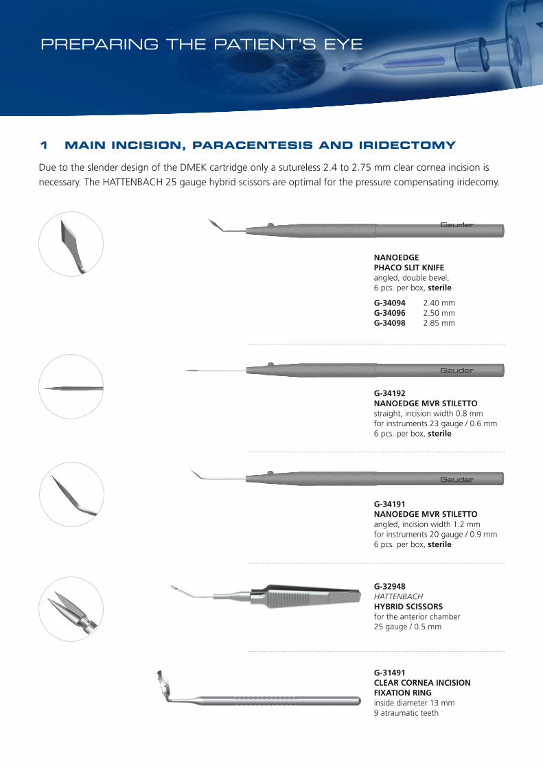
1 MAIN INCISION, PARACENTESIS AND IRIDECTOMY
Due to the slender design of the DMEK cartridge only a sutureless 2.4 to 2.75 mm clear cornea incision is
necessary. The HATTENBACH 25 gauge hybrid scissors are optimal for the pressure compensating iridecomy.
NANOEDGEPHACO SLIT KNIFEangled, double bevel,6 pcs. per box, sterile
G-34094 2.40 mmG-34096 2.50 mmG-34098 2.85 mm
G-34191NANOEDGE MVR STILETTOangled, incision width 1.2 mmfor instruments 20 gauge / 0.9 mm6 pcs. per box, sterile
G-34192NANOEDGE MVR STILETTOstraight, incision width 0.8 mmfor instruments 23 gauge / 0.6 mm6 pcs. per box, sterile
G-31491CLEAR CORNEA INCISION FIXATION RINGinside diameter 13 mm9 atraumatic teeth
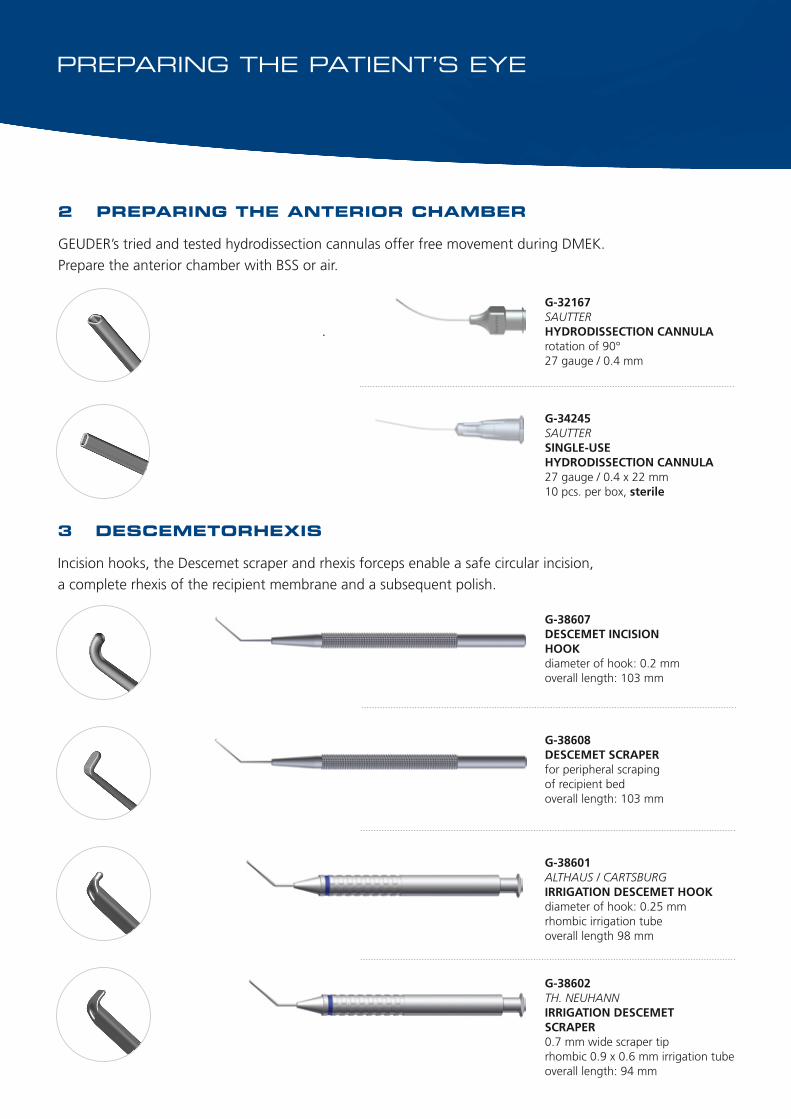
G-34245SAUTTERSINGLE-USEHYDRODISSECTION CANNULA27 gauge / 0.4 x 22 mm10 pcs. per box, sterile
G-32167SAUTTERHYDRODISSECTION CANNULArotation of 90°27 gauge / 0.4 mm
2 PREPARING THE ANTERIOR CHAMBER
GEUDER’s tried and tested hydrodissection cannulas offer free movement during DMEK.
Prepare the anterior chamber with BSS or air.
PREPARING THE PATIENT’S EYE
G-38607DESCEMET INCISIONHOOKdiameter of hook: 0.2 mmoverall length: 103 mm
3 DESCEMETORHEXIS
Incision hooks, the Descemet scraper and rhexis forceps enable a safe circular incision,
a complete rhexis of the recipient membrane and a subsequent polish.
G-38608DESCEMET SCRAPERfor peripheral scraping of recipient bedoverall length: 103 mm
G-38601ALTHAUS / CARTSBURGIRRIGATION DESCEMET HOOKdiameter of hook: 0.25 mmrhombic irrigation tubeoverall length 98 mm
G-38602TH. NEUHANNIRRIGATION DESCEMET SCRAPER0.7 mm wide scraper tiprhombic 0.9 x 0.6 mm irrigation tubeoverall length: 94 mm
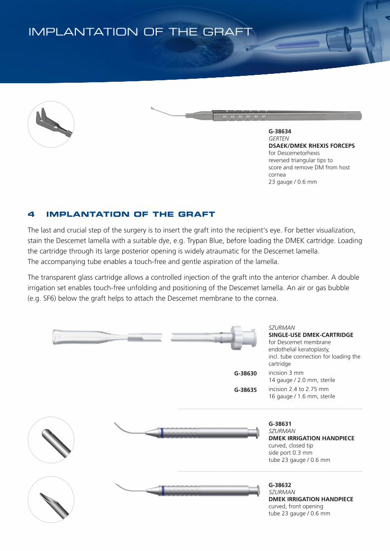
4 IMPLANTATION OF THE GRAFT
The last and crucial step of the surgery is to insert the graft into the recipient‘s eye. For better visualization,
stain the Descemet lamella with a suitable dye, e.g. Trypan Blue, before loading the DMEK cartridge. Loading
the cartridge through its large posterior opening is widely atraumatic for the Descemet lamella.
The accompanying tube enables a touch-free and gentle aspiration of the lamella.
The transparent glass cartridge allows a controlled injection of the graft into the anterior chamber. A double
irrigation set enables touch-free unfolding and positioning of the Descemet lamella. An air or gas bubble
(e.g. SF6) below the graft helps to attach the Descemet membrane to the cornea.
SZURMAN SINGLE-USE DMEK-CARTRIDGE for Descemet membrane endothelial keratoplasty, incl. tube connection for loading the cartridge
incision 3 mm 14 gauge / 2.0 mm, sterile
incision 2.4 to 2.75 mm 16 gauge / 1.6 mm, sterile
G-38630
G-38635
IMPLANTATION OF THE GRAFT
G-38631SZURMANDMEK IRRIGATION HANDPIECEcurved, closed tipside port 0.3 mmtube 23 gauge / 0.6 mm
G-38632SZURMAN DMEK IRRIGATION HANDPIECEcurved, front openingtube 23 gauge / 0.6 mm
G-38634GERTENDSAEK/DMEK RHEXIS FORCEPSfor Descemetorhexisreversed triangular tips toscore and remove DM from host cornea23 gauge / 0.6 mm

2017
-09
69
190
Phone: +49 6221 3066Fax: +49 6221 [email protected]
GEUDER AGHertzstrasse 469126 HeidelbergGermany
GEUDER AG reserves the right to make changes to technical details in response to recent developments. GEUDER does not assume liability for the accuracy of each individual statement.
Illustrations not drawn to scale (some illustrations are reduced to 80%).
Related Documents