ORIGINAL ARTICLE Rabbit anti-rat thymocyte immunoglobulin preserves renal function during ischemia/reperfusion injury in rat kidney transplantation Sistiana Aiello, 1 Paola Cassis, 1 Marilena Mister, 1 Samantha Solini, 1 Federica Rocchetta, 1 Mauro Abbate, 2 Elena Gagliardini, 2 Ariela Benigni, 2 Giuseppe Remuzzi 3 and Marina Noris 1 1 Transplant Research Center, ‘‘Chiara Cucchi De Alessandri & Gilberto Crespi’’, Mario Negri Institute for Pharmacological Research, Ranica, Bergamo, Italy 2 Mario Negri Institute for Pharmacological Research, Centro Anna Maria Astori, Science and Technology Park Kilometro Rosso, Bergamo, Italy 3 Department of Immunology and Organ Transplantation, Ospedali Riuniti – Mario Negri Institute for Pharmacological Research, Bergamo, Italy Introduction Long-term preservation of graft function has been one of the most important concerns since the beginning of organ transplantation. Solid organ transplantation is inevitably associated with a period of ischemia starting at the recov- ery of organs from the donor until their reperfusion in the recipient. The reintroduction of blood flow to the ischemic organ, although necessary to rescue the organ from necrosis and permanent loss of function, may cause acute cellular injury [1]. Ischemia/reperfusion (I/R) injury is an important cause of renal graft dysfunction, leading to a higher risk of early post-transplant complications including delayed graft function (DGF) and acute rejec- tion [2,3]. Cold-ischemia time also negatively impacts long-term kidney graft outcome and significantly predicts long-term graft loss in humans [4]. The improvement in understanding the pathophysiol- ogy of renal I/R injury has contributed to the develop- ment of potential strategies to limit the consequent graft dysfunction [5–7]. However, the prevention and treat- ment of postischemic injury remain difficult areas of kid- ney transplant medicine with modest achievements in the last 20 years. Keywords apoptosis, ischemia/reperfusion, kidney transplant, oxidative stress, Thymoglobuline. Correspondence Marina Noris PhD, Mario Negri Institute for Pharmacological Research, Via Camozzi, 3, 24020 Ranica, Italy. Tel.: +39-035-453-5362; fax: +39-035-453-5377; e-mail: marina. [email protected] Conflicts of Interest All the authors declared no competing interests. SA and PC equally contributed to the paper. Received: 21 January 2011 Revision requested: 15 February 2011 Accepted: 31 March 2011 Published online: 5 May 2011 doi:10.1111/j.1432-2277.2011.01263.x Summary Ischemia/reperfusion (I/R) injury is an important cause of renal graft dysfunc- tion in humans. Increases in cold and warm ischemia times lead to a higher risk of early post-transplant complications including delayed graft function and acute rejection. Moreover, prolonged cold ischemia is a predictor of long-term kidney graft loss. The protective effect of rabbit anti-rat thymocyte immuno- globulin (rATG) was evaluated in a rat model of I/R injury following syngeneic kidney transplantation. Serum creatinine concentration was evaluated at 16 h and 24 h post-transplant. Animals were sacrificed 24 h post-transplant for eval- uation of histology, infiltrating leukocytes, nitrotyrosine staining, and apopto- sis. rATG was effective in preventing renal function impairment, tissue damage and tubular apoptosis associated with I/R only when was given 2 h before transplantation but not at the time of reperfusion. Pretransplant rATG treat- ment of recipient animals effectively reduced the amount of macrophages, CD4 + , CD8 + T cells and LFA-1 + cells infiltrating renal graft subjected to cold ischemia as well as granzyme-B expression within ischemic kidney. On the other hand, granulocyte infiltration and oxidative stress were not modified by rATG. If these results will be translated into the clinical setting, pretransplant administration of Thymoglobuline Ò could offer the additional advantage over peri-transplant administration of limiting I/R-mediated kidney graft damage. Transplant International ISSN 0934-0874 ª 2011 The Authors Transplant International ª 2011 European Society for Organ Transplantation 24 (2011) 829–838 829

Welcome message from author
This document is posted to help you gain knowledge. Please leave a comment to let me know what you think about it! Share it to your friends and learn new things together.
Transcript

ORIGINAL ARTICLE
Rabbit anti-rat thymocyte immunoglobulin preserves renalfunction during ischemia/reperfusion injury in rat kidneytransplantationSistiana Aiello,1 Paola Cassis,1 Marilena Mister,1 Samantha Solini,1 Federica Rocchetta,1
Mauro Abbate,2 Elena Gagliardini,2 Ariela Benigni,2 Giuseppe Remuzzi3 and Marina Noris1
1 Transplant Research Center, ‘‘Chiara Cucchi De Alessandri & Gilberto Crespi’’, Mario Negri Institute for Pharmacological Research, Ranica,
Bergamo, Italy
2 Mario Negri Institute for Pharmacological Research, Centro Anna Maria Astori, Science and Technology Park Kilometro Rosso, Bergamo, Italy
3 Department of Immunology and Organ Transplantation, Ospedali Riuniti – Mario Negri Institute for Pharmacological Research, Bergamo,
Italy
Introduction
Long-term preservation of graft function has been one of
the most important concerns since the beginning of organ
transplantation. Solid organ transplantation is inevitably
associated with a period of ischemia starting at the recov-
ery of organs from the donor until their reperfusion in
the recipient. The reintroduction of blood flow to the
ischemic organ, although necessary to rescue the organ
from necrosis and permanent loss of function, may cause
acute cellular injury [1]. Ischemia/reperfusion (I/R) injury
is an important cause of renal graft dysfunction, leading
to a higher risk of early post-transplant complications
including delayed graft function (DGF) and acute rejec-
tion [2,3]. Cold-ischemia time also negatively impacts
long-term kidney graft outcome and significantly predicts
long-term graft loss in humans [4].
The improvement in understanding the pathophysiol-
ogy of renal I/R injury has contributed to the develop-
ment of potential strategies to limit the consequent graft
dysfunction [5–7]. However, the prevention and treat-
ment of postischemic injury remain difficult areas of kid-
ney transplant medicine with modest achievements in the
last 20 years.
Keywords
apoptosis, ischemia/reperfusion, kidney
transplant, oxidative stress, Thymoglobuline.
Correspondence
Marina Noris PhD, Mario Negri Institute for
Pharmacological Research, Via Camozzi, 3,
24020 Ranica, Italy. Tel.: +39-035-453-5362;
fax: +39-035-453-5377; e-mail: marina.
Conflicts of Interest
All the authors declared no competing
interests.
SA and PC equally contributed to the paper.
Received: 21 January 2011
Revision requested: 15 February 2011
Accepted: 31 March 2011
Published online: 5 May 2011
doi:10.1111/j.1432-2277.2011.01263.x
Summary
Ischemia/reperfusion (I/R) injury is an important cause of renal graft dysfunc-
tion in humans. Increases in cold and warm ischemia times lead to a higher
risk of early post-transplant complications including delayed graft function and
acute rejection. Moreover, prolonged cold ischemia is a predictor of long-term
kidney graft loss. The protective effect of rabbit anti-rat thymocyte immuno-
globulin (rATG) was evaluated in a rat model of I/R injury following syngeneic
kidney transplantation. Serum creatinine concentration was evaluated at 16 h
and 24 h post-transplant. Animals were sacrificed 24 h post-transplant for eval-
uation of histology, infiltrating leukocytes, nitrotyrosine staining, and apopto-
sis. rATG was effective in preventing renal function impairment, tissue damage
and tubular apoptosis associated with I/R only when was given 2 h before
transplantation but not at the time of reperfusion. Pretransplant rATG treat-
ment of recipient animals effectively reduced the amount of macrophages,
CD4+, CD8+ T cells and LFA-1+ cells infiltrating renal graft subjected to cold
ischemia as well as granzyme-B expression within ischemic kidney. On the
other hand, granulocyte infiltration and oxidative stress were not modified by
rATG. If these results will be translated into the clinical setting, pretransplant
administration of Thymoglobuline� could offer the additional advantage over
peri-transplant administration of limiting I/R-mediated kidney graft damage.
Transplant International ISSN 0934-0874
ª 2011 The Authors
Transplant International ª 2011 European Society for Organ Transplantation 24 (2011) 829–838 829
The mechanism of injury in I/R involves activation of
endothelial cells which are induced to express high levels
of surface adhesion molecules and produce cytokines and
chemokines capable to attract inflammatory leukocytes, as
to create a nonspecific local host inflammatory response
[8]. Such a cascade of events eventually ends in tubular
cell apoptosis and necrosis [9].
Thymoglobuline� (Genzyme Corporation, Cambridge,
MA, USA) is a purified fraction of IgG obtained from
sera of rabbits immunized against human thymocytes and
it is commonly used in clinical transplant setting as
induction therapy [10].
Previous in vitro studies on cultured human peripheral
blood leukocytes showed that antibodies present in Thy-
moglobuline affected the binding and/or the surface
expression of leukocyte integrins (LFA-1, VLA-4) and
ligands (ICAM-1) involved in leukocyte/endothelial cell
interaction [11]. In addition, Thymoglobuline contains
anti-CCR7, anti-CXCR4 and anti-CCR5 antibodies that
inhibit leukocyte response to chemoattractants by compe-
tition with and by down-modulation of the correspond-
ing antigen [11]. Altogether these data suggest that the
use of Thymoglobuline might contribute to decrease graft
cellular infiltration occurring after I/R, thus limiting acute
and chronic graft dysfunction.
The clinical need of effective strategies to limit post-
transplant I/R-induced tissue injury has prompted us to
design a study in an experimental model of syngeneic rat
kidney transplantation with the aim to evaluate whether
treatment with rabbit anti-rat thymocyte immunoglobulin
(rATG) limits I/R injury and facilitates immediate graft
function.
Methods
Animals
Inbred adult male Lewis (LW) rats (RT1l, Charles River
Italia Spa, Calco, Italy), were used as donors and recipi-
ents in syngeneic kidney transplants. Animal care and
treatment have been conducted in accordance with insti-
tutional guidelines in compliance with national (D.L.
116,18/02/92) and international low and policies
(E.E.C.C.D. 86/609, OJ L 358,1/12/97; Guide for Care and
Use of Laboratory Animals, 1996).
Generation and characterization of ratATG
Rabbit anti-rat thymocyte globulin (rATG) was provided
by Genzyme Corporation and generated in a manner
analogous to the commercial ATG product (Thymoglobu-
line�). Briefly, rabbits were immunized with a mixture of
thymocytes from four different strains of rats [Sprague
Dawley, F344 (Fischer), Lewis and Long Evans]. Thymo-
cyte suspensions were prepared from thymi extracted
from the various donor rats. Fifty New Zealand White
rabbits were immunized twice, 2 weeks apart, and termi-
nally bled 2 weeks following the second immunization.
Total rabbit IgG from the resulting serum was pooled
and purified with a process analogous to Thymoglobu-
line�. Control rabbit IgG was similarly purified from
whole normal rabbit serum. We verified whether the dose
of 22 mg/kg rATG (suggested by manufacturer, Genzyme
Corporation) efficiently depleted T cells in Lewis rats. As
shown in Table 1, in the rats injected with 22 mg/kg
rATG, total T cells dropped from 45% (at time 0) to
15%, 2% and 1% as measured at 30 min, 16 h and 24 h
post-rATG injection, respectively. As for T-cell subsets,
peripheral CD4+ as well as CD8+ T cells were almost
completely absent at both 16 h and 24 h post-rATG infu-
sion (Table 1). Of note, early after infusion (at 30 min)
rATG depleted CD8+ T cells more efficiently than CD4+
T cells (Table 1).
Experimental design
The following experimental groups were studied:
1 Pretransplant rATG group (n = 5): recipient Lewis rats
were treated with rATG (i.v., 22 mg/kg) 2 h before
starting the surgery and then were given a syngeneic
kidney, previously exposed to 7 h of cold ischemia.
2 Peri-transplant rATG group (n = 5): recipient Lewis
rats were given a syngeneic kidney, previously exposed
to 7 h of cold ischemia. Recipient rats were treated
with rATG (i.v., 22 mg/kg) at the end of surgery right
at the time of reperfusion.
3 Pretransplant control (ctr) IgG group (n = 5): recipi-
ent Lewis rats were treated with rabbit ctr IgG (i.v.,
22 mg/kg) 2 h before starting the surgery and then
were given a syngeneic kidney, previously exposed to
7 h of cold ischemia.
4 Peri-transplant ctr IgG group (n = 5): recipient Lewis
rats were given a syngeneic kidney, previously exposed
to 7 h of cold ischemia. Recipient rats were treated
with rabbit ctr IgG (i.v., 22 mg/kg) at the end of
surgery right at the time of reperfusion.
Table 1. FACS analysis of peripheral blood cells.
Basal
Post-rATG
(30 min)
Post-rATG
(16 h)
Post-rATG
(24 h)
CD3 (%) 45 ± 11 15 ± 11 4 ± 1 1 ± 1
CD4 (%) 33 ± 8 14 ± 8 4 ± 1 1 ± 1
CD8 (%) 13 ± 4 1 ± 1 0 0
Percentages of peripheral CD3+, CD4+ and CD8+ cells (mean ± SD,
n = 4).
rATG, rabbit anti-rat thymocyte immunoglobulin.
Thymoglobuline in post-transplant I/R injury Aiello et al.
ª 2011 The Authors
830 Transplant International ª 2011 European Society for Organ Transplantation 24 (2011) 829–838
5 No cold ischemia group (no CI n = 5): Lewis rats
received a syngeneic kidney not subjected to cold
ischemia (just the time of surgical procedure).
Warm ischemia time was standardized to 37 min for
all groups. Infusion of rATG and ctr IgG intravenously
took 1 min on average.
Creatinine has been measured at 16 h and 24 h after
transplantation in whole blood using an auto analyzer. In
preliminary transplant studies, serum creatinine levels
recorded 16 h and 24 h after transplantation in three
untreated rats receiving a syngeneic kidney graft with the
above protocol (7 h cold ischemia + 37 min warm ische-
mia) were 2.18 ± 0.52 mg/dl and 2.22 ± 0.48 mg/dl
respectively.
All the animals were sacrificed after 24 h. The kidney
grafts were removed, cut in slices and put in Duboscq-
Brazil solution for the analysis of conventional histology
by light microscopy. Additional kidney fragments were
frozen in liquid nitrogen and used for immunohisto-
chemical analysis of inflammatory cell infiltrate (granulo-
cytes, macrophages, CD8 and CD4 lymphocytes, LFA-1+
cells), Inter-Cellular Adhesion Molecule 1 (ICAM-1)
staining and protein extraction for Western blot analysis.
Other portions of the kidney tissue were formalin fixed
and paraffin embedded for analysis of apoptotic cells
using TUNEL assay and of oxidative stress by nitro-tyro-
sine staining.
Kidney transplantation
Kidney transplantation was performed as described previ-
ously [12,13]. Donor animals were anesthetized and the
left kidney was prepared by freeing the ureter from
the attachments. The renal artery was separated from the
renal vein by blunt dissection. The donor kidney and ure-
ter were removed en bloc and flushed with Belzer (UW)
containing 1000 U/ml heparin. Then the kidney was
placed in an iced Belzer (UW) solution for 7 h (cold
ischemia) until transplant. Recipient was prepared by
removal of the left kidney. Kidney grafts were washed
with saline solution before transplant. An anastomosis
was created between the donor and recipient renal artery
as well as renal vein with end-to-end anastomosis. Vascu-
lar clamps were released after 37 min (warm ischemia).
Donor and recipient ureters were attached end-to-end.
The native right kidney was then removed. Animals were
placed in individual metabolic cages for measurements of
daily urine output as an index of renal function recovery.
Morphologic evaluation
Kidney specimens were fixed with Duboscq-Brazil. After
paraffin embedding, 3-lm sections in thickness were
stained with periodic acid-Schiff reagent and hematoxylin
eosin.
Tubular damage consisted of epithelial cell degenera-
tion, brush border loss, cell detachment, luminal cell deb-
ris, luminal casts and was evaluated by a semiquantitative
score accordingly to Dragun et al. [14]. Evaluation and
scoring were performed by two blinded investigators.
Immunofluorescence detection of infiltrating cells, LFA-1
and ICAM-1 in the graft
Intragraft infiltrating cells, integrin LFA-1 and integrin
ligand ICAM-1 were analyzed in situ by indirect immuno-
fluorescence technique on frozen tissue section (3 lm
thick). A mouse anti-rat granulocyte monoclonal anti-
body (clone MOM/3F12/F2; Valter Occhiena, Torino,
Italy) was used to stain infiltrating granulocytes. Mouse
monoclonal antibodies were used for the detection of the
following antigens: ED1 macrophage antigen (Chemicon,
Temecula, CA, USA); CD8 (OX8; Serotec, Oxford, UK);
CD4 (W3/25; Serotec); Lymphocyte function-associated
antigen 1 (LFA-1, CD11a, clone WT.1; Biolegend, San
Diego, CA, USA) and Inter-Cellular Adhesion Molecule 1
(ICAM-1, CD54, clone 1A29; Biolegend).
The sections were acetone fixed, blocked with PBS/1%
BSA and incubated overnight at 4 �C with the primary
antibody (MOM, 1:10; ED-1, 10 lg/ml; OX8, 5 lg/ml;
W3/25, 40 lg/ml; LFA-1, 15 lg/ml; ICAM-1, 4 lg/ml).
The sections were then washed with PBS and incubated
with Cy3-conjugated donkey anti-mouse IgG antibodies
(5 lg/ml in PBS; Jackson Immuno-Research, West Grove,
PA, USA) for 1 h at room temperature. For infiltrating
cells and LFA-1+ cells, the number of cells was counted in
at least 20 randomly selected high power microscope fields
(400·) for each animal. For ICAM-1 staining a semiquan-
titative score was evaluated. The score (0 = absent;
1 = faint; 2 = moderate; 3 = intense) was calculated as a
weighted mean in at least 20 nonoverlapping fields (400·)
for each section by two blinded investigators.
Nitrotyrosine staining
Oxidative damage was localized using a specific mouse
monoclonal antibody against nitrotyrosine (Upstate Bio-
technology Inc, Lake Placid, NY, USA). Briefly, 3-lm for-
malin fixed and paraffin embedded sections were
incubated with primary antibody (1:300), followed by
biotinylated secondary antibodies (horse anti-mouse IgG,
1:200; Vector Laboratories, Burlingame, CA, USA). The
signals were developed with diaminobenzidine-Nickel
(Vector Laboratories). The score (0 = absent; 1 = faint;
2 = moderate; 3 = intense) was calculated as a weighted
Aiello et al. Thymoglobuline in post-transplant I/R injury
ª 2011 The Authors
Transplant International ª 2011 European Society for Organ Transplantation 24 (2011) 829–838 831
mean. At least 20 nonoverlapping fields (400·) for each
section were examined by two blinded investigators.
TUNEL staining
For analysis of apoptosis, terminal-deoxynucleotidyl-
transferase-mediated dUTP nick and labeling (TUNEL)
was used (in Situ Cell Detection Kit, POD; Roche Applied
Science, Indianapolis, IN, USA) according to the manu-
facturer’s protocol. The percentages of the numbers of
TUNEL-positive nuclei to the numbers of total cell nuclei
were counted in 20 nonoverlapping random areas (400·)
per section by two blinded investigators.
Western blot analysis
A portion of frozen kidneys was resuspended in 0.5 ml
lysis buffer (50 mm b glicerolphosphate, 2 mm MgCl2,
1 mm EGTA, 0.5% Triton X-100, 0.5% NP-40, 1 mm
DTT, 1 mm pefabloc, 20 mm pepstatin, 20 mm leupeptin,
1000 U/ml aprotinin), minced by ultraturrax and soni-
cated (cortex/medulla ratio was similar in each tissue
sample). The proteins (20 lg for each lane) were sepa-
rated on denaturating sodium dodecyl sulfate polyacryl-
amide gel by electrophoresis and then blotted to PVDF
membrane, blocked with 5% milk and incubated with
primary antibody (anti-granzyme-B, C-19 sc-1968; Santa
Cruz, Santa Cruz, CA, USA; or anti-actin, aa20–33,
Sigma-Aldrich, St Louis, MO, USA). ECL Advance
(Amersham Biosciences, Piscataway, NJ, USA) was used
for detection.
Fluorescence-activated cell sorter (FACS) analysis
Analysis of CD3+, CD4+ and CD8+ peripheral cells have
been performed on whole blood, after red cell lysis, by
FACS (FACSAria; Becton Dickinson & Co, FranklinLake,
NJ, USA). Monoclonal antibodies specific for rat determi-
nant included PE-conjugated anti-CD3 (eBioscience, San
Diego, CA, USA), APC-conjugated anti-CD4 (Biolegend),
fluorescein isothiocyanate (FITC)-conjugated anti-CD8
(Caltag, South San Francisco, CA, USA). All staining
included negative control with control isotype IgG.
Statistical analysis
Results were given as mean ± SE. For all parameters, the
significance level of difference between individual groups
was analyzed using one-way anova. Variations of the
various parameters over time were evaluated by anova
for repeated measures. Statistical significance was defined
as P < 0.05.
Results
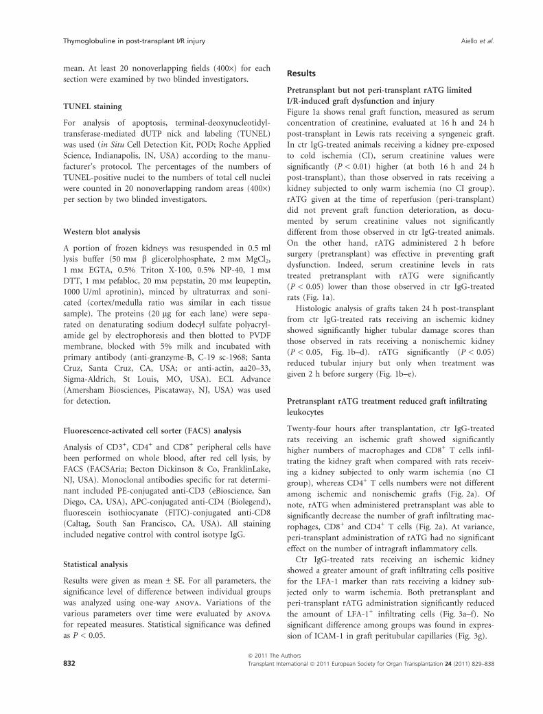
Pretransplant but not peri-transplant rATG limited
I/R-induced graft dysfunction and injury
Figure 1a shows renal graft function, measured as serum
concentration of creatinine, evaluated at 16 h and 24 h
post-transplant in Lewis rats receiving a syngeneic graft.
In ctr IgG-treated animals receiving a kidney pre-exposed
to cold ischemia (CI), serum creatinine values were
significantly (P < 0.01) higher (at both 16 h and 24 h
post-transplant), than those observed in rats receiving a
kidney subjected to only warm ischemia (no CI group).
rATG given at the time of reperfusion (peri-transplant)
did not prevent graft function deterioration, as docu-
mented by serum creatinine values not significantly
different from those observed in ctr IgG-treated animals.
On the other hand, rATG administered 2 h before
surgery (pretransplant) was effective in preventing graft
dysfunction. Indeed, serum creatinine levels in rats
treated pretransplant with rATG were significantly
(P < 0.05) lower than those observed in ctr IgG-treated
rats (Fig. 1a).
Histologic analysis of grafts taken 24 h post-transplant
from ctr IgG-treated rats receiving an ischemic kidney
showed significantly higher tubular damage scores than
those observed in rats receiving a nonischemic kidney
(P < 0.05, Fig. 1b–d). rATG significantly (P < 0.05)
reduced tubular injury but only when treatment was
given 2 h before surgery (Fig. 1b–e).
Pretransplant rATG treatment reduced graft infiltrating
leukocytes
Twenty-four hours after transplantation, ctr IgG-treated
rats receiving an ischemic graft showed significantly
higher numbers of macrophages and CD8+ T cells infil-
trating the kidney graft when compared with rats receiv-
ing a kidney subjected to only warm ischemia (no CI
group), whereas CD4+ T cells numbers were not different
among ischemic and nonischemic grafts (Fig. 2a). Of
note, rATG when administered pretransplant was able to
significantly decrease the number of graft infiltrating mac-
rophages, CD8+ and CD4+ T cells (Fig. 2a). At variance,
peri-transplant administration of rATG had no significant
effect on the number of intragraft inflammatory cells.
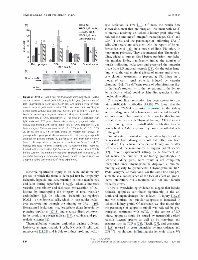
Ctr IgG-treated rats receiving an ischemic kidney
showed a greater amount of graft infiltrating cells positive
for the LFA-1 marker than rats receiving a kidney sub-
jected only to warm ischemia. Both pretransplant and
peri-transplant rATG administration significantly reduced
the amount of LFA-1+ infiltrating cells (Fig. 3a–f). No
significant difference among groups was found in expres-
sion of ICAM-1 in graft peritubular capillaries (Fig. 3g).
Thymoglobuline in post-transplant I/R injury Aiello et al.
ª 2011 The Authors
832 Transplant International ª 2011 European Society for Organ Transplantation 24 (2011) 829–838
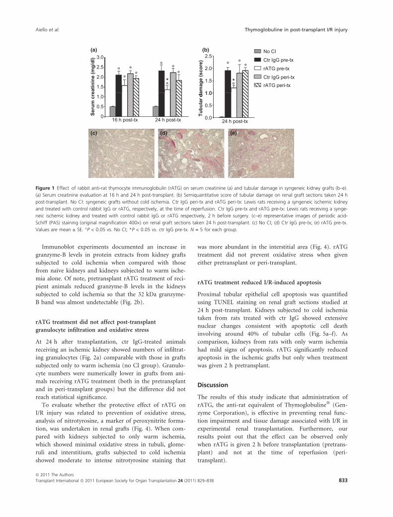
Immunoblot experiments documented an increase in
granzyme-B levels in protein extracts from kidney grafts
subjected to cold ischemia when compared with those
from naı̈ve kidneys and kidneys subjected to warm ische-
mia alone. Of note, pretransplant rATG treatment of reci-
pient animals reduced granzyme-B levels in the kidneys
subjected to cold ischemia so that the 32 kDa granzyme-
B band was almost undetectable (Fig. 2b).
rATG treatment did not affect post-transplant
granulocyte infiltration and oxidative stress
At 24 h after transplantation, ctr IgG-treated animals
receiving an ischemic kidney showed numbers of infiltrat-
ing granulocytes (Fig. 2a) comparable with those in grafts
subjected only to warm ischemia (no CI group). Granulo-
cyte numbers were numerically lower in grafts from ani-
mals receiving rATG treatment (both in the pretransplant
and in peri-transplant groups) but the difference did not
reach statistical significance.
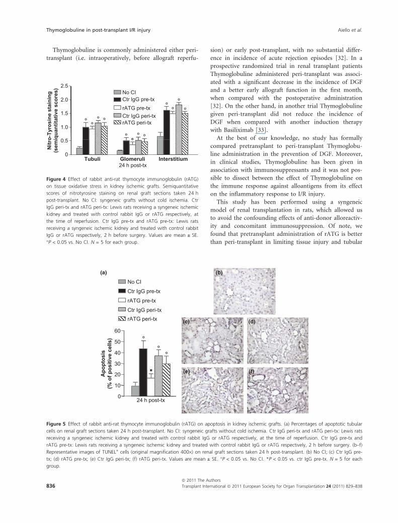
To evaluate whether the protective effect of rATG on
I/R injury was related to prevention of oxidative stress,
analysis of nitrotyrosine, a marker of peroxynitrite forma-
tion, was undertaken in renal grafts (Fig. 4). When com-
pared with kidneys subjected to only warm ischemia,
which showed minimal oxidative stress in tubuli, glome-
ruli and interstitium, grafts subjected to cold ischemia
showed moderate to intense nitrotyrosine staining that
was more abundant in the interstitial area (Fig. 4). rATG
treatment did not prevent oxidative stress when given
either pretransplant or peri-transplant.
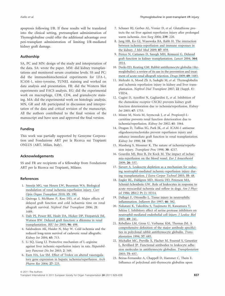
rATG treatment reduced I/R-induced apoptosis
Proximal tubular epithelial cell apoptosis was quantified
using TUNEL staining on renal graft sections studied at
24 h post-transplant. Kidneys subjected to cold ischemia
taken from rats treated with ctr IgG showed extensive
nuclear changes consistent with apoptotic cell death
involving around 40% of tubular cells (Fig. 5a–f). As
comparison, kidneys from rats with only warm ischemia
had mild signs of apoptosis. rATG significantly reduced
apoptosis in the ischemic grafts but only when treatment
was given 2 h pretransplant.
Discussion
The results of this study indicate that administration of
rATG, the anti-rat equivalent of Thymoglobuline� (Gen-
zyme Corporation), is effective in preventing renal func-
tion impairment and tissue damage associated with I/R in
experimental renal transplantation. Furthermore, our
results point out that the effect can be observed only
when rATG is given 2 h before transplantation (pretrans-
plant) and not at the time of reperfusion (peri-
transplant).
No CI
1.5
2.0
2.5
3.0
Seru
m c
reat
inin
e (m
g/dl
)°
°°*
Ctr IgG pre-tx
rATG pre-tx
Ctr IgG peri-txrATG peri-tx
1 0
1.5
2.0
2.5
dam
age
(sco
re)
°°°
°°
°
°
*
°°
0
0.5
1.0
16 h post-tx 24 h post-tx 0.0
0.5
1.0
Tubu
lard
24 h post-tx
(a) (b)
(c) (d) (e)
Figure 1 Effect of rabbit anti-rat thymocyte immunoglobulin (rATG) on serum creatinine (a) and tubular damage in syngeneic kidney grafts (b–e).
(a) Serum creatinine evaluation at 16 h and 24 h post-transplant. (b) Semiquantitative score of tubular damage on renal graft sections taken 24 h
post-transplant. No CI: syngeneic grafts without cold ischemia. Ctr IgG peri-tx and rATG peri-tx: Lewis rats receiving a syngeneic ischemic kidney
and treated with control rabbit IgG or rATG, respectively, at the time of reperfusion. Ctr IgG pre-tx and rATG pre-tx: Lewis rats receiving a synge-
neic ischemic kidney and treated with control rabbit IgG or rATG respectively, 2 h before surgery. (c–e) representative images of periodic acid-
Schiff (PAS) staining (original magnification 400·) on renal graft sections taken 24 h post-transplant. (c) No CI; (d) Ctr IgG pre-tx; (e) rATG pre-tx.
Values are mean ± SE. �P < 0.05 vs. No CI; *P < 0.05 vs. ctr IgG pre-tx. N = 5 for each group.
Aiello et al. Thymoglobuline in post-transplant I/R injury
ª 2011 The Authors
Transplant International ª 2011 European Society for Organ Transplantation 24 (2011) 829–838 833
Ischemia/reperfusion injury is an acute inflammatory
process in which the tissue is damaged first by temporary
ischemia, hypoxia and accumulation of toxic metabolites
and later during reperfusion [15,16]. Ischemia increases
vascular permeability and facilitates extravasation of leu-
kocytes by interrupting the integrity of renal vascular
endothelium [8]. In addition, ischemia up-regulates
ICAM-1 on endothelial cells, which in turn guides leuko-
cyte extravasation through the binding to LFA-1 [16].
Extravasated leukocytes may exacerbate tissue hypoxia by
plugging capillaries [17,18] and mediate direct cytotoxic-
ity by producing oxygen radicals [19], cytokines and pro-
teolytic enzymes [20].
Thymoglobuline contains antibodies against different
leukocyte antigens (mainly T cells, NK cells, B cells, and
monocytes) [21,22] and is able to induce profound leuko-
cyte depletion in vivo [10]. Of note, the results here
shown document that pretransplant treatment with rATG
of animals receiving an ischemic kidney graft effectively
reduced the amount of intragraft macrophages, CD8+ and
CD4+ T cells and the percentage of infiltrating LFA-1+
cells. Our results are consistent with the report of Beiras-
Fernandez et al. [23] in a model of limb I/R injury in
nonhuman primates. They documented that Thymoglob-
uline, added to human blood before perfusion into ische-
mic monkey limbs, significantly limited the number of
muscle infiltrating leukocytes and preserved the muscular
tissue from I/R-induced necrosis [23]. On the other hand,
Jang et al. showed minimal effects of mouse anti-thymo-
cyte globulin treatment in preventing I/R injury in a
model of warm renal ischemia induced by vascular
clamping [24]. The different route of administration (i.p.
in the Jang’s studies, i.v. in the present and in the Beiras-
Fernandez’s studies) could explain discrepancies in thy-
moglobuline efficacy.
Thymoglobuline preparation has been shown to con-
tain anti-ICAM-1 antibodies [18,19]. We found that the
increase in ICAM-1 expression occurring in the kidney
grafts undergoing cold ischemia was not affected by rATG
administration. One possible explanation for this finding
is that, at variance with Thymoglobuline, rATG does not
contain enough titer of anti-ICAM-1 antibodies to effi-
ciently bind ICAM-1 expressed by donor endothelial cells
in the graft.
Granulocytes, recruited in huge numbers by chemokin-
es released from damaged endothelial cells, have been
considered key cellular mediators of kidney injury after
ischemia and the main source of oxygen radical species
[12]. In our experimental setting, rATG treatment did
not reduce the number of infiltrating granulocytes in
ischemic kidney grafts. Such result is not completely
unexpected since Thymoglobuline displayed a minimal
binding capacity to granulocytes (Thymoglobuline BLA,
1998; Genzyme Corporation). On the same line and pre-
sumably as a consequence of the lack of effect on granu-
locyte infiltration, rATG treatment did not limit tubular
oxidative stress.
There is overwhelming evidence to suggest that besides
necrosis, apoptosis contributes significantly to the cell
death and organ damage that follows I/R injury [16,25]
and we confirm that tubular apoptosis is increased in
ischemic kidney grafts. Of relevance, we also found that
the percentage of apoptotic tubuli was reduced by pre-
transplant treatment with rATG. In the context of I/R
injury, apoptosis could be caused by neutrophil-derived
reactive oxygen species as well as by cytokines and
enzymes such as TNF-a [26], TRAIL [27], and granzyme-
B [28] released in great quantities by macrophages and
CD8+ T lymphocytes infiltrating the ischemic tissue. We
100
120
40
60
80
Cel
ls/H
PF
0
20
Macrophages CD8 GranulocytesCD424 h post-tx
No CICtr IgG pre-txrATG pre-txCtr IgG peri-txrATGiperi-tx
°
° ° °°
°
**
*
Granzyme-B
1 2 3 4 5 6
Actin
(a)
(b)
Figure 2 Effect of rabbit anti-rat thymocyte immunoglobulin (rATG)
on the number of renal graft infiltrating leukocytes. (a) Numbers of
ED1+ macrophages, CD4+ cells, CD8+ cells and granulocytes for each
animal on renal graft sections taken 24 h post-transplant. No CI: syn-
geneic grafts without cold ischemia. Ctr IgG peri-tx and rATG peri-tx:
Lewis rats receiving a syngeneic ischemic kidney and treated with con-
trol rabbit IgG or rATG respectively, at the time of reperfusion. Ctr
IgG pre-tx and rATG pre-tx: Lewis rats receiving a syngeneic ischemic
kidney and treated with control rabbit IgG or rATG respectively, 2 h
before surgery. Values are mean ± SE. �P < 0.05 vs. No CI; *P < 0.05
vs. ctr IgG pre-tx. N = 5 for each group. (b) Western blot analysis of
granzyme-B. Upper panel shows Western blot with anti-granzyme-B
antibody on protein extracts (20 lg for each lane) from naı̈ve kidney
(lane 1), kidneys subjected to warm ischemia alone (lanes 2 and 3),
kidneys subjected to cold ischemia and transplanted into recipients
treated with control rabbit IgG (lane 4) or rATG (lanes 5 and 6) 2 h
before surgery. The membrane has been stripped and re-probed with
anti-actin antibody as housekeeping (lower panel). In figure is shown
a representative Western blot of three experiments.
Thymoglobuline in post-transplant I/R injury Aiello et al.
ª 2011 The Authors
834 Transplant International ª 2011 European Society for Organ Transplantation 24 (2011) 829–838
focused on granzyme-B based on recent findings that this
molecule has been involved in mediating postischemic
neuronal death in a rat model of CD8+ dependent focal
cerebral ischemia [29]. Our results documenting that
granzyme-B levels, increased in ischemic renal tissue, were
completely dampened by rATG treatment, would indicate
that pretransplant rATG administration, by reducing the
numbers of infiltrating CD8+ T cells, limited the intra-
graft release of granzyme-B resulting in less tubular apop-
tosis.
In clinical transplant setting, Thymoglobuline is given
as induction therapy to effectively prevent acute cellular-
mediated rejection [10]. Evidence is also emerging that
Thymoglobuline could be of benefit to limit I/R injury
and the consequent DGF. In two retrospective studies
comparing kidney transplant patients receiving or not
Thymoglobuline induction therapy, Thymoglobuline-
treated patients either did not experience DGF [30] or
had a decrease in the duration of anuria and faster recov-
ery of DGF [31].
(b)
(c) (d)
(e) (f)
No CI
Ctr IgG pre-tx
rATG pre-tx
Ctr IgG peri-tx
rATG peri-tx
(a)
24 h post-tx
25
30
35°
LFA
-1 (c
ells
/HPF
)
0
5
10
15
20 °
*§
2.0No CI
Ctr IgG pre-tx
1.0
0.5
1.5
ICA
M-1
sta
inin
g(s
emiq
uant
itativ
e sc
ores
)
rATG pre-tx
Ctr IgG peri-tx
rATG peri-tx
0Peritubular capillaries
24 h post-tx
(g)
Figure 3 Effect of rabbit anti-rat thymocyte immunoglobulin (rATG) on LFA-1 (a–f) and Inter-Cellular Adhesion Molecule 1 (ICAM-1) (g) expres-
sion in kidney ischemic grafts. (a) Numbers of LFA-1+ cells per high power field (HPF) in renal graft sections taken 24 h post-transplant. (g) Semi-
quantitative score of ICAM-1 staining in peritubular capillaries of renal graft sections taken 24 h post-transplant. No CI: syngeneic grafts without
cold ischemia. Ctr IgG peri-tx and rATG peri-tx: Lewis rats receiving a syngeneic ischemic kidney and treated with control rabbit IgG or rATG
respectively, at the time of reperfusion. Ctr IgG pre-tx and rATG pre-tx: Lewis rats receiving a syngeneic ischemic kidney and treated with control
rabbit IgG or rATG respectively, 2 h before surgery. (b–f) representative images of LFA-1 immunofluorescence staining (original magnification
400·) in renal graft sections taken 24 h post-transplant. (b) No CI; (c) Ctr IgG pre-tx; (d) rATG pre-tx; (e) Ctr IgG peri-tx; (f) rATG peri-tx. Values
are mean ± SE. �P < 0.05 vs. No CI; *P < 0.05 vs. ctr IgG pre-tx; §P < 0.05 vs. rATG peri-tx. N = 5 for each group.
Aiello et al. Thymoglobuline in post-transplant I/R injury
ª 2011 The Authors
Transplant International ª 2011 European Society for Organ Transplantation 24 (2011) 829–838 835
Thymoglobuline is commonly administered either peri-
transplant (i.e. intraoperatively, before allograft reperfu-
sion) or early post-transplant, with no substantial differ-
ence in incidence of acute rejection episodes [32]. In a
prospective randomized trial in renal transplant patients
Thymoglobuline administered peri-transplant was associ-
ated with a significant decrease in the incidence of DGF
and a better early allograft function in the first month,
when compared with the postoperative administration
[32]. On the other hand, in another trial Thymoglobuline
given peri-transplant did not reduce the incidence of
DGF when compared with another induction therapy
with Basiliximab [33].
At the best of our knowledge, no study has formally
compared pretransplant to peri-transplant Thymoglobu-
line administration in the prevention of DGF. Moreover,
in clinical studies, Thymoglobuline has been given in
association with immunosuppressants and it was not pos-
sible to dissect between the effect of Thymoglobuline on
the immune response against alloantigens from its effect
on the inflammatory response to I/R injury.
This study has been performed using a syngeneic
model of renal transplantation in rats, which allowed us
to avoid the confounding effects of anti-donor alloreactiv-
ity and concomitant immunosuppression. Of note, we
found that pretransplant administration of rATG is better
than peri-transplant in limiting tissue injury and tubular
2.5No CICtr lgG pre-tx
0 5
1.0
1.5
2.0 °°rATG pre-txCtr IgG peri-txrATG peri-tx
0
.
Tubuli Glomeruli Interstitium
Nitr
o-Ty
rosi
ne s
tain
ing
(sem
iqua
ntita
tive
scor
es)
24 h post-tx
° °° °
° ° °
°°
°°
Figure 4 Effect of rabbit anti-rat thymocyte immunoglobulin (rATG)
on tissue oxidative stress in kidney ischemic grafts. Semiquantitative
scores of nitrotyrosine staining on renal graft sections taken 24 h
post-transplant. No CI: syngeneic grafts without cold ischemia. Ctr
IgG peri-tx and rATG peri-tx: Lewis rats receiving a syngeneic ischemic
kidney and treated with control rabbit IgG or rATG respectively, at
the time of reperfusion. Ctr IgG pre-tx and rATG pre-tx: Lewis rats
receiving a syngeneic ischemic kidney and treated with control rabbit
IgG or rATG respectively, 2 h before surgery. Values are mean ± SE.
�P < 0.05 vs. No CI. N = 5 for each group.
No CI
Ctr IgG pre-tx
rATG pre-tx
Ctr IgG peri-txrATG peri-tx
30
40
50
60
Apo
ptos
is(%
of p
ositi
ve c
ells
)
p
0
10
20
24 h post-tx
°°
°
**
(a) (b)
(c) (d)
(e) (f)
Figure 5 Effect of rabbit anti-rat thymocyte immunoglobulin (rATG) on apoptosis in kidney ischemic grafts. (a) Percentages of apoptotic tubular
cells on renal graft sections taken 24 h post-transplant. No CI: syngeneic grafts without cold ischemia. Ctr IgG peri-tx and rATG peri-tx: Lewis rats
receiving a syngeneic ischemic kidney and treated with control rabbit IgG or rATG respectively, at the time of reperfusion. Ctr IgG pre-tx and
rATG pre-tx: Lewis rats receiving a syngeneic ischemic kidney and treated with control rabbit IgG or rATG respectively, 2 h before surgery. (b–f)
Representative images of TUNEL+ cells (original magnification 400·) on renal graft sections taken 24 h post-transplant. (b) No CI; (c) Ctr IgG pre-
tx; (d) rATG pre-tx; (e) Ctr IgG peri-tx; (f) rATG peri-tx. Values are mean ± SE. �P < 0.05 vs. No CI. *P < 0.05 vs. ctr IgG pre-tx. N = 5 for each
group.
Thymoglobuline in post-transplant I/R injury Aiello et al.
ª 2011 The Authors
836 Transplant International ª 2011 European Society for Organ Transplantation 24 (2011) 829–838
apoptosis following I/R. If these results will be translated
into the clinical setting, pretransplant administration of
Thymoglobuline could offer the additional advantage over
peri-transplant administration of limiting I/R-mediated
kidney graft damage.
Authorship
SA, PC and MN: design of the study and interpretation of
the data. SA: wrote the paper. MM: did kidney transplan-
tations and monitored serum creatinine levels. SS and PC:
did the immunohistochemical experiments for LFA-1,
ICAM-1, nitro-tyrosine, TUNEL staining and worked on
data analysis and presentation. FR: did the Western blot
experiments and FACS analysis. EG: did the experimental
work on macrophage, CD8, CD4, and granulocyte stain-
ing. MA: did the experimental work on histologic analysis.
MN, GR and AB: participated in discussion and interpre-
tation of the data and critical revision of the manuscript.
All the authors contributed to the final version of the
manuscript and have seen and approved the final version.
Funding
This work was partially supported by Genzyme Corpora-
tion and Fondazione ART per la Ricerca sui Trapianti
ONLUS (ART, Milan, Italy).
Acknowledgements
SS and FR are recipients of a fellowship from Fondazione
ART per la Ricerca sui Trapianti, Milano.
References
1. Snoeijs MG, van Heurn LW, Buurman WA. Biological
modulation of renal ischemia-reperfusion injury. Curr
Opin Organ Transplant 2010; 15: 190.
2. Quiroga I, McShane P, Koo DD, et al. Major effects of
delayed graft function and cold ischaemia time on renal
allograft survival. Nephrol Dial Transplant 2006; 21:
1689.
3. Daly PJ, Power RE, Healy DA, Hickey DP, Fitzpatrick JM,
Watson RW. Delayed graft function: a dilemma in renal
transplantation. BJU Int 2005; 96: 498.
4. Salahudeen AK, Haider N, May W. Cold ischemia and the
reduced long-term survival of cadaveric renal allografts.
Kidney Int 2004; 65: 713.
5. Li SQ, Liang LJ. Protective mechanism of L-arginine
against liver ischemic-reperfusion injury in rats. Hepatobil-
iary Pancreat Dis Int 2003; 2: 549.
6. Eum HA, Lee SM. Effect of Trolox on altered vasoregula-
tory gene expression in hepatic ischemia/reperfusion. Arch
Pharm Res 2004; 27: 225.
7. Schauer RJ, Gerbes AL, Vonier D, et al. Glutathione pro-
tects the rat liver against reperfusion injury after prolonged
warm ischemia. Ann Surg 2004; 239: 220.
8. Jang HR, Ko GJ, Wasowska BA, Rabb H. The interaction
between ischemia-reperfusion and immune responses in
the kidney. J Mol Med 2009; 87: 859.
9. Perico N, Cattaneo D, Sayegh MH, Remuzzi G. Delayed
graft function in kidney transplantation. Lancet 2004; 364:
1814.
10. Deeks ED, Keating GM. Rabbit antithymocyte globulin (thy-
moglobulin): a review of its use in the prevention and treat-
ment of acute renal allograft rejection. Drugs 2009; 69: 1483.
11. Mehrabi A, Mood Zh A, Sadeghi M, et al. Thymoglobulin
and ischemia reperfusion injury in kidney and liver trans-
plantation. Nephrol Dial Transplant 2007; 22 (Suppl. 8):
VIII54.
12. Cugini D, Azzollini N, Gagliardini E, et al. Inhibition of
the chemokine receptor CXCR2 prevents kidney graft
function deterioration due to ischemia/reperfusion. Kidney
Int 2005; 67: 1753.
13. Mister M, Noris M, Szymczuk J, et al. Propionyl-L-
carnitine prevents renal function deterioration due to
ischemia/reperfusion. Kidney Int 2002; 61: 1064.
14. Dragun D, Tullius SG, Park JK, et al. ICAM-1 antisense
oligodesoxynucleotides prevent reperfusion injury and
enhance immediate graft function in renal transplantation.
Kidney Int 1998; 54: 590.
15. Massberg S, Messmer K. The nature of ischemia/reperfu-
sion injury. Transplant Proc 1998; 30: 4217.
16. Gourdin MJ, Bree B, De Kock M. The impact of ischae-
mia-reperfusion on the blood vessel. Eur J Anaesthesiol
2009; 26: 537.
17. Sievert A. Leukocyte depletion as a mechanism for reduc-
ing neutrophil-mediated ischemic-reperfusion injury dur-
ing transplantation. J Extra Corpor Technol 2003; 35: 48.
18. Engler RL, Dahlgren MD, Morris DD, Peterson MA,
Schmid-Schonbein GW. Role of leukocytes in response to
acute myocardial ischemia and reflow in dogs. Am J Phys-
iol 1986; 251(2 Pt 2): H314.
19. Dallegri F, Ottonello L. Tissue injury in neutrophilic
inflammation. Inflamm Res 1997; 46: 382.
20. Nakatani K, Takeshita S, Tsujimoto H, Kawamura Y,
Sekine I. Inhibitory effect of serine protease inhibitors on
neutrophil-mediated endothelial cell injury. J Leukoc Biol
2001; 69: 241.
21. Rebellato LM, Gross U, Verbanac KM, Thomas JM. A
comprehensive definition of the major antibody specifici-
ties in polyclonal rabbit antithymocyte globulin. Trans-
plantation 1994; 57: 685.
22. Michallet MC, Preville X, Flacher M, Fournel S, Genestier
L, Revillard JP. Functional antibodies to leukocyte adhe-
sion molecules in antithymocyte globulins. Transplantation
2003; 75: 657.
23. Beiras-Fernandez A, Chappell D, Hammer C, Thein E.
Influence of polyclonal anti-thymocyte globulins upon
Aiello et al. Thymoglobuline in post-transplant I/R injury
ª 2011 The Authors
Transplant International ª 2011 European Society for Organ Transplantation 24 (2011) 829–838 837
ischemia-reperfusion injury in a non-human primate
model. Transpl Immunol 2006; 15: 273.
24. Jang HR, Gandolfo MT, Ko GJ, Racusen L, Rabb H. The
effect of murine anti-thymocyte globulin on experimental
kidney warm ischemia-reperfusion injury in mice. Transpl
Immunol 2009; 22: 44.
25. Lopez-Neblina F, Toledo AH, Toledo-Pereyra LH. Molecu-
lar biology of apoptosis in ischemia and reperfusion.
J Invest Surg 2005; 18: 335.
26. Bertazza L, Mocellin S. Tumor necrosis factor (TNF)
biology and cell death. Front Biosci 2008; 13: 2736.
27. Mellier G, Huang S, Shenoy K, Pervaiz S. TRAILing death
in cancer. Mol Aspects Med 2010; 31: 93.
28. Bolitho P, Voskoboinik I, Trapani JA, Smyth MJ. Apopto-
sis induced by the lymphocyte effector molecule perforin.
Curr Opin Immunol 2007; 19: 339.
29. Chaitanya GV, Schwaninger M, Alexander JS, Prakash
Babu P. Granzyme-B is involved in mediating
post-ischemic neuronal death during focal cerebral
ischemia in rat model. Neuroscience 2010; 165: 1203.
30. Hardinger KL, Schnitzler MA, Koch MJ, et al. Thymoglob-
ulin induction is safe and effective in live-donor renal
transplantation: a single center experience. Transplantation
2006; 81: 1285.
31. Cravedi P, Codreanu I, Satta A, et al. Cyclosporine pro-
longs delayed graft function in kidney transplantation: are
rabbit anti-human thymocyte globulins the answer?
Nephron Clin Pract 2005; 101: c65.
32. Goggins WC, Pascual MA, Powelson JA, et al. A
prospective, randomized, clinical trial of intraoperative
versus postoperative Thymoglobulin in adult cadaveric
renal transplant recipients. Transplantation 2003; 76:
798.
33. Brennan DC, Daller JA, Lake KD, Cibrik D, Del Castillo
D. Rabbit antithymocyte globulin versus basiliximab in
renal transplantation. N Engl J Med 2006; 355: 1967.
Thymoglobuline in post-transplant I/R injury Aiello et al.
ª 2011 The Authors
838 Transplant International ª 2011 European Society for Organ Transplantation 24 (2011) 829–838
Related Documents