pharmacy Review Quinolone Allergy Edoabasi U. McGee 1 , Essie Samuel 1 , Bernadett Boronea 1 , Nakoasha Dillard 1 , Madison N. Milby 2 and Susan J. Lewis 3,4, * 1 School of Pharmacy, Philadelphia College of Osteopathic Medicine, Suwanee, GA 30024, USA 2 College of Pharmacy, University of Findlay, Findlay, OH 45840, USA 3 Department of Pharmacy Practice, College of Pharmacy, University of Findlay, Findlay, OH 45840, USA 4 Mercy Health—St. Anne Hospital, Toledo, OH 43623, USA * Correspondence: slewis@findlay.edu; Tel.: +1-419-434-5948 Received: 28 June 2019; Accepted: 17 July 2019; Published: 19 July 2019 Abstract: Quinolones are the second most common antibiotic class associated with drug-induced allergic reactions, but data on quinolone allergy are scarce. This review article discusses the available evidence on quinolone allergy, including prevalence, risk factors, diagnosis, clinical manifestations, cross-reactivity, and management of allergic reactions. Although the incidence of quinolone allergy is still lower than beta-lactams, it has been increasingly reported in recent decades, most likely from its expanded use and the introduction of moxifloxacin. Thorough patient history remains essential in the evaluation of quinolone allergy. Many diagnostic tools have been investigated, but skin tests can yield false-positive results and in vitro tests have not been validated. The drug provocation test is considered the test of choice to confirm a quinolone allergy but is not without risk. Evidence regarding cross-reactivity among the quinolones is limited and conflicting. Quinolone allergy can be manifested either as an immediate or delayed reaction, but is not uniform across the class, with moxifloxacin posing the highest risk of anaphylaxis. Quinolone should be discontinued when an allergic reaction occurs and avoided in future scenarios, but desensitization may be warranted if no alternatives are available. Keywords: quinolones; fluoroquinolones; allergy; hypersensitivity; cross-reactivity 1. Introduction Quinolones, highly effective synthetic antibiotics, are one of the most commonly prescribed antibiotic classes in outpatient [1] and in acute care hospital settings [2]. They inhibit DNA gyrase in Gram-negative bacteria and topoisomerase IV in Gram-positive bacteria, promoting the DNA cleavage and rapid killing of susceptible bacteria [3]. With their broad-spectrum activity against both Gram-positive and Gram-negative aerobic and anaerobic bacteria, excellent tissue and intracellular penetration, high bioavailability, and generally good oral tolerability, quinolones have a broad array of indications including treatment of urinary tract infections (UTIs), sexually transmitted diseases, gastrointestinal and abdominal infections, respiratory tract infections, bone and joint infections, and skin and soft tissue infections in adults. Although considered well-tolerated in general, quinolones can induce allergic reactions. In fact, they are the second most common antibiotics associated with allergic reactions, following beta-lactams [4–6]. Currently, most of the data regarding antibiotic allergy have been published for patients with a beta-lactam allergy and the paucity of data exist regarding quinolone allergy. 2. Structures Quinolones are classified into four generations based on chemical structure as well as antibacterial activity and pharmacokinetic properties [6]. The basic structure of quinolones consists of a bicyclic Pharmacy 2019, 7, 97; doi:10.3390/pharmacy7030097 www.mdpi.com/journal/pharmacy

Welcome message from author
This document is posted to help you gain knowledge. Please leave a comment to let me know what you think about it! Share it to your friends and learn new things together.
Transcript
pharmacy
Review
Quinolone Allergy
Edoabasi U. McGee 1, Essie Samuel 1, Bernadett Boronea 1, Nakoasha Dillard 1,Madison N. Milby 2 and Susan J. Lewis 3,4,*
1 School of Pharmacy, Philadelphia College of Osteopathic Medicine, Suwanee, GA 30024, USA2 College of Pharmacy, University of Findlay, Findlay, OH 45840, USA3 Department of Pharmacy Practice, College of Pharmacy, University of Findlay, Findlay, OH 45840, USA4 Mercy Health—St. Anne Hospital, Toledo, OH 43623, USA* Correspondence: [email protected]; Tel.: +1-419-434-5948
Received: 28 June 2019; Accepted: 17 July 2019; Published: 19 July 2019�����������������
Abstract: Quinolones are the second most common antibiotic class associated with drug-inducedallergic reactions, but data on quinolone allergy are scarce. This review article discusses the availableevidence on quinolone allergy, including prevalence, risk factors, diagnosis, clinical manifestations,cross-reactivity, and management of allergic reactions. Although the incidence of quinolone allergy isstill lower than beta-lactams, it has been increasingly reported in recent decades, most likely fromits expanded use and the introduction of moxifloxacin. Thorough patient history remains essentialin the evaluation of quinolone allergy. Many diagnostic tools have been investigated, but skin testscan yield false-positive results and in vitro tests have not been validated. The drug provocation testis considered the test of choice to confirm a quinolone allergy but is not without risk. Evidenceregarding cross-reactivity among the quinolones is limited and conflicting. Quinolone allergy canbe manifested either as an immediate or delayed reaction, but is not uniform across the class, withmoxifloxacin posing the highest risk of anaphylaxis. Quinolone should be discontinued when anallergic reaction occurs and avoided in future scenarios, but desensitization may be warranted if noalternatives are available.
Keywords: quinolones; fluoroquinolones; allergy; hypersensitivity; cross-reactivity
1. Introduction
Quinolones, highly effective synthetic antibiotics, are one of the most commonly prescribedantibiotic classes in outpatient [1] and in acute care hospital settings [2]. They inhibit DNA gyrasein Gram-negative bacteria and topoisomerase IV in Gram-positive bacteria, promoting the DNAcleavage and rapid killing of susceptible bacteria [3]. With their broad-spectrum activity against bothGram-positive and Gram-negative aerobic and anaerobic bacteria, excellent tissue and intracellularpenetration, high bioavailability, and generally good oral tolerability, quinolones have a broad arrayof indications including treatment of urinary tract infections (UTIs), sexually transmitted diseases,gastrointestinal and abdominal infections, respiratory tract infections, bone and joint infections, andskin and soft tissue infections in adults. Although considered well-tolerated in general, quinolonescan induce allergic reactions. In fact, they are the second most common antibiotics associated withallergic reactions, following beta-lactams [4–6]. Currently, most of the data regarding antibiotic allergyhave been published for patients with a beta-lactam allergy and the paucity of data exist regardingquinolone allergy.
2. Structures
Quinolones are classified into four generations based on chemical structure as well as antibacterialactivity and pharmacokinetic properties [6]. The basic structure of quinolones consists of a bicyclic
Pharmacy 2019, 7, 97; doi:10.3390/pharmacy7030097 www.mdpi.com/journal/pharmacy
Pharmacy 2019, 7, 97 2 of 12
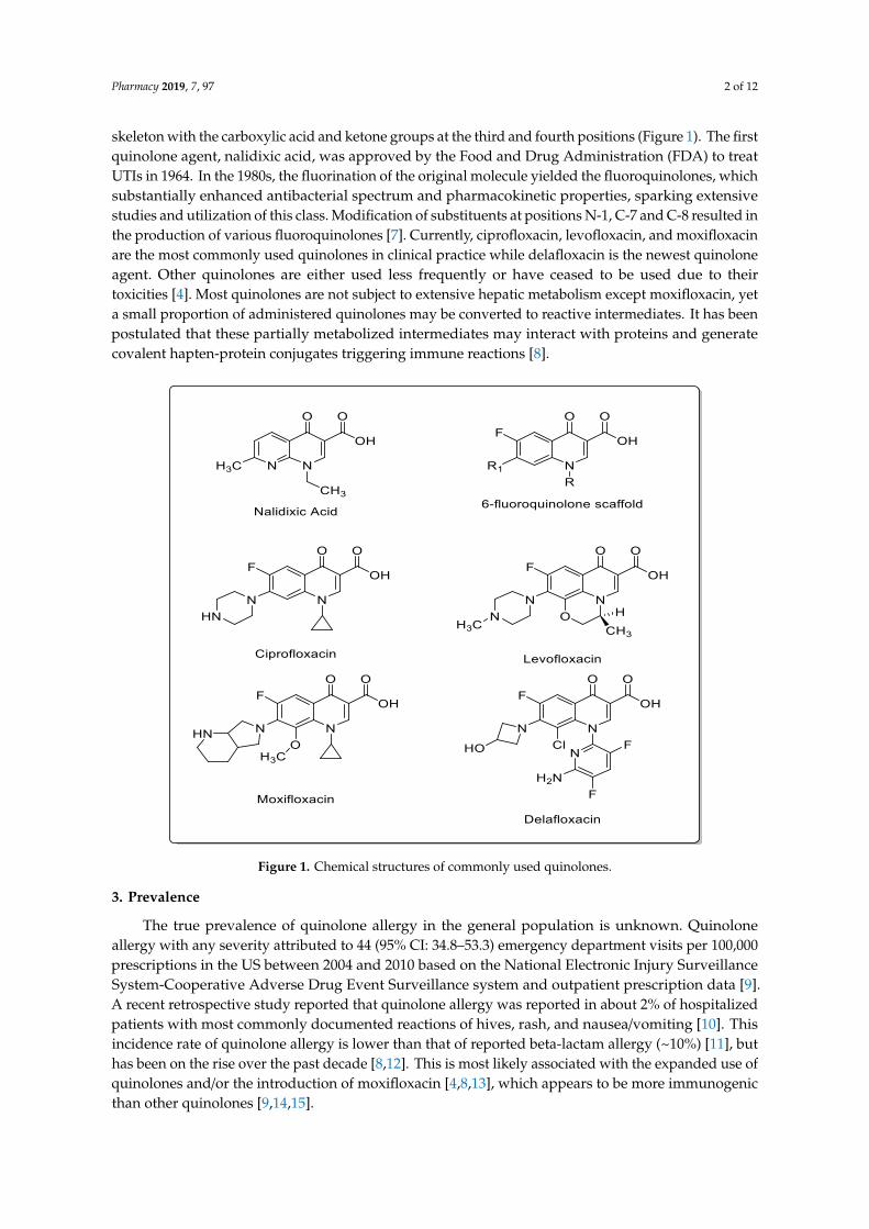
skeleton with the carboxylic acid and ketone groups at the third and fourth positions (Figure 1). The firstquinolone agent, nalidixic acid, was approved by the Food and Drug Administration (FDA) to treatUTIs in 1964. In the 1980s, the fluorination of the original molecule yielded the fluoroquinolones, whichsubstantially enhanced antibacterial spectrum and pharmacokinetic properties, sparking extensivestudies and utilization of this class. Modification of substituents at positions N-1, C-7 and C-8 resulted inthe production of various fluoroquinolones [7]. Currently, ciprofloxacin, levofloxacin, and moxifloxacinare the most commonly used quinolones in clinical practice while delafloxacin is the newest quinoloneagent. Other quinolones are either used less frequently or have ceased to be used due to theirtoxicities [4]. Most quinolones are not subject to extensive hepatic metabolism except moxifloxacin, yeta small proportion of administered quinolones may be converted to reactive intermediates. It has beenpostulated that these partially metabolized intermediates may interact with proteins and generatecovalent hapten-protein conjugates triggering immune reactions [8].
Pharmacy 2019, 7, x FOR PEER REVIEW 2 of 12
2
Quinolones are classified into four generations based on chemical structure as well as
antibacterial activity and pharmacokinetic properties [6]. The basic structure of quinolones consists
of a bicyclic skeleton with the carboxylic acid and ketone groups at the third and fourth positions
(Figure 1). The first quinolone agent, nalidixic acid, was approved by the Food and Drug
Administration (FDA) to treat UTIs in 1964. In the 1980s, the fluorination of the original molecule
yielded the fluoroquinolones, which substantially enhanced antibacterial spectrum and
pharmacokinetic properties, sparking extensive studies and utilization of this class. Modification of
substituents at positions N-1, C-7 and C-8 resulted in the production of various fluoroquinolones [7].
Currently, ciprofloxacin, levofloxacin, and moxifloxacin are the most commonly used quinolones in
clinical practice while delafloxacin is the newest quinolone agent. Other quinolones are either used
less frequently or have ceased to be used due to their toxicities [4]. Most quinolones are not subject to
extensive hepatic metabolism except moxifloxacin, yet a small proportion of administered
quinolones may be converted to reactive intermediates. It has been postulated that these partially
metabolized intermediates may interact with proteins and generate covalent hapten-protein
conjugates triggering immune reactions [8].
Figure 1. Chemical structures of commonly used quinolones.
3. Prevalence
The true prevalence of quinolone allergy in the general population is unknown. Quinolone
allergy with any severity attributed to 44 (95% CI: 34.8-53.3) emergency department visits per
100,000 prescriptions in the US between 2004 and 2010 based on the National Electronic Injury
Surveillance System-Cooperative Adverse Drug Event Surveillance system and outpatient
prescription data [9]. A recent retrospective study reported that quinolone allergy was reported in
about 2% of hospitalized patients with most commonly documented reactions of hives, rash, and
nausea/vomiting [10]. This incidence rate of quinolone allergy is lower than that of reported
beta-lactam allergy (~10%) [11], but has been on the rise over the past decade [8,12]. This is most
likely associated with the expanded use of quinolones and/or the introduction of moxifloxacin
[4,8,13], which appears to be more immunogenic than other quinolones [9,14,15].
Figure 1. Chemical structures of commonly used quinolones.
3. Prevalence
The true prevalence of quinolone allergy in the general population is unknown. Quinoloneallergy with any severity attributed to 44 (95% CI: 34.8–53.3) emergency department visits per 100,000prescriptions in the US between 2004 and 2010 based on the National Electronic Injury SurveillanceSystem-Cooperative Adverse Drug Event Surveillance system and outpatient prescription data [9].A recent retrospective study reported that quinolone allergy was reported in about 2% of hospitalizedpatients with most commonly documented reactions of hives, rash, and nausea/vomiting [10]. Thisincidence rate of quinolone allergy is lower than that of reported beta-lactam allergy (~10%) [11], buthas been on the rise over the past decade [8,12]. This is most likely associated with the expanded use ofquinolones and/or the introduction of moxifloxacin [4,8,13], which appears to be more immunogenicthan other quinolones [9,14,15].
Pharmacy 2019, 7, 97 3 of 12
Quinolone allergy can be largely classified into two types: Ig-E mediated immediate reactions(IRs), which occur less than one hour after administration, and T-cell mediated delayed reactions (DRs),which occur more than an hour after administration. The most common quinolone allergies are IRsand ~70% of those cases are severe [8]. Anaphylaxis is an uncommon IR to quinolones and most caseswere reported in the post-marketing stage. An estimated incidence of quinolone-induced anaphylaxisis 1.8–2.3 per 10,000,000 days of treatment [16]. Quinolones were reported to be responsible for 4.5% of333 drug-induced anaphylaxis cases [17]. The risk of anaphylaxis may be different among quinolones.An in vitro laboratory evaluation and spontaneous adverse drug reaction reports in Europe has shownthat moxifloxacin was most frequently involved in anaphylaxis (52.1–63%), followed by levofloxacin(13–35.7%) and ciprofloxacin (7.1–28.9%) [12,15,18]; however, a cohort study performed in the U.S.using a large insurance claim database found no difference in the incidence of anaphylaxis acrossthe quinolone class [19], whereas the highest incidence of DRs has been observed with ciprofloxacin(33.3–34.9%), followed by levofloxacin (19.9–32.3%) and moxifloxacin (13.5–20.4%) [18,20].
4. Risk Factors
Information regarding common risk factors associated with quinolone allergies are limited.Blanca-Lopez and colleagues identified a previous history of beta-lactam allergy as a strong risk factor(OR:4.571; 95% CI: 0.987–21.171; adjusted OR: 23.654; 95% CI: 1.529–365.853) to develop a quinoloneallergy [8]. Patients with a history of drug allergy were noted to be more susceptible to allergic reactionsto a non-chemically related drug [21]. Another study displayed that 21% of patients with a history ofIgE-mediated penicillin allergy reported allergic reactions to non-beta-lactams, compared to only 1%of patients with no drug allergy history [22].
Although the underlying mechanism is unknown, a genetic predisposition, or the fact thatquinolones are likely to be selected as alternatives to treat those with a history of beta-lactam allergy,may explain these findings [23]. This is particularly of concern because beta-lactams and quinolonesare often recommended as the first-line agents for many infections. Patients who are allergic to bothclasses are subject to receive inferior antibiotic therapy resulting in less favorable patient outcomes [6].Additionally, a higher incidence of intravenous contrast allergy was noted in patients with a quinoloneallergy (6%) compared to those with a penicillin allergy (2%) [10,24].
5. Evaluation and Diagnosis
The evaluation of drug allergy typically begins with a patient history and if a true allergy is suspected,appropriate tests are performed to confirm the diagnosis. However, the pathogenic mechanism ofquinolone allergy is not clearly comprehended and no standardized or validated diagnostic tests havebeen determined, making its diagnosis challenging [25]. Thus, patient history remains an integralpart of the quinolone allergy evaluation. Patient history should include the details of the symptoms,the timing of the reaction in relation to the causative agent, the timing since the reaction, and anyrelevant ingestions, taken concurrently and/or since the reaction [26]. Additionally, the patient’s currentmedication list, allergy history, and previous exposure to the same or any quinolone agent may bereviewed. A thorough patient history can aid to identify a true allergy, the type and severity of allergicreactions, and the need for diagnostic tests. Many patients who report quinolone allergy may nothave a true allergy [26] but experience non-allergic adverse drug reactions such as gastrointestinalsymptoms, central nervous system reactions, myalgias, and tendon rupture, which may not requirefurther evaluation. The type of allergic reaction can be determined based on the time interval betweenthe administration of the causative agent and the reaction [6].
In cases where patient history is not reliable, other diagnostic tests may be necessary to assessquinolone allergy. In recent years, multiple studies have been conducted to evaluate various diagnostictests including skin tests, drug provocation tests (DPT), and in-vitro laboratory tests [24,27–38]. However,skin tests and in vitro tests have displayed low sensitivity and specificity, limiting their diagnosticutility [24,30,32,33,35,36,38].
Pharmacy 2019, 7, 97 4 of 12
5.1. Skin Tests
The value of skin testing remains a subject of debate. Both skin prick and intradermal skin testsare used for IRs. Skin prick tests are the safest and easiest but only moderately sensitive, while theintradermal skin tests are more sensitive but have a higher risk for inducing irritative and false-positivereactions. This can also lead to an anaphylactic reaction in IgE-dependent reactions [28]. Althoughsome consider skin prick and intradermal tests useful, most studies show that quinolones can inducefalse-positive results, probably because of the capacity of some quinolones to directly induce histaminerelease because of mast cell activation [15,33]. It is also important to note that late or delayed reactions canoccur with intradermal skin tests and should always be examined [28]. For prick and intradermal skintests, widely divergent non-irritant test concentrations have been recommended, and desensitizationmay be possible in selected patients [31]. For patients with DRs, patch tests were performed on theupper back on unaffected, untreated, and uncleaned skin (no prior rinsing with alcohol) [28,33]. Thispatch was removed after one day and the test was read on days two, three, and four [28].
5.2. Drug Provocation Test
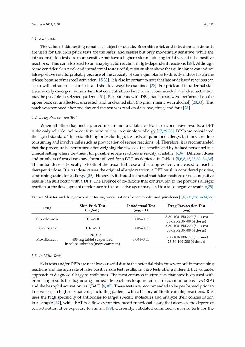
When all other diagnostic procedures are not available or lead to inconclusive results, a DPTis the only reliable tool to confirm or to rule out a quinolone allergy [27,29,33]. DPTs are consideredthe “gold standard” for establishing or excluding diagnosis of quinolone allergy, but they are timeconsuming and involve risks such as provocation of severe reactions [6]. Therefore, it is recommendedthat the procedure be performed after weighing the risks vs. the benefits and by trained personnel in aclinical setting where treatment for possible severe reactions is readily available [6,36]. Different dosesand numbers of test doses have been utilized for a DPT, as depicted in Table 1 [5,6,8,15,25,32–34,36].The initial dose is typically 1/100th of the usual full dose and is progressively increased to reach atherapeutic dose. If a test dose causes the original allergic reaction, a DPT result is considered positive,confirming quinolone allergy [29]. However, it should be noted that false-positive or false-negativeresults can still occur with a DPT. The absence of co-factors that contributed to the previous allergicreaction or the development of tolerance to the causative agent may lead to a false-negative result [6,29].
Table 1. Skin test and drug provocation testing concentrations for commonly used quinolones [5,6,8,15,25,32–34,36].
Drug Skin Prick Test(mg/mL)
Intradermal Test(mg/mL)
Drug Provocation Test(mg)
Ciprofloxacin 0.02–5.0 0.005–0.05 5-50-100-150-200 (5 doses)50-125-250-500 (4 doses)
Levofloxacin 0.025–5.0 0.005–0.05 5-50-100-150-200 (5 doses)50-125-250-500 (4 doses)
Moxifloxacin1.0–20.0 or
400 mg tablet suspendedin saline solution (more common)
0.004–0.05 5-50-100-100-150 (5 doses)25-50-100-200 (4 doses)
5.3. In Vitro Tests
Skin tests and/or DPTs are not always useful due to the potential risks for severe or life-threateningreactions and the high rate of false positive skin test results. In vitro tests offer a different, but valuable,approach to diagnose allergy to antibiotics. The most common in vitro tests that have been used withpromising results for diagnosing immediate reactions to quinolones are radioimmunoassays (RIA)and the basophil activation test (BAT) [6,38]. These tests are recommended to be performed prior toin vivo tests in high-risk patients, including patients with a history of life-threatening reactions. RIAuses the high specificity of antibodies to target specific molecules and analyze their concentrationin a sample [37], while BAT is a flow-cytometry-based functional assay that assesses the degree ofcell activation after exposure to stimuli [38]. Currently, validated commercial in vitro tests for the
Pharmacy 2019, 7, 97 5 of 12
evaluation of quinolone allergy are not available. Therefore, most studies have utilized tests that areproduced in-house [6].
RIA has been shown to have low sensitivity for quinolones, varying from 31.6% to 54.5%, and highspecificity [6]. This may be due to the different quinolones involved in each study and the severity of thereactions, as well as the total IgE levels, the time interval between the reaction and the performance ofthe test. Higher IgE levels were found in patients evaluated within a few months after the reaction, whilepatients showing negative results were generally evaluated after a longer time period [6]. These assaysdo not use enzymes and therefore reduce the risk of interference from the sample itself. The drawbacksof RIA include the use of a radiolabel and its short shelf life [37].
The utilization of BAT for drug allergy must be optimized for each drug because of the possibledifferences in the stimulation mechanism that leads to the upregulation of different activationmarkers [33]. Studies have shown the utility of BAT for evaluating quinolone-allergic reactionswith sensitivity ranging from 50% to 100%, which can be explained by different factors [38]. Intrinsicfactors such as patient medication therapy and the choice of activation markers can play a role in theresults of BAT [33]. Sensitivity can be affected by both the quinolones involved in the reaction and thequinolones used for the test [38].
6. Cross-Reactivity of Quinolones
Cross-reactivity within the quinolone drug class has been reported in the literature, but theevidence is conflicting. A report by Dávila and colleagues was one of the initial reports describingcross-reactivity among quinolones with a recommendation to avoid any quinolone among patientswho have had a reaction to one of them [39]. González and colleagues explored hypersensitivityamong quinolones using skin tests and concluded that skin tests can predict group hypersensitivity,but not specific tolerance to each drug. This study found a high degree of cross-reactivity amongfluoroquinolones, including moxifloxacin, which is chemically different from other quinolones [40].Another case report described in vitro cross-reactivity between ciprofloxacin and ofloxacin, whichcould be due to the similarity in the structures of these drugs [41]. Anovadiya and colleagues describeda cross-reactivity reaction between ciprofloxacin and levofloxacin in a seven-year-old male childwith subacute appendicitis who was treated with ciprofloxacin and immediately developed multipleerythematous papules. An allergic reaction developed again when this patient was treated withlevofloxacin. IgE binding at the seventh position of core structure of quinolones is likely to be themechanism of a hypersensitivity reaction [42].
Conversely, a review of three case reports of IRs to moxifloxacin demonstrated a lack ofcross-reactivity among moxifloxacin and ciprofloxacin. In the case reports, all three patients whodeveloped IRs to moxifloxacin were able to tolerate the oral challenge tests to ciprofloxacin as well as afull course of ciprofloxacin. This lack of cross-reactivity may be explained by moxifloxacin’s uniqueside chain [43]. Another study reviewed 12 patients who had experienced an IR (four anaphylaxisand eight urticaria/angioedema) after oral administration of quinolones. Most of the ciprofloxacin-reactive patients tolerated levofloxacin and the majority of the levofloxacin-reactive patients toleratedciprofloxacin. In addition, the patients who reacted to moxifloxacin tolerated ciprofloxacin andlevofloxacin. This study highlighted the lack of cross-reactivity among quinolones and suggested thatlevofloxacin could be a safer alternative in cases of reaction to first-, second-, or fourth-generationquinolones [34].
Several studies have reviewed cross-reactivity within the quinolone drug class, but few haveobserved cross-reactivity among quinolones and other classes of antibiotics. A case was publishedto report a rare type of IR and a late phase reaction to an anti-tubercular therapy that consisted ofethambutol and levofloxacin. In this report, intradermal skin tests were performed; an immediatedrug reaction was experienced with ethambutol within one hour, and a flare reaction occurred atthe levofloxacin injection site 15 min after the test was administered and disappeared six hours later.The findings of this case suggest that drug eruptions are not necessarily caused by a single agent, andthat multiple types of allergic reaction may occur consecutively in a single case [44].
Pharmacy 2019, 7, 97 6 of 12
7. Types and Manifestations of Quinolone Allergy
As previously mentioned, the mechanism of a quinolone allergy has been described mainly by twopathways based on the Coombs and Gell classification: IgE-mediated IR, also known as type I, andT-cell-mediated DR, also known as type IV responses [45]. The IgE-mediated pathological characteristicis mast-cell degranulation. Examples of IgE-mediated responses include urticaria, anaphylaxis, asthma,rhinitis and angioedema. In contrast, T-cell-mediated pathological characteristics may be induced bythe subclassifications of type IV responses: Type IVa (monocyte activation), Type IVb (eosinophilicinflammation), Type IVc (CD4 or CD8-mediated killing of cells), Type IVd (neutrophil activation) andType IVe (CD4 and CD8 activated T-cells). Examples of T-cell-mediated responses include contact eczema,maculopapular exanthema, bullous exanthema, Stevens–Johnson Syndrome Toxic epidermal necrolysis(SJS-TEN), fixed drug eruption (FDE), acute generalized exanthematous pustulosis (AGEP), and delayedurticaria. Other T-cell-mediated responses without a definitive Coombs and Gell classification includedrug rash with eosinophilia and systemic symptoms (DRESS) and organ-specific T-cell-mediatedresponses like hepatitis and pneumonitis [45]. In addition to the two aforementioned pathways, anovel mechanism has recently been described of a non-IgE-mediated, pseudo-allergic anaphylactoidreaction of quinolone-induced anaphylaxis [46–48]. In this novel pathway, mast cells are activated viaMAS-related G protein-coupled receptor-X2 (MRGPRX2), rather than Fcε receptors, which is linked toIgE-mediated IR [49]. Quinolones can activate the MRGPRX2, inducing non-IgE-mediated anaphylaxis,with a higher risk for patients with mastocytosis [46]. However, allergy manifestations due to quinoloneuse are not uniform across the class [24].
7.1. Moxifloxacin-Induced Allergy Manifestation
Among the quinolones, moxifloxacin has been reported to have the highest incidence of immediateallergic reactions, specifically anaphylaxis [4]. Published data of IgE mediated IR due to moxifloxacinhave mainly consisted of case reports and short series. Urticaria was more frequently reported at a rateof 31.6–85% for patients on moxifloxacin as compared to anaphylaxis reported at a rate of 13–42% [4].In a cross-sectional study of 54 patients in Istanbul, 17.5% of patients experienced an IR to moxifloxacin,of which 50% were anaphylaxis. In a Thailand retrospective review by Kulthanan and colleagues, apattern of cutaneous reaction related to the administration of various quinolones was demonstrated.Three of 151 (2%) patients developed an allergic reaction to moxifloxacin; of those, one of three had anIR presenting with urticaria [50]. Interestingly, one female patient developed both an IR and delayedanaphylaxis due to moxifloxacin when orally provoked after a skin test [24].
T-cell-mediated DR induced by moxifloxacin was described in the case report where a patientwith multiple drug allergies previously treated with levofloxacin and ciprofloxacin was prescribedmoxifloxacin for a 10-day regimen. After four days of treatment, the patient developed a rash on thelower extremities, which progressed to oral thrush, skin sloughing, and shortness of breath [51]. In theaforementioned retrospective review by Kulthanan and colleagues where three of 151 (2%) patientsdeveloped a reaction to moxifloxacin, two developed DRs, with one developing SJS-TEN and the othera maculopapular rash [50].
7.2. Ciprofloxacin-Induced Allergy Manifestation
The literature suggests that ciprofloxacin is more commonly associated with DRs. Various reactions,including SJS-TEN, eczema, FDE, erythroderma, erythema multiforme and maculopapular rashwere reported in 59 of 151 (39%) patients [50]. In addition, other cutaneous DR, such as demarcatederythematous plaques, vesicular and bullous lesions have also been reported in various case reports [52–55].However, Kulthanan and colleagues described IRs due to ciprofloxacin in 20 of 151 (13%) patients, withurticaria being the most common, as observed in 12 of 20 (60%) patients [50].
Apart from the commonly noted IRs and DRs, Sim and colleagues describe a type II IgG-mediatedallergic reaction which involved a patient who developed ciprofloxacin-induced thrombocytopenia.
Pharmacy 2019, 7, 97 7 of 12
Immediately after two administered doses of intravenous ciprofloxacin, the patient’s platelet countdecreased to 6000/µL from 220,000/µL and the platelet count continued to drop with five days ofciprofloxacin treatment. After the patient was switched to another antibiotic, the platelet countnormalized (245,000/µL) during hospitalization and after discharge [56].
Giavina-Bianchi and colleagues recently reported a case on a woman with recurrent anaphylaxiswho had five severe cases over a seven-year period. The first three episodes were induced bystinging insects while the other two were associated with ciprofloxacin. The reactions associated withciprofloxacin included flushing, laryngeal edema and syncope. The patient was subsequently diagnosedseven years later with systemic mastocytosis, a disease associated with anomalous proliferation andaccumulation of mast cells in different tissues, with a lifetime anaphylaxis risk of 50%. The authorsconcluded that the excess of mast cells and activation of MRGPRX2 receptors in patients withmastocytosis may induce anaphylaxis [46].
7.3. Levofloxacin-Induced Allergy Manifestation
The Thailand retrospective review noted that five of the 151 (3%) patients had an IR to levofloxacinwith symptoms of urticaria, and nine of the 151 (6%) had a DR with a maculopapular rash andSJS/TEN [50]. Matsumoto and colleagues described a patient case in which the patient had both an IRand DR to levofloxacin [57]. Initially the patient received intravenous levofloxacin and skin pruritusoccurred 30 min after initiation. Subsequently, levofloxacin 500 mg oral once daily was started and theskin pruritus resolved. However, three weeks later, the patient was hospitalized and found to haveerythroderma, lymphadenopathy, pulmonary lesions and eosinophilia. Levofloxacin was discontinued,and supportive treatment was given, resulting in remission of all symptoms [57].
Nunez and colleagues reported a rather uncommon allergic reaction where a patient developedan IR to levofloxacin with generalized angioedema, urticaria and atypical symptoms of acute coronarysyndrome evidenced by ST-segment decline [58]. Despite being previously healthy, the patientexhibited symptoms of severe heart failure and was diagnosed with type I Kounis syndrome, a caseof a histamine-induced coronary artery spasm as a consequence of extensive vasodilation and lowcardiac output [58].
7.4. Delafloxacin-Induced Allergy Manifestation
Delafloxacin, a newer generation quinolone, appears to cause similar allergic reactions in patientsas other quinolones. In phase III clinical trials, seven of 741 patients treated with delafloxacin presentedcutaneous tissue disorder such as pruritus, urticaria, dermatitis, and rash. No post-marketing data hasbeen published regarding allergic reactions with delafloxacin [59].
8. Management
Quinolone allergy management is based on three foundational principles: discontinuation of theoffending agent, initiation of alternative agent, and supportive care such as corticosteroid therapy,fluid replacement with electrolytes and albumin substitution. In addition, rescue agents such ascorticosteroids, histamine antagonist, anti-IgE antibody, or short acting beta-adrenergic agonist can beutilized based on the clinical severity of the manifestation. A compromised airway would necessitatethe use of an intravenous route as opposed to the oral route for treatment of the reaction. A seriesof case reports demonstrated successful management of quinolone allergies by the previously listedrescue agents. Moghaddam and colleagues described a DR to ciprofloxacin with progression to SJSwhere an oral prednisone taper starting at 60 mg daily was administered along with oral famotidineand diphenhydramine. Resolution was seen seven days after completion [60]. Other studies havedocumented resolution of allergic reaction due to ciprofloxacin using intramuscular epinephrine,intravenous corticosteroids, and oral chlorpheniramine [61,62]. Uzun and colleagues described the useof anti-IgE antibody in addition to standard rescue therapy to treat a patient with a past medical history(PMH) of Hepatitis C (HCV) who developed SJS-TEN after receiving one dose of levofloxacin [63].
Pharmacy 2019, 7, 97 8 of 12
The patient was treated with intravenous methylprednisolone 500 mg, human albumin, and intravenousomalizumab 300 mg, an anti-IgE monoclonal antibody [63]. TEN is typically related to an allergic drugreaction, however may occur occasionally after infections such as hepatitis. The authors hypothesizedthat having a PMH of HCV infection may have facilitated the immunologic reaction, causing an elevatedeosinophil cationic peptide (ECP). They believed that ECP may have played an important role in therelationship between TEN and the immunologic reaction. Serum levels of IgE were monitored before andafter omalizumab administration; dosing was based on pre-treatment serum levels of IgE. Symptomswere resolved after administration of omalizumab [63]. The novel mechanisms of drug-induced mast celldegranulation mediated by MRGPRX2 may modify the current management of drug hypersensitivityreactions potentially via personalized medicine. However, based on Giavina-Bianchi and colleagues’report, patients with mastocytosis should avoid the use of quinolones due to their increased risk ofhypesensitivity of up to 50% [46].
Utilizing these three foundational principles may work as treatment for most patients. However,in some cases with multiple confirmed antibiotic allergies, quinolones may be the only therapeuticoption available, making de-sensitization a necessity. Desensitization has been performed for bothIRs and DRs, but mainly IRs due to ciprofloxacin [64]. To avoid lapses in drug administration and thedevelopment of a systemic reaction, it is recommended to administer the quinolone in progressivedoses at intervals of 30–60 min until the therapeutic dose is achieved [64–67]. Lastly, serious delayedcutaneous reactions may require wound care management or surgical debridement based on the extentof the severity [45].
9. Summary and Recommendations
Quinolones are the second most common antibiotic class associated with allergic reactions. The trueprevalence of quinolone allergies still remains unknown, but its incidence has increased in recentdecades, likely due to the extensive utilization and the introduction of moxifloxacin. The literaturesuggests that individuals with a history of allergy to beta-lactams allergy or intravenous contrast aremore prone to developing a quinolone allergy. Diagnosis of a quinolone allergy is difficult due to itsunknown pathogenic mechanism and lack of validated diagnostic tests. Skin prick tests can yield falsepositive results and in vitro tests such as RIA and BAT have not been validated for routine clinical use.DPTs are the diagnostic tests of choice for establishing or excluding a diagnosis of quinolone allergy butare time consuming and also involve risks. A thorough patient history is therefore essential to assessquinolone allergy. There is conflicting evidence regarding cross-reactivity among the quinolone drugclass. Several studies have established cross-reactivity among different quinolones, while toleranceto a different quinolone in patients with a reported allergy have been published as well. Allergicmanifestations due to quinolone use is not uniform across the class, though moxifloxacin has beenimplicated in causing anaphylaxis more frequently than others. Overall, IR has frequently beenreported as most common in the previous literature, but many case reports and case series describeDR induced by quinolones. In terms of management of quinolone allergy, the mainstay is to ensurediscontinuation of the offending agent and appropriate documentation in patient medical record.Initiating an alternative agent and providing supportive care may also be warranted. Patients with aknown history of mastocytosis may avoid quinolones. Desensitization may be required in patientswith no other antibiotic options.
Author Contributions: Conceptualization, S.J.L., E.U.M., E.S., M.N.M., N.D., B.B.; methodology, S.J.L., E.U.M., E.S.,M.N.M., N.D., B.B.; writing—original draft preparation, S.J.L., E.U.M., E.S., M.N.M., N.D., B.B.; writing—reviewand editing, S.J.L., E.U.M., E.S., M.N.M., N.D., B.B.
Funding: This research received no external funding.
Acknowledgments: We thank Richard W. Dudley for help with the production of quinolone structures.
Conflicts of Interest: The authors declare no conflict of interest.
Pharmacy 2019, 7, 97 9 of 12
References
1. Centers for Disease Control and Prevention. Antibiotic Prescribing and Use in Doctor’s Offices. Availableonline: https://www.cdc.gov/antibiotic-use/community/programs-measurement/state-local-activities/outpatient-antibiotic-prescriptions-US-2016.html (accessed on 24 June 2019).
2. Magill, S.S.; Edwards, J.R.; Beldavs, Z.G.; Dumyati, G.; Janelle, S.J.; Kainer, M.A.; Lynfield, R.; Nadle, J.;Neuhauser, M.M.; Ray, S.M.; et al. Emerging infections program healthcare-associated infections andantimicrobial use prevalence survey team. Prevalence of antimicrobial use in US acute care hospitals,May–September 2011. JAMA 2014, 312, 1438–1446. [CrossRef] [PubMed]
3. Hooper, D.C. Mechanism of action of antimicrobials: Focus on fluoroquinolones. Clin. Infect. Dis. 2001, 32,S9–S15. [CrossRef] [PubMed]
4. Blanca-Lopez, N.; Andreu, I.; Torres Jaen, M.J. Hypersensitivity reactions to quinolones. Curr. Opin. AllergyClin. Immunol. 2011, 11, 285–291. [CrossRef] [PubMed]
5. González-Gregori, R.; De Rojas, M.D.H.F.; López-Salgueiro, R.; Díaz-Palacios, M.; García, A.N. Allergyalerts in electronic health records for hospitalized patients. Ann. Allergy Asthma Immunol. 2012, 109, 137–140.[CrossRef] [PubMed]
6. Doña, I.; Moreno, E.; Pérez-Sánchez, N.; Andreu, I.; de Rojas, D.H.F.; Torres, M.J. Update on QuinoloneAllergy. Curr. Allergy Asthma Rep. 2017, 17, 56. [CrossRef] [PubMed]
7. Núñez Otero, V.; Limeres Posse, J.; Carmona, I.T.; Diz Dios, P. Efficacy of fluoroquinolones against pathogenicoral bacteria. Mini Rev. Med. Chem. 2009, 9, 1147–1158. [CrossRef] [PubMed]
8. Blanca-Lopez, N.; Ariza, A.; Dona, I.; Mayorga, C.; Montanez, M.I.; Garcia-Campos, J.; Gomez, F.; Rondon, C.;Blanca, M.; Torres, M.J. Hypersensitivity reactions to fluoroquinolones: Analysis of the factors involved.Clin. Exp. Allergy 2013, 43, 560–567. [CrossRef] [PubMed]
9. Jones, S.C.; Budnitz, D.; Sorbello, A.; Mehta, H. US-based emergency department visits for fluoroquinolone-associated hypersensitivity reactions. Pharmacoepidemiol. Drug Saf. 2013, 22, 1099–1106. [CrossRef]
10. Wall, G.C.; Taylor, M.J.; Smith, H.L. Prevalence and characteristics of hospital inpatients with reportedfluoroquinolone allergy. Int. J. Clin. Pharm. 2018, 40, 890–894. [CrossRef]
11. Solensky, R. Allergy to β-lactam antibiotics. J Allergy Clin. Immunol. 2012, 130, 1442-2.e5. [CrossRef]12. Sachs, B.; Riegel, S.; Seebeck, J.; Beier, R.; Schichler, D.; Barger, A.; Merk, H.F.; Erdmann, S. Fluoroquinolone-
associated anaphylaxis in spontaneous adverse drug reaction reports in Germany: Differences in reportingrates between individual fluoroquinolones and occurrence after first-ever use. Drug Saf. 2006, 29, 1087–1100.[CrossRef] [PubMed]
13. Salas, M.; Barrionuevo, E.; Fernandez, T.D.; Ruiz, A.; Andreu, I.; Torres, M.J.; Mayorga, C. Hypersensitivityreactions to fluoroquinolones. Curr. Treat. Opt. Allergy 2016, 3, 129–146. [CrossRef]
14. Salvo, F.; Polimeni, G.; Cutroneo, P.M.; Leone, R.; Confortic, A.; Moretti, U.; Motola, D.; Tuccori, M.;Caputi, A.P. Allergic reactions to oral drugs: A case/non-case study from an Italian spontaneous reportingdatabase (GIF). Pharmacol. Res. 2008, 58, 202–207. [CrossRef]
15. Aranda, A.; Mayorga, C.; Ariza, A.; Dona, I.; Rosado, A.; Blanca-Lopez, N.; Andreu, I.; Torres, M.J. In vitroevaluation of IgE-mediated hypersensitivity reactions to quinolones. Allergy 2011, 66, 247–254. [CrossRef][PubMed]
16. Blayac, J.P.; Hillaire-Buys, D.; Pinzani, V. Fluoroquinolones and anaphylaxis. Therapie 1996, 51, 417–418.17. Renaudin, J.M.; Beaudouin, E.; Ponvert, C.; Demoly, P.; Moneret-Vautrin, D.A. Severe drug-induced
anaphylaxis: Analysis of 333 cases recorded by the allergy vigilance network from 2002 to 2010. Allergy 2013,68, 929–937. [CrossRef] [PubMed]
18. Lapi, F.; Tuccori, M.; Motola, D.; Pugi, A.; Vietri, M.; Montanaro, N.; Vaccheri, A.; Leoni, O.; Cocci, A.;Leone, R.; et al. Safety profile of the fluoroquinolones: Analysis of adverse drug reactions in relation toprescription data using four regional pharmacovigilance databases in Italy. Drug Saf. 2010, 33, 789–799.[CrossRef]
19. Johannes, C.B.; Ziyadeh, N.; Seeger, J.D.; Tucker, E.; Reiter, C.; Faich, G. Incidence of allergic reactionsassociated with antibacterial use in a large, managed care organisation. Drug Saf. 2007, 30, 705–713. [CrossRef][PubMed]
Pharmacy 2019, 7, 97 10 of 12
20. Leone, R.; Venegoni, M.; Motola, D.; Moretti, U.; Piazzetta, V.; Cocci, A.; Resi, D.; Mozzo, F.; Velo, G.;Burzilleri, L.; et al. Adverse drug reactions related to the use of fluoroquinolone antimicrobials: An analysisof spontaneous reports and fluoroquinolone consumption data from three italian regions. Drug Saf. 2003, 26,109–120. [CrossRef]
21. Smith, J.W.; Johnson, J.E.; Cliff, L.E. Studies on the epidemiology of adverse drug reactions—An evaluationof penicillin allergy. N. Engl. J. Med. 1966, 274, 998–1002. [CrossRef]
22. Sullivan, T.J.; Ong, R.C.; Gilliam, L.K. Studies of the multiple drug allergy syndrome. J. Allergy Clin. Immunol.1989, 83, 270.
23. Doña, I.; Barrionuevo, E.; Blanca-Lopez, N.; Torres, M.J.; Fernandez, T.D.; Mayorga, C.; Canto, G.; Blanca, M.Trends in hypersensitivity drug reactions: More drugs, more response patterns, more heterogeneity. J. Investig.Allergol. Clin. Immunol. 2014, 24, 143–153. [PubMed]
24. Demir, S.; Unal, D.; Olgac, M.; Akdeniz, N.; Aktas-Cetin, E.; Gelincik, A.; Colakoglu, B.; Buyukozturk, S. Anunusual dual hypersensitivity reaction to moxifloxacin in a patient. Asia Pac. Allergy 2018, 8, e26. [CrossRef][PubMed]
25. Demir, S.G.A.; Akdeniz, N.; Aktas-Cetin, E.; Olgac, M.; Unal, D.; Ertek, B.; Coskun, R.; Colakoglu, B.; Deniz, G.;Buyukozturk, S. Usefulness of in vivo and in vitro diagnostic tests in the diagnosis of hypersensitivityreactions to quinolones and in the evaluation of cross-reactivity: A comprehensive study including the latestquinolone gemifloxacin. Allergy Asthma Immunol. Res. 2017, 9, 347–359. [CrossRef] [PubMed]
26. Blumenthal, K.G.; Peter, J.G.; Trubiano, J.A.; Phillips, E.J. Antibiotic allergy. Lancet 2019, 393, 183–198.[CrossRef]
27. Valdivieso, R.; Pola, J.; Losada, E.; Subiza, J.; Armentia, A.; Zapata, C. Severe anaphylactoid reaction tonalidixic acid. Allergy 1988, 43, 71–73. [CrossRef] [PubMed]
28. Brockow, K.; Romano, A.; Blanca, M.; Ring, J.; Pichler, W.; Demoly, P. General considerations for skin testprocedures in the diagnosis of drug hypersensitivity. Allergy 2002, 57, 45–51.
29. Aberer, W.; Bircher, A.; Romano, A.; Blanca, M.; Campi, P.; Fernandez, J.; Brockow, K.; Pichler, W.J.; Demoly, P.Drug provocation testing in the diagnosis of drug hypersensitivity reactions: General considerations. Allergy2003, 58, 854–863. [CrossRef]
30. Manfredi, M.; Severino, M.; Testi, S.; Macchia, D.; Ermini, G.; Pichler, W.J.; Campi, P. Detection of specific IgEto quinolones. J. Allergy Clin. Immunol. 2004, 113, 155–160. [CrossRef]
31. Scherer, K.; Bircher, A.J. Hypersensitivity reactions to fluoroquinolones. Curr. Allergy Asthma Rep. 2005, 5,15–21. [CrossRef]
32. Venturini Diaz, M.; Lobera Labairu, T.; del Pozo Gil, M.D.; Blasco Sarramian, A.; Gonzalez, M.I. In vivodiagnostic tests in adverse reactions to quinolones. J. Investig. Allergol. Clin. Immunol. 2007, 17, 393–398.[PubMed]
33. Seitz, C.S.; Brocker, E.B.; Trautmann, A. Diagnostic testing in suspected fluoroquinolone hypersensitivity.Clin. Exp. Allergy 2009, 39, 1738–1745. [CrossRef] [PubMed]
34. Lobera, T.; Audicana, M.T.; Alarcon, E.; Longo, N.; Navarro, B.; Munoz, D. Allergy to quinolones: Lowcross-reactivity to levofloxacin. J. Investig. Allergol. Clin. Immunol. 2010, 20, 607–611. [PubMed]
35. Ebo, D.G.; Leysen, J.; Mayorga, C.; Rozieres, A.; Knol, E.F.; Terreehorst, I. The in vitro diagnosis of drugallergy: Status and perspectives. Allergy 2011, 66, 1275–1286. [CrossRef] [PubMed]
36. Perez, E.; Callero, A.; Martinez-Tadeo, J.A.; Hernandez, G.; Rodríguez Plata, E.; Almeida, Z.; Garcia-Robaina, J.C.Are skin tests useful in fluoroquinolone hypersensitivity diagnosis? Ann. Allergy Asthma Immunol. 2013, 111,423–425. [CrossRef] [PubMed]
37. Grange, R.D.; Thompson, J.P.; Lambert, D.G. Radioimmunoassay, enzyme and non-enzyme-basedimmunoassays. Br. J. Anaesth. 2014, 112, 213–216. [CrossRef] [PubMed]
38. Fernandez, T.D.; Ariza, A.; Palomares, F.; Montanez, M.I.; Salas, M.; Martin-Serrano, A.; Fernandez, R.;Ruiz, A.; Blanca, M.; Moyorga, C.; et al. Hypersensitivity to fluoroquinolones: The expression of basophilactivation markers depends on the clinical entity and the culprit fluoroquinolone. Medicine 2016, 95, e3679.[CrossRef] [PubMed]
39. Dávila, I.; Diez, M.L.; Quirce, S.; Fraj, J.; De La Hoz, B.; Lazaro, M. Cross-reactivity between quinolones.Report of three cases. Allergy 1993, 48, 388–390. [CrossRef]
40. González, I.; Lobera, T.; Blasco, A.; Del Pozo, M.D. Immediate hypersensitivity to quinolones: Moxifloxacincross-reactivity. J. Investig. Allergol. Clin. Immunol. 2005, 15, 146–149.
Pharmacy 2019, 7, 97 11 of 12
41. Rönnau, A.C.; Sachs, B.; Von Schmiedeberg, S.; Hunzelmann, N.; Ruzicka, T.; Gleichmann, E.; Schuppe, H.C.Cutaneous adverse reaction to ciprofloxacin: Demonstration of specific lymphocyte proliferation andcross-reactivity to ofloxacin in vitro. Acta Derm. Venereol. 1997, 77, 285–288.
42. Anovadiya, A.; Barvaliya, M.; Patel, T.; Tripathi, C. Cross sensitivity between ciprofloxacin and levofloxacinfor an immediate hypersensitivity reaction. J. Pharmacol. Pharmacother. 2011, 2, 187–188. [PubMed]
43. Chang, B.; Knowles, S.; Weber, E. Immediate hypersensitivity to moxifloxacin with tolerance to ciprofloxacin:Report of three cases and review of the literature. Ann. Pharmacother. 2010, 44, 740–745. [CrossRef] [PubMed]
44. Kato, Y.; Sato, Y.; Nakasu, M.; Tsuboi, R. Immediate type hypersensitivity and late phase reaction occurredconsecutively in a patient receiving ethambutol and levofloxacin. Allergy Asthma Clin. Immunol. 2018, 14, 13.[CrossRef] [PubMed]
45. Celik, G.E.; Pichler, W.J.; Adkinson, N.F. Drug allergy. In Middleton’s Allergy: Principles and Practice, 8th ed.;Adkinson, N.F., Bochner, B.S., Burks, A.W., Busse, W.W., Holgate, S.T., Lemanske, R.F., O’Hehir, R.E., Eds.;Elsevier Saunders: Philadelphia, PA, USA, 2014; Volume 2, pp. 1280–1282.
46. Giavina-Bianchi, P.; Gonçalves, D.G.; Zanandréa, A.; Borges de Castro, R.; Garro, L.S.; Kalil, J.; Castells, M.Anaphylaxis to quinolones in mastocytosis: Hypothesis on the mechanism. J. Allergy Clin. Immunol. Pract.2019, 7, 2089–2090. [CrossRef] [PubMed]
47. McNeil, B.D.; Pundir, P.; Meeker, S.; Han, L.; Undem, B.J.; Kulka, M.; Dong, X. Identification of a mast-cell-specific receptor crucial for pseudo-allergic drug reactions. Nature 2015, 519, 237–241. [CrossRef] [PubMed]
48. Porebski, G.; Kwiecien, K.; Pawica, M.; Kwitniewski, M. Mas-Related G Protein-Coupled Receptor-X2(MRGPRX2) in Drug Hypersensitivity Reactions. Front. Immunol. 2018, 9, 3027. [CrossRef] [PubMed]
49. Olivera, A.; Beaven, M.A.; Metcalfe, D.D. Mast cells signal their importance in health and disease. J. AllergyClin. Immunol. 2018, 142, 381–393. [CrossRef] [PubMed]
50. Kulthanan, K.; Chularojanamontri, L.; Manapajon, A.; Dhana, N.; Jongjarearnprasert, K. Cutaneous adversereactions to fluoroquinolones. Dermatitis 2011, 22, 155–160. [PubMed]
51. Howard-Thompson, A.; Cartmell, B.; Suda, K.J. Toxic epidermal necrolysis reaction associated with use ofmoxifloxacin. Int. J. Antimicrob. Agents 2014, 44, 178–179. [CrossRef] [PubMed]
52. Mendes-Bastos, P.; Carvalho, R.; Cunha, D.; Cardoso, J. Ciprofloxacin: An uncommon drug reaction to acommonly used drug. Korean J. Intern. Med. 2014, 29, 263–264. [CrossRef] [PubMed]
53. Cozzani, E.; Chinazzo, C.; Burlando, M.; Romagnoli, M.; Parodi, A. Ciprofloxacin as a trigger for bullouspemphigoid: The second case in the literature. Am. J. Ther. 2016, 23, e1202–e1204. [CrossRef] [PubMed]
54. Iliyas, M.; Reddy, M.; Devi, U. Ciprofloxacin-induced generalised non-bullous fixed drug eruption.BMJ Case Rep. 2018. [CrossRef] [PubMed]
55. Sobrino-García, M.; Gómez-Cardeñosa, A.; Moreno-Rodilla, E.; Muñoz-Bellido, F.J.; Lázaro-Sastre, M.;Dávila, I. A case report of fixed drug eruption caused by several drugs because of cross-reactivity andco-sensitization. Contact Dermat. 2019, 80, 56–57. [CrossRef] [PubMed]
56. Sim, D.W.; Yu, J.E.; Jeong, J.; Koh, Y.I. Ciprofloxacin-induced immune-mediated thrombocytopenia: Nocross-reactivity with gemifloxacin. J. Clin. Pharm. Ther. 2018, 43, 134–136. [CrossRef] [PubMed]
57. Matsumoto, T.; Horino, T.; Inotani, S.; Ichii, O.; Terada, Y. Drug reaction with eosinophilia and systemicsymptoms/drug-induced hypersensitivity syndrome (DRESS/DIHS) caused by levofloxacin in a patientwith systemic scleroderma, rheumatoid arthritis, and Sjogren syndrome. Contact Dermat. 2019, 80, 253–254.[CrossRef] [PubMed]
58. Nunez, G.; Marmol, A.; Villarejo, B.; Vergara, S.; Gonzalez, E.; Ariza, R. Kounis syndrome after levofloxacinintake: A clinical report and cross-reactivity study. J. Investig. Allergol. Clin. Immunol. 2016, 26, 335–336.[CrossRef] [PubMed]
59. Bassetti, M.; Hooper, D.; Tillotson, G. Analysis of pooled phase 3 safety data for delafloxacin in acute bacterialskin and skin structure infections. Clin. Infect. Dis. 2019, 68, S233–S240. [CrossRef]
60. Moghaddam, S.; Connolly, D.J. Photo-induced Stevens-Johnson syndrome. Am. Acad. Dermatol. 2014, 71,e82–e83. [CrossRef]
61. Burke, P.; Burne, S.R. Allergy associated with ciprofloxacin. BMJ 2000, 320, 679. [CrossRef]62. Foti, C.; Romita, P.; Zanframundo, G.; Mastrolonardo, M.; Angelini, G.; Calogiuri, G.; Nettis, E.; Bonamonte, D.
Ciprofloxacin induced acute generalized exanthematous pustulosis. Indian J. Pharmacol. 2017, 49, 119–120.
Pharmacy 2019, 7, 97 12 of 12
63. Uzun, R.; Yalcin, A.D.; Celik, B.; Bulut, T.; Yalcin, A.N. Levofloxacin induced toxic epidermal necrolysis:Successful therapy with omalizumab (Anti-IgE) and pulse prednisolone. Am. J. Case Rep. 2016, 17, 666–671.[CrossRef] [PubMed]
64. Lantner, R.R. Ciprofloxacin desensitization in a patient with cystic fibrosis. J. Allergy Clin. Immunol. 1995, 96,1001–1002. [CrossRef]
65. Gea-Banacloche, J.G.; Metcalfe, D.D. Ciprofloxacin desensitization. J. Allergy Clin. Immunol. 1996, 97, 1426–1427.[CrossRef]
66. Erdem, G.; Staat, M.A.; Connelly, B.L. Anaphylactic reaction to ciprofloxacin in a toddler: Successfuldesensitization. Pediatr. Infect. Dis. J. 1999, 18, 563–564. [CrossRef] [PubMed]
67. Bircher, A.J.; Rutishauser, M. Oral “desensitization” of maculopapular exanthema from ciprofloxacin. Allergy1997, 52, 1246–1248. [CrossRef] [PubMed]
© 2019 by the authors. Licensee MDPI, Basel, Switzerland. This article is an open accessarticle distributed under the terms and conditions of the Creative Commons Attribution(CC BY) license (http://creativecommons.org/licenses/by/4.0/).
Related Documents