ARTICLE Structural basis of the stereoselective formation of the spirooxindole ring in the biosynthesis of citrinadins Zhiwen Liu 1,11 , Fanglong Zhao 1,11 , Boyang Zhao 2,11 , Jie Yang 1 , Joseph Ferrara 3 , Banumathi Sankaran 4 , B. V. Venkataram Prasad 2,5 , Biki Bapi Kundu 6 , George N. Phillips Jr. 7,8 , Yang Gao 7 , Liya Hu 5 , Tong Zhu 9 ✉ & Xue Gao 1,8,10 ✉ Prenylated indole alkaloids featuring spirooxindole rings possess a 3R or 3S carbon stereo- center, which determines the bioactivities of these compounds. Despite the stereoselective advantages of spirooxindole biosynthesis compared with those of organic synthesis, the biocatalytic mechanism for controlling the 3R or 3S-spirooxindole formation has been elusive. Here, we report an oxygenase/semipinacolase CtdE that specifies the 3S-spirooxindole construction in the biosynthesis of 21R-citrinadin A. High-resolution X-ray crystal structures of CtdE with the substrate and cofactor, together with site-directed mutagenesis and com- putational studies, illustrate the catalytic mechanisms for the possible β-face epoxidation followed by a regioselective collapse of the epoxide intermediate, which triggers semipinacol rearrangement to form the 3S-spirooxindole. Comparing CtdE with PhqK, which catalyzes the formation of the 3R-spirooxindole, we reveal an evolutionary branch of CtdE in specific3S spirocyclization. Our study provides deeper insights into the stereoselective catalytic machinery, which is important for the biocatalysis design to synthesize spirooxindole pharmaceuticals. https://doi.org/10.1038/s41467-021-24421-0 OPEN 1 Department of Chemical and Biomolecular Engineering, Rice University, Houston, TX, USA. 2 Department of Molecular Virology and Microbiology, Baylor College of Medicine, Houston, TX, USA. 3 Rigaku Americas Corporation, The Woodlands, TX, USA. 4 Department of Molecular Biophysics and Integrated Bioimaging, Berkeley Center for Structural Biology, Lawrence Berkeley National Laboratory, Berkeley, CA, USA. 5 Verna and Marrs McLean Department of Biochemistry and Molecular Biology, Baylor College of Medicine, Houston, TX, USA. 6 PhD Program in Systems, Synthetic, and Physical Biology, Rice University, Houston, TX, USA. 7 Department of Biosciences, Rice University, Houston, TX, USA. 8 Department of Chemistry, Rice University, Houston, TX, USA. 9 Shanghai Engineering Research Center of Molecular Therapeutics & New Drug Development, School of Chemistry and Molecular Engineering, East China Normal University, Shanghai, China. 10 Department of Bioengineering, Rice University, Houston, TX, USA. 11 These authors contributed equally: Zhiwen Liu, Fanglong Zhao, Boyang Zhao. ✉ email: [email protected]; [email protected] NATURE COMMUNICATIONS | (2021)12:4158 | https://doi.org/10.1038/s41467-021-24421-0 | www.nature.com/naturecommunications 1 1234567890():,;

Welcome message from author
This document is posted to help you gain knowledge. Please leave a comment to let me know what you think about it! Share it to your friends and learn new things together.
Transcript
ARTICLE
Structural basis of the stereoselective formation ofthe spirooxindole ring in the biosynthesis ofcitrinadinsZhiwen Liu1,11, Fanglong Zhao1,11, Boyang Zhao2,11, Jie Yang1, Joseph Ferrara 3, Banumathi Sankaran4,
B. V. Venkataram Prasad 2,5, Biki Bapi Kundu 6, George N. Phillips Jr. 7,8, Yang Gao7, Liya Hu 5,
Tong Zhu 9✉ & Xue Gao 1,8,10✉
Prenylated indole alkaloids featuring spirooxindole rings possess a 3R or 3S carbon stereo-
center, which determines the bioactivities of these compounds. Despite the stereoselective
advantages of spirooxindole biosynthesis compared with those of organic synthesis, the
biocatalytic mechanism for controlling the 3R or 3S-spirooxindole formation has been elusive.
Here, we report an oxygenase/semipinacolase CtdE that specifies the 3S-spirooxindole
construction in the biosynthesis of 21R-citrinadin A. High-resolution X-ray crystal structures
of CtdE with the substrate and cofactor, together with site-directed mutagenesis and com-
putational studies, illustrate the catalytic mechanisms for the possible β-face epoxidation
followed by a regioselective collapse of the epoxide intermediate, which triggers semipinacol
rearrangement to form the 3S-spirooxindole. Comparing CtdE with PhqK, which catalyzes the
formation of the 3R-spirooxindole, we reveal an evolutionary branch of CtdE in specific 3S
spirocyclization. Our study provides deeper insights into the stereoselective catalytic
machinery, which is important for the biocatalysis design to synthesize spirooxindole
pharmaceuticals.
https://doi.org/10.1038/s41467-021-24421-0 OPEN
1 Department of Chemical and Biomolecular Engineering, Rice University, Houston, TX, USA. 2Department of Molecular Virology and Microbiology, BaylorCollege of Medicine, Houston, TX, USA. 3 Rigaku Americas Corporation, The Woodlands, TX, USA. 4 Department of Molecular Biophysics and IntegratedBioimaging, Berkeley Center for Structural Biology, Lawrence Berkeley National Laboratory, Berkeley, CA, USA. 5 Verna and Marrs McLean Department ofBiochemistry and Molecular Biology, Baylor College of Medicine, Houston, TX, USA. 6 PhD Program in Systems, Synthetic, and Physical Biology, RiceUniversity, Houston, TX, USA. 7Department of Biosciences, Rice University, Houston, TX, USA. 8Department of Chemistry, Rice University, Houston, TX,USA. 9 Shanghai Engineering Research Center of Molecular Therapeutics & New Drug Development, School of Chemistry and Molecular Engineering, EastChina Normal University, Shanghai, China. 10 Department of Bioengineering, Rice University, Houston, TX, USA. 11These authors contributed equally: ZhiwenLiu, Fanglong Zhao, Boyang Zhao. ✉email: [email protected]; [email protected]
NATURE COMMUNICATIONS | (2021) 12:4158 | https://doi.org/10.1038/s41467-021-24421-0 | www.nature.com/naturecommunications 1
1234
5678
90():,;
The efficacy and safety of chiral pharmaceuticals often cri-tically depend on their specific stereochemistry. Therefore,the asymmetric synthesis of chiral molecules is important
in pharmaceutical research and development1. While it remainsvery challenging in organic synthesis to rigidly control the ste-reochemistry of small molecules with multiple stereocenters,nature has evolved many fascinating enzymes catalyzing stereo-selective chemical transformations2,3. Discovering new biocata-lysts featuring high stereoselectivity and understanding theirmolecular mechanisms will provide new insights into powerfulbiocatalyst development for the manufacture of structurallycomplex pharmaceuticals.
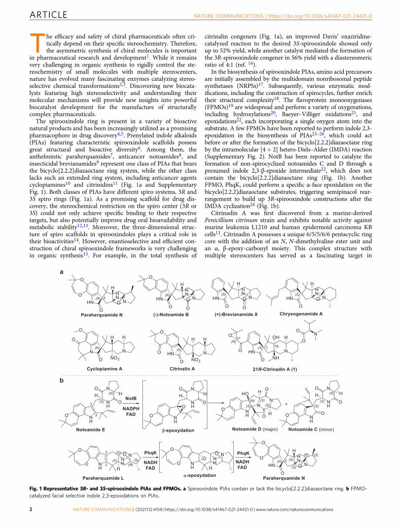
The spirooxindole ring is present in a variety of bioactivenatural products and has been increasingly utilized as a promisingpharmacophore in drug discovery4,5. Prenylated indole alkaloids(PIAs) featuring characteristic spirooxindole scaffolds possessgreat structural and bioactive diversity6. Among them, theanthelmintic paraherquamides7, anticancer notoamides8, andinsecticidal brevianamides9 represent one class of PIAs that bearsthe bicyclo[2.2.2]diazaoctane ring system, while the other classlacks such an extended ring system, including anticancer agentscyclopiamines10 and citrinidins11 (Fig. 1a and SupplementaryFig. 1). Both classes of PIAs have different spiro systems, 3R and3S spiro rings (Fig. 1a). As a promising scaffold for drug dis-covery, the stereochemical restriction on the spiro center (3R or3S) could not only achieve specific binding to their respectivetargets, but also potentially improve drug oral bioavailability andmetabolic stability12,13. Moreover, the three-dimensional struc-ture of spiro scaffolds in spirooxindoles plays a critical role intheir bioactivities14. However, enantioselective and efficient con-struction of chiral spirooxindole frameworks is very challengingin organic synthesis15. For example, in the total synthesis of
citrinalin congeners (Fig. 1a), an improved Davis’ oxaziridine-catalyzed reaction to the desired 3S-spirooxindole showed onlyup to 52% yield, while another catalyst mediated the formation ofthe 3R-spirooxindole congener in 56% yield with a diastereomericratio of 4:1 (ref. 16).
In the biosynthesis of spirooxindole PIAs, amino acid precursorsare initially assembled by the multidomain nonribosomal peptidesynthetases (NRPSs)17. Subsequently, various enzymatic mod-ifications, including the construction of spirocycles, further enrichtheir structural complexity18. The flavoprotein monooxygenases(FPMOs)19 are widespread and perform a variety of oxygenations,including hydroxylations20, Baeyer–Villiger oxidations21, andepoxidations22, each incorporating a single oxygen atom into thesubstrate. A few FPMOs have been reported to perform indole 2,3-epoxidation in the biosynthesis of PIAs23–26, which could actbefore or after the formation of the bicyclo[2.2.2]diazaoctane ringby the intramolecular [4+ 2] hetero-Diels–Alder (IMDA) reaction(Supplementary Fig. 2). NotB has been reported to catalyze theformation of non-spirocyclized notoamides C and D through apresumed indole 2,3-β-epoxide intermediate22, which does notcontain the bicyclo[2.2.2]diazaoctane ring (Fig. 1b). AnotherFPMO, PhqK, could perform a specific α-face epoxidation on thebicyclo[2.2.2]diazaoctane substrates, triggering semipinacol rear-rangement to build up 3R-spirooxindole constructions after theIMDA cyclization24 (Fig. 1b).
Citrinadin A was first discovered from a marine-derivedPenicillium citrinum strain and exhibits notable activity againstmurine leukemia L1210 and human epidermoid carcinoma KBcells11. Citrinadin A possesses a unique 6/5/5/6/6 pentacyclic ringcore with the addition of an N, N-dimethylvaline ester unit andan α, β-epoxy-carbonyl moiety. This complex structure withmultiple stereocenters has served as a fascinating target in
Fig. 1 Representative 3R- and 3S-spirooxindole PIAs and FPMOs. a Spirooxindole PIAs contain or lack the bicyclo[2.2.2]diazaoctane ring. b FPMO-catalyzed facial selective indole 2,3-epoxidations on PIAs.
ARTICLE NATURE COMMUNICATIONS | https://doi.org/10.1038/s41467-021-24421-0
2 NATURE COMMUNICATIONS | (2021) 12:4158 | https://doi.org/10.1038/s41467-021-24421-0 | www.nature.com/naturecommunications
subsequent synthetic studies27–31. The absolute configuration ofcitrinadin A was corrected to be 3S spirocycle via the firstenantioselective total synthesis in 2013 (refs. 31,32, Fig. 1a), thesame as citrinalin16 and chrysogenamide A33 (Fig. 1a). A recentbiosynthetic study has demonstrated that (2S, 6S)-6-methylpipecolate is a key precursor in building up the L-pipecolatemoiety in citrinadin A34. However, the later biosynthetic steps forcitrinadins, including the formation of the spirooxindole ring,remain elusive. Unlike paraherquamides24, notoamides23, andbrevianamides25, citrinadins do not contain the bicyclo[2.2.2]diazaoctane ring. Notably, a close examination of the stereo-centers in citrinadins showed that they feature 3S-spirooxindole,which is opposite to those in paraherquamides24, indicating thepossible presence of unique stereocontrol for the 3S spirocycleformation in citrinadin biosynthesis.
Herein, we report the identification of a distinct FPMO, CtdE,that stereoselectively catalyzes the 3S-spirooxindole formation inthe 21R-citrinadin A (1) biosynthesis. Based on thorough ana-lyses of the high-resolution X-ray crystal structures of CtdEcomplex containing substrate and cofactor flavin adenine dinu-cleotide (FAD), together with the site-directed mutagenesis andcomputational study, we revealed the molecular basis for thestereoselective catalytic mechanism that CtdE exploits for thepossible β-facial epoxidation, triggering semipinacol rearrange-ment to yield 3S-spirooxindole PIAs. Our discovery of the ste-reoselective formation of the 3S spirocycle and deciphering of themechanistic details of CtdE are important in the spirooxindolepharmaceutical research and development.
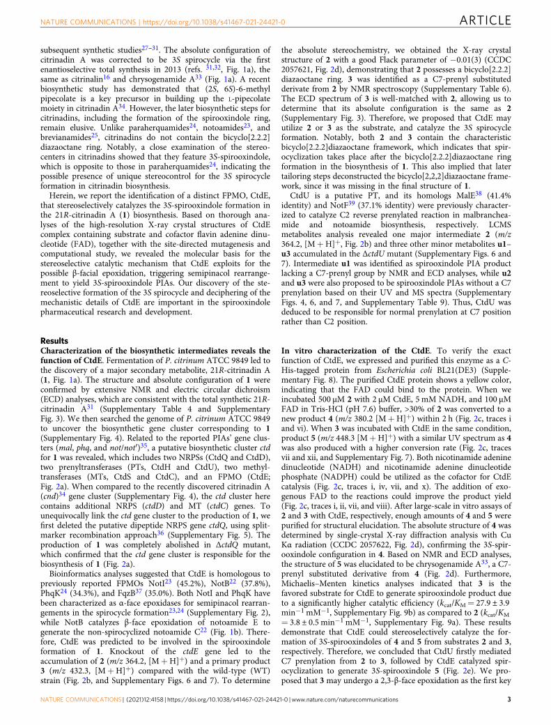
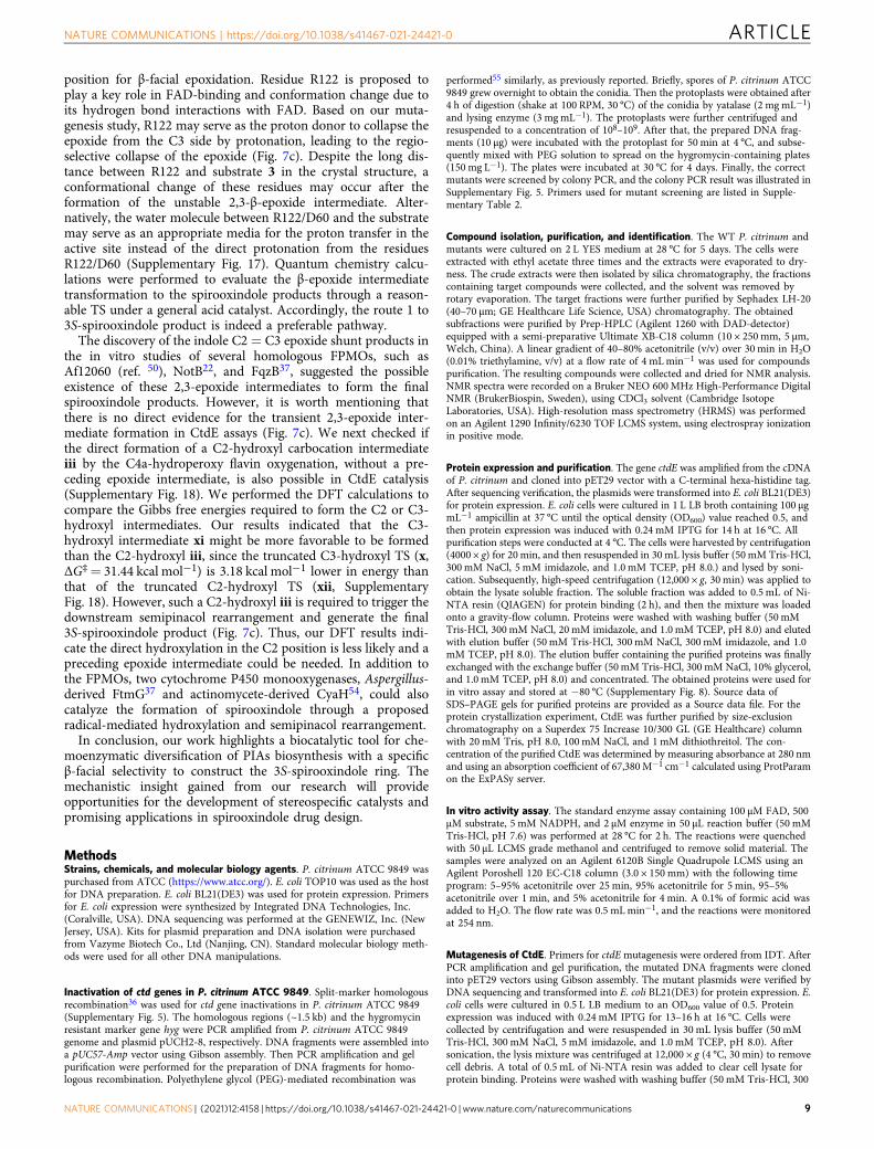
ResultsCharacterization of the biosynthetic intermediates reveals thefunction of CtdE. Fermentation of P. citrinum ATCC 9849 led tothe discovery of a major secondary metabolite, 21R-citrinadin A(1, Fig. 1a). The structure and absolute configuration of 1 wereconfirmed by extensive NMR and electric circular dichroism(ECD) analyses, which are consistent with the total synthetic 21R-citrinadin A31 (Supplementary Table 4 and SupplementaryFig. 3). We then searched the genome of P. citrinum ATCC 9849to uncover the biosynthetic gene cluster corresponding to 1(Supplementary Fig. 4). Related to the reported PIAs’ gene clus-ters (mal, phq, and not/not′)35, a putative biosynthetic cluster ctdfor 1 was revealed, which includes two NRPSs (CtdQ and CtdD),two prenyltransferases (PTs, CtdH and CtdU), two methyl-transferases (MTs, CtdS and CtdC), and an FPMO (CtdE;Fig. 2a). When compared to the recently discovered citrinadin A(cnd)34 gene cluster (Supplementary Fig. 4), the ctd cluster herecontains additional NRPS (ctdD) and MT (ctdC) genes. Tounequivocally link the ctd gene cluster to the production of 1, wefirst deleted the putative dipeptide NRPS gene ctdQ, using split-marker recombination approach36 (Supplementary Fig. 5). Theproduction of 1 was completely abolished in ΔctdQ mutant,which confirmed that the ctd gene cluster is responsible for thebiosynthesis of 1 (Fig. 2a).
Bioinformatics analyses suggested that CtdE is homologous topreviously reported FPMOs NotI23 (45.2%), NotB22 (37.8%),PhqK24 (34.3%), and FqzB37 (35.0%). Both NotI and PhqK havebeen characterized as α-face epoxidases for semipinacol rearran-gements in the spirocycle formation23,24 (Supplementary Fig. 2),while NotB catalyzes β-face epoxidation of notoamide E togenerate the non-spirocyclized notoamide C22 (Fig. 1b). There-fore, CtdE was predicted to be involved in the spirooxindoleformation of 1. Knockout of the ctdE gene led to theaccumulation of 2 (m/z 364.2, [M+H]+) and a primary product3 (m/z 432.3, [M+H]+) compared with the wild-type (WT)strain (Fig. 2b, and Supplementary Figs. 6 and 7). To determine
the absolute stereochemistry, we obtained the X-ray crystalstructure of 2 with a good Flack parameter of −0.01(3) (CCDC2057621, Fig. 2d), demonstrating that 2 possesses a bicyclo[2.2.2]diazaoctane ring. 3 was identified as a C7-prenyl substitutedderivate from 2 by NMR spectroscopy (Supplementary Table 6).The ECD spectrum of 3 is well-matched with 2, allowing us todetermine that its absolute configuration is the same as 2(Supplementary Fig. 3). Therefore, we proposed that CtdE mayutilize 2 or 3 as the substrate, and catalyze the 3S spirocycleformation. Notably, both 2 and 3 contain the characteristicbicyclo[2.2.2]diazaoctane framework, which indicates that spir-ocyclization takes place after the bicyclo[2.2.2]diazaoctane ringformation in the biosynthesis of 1. This also implied that latertailoring steps deconstructed the bicyclo[2,2,2]diazaoctane frame-work, since it was missing in the final structure of 1.
CtdU is a putative PT, and its homologs MalE38 (41.4%identity) and NotF39 (37.1% identity) were previously character-ized to catalyze C2 reverse prenylated reaction in malbranchea-mide and notoamide biosynthesis, respectively. LCMSmetabolites analysis revealed one major intermediate 2 (m/z364.2, [M+H]+, Fig. 2b) and three other minor metabolites u1–u3 accumulated in the ΔctdU mutant (Supplementary Figs. 6 and7). Intermediate u1 was identified as spirooxindole PIA productlacking a C7-prenyl group by NMR and ECD analyses, while u2and u3 were also proposed to be spirooxindole PIAs without a C7prenylation based on their UV and MS spectra (SupplementaryFigs. 4, 6, and 7, and Supplementary Table 9). Thus, CtdU wasdeduced to be responsible for normal prenylation at C7 positionrather than C2 position.
In vitro characterization of the CtdE. To verify the exactfunction of CtdE, we expressed and purified this enzyme as a C-His-tagged protein from Escherichia coli BL21(DE3) (Supple-mentary Fig. 8). The purified CtdE protein shows a yellow color,indicating that the FAD could bind to the protein. When weincubated 500 μM 2 with 2 μM CtdE, 5 mM NADH, and 100 μMFAD in Tris-HCl (pH 7.6) buffer, >30% of 2 was converted to anew product 4 (m/z 380.2 [M+H]+) within 2 h (Fig. 2c, traces iand vi). When 3 was incubated with CtdE in the same condition,product 5 (m/z 448.3 [M+H]+) with a similar UV spectrum as 4was also produced with a higher conversion rate (Fig. 2c, tracesvii and xii, and Supplementary Fig. 7). Both nicotinamide adeninedinucleotide (NADH) and nicotinamide adenine dinucleotidephosphate (NADPH) could be utilized as the cofactor for CtdEcatalysis (Fig. 2c, traces i, iv, vii, and x). The addition of exo-genous FAD to the reactions could improve the product yield(Fig. 2c, traces i, ii, vii, and viii). After large-scale in vitro assays of2 and 3 with CtdE, respectively, enough amounts of 4 and 5 werepurified for structural elucidation. The absolute structure of 4 wasdetermined by single-crystal X-ray diffraction analysis with CuKα radiation (CCDC 2057622, Fig. 2d), confirming the 3S-spir-ooxindole configuration in 4. Based on NMR and ECD analyses,the structure of 5 was elucidated to be chrysogenamide A33, a C7-prenyl substituted derivative from 4 (Fig. 2d). Furthermore,Michaelis–Menten kinetics analyses indicated that 3 is thefavored substrate for CtdE to generate spirooxindole product dueto a significantly higher catalytic efficiency (kcat/KM= 27.9 ± 3.9min−1 mM−1, Supplementary Fig. 9b) as compared to 2 (kcat/KM
= 3.8 ± 0.5 min−1 mM−1, Supplementary Fig. 9a). These resultsdemonstrate that CtdE could stereoselectively catalyze the for-mation of 3S-spirooxindoles of 4 and 5 from substrates 2 and 3,respectively. Therefore, we concluded that CtdU firstly mediatedC7 prenylation from 2 to 3, followed by CtdE catalyzed spir-ocyclization to generate 3S-spirooxindole 5 (Fig. 2e). We pro-posed that 3 may undergo a 2,3-β-face epoxidation as the first key
NATURE COMMUNICATIONS | https://doi.org/10.1038/s41467-021-24421-0 ARTICLE
NATURE COMMUNICATIONS | (2021) 12:4158 | https://doi.org/10.1038/s41467-021-24421-0 | www.nature.com/naturecommunications 3
step catalyzed by CtdE and followed by the regioselective openingof the epoxide ring that triggers the semipinacol rearrangement toform the 3S-spirooxindole 5. Notably, CtdE revealed a divergentevolutionary process in PIAs biosynthesis (SupplementaryFig. 10). In the mal biosynthetic pathway, (+)-pre-malbrancheamide is synthesized via an IMDA reaction40; furtherbiosynthesis of spirooxindole rings was not found in the mal/mal′gene cluster38,41. In contrast, PhqK transforms the bicyclo[2,2,2]diazaoctane substrate to 3R-spirooxindole24. Here, we show thatCtdE catalyzes the β-face epoxidation of substrate 3 to construct a3S spirocycle. Moreover, the bvn system represents another bio-synthetic branch, in which BvnB catalyzes the β-face epoxidationof deoxybrevianamide E to generate 3β-hydroxyindolenine pro-duct before the bicyclo[2,2,2]diazaoctane ring formation25.
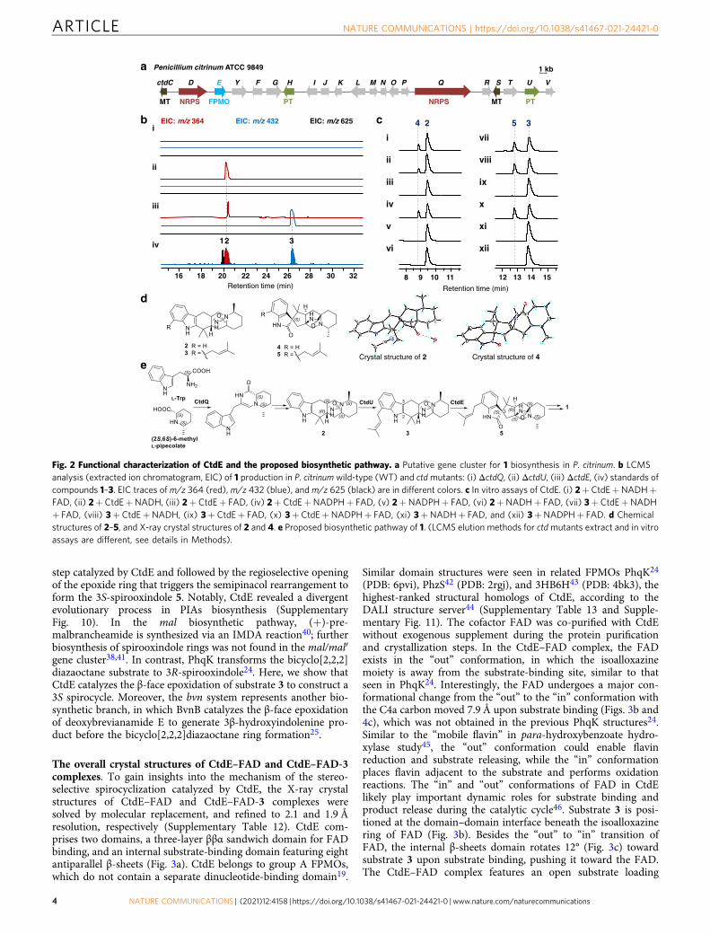
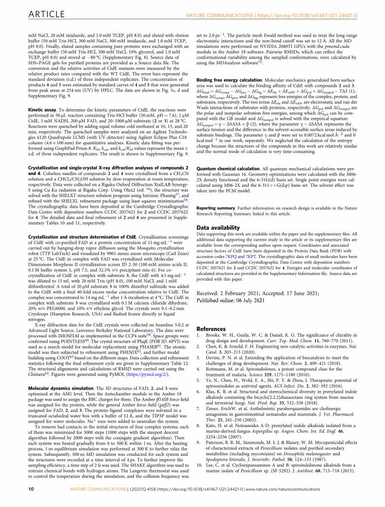
The overall crystal structures of CtdE–FAD and CtdE–FAD-3complexes. To gain insights into the mechanism of the stereo-selective spirocyclization catalyzed by CtdE, the X-ray crystalstructures of CtdE–FAD and CtdE–FAD-3 complexes weresolved by molecular replacement, and refined to 2.1 and 1.9 Åresolution, respectively (Supplementary Table 12). CtdE com-prises two domains, a three-layer ββα sandwich domain for FADbinding, and an internal substrate-binding domain featuring eightantiparallel β-sheets (Fig. 3a). CtdE belongs to group A FPMOs,which do not contain a separate dinucleotide-binding domain19.
Similar domain structures were seen in related FPMOs PhqK24
(PDB: 6pvi), PhzS42 (PDB: 2rgj), and 3HB6H43 (PDB: 4bk3), thehighest-ranked structural homologs of CtdE, according to theDALI structure server44 (Supplementary Table 13 and Supple-mentary Fig. 11). The cofactor FAD was co-purified with CtdEwithout exogenous supplement during the protein purificationand crystallization steps. In the CtdE–FAD complex, the FADexists in the “out” conformation, in which the isoalloxazinemoiety is away from the substrate-binding site, similar to thatseen in PhqK24. Interestingly, the FAD undergoes a major con-formational change from the “out” to the “in” conformation withthe C4a carbon moved 7.9 Å upon substrate binding (Figs. 3b and4c), which was not obtained in the previous PhqK structures24.Similar to the “mobile flavin” in para-hydroxybenzoate hydro-xylase study45, the “out” conformation could enable flavinreduction and substrate releasing, while the “in” conformationplaces flavin adjacent to the substrate and performs oxidationreactions. The “in” and “out” conformations of FAD in CtdElikely play important dynamic roles for substrate binding andproduct release during the catalytic cycle46. Substrate 3 is posi-tioned at the domain–domain interface beneath the isoalloxazinering of FAD (Fig. 3b). Besides the “out” to “in” transition ofFAD, the internal β-sheets domain rotates 12° (Fig. 3c) towardsubstrate 3 upon substrate binding, pushing it toward the FAD.The CtdE–FAD complex features an open substrate loading
1
EIC: m/z 364 EIC: m/z 432
2018 2422 26 28 30 3216
2 3
EIC: m/z 625i
ii
iii
iv
Retention time (min)
b
a Penicillium citrinum ATCC 9849
c
e
d
1 kb
HctdC MI J N OLD E Y F G K TP Q U VSR
FPMO NRPS PTPTMT MTNRPS
Retention time (min)
1098 11
24
i
ii
iv
v
vi
iii
35
141312 15
vii
viii
ix
x
xi
xii
Crystal structure of 2 Crystal structure of 4
Fig. 2 Functional characterization of CtdE and the proposed biosynthetic pathway. a Putative gene cluster for 1 biosynthesis in P. citrinum. b LCMSanalysis (extracted ion chromatogram, EIC) of 1 production in P. citrinum wild-type (WT) and ctdmutants: (i) ΔctdQ, (ii) ΔctdU, (iii) ΔctdE, (iv) standards ofcompounds 1–3. EIC traces of m/z 364 (red), m/z 432 (blue), and m/z 625 (black) are in different colors. c In vitro assays of CtdE. (i) 2+ CtdE+NADH+FAD, (ii) 2+ CtdE+NADH, (iii) 2+ CtdE+ FAD, (iv) 2+ CtdE+NADPH+ FAD, (v) 2+NADPH+ FAD, (vi) 2+NADH+ FAD, (vii) 3+ CtdE+NADH+ FAD, (viii) 3+CtdE+NADH, (ix) 3+CtdE+ FAD, (x) 3+CtdE+NADPH+ FAD, (xi) 3+NADH+ FAD, and (xii) 3+NADPH+ FAD. d Chemicalstructures of 2–5, and X-ray crystal structures of 2 and 4. e Proposed biosynthetic pathway of 1. (LCMS elution methods for ctdmutants extract and in vitroassays are different, see details in Methods).
ARTICLE NATURE COMMUNICATIONS | https://doi.org/10.1038/s41467-021-24421-0
4 NATURE COMMUNICATIONS | (2021) 12:4158 | https://doi.org/10.1038/s41467-021-24421-0 | www.nature.com/naturecommunications
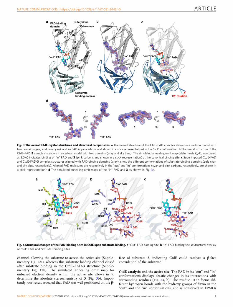
channel, allowing the substrate to access the active site (Supple-mentary Fig. 12a), whereas this substrate loading channel closedafter substrate binding in the CtdE–FAD-3 structure (Supple-mentary Fig. 12b). The simulated annealing omit map forunbiased electron density within the active site allows us todetermine the absolute stereochemistry of 3 (Fig. 3b). Impor-tantly, our result revealed that FAD was well positioned on the β-
face of substrate 3, indicating CtdE could catalyze a β-faceepoxidation of the substrate.
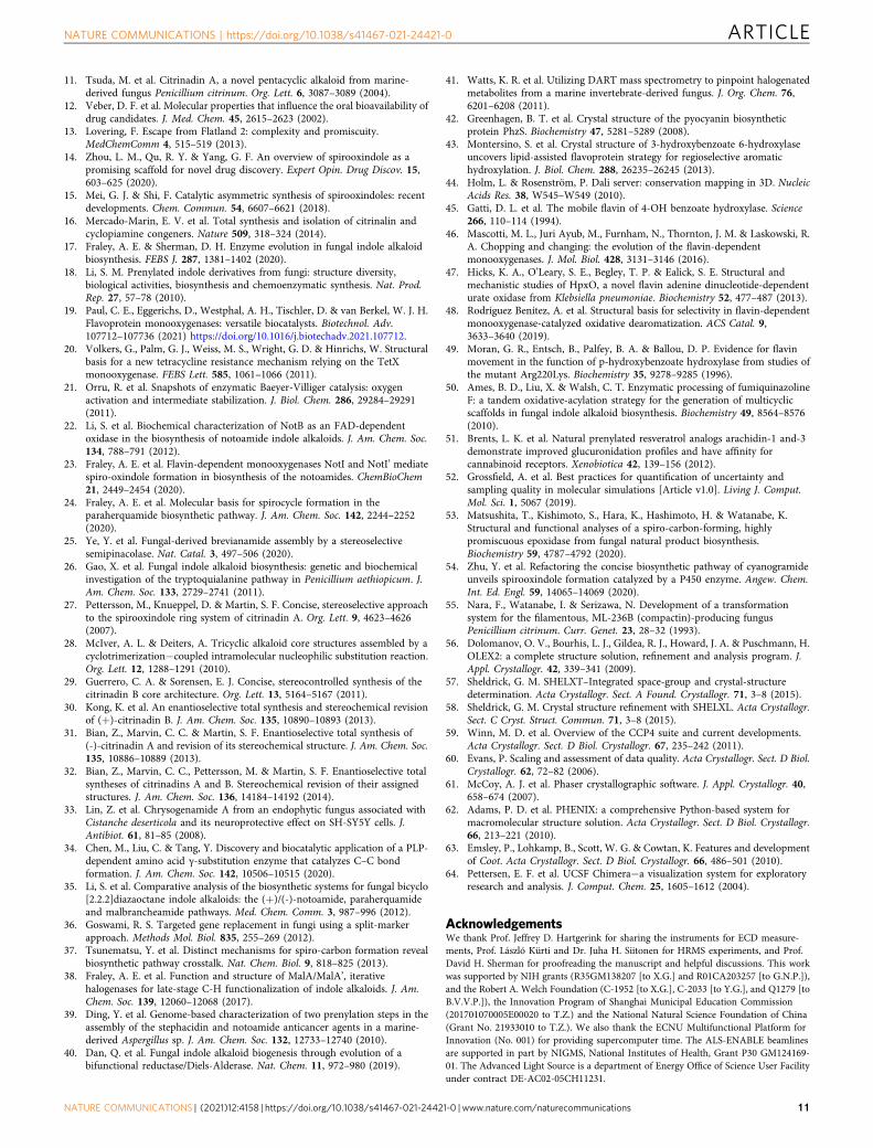
CtdE catalysis and the active site. The FAD in its “out” and “in”conformations displays drastic changes in its interactions withsurrounding residues (Fig. 4a, b). The residue R122 forms dif-ferent hydrogen bonds with the hydroxy groups of flavin in the“out” and the “in” conformations, and is conserved in FPMOs
a
α13
α14
b
“in” FAD
3
c
“out” FAD “in” FAD
3
12 rotation
90 3 3
“in” FAD “in” FAD
90
Substrate-binding domain
N-terminus
C-terminusFAD-binding domain
α1
β6
β1 β2
β3
α2
α3
α4
β4 β5
β7 β8 β9
β10
α5
β11
α6
α7
β12 β13
β14
β15
α8
α9
β16
β17
α10
α11
α12
“out” FAD
d
Fig. 3 The overall CtdE crystal structures and structural comparisons. a The overall structure of the CtdE–FAD complex shown in a cartoon model withtwo domains (gray and pale cyan), and an FAD (cyan carbons and shown in a stick representation) in the “out” conformation. b The overall structure of theCtdE–FAD-3 complex is shown in a cartoon model with two domains (gray and sky blue). The simulated annealing omit map (slate mesh, Fo–Fc, contouredat 3.0 σ) indicates binding of “in” FAD and 3 (pink carbons and shown in a stick representation) at the canonical binding site. c Superimposed CtdE–FADand CtdE–FAD-3 complex structures aligned with FAD-binding domains (gray), show the different conformations of substrate-binding domains (pale cyanand sky blue, respectively). Aligned FAD molecules are respectively in the “out” and “in” conformations (cyan and pink carbons, respectively, are shown ina stick representation). d The simulated annealing omit maps of the “in” FAD and 3 as shown in Fig. 3b.
a
R122
3.0Å
2.9Å
R200 D60
2.6Å
M56
W264
I58
H57
S63
N338
H301
D324
N338
b
R122
R200
2.7Å
2.7Å
2.8Å3.4Å
2.7Å
H57
M56
W264
I58
H301
D60S63
“in” FAD
D324
“out” FADc
“in” FAD
“out” FAD
7.9Å
3
C4a
3
C4a
5.6Å 5.5Å
C2C3
Fig. 4 Structural changes of the FAD-binding sites in CtdE upon substrate binding. a “Out” FAD-binding site. b “In” FAD-binding site. c Structural overlayof “out” FAD and “in” FAD-binding sites.
NATURE COMMUNICATIONS | https://doi.org/10.1038/s41467-021-24421-0 ARTICLE
NATURE COMMUNICATIONS | (2021) 12:4158 | https://doi.org/10.1038/s41467-021-24421-0 | www.nature.com/naturecommunications 5
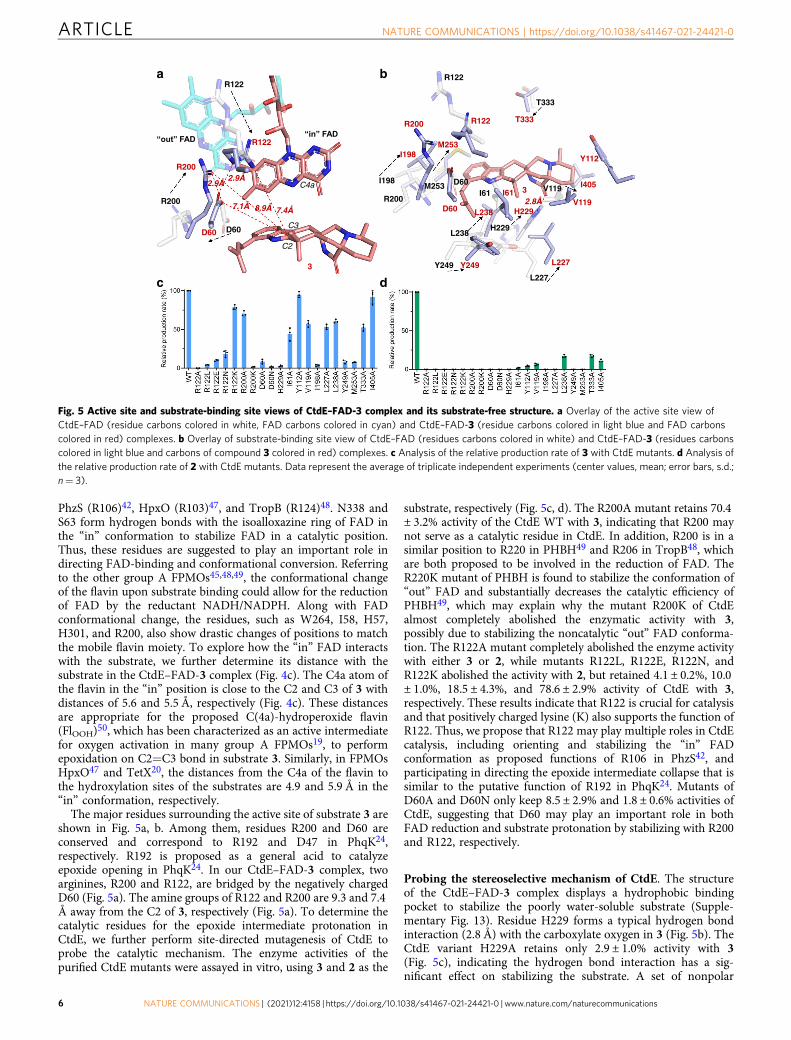
PhzS (R106)42, HpxO (R103)47, and TropB (R124)48. N338 andS63 form hydrogen bonds with the isoalloxazine ring of FAD inthe “in” conformation to stabilize FAD in a catalytic position.Thus, these residues are suggested to play an important role indirecting FAD-binding and conformational conversion. Referringto the other group A FPMOs45,48,49, the conformational changeof the flavin upon substrate binding could allow for the reductionof FAD by the reductant NADH/NADPH. Along with FADconformational change, the residues, such as W264, I58, H57,H301, and R200, also show drastic changes of positions to matchthe mobile flavin moiety. To explore how the “in” FAD interactswith the substrate, we further determine its distance with thesubstrate in the CtdE–FAD-3 complex (Fig. 4c). The C4a atom ofthe flavin in the “in” position is close to the C2 and C3 of 3 withdistances of 5.6 and 5.5 Å, respectively (Fig. 4c). These distancesare appropriate for the proposed C(4a)-hydroperoxide flavin(FlOOH)50, which has been characterized as an active intermediatefor oxygen activation in many group A FPMOs19, to performepoxidation on C2=C3 bond in substrate 3. Similarly, in FPMOsHpxO47 and TetX20, the distances from the C4a of the flavin tothe hydroxylation sites of the substrates are 4.9 and 5.9 Å in the“in” conformation, respectively.
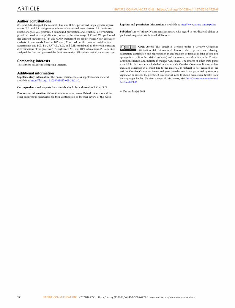
The major residues surrounding the active site of substrate 3 areshown in Fig. 5a, b. Among them, residues R200 and D60 areconserved and correspond to R192 and D47 in PhqK24,respectively. R192 is proposed as a general acid to catalyzeepoxide opening in PhqK24. In our CtdE–FAD-3 complex, twoarginines, R200 and R122, are bridged by the negatively chargedD60 (Fig. 5a). The amine groups of R122 and R200 are 9.3 and 7.4Å away from the C2 of 3, respectively (Fig. 5a). To determine thecatalytic residues for the epoxide intermediate protonation inCtdE, we further perform site-directed mutagenesis of CtdE toprobe the catalytic mechanism. The enzyme activities of thepurified CtdE mutants were assayed in vitro, using 3 and 2 as the
substrate, respectively (Fig. 5c, d). The R200A mutant retains 70.4± 3.2% activity of the CtdE WT with 3, indicating that R200 maynot serve as a catalytic residue in CtdE. In addition, R200 is in asimilar position to R220 in PHBH49 and R206 in TropB48, whichare both proposed to be involved in the reduction of FAD. TheR220K mutant of PHBH is found to stabilize the conformation of“out” FAD and substantially decreases the catalytic efficiency ofPHBH49, which may explain why the mutant R200K of CtdEalmost completely abolished the enzymatic activity with 3,possibly due to stabilizing the noncatalytic “out” FAD conforma-tion. The R122A mutant completely abolished the enzyme activitywith either 3 or 2, while mutants R122L, R122E, R122N, andR122K abolished the activity with 2, but retained 4.1 ± 0.2%, 10.0± 1.0%, 18.5 ± 4.3%, and 78.6 ± 2.9% activity of CtdE with 3,respectively. These results indicate that R122 is crucial for catalysisand that positively charged lysine (K) also supports the function ofR122. Thus, we propose that R122 may play multiple roles in CtdEcatalysis, including orienting and stabilizing the “in” FADconformation as proposed functions of R106 in PhzS42, andparticipating in directing the epoxide intermediate collapse that issimilar to the putative function of R192 in PhqK24. Mutants ofD60A and D60N only keep 8.5 ± 2.9% and 1.8 ± 0.6% activities ofCtdE, suggesting that D60 may play an important role in bothFAD reduction and substrate protonation by stabilizing with R200and R122, respectively.
Probing the stereoselective mechanism of CtdE. The structureof the CtdE–FAD-3 complex displays a hydrophobic bindingpocket to stabilize the poorly water-soluble substrate (Supple-mentary Fig. 13). Residue H229 forms a typical hydrogen bondinteraction (2.8 Å) with the carboxylate oxygen in 3 (Fig. 5b). TheCtdE variant H229A retains only 2.9 ± 1.0% activity with 3(Fig. 5c), indicating the hydrogen bond interaction has a sig-nificant effect on stabilizing the substrate. A set of nonpolar
7.4Å8.9ÅR200
R122“out” FAD
3
C2
C3
C4a
D60
R122
7.1Å
L227
a b R122
R122R200
R200 V119
V119
H229
H229
L227
Y249Y249
L238
L238
D60
D60
M253
M253
D60
T333
T333
I61I61
2.9Å3
2.9Å
R200
I198
I198
“in” FAD
c d
Y112
2.8Å
I405
Fig. 5 Active site and substrate-binding site views of CtdE–FAD-3 complex and its substrate-free structure. a Overlay of the active site view ofCtdE–FAD (residue carbons colored in white, FAD carbons colored in cyan) and CtdE–FAD-3 (residue carbons colored in light blue and FAD carbonscolored in red) complexes. b Overlay of substrate-binding site view of CtdE–FAD (residues carbons colored in white) and CtdE–FAD-3 (residues carbonscolored in light blue and carbons of compound 3 colored in red) complexes. c Analysis of the relative production rate of 3 with CtdE mutants. d Analysis ofthe relative production rate of 2 with CtdE mutants. Data represent the average of triplicate independent experiments (center values, mean; error bars, s.d.;n= 3).
ARTICLE NATURE COMMUNICATIONS | https://doi.org/10.1038/s41467-021-24421-0
6 NATURE COMMUNICATIONS | (2021) 12:4158 | https://doi.org/10.1038/s41467-021-24421-0 | www.nature.com/naturecommunications
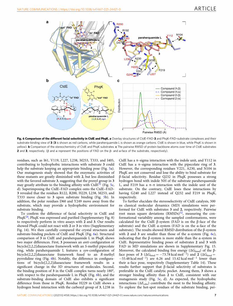
residues, such as I61, V119, L227, L238, M253, T333, and I405,contributing to hydrophobic interactions with substrate 3 couldhelp the substrate keeping an appropriate binding pose (Fig. 5a).Our mutagenesis study showed that the enzymatic activities ofthese mutants are greatly diminished with 2, but less diminishedwith the favored substrate 3, suggesting that the prenyl group in 3may greatly attribute to the binding affinity with CtdE51 (Fig. 5c,d). Superimposing the CtdE–FAD complex onto the CtdE–FAD-3 revealed that the residues R122, R200, H229, L238, M253, andT333 move closer to 3 upon substrate binding (Fig. 5b). Inaddition, the polar residues D60 and Y249 move away from thesubstrate, which may provide a hydrophobic environment forsubstrate binding.
To confirm the difference of facial selectivity in CtdE andPhqK24, PhqK was expressed and purified (Supplementary Fig. 8)to respectively perform in vitro assays with 2 and 3. Our resultsshowed PhqK could not react with 2 or 3 in vitro (SupplementaryFig. 14). We then carefully compared the crystal structures andsubstrate-binding pockets of CtdE and PhqK (Fig. 6a). Structuralcomparison of 3 in CtdE and paraherquamide L in PhqK showstwo major differences. First, 3 possesses an anti-configuration ofbicyclo[2,2,2]diazaoctane framework with an S-methyl pipecolatering, while paraherquamide L features a syn-configuration ofbicyclo[2,2,2]diazaoctane framework fused to an R-methylpyrrolidine ring (Fig. 6b). Notably, the difference in configura-tions of bicyclo[2,2,2]diazaoctane framework results in asignificant change in the 3D structures of molecules. Second,the binding position of 3 in the CtdE complex turns nearly 180°,with respect to the paraherquamide L in PhqK (Fig. 6b), and thesubstrate-binding domain of CtdE also exhibits a significantdifference from those in PhqK. Residue H229 in CtdE shows ahydrogen bond interaction with the carboxyl group of 3, L238 in
CtdE has a π–sigma interaction with the indole unit, and Y112 inCtdE has a π–sigma interaction with the pipecolate ring of 3.However, the corresponding residues V221, A230, and N104 inPhqK are not conserved and lose the ability to bind substrate forβ-facial selectivity. Residue Q232 in PhqK possesses a stronghydrogen bond with indole NH of the substrate paraherquamideL, and F219 has a π–π interaction with the indole unit of thesubstrate. On the contrary, CtdE loses these interactions byhaving G240 and L227 instead of Q232 and F219 in PhqK,respectively.
To further elucidate the stereoselectivity of CtdE catalysis, 500ns classical molecular dynamics (MD) simulations were per-formed for CtdE with substrates 3 and 2, respectively. Pairwiseroot mean square deviations (RMSDs)52, measuring the con-formational variability among the sampled conformations, werecalculated for the CtdE β-system (FAD is on the β-face of thesubstrate) and the CtdE α-system (FAD is on the α-face of thesubstrate). The results showed RMSD distribution of the β-systemwith 2 and 3 are smaller than those of the α-system (Fig. 6c),indicating that the β-system is more stable than the α-system inCtdE. Representative binding poses of substrates 2 and 3 withFAD in MD simulations are shown in Supplementary Fig. 15.Moreover, the calculated binding free energy (ΔGbind) of the β-face poses of 3 (ΔGbind=−73.78 kcal mol−1) and 2 (ΔGbind=−55.48 kcal mol−1) are 6.26 and 15.42 kcal mol−1 lower thantheir α-face pose, respectively (Supplementary Table 14). Theseresults further support that β-facial selectivity of substrates ispreferable in the CtdE catalytic pocket. Among them, 3 shows astronger binding affinity than 2 to CtdE, consistent with ourmutagenesis study (Fig. 5c, d). As expected, the hydrophobicinteractions (ΔEvdw) contribute the most to the binding affinity.To explore the hot-spot residues of the substrate binding, per-
b
R200
R192
Q232
G240 L227
Y112
L238 A230
H229
N104
F219V221
a
D60
D47
3
paraherquamide L
3
paraherquamide L
in FAD
out FAD
c
Fig. 6 Comparison of the different facial selectivity in CtdE and PhqK. a Overlay structures of CtdE–FAD-3 and PhqK–FAD–substrate complexes and theirsubstrate-binding view of 3 (3 is shown as red carbons, while paraherquamide L is shown as orange carbons. CtdE is shown in blue, while PhqK is shown inyellow). b Comparison of the stereochemistry of CtdE and PhqK substrates. c The pairwise RMSD of protein backbone atoms over time of CtdE substrates2 and 3, respectively. (β and α represent the positions of FAD on the β- and α-face of the substrate, respectively).
NATURE COMMUNICATIONS | https://doi.org/10.1038/s41467-021-24421-0 ARTICLE
NATURE COMMUNICATIONS | (2021) 12:4158 | https://doi.org/10.1038/s41467-021-24421-0 | www.nature.com/naturecommunications 7
residue free energy decomposition was performed. The resultsshowed that the residues L238, H229, L227, R200, and I405provide the major contribution to substrate binding in the CtdEactive pocket (Supplementary Fig. 16).
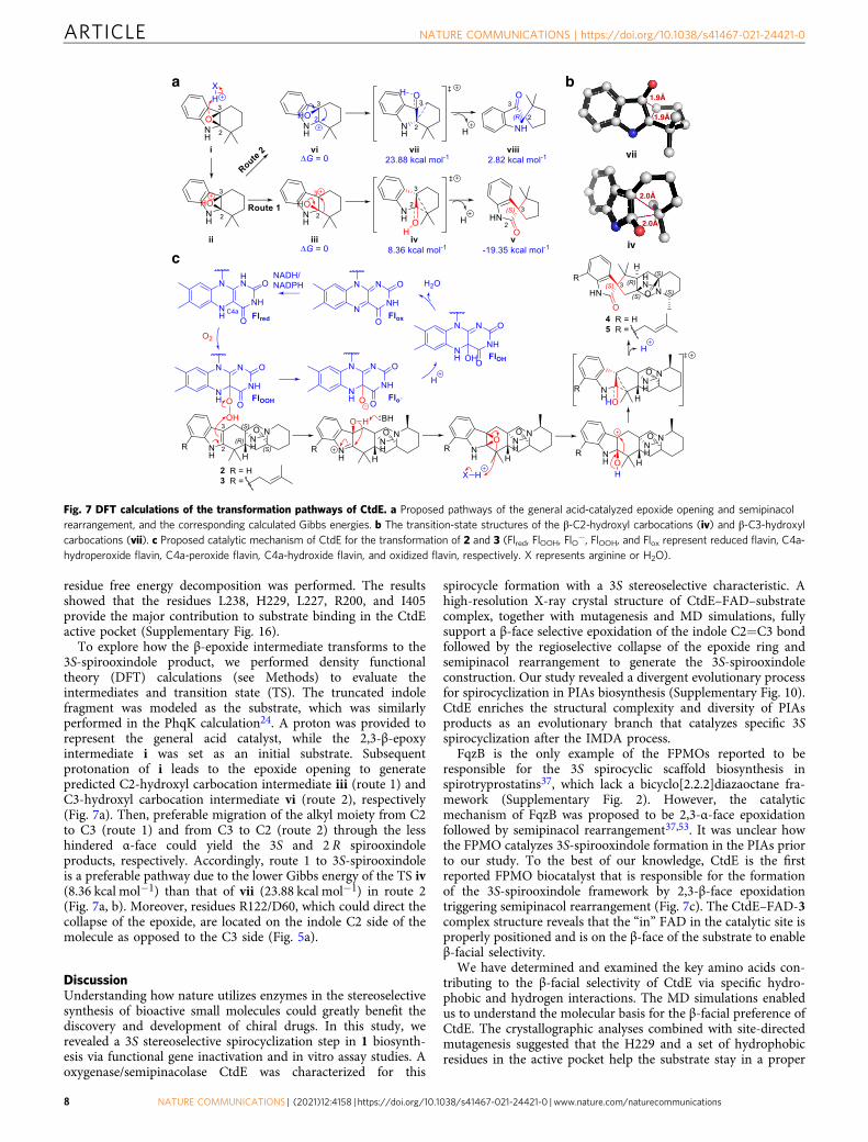
To explore how the β-epoxide intermediate transforms to the3S-spirooxindole product, we performed density functionaltheory (DFT) calculations (see Methods) to evaluate theintermediates and transition state (TS). The truncated indolefragment was modeled as the substrate, which was similarlyperformed in the PhqK calculation24. A proton was provided torepresent the general acid catalyst, while the 2,3-β-epoxyintermediate i was set as an initial substrate. Subsequentprotonation of i leads to the epoxide opening to generatepredicted C2-hydroxyl carbocation intermediate iii (route 1) andC3-hydroxyl carbocation intermediate vi (route 2), respectively(Fig. 7a). Then, preferable migration of the alkyl moiety from C2to C3 (route 1) and from C3 to C2 (route 2) through the lesshindered α-face could yield the 3S and 2 R spirooxindoleproducts, respectively. Accordingly, route 1 to 3S-spirooxindoleis a preferable pathway due to the lower Gibbs energy of the TS iv(8.36 kcal mol−1) than that of vii (23.88 kcal mol−1) in route 2(Fig. 7a, b). Moreover, residues R122/D60, which could direct thecollapse of the epoxide, are located on the indole C2 side of themolecule as opposed to the C3 side (Fig. 5a).
DiscussionUnderstanding how nature utilizes enzymes in the stereoselectivesynthesis of bioactive small molecules could greatly benefit thediscovery and development of chiral drugs. In this study, werevealed a 3S stereoselective spirocyclization step in 1 biosynth-esis via functional gene inactivation and in vitro assay studies. Aoxygenase/semipinacolase CtdE was characterized for this
spirocycle formation with a 3S stereoselective characteristic. Ahigh-resolution X-ray crystal structure of CtdE–FAD–substratecomplex, together with mutagenesis and MD simulations, fullysupport a β-face selective epoxidation of the indole C2=C3 bondfollowed by the regioselective collapse of the epoxide ring andsemipinacol rearrangement to generate the 3S-spirooxindoleconstruction. Our study revealed a divergent evolutionary processfor spirocyclization in PIAs biosynthesis (Supplementary Fig. 10).CtdE enriches the structural complexity and diversity of PIAsproducts as an evolutionary branch that catalyzes specific 3Sspirocyclization after the IMDA process.
FqzB is the only example of the FPMOs reported to beresponsible for the 3S spirocyclic scaffold biosynthesis inspirotryprostatins37, which lack a bicyclo[2.2.2]diazaoctane fra-mework (Supplementary Fig. 2). However, the catalyticmechanism of FqzB was proposed to be 2,3-α-face epoxidationfollowed by semipinacol rearrangement37,53. It was unclear howthe FPMO catalyzes 3S-spirooxindole formation in the PIAs priorto our study. To the best of our knowledge, CtdE is the firstreported FPMO biocatalyst that is responsible for the formationof the 3S-spirooxindole framework by 2,3-β-face epoxidationtriggering semipinacol rearrangement (Fig. 7c). The CtdE–FAD-3complex structure reveals that the “in” FAD in the catalytic site isproperly positioned and is on the β-face of the substrate to enableβ-facial selectivity.
We have determined and examined the key amino acids con-tributing to the β-facial selectivity of CtdE via specific hydro-phobic and hydrogen interactions. The MD simulations enabledus to understand the molecular basis for the β-facial preference ofCtdE. The crystallographic analyses combined with site-directedmutagenesis suggested that the H229 and a set of hydrophobicresidues in the active pocket help the substrate stay in a proper
2.0Å
2.0Å
1.9Å
1.9Å
ba
vii
iv
c
Fig. 7 DFT calculations of the transformation pathways of CtdE. a Proposed pathways of the general acid-catalyzed epoxide opening and semipinacolrearrangement, and the corresponding calculated Gibbs energies. b The transition-state structures of the β-C2-hydroxyl carbocations (iv) and β-C3-hydroxylcarbocations (vii). c Proposed catalytic mechanism of CtdE for the transformation of 2 and 3 (Flred, FlOOH, FlO−, FlOOH, and Flox represent reduced flavin, C4a-hydroperoxide flavin, C4a-peroxide flavin, C4a-hydroxide flavin, and oxidized flavin, respectively. X represents arginine or H2O).
ARTICLE NATURE COMMUNICATIONS | https://doi.org/10.1038/s41467-021-24421-0
8 NATURE COMMUNICATIONS | (2021) 12:4158 | https://doi.org/10.1038/s41467-021-24421-0 | www.nature.com/naturecommunications
position for β-facial epoxidation. Residue R122 is proposed toplay a key role in FAD-binding and conformation change due toits hydrogen bond interactions with FAD. Based on our muta-genesis study, R122 may serve as the proton donor to collapse theepoxide from the C3 side by protonation, leading to the regio-selective collapse of the epoxide (Fig. 7c). Despite the long dis-tance between R122 and substrate 3 in the crystal structure, aconformational change of these residues may occur after theformation of the unstable 2,3-β-epoxide intermediate. Alter-natively, the water molecule between R122/D60 and the substratemay serve as an appropriate media for the proton transfer in theactive site instead of the direct protonation from the residuesR122/D60 (Supplementary Fig. 17). Quantum chemistry calcu-lations were performed to evaluate the β-epoxide intermediatetransformation to the spirooxindole products through a reason-able TS under a general acid catalyst. Accordingly, the route 1 to3S-spirooxindole product is indeed a preferable pathway.
The discovery of the indole C2 = C3 epoxide shunt products inthe in vitro studies of several homologous FPMOs, such asAf12060 (ref. 50), NotB22, and FqzB37, suggested the possibleexistence of these 2,3-epoxide intermediates to form the finalspirooxindole products. However, it is worth mentioning thatthere is no direct evidence for the transient 2,3-epoxide inter-mediate formation in CtdE assays (Fig. 7c). We next checked ifthe direct formation of a C2-hydroxyl carbocation intermediateiii by the C4a-hydroperoxy flavin oxygenation, without a pre-ceding epoxide intermediate, is also possible in CtdE catalysis(Supplementary Fig. 18). We performed the DFT calculations tocompare the Gibbs free energies required to form the C2 or C3-hydroxyl intermediates. Our results indicated that the C3-hydroxyl intermediate xi might be more favorable to be formedthan the C2-hydroxyl iii, since the truncated C3-hydroxyl TS (x,ΔG‡= 31.44 kcal mol−1) is 3.18 kcal mol−1 lower in energy thanthat of the truncated C2-hydroxyl TS (xii, SupplementaryFig. 18). However, such a C2-hydroxyl iii is required to trigger thedownstream semipinacol rearrangement and generate the final3S-spirooxindole product (Fig. 7c). Thus, our DFT results indi-cate the direct hydroxylation in the C2 position is less likely and apreceding epoxide intermediate could be needed. In addition tothe FPMOs, two cytochrome P450 monooxygenases, Aspergillus-derived FtmG37 and actinomycete-derived CyaH54, could alsocatalyze the formation of spirooxindole through a proposedradical-mediated hydroxylation and semipinacol rearrangement.
In conclusion, our work highlights a biocatalytic tool for che-moenzymatic diversification of PIAs biosynthesis with a specificβ-facial selectivity to construct the 3S-spirooxindole ring. Themechanistic insight gained from our research will provideopportunities for the development of stereospecific catalysts andpromising applications in spirooxindole drug design.
MethodsStrains, chemicals, and molecular biology agents. P. citrinum ATCC 9849 waspurchased from ATCC (https://www.atcc.org/). E. coli TOP10 was used as the hostfor DNA preparation. E. coli BL21(DE3) was used for protein expression. Primersfor E. coli expression were synthesized by Integrated DNA Technologies, Inc.(Coralville, USA). DNA sequencing was performed at the GENEWIZ, Inc. (NewJersey, USA). Kits for plasmid preparation and DNA isolation were purchasedfrom Vazyme Biotech Co., Ltd (Nanjing, CN). Standard molecular biology meth-ods were used for all other DNA manipulations.
Inactivation of ctd genes in P. citrinum ATCC 9849. Split-marker homologousrecombination36 was used for ctd gene inactivations in P. citrinum ATCC 9849(Supplementary Fig. 5). The homologous regions (~1.5 kb) and the hygromycinresistant marker gene hyg were PCR amplified from P. citrinum ATCC 9849genome and plasmid pUCH2-8, respectively. DNA fragments were assembled intoa pUC57-Amp vector using Gibson assembly. Then PCR amplification and gelpurification were performed for the preparation of DNA fragments for homo-logous recombination. Polyethylene glycol (PEG)-mediated recombination was
performed55 similarly, as previously reported. Briefly, spores of P. citrinum ATCC9849 grew overnight to obtain the conidia. Then the protoplasts were obtained after4 h of digestion (shake at 100 RPM, 30 °C) of the conidia by yatalase (2 mgmL−1)and lysing enzyme (3 mgmL−1). The protoplasts were further centrifuged andresuspended to a concentration of 108–109. After that, the prepared DNA frag-ments (10 µg) were incubated with the protoplast for 50 min at 4 °C, and subse-quently mixed with PEG solution to spread on the hygromycin-containing plates(150 mg L−1). The plates were incubated at 30 °C for 4 days. Finally, the correctmutants were screened by colony PCR, and the colony PCR result was illustrated inSupplementary Fig. 5. Primers used for mutant screening are listed in Supple-mentary Table 2.
Compound isolation, purification, and identification. The WT P. citrinum andmutants were cultured on 2 L YES medium at 28 °C for 5 days. The cells wereextracted with ethyl acetate three times and the extracts were evaporated to dry-ness. The crude extracts were then isolated by silica chromatography, the fractionscontaining target compounds were collected, and the solvent was removed byrotary evaporation. The target fractions were further purified by Sephadex LH-20(40–70 μm; GE Healthcare Life Science, USA) chromatography. The obtainedsubfractions were purified by Prep-HPLC (Agilent 1260 with DAD-detector)equipped with a semi-preparative Ultimate XB-C18 column (10 × 250mm, 5 µm,Welch, China). A linear gradient of 40–80% acetonitrile (v/v) over 30 min in H2O(0.01% triethylamine, v/v) at a flow rate of 4 mLmin−1 was used for compoundspurification. The resulting compounds were collected and dried for NMR analysis.NMR spectra were recorded on a Bruker NEO 600MHz High-Performance DigitalNMR (BrukerBiospin, Sweden), using CDCl3 solvent (Cambridge IsotopeLaboratories, USA). High-resolution mass spectrometry (HRMS) was performedon an Agilent 1290 Infinity/6230 TOF LCMS system, using electrospray ionizationin positive mode.
Protein expression and purification. The gene ctdE was amplified from the cDNAof P. citrinum and cloned into pET29 vector with a C-terminal hexa-histidine tag.After sequencing verification, the plasmids were transformed into E. coli BL21(DE3)for protein expression. E. coli cells were cultured in 1 L LB broth containing 100 μgmL−1 ampicillin at 37 °C until the optical density (OD600) value reached 0.5, andthen protein expression was induced with 0.24 mM IPTG for 14 h at 16 °C. Allpurification steps were conducted at 4 °C. The cells were harvested by centrifugation(4000 × g) for 20min, and then resuspended in 30mL lysis buffer (50mM Tris-HCl,300mM NaCl, 5 mM imidazole, and 1.0mM TCEP, pH 8.0.) and lysed by soni-cation. Subsequently, high-speed centrifugation (12,000 × g, 30 min) was applied toobtain the lysate soluble fraction. The soluble fraction was added to 0.5mL of Ni-NTA resin (QIAGEN) for protein binding (2 h), and then the mixture was loadedonto a gravity-flow column. Proteins were washed with washing buffer (50mMTris-HCl, 300mM NaCl, 20mM imidazole, and 1.0 mM TCEP, pH 8.0) and elutedwith elution buffer (50mM Tris-HCl, 300mM NaCl, 300mM imidazole, and 1.0mM TCEP, pH 8.0). The elution buffer containing the purified proteins was finallyexchanged with the exchange buffer (50mM Tris-HCl, 300mM NaCl, 10% glycerol,and 1.0mM TCEP, pH 8.0) and concentrated. The obtained proteins were used forin vitro assay and stored at −80 °C (Supplementary Fig. 8). Source data ofSDS–PAGE gels for purified proteins are provided as a Source data file. For theprotein crystallization experiment, CtdE was further purified by size-exclusionchromatography on a Superdex 75 Increase 10/300 GL (GE Healthcare) columnwith 20 mM Tris, pH 8.0, 100mM NaCl, and 1 mM dithiothreitol. The con-centration of the purified CtdE was determined by measuring absorbance at 280 nmand using an absorption coefficient of 67,380M−1 cm−1 calculated using ProtParamon the ExPASy server.
In vitro activity assay. The standard enzyme assay containing 100 μM FAD, 500μM substrate, 5 mM NADPH, and 2 μM enzyme in 50 μL reaction buffer (50 mMTris-HCl, pH 7.6) was performed at 28 °C for 2 h. The reactions were quenchedwith 50 μL LCMS grade methanol and centrifuged to remove solid material. Thesamples were analyzed on an Agilent 6120B Single Quadrupole LCMS using anAgilent Poroshell 120 EC-C18 column (3.0 × 150 mm) with the following timeprogram: 5–95% acetonitrile over 25 min, 95% acetonitrile for 5 min, 95–5%acetonitrile over 1 min, and 5% acetonitrile for 4 min. A 0.1% of formic acid wasadded to H2O. The flow rate was 0.5 mLmin−1, and the reactions were monitoredat 254 nm.
Mutagenesis of CtdE. Primers for ctdE mutagenesis were ordered from IDT. AfterPCR amplification and gel purification, the mutated DNA fragments were clonedinto pET29 vectors using Gibson assembly. The mutant plasmids were verified byDNA sequencing and transformed into E. coli BL21(DE3) for protein expression. E.coli cells were cultured in 0.5 L LB medium to an OD600 value of 0.5. Proteinexpression was induced with 0.24 mM IPTG for 13–16 h at 16 °C. Cells werecollected by centrifugation and were resuspended in 30 mL lysis buffer (50 mMTris-HCl, 300 mM NaCl, 5 mM imidazole, and 1.0 mM TCEP, pH 8.0). Aftersonication, the lysis mixture was centrifuged at 12,000 × g (4 °C, 30 min) to removecell debris. A total of 0.5 mL of Ni-NTA resin was added to clear cell lysate forprotein binding. Proteins were washed with washing buffer (50 mM Tris-HCl, 300
NATURE COMMUNICATIONS | https://doi.org/10.1038/s41467-021-24421-0 ARTICLE
NATURE COMMUNICATIONS | (2021) 12:4158 | https://doi.org/10.1038/s41467-021-24421-0 | www.nature.com/naturecommunications 9
mM NaCl, 20 mM imidazole, and 1.0 mM TCEP, pH 8.0) and eluted with elutionbuffer (50 mM Tris-HCl, 300 mM NaCl, 300 mM imidazole, and 1.0 mM TCEP,pH 8.0). Finally, eluted samples containing pure proteins were exchanged with anexchange buffer (50 mM Tris-HCl, 300 mM NaCl, 10% glycerol, and 1.0 mMTCEP, pH 8.0) and stored at −80 °C (Supplementary Fig. 8). Source data ofSDS–PAGE gels for purified proteins are provided as a Source data file. Theconversion and the relative activities of CtdE mutants were measured by therelative product rates compared with the WT CtdE. The error bars represent thestandard deviation (s.d.) of three independent replicates. The concentration ofproducts 4 and 5 were estimated by standard carves of 4 and 5 that were generatedfrom peak areas at 254 nm (UV) by HPLC. The data are shown in Fig. 5c, d andSupplementary Fig. 9.
Kinetic assay. To determine the kinetic parameters of CtdE, the reactions wereperformed in 50 µL reaction containing Tris-HCl buffer (50 mM, pH= 7.6), 2 μMCtdE, 5 mM NADH, 200 μM FAD, and 20–1000 μM substrate (2 or 3) at 28 °C.Reactions were quenched by adding equal volume cold methanol at 10, 15, and 20min, respectively. The quenched samples were analyzed on an Agilent Technolo-gies 6120 Quadrupole LCMS (with UV-detector) using Agilent Eclipse Plus C18column (4.6 × 100 mm) for quantitative analyses. Kinetic data fitting was per-formed using GraphPad Prism 8. KM, kcat, and kcat/KM values represent the mean ±s.d. of three independent replicates. The result is shown in Supplementary Fig. 9.
Crystallization and single-crystal X-ray diffraction analyses of compounds 2and 4. Colorless needles of compounds 2 and 4 were crystallized from a CH3CNsolution and a CHCl3/CH3OH solution by slow evaporation at room temperature,respectively. Data were collected on a Rigaku Oxford Diffraction XtalLAB Synergy-S using Cu Kα radiation at Rigaku Corp. Using Olex2 (ref. 56), the structure wassolved with the SHELXT structure solution program using Intrinsic Phasing57, andrefined with the SHELXL refinement package using least squares minimization58.The crystallographic data have been deposited at the Cambridge CrystallographicData Centre with deposition numbers CCDC 2057621 for 2 and CCDC 2057622for 4. The detailed data and final refinement of 2 and 4 are presented in Supple-mentary Tables 10 and 11, respectively.
Crystallization and structure determination of CtdE. Crystallization screeningsof CtdE with co-purified FAD at a protein concentration of 11 mgmL−1 werecarried out by hanging-drop vapor diffusion using the Mosquito crystallizationrobot (TTP LabTech) and visualized by 9901 stereo zoom microscope (Carl Zeiss)at 25 °C. The CtdE in complex with FAD was crystallized with MolecularDimensions Morpheus II crystallization screen ID 2-30 (100 mM amino acids II,0.1 M buffer system 5, pH 7.5, and 32.5% v/v precipitant mix 6). For co-crystallization of CtdE in complex with substrate 3, the CtdE with 4.5 mg mL−1
was diluted to 15 mL with 20 mM Tris (pH 8.0), 100 mM NaCl, and 1 mMdithiothreitol. A total of 20 μM substrate 3 in 100% dimethyl sulfoxide was addedto the CtdE with a final 40-fold excess molar concentration relative to CtdE. Thecomplex was concentrated to 14 mgmL−1 after 1-h incubation at 4 °C. The CtdE incomplex with substrate 3 was crystallized with 0.1 M calcium chloride dihydrate,20% w/v PEG6000, and 10% v/v ethylene glycol. The crystals were 0.1–0.2 mmCryoloops (Hampton Research, USA) and flashed frozen directly in liquidnitrogen.
X-ray diffraction data for the CtdE crystals were collected on beamline 5.0.2 atAdvanced Light Source, Lawrence Berkeley National Laboratory. The data wereprocessed with IMOSFLM as implemented in the CCP4 suite59. Space groups wereconfirmed using POINTLESS60. The crystal structure of PhqK (PDB ID: 6PVI) wasused as a search model for molecular replacement using PHASER61. The atomicmodel was then subjected to refinement using PHENIX62, and further modelbuilding using COOT63 based on the different maps. Data collection and refinementstatistics following the final refinement cycle are given in Supplementary Table 12.The structural alignments and calculations of RMSD were carried out using theChimera64. Figures were generated using PyMOL (https://pymol.org/2/).
Molecular dynamics simulation. The 3D structures of FAD, 2, and 3 wereoptimized at the AM1 level. Then the Antechamber module in the Amber 18package was used to assign the BBC charges for them. The Amber ff14SB force fieldwas assigned for the protein, while the general Amber force field (gaff2) wasassigned for FAD, 2, and 3. The protein–ligand complexes were solvated in atruncated octahedral water box with a buffer of 12 Å, and the TIP3P model wasassigned for water molecules. Na+ ions were added to neutralize the system.
To remove bad contacts in the initial structures of four complex systems, eachof them was minimized for 3000 steps (1000 steps with the steepest descentalgorithm followed by 2000 steps with the conjugate gradient algorithm). Theneach system was heated gradually from 0 to 300 K within 1 ns. After the heatingprocess, 1 ns equilibrium simulation was performed at 300 K to further relax thesystem. Subsequently, 500 ns MD simulation was conducted for each system andthe structures were recorded at a time interval of 4 ps. To further improve thesampling efficiency, a time step of 2 fs was used. The SHAKE algorithm was used torestrain chemical bonds with hydrogen atoms. The Langevin thermostat was usedto control the temperature during the simulation, and the collision frequency was
set to 2.0 ps−1. The particle mesh Ewald method was used to treat the long-rangeelectrostatic interactions and the non-bond cutoff was set to 12 Å. All the MDsimulations were performed on NVIDIA 2080TI GPUs with the pmemd.cudamodule in the Amber 18 software. Pairwise RMSDs, which can reflect theconformational variability among the sampled conformations, were calculated byusing the MDAnalysis software52.
Binding free energy calculation. Molecular mechanics generalized born surfacearea was used to calculate the binding affinity of CdtE with compounds 2 and 3.ΔGbind= ΔGcomp− ΔGpro− ΔGlig= ΔEele+ ΔEvdW+ ΔGpol+ ΔGnonpol− TΔS (1),where ΔGcomp, ΔGpro, and ΔGlig, represent free energies of the complex, protein, andsubstrates, respectively. The two terms ΔEele and ΔEvdW are electrostatic and van derWaals interactions of substrates with proteins, respectively. ΔGpol and ΔGnonpol arethe polar and nonpolar solvation free energies, among which ΔGpol can be com-puted with the GB model and ΔGnonpol is solved with the empirical equation:ΔGnonpol= γ− ΔSASA+ β (2), where the parameter γ− ΔSASA represents thesurface tension and the difference in the solvent-accessible surface areas induced bysubstrate bindings. The parameter γ and β were set to 0.0072 kcal mol Å−2 and 0kcal mol−1 in our work, separately. We neglected the calculation of the entropychange because the structures of the compounds in this work are relatively similarand the normal mode of calculation is very time-consuming.
Quantum chemical calculation. All quantum mechanical calculations were per-formed with Gaussian 16. Geometry optimizations were calculated with the M06-2X density functional and the 6-31G(d) basis set. Single point energies were cal-culated using M06-2X and the 6-311++G(d,p) basis set. The solvent effect wastaken into the PCM model.
Reporting summary. Further information on research design is available in the NatureResearch Reporting Summary linked to this article.
Data availabilityData supporting this work are available within the paper and the supplementary files. Alladditional data supporting the current study in the article or its supplementary files areavailable from the corresponding author upon request. Coordinates and associatedstructure factors of CtdE have been deposited in the Protein Data Bank (PDB) withaccession codes 7KPQ and 7KPT. The crystallographic data of small molecules have beendeposited at the Cambridge Crystallographic Data Centre with deposition numbersCCDC 2057621 for 2 and CCDC 2057622 for 4. Energies and molecular coordinates ofcalculated structures are provided in the Supplementary Information file. Source data areprovided with this paper.
Received: 2 February 2021; Accepted: 17 June 2021;
References1. Brooks, W. H., Guida, W. C. & Daniel, K. G. The significance of chirality in
drug design and development. Curr. Top. Med. Chem. 11, 760–770 (2011).2. Chen, K. & Arnold, F. H. Engineering new catalytic activities in enzymes. Nat.
Catal. 3, 203–213 (2020).3. Devine, P. N. et al. Extending the application of biocatalysis to meet the
challenges of drug development. Nat. Rev. Chem. 2, 409–421 (2018).4. Rottmann, M. et al. Spiroindolones, a potent compound class for the
treatment of malaria. Science 329, 1175–1180 (2010).5. Ye, N., Chen, H., Wold, E. A., Shi, P. Y. & Zhou, J. Therapeutic potential of
spirooxindoles as antiviral agents. ACS Infect. Dis. 2, 382–392 (2016).6. Klas, K. R. et al. Structural and stereochemical diversity in prenylated indole
alkaloids containing the bicyclo[2.2.2]diazaoctane ring system from marineand terrestrial fungi. Nat. Prod. Rep. 35, 532–558 (2018).
7. Zinser, ErichW. et al. Anthelmintic paraherquamides are cholinergicantagonists in gastrointestinal nematodes and mammals. J. Vet. Pharmacol.Ther. 25, 241–250 (2002).
8. Kato, H. et al. Notoamides A-D: prenylated indole alkaloids isolated from amarine-derived fungus Aspergillus sp. Angew. Chem. Int. Ed. Engl. 46,2254–2256 (2007).
9. Paterson, R. R. M., Simmonds, M. S. J. & Blaney, W. M. Mycopesticidal effectsof characterized extracts of Penicillium isolates and purified secondarymetabolites (including mycotoxins) on Drosophila melanogaster andSpodoptora littoralis. J. Invertebr. Pathol. 50, 124–133 (1987).
10. Lee, C. et al. Cycloexpansamines A and B: spiroindolinone alkaloids from amarine isolate of Penicillium sp. (SF-5292). J. Antibiot. 68, 715–718 (2015).
ARTICLE NATURE COMMUNICATIONS | https://doi.org/10.1038/s41467-021-24421-0
10 NATURE COMMUNICATIONS | (2021) 12:4158 | https://doi.org/10.1038/s41467-021-24421-0 | www.nature.com/naturecommunications
11. Tsuda, M. et al. Citrinadin A, a novel pentacyclic alkaloid from marine-derived fungus Penicillium citrinum. Org. Lett. 6, 3087–3089 (2004).
12. Veber, D. F. et al. Molecular properties that influence the oral bioavailability ofdrug candidates. J. Med. Chem. 45, 2615–2623 (2002).
13. Lovering, F. Escape from Flatland 2: complexity and promiscuity.MedChemComm 4, 515–519 (2013).
14. Zhou, L. M., Qu, R. Y. & Yang, G. F. An overview of spirooxindole as apromising scaffold for novel drug discovery. Expert Opin. Drug Discov. 15,603–625 (2020).
15. Mei, G. J. & Shi, F. Catalytic asymmetric synthesis of spirooxindoles: recentdevelopments. Chem. Commun. 54, 6607–6621 (2018).
16. Mercado-Marin, E. V. et al. Total synthesis and isolation of citrinalin andcyclopiamine congeners. Nature 509, 318–324 (2014).
17. Fraley, A. E. & Sherman, D. H. Enzyme evolution in fungal indole alkaloidbiosynthesis. FEBS J. 287, 1381–1402 (2020).
18. Li, S. M. Prenylated indole derivatives from fungi: structure diversity,biological activities, biosynthesis and chemoenzymatic synthesis. Nat. Prod.Rep. 27, 57–78 (2010).
19. Paul, C. E., Eggerichs, D., Westphal, A. H., Tischler, D. & van Berkel, W. J. H.Flavoprotein monooxygenases: versatile biocatalysts. Biotechnol. Adv.107712–107736 (2021) https://doi.org/10.1016/j.biotechadv.2021.107712.
20. Volkers, G., Palm, G. J., Weiss, M. S., Wright, G. D. & Hinrichs, W. Structuralbasis for a new tetracycline resistance mechanism relying on the TetXmonooxygenase. FEBS Lett. 585, 1061–1066 (2011).
21. Orru, R. et al. Snapshots of enzymatic Baeyer-Villiger catalysis: oxygenactivation and intermediate stabilization. J. Biol. Chem. 286, 29284–29291(2011).
22. Li, S. et al. Biochemical characterization of NotB as an FAD-dependentoxidase in the biosynthesis of notoamide indole alkaloids. J. Am. Chem. Soc.134, 788–791 (2012).
23. Fraley, A. E. et al. Flavin-dependent monooxygenases NotI and NotI’ mediatespiro-oxindole formation in biosynthesis of the notoamides. ChemBioChem21, 2449–2454 (2020).
24. Fraley, A. E. et al. Molecular basis for spirocycle formation in theparaherquamide biosynthetic pathway. J. Am. Chem. Soc. 142, 2244–2252(2020).
25. Ye, Y. et al. Fungal-derived brevianamide assembly by a stereoselectivesemipinacolase. Nat. Catal. 3, 497–506 (2020).
26. Gao, X. et al. Fungal indole alkaloid biosynthesis: genetic and biochemicalinvestigation of the tryptoquialanine pathway in Penicillium aethiopicum. J.Am. Chem. Soc. 133, 2729–2741 (2011).
27. Pettersson, M., Knueppel, D. & Martin, S. F. Concise, stereoselective approachto the spirooxindole ring system of citrinadin A. Org. Lett. 9, 4623–4626(2007).
28. McIver, A. L. & Deiters, A. Tricyclic alkaloid core structures assembled by acyclotrimerization−coupled intramolecular nucleophilic substitution reaction.Org. Lett. 12, 1288–1291 (2010).
29. Guerrero, C. A. & Sorensen, E. J. Concise, stereocontrolled synthesis of thecitrinadin B core architecture. Org. Lett. 13, 5164–5167 (2011).
30. Kong, K. et al. An enantioselective total synthesis and stereochemical revisionof (+)-citrinadin B. J. Am. Chem. Soc. 135, 10890–10893 (2013).
31. Bian, Z., Marvin, C. C. & Martin, S. F. Enantioselective total synthesis of(-)-citrinadin A and revision of its stereochemical structure. J. Am. Chem. Soc.135, 10886–10889 (2013).
32. Bian, Z., Marvin, C. C., Pettersson, M. & Martin, S. F. Enantioselective totalsyntheses of citrinadins A and B. Stereochemical revision of their assignedstructures. J. Am. Chem. Soc. 136, 14184–14192 (2014).
33. Lin, Z. et al. Chrysogenamide A from an endophytic fungus associated withCistanche deserticola and its neuroprotective effect on SH-SY5Y cells. J.Antibiot. 61, 81–85 (2008).
34. Chen, M., Liu, C. & Tang, Y. Discovery and biocatalytic application of a PLP-dependent amino acid γ-substitution enzyme that catalyzes C–C bondformation. J. Am. Chem. Soc. 142, 10506–10515 (2020).
35. Li, S. et al. Comparative analysis of the biosynthetic systems for fungal bicyclo[2.2.2]diazaoctane indole alkaloids: the (+)/(-)-notoamide, paraherquamideand malbrancheamide pathways. Med. Chem. Comm. 3, 987–996 (2012).
36. Goswami, R. S. Targeted gene replacement in fungi using a split-markerapproach. Methods Mol. Biol. 835, 255–269 (2012).
37. Tsunematsu, Y. et al. Distinct mechanisms for spiro-carbon formation revealbiosynthetic pathway crosstalk. Nat. Chem. Biol. 9, 818–825 (2013).
38. Fraley, A. E. et al. Function and structure of MalA/MalA’, iterativehalogenases for late-stage C-H functionalization of indole alkaloids. J. Am.Chem. Soc. 139, 12060–12068 (2017).
39. Ding, Y. et al. Genome-based characterization of two prenylation steps in theassembly of the stephacidin and notoamide anticancer agents in a marine-derived Aspergillus sp. J. Am. Chem. Soc. 132, 12733–12740 (2010).
40. Dan, Q. et al. Fungal indole alkaloid biogenesis through evolution of abifunctional reductase/Diels-Alderase. Nat. Chem. 11, 972–980 (2019).
41. Watts, K. R. et al. Utilizing DART mass spectrometry to pinpoint halogenatedmetabolites from a marine invertebrate-derived fungus. J. Org. Chem. 76,6201–6208 (2011).
42. Greenhagen, B. T. et al. Crystal structure of the pyocyanin biosyntheticprotein PhzS. Biochemistry 47, 5281–5289 (2008).
43. Montersino, S. et al. Crystal structure of 3-hydroxybenzoate 6-hydroxylaseuncovers lipid-assisted flavoprotein strategy for regioselective aromatichydroxylation. J. Biol. Chem. 288, 26235–26245 (2013).
44. Holm, L. & Rosenström, P. Dali server: conservation mapping in 3D. NucleicAcids Res. 38, W545–W549 (2010).
45. Gatti, D. L. et al. The mobile flavin of 4-OH benzoate hydroxylase. Science266, 110–114 (1994).
46. Mascotti, M. L., Juri Ayub, M., Furnham, N., Thornton, J. M. & Laskowski, R.A. Chopping and changing: the evolution of the flavin-dependentmonooxygenases. J. Mol. Biol. 428, 3131–3146 (2016).
47. Hicks, K. A., O’Leary, S. E., Begley, T. P. & Ealick, S. E. Structural andmechanistic studies of HpxO, a novel flavin adenine dinucleotide-dependenturate oxidase from Klebsiella pneumoniae. Biochemistry 52, 477–487 (2013).
48. Rodríguez Benítez, A. et al. Structural basis for selectivity in flavin-dependentmonooxygenase-catalyzed oxidative dearomatization. ACS Catal. 9,3633–3640 (2019).
49. Moran, G. R., Entsch, B., Palfey, B. A. & Ballou, D. P. Evidence for flavinmovement in the function of p-hydroxybenzoate hydroxylase from studies ofthe mutant Arg220Lys. Biochemistry 35, 9278–9285 (1996).
50. Ames, B. D., Liu, X. & Walsh, C. T. Enzymatic processing of fumiquinazolineF: a tandem oxidative-acylation strategy for the generation of multicyclicscaffolds in fungal indole alkaloid biosynthesis. Biochemistry 49, 8564–8576(2010).
51. Brents, L. K. et al. Natural prenylated resveratrol analogs arachidin-1 and-3demonstrate improved glucuronidation profiles and have affinity forcannabinoid receptors. Xenobiotica 42, 139–156 (2012).
52. Grossfield, A. et al. Best practices for quantification of uncertainty andsampling quality in molecular simulations [Article v1.0]. Living J. Comput.Mol. Sci. 1, 5067 (2019).
53. Matsushita, T., Kishimoto, S., Hara, K., Hashimoto, H. & Watanabe, K.Structural and functional analyses of a spiro-carbon-forming, highlypromiscuous epoxidase from fungal natural product biosynthesis.Biochemistry 59, 4787–4792 (2020).
54. Zhu, Y. et al. Refactoring the concise biosynthetic pathway of cyanogramideunveils spirooxindole formation catalyzed by a P450 enzyme. Angew. Chem.Int. Ed. Engl. 59, 14065–14069 (2020).
55. Nara, F., Watanabe, I. & Serizawa, N. Development of a transformationsystem for the filamentous, ML-236B (compactin)-producing fungusPenicillium citrinum. Curr. Genet. 23, 28–32 (1993).
56. Dolomanov, O. V., Bourhis, L. J., Gildea, R. J., Howard, J. A. & Puschmann, H.OLEX2: a complete structure solution, refinement and analysis program. J.Appl. Crystallogr. 42, 339–341 (2009).
57. Sheldrick, G. M. SHELXT–Integrated space-group and crystal-structuredetermination. Acta Crystallogr. Sect. A Found. Crystallogr. 71, 3–8 (2015).
58. Sheldrick, G. M. Crystal structure refinement with SHELXL. Acta Crystallogr.Sect. C Cryst. Struct. Commun. 71, 3–8 (2015).
59. Winn, M. D. et al. Overview of the CCP4 suite and current developments.Acta Crystallogr. Sect. D Biol. Crystallogr. 67, 235–242 (2011).
60. Evans, P. Scaling and assessment of data quality. Acta Crystallogr. Sect. D Biol.Crystallogr. 62, 72–82 (2006).
61. McCoy, A. J. et al. Phaser crystallographic software. J. Appl. Crystallogr. 40,658–674 (2007).
62. Adams, P. D. et al. PHENIX: a comprehensive Python-based system formacromolecular structure solution. Acta Crystallogr. Sect. D Biol. Crystallogr.66, 213–221 (2010).
63. Emsley, P., Lohkamp, B., Scott, W. G. & Cowtan, K. Features and developmentof Coot. Acta Crystallogr. Sect. D Biol. Crystallogr. 66, 486–501 (2010).
64. Pettersen, E. F. et al. UCSF Chimera−a visualization system for exploratoryresearch and analysis. J. Comput. Chem. 25, 1605–1612 (2004).
AcknowledgementsWe thank Prof. Jeffrey D. Hartgerink for sharing the instruments for ECD measure-ments, Prof. László Kürti and Dr. Juha H. Siitonen for HRMS experiments, and Prof.David H. Sherman for proofreading the manuscript and helpful discussions. This workwas supported by NIH grants (R35GM138207 [to X.G.] and R01CA203257 [to G.N.P.]),and the Robert A. Welch Foundation (C-1952 [to X.G.], C-2033 [to Y.G.], and Q1279 [toB.V.V.P.]), the Innovation Program of Shanghai Municipal Education Commission(201701070005E00020 to T.Z.) and the National Natural Science Foundation of China(Grant No. 21933010 to T.Z.). We also thank the ECNU Multifunctional Platform forInnovation (No. 001) for providing supercomputer time. The ALS-ENABLE beamlinesare supported in part by NIGMS, National Institutes of Health, Grant P30 GM124169-01. The Advanced Light Source is a department of Energy Office of Science User Facilityunder contract DE-AC02-05CH11231.
NATURE COMMUNICATIONS | https://doi.org/10.1038/s41467-021-24421-0 ARTICLE
NATURE COMMUNICATIONS | (2021) 12:4158 | https://doi.org/10.1038/s41467-021-24421-0 | www.nature.com/naturecommunications 11
Author contributionsZ.L. and X.G. designed the research. F.Z. and B.B.K. performed fungal genetic experi-ments. Z.L. and F.Z. did genome mining of the related gene clusters. F.Z. performedkinetic analyses. Z.L. performed compound purification and structural determination,protein expression, and purification, as well as in vitro assays. F.Z. and Z.L. performedsite-directed mutagenesis. J.F. and G.N.P. performed the single-crystal X-ray diffractionanalysis of compounds 2 and 4. B.Z. and J.Y. carried out the protein crystallizationexperiments, and B.Z., B.S., B.V.V.P., Y.G., and L.H. contributed to the crystal structuredeterminations of the proteins. T.Z. performed MD and DFT calculations. Z.L. and X.G.analyzed the data and prepared the draft manuscript. All authors revised the manuscript.
Competing interestsThe authors declare no competing interests.
Additional informationSupplementary information The online version contains supplementary materialavailable at https://doi.org/10.1038/s41467-021-24421-0.
Correspondence and requests for materials should be addressed to T.Z. or X.G.
Peer review information Nature Communications thanks Orlando Acevedo and theother anonymous reviewer(s) for their contribution to the peer review of this work.
Reprints and permission information is available at http://www.nature.com/reprints
Publisher’s note Springer Nature remains neutral with regard to jurisdictional claims inpublished maps and institutional affiliations.
Open Access This article is licensed under a Creative CommonsAttribution 4.0 International License, which permits use, sharing,
adaptation, distribution and reproduction in any medium or format, as long as you giveappropriate credit to the original author(s) and the source, provide a link to the CreativeCommons license, and indicate if changes were made. The images or other third partymaterial in this article are included in the article’s Creative Commons license, unlessindicated otherwise in a credit line to the material. If material is not included in thearticle’s Creative Commons license and your intended use is not permitted by statutoryregulation or exceeds the permitted use, you will need to obtain permission directly fromthe copyright holder. To view a copy of this license, visit http://creativecommons.org/licenses/by/4.0/.
© The Author(s) 2021
ARTICLE NATURE COMMUNICATIONS | https://doi.org/10.1038/s41467-021-24421-0
12 NATURE COMMUNICATIONS | (2021) 12:4158 | https://doi.org/10.1038/s41467-021-24421-0 | www.nature.com/naturecommunications
Related Documents