Purpose of Ophthalmoscopy • An ophthalmoscope is used to examine the inner eye, also called the retina or the fundus – It is the only way (and place in the body) that veins and arteries can be seen in their natural state, non-invasively (in vivo). • The funduscopic exam is (potentially) valuable clinically because many disease states can be diagnosed based on evidence seen in the inner eye. – includes both eye-specific disorders, as well as systemic and neurological conditions affecting the body in general – diseases leave clues or "footprints" which appear as changes locally in the vessels and nerves of the fundus. • Observing the vessels in the fundus can provide an excellent idea of the state of the vasculature of other organs in the body.

Welcome message from author
This document is posted to help you gain knowledge. Please leave a comment to let me know what you think about it! Share it to your friends and learn new things together.
Transcript
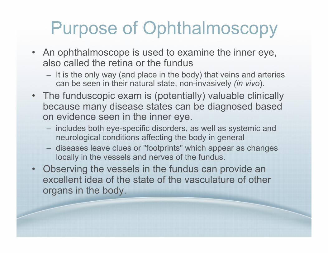
Purpose of Ophthalmoscopy • An ophthalmoscope is used to examine the inner eye,
also called the retina or the fundus – It is the only way (and place in the body) that veins and arteries
can be seen in their natural state, non-invasively (in vivo). • The funduscopic exam is (potentially) valuable clinically
because many disease states can be diagnosed based on evidence seen in the inner eye. – includes both eye-specific disorders, as well as systemic and
neurological conditions affecting the body in general – diseases leave clues or "footprints" which appear as changes
locally in the vessels and nerves of the fundus. • Observing the vessels in the fundus can provide an
excellent idea of the state of the vasculature of other organs in the body.

• Diseases which Manifest in the Fundus – AIDS (CMV retinitis), hypertension, and diabetes are
systemic diseases that manifest themselves in the fundus and thus ophthalmoscopy can provide valuable clues to their diagnosis.
– Papilledema is a sign of concussion or increased intra-cranial pressure.
– Other important eye disorders such as Glaucoma, Age-related Macular Degeneration, and Diabetic Retinopathy can be detected.
– Vein/Artery Occlusions -- related to Stroke – Tumors in the retina (melanoma)
Purpose of Ophthalmoscopy
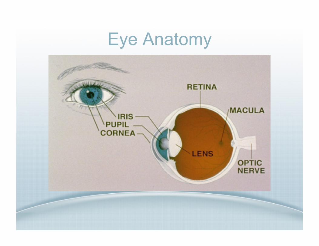
Eye Anatomy
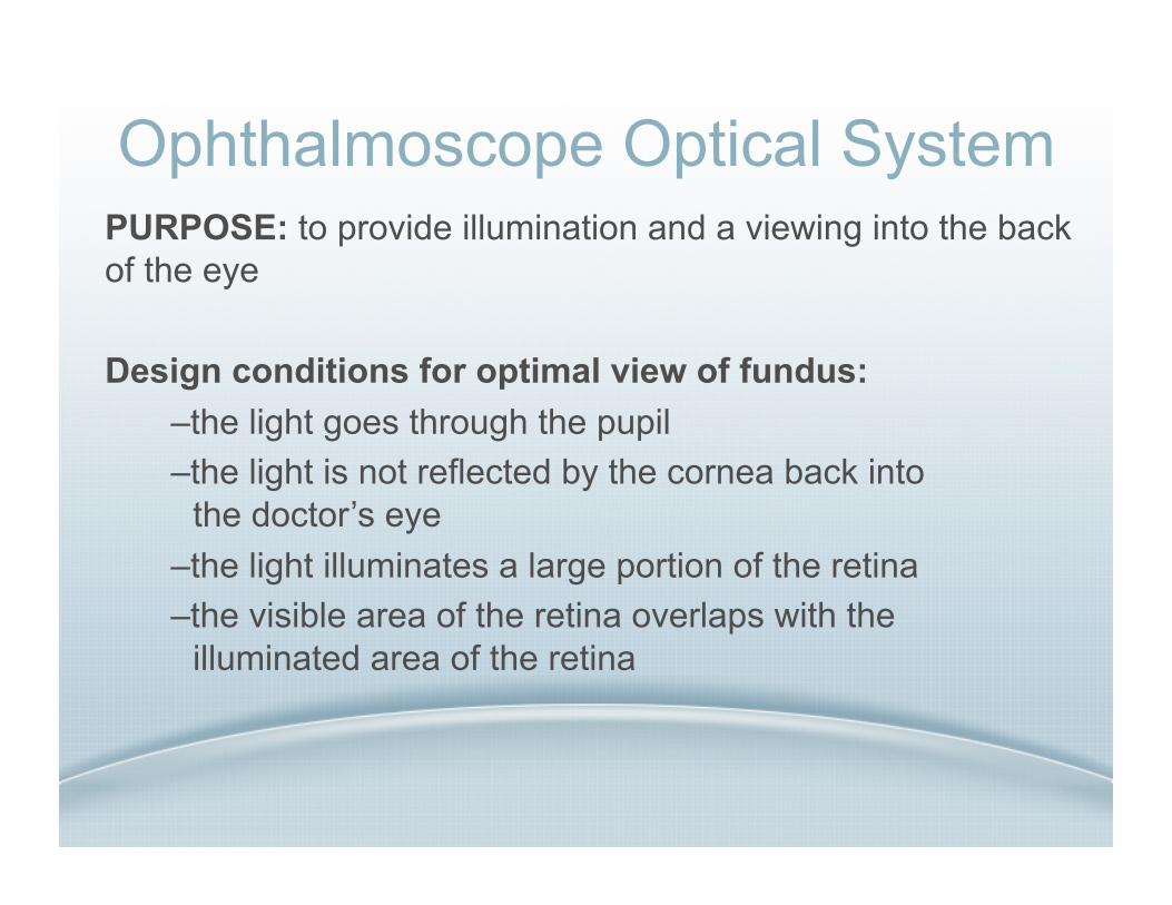
Ophthalmoscope Optical System PURPOSE: to provide illumination and a viewing into the back of the eye
Design conditions for optimal view of fundus: – the light goes through the pupil – the light is not reflected by the cornea back into the doctor’s eye
– the light illuminates a large portion of the retina – the visible area of the retina overlaps with the illuminated area of the retina
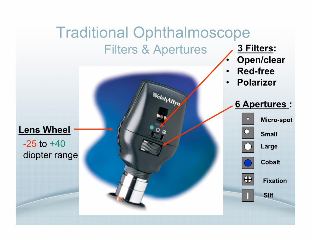
Traditional Ophthalmoscope Filters & Apertures
• Open/clear • Red-free • Polarizer
6 Apertures :
l
Micro-spot
Small
Large
Cobalt
Fixation
Slit
3 Filters:
Lens Wheel -25 to +40 diopter range

Ophthalmoscope Technique
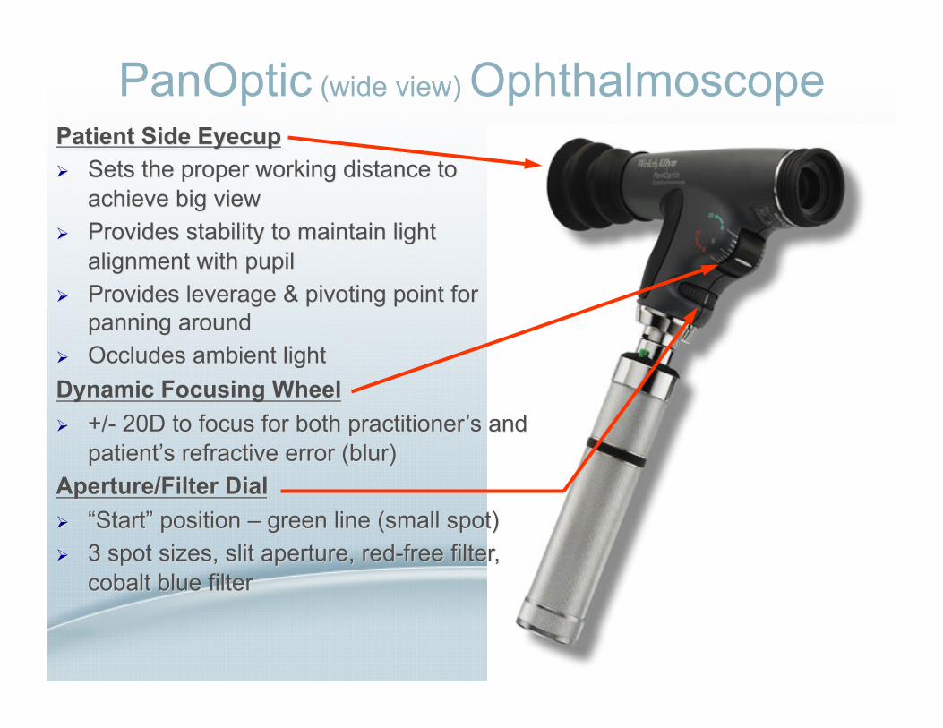
PanOptic (wide view) Ophthalmoscope

1. SET TO SMALL SPOT (green line on aperture dial) 2. FOCUS ACROSS THE ROOM on something 10 ft. away 3. EXAM POSITION
6 inches away and at a 15 to 20 degree angle on the temporal side of your patient
Looking through the scope, shine light at patient’s eye and look for the red retinal reflex
4. APPROACH & MAKE CONTACT Carefully follow the red reflex into the pupil The eyecup must contact patient’s brow to achieve view
5. COMPRESS EYECUP TO MAXIMIZE VIEW Make minor, fine-tuning adjustments to the focus if needed
PanOptic Technique

Ophthalmoscope for Disease Management In primary care, the ophthalmoscope is most often
used on patients who: • Are known or suspected to have Diabetes • Are known or suspected to have
Hypertension • Complain of Headaches • Have had any kind of Head trauma/injury • Have loss of Vision or degraded vision
(flashes, distortion, shadows) • Are undergoing a routine Physical
Examination • Have eye Pain – including abrasions
(scratches) and foreign bodies in the eye .
PREVELANT DISEASES DIAGNOSED SPECIALTY
Diabetic Retinopathy only 30% of diabetic patients see the ophthalmogist for annual exam
Internal Medicine, Family Medicine, Nephrology
Hypertensive Retinopathy useful in finding undetected hypertension helps monitor control & management of disease
Internal Medicine, Family Medicine, Nephrology
Papilledema/Head Trauma patient complaint of headaches
Internal Medicine, Family Medicine, Emergency Medicine
Central Nervous System Problems manifestation commonly seen in retina
Neurology
Shaken Baby Syndrome can visualize retinal hemorrhages consistent with syndrome
Pediatrics, Family Medicine
Foreign Bodies/Corneal Abrasions Internal Medicine, Family Medicine, Emergency Medicine, Pediatrics

Ophthalmoscope for Disease Management
– Clinically valuable • many systemic disease states manifest in the
fundus • AIDS, hypertension, diabetes
– Eye is extension of Central Nervous System • neurological disorders
– Looking for? Dots, Blots, Spots • + Abnormal blood vessel growth
(neovascularization)

Ophthalmoscope for Disease Management
• Normal Fundus

Ophthalmoscope for Disease Management • Diabetic Retinopathy
– After 10 years, majority of diabetics will have some form of Diabetic Retinopathy
– After 25 years, 85% will – Leading cause of
blindness! – Neovascularization
(abnormal blood vessel growth) proliferative stage
– Hemorrhages & micro aneurysms (red Blots)

Ophthalmoscope for Disease Management
• Hypertension – Vessel walls become
thickened and sclerotic – Vessel light reflex looks
like copper wiring – When artery crosses
vein, vein appears to disappear abruptly • “A/V nicking”
– Hemorrhages – Cotton-wool Spots
(retinal ischemia)
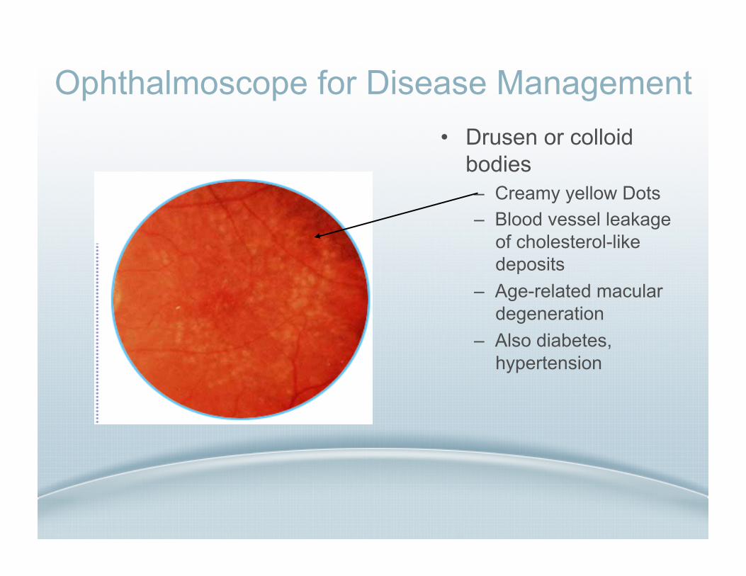
Ophthalmoscope for Disease Management • Drusen or colloid
bodies – Creamy yellow Dots – Blood vessel leakage
of cholesterol-like deposits
– Age-related macular degeneration
– Also diabetes, hypertension

Ophthalmoscope for Disease Management • Papilledema
– Increased pressure in the central nervous system
– Increased intra-cranial pressure
– Head injuries - headache
– Engorged veins – Disc elevation
Ophthalmoscope for Disease Management
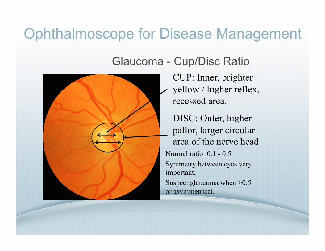
Glaucoma - Cup/Disc Ratio
Normal ratio: 0.1 - 0.5 Symmetry between eyes very important. Suspect glaucoma when >0.5 or asymmetrical.
CUP: Inner, brighter yellow / higher reflex, recessed area.
DISC: Outer, higher pallor, larger circular area of the nerve head.

Ophthalmoscope for Disease Management
• Central Retinal Vein Occlusion – Painless loss of vision – The classical “pizza
thrown against the wall” appearance of the fundus in this serious condition.
– Causes: hypertension, glaucoma, atherosclerosis, diabetes

Ophthalmoscope for Disease Management
• Chorioretinal Scar (Toxoplasmosis) – Congenital – Early lesion heals and
leaves scar in a cyst.

Ophthalmoscope for Disease Management • Detached Retina
– The thin wall of the retina is pulled away by the retracting of the vitreous gel.
Ophthalmoscope for Disease Management
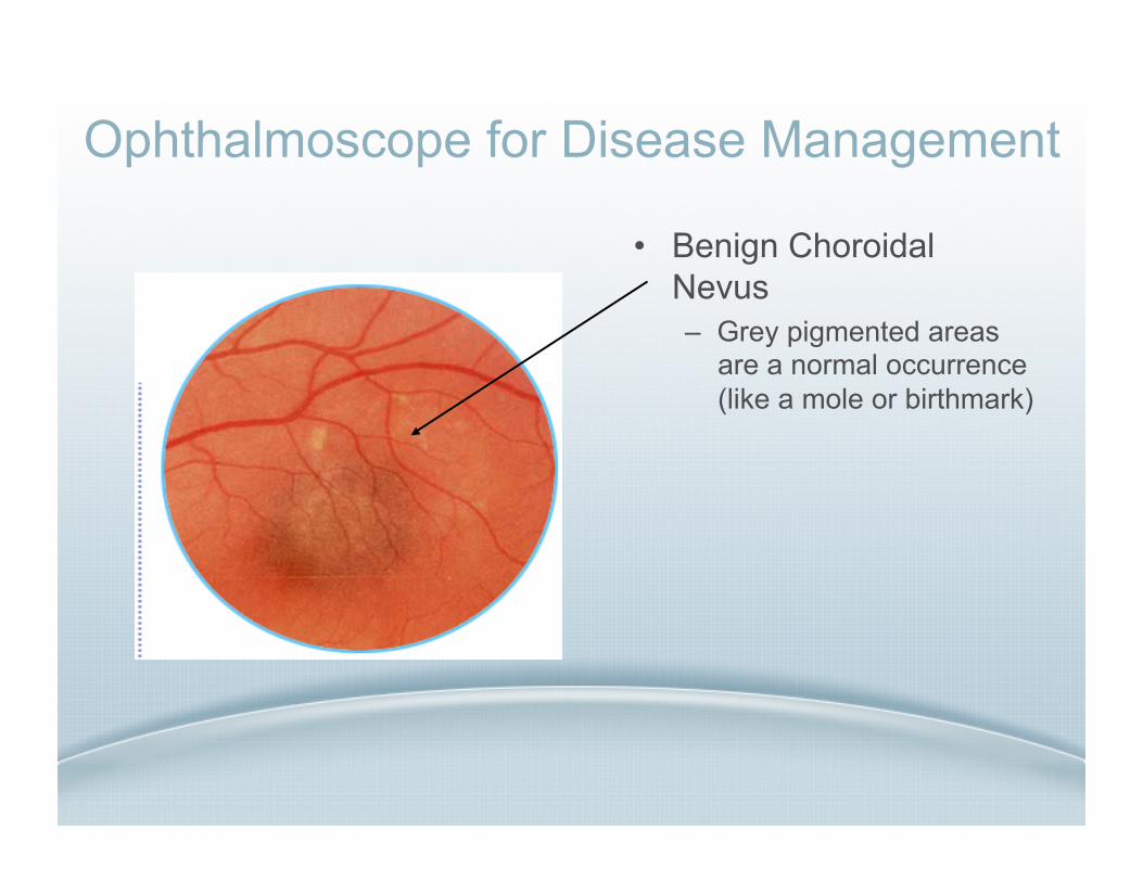
• Benign Choroidal Nevus – Grey pigmented areas
are a normal occurrence (like a mole or birthmark)
Related Documents