Proteolysis of multiple myelin basic protein isoforms after neurotrauma: characterization by mass spectrometry Andrew K. Ottens,* , Erin C. Golden, Liliana Bustamante,* Ronald L. Hayes,* , Nancy D. Denslowà and Kevin K. W. Wang* , *Departments of Psychiatry, Centers for Neuroproteomics and Biomarker Research and Traumatic Brain Injury Studies at the McKnight Brain Institute of the University of Florida Gainesville, Florida, USA Departments of Neuroscience, Centers for Neuroproteomics and Biomarker Research and Traumatic Brain Injury Studies at the McKnight Brain Institute of the University of Florida Gainesville, Florida, USA àDepartments of Physiological Sciences, Centers for Neuroproteomics and Biomarker Research and Traumatic Brain Injury Studies at the McKnight Brain Institute of the University of Florida Gainesville, Florida, USA Myelin is a multilamellar membrane that sheaths axons in the white matter of the CNS, which serves to protect and improve signal conductance (Arroyo and Scherer 2000). The membrane is composed of lipid layers interspersed between protein layers comprised near equally by myelin basic protein (MBP), proteolipid protein, and myelin-oligodendro- cyte glycoprotein (Norton and Cammer 1984). Each of the three myelin proteins is expressed as multiple isoforms (Voskuhl et al. 1994) that speculatively have unique func- tions in the formation and maintenance of myelin. In particular, MBP isoform dynamics vary in association with myelin formation, compaction and stabilization from embry- onic development on throughout life (Kruger et al. 1999), and is of interest to the study of neurological development (Boggs 2006), neurodegenerative disease (Schaecher et al. 2001; Boggs 2006) and neurotrauma (Liu et al. 2006a). Myelin basic protein isoforms are derived from differential splicing of the MBP gene (Harauz et al. 2004; Boggs 2006) with variations in the translation of exons 2, 5, and 6 (Kimura et al. 1998). Eight possible isoforms (Fig. 1) are derived, of which all have been identified in mice at the mRNA level (Nakajima et al. 1993), and five (21.6, 18.6, 17.3, 17.2, and 14.0 kDa) at the protein level (Givogri et al. 2000). Rat and Received July 18, 2007; revised manuscript received October 16, 2007; accepted October 17, 2007. Address correspondence and reprint requests to Andrew K. Ottens, PO Box 100256, Gainesville, FL 32610-0256, USA. E-mail: [email protected]fl.edu Abbreviations used: IDA, isotope dilution analysis; M a , apparent molecular mass; MBP, myelin basic protein; MS, multiple sclerosis; MSMS, tandem mass spectrometry; RPLC, reversed-phase liquid chro- matography; TBI, traumatic brain injury. Abstract Neurotrauma, as in the case of traumatic brain injury, pro- motes protease over-activation characterized by the select fragmentation of brain proteins. The resulting polypeptides are indicators of biochemical processes, which can be used to study post-injury dynamics and may also be developed into biomarkers. To this end, we devised a novel mass spec- trometry approach to characterize post-injury calpain proteo- lytic processing of myelin basic protein (MBP), a biomarker of brain injury that denotes white matter damage and recovery. Our approach exceeds conventional immunological assays in its deconvolution of multiple protein isoforms, its absolute quantification of proteolytic fragments and its polypeptide selectivity. We quantified and characterized post-injury pro- teolytic processing of all MBP isoforms identified in adult rat cortex. Further, the translation of calpain-cleaved MBP into CSF was verified following brain injury. We ascertained that the exon-6 sequence of MBP resulted in a characteristic shift in gel migration for intact and fragmented protein alike. We also found evidence for a second post-TBI cleavage event within exon-2 and for the dimerization of the post-TBI 4.3 kDa fragment. Ultimately, the novel methodology described here can be used to study MBP dynamics and other similar pro- teolytic events of relevance to brain injury and other CNS processes. Keywords: biomarkers, mass spectrometry, myelin basic protein, neurotrauma, proteolysis, quantification. J. Neurochem. (2008) 104, 1404–1414. d JOURNAL OF NEUROCHEMISTRY | 2008 | 104 | 1404–1414 doi: 10.1111/j.1471-4159.2007.05086.x 1404 Journal Compilation ȑ 2007 International Society for Neurochemistry, J. Neurochem. (2008) 104, 1404–1414 ȑ 2007 The Authors

Welcome message from author
This document is posted to help you gain knowledge. Please leave a comment to let me know what you think about it! Share it to your friends and learn new things together.
Transcript
Proteolysis of multiple myelin basic protein isoforms afterneurotrauma: characterization by mass spectrometry
Andrew K. Ottens,*,� Erin C. Golden,� Liliana Bustamante,* Ronald L. Hayes,*,�Nancy D. Denslow� and Kevin K. W. Wang*,�
*Departments of Psychiatry, Centers for Neuroproteomics and Biomarker Research and Traumatic Brain Injury Studies at the
McKnight Brain Institute of the University of Florida Gainesville, Florida, USA
�Departments of Neuroscience, Centers for Neuroproteomics and Biomarker Research and Traumatic Brain Injury Studies at the
McKnight Brain Institute of the University of Florida Gainesville, Florida, USA
�Departments of Physiological Sciences, Centers for Neuroproteomics and Biomarker Research and Traumatic Brain Injury Studies
at the McKnight Brain Institute of the University of Florida Gainesville, Florida, USA
Myelin is a multilamellar membrane that sheaths axons in thewhite matter of the CNS, which serves to protect andimprove signal conductance (Arroyo and Scherer 2000). Themembrane is composed of lipid layers interspersed betweenprotein layers comprised near equally by myelin basicprotein (MBP), proteolipid protein, and myelin-oligodendro-cyte glycoprotein (Norton and Cammer 1984). Each of thethree myelin proteins is expressed as multiple isoforms(Voskuhl et al. 1994) that speculatively have unique func-tions in the formation and maintenance of myelin. Inparticular, MBP isoform dynamics vary in association withmyelin formation, compaction and stabilization from embry-onic development on throughout life (Kruger et al. 1999),and is of interest to the study of neurological development(Boggs 2006), neurodegenerative disease (Schaecher et al.2001; Boggs 2006) and neurotrauma (Liu et al. 2006a).
Myelin basic protein isoforms are derived from differentialsplicing of the MBP gene (Harauz et al. 2004; Boggs 2006)with variations in the translation of exons 2, 5, and 6 (Kimuraet al. 1998). Eight possible isoforms (Fig. 1) are derived, ofwhich all have been identified in mice at the mRNA level(Nakajima et al. 1993), and five (21.6, 18.6, 17.3, 17.2, and14.0 kDa) at the protein level (Givogri et al. 2000). Rat and
Received July 18, 2007; revised manuscript received October 16, 2007;accepted October 17, 2007.Address correspondence and reprint requests to Andrew K. Ottens, PO
Box 100256, Gainesville, FL 32610-0256, USA.E-mail: [email protected] used: IDA, isotope dilution analysis; Ma, apparent
molecular mass; MBP, myelin basic protein; MS, multiple sclerosis;MSMS, tandem mass spectrometry; RPLC, reversed-phase liquid chro-matography; TBI, traumatic brain injury.
Abstract
Neurotrauma, as in the case of traumatic brain injury, pro-
motes protease over-activation characterized by the select
fragmentation of brain proteins. The resulting polypeptides are
indicators of biochemical processes, which can be used to
study post-injury dynamics and may also be developed into
biomarkers. To this end, we devised a novel mass spec-
trometry approach to characterize post-injury calpain proteo-
lytic processing of myelin basic protein (MBP), a biomarker of
brain injury that denotes white matter damage and recovery.
Our approach exceeds conventional immunological assays in
its deconvolution of multiple protein isoforms, its absolute
quantification of proteolytic fragments and its polypeptide
selectivity. We quantified and characterized post-injury pro-
teolytic processing of all MBP isoforms identified in adult rat
cortex. Further, the translation of calpain-cleaved MBP into
CSF was verified following brain injury. We ascertained that
the exon-6 sequence of MBP resulted in a characteristic shift
in gel migration for intact and fragmented protein alike. We
also found evidence for a second post-TBI cleavage event
within exon-2 and for the dimerization of the post-TBI 4.3 kDa
fragment. Ultimately, the novel methodology described here
can be used to study MBP dynamics and other similar pro-
teolytic events of relevance to brain injury and other CNS
processes.
Keywords: biomarkers, mass spectrometry, myelin basic
protein, neurotrauma, proteolysis, quantification.
J. Neurochem. (2008) 104, 1404–1414.
d JOURNAL OF NEUROCHEMISTRY | 2008 | 104 | 1404–1414 doi: 10.1111/j.1471-4159.2007.05086.x
1404 Journal Compilation � 2007 International Society for Neurochemistry, J. Neurochem. (2008) 104, 1404–1414� 2007 The Authors
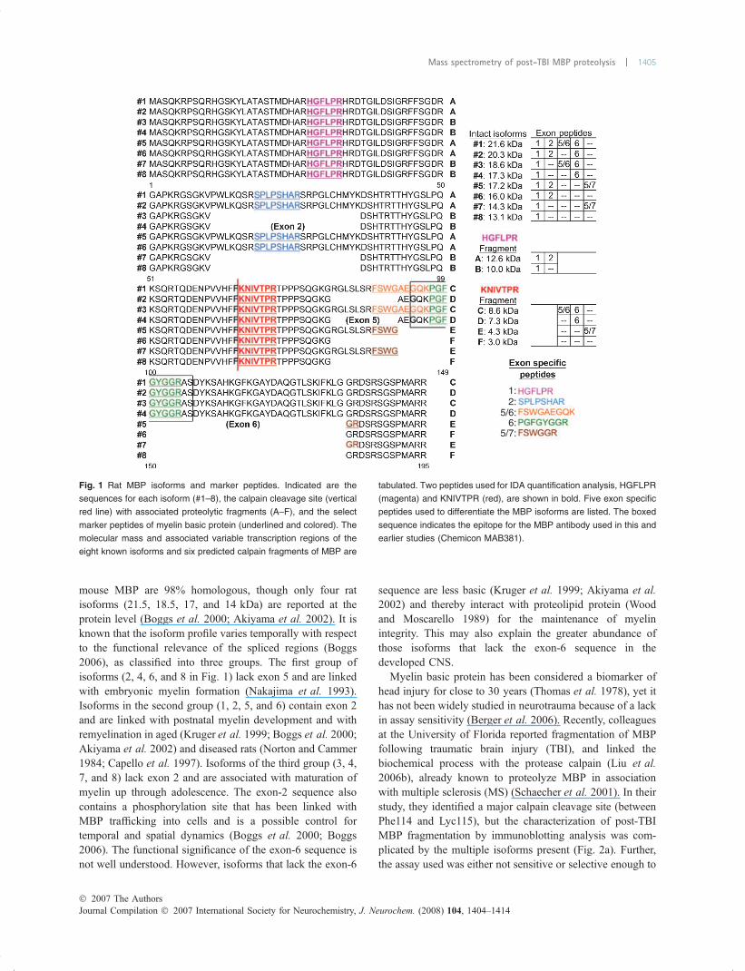
mouse MBP are 98% homologous, though only four ratisoforms (21.5, 18.5, 17, and 14 kDa) are reported at theprotein level (Boggs et al. 2000; Akiyama et al. 2002). It isknown that the isoform profile varies temporally with respectto the functional relevance of the spliced regions (Boggs2006), as classified into three groups. The first group ofisoforms (2, 4, 6, and 8 in Fig. 1) lack exon 5 and are linkedwith embryonic myelin formation (Nakajima et al. 1993).Isoforms in the second group (1, 2, 5, and 6) contain exon 2and are linked with postnatal myelin development and withremyelination in aged (Kruger et al. 1999; Boggs et al. 2000;Akiyama et al. 2002) and diseased rats (Norton and Cammer1984; Capello et al. 1997). Isoforms of the third group (3, 4,7, and 8) lack exon 2 and are associated with maturation ofmyelin up through adolescence. The exon-2 sequence alsocontains a phosphorylation site that has been linked withMBP trafficking into cells and is a possible control fortemporal and spatial dynamics (Boggs et al. 2000; Boggs2006). The functional significance of the exon-6 sequence isnot well understood. However, isoforms that lack the exon-6
sequence are less basic (Kruger et al. 1999; Akiyama et al.2002) and thereby interact with proteolipid protein (Woodand Moscarello 1989) for the maintenance of myelinintegrity. This may also explain the greater abundance ofthose isoforms that lack the exon-6 sequence in thedeveloped CNS.
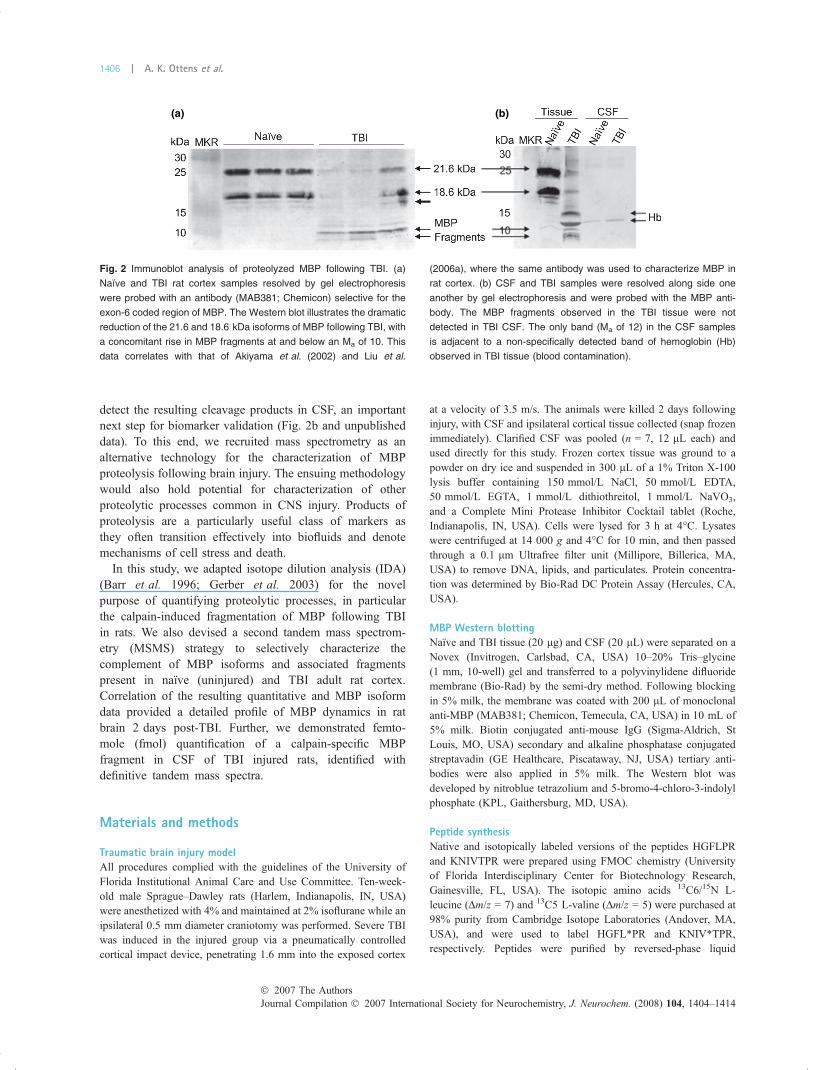
Myelin basic protein has been considered a biomarker ofhead injury for close to 30 years (Thomas et al. 1978), yet ithas not been widely studied in neurotrauma because of a lackin assay sensitivity (Berger et al. 2006). Recently, colleaguesat the University of Florida reported fragmentation of MBPfollowing traumatic brain injury (TBI), and linked thebiochemical process with the protease calpain (Liu et al.2006b), already known to proteolyze MBP in associationwith multiple sclerosis (MS) (Schaecher et al. 2001). In theirstudy, they identified a major calpain cleavage site (betweenPhe114 and Lyc115), but the characterization of post-TBIMBP fragmentation by immunoblotting analysis was com-plicated by the multiple isoforms present (Fig. 2a). Further,the assay used was either not sensitive or selective enough to
1:2:
5/6:
5/7:6:
Fig. 1 Rat MBP isoforms and marker peptides. Indicated are the
sequences for each isoform (#1–8), the calpain cleavage site (vertical
red line) with associated proteolytic fragments (A–F), and the select
marker peptides of myelin basic protein (underlined and colored). The
molecular mass and associated variable transcription regions of the
eight known isoforms and six predicted calpain fragments of MBP are
tabulated. Two peptides used for IDA quantification analysis, HGFLPR
(magenta) and KNIVTPR (red), are shown in bold. Five exon specific
peptides used to differentiate the MBP isoforms are listed. The boxed
sequence indicates the epitope for the MBP antibody used in this and
earlier studies (Chemicon MAB381).
� 2007 The AuthorsJournal Compilation � 2007 International Society for Neurochemistry, J. Neurochem. (2008) 104, 1404–1414
Mass spectrometry of post-TBI MBP proteolysis | 1405
detect the resulting cleavage products in CSF, an importantnext step for biomarker validation (Fig. 2b and unpublisheddata). To this end, we recruited mass spectrometry as analternative technology for the characterization of MBPproteolysis following brain injury. The ensuing methodologywould also hold potential for characterization of otherproteolytic processes common in CNS injury. Products ofproteolysis are a particularly useful class of markers asthey often transition effectively into biofluids and denotemechanisms of cell stress and death.
In this study, we adapted isotope dilution analysis (IDA)(Barr et al. 1996; Gerber et al. 2003) for the novelpurpose of quantifying proteolytic processes, in particularthe calpain-induced fragmentation of MBP following TBIin rats. We also devised a second tandem mass spectrom-etry (MSMS) strategy to selectively characterize thecomplement of MBP isoforms and associated fragmentspresent in naıve (uninjured) and TBI adult rat cortex.Correlation of the resulting quantitative and MBP isoformdata provided a detailed profile of MBP dynamics in ratbrain 2 days post-TBI. Further, we demonstrated femto-mole (fmol) quantification of a calpain-specific MBPfragment in CSF of TBI injured rats, identified withdefinitive tandem mass spectra.
Materials and methods
Traumatic brain injury modelAll procedures complied with the guidelines of the University of
Florida Institutional Animal Care and Use Committee. Ten-week-
old male Sprague–Dawley rats (Harlem, Indianapolis, IN, USA)
were anesthetized with 4% and maintained at 2% isoflurane while an
ipsilateral 0.5 mm diameter craniotomy was performed. Severe TBI
was induced in the injured group via a pneumatically controlled
cortical impact device, penetrating 1.6 mm into the exposed cortex
at a velocity of 3.5 m/s. The animals were killed 2 days following
injury, with CSF and ipsilateral cortical tissue collected (snap frozen
immediately). Clarified CSF was pooled (n = 7, 12 lL each) and
used directly for this study. Frozen cortex tissue was ground to a
powder on dry ice and suspended in 300 lL of a 1% Triton X-100
lysis buffer containing 150 mmol/L NaCl, 50 mmol/L EDTA,
50 mmol/L EGTA, 1 mmol/L dithiothreitol, 1 mmol/L NaVO3,
and a Complete Mini Protease Inhibitor Cocktail tablet (Roche,
Indianapolis, IN, USA). Cells were lysed for 3 h at 4�C. Lysateswere centrifuged at 14 000 g and 4�C for 10 min, and then passed
through a 0.1 lm Ultrafree filter unit (Millipore, Billerica, MA,
USA) to remove DNA, lipids, and particulates. Protein concentra-
tion was determined by Bio-Rad DC Protein Assay (Hercules, CA,
USA).
MBP Western blottingNaıve and TBI tissue (20 lg) and CSF (20 lL) were separated on a
Novex (Invitrogen, Carlsbad, CA, USA) 10–20% Tris–glycine
(1 mm, 10-well) gel and transferred to a polyvinylidene difluoride
membrane (Bio-Rad) by the semi-dry method. Following blocking
in 5% milk, the membrane was coated with 200 lL of monoclonal
anti-MBP (MAB381; Chemicon, Temecula, CA, USA) in 10 mL of
5% milk. Biotin conjugated anti-mouse IgG (Sigma-Aldrich, St
Louis, MO, USA) secondary and alkaline phosphatase conjugated
streptavadin (GE Healthcare, Piscataway, NJ, USA) tertiary anti-
bodies were also applied in 5% milk. The Western blot was
developed by nitroblue tetrazolium and 5-bromo-4-chloro-3-indolyl
phosphate (KPL, Gaithersburg, MD, USA).
Peptide synthesisNative and isotopically labeled versions of the peptides HGFLPR
and KNIVTPR were prepared using FMOC chemistry (University
of Florida Interdisciplinary Center for Biotechnology Research,
Gainesville, FL, USA). The isotopic amino acids 13C6/15N L-
leucine (Dm/z = 7) and 13C5 L-valine (Dm/z = 5) were purchased at
98% purity from Cambridge Isotope Laboratories (Andover, MA,
USA), and were used to label HGFL*PR and KNIV*TPR,
respectively. Peptides were purified by reversed-phase liquid
(a) (b)
Fig. 2 Immunoblot analysis of proteolyzed MBP following TBI. (a)
Naıve and TBI rat cortex samples resolved by gel electrophoresis
were probed with an antibody (MAB381; Chemicon) selective for the
exon-6 coded region of MBP. The Western blot illustrates the dramatic
reduction of the 21.6 and 18.6 kDa isoforms of MBP following TBI, with
a concomitant rise in MBP fragments at and below an Ma of 10. This
data correlates with that of Akiyama et al. (2002) and Liu et al.
(2006a), where the same antibody was used to characterize MBP in
rat cortex. (b) CSF and TBI samples were resolved along side one
another by gel electrophoresis and were probed with the MBP anti-
body. The MBP fragments observed in the TBI tissue were not
detected in TBI CSF. The only band (Ma of 12) in the CSF samples
is adjacent to a non-specifically detected band of hemoglobin (Hb)
observed in TBI tissue (blood contamination).
Journal Compilation � 2007 International Society for Neurochemistry, J. Neurochem. (2008) 104, 1404–1414� 2007 The Authors
1406 | A. K. Ottens et al.
chromatography (RPLC) to 99% purity as confirmed by mass
spectrometry.
Gel electrophoresisSamples mixed with Laemmli buffer (Bio-Rad) and 10 mmol/L
dithiothreitol were heated to 80�C for 2 min prior to separation on
Invitrogen Tris–glycine Novex polyacrylamide gels (1 mm, 10-well).
Rainbow molecular mass marker (GE Healthcare) was used to
indicate apparent molecular mass (Ma), as calculated for extracted gel
slices using Phoretix 1D gel processing software (Nonlinear USA,
Durham, NC, USA). Bovine MBP (Sigma Aldrich) was purified by
cation exchange chromatography to isolate the 18.6 kDa MBP
isoform as a marker and positive control. Gels were visualized by
staining with concentrated Bio-Rad Coomassie Blue R250 for 20 min
and destained in 40% ethanol/10% acetic acid in water for 2 h.
In-gel clostripain digestionExcised gel slices were cut into 1-mm cubes and washed in HPLC
grade water (Burdick & Jackson, Muskegon, MI, USA), destained in
50 : 50 100 mmol/L NH4HCO3/spectroscopy grade acetonitrile
(Burdick & Jackson) and dried in 100% acetonitrile followed by
speed vacuum. Dried gel pieces were re-hydrated with 20 lL of a
25 ng/lL clostripain solution (Worthington Biochemical, Lake-
wood, NJ, USA) made with an NH4HCO3 buffer (100 mmol/L)
with CaCl2 (20 mmol/L) and the two isotopically labeled internal
standard peptides (10 nmol/L). Digestion was performed overnight
at 37�C. The resulting peptide supernatant and two 50% acetonitrile
extractions were pooled and dried by speed vacuum. Peptides were
resuspended in 20 lL of HPLC grade 4% acetonitrile/0.1% formic
acid (J. T. Baker, Phillipsburg, NJ, USA).
Capillary reversed-phase liquid chromatographyTwo microliters of each sample was loaded by autosampler onto a
100 lm diameter · 5 cm long column of 3-lm C-18 reversed-phase
particles (Agilent, Palo Alto, CA, USA) packed behind an Upchurch
(Oak Harbor, WA, USA) 0.5-lm polyetherketone microfilter
assembly operated at a flow rate of 600 nL/min. A rapid 10 min
gradient separation from 4% to 30% acetonitrile (0.1% formic acid)
was performed for quantitative analysis. A longer 40 min gradient
separation from 4% to 60% acetonitrile was performed for MBP
isoform characterization. Chromatographic effluent was directly
electrosprayed into a Thermo Electron (San Jose, CA, USA) LCQ
Deca XP Plus� ion trap mass spectrometer.
Quantitative isotope dilution analysisControl and TBI samples (50 lg for cortical tissue and 20 lL for
CSF) were resolved by gel electrophoresis. Each gel lane was sliced
into 1-mm bands below a relative molecular mass of 30. For each
gel slice, we performed an in-gel digestion using clostripain to
which a fixed amount of isotopically labeled HGFLPR and
KNIVTPR peptides was added as internal standards. A custom
mass spectrometry method was developed to sequentially perform
MSMS analysis for the endogenous and isotopically labeled
HGFLPR and KNIVTPR peptides. Chromatographic peaks were
identified after filtering for the doubly charged precursor ion and the
respective dominant singly charged fragment ion (the y5 ion of
HGFLPR and y6 ion of KNIVTPR). Peptide selectivity was
confirmed by the observed tandem mass spectrum. We then
multiplied the peak area ratio of endogenous-to-isotopically labeled
peptide by the amount of internal standard (14 fmol for tissue and
18.6 fmol for CSF) to determine the quantity of endogenous peptide
sampled (one-tenth of total digest). Data was reported as the mean
with the range indicated by error bars. Statistical evaluation was
performed with a non-parametric one-tailed Mann–Whitney U-test(n of 3), anticipating a decrease in intact MBP following injury.
MBP isoform characterizationAs above, 50 lg of control and TBI samples were resolved by gel
electrophoresis, with gel slices extracted below an Ma of 30. In-gel
trypsin (Promega, Madison, WI, USA) digestion was performed as
described for clostripain, but without CaCl2 and internal standard
peptides. Five of 30 lL for each digest was analyzed by RPLC-
MSMS. A custom MSMS method was devised to sequentially
analyze five target peptides selective for the variable transcription
regions of MBP: exon encoding sequences 1, HGFLPR; 2,
SPLPSHAR; tandem 5/6, FSWGAEGQK; 6, PGFGYGGR; tandem
5/7, FSWGGR (the short exon-5 coded region was best character-
ized by peptides overlapping with adjacent sequences). Chromato-
graphic peaks were identified after filtering for the precursor ion and
dominant fragment ion of each peptide (confirmed by tandem mass
spectra). Peak areas for each target peptide were averaged from
replicate digests (same Ma, n of 3) and normalized across all
samples to adjust for differences in extraction, separation, and
ionization efficiencies between peptides.
Determination of peptide specificity and tandem mass spectrumselectivityPeptide sequence specificity was evaluated by BLAST analysis
(NCBI, Bethesda, MD, USA) with a word size of 2 and an expect
value of 20 000 searched against a non-redundant RefSeq (NCBI)
database (Release 22) filtered for rattus norvegicus proteins. Sepa-
rately, tandem mass spectrum selectivity was evaluated by Mascot
search (Matrix Science, Boston, MA, USA). Raw MSMS data were
converted to DTA file format (extract_msn.exe; Thermo Electron) for
each peptide.Mascot parameters includedArg-C (clostripain) enzyme
selectivity and a mass tolerance of 0.75 Da for peptide and MSMS
values. Searches were performed against an Integr8 non-redundant
rattus norvegicus (species 122) protein database (Release 59).
Results
Isotope dilution mass spectrometry quantificationWe adapted IDA to selectively quantify proteolytic cleavageevents, in our case calpain-induced MBP fragmentation, bytargeting the resulting N-terminal peptide (KNIVTPR). It isfirst important to demonstrate that the target peptide is readilydetected. We analyzed tryptic digests of purified MBP bydata-dependent mass spectrometry, which biases detection ofpeptides that ionize efficiently (Dongre et al. 1997; Aeber-sold and Mann 2003). We observed a strong signal anddetailed tandem mass spectrum for the related NIVTPRpeptide. HGFLPR was another peptide that was readilydetected, whichwas common to all isoforms ofMBP (exon-1).
� 2007 The AuthorsJournal Compilation � 2007 International Society for Neurochemistry, J. Neurochem. (2008) 104, 1404–1414
Mass spectrometry of post-TBI MBP proteolysis | 1407
Pre-evaluation, however, may prove more difficult for othercleavage specific peptides, as discussed later. Native andisotopically labeled forms of the KNIVTPR peptide were
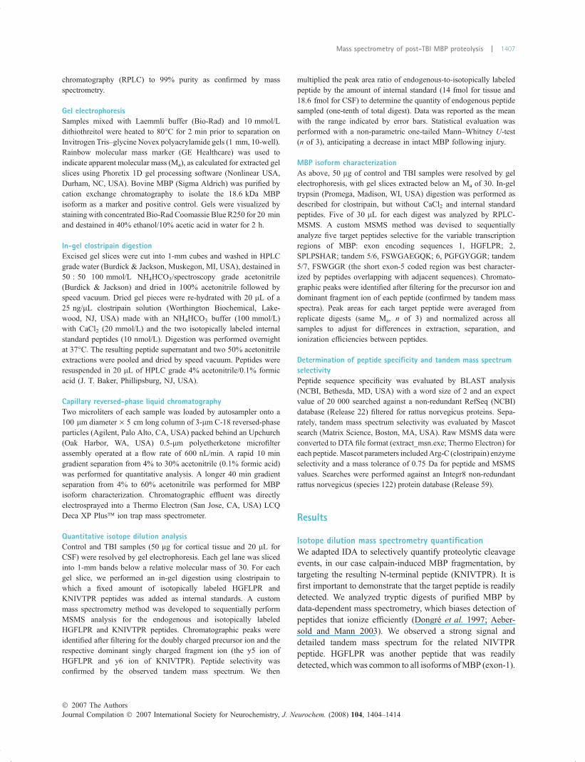
synthesized with a mass difference of 5 Da (easily resolvedby the mass spectrometer despite simultaneous chromato-graphic elution) (Gerber et al. 2003). We also synthesizednative and isotopically labeled forms (7 Da mass difference)of the peptide HGFLPR to quantify intact MBP and MBPfragments complementary to those detected with KNIVTPR.We used the native peptides for assay development, while theisotopically labeled peptides served as internal standards forquantification. RPLC and MSMS parameters were optimizedwith mixtures of the four synthesized peptides to produce arapid 10 min RPLC gradient that resolved HGFLPR andKNIVTPR (Fig. 3a) with subsequent selective detection(Fig. 3b). Data were filtered by the mass-to-charge (m/z)values for the intact peptide ion (precursor ion) and mostabundant fragment ion to generate a chromatographic peakfor each peptide. The moles of native peptide were quantifiedby multiplying the peak area ratio (native to internalstandard) by the known concentration of internal standard.The method also assumed a linear dynamic range, which wasvalidated across four orders in magnitude and with adetection limit of less than 1 fmol (Fig. 3c).
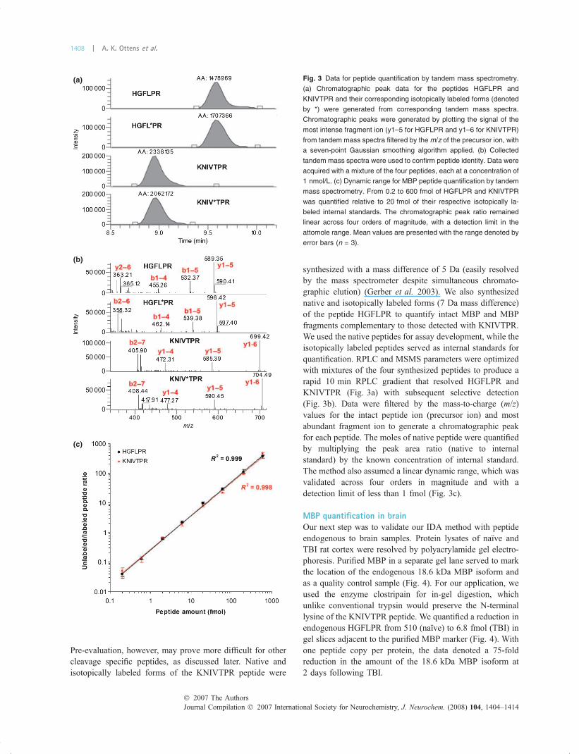
MBP quantification in brainOur next step was to validate our IDA method with peptideendogenous to brain samples. Protein lysates of naıve andTBI rat cortex were resolved by polyacrylamide gel electro-phoresis. Purified MBP in a separate gel lane served to markthe location of the endogenous 18.6 kDa MBP isoform andas a quality control sample (Fig. 4). For our application, weused the enzyme clostripain for in-gel digestion, whichunlike conventional trypsin would preserve the N-terminallysine of the KNIVTPR peptide. We quantified a reduction inendogenous HGFLPR from 510 (naıve) to 6.8 fmol (TBI) ingel slices adjacent to the purified MBP marker (Fig. 4). Withone peptide copy per protein, the data denoted a 75-foldreduction in the amount of the 18.6 kDa MBP isoform at2 days following TBI.
(b)
(a)
(c)
Fig. 3 Data for peptide quantification by tandem mass spectrometry.
(a) Chromatographic peak data for the peptides HGFLPR and
KNIVTPR and their corresponding isotopically labeled forms (denoted
by *) were generated from corresponding tandem mass spectra.
Chromatographic peaks were generated by plotting the signal of the
most intense fragment ion (y1–5 for HGFLPR and y1–6 for KNIVTPR)
from tandem mass spectra filtered by the m/z of the precursor ion, with
a seven-point Gaussian smoothing algorithm applied. (b) Collected
tandem mass spectra were used to confirm peptide identity. Data were
acquired with a mixture of the four peptides, each at a concentration of
1 nmol/L. (c) Dynamic range for MBP peptide quantification by tandem
mass spectrometry. From 0.2 to 600 fmol of HGFLPR and KNIVTPR
was quantified relative to 20 fmol of their respective isotopically la-
beled internal standards. The chromatographic peak ratio remained
linear across four orders of magnitude, with a detection limit in the
attomole range. Mean values are presented with the range denoted by
error bars (n = 3).
Journal Compilation � 2007 International Society for Neurochemistry, J. Neurochem. (2008) 104, 1404–1414� 2007 The Authors
1408 | A. K. Ottens et al.
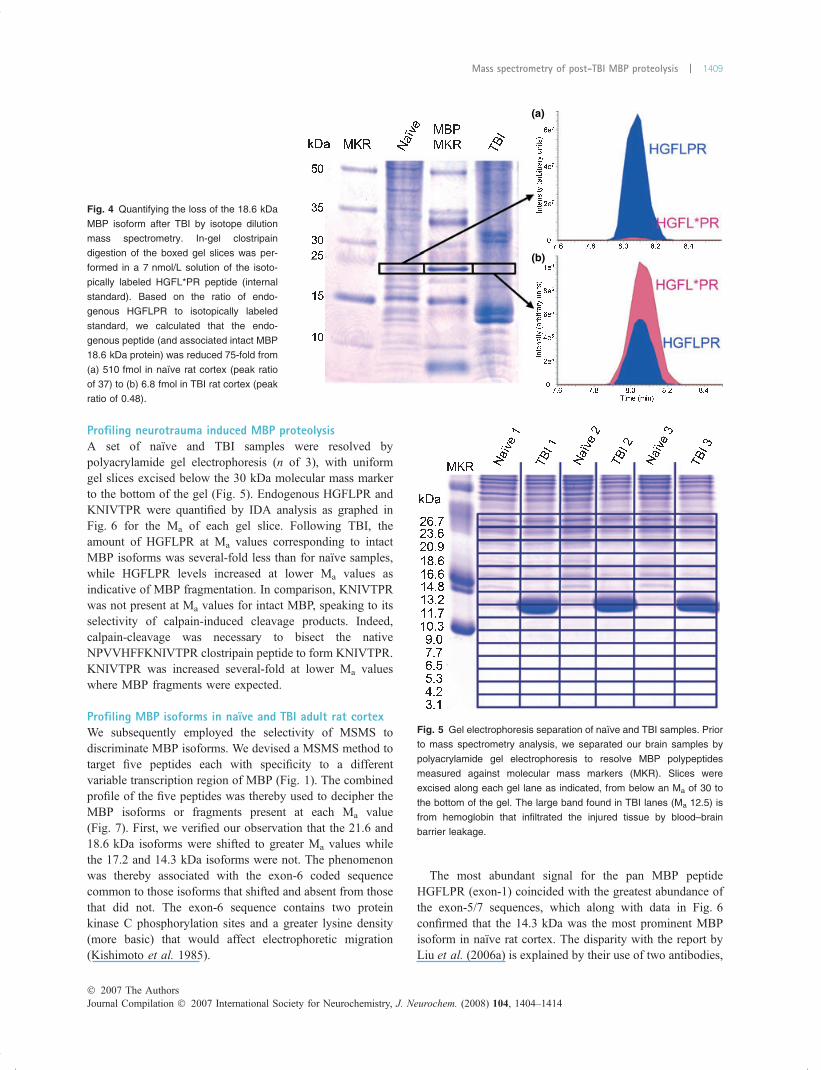
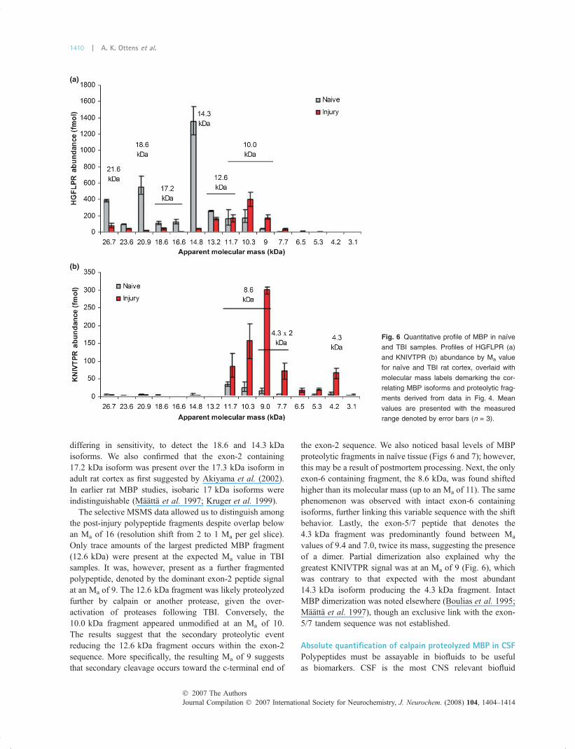
Profiling neurotrauma induced MBP proteolysisA set of naıve and TBI samples were resolved bypolyacrylamide gel electrophoresis (n of 3), with uniformgel slices excised below the 30 kDa molecular mass markerto the bottom of the gel (Fig. 5). Endogenous HGFLPR andKNIVTPR were quantified by IDA analysis as graphed inFig. 6 for the Ma of each gel slice. Following TBI, theamount of HGFLPR at Ma values corresponding to intactMBP isoforms was several-fold less than for naıve samples,while HGFLPR levels increased at lower Ma values asindicative of MBP fragmentation. In comparison, KNIVTPRwas not present at Ma values for intact MBP, speaking to itsselectivity of calpain-induced cleavage products. Indeed,calpain-cleavage was necessary to bisect the nativeNPVVHFFKNIVTPR clostripain peptide to form KNIVTPR.KNIVTPR was increased several-fold at lower Ma valueswhere MBP fragments were expected.
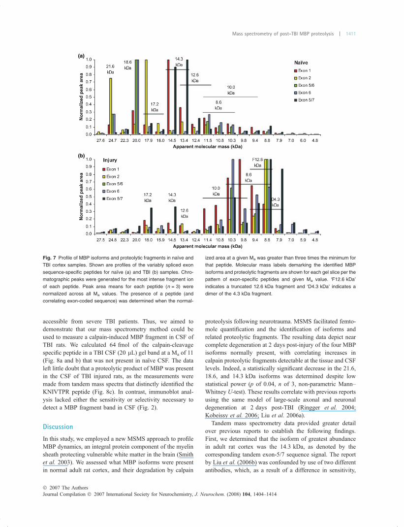
Profiling MBP isoforms in naıve and TBI adult rat cortexWe subsequently employed the selectivity of MSMS todiscriminate MBP isoforms. We devised a MSMS method totarget five peptides each with specificity to a differentvariable transcription region of MBP (Fig. 1). The combinedprofile of the five peptides was thereby used to decipher theMBP isoforms or fragments present at each Ma value(Fig. 7). First, we verified our observation that the 21.6 and18.6 kDa isoforms were shifted to greater Ma values whilethe 17.2 and 14.3 kDa isoforms were not. The phenomenonwas thereby associated with the exon-6 coded sequencecommon to those isoforms that shifted and absent from thosethat did not. The exon-6 sequence contains two proteinkinase C phosphorylation sites and a greater lysine density(more basic) that would affect electrophoretic migration(Kishimoto et al. 1985).
The most abundant signal for the pan MBP peptideHGFLPR (exon-1) coincided with the greatest abundance ofthe exon-5/7 sequences, which along with data in Fig. 6confirmed that the 14.3 kDa was the most prominent MBPisoform in naıve rat cortex. The disparity with the report byLiu et al. (2006a) is explained by their use of two antibodies,
(a)
(b)
Fig. 4 Quantifying the loss of the 18.6 kDa
MBP isoform after TBI by isotope dilution
mass spectrometry. In-gel clostripain
digestion of the boxed gel slices was per-
formed in a 7 nmol/L solution of the isoto-
pically labeled HGFL*PR peptide (internal
standard). Based on the ratio of endo-
genous HGFLPR to isotopically labeled
standard, we calculated that the endo-
genous peptide (and associated intact MBP
18.6 kDa protein) was reduced 75-fold from
(a) 510 fmol in naıve rat cortex (peak ratio
of 37) to (b) 6.8 fmol in TBI rat cortex (peak
ratio of 0.48).
Fig. 5 Gel electrophoresis separation of naıve and TBI samples. Prior
to mass spectrometry analysis, we separated our brain samples by
polyacrylamide gel electrophoresis to resolve MBP polypeptides
measured against molecular mass markers (MKR). Slices were
excised along each gel lane as indicated, from below an Ma of 30 to
the bottom of the gel. The large band found in TBI lanes (Ma 12.5) is
from hemoglobin that infiltrated the injured tissue by blood–brain
barrier leakage.
� 2007 The AuthorsJournal Compilation � 2007 International Society for Neurochemistry, J. Neurochem. (2008) 104, 1404–1414
Mass spectrometry of post-TBI MBP proteolysis | 1409
differing in sensitivity, to detect the 18.6 and 14.3 kDaisoforms. We also confirmed that the exon-2 containing17.2 kDa isoform was present over the 17.3 kDa isoform inadult rat cortex as first suggested by Akiyama et al. (2002).In earlier rat MBP studies, isobaric 17 kDa isoforms wereindistinguishable (Maatta et al. 1997; Kruger et al. 1999).
The selective MSMS data allowed us to distinguish amongthe post-injury polypeptide fragments despite overlap belowan Ma of 16 (resolution shift from 2 to 1 Ma per gel slice).Only trace amounts of the largest predicted MBP fragment(12.6 kDa) were present at the expected Ma value in TBIsamples. It was, however, present as a further fragmentedpolypeptide, denoted by the dominant exon-2 peptide signalat an Ma of 9. The 12.6 kDa fragment was likely proteolyzedfurther by calpain or another protease, given the over-activation of proteases following TBI. Conversely, the10.0 kDa fragment appeared unmodified at an Ma of 10.The results suggest that the secondary proteolytic eventreducing the 12.6 kDa fragment occurs within the exon-2sequence. More specifically, the resulting Ma of 9 suggeststhat secondary cleavage occurs toward the c-terminal end of
the exon-2 sequence. We also noticed basal levels of MBPproteolytic fragments in naıve tissue (Figs 6 and 7); however,this may be a result of postmortem processing. Next, the onlyexon-6 containing fragment, the 8.6 kDa, was found shiftedhigher than its molecular mass (up to an Ma of 11). The samephenomenon was observed with intact exon-6 containingisoforms, further linking this variable sequence with the shiftbehavior. Lastly, the exon-5/7 peptide that denotes the4.3 kDa fragment was predominantly found between Ma
values of 9.4 and 7.0, twice its mass, suggesting the presenceof a dimer. Partial dimerization also explained why thegreatest KNIVTPR signal was at an Ma of 9 (Fig. 6), whichwas contrary to that expected with the most abundant14.3 kDa isoform producing the 4.3 kDa fragment. IntactMBP dimerization was noted elsewhere (Boulias et al. 1995;Maatta et al. 1997), though an exclusive link with the exon-5/7 tandem sequence was not established.
Absolute quantification of calpain proteolyzed MBP in CSFPolypeptides must be assayable in biofluids to be usefulas biomarkers. CSF is the most CNS relevant biofluid
(a)
(b)
Fig. 6 Quantitative profile of MBP in naıve
and TBI samples. Profiles of HGFLPR (a)
and KNIVTPR (b) abundance by Ma value
for naıve and TBI rat cortex, overlaid with
molecular mass labels demarking the cor-
relating MBP isoforms and proteolytic frag-
ments derived from data in Fig. 4. Mean
values are presented with the measured
range denoted by error bars (n = 3).
Journal Compilation � 2007 International Society for Neurochemistry, J. Neurochem. (2008) 104, 1404–1414� 2007 The Authors
1410 | A. K. Ottens et al.
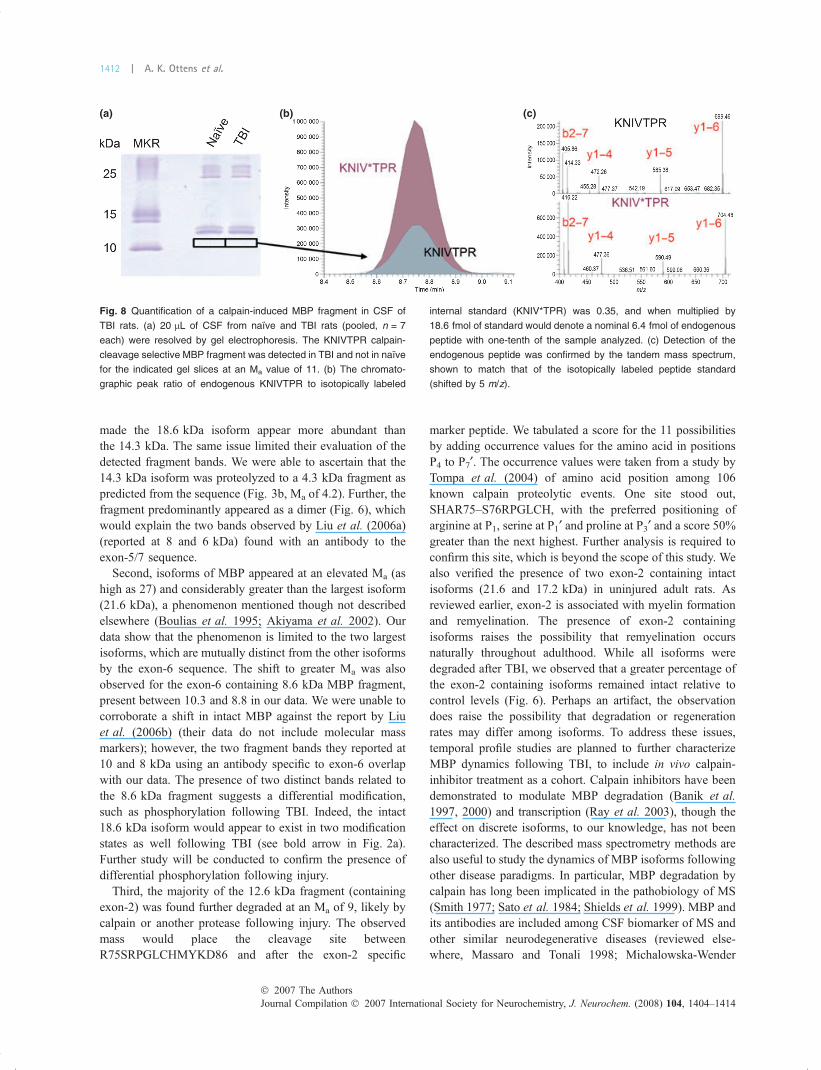
accessible from severe TBI patients. Thus, we aimed todemonstrate that our mass spectrometry method could beused to measure a calpain-induced MBP fragment in CSF ofTBI rats. We calculated 64 fmol of the calpain-cleavagespecific peptide in a TBI CSF (20 lL) gel band at a Ma of 11(Fig. 8a and b) that was not present in naıve CSF. The dataleft little doubt that a proteolytic product of MBP was presentin the CSF of TBI injured rats, as the measurements weremade from tandem mass spectra that distinctly identified theKNIVTPR peptide (Fig. 8c). In contrast, immunoblot anal-ysis lacked either the sensitivity or selectivity necessary todetect a MBP fragment band in CSF (Fig. 2).
Discussion
In this study, we employed a new MSMS approach to profileMBP dynamics, an integral protein component of the myelinsheath protecting vulnerable white matter in the brain (Smithet al. 2003). We assessed what MBP isoforms were presentin normal adult rat cortex, and their degradation by calpain
proteolysis following neurotrauma. MSMS facilitated femto-mole quantification and the identification of isoforms andrelated proteolytic fragments. The resulting data depict nearcomplete degeneration at 2 days post-injury of the four MBPisoforms normally present, with correlating increases incalpain proteolytic fragments detectable at the tissue and CSFlevels. Indeed, a statistically significant decrease in the 21.6,18.6, and 14.3 kDa isoforms was determined despite lowstatistical power (p of 0.04, n of 3, non-parametric Mann–Whitney U-test). These results correlate with previous reportsusing the same model of large-scale axonal and neuronaldegeneration at 2 days post-TBI (Ringger et al. 2004;Kobeissy et al. 2006; Liu et al. 2006a).
Tandem mass spectrometry data provided greater detailover previous reports to establish the following findings.First, we determined that the isoform of greatest abundancein adult rat cortex was the 14.3 kDa, as denoted by thecorresponding tandem exon-5/7 sequence signal. The reportby Liu et al. (2006b) was confounded by use of two differentantibodies, which, as a result of a difference in sensitivity,
(a)
(b)
Fig. 7 Profile of MBP isoforms and proteolytic fragments in naıve and
TBI cortex samples. Shown are profiles of the variably spliced exon
sequence-specific peptides for naıve (a) and TBI (b) samples. Chro-
matographic peaks were generated for the most intense fragment ion
of each peptide. Peak area means for each peptide (n = 3) were
normalized across all Ma values. The presence of a peptide (and
correlating exon-coded sequence) was determined when the normal-
ized area at a given Ma was greater than three times the minimum for
that peptide. Molecular mass labels demarking the identified MBP
isoforms and proteolytic fragments are shown for each gel slice per the
pattern of exon-specific peptides and given Ma value. ‘F12.6 kDa’
indicates a truncated 12.6 kDa fragment and ‘D4.3 kDa’ indicates a
dimer of the 4.3 kDa fragment.
� 2007 The AuthorsJournal Compilation � 2007 International Society for Neurochemistry, J. Neurochem. (2008) 104, 1404–1414
Mass spectrometry of post-TBI MBP proteolysis | 1411
made the 18.6 kDa isoform appear more abundant thanthe 14.3 kDa. The same issue limited their evaluation of thedetected fragment bands. We were able to ascertain that the14.3 kDa isoform was proteolyzed to a 4.3 kDa fragment aspredicted from the sequence (Fig. 3b, Ma of 4.2). Further, thefragment predominantly appeared as a dimer (Fig. 6), whichwould explain the two bands observed by Liu et al. (2006a)(reported at 8 and 6 kDa) found with an antibody to theexon-5/7 sequence.
Second, isoforms of MBP appeared at an elevated Ma (ashigh as 27) and considerably greater than the largest isoform(21.6 kDa), a phenomenon mentioned though not describedelsewhere (Boulias et al. 1995; Akiyama et al. 2002). Ourdata show that the phenomenon is limited to the two largestisoforms, which are mutually distinct from the other isoformsby the exon-6 sequence. The shift to greater Ma was alsoobserved for the exon-6 containing 8.6 kDa MBP fragment,present between 10.3 and 8.8 in our data. We were unable tocorroborate a shift in intact MBP against the report by Liuet al. (2006b) (their data do not include molecular massmarkers); however, the two fragment bands they reported at10 and 8 kDa using an antibody specific to exon-6 overlapwith our data. The presence of two distinct bands related tothe 8.6 kDa fragment suggests a differential modification,such as phosphorylation following TBI. Indeed, the intact18.6 kDa isoform would appear to exist in two modificationstates as well following TBI (see bold arrow in Fig. 2a).Further study will be conducted to confirm the presence ofdifferential phosphorylation following injury.
Third, the majority of the 12.6 kDa fragment (containingexon-2) was found further degraded at an Ma of 9, likely bycalpain or another protease following injury. The observedmass would place the cleavage site betweenR75SRPGLCHMYKD86 and after the exon-2 specific
marker peptide. We tabulated a score for the 11 possibilitiesby adding occurrence values for the amino acid in positionsP4 to P7¢. The occurrence values were taken from a study byTompa et al. (2004) of amino acid position among 106known calpain proteolytic events. One site stood out,SHAR75–S76RPGLCH, with the preferred positioning ofarginine at P1, serine at P1¢ and proline at P3¢ and a score 50%greater than the next highest. Further analysis is required toconfirm this site, which is beyond the scope of this study. Wealso verified the presence of two exon-2 containing intactisoforms (21.6 and 17.2 kDa) in uninjured adult rats. Asreviewed earlier, exon-2 is associated with myelin formationand remyelination. The presence of exon-2 containingisoforms raises the possibility that remyelination occursnaturally throughout adulthood. While all isoforms weredegraded after TBI, we observed that a greater percentage ofthe exon-2 containing isoforms remained intact relative tocontrol levels (Fig. 6). Perhaps an artifact, the observationdoes raise the possibility that degradation or regenerationrates may differ among isoforms. To address these issues,temporal profile studies are planned to further characterizeMBP dynamics following TBI, to include in vivo calpain-inhibitor treatment as a cohort. Calpain inhibitors have beendemonstrated to modulate MBP degradation (Banik et al.1997, 2000) and transcription (Ray et al. 2003), though theeffect on discrete isoforms, to our knowledge, has not beencharacterized. The described mass spectrometry methods arealso useful to study the dynamics of MBP isoforms followingother disease paradigms. In particular, MBP degradation bycalpain has long been implicated in the pathobiology of MS(Smith 1977; Sato et al. 1984; Shields et al. 1999). MBP andits antibodies are included among CSF biomarker of MS andother similar neurodegenerative diseases (reviewed else-where, Massaro and Tonali 1998; Michalowska-Wender
(a) (b) (c)
Fig. 8 Quantification of a calpain-induced MBP fragment in CSF of
TBI rats. (a) 20 lL of CSF from naıve and TBI rats (pooled, n = 7
each) were resolved by gel electrophoresis. The KNIVTPR calpain-
cleavage selective MBP fragment was detected in TBI and not in naıve
for the indicated gel slices at an Ma value of 11. (b) The chromato-
graphic peak ratio of endogenous KNIVTPR to isotopically labeled
internal standard (KNIV*TPR) was 0.35, and when multiplied by
18.6 fmol of standard would denote a nominal 6.4 fmol of endogenous
peptide with one-tenth of the sample analyzed. (c) Detection of the
endogenous peptide was confirmed by the tandem mass spectrum,
shown to match that of the isotopically labeled peptide standard
(shifted by 5 m/z).
Journal Compilation � 2007 International Society for Neurochemistry, J. Neurochem. (2008) 104, 1404–1414� 2007 The Authors
1412 | A. K. Ottens et al.
et al. 2001), though the particular proteolytic fragmentquantified in our study has not been described. Future effortsmight ascertain whether this fragment is present in the CSFof MS patients, for potential utility as a alternative biomarkerof degradative myelin processing.
The presented methods hinge on the use of MSMSspectra to selectively characterize peptides specific to thecalpain cleavage site and alternative spliced coding regionsof MBP. All target peptides are unique to MBP among allother proteins in a rat proteome database as determined byBLAST analysis. We evaluated the selectivity of corre-sponding MSMS spectra by using Mascot software tocalculate the probability (p) that the match of the spectrawith the correct peptide sequences would occur by chance(Perkins et al. 1999). For example, the probability for achance match of the HGFLPR and KNIVTPR MSMS datawas p = 0.00050 (1 in 2000) and p = 0.00016 (1 in 6250),respectively, when evaluated against a rat protein database.In contrast, the next closest peptide sequences had a greater,1 in 8, probability of being a chance match of the MSMSspectra (p = 0.13). With significance often defined as a lessthan 1 in 20 chance occurrence (i.e. p < 0.05), the collectedMSMS spectra are highly significant in their selectivity ofthe target peptide.
Presently, our application of the reported approach isrestricted by the speed of our mass spectrometer in thatchromatographic peaks are defined only by four or five datapoints, and IDA and isoform characterization were per-formed separately. More current equipment, such as thefaster, more sensitive linear ion trap (Blackler et al. 2006),would improve peak characterization and still allow forrapid collection of tandem mass spectra. Parameters wereoptimized in our study to quantify MBP peptides; however,the approach is generally applicable to other proteolyzedproteins. Likewise, isoforms of other proteins can bedelineated using the presented method, in combination orseparate from IDA quantification, which is significant in thatwe now anticipate most proteins to have multiple isoformswith unique functions or spatial/temporal dynamics impor-tant for the regulation of cellular processes (Godovac-Zimmermann et al. 2005; Yura et al. 2006). The generalapplicability of the approach is nonetheless expressed in lightof limitations to peptide analysis by mass spectrometry.Peptide coelution and poor ionization efficiency are con-founds that limit sensitivity and tandem mass spectrumquality. Application of our mass spectrometry approach toother peptides would thereby require individual consider-ation and optimization of gel electrophoresis or RPLCseparations (possibly adding analysis time). To this end,separation and detection efficiencies could be checkedquickly through test analysis of a synthesized targetpeptide(s) within the complex mixture of interest. Overall,the upfront time and cost for this test is less than required fornovel antibody generation as the alternative.
In summary, we present mass spectrometry methodologyto characterize and quantify proteolytic processing of multi-isoform proteins, exemplified in this study by calpainproteolysis of MBP after neurotrauma. Intact and fragmentedMBP isoforms were deconvoluted using sequence specificpeptide MSMS data, and quantified at femtomole amounts intissue and CSF using IDA. We demonstrated by theselectivity of MSMS that the 14.3 kDa MBP isoform waspredominant in our adult rats and quantified the degradationof four predominant isoforms (21.6, 18.6, 17.2, and14.3 kDa) following TBI. We ascertained that the presenceof the MBP exon-6 sequence resulted in a characteristic shiftin gel migration for intact and fragmented MBP alike. Wealso found evidence for a second post-TBI cleavage eventwithin the exon-2 MBP sequence, and that the post-TBI4.3 kDa fragment forms a dimer. Overall, we expect that thepresented methodology will be broadly applicable formonitoring dynamics of other multi-isoform proteins andproteolytic events of significance in the study of the CNS,with particular relevance to biomarker research.
Acknowledgements
This work was performed with support of the Department of
Defense grant DAMD17-03-1-0066 and the National Institute for
Neurological Disorders and Stroke grant K25NS055012. We thank
W. Harrison for critical reading of the manuscript, A. Chung for
peptide synthesis, M. C. Liu for insightful discussions on MBP
immunoblot analysis, and R. Tivis, SJ. Nixon, and M. Lewis for
assistance with statistical evaluation. RLH, NDD, and KKWW hold
equity in Banyan Biomarkers, Inc, a company commercializing
biomarker technology in brain injury.
References
Aebersold R. and Mann M. (2003) Mass spectrometry-based proteomics.Nature 422, 198–207.
Akiyama K., Ichinose S., Omori A., Sakurai Y. and Asou H. (2002)Study of expression of myelin basic proteins (MBPs) in developingrat brain using a novel antibody reacting with four major isoformsof MBP. J. Neruosci. Res. 68, 19–28.
Arroyo E. J. and Scherer S. S. (2000) On the molecular architecture ofmyelinated fibers. Histochem. Cell Biol. 113, 1–18.
Banik N. L., Matzelle D., Terry E. and Hogan E. L. (1997) A new mech-anism of methylprednisolone and other corticosteroids action dem-onstrated in vitro: inhibition of a proteinase (calpain) preventsmyelinand cytoskeletal protein degradation. Brain Res. 748, 205–210.
Banik N. L., Matzelle D., Terry E., Gantt-Wilford G. and Hogan E. L.(2000) Inhibition of proteolysis by a cyclooxygenase inhibitor,indomethacin. Neurochem. Res. 25, 1509–1515.
Barr J. R., Maggio V. L., Patterson Jr D. G. et al. (1996) Isotopedilution-mass spectrometric quantification of specific proteins:model application with apolipoprotein A-I. Clin. Chem. 42, 1676–1682.
Berger R. P., Adelson P. D., Richichi R. and Kochanek P. M. (2006)Serum biomarkers after traumatic and hypoxemic brain injuries:insight into the biochemical response of the pediatric brain to inflictbrain injury. Dev. Neurosci. 28, 327–335.
� 2007 The AuthorsJournal Compilation � 2007 International Society for Neurochemistry, J. Neurochem. (2008) 104, 1404–1414
Mass spectrometry of post-TBI MBP proteolysis | 1413
Blackler A. R., Klammer A. A., MacCoss M. J. and Wu C. C. (2006)Qualitative comparison of proteomic data quality between a 2Dand 3D quadrupole ion trap. Anal. Chem. 78, 1337–1344.
Boggs J. M. (2006) Myelin basic protein: a multifunctional protein. Cell.Mol. Life Sci. 63, 1945–1961.
Boggs J. M., Rangaraj G., Koshy K. M. and Mueller J. P. (2000)Adhesion of acidic lipid vesicles by 21.5 kDa (recombinant) and18.5 kDa isoforms of myelin basic protein. Biochem. Biophys.Acta 1463, 81–87.
Boulias C., Pang H., Mastronardi F. and Moscarello M. A. (1995) Theisolation and characterization of four myelin basic proteins fromthe unbound fraction during CM52 chromatography. Arch. Bio-chem. Biophys. 322, 174–182.
Capello E., Voskuhl R. R., Mctarland H. F. and Rane C. S. (1997)Multiple sclerosis: re-expression of developmetnal gene in chromiclesions correlates with remyelination. Ann. Neurol. 41, 797–805.
Dongre A. R., Eng J. K. and Yates III J. R. (1997) Emerging tandem-mass-spectrometry techniques for the rapid identification of pro-teins. Trends Biotechnol. 15, 418–425.
Gerber S. A., Rush J., Stemman O., Kirschner M. W. and Gygi S. P.(2003) Absolute quantification of proteins and phosphoproteinsfrom cell lysates by tandem MS. Proc. Natl Acad. Sci. USA 100,6940–6945.
Givogri M. I., Bongarzone E. R. and Campagnoni A. T. (2000) Newinsights on the biology of myelin basic protein gene: the neural-immune connection. J. Neurosci. Res. 59, 153–159.
Godovac-Zimmermann J., Kleiner O., Brown L. R. and Drukier A. K.(2005) Perspectives in spicing up proteomics with splicing.Proteomics 5, 699–709.
Harauz G., Ishiyama N., Hill C. M., Bates I. R., Libich D. S. and FaresC. (2004) Myelin basic protein – diverse conformational states ofan intrinsically unstructured protein and its roles in myelinassembly and multiple sclerosis. Micron 35, 503–542.
Kimura M., Sato M., Akatsuka A., Saito S., Ando K., Yokoyama M. andKatsuki M. (1998) Overexpression of a minor component ofmyelin basic protein isoform (17.2 kDa) can restore myelinogen-esis in transgenic shiverer mice. Brain Res. 785, 245–252.
Kishimoto A., Nishiyama K., Nakanishi H., Uratsuji Y., Nomura H.,Takeyama Y. and Nishizuka Y. (1985) Studies on the phosphory-lation of myelin basic protein by protein kinase C and adenosine3¢,5¢-monophosphate-dependent protein kinase. J. Biol. Chem. 260,12492–12499.
Kobeissy F. H., Ottens A. K., Zhang Z., Liu M. C., Denslow N. D., DaveJ. R., Tortella F. C., Hayes R. L. and Wang K. K. (2006) Noveldifferential neuroproteomics analysis of traumatic brain injury inrats. Mol. Cell. Protoemics 5, 1887–1898.
Kruger G. M., Diemel L. T., Copelman C. A. and Cuzner M. L. (1999)Myelin basic protein isoforms in myelinating and remyelinating ratbrain aggregate cultures. J. Neurosci. Res. 56, 241–247.
Liu M. C., Akle V., Zheng W. et al. (2006a) Extensive degradation ofmyelin basic protein isoforms by calpain following traumatic braininjury. J. Neurochem. 98, 700–712.
Liu M. C., Akle V., Zheng W., Dave J. R., Tortella F. C., Hayes R. L. andWang K. K. (2006b) Comparing calpain- and caspase-3-mediateddegradation patterns in traumatic brain injury by differential pro-teome analysis. Biochem. J. 394, 715–725.
Maatta J. A., Coffey E. T., Hermonen J. A., Salmi A. A. and Hin-kkanen A. E. (1997) Detection of myelin basic protein isoforms
by organic concentration. Biochem. Biophys. Res. Commun. 238,498–502.
Massaro A. R. and Tonali P. (1998) Cerebrospinal fluid markers inmultiple sclerosis: an overview. Mult. Scler. 4, 1–4.
Michalowska-Wender G., Losy J. and Wender M. (2001) Biologicalmarkers to confirm diagnosis and monitor the therapy in multiplesclerosis patients. Folia Neuropathol. 39, 1–5.
Nakajima K., Ikenaka K., Kagawa T., Aruga J., Nakao J., Nakahira K.,Shiota C., Kim S. U. and Mikoshiba K. (1993) Novel isoforms ofmouse myelin basic protein predominantly expressed in embryonicstage. J. Neurochem. 60, 1554–1563.
Norton W. T. and Cammer W. (1984) Isolation and characterization ofmyelin, in Myelin, 2nd ed (Morell P., ed.), pp. 147–180. PlenumPress, New York.
Perkins D. N., Pappin D. J. C., Creasy D. M. and Cottrell J. S. (1999)Probability-based protein identification by searching sequence da-tabases using mass spectrometry data. Electrophoresis 20,3551–3567.
Ray S. K., Matzelle D. D., Sribnick E. A., Guyton K., Wingrave J. M.and Banik N. L. (2003) Calpain inhibitor prevented apoptosis andmaintained transcription of proteolipid protein and myelin basicprotein genes in rat spinal cord injury. J. Chem. Neuroanat. 26,119–124.
Ringger N. C., O’Steen B. E., Brabham J. G., Silver X., Pineda J., WangK. K., Hayes R. L. and Papa L. (2004) A novel marker of traumaticbrain injury: CSF alphaII-spectrin breakdown product levels.J. Neurotruama 21, 1443–1456.
Sato S., Quarles R. H., Brady R. O. and Tourtellotte W. W. (1984)Elevated neutral protease activity in myelin from brains of patientswith multiple sclerosis. Ann. Neurol. 15, 264–267.
Schaecher K. E., Shields D. C. and Banik N. L. (2001) Mechanism ofmyelin breakdown in experimental demyelination: a putative rolefor calpain. Neurochem. Res. 26, 731–737.
Shields D. C., Schaecher K. E., Saido T. C. and Banik N. L. (1999) Aputative mechanism of demyelination in multiple sclerosis bya proteolytic enzume, calpain. Proc. Natl Acad. Sci. 96,11486–11491.
Smith M. E. (1977) The role of proteolytic enzymes in experimentalallergic encephalomyelitis. Neurochemistry 2, 233–246.
Smith D. H., Meaney D. F. and Shull W. H. (2003) Diffuse axonal injuryin head trauma. J. Head Trauma Rehabil. 18, 307–316.
Thomas D. G., Palfreyman J. W. and Ratcliffe J. G. (1978) Serum-myelin-basic-protein assay in diagnosis and prognosis of patientswith head injury. Lancet 1, 113–115.
Tompa P., Buzder-Lantos P., Tantos A., Farkas A., Szilagyi A., BanocziZ., Hudecz F. and Friedrich P. (2004) On the sequential determi-nants of calpain cleavage. J. Biol. Chem. 279, 20775–20785.
Voskuhl R. R., Robinson E. D., Segal B. M., Tranquill L., CamphausenK., Albert P. S., Richert J. R. and McFarland H. F. (1994) HLArestriction and TCR usage of T lymphocytes specific for a novelcandidate autoantigen, X2 MBP, in multiple sclerosis. J. Immunol.153, 4834–4844.
Wood D. D. and Moscarello M. A. (1989) The isolation, characteriza-tion, and lipid-aggregating properties of citrulline containingmyelin basic protein. J. Biol. Chem. 25, 5121–5127.
Yura K., Shionyu M., Hagino K. et al. (2006) Alternative splicing inhuman transcriptome: functional and structural influence on pro-teins. Gene 380, 63–71.
Journal Compilation � 2007 International Society for Neurochemistry, J. Neurochem. (2008) 104, 1404–1414� 2007 The Authors
1414 | A. K. Ottens et al.
Related Documents