Protein Synthesis From Gene to Protein Unit 3

Welcome message from author
This document is posted to help you gain knowledge. Please leave a comment to let me know what you think about it! Share it to your friends and learn new things together.
Transcript
Protein SynthesisFrom Gene to
ProteinUnit 3
Protein synthesis The information content of DNA
Is in the form of specific sequences of nucleotides along the DNA strands
The DNA inherited by an organism Leads to specific traits by dictating the synthesis of
proteins The process by which DNA directs protein synthesis, gene
expression Includes two stages, called transcription and translation
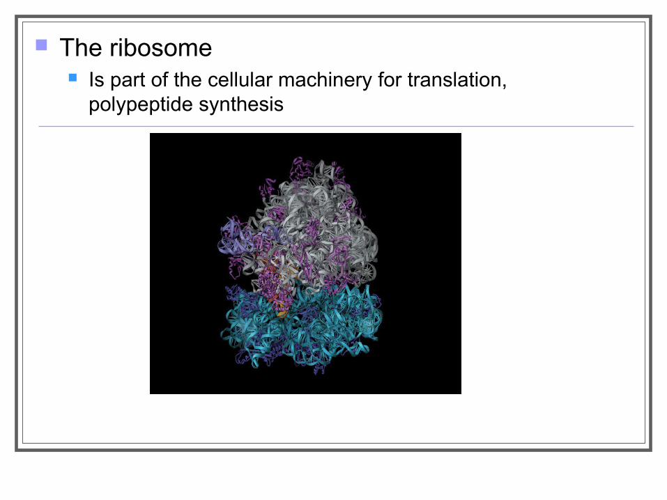
The ribosome Is part of the cellular machinery for translation,
polypeptide synthesis
Genes specify proteins via transcription and translation
Transcription involves the transfer of genetic information from DNA into an RNA molecule while translation involves the transfer of the information in the RNA to the synthesis of a protein
Evidence from the Study of Metabolic Defects The relationship between genes and proteins was first proposed in 1909 by an English physician Archibald Garrod
He was the first to suggest that genes dictate phenotypes through enzymes which are proteins that catalyze specific chemical reactions in the cell.
He hypothesized that inherited diseases reflect a person’s inability to make a particular enzyme.
Citing the disease alkaptonuria where urine appears dark red due to the presence of alkapton as an example, Garrod reasoned that normal individuals have an enzyme that breaks down alkapton while alkaptonuric individuals lack the enzyme
Garrod’s hypothesis was ahead of its time but research decades later proved him right
Nutritional Mutants in Neurospora: Scientific Inquiry
In 1940s, George Beadle and Edward Tatum proved the relationship between genes and enzymes by using the bread mold, Neurospora crassa.
Beadle and Tatum studied strains of the mold that were unable to grow on the usual minimal growth medium. These strains were mutants created using X-ray radiation.
Each of these mutants lacked an enzyme in a metabolic pathway and therefore were unable to produce a particular molecule such as an amino acid.
They showed that each mutant was defective in a single gene and hypothesized that one gene controlled the production of one specific enzyme.
This hypothesis has now been modified from one gene-one enzyme to one gene-one protein to one gene–one polypeptide.
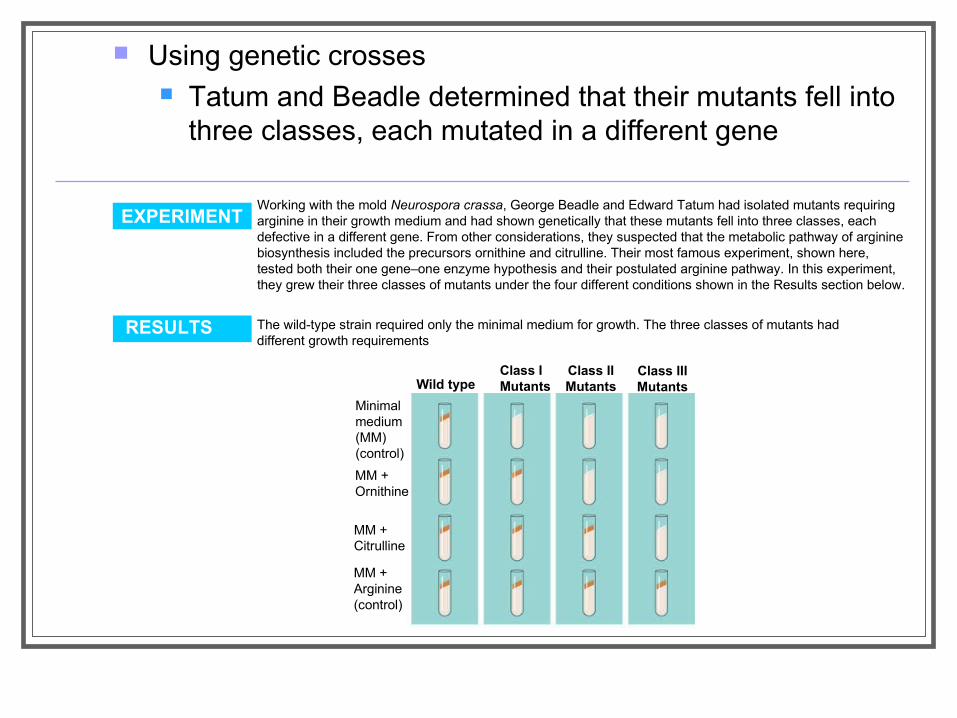
Using genetic crosses Tatum and Beadle determined that their mutants fell into
three classes, each mutated in a different gene
Working with the mold Neurospora crassa, George Beadle and Edward Tatum had isolated mutants requiring arginine in their growth medium and had shown genetically that these mutants fell into three classes, each defective in a different gene. From other considerations, they suspected that the metabolic pathway of arginine biosynthesis included the precursors ornithine and citrulline. Their most famous experiment, shown here, tested both their one gene–one enzyme hypothesis and their postulated arginine pathway. In this experiment, they grew their three classes of mutants under the four different conditions shown in the Results section below.
The wild-type strain required only the minimal medium for growth. The three classes of mutants had different growth requirements
EXPERIMENT
RESULTS
Class IMutants
Class IIMutants
Class IIIMutantsWild type
Minimal medium(MM)(control)
MM +Ornithine
MM +Citrulline
MM +Arginine(control)
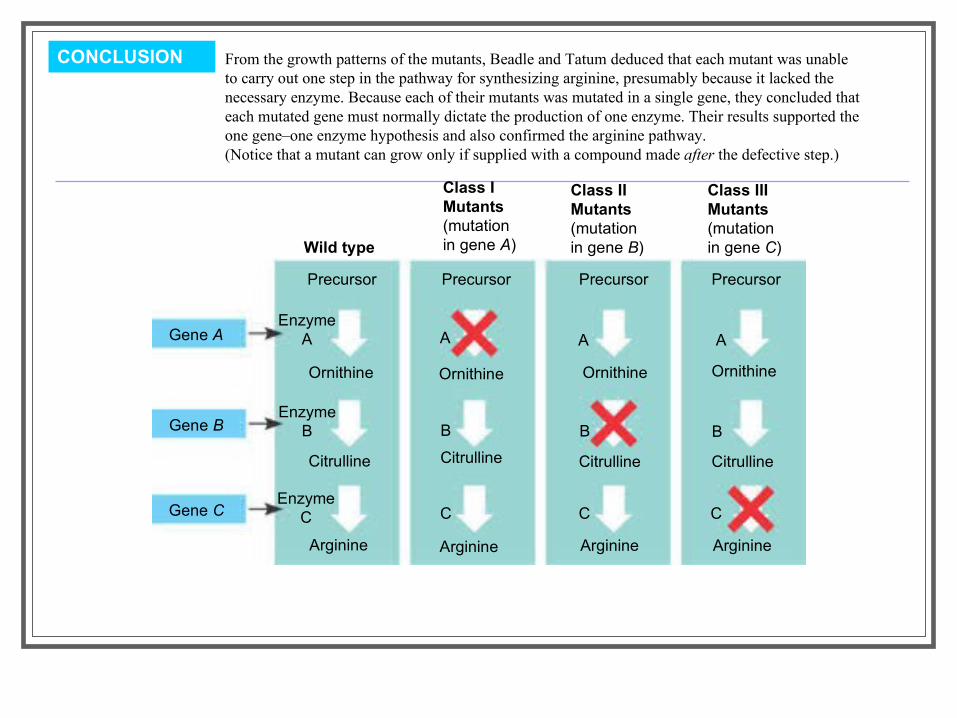
CONCLUSION From the growth patterns of the mutants, Beadle and Tatum deduced that each mutant was unable to carry out one step in the pathway for synthesizing arginine, presumably because it lacked the necessary enzyme. Because each of their mutants was mutated in a single gene, they concluded that each mutated gene must normally dictate the production of one enzyme. Their results supported the one gene–one enzyme hypothesis and also confirmed the arginine pathway. (Notice that a mutant can grow only if supplied with a compound made after the defective step.)
Class IMutants(mutationin gene A)
Class IIMutants(mutationin gene B)
Class IIIMutants(mutationin gene C)Wild type
Gene A
Gene B
Gene C
Precursor Precursor Precursor Precursor
Ornithine Ornithine Ornithine Ornithine
Citrulline Citrulline Citrulline Citrulline
Arginine Arginine Arginine Arginine
EnzymeA
EnzymeB
EnzymeC
A A A
B B B
C C C
Beadle and Tatum developed the “one gene–one enzyme hypothesis” Which states that the function of a gene is to dictate the production of a specific
enzyme As researchers learned more about proteins
They made minor revision to the one gene–one enzyme hypothesis Genes are now known to code for polypeptide chains or for RNA molecules.
The Products of Gene Expression: A Developing Story
Basic Principles of Transcription and Translation
Transcription Is the synthesis of RNA under the direction of DNA Produces messenger RNA (mRNA)
Translation Is the actual synthesis of a polypeptide, which
occurs under the direction of mRNA Occurs on ribosomes
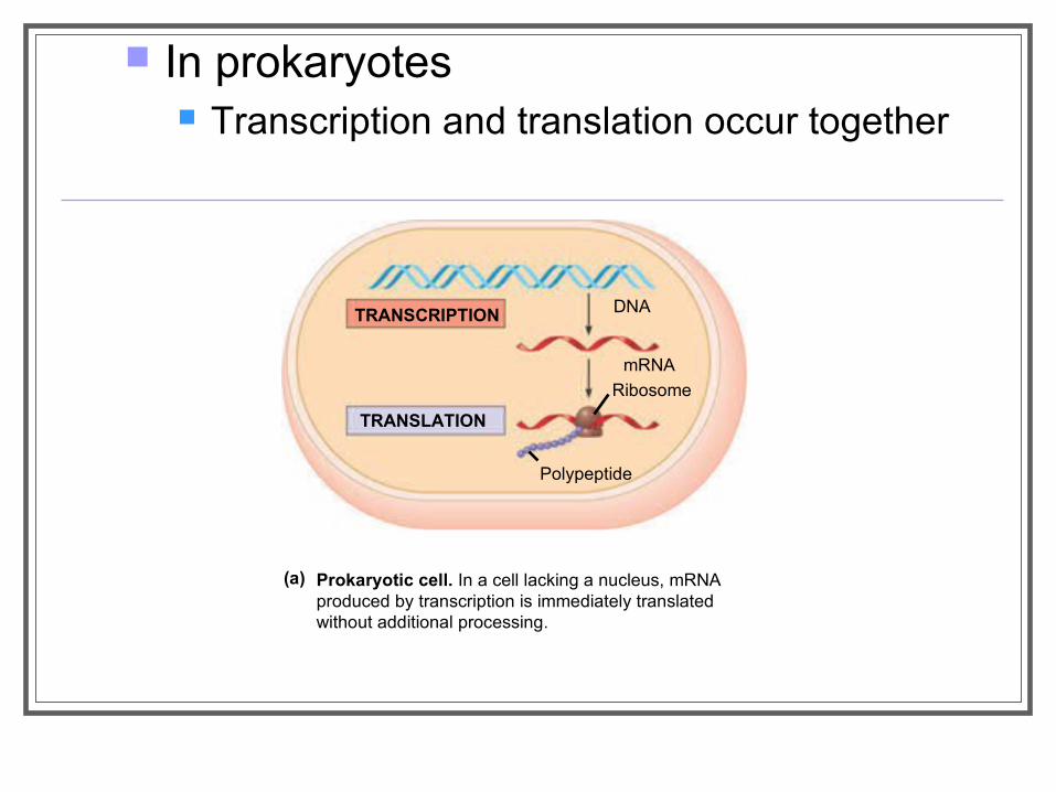
In prokaryotes Transcription and translation occur together
Prokaryotic cell. In a cell lacking a nucleus, mRNAproduced by transcription is immediately translatedwithout additional processing.
(a)
TRANSLATION
TRANSCRIPTION DNA
mRNA
Ribosome
Polypeptide
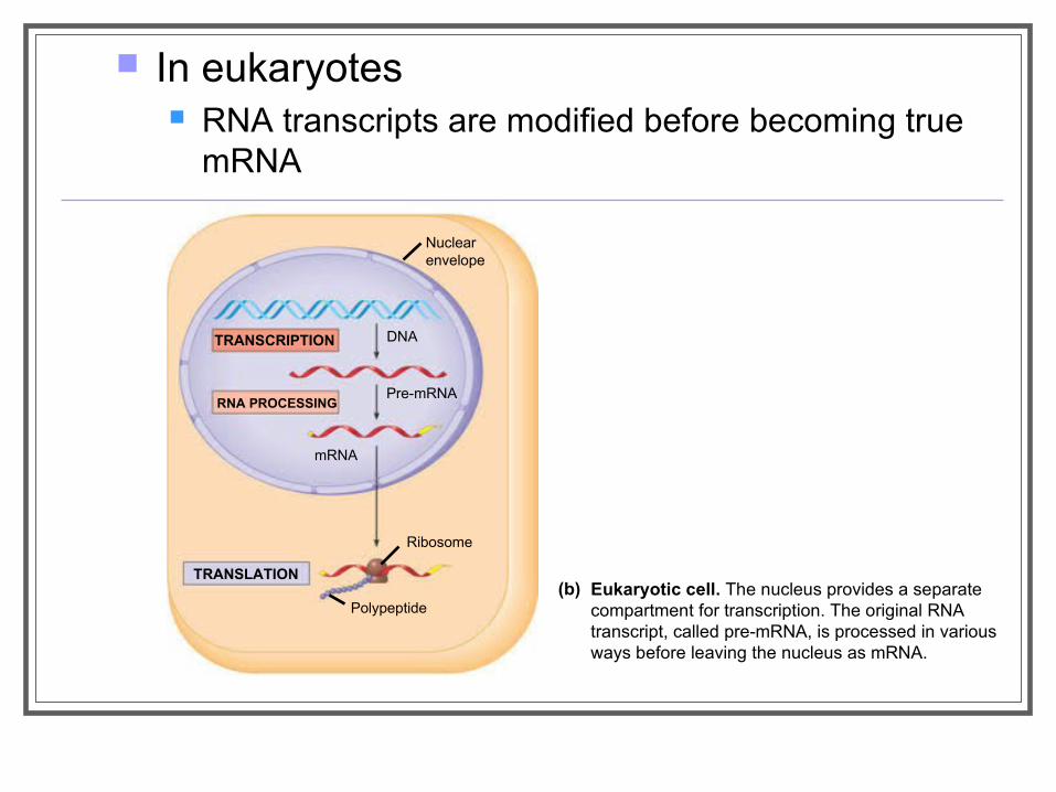
In eukaryotes RNA transcripts are modified before becoming true
mRNA
Eukaryotic cell. The nucleus provides a separatecompartment for transcription. The original RNAtranscript, called pre-mRNA, is processed in various ways before leaving the nucleus as mRNA.
(b)
TRANSCRIPTION
RNA PROCESSING
TRANSLATION
mRNA
DNA
Pre-mRNA
Polypeptide
Ribosome
Nuclearenvelope
Cells are governed by a cellular chain of command DNA → RNA → protein
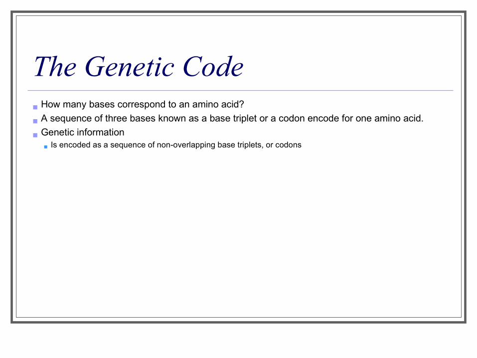
The Genetic Code How many bases correspond to an amino acid?
A sequence of three bases known as a base triplet or a codon encode for one amino acid.
Genetic information Is encoded as a sequence of non-overlapping base triplets, or codons
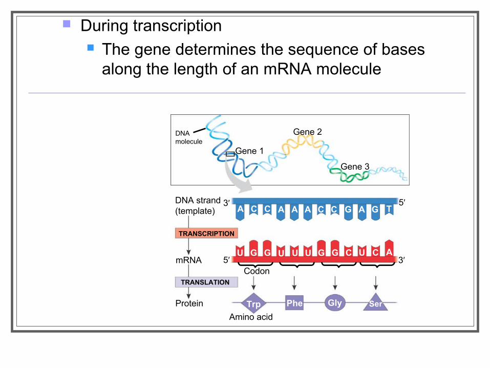
During transcription The gene determines the sequence of bases
along the length of an mRNA molecule
DNAmolecule
Gene 1
Gene 2
Gene 3
DNA strand(template)
TRANSCRIPTION
mRNA
Protein
TRANSLATION
Amino acid
A C C A A A C C G A G T
U G G U U U G G C U C A
Trp Phe Gly Ser
Codon
3′ 5′
3′5′
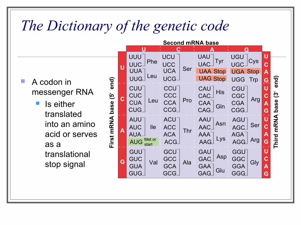
The Dictionary of the genetic code
A codon in messenger RNA Is either
translated into an amino acid or serves as a translational stop signal
Second mRNA baseU C A G
U
C
A
G
UUUUUCUUAUUG
CUUCUCCUACUG
AUUAUCAUAAUG
GUUGUCGUAGUG
Met orstart
Phe
Leu
Leu
lle
Val
UCUUCCUCAUCG
CCUCCCCCACCG
ACUACCACAACG
GCUGCCGCAGCG
Ser
Pro
Thr
Ala
UAUUAC
UGUUGC
Tyr Cys
CAUCACCAACAG
CGUCGCCGACGG
AAUAACAAAAAG
AGUAGCAGAAGG
GAUGACGAAGAG
GGUGGCGGAGGG
UGGUAAUAG Stop
Stop UGA StopTrp
His
Gln
Asn
Lys
Asp
Arg
Ser
Arg
Gly
U
CA
GUCAG
UCAG
UCAG
Fir
st m
RN
A b
ase
(5′
end
)
Th
ird
mR
NA
bas
e (3
′ en
d)
Glu
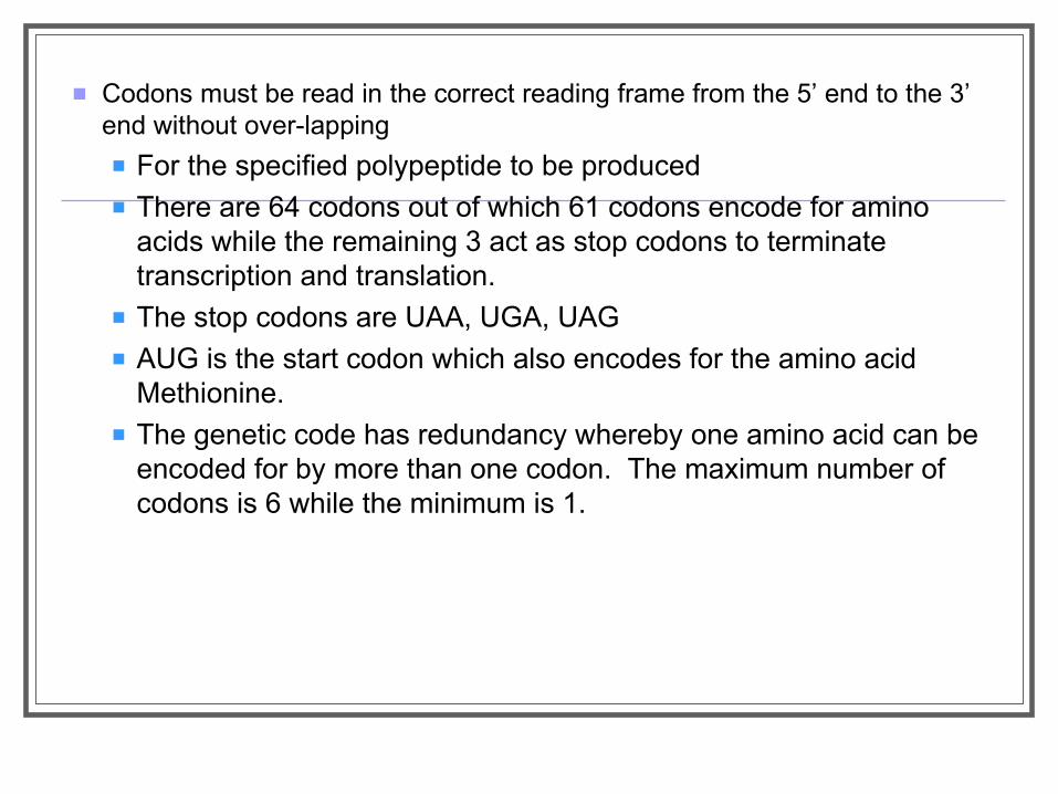
Codons must be read in the correct reading frame from the 5’ end to the 3’ end without over-lapping
For the specified polypeptide to be produced There are 64 codons out of which 61 codons encode for amino
acids while the remaining 3 act as stop codons to terminate transcription and translation.
The stop codons are UAA, UGA, UAG AUG is the start codon which also encodes for the amino acid
Methionine. The genetic code has redundancy whereby one amino acid can be
encoded for by more than one codon. The maximum number of codons is 6 while the minimum is 1.
Evolution of the Genetic Code The genetic code is nearly universal
Shared by organisms from the simplest bacteria to the most complex animals
In laboratory experiments Genes can be transcribed and translated after being transplanted from one
species to another
Transcription Transcription is the DNA-directed synthesis
of mRNA: a closer look

Molecular Components of Transcription
mRNA synthesis Is catalyzed by RNA polymerase, which pries the
DNA strands apart and hooks together the RNA nucleotides
Follows the same base-pairing rules as DNA, except that in RNA, uracil substitutes for thymine
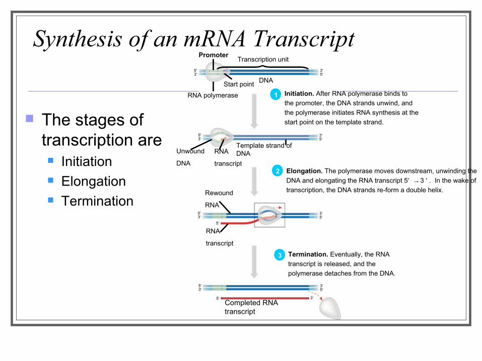
Synthesis of an mRNA Transcript
The stages of transcription are Initiation Elongation Termination
PromoterTranscription unit
RNA polymerase
Start point
5′3′
3′5′
3′5′
5′3′
5′3′
3′5′
5′3′
3′5′
5′
5′
Rewound
RNA
RNA
transcript
3′
3′Completed RNA transcript
Unwound
DNA
RNA
transcript
Template strand of DNA
DNA
1 Initiation. After RNA polymerase binds to
the promoter, the DNA strands unwind, and
the polymerase initiates RNA synthesis at the
start point on the template strand.
2 Elongation. The polymerase moves downstream, unwinding the
DNA and elongating the RNA transcript 5′ → 3 ′ . In the wake of
transcription, the DNA strands re-form a double helix.
3 Termination. Eventually, the RNA
transcript is released, and the
polymerase detaches from the DNA.
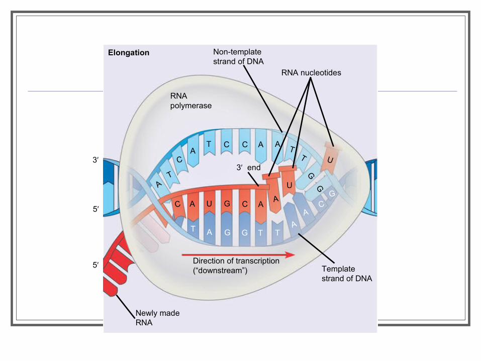
Elongation
RNApolymerase
Non-templatestrand of DNA
RNA nucleotides
3′ end
C A U G C AA
U
T A G G T TA
AC
G
U
AT
CA
T C C A AT
T
GG
3′
5′
5′
Newly madeRNA
Direction of transcription(“downstream”) Template
strand of DNA
Elongation of the RNA Strand As RNA polymerase moves along the DNA
It continues to untwist the double helix, exposing about 10 to 20 DNA bases at a time for pairing with RNA nucleotides
Termination of Transcription The mechanisms of termination
Are different in prokaryotes and eukaryotes
Eukaryotic cells modify RNA after transcription
Enzymes in the eukaryotic nucleus Modify pre-mRNA in specific ways before the
genetic messages are dispatched to the cytoplasm They modify the 5’ and 3’ ends and also remove the
introns to splice the exons together to form a continuous reading frame.
RNA Processing/Post-Transcriptional Modification
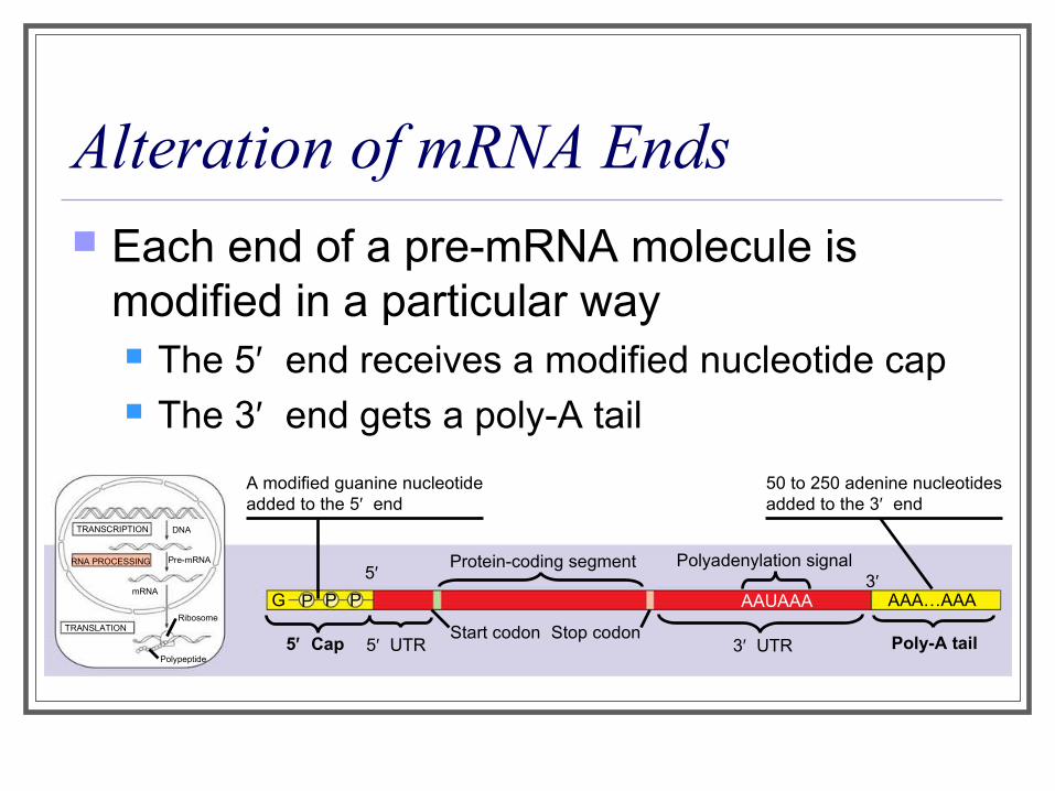
Alteration of mRNA Ends Each end of a pre-mRNA molecule is
modified in a particular way The 5′ end receives a modified nucleotide cap The 3′ end gets a poly-A tail
A modified guanine nucleotideadded to the 5′ end
50 to 250 adenine nucleotidesadded to the 3′ end
Protein-coding segment Polyadenylation signal
Poly-A tail3′ UTRStop codonStart codon
5′ Cap 5′ UTR
AAUAAA AAA…AAA
TRANSCRIPTION
RNA PROCESSING
DNA
Pre-mRNA
mRNA
TRANSLATIONRibosome
Polypeptide
G P P P
5′ 3′
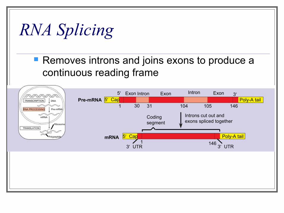
RNA Splicing
Removes introns and joins exons to produce a continuous reading frame
TRANSCRIPTION
RNA PROCESSING
DNA
Pre-mRNA
mRNA
TRANSLATION
Ribosome
Polypeptide
5′ CapExon Intron
1
5′
30 31
Exon Intron
104 105 146
Exon 3′Poly-A tail
Poly-A tail
Introns cut out andexons spliced together
Codingsegment
5′ Cap1 146
3′ UTR3′ UTR
Pre-mRNA
mRNA
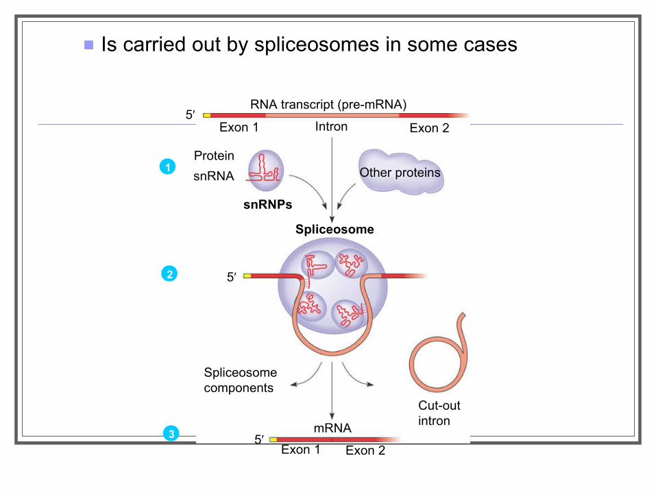
Is carried out by spliceosomes in some cases
RNA transcript (pre-mRNA)
Exon 1 Intron Exon 2
Other proteinsProtein
snRNA
snRNPs
Spliceosome
Spliceosomecomponents
Cut-outintron
mRNA
Exon 1 Exon 2
5′
5′
5′
1
2
3
Translation Translation is the RNA-directed synthesis
of a polypeptide: a closer look
Molecular Components of Translation A cell translates an mRNA message into
protein With the help of transfer RNA (tRNA)
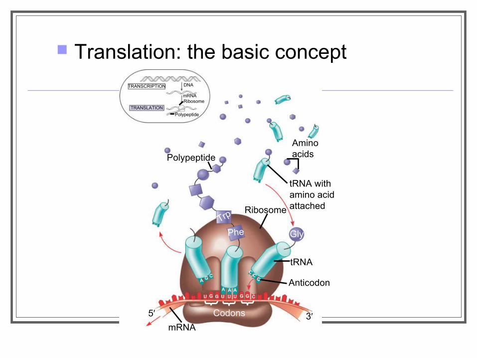
Translation: the basic concept
TRANSCRIPTION
TRANSLATION
DNA
mRNARibosome
Polypeptide
Polypeptide
Aminoacids
tRNA withamino acidattachedRibosome
tRNA
Anticodon
mRNA
Trp
Phe Gly
A G C
A A A
CC
G
U G G U U U G G C
Codons5′ 3′
Molecules of tRNA are not all identical Each carries a specific amino acid on one end (3’
end) Each has an anticodon on the other end which is
complementary to a codon of mRNA
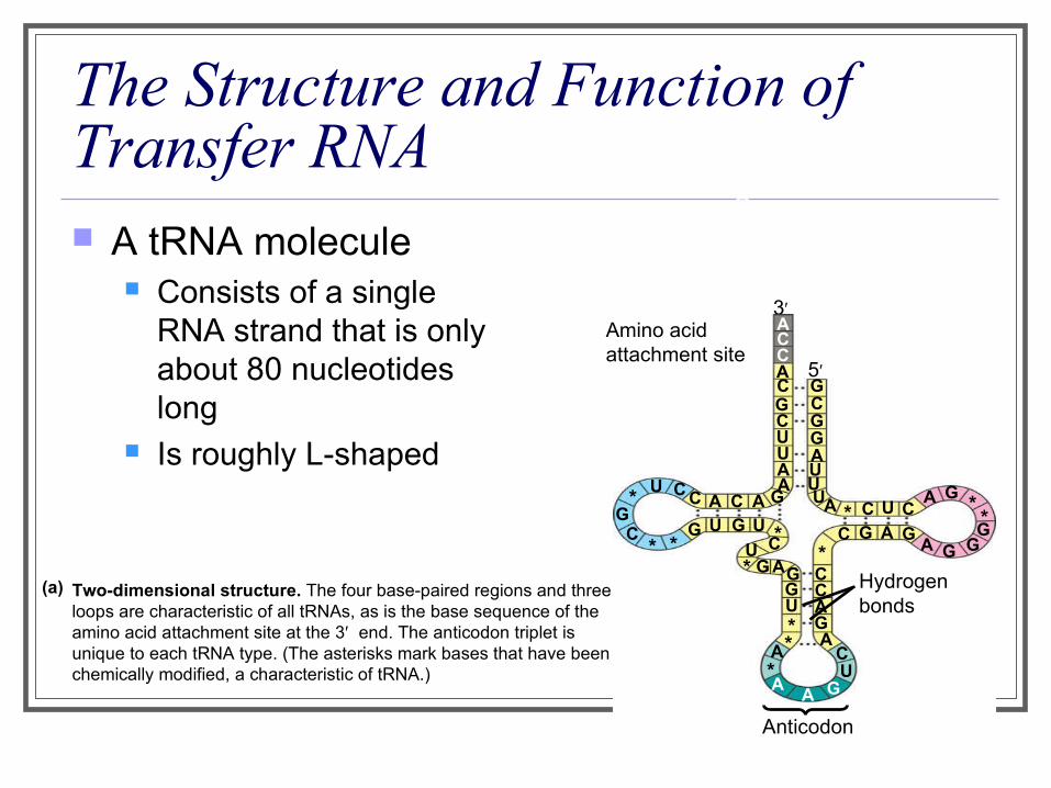
The Structure and Function of Transfer RNA A tRNA molecule
Consists of a single RNA strand that is only about 80 nucleotides long
Is roughly L-shaped
ACC
Two-dimensional structure. The four base-paired regions and three loops are characteristic of all tRNAs, as is the base sequence of the amino acid attachment site at the 3′ end. The anticodon triplet is unique to each tRNA type. (The asterisks mark bases that have been chemically modified, a characteristic of tRNA.)
(a)
3′
CCACGCUUAA
GACACCU*
GC
* *G U G U *CU
* G AGGU**A
*A
A GUC
AGACC*
C G A GA G G
G*
*GA
CUC*AUUUAGGCG5′
Amino acidattachment site
Hydrogenbonds
Anticodon
A
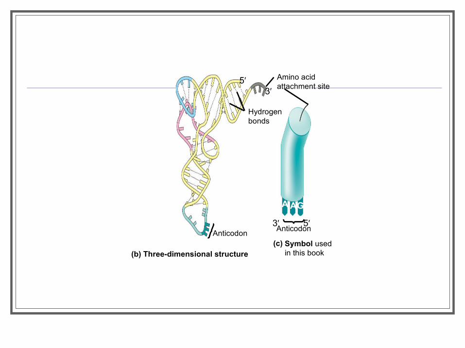
(b) Three-dimensional structureSymbol used in this book
Amino acidattachment site
Hydrogen bonds
AnticodonAnticodon
A AG
5′3′
3′ 5′
(c)

A specific enzyme called an aminoacyl-tRNA synthetase Joins each amino acid to the correct tRNA
Amino acid
ATP
Adenosine
Pyrophosphate
Adenosine
Adenosine
Phosphates
tRNA
P P P
P
P Pi
Pi
Pi
P
AMP
Aminoacyl tRNA(an “activatedamino acid”)
Aminoacyl-tRNAsynthetase (enzyme)
Active site binds theamino acid and ATP. 1
ATP loses two P groupsand joins amino acid as AMP.2
3 AppropriatetRNA covalentlyBonds to aminoAcid, displacingAMP.
Activated amino acidis released by the enzyme.4
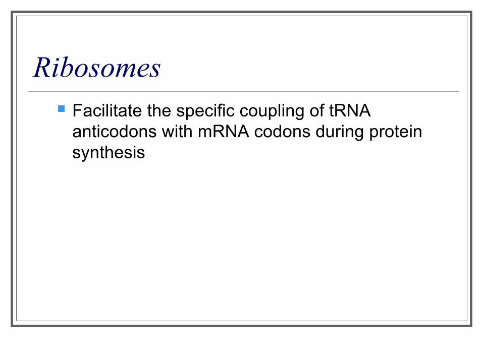
Ribosomes Facilitate the specific coupling of tRNA
anticodons with mRNA codons during protein synthesis
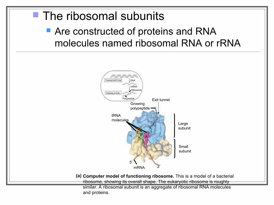
The ribosomal subunits Are constructed of proteins and RNA
molecules named ribosomal RNA or rRNA
TRANSCRIPTION
TRANSLATION
DNA
mRNA
Ribosome
Polypeptide Exit tunnelGrowingpolypeptide
tRNAmolecules
EP A
Largesubunit
Smallsubunit
mRNA
Computer model of functioning ribosome. This is a model of a bacterial ribosome, showing its overall shape. The eukaryotic ribosome is roughly similar. A ribosomal subunit is an aggregate of ribosomal RNA molecules and proteins.
(a)
5′3′

The ribosome has three binding sites for tRNA The P site The A site The E site
E P A
P site (Peptidyl-tRNAbinding site)
E site (Exit site)
mRNAbinding site
A site (Aminoacyl-tRNA binding site)
Largesubunit
Smallsubunit
Schematic model showing binding sites. A ribosome has an mRNA binding site and three tRNA binding sites, known as the A, P, and E sites. This schematic ribosome will appear in later diagrams.
(b)
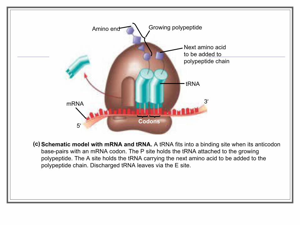
Amino end Growing polypeptide
Next amino acidto be added topolypeptide chain
tRNA
mRNA
Codons
3′
5′
Schematic model with mRNA and tRNA. A tRNA fits into a binding site when its anticodon base-pairs with an mRNA codon. The P site holds the tRNA attached to the growing polypeptide. The A site holds the tRNA carrying the next amino acid to be added to the polypeptide chain. Discharged tRNA leaves via the E site.
(c)
Building a Polypeptide We can divide translation into three stages
Initiation Elongation Termination
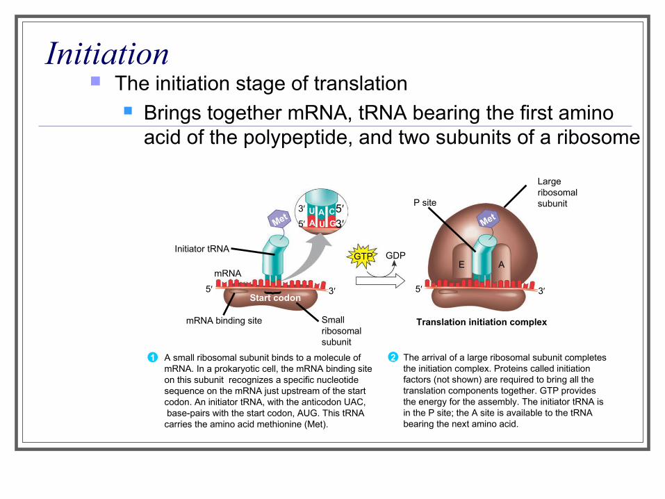
Initiation The initiation stage of translation
Brings together mRNA, tRNA bearing the first amino acid of the polypeptide, and two subunits of a ribosome
Largeribosomalsubunit
The arrival of a large ribosomal subunit completes the initiation complex. Proteins called initiationfactors (not shown) are required to bring all the translation components together. GTP provides the energy for the assembly. The initiator tRNA is in the P site; the A site is available to the tRNA bearing the next amino acid.
2
Initiator tRNA
mRNA
mRNA binding site Smallribosomalsubunit
Translation initiation complex
P site
GDPGTP
Start codon
A small ribosomal subunit binds to a molecule of mRNA. In a prokaryotic cell, the mRNA binding site on this subunit recognizes a specific nucleotide sequence on the mRNA just upstream of the start codon. An initiator tRNA, with the anticodon UAC, base-pairs with the start codon, AUG. This tRNA carries the amino acid methionine (Met).
1
MetMet
U A CA U G
E A
3′
5′5′3′
3′5′ 3′5′
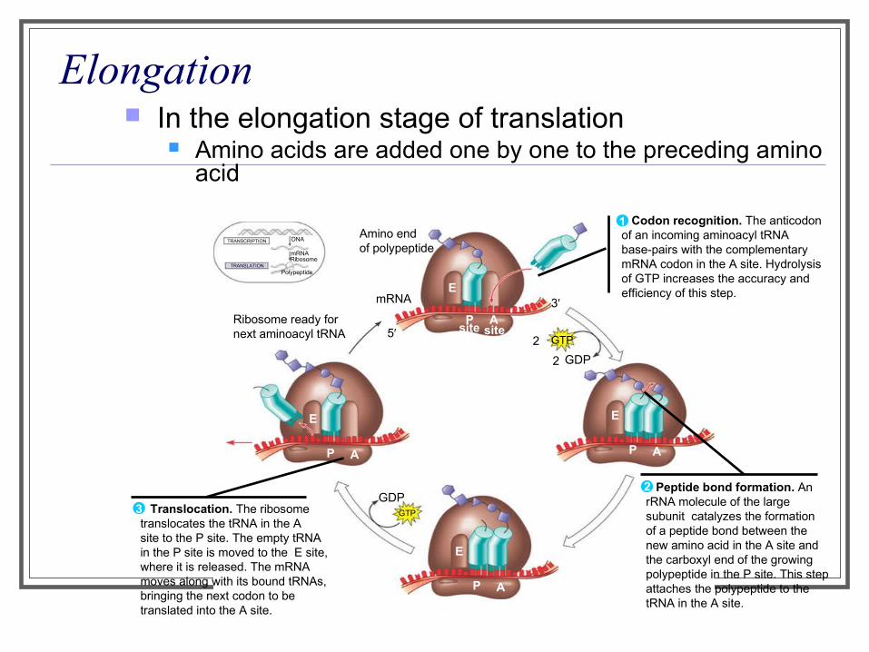
Elongation In the elongation stage of translation
Amino acids are added one by one to the preceding amino acid
Amino endof polypeptide
mRNA
Ribosome ready fornext aminoacyl tRNA
E
P A
E
P A
E
P A
E
P A
GDPGTP
GTP
GDP
2
2
site site5′
3′
TRANSCRIPTION
TRANSLATION
DNA
mRNARibosome
Polypeptide
Codon recognition. The anticodon of an incoming aminoacyl tRNA base-pairs with the complementary mRNA codon in the A site. Hydrolysisof GTP increases the accuracy andefficiency of this step.
1
Peptide bond formation. An rRNA molecule of the large subunit catalyzes the formation of a peptide bond between the new amino acid in the A site and the carboxyl end of the growing polypeptide in the P site. This step attaches the polypeptide to the tRNA in the A site.
2
Translocation. The ribosome translocates the tRNA in the A site to the P site. The empty tRNA in the P site is moved to the E site, where it is released. The mRNA moves along with its bound tRNAs,bringing the next codon to be translated into the A site.
3
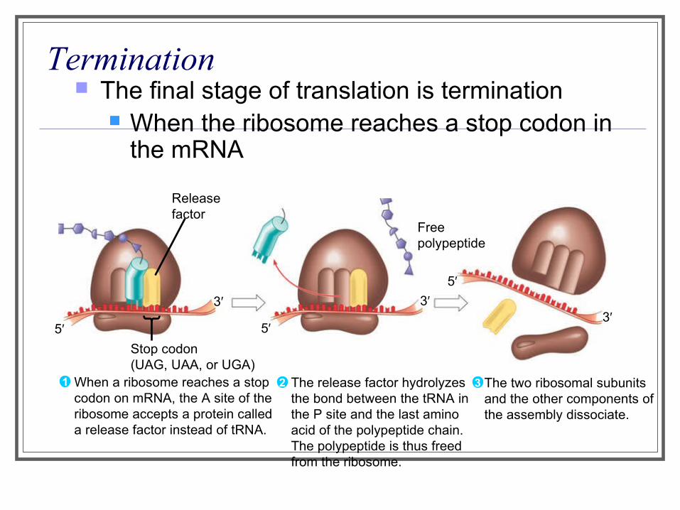
Termination The final stage of translation is termination
When the ribosome reaches a stop codon in the mRNA
Release factor
Freepolypeptide
Stop codon(UAG, UAA, or UGA)
5′
3′ 3′5′
3′5′
When a ribosome reaches a stop codon on mRNA, the A site of the ribosome accepts a protein called a release factor instead of tRNA.
1 The release factor hydrolyzes the bond between the tRNA in the P site and the last amino acid of the polypeptide chain. The polypeptide is thus freed from the ribosome.
2 3 The two ribosomal subunits and the other components of the assembly dissociate.
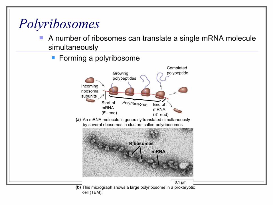
Polyribosomes A number of ribosomes can translate a single mRNA molecule
simultaneously Forming a polyribosome
Growingpolypeptides
Completedpolypeptide
Incomingribosomalsubunits
Start of mRNA(5′ end)
End of mRNA(3′ end)
Polyribosome
An mRNA molecule is generally translated simultaneously by several ribosomes in clusters called polyribosomes.
(a)
Ribosomes
mRNA
This micrograph shows a large polyribosome in a prokaryotic cell (TEM).
0.1 µm(b)
Completing and Targeting the Functional Protein Polypeptide chains
Undergo modifications after the translation process
Protein Folding and Post-Translational Modifications
After translation Proteins may be modified in ways that affect their
three-dimensional shape
Targeting Polypeptides to Specific Locations Two populations of ribosomes are evident
in cells Free and bound
Free ribosomes in the cytosol Initiate the synthesis of all proteins
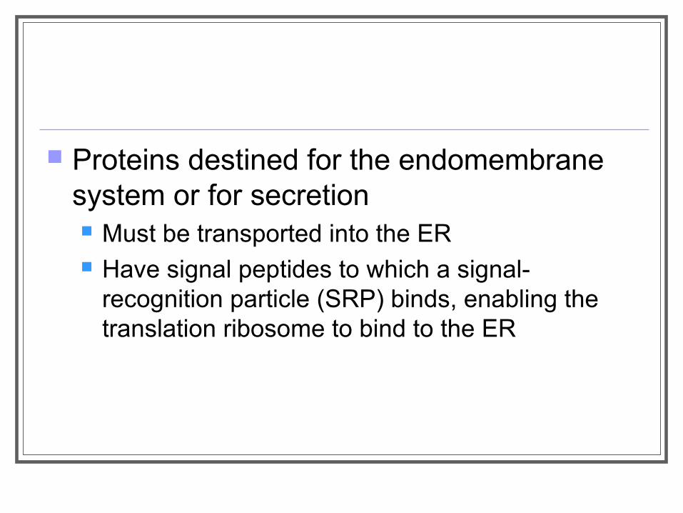
Proteins destined for the endomembrane system or for secretion Must be transported into the ER Have signal peptides to which a signal-
recognition particle (SRP) binds, enabling the translation ribosome to bind to the ER
Ribosome
mRNASignalpeptide
Signal-recognitionparticle(SRP) SRP
receptorprotein
Translocationcomplex
CYTOSOL
Signalpeptideremoved
ERmembrane
Protein
ERLUMEN
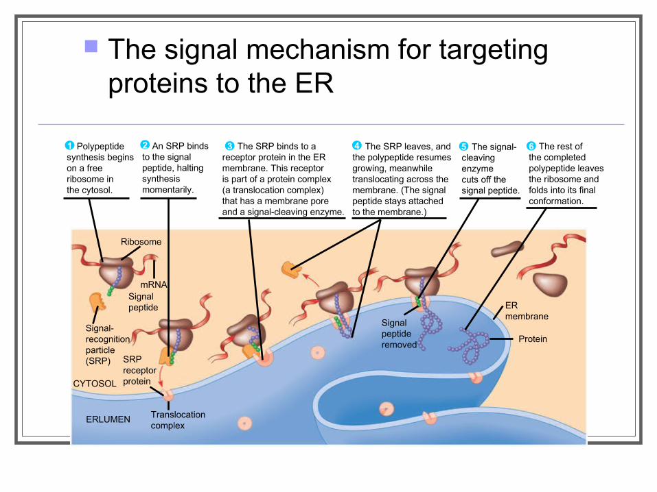
The signal mechanism for targeting proteins to the ER
Polypeptidesynthesis beginson a freeribosome inthe cytosol.
1 An SRP binds to the signal peptide, halting synthesismomentarily.
2 The SRP binds to areceptor protein in the ERmembrane. This receptoris part of a protein complex(a translocation complex)that has a membrane poreand a signal-cleaving enzyme.
3 The SRP leaves, andthe polypeptide resumesgrowing, meanwhiletranslocating across themembrane. (The signalpeptide stays attachedto the membrane.)
4 The signal-cleaving enzymecuts off thesignal peptide.
5 The rest ofthe completedpolypeptide leaves the ribosome andfolds into its finalconformation.
6
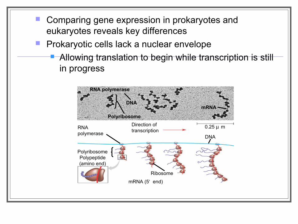
Comparing gene expression in prokaryotes and eukaryotes reveals key differences
Prokaryotic cells lack a nuclear envelope Allowing translation to begin while transcription is still
in progress
DNA
Polyribosome
mRNA
Direction oftranscription
0.25 µ mRNApolymerase
Polyribosome
Ribosome
DNA
mRNA (5′ end)
RNA polymerase
Polypeptide(amino end)

In a eukaryotic cell The nuclear envelope separates transcription
from translation Extensive RNA processing occurs in the nucleus
Point Mutations
Point mutations can affect protein structure and function
Mutations Are changes in the genetic material of a cell
Point mutations Are changes in just one base pair of a gene
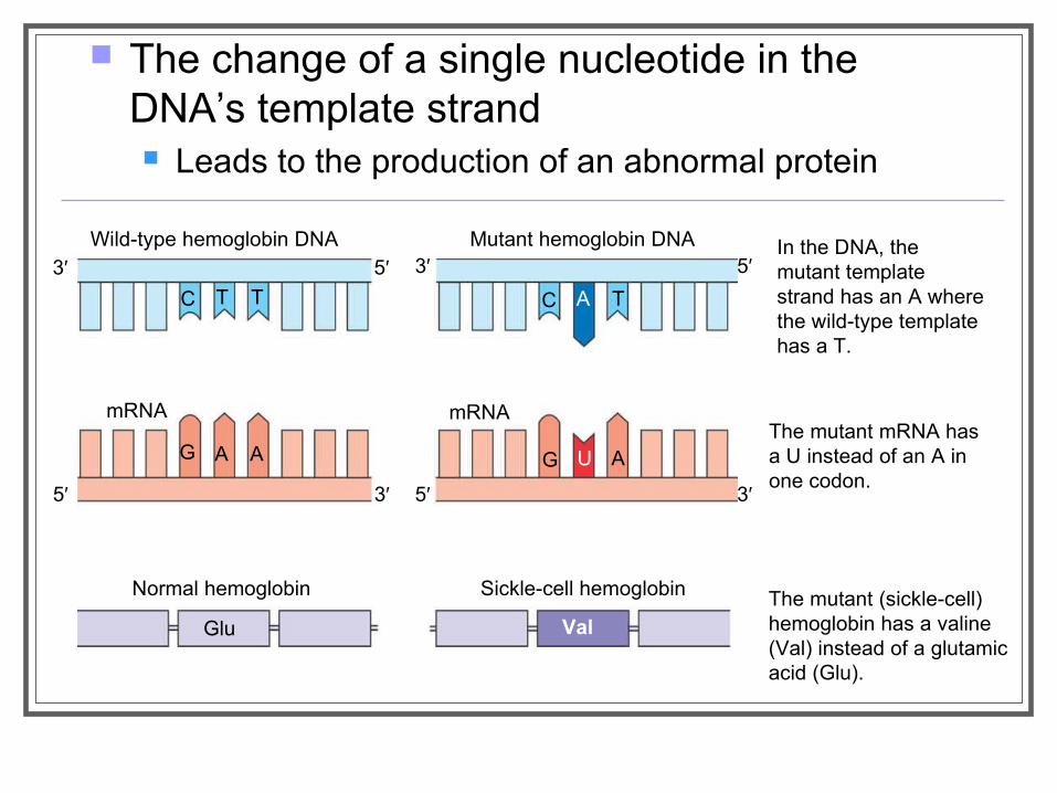
The change of a single nucleotide in the DNA’s template strand Leads to the production of an abnormal protein
In the DNA, themutant templatestrand has an A where the wild-type template has a T.
The mutant mRNA has a U instead of an A in one codon.
The mutant (sickle-cell) hemoglobin has a valine (Val) instead of a glutamic acid (Glu).
Mutant hemoglobin DNAWild-type hemoglobin DNA
mRNA mRNA
Normal hemoglobin Sickle-cell hemoglobin
Glu Val
C T T C A T
G A A G U A
3′ 5′ 3′ 5′
5′ 3′5′ 3′
Types of Point Mutations Point mutations within a gene can be
divided into two general categories Base-pair substitutions Base-pair insertions or deletions
Substitutions
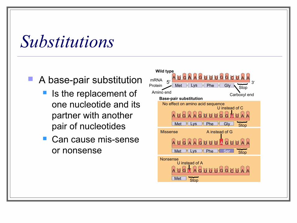
A base-pair substitution Is the replacement of
one nucleotide and its partner with another pair of nucleotides
Can cause mis-sense or nonsense
Wild type
A U G A A G U U U G G C U A AmRNA 5′Protein Met Lys Phe Gly Stop
Carboxyl endAmino end
3′
A U G A A G U U U G G U U A A
Met Lys Phe Gly
Base-pair substitutionNo effect on amino acid sequence
U instead of C
Stop
A U G A A G U U U A G U U A A
Met Lys Phe Ser Stop
A U G U A G U U U G G C U A A
Met Stop
Missense A instead of G
NonsenseU instead of A
Insertions and Deletions
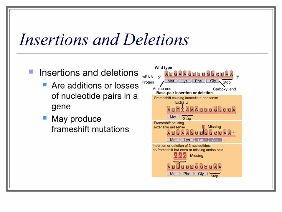
Insertions and deletions Are additions or losses
of nucleotide pairs in a gene
May produce frameshift mutations
mRNAProtein
Wild type
A U G A A G U U U G G C U A A5′
Met Lys Phe Gly
Amino end Carboxyl end
Stop
Base-pair insertion or deletionFrameshift causing immediate nonsense
A U G U A A G U U U G G C U A
A U G A A G U U G G C U A A
A U G U U U G G C U A A
Met Stop
U
Met Lys Leu Ala
Met Phe GlyStop
MissingA A G
Missing
Extra U
Frameshift causing extensive missense
Insertion or deletion of 3 nucleotides:no frameshift but extra or missing amino acid
3′
Mutagens Spontaneous mutations
Can occur during DNA replication, recombination, or repair
Mutagens Are physical or chemical agents that can cause
mutations

What is a gene? revisiting the question A gene
Is a region of DNA whose final product is either a polypeptide or an RNA molecule
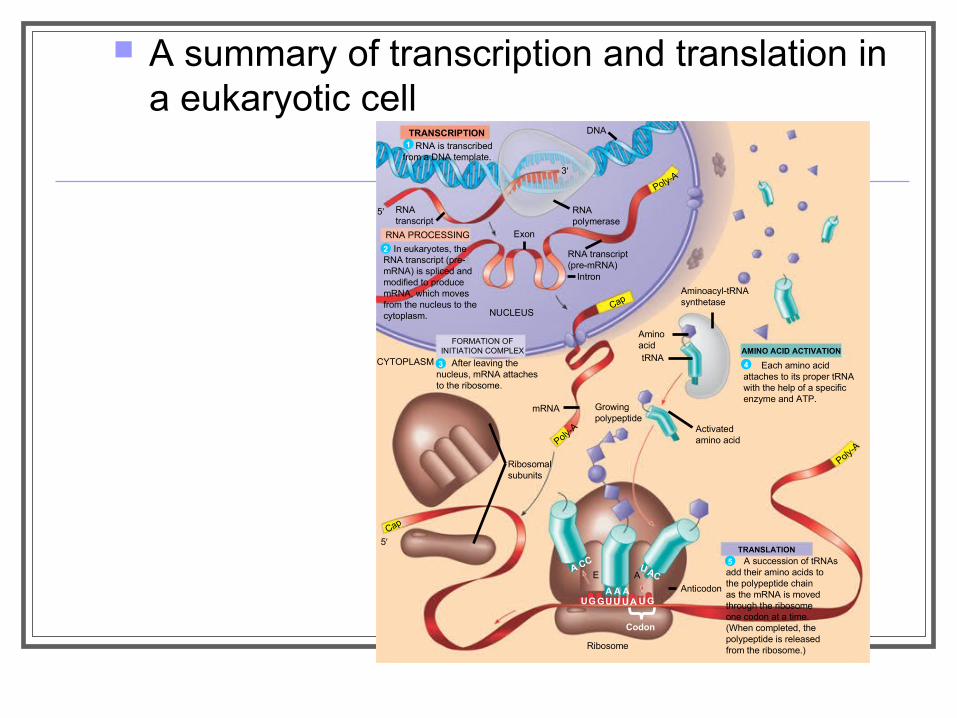
A summary of transcription and translation in a eukaryotic cell
TRANSCRIPTION RNA is transcribedfrom a DNA template.
DNA
RNApolymerase
RNAtranscript
RNA PROCESSING
In eukaryotes, theRNA transcript (pre-mRNA) is spliced andmodified to producemRNA, which movesfrom the nucleus to thecytoplasm.
Exon
Poly-A
RNA transcript(pre-mRNA)
Intron
NUCLEUSCap
FORMATION OFINITIATION COMPLEX
After leaving thenucleus, mRNA attachesto the ribosome.
CYTOPLASM
mRNA
Poly-A
Growingpolypeptide
Ribosomalsubunits
Cap
Aminoacyl-tRNAsynthetase
AminoacidtRNA
AMINO ACID ACTIVATION
Each amino acidattaches to its proper tRNAwith the help of a specificenzyme and ATP.
Activatedamino acid
TRANSLATION
A succession of tRNAsadd their amino acids tothe polypeptide chainas the mRNA is movedthrough the ribosomeone codon at a time.(When completed, thepolypeptide is releasedfrom the ribosome.)
Anticodon
A CC
A A AUG GUU UA U G
UACE A
Ribosome
1
Poly-A
5′
5′
3′
Codon
2
3 4
5

Try this! 1. What are transcription and translation? 2. How many nucleotides are necessary to code for a polypeptide that is
100 amino acids long? 3. An mRNA molecule contains the nucleotide sequence
CCAUUUACG. Using the dictionary of the genetic code, translate this sequence into the corresponding amino acid sequence.
4. What is an anticodon? 5. What is the function of the ribosome in protein synthesis? 6. Which of the following does not participate directly in translation:
ribosomes, tRNA, mRNA, DNA, enzymes and ATP?
Related Documents