Propionyl-L-Carnitine is Efficacious in Ulcerative Colitis Through its Action on the Immune Function and Microvasculature Maria Giovanna Scioli, PhD 1,5 , Maria Antonietta Stasi, BSc 2,5 , Daniela Passeri, PhD 1 , Elena Doldo, PhD 1 , Gaetana Costanza, BSc 1 , Roberto Camerini, MD, PhD 2 , Paolo Fociani, MD 3 , Gaetano Arcuri, PhD 4 , Katia Lombardo, BSc 2 , Silvia Pace, BSc 2 , Franco Borsini, PhD 2 and Augusto Orlandi, MD 1 OBJECTIVES: Microvascular endothelial dysfunction characterizes ulcerative colitis (UC), the most widespread form of inflammatory bowel disease. Intestinal mucosal microvessels in UC display aberrant expression of cell adhesion molecules (CAMs) and increased inflammatory cell recruitment. Propionyl-L-carnitine (PLC), an ester of L-carnitine required for the mitochondrial transport of fatty acids, ameliorates propionyl-CoA bioavailability and reduces oxidative stress in ischemic tissues. The present study aimed to document the efficacy of anti-oxidative stress properties of PLC in counteracting intestinal microvascular endothelial dysfunction and inflammation. METHODS: To evaluate the efficacy in vivo, we analyzed the effects in intestinal biopsies of patients with mild-to-moderate UC receiving oral PLC co-treatment and in rat TNBS-induced colitis; in addition, we investigated antioxidant PLC action in TNF-a- stimulated human intestinal microvascular endothelial cells (HIMECs) in vitro. RESULTS: Four-week PLC co-treatment reduced intestinal mucosal polymorph infiltration and CD4 þ lymphocytes, ICAM-1 þ and iNOS þ microvessels compared with placebo-treated patients with UC. Oral and intrarectal administration of PLC but not L-carnitine or propionate reduced intestinal damage and microvascular dysfunction in rat TNBS-induced acute and reactivated colitis. In cultured TNF-a-stimulated HIMECs, PLC restored b-oxidation and counteracted NADPH oxidase 4-generated oxidative stress-induced CAM expression and leukocyte adhesion. Inhibition of b-oxidation by L-aminocarnitine increased reactive oxygen species production and PLC beneficial effects on endothelial dysfunction and leukocyte adhesion. Finally, PLC reduced iNOS activity and nitric oxide accumulation in rat TNBS-induced colitis and in HIMEC cultures. CONCLUSIONS: Our results show that the beneficial antioxidant effect of PLC targeting intestinal microvasculature restores endothelial b-oxidation and function, and reduces mucosal inflammation in UC patients. Clinical and Translational Gastroenterology (2014) 5, e55; doi:10.1038/ctg.2014.4; published online 20 March 2014 Subject Category: Inflammatory Bowel Disease INTRODUCTION Ulcerative colitis (UC) and Crohn disease are the two major forms of inflammatory bowel disease (IBD). 1 UC is the most common, with an incidence of 1.2–20.3 cases for 100,000 people per year. 1,2 Genetic and environmental factors contribute to the deregulation of mucosal inflammatory response in patients with IBD, and both immune and non- immune regulatory pathways contribute to UC pathogen- esis. 1,3 Microvascular endothelium has a crucial role in the initiation and the progression of the inflammatory response and consequent tissue remodeling of UC. Intestinal micro- vascular endothelial cells regulate the influx of leukocytes through the expression of cell adhesion molecules (CAMs) and chemokine secretion. 4 Chronically inflamed intestinal microvessels in UC patients display aberrant CAM expression and enhanced susceptibility to adherence, migration, and recruitment of leukocytes. 1,5 Our hypothesis was that intestinal mucosal microvascular endothelial dysfunction has a primary role in governing mucosal inflammation in UC patients, thus representing a consistent therapeutic target in UC patients. 6 Experimental models and colonic mucosal biopsies from UC patients documented metabolic changes and an impairment of b-oxidation. 7,8 Propionyl-L-carnitine (PLC) is an ester of L-carnitine that is required for the transport of fatty acids into the mitochondria. 9 PLC has been docu- mented to be capable of reducing membrane lipid peroxida- tion and the effects of hypoxia in cardiomyocytes as a sulfoxide scavenger. 10,11 PLC has been introduced among non-interventional medical regimens to counteract adverse effects of peripheral arterial disease. 12,13 Oxidative stress is characterized by reactive oxygen species (ROS) overproduc- tion causing cell damage and death. 14 PLC appeared effective in the reduction of endothelial dysfunction induced by ROS accumulation in ischemic rabbit limbs. 15 Consequently, the 1 Anatomic Pathology, Tor Vergata University, Rome, Italy; 2 Sigma-Tau, Pomezia, Rome, Italy; 3 Anatomic Pathology, Luigi Sacco Hospital, Milan, Italy and 4 Experimental Medicine and Biochemical Sciences, Tor Vergata University of Rome, Rome, Italy Correspondence: Augusto Orlandi, MD, Institute of Anatomic Pathology, Dept. Biomedicine and Prevention, Tor Vergata University of Rome, Via Montpellier, Rome 00133, Italy. E-mail: [email protected] 5 These authors equally contributed as first Authors. Received 27 June 2013; revised 5 December 2013; accepted 30 December 2013 Citation: Clinical and Translational Gastroenterology (2014) 5, e55; doi:10.1038/ctg.2014.4 & 2014 the American College of Gastroenterology All rights reserved 2155-384X/14 www.nature.com/ctg

Welcome message from author
This document is posted to help you gain knowledge. Please leave a comment to let me know what you think about it! Share it to your friends and learn new things together.
Transcript
Propionyl-L-Carnitine is Efficacious in UlcerativeColitis Through its Action on the Immune Function andMicrovasculature
Maria Giovanna Scioli, PhD1,5, Maria Antonietta Stasi, BSc2,5, Daniela Passeri, PhD1, Elena Doldo, PhD1, Gaetana Costanza, BSc1,Roberto Camerini, MD, PhD2, Paolo Fociani, MD3, Gaetano Arcuri, PhD4, Katia Lombardo, BSc2, Silvia Pace, BSc2,Franco Borsini, PhD2 and Augusto Orlandi, MD1
OBJECTIVES: Microvascular endothelial dysfunction characterizes ulcerative colitis (UC), the most widespread form ofinflammatory bowel disease. Intestinal mucosal microvessels in UC display aberrant expression of cell adhesion molecules(CAMs) and increased inflammatory cell recruitment. Propionyl-L-carnitine (PLC), an ester of L-carnitine required for themitochondrial transport of fatty acids, ameliorates propionyl-CoA bioavailability and reduces oxidative stress in ischemictissues. The present study aimed to document the efficacy of anti-oxidative stress properties of PLC in counteracting intestinalmicrovascular endothelial dysfunction and inflammation.METHODS: To evaluate the efficacy in vivo, we analyzed the effects in intestinal biopsies of patients with mild-to-moderateUC receiving oral PLC co-treatment and in rat TNBS-induced colitis; in addition, we investigated antioxidant PLC action in TNF-a-stimulated human intestinal microvascular endothelial cells (HIMECs) in vitro.RESULTS: Four-week PLC co-treatment reduced intestinal mucosal polymorph infiltration and CD4þ lymphocytes, ICAM-1þ
and iNOSþ microvessels compared with placebo-treated patients with UC. Oral and intrarectal administration of PLC but notL-carnitine or propionate reduced intestinal damage and microvascular dysfunction in rat TNBS-induced acute and reactivatedcolitis. In cultured TNF-a-stimulated HIMECs, PLC restored b-oxidation and counteracted NADPH oxidase 4-generated oxidativestress-induced CAM expression and leukocyte adhesion. Inhibition of b-oxidation by L-aminocarnitine increased reactiveoxygen species production and PLC beneficial effects on endothelial dysfunction and leukocyte adhesion. Finally, PLC reducediNOS activity and nitric oxide accumulation in rat TNBS-induced colitis and in HIMEC cultures.CONCLUSIONS: Our results show that the beneficial antioxidant effect of PLC targeting intestinal microvasculature restoresendothelial b-oxidation and function, and reduces mucosal inflammation in UC patients.Clinical and Translational Gastroenterology (2014) 5, e55; doi:10.1038/ctg.2014.4; published online 20 March 2014Subject Category: Inflammatory Bowel Disease
INTRODUCTIONUlcerative colitis (UC) and Crohn disease are the two majorforms of inflammatory bowel disease (IBD).1 UC is the mostcommon, with an incidence of 1.2–20.3 cases for 100,000people per year.1,2 Genetic and environmental factorscontribute to the deregulation of mucosal inflammatoryresponse in patients with IBD, and both immune and non-immune regulatory pathways contribute to UC pathogen-esis.1,3 Microvascular endothelium has a crucial role in theinitiation and the progression of the inflammatory responseand consequent tissue remodeling of UC. Intestinal micro-vascular endothelial cells regulate the influx of leukocytesthrough the expression of cell adhesion molecules (CAMs)and chemokine secretion.4 Chronically inflamed intestinalmicrovessels in UC patients display aberrant CAM expressionand enhanced susceptibility to adherence, migration, andrecruitment of leukocytes.1,5 Our hypothesis was that
intestinal mucosal microvascular endothelial dysfunction hasa primary role in governing mucosal inflammation in UCpatients, thus representing a consistent therapeutic target inUC patients.6 Experimental models and colonic mucosalbiopsies from UC patients documented metabolic changesand an impairment of b-oxidation.7,8 Propionyl-L-carnitine(PLC) is an ester of L-carnitine that is required for the transportof fatty acids into the mitochondria.9 PLC has been docu-mented to be capable of reducing membrane lipid peroxida-tion and the effects of hypoxia in cardiomyocytes as asulfoxide scavenger.10,11 PLC has been introduced amongnon-interventional medical regimens to counteract adverseeffects of peripheral arterial disease.12,13 Oxidative stress ischaracterized by reactive oxygen species (ROS) overproduc-tion causing cell damage and death.14 PLC appeared effectivein the reduction of endothelial dysfunction induced by ROSaccumulation in ischemic rabbit limbs.15 Consequently, the
1Anatomic Pathology, Tor Vergata University, Rome, Italy; 2Sigma-Tau, Pomezia, Rome, Italy; 3Anatomic Pathology, Luigi Sacco Hospital, Milan, Italy and4Experimental Medicine and Biochemical Sciences, Tor Vergata University of Rome, Rome, ItalyCorrespondence: Augusto Orlandi, MD, Institute of Anatomic Pathology, Dept. Biomedicine and Prevention, Tor Vergata University of Rome, Via Montpellier, Rome00133, Italy. E-mail: [email protected] authors equally contributed as first Authors.Received 27 June 2013; revised 5 December 2013; accepted 30 December 2013
Citation: Clinical and Translational Gastroenterology (2014) 5, e55; doi:10.1038/ctg.2014.4
& 2014 the American College of Gastroenterology All rights reserved 2155-384X/14
www.nature.com/ctg
reduction of oxidative stress could explain the clinicaladvantage of PLC treatment in patients with peripheral arterialdisease.16 Interestingly, plasma levels of PLC but not ofL-carnitine are reduced in UC patients.17 Preliminary datareported the improvement of inflammation in patients with mildUC receiving topical irrigation of PLC.18 Moreover, a multi-centric phase II double-blind trial documented the clinicalefficacy of PLC in patients with mild-to-moderate UC underoral stable aminosalicylate (5-ASA) therapy.19 Here we reportthat the anti-inflammatory efficacy of adjuvant therapy withPLC in UC patients is mediated by microvascular endothelialdysfunction-targeted antioxidant action.
METHODS
A detailed description is available in the SupplementaryMaterials and Methods online.
Clinical and microscopic study. A randomized, double-blind, placebo-controlled, multicenter clinical study19 wasapproved by the Independent Ethics Committee and con-ducted in accordance with good clinical practice and theDeclaration of Helsinki. From that study, 23 patients, affectedfrom mild-to-moderate UC, were randomly selected (seeSupplementary Table 1) from three groups receiving stableoral aminosalicylate therapy (5-ASA; 3.2 g/day) with orwithout orally PLC co-treatment (Chemical Department ofSigma-Tau S.p.A., Italy) or placebo. Microscopic analysis ofintestinal biopsies was performed at baseline and after a4-week-follow up by using semiquantitative methods (seeSupplementary Materials and Methods).
Induction of experimental colitis in rats. Male Sprague–Dawley rats, weighing 200-250 g, were provided by CharlesRiver (Calco, Lecco, Italy). Experiments were performedaccording to international guidelines for animal experimentsand approved by the Company veterinarian and ItalianMinistry of Health. A set of experiments were performed tounderstand the efficacy of PLC by using different adminis-tration routes, dosages, and days of treatment and theirfrequencies (see Supplementary Materials and Methods).Acute colitis was induced by intrarectal instillation of 2,4,6-trinitrobenzene sulfonic acid (TNBS, 120 mg/ml/kg), underhalothane anesthesia. PLC and 5-ASA (120 mg/kg; Sigma-Aldrich, Milan, Italy) were administered by intrarectal instilla-tion. For the reactivated colitis, 42 days after the first intracolonTNBS instillation, 10 mg/kg TNBS was administered sub-cutaneously twice daily for three consecutive days. Twohours before killing, rats received intraperitoneally 30 mg/kgof Bromodeoxyuridine (BrdU, Sigma-Aldrich).20 After eutha-nasia, the colon segments were excised, opened longitudi-nally, photographed, sampled, and analyzed by morphometricmethods (see Supplementary Materials and Methods).
Measurement of plasma carnitine levels. Rat bloodvenous samples were collected before intrarectal instillationof TNBS to assess the baseline concentration and after6 days of treatments. On day 6, blood samples werecollected (time 0) and 1, 4, and 7 h after the last vehicle orPLC administration. Plasma carnitine concentrations were
determined by high-performance liquid chromatography-mass spectrometry (see Supplementary Materials andMethods).
Immunohistochemistry and morphometric analysis. Forimmunohistochemistry,21 4-mm thick serial sections weredeparaffinized, rehydrated and, after antigen retrieval andnonspecific peroxidase blocking, incubated with mousemonoclonal anti-ICAM-1 (Pierce, IL, USA), anti-eNOS (Pierce),anti-Ki67 (Ventana Medical Systems, AZ, USA), anti-BrdU(YLEM, Avezzano, Italy), anti-human CD31 (Ventana), anti-CD4 (Ventana), and rabbit polyclonal anti-VCAM-1 (Abcam,CB, UK), anti-iNOS (Pierce), and anti-PlGF (Abcam). For rattissues, mouse monoclonal anti-rat CD31 (BD Pharmingen,NJ, USA), was used. Morphometric evaluation of immuno-reactivity was performed according to defined criteria (seeSupplementary Materials and Methods).
Ultrastructural study. For transmission electron micro-scopy, small rat colon samples were fixed in Karnovsky(2% paraformaldehyde, 2.5% glutaraldehyde) in 0.1 Mcacodylate buffer, pH 7.35 for 24 h at 4 1C, post-fixed in 1%OsO4 for 2 h, and dehydrated through an alcohol series andpropylene oxide before embedding in EPON 812. Ultrathinsections were counterstained with uranyl acetate andlead citrate, and photographed with a Philips 301 electronmicroscope.
Cell culture and leukocyte adhesion assay. First-thirdpassage human intestinal microvascular endothelial cells(HIMECs, Innoprot, Spain) and human umbilical vein endo-thelial cells (HUVECs, Lonza, Italy) were treated with TNF-a(5 ng/ml; Sigma-Aldrich), PLC (1 mM), L-aminocarnitine, aninhibitor of carnitine-palmitoyltransferase-2 (1 mM, Sigma-Tau), or desipramine, inhibitor of the acidic sphingomyeli-nase (5mM; Sigma-Aldrich). For in vitro assays, cells werepretreated with PLC before adding TNF-a or, in someexperiences, PLC was added after TNF-a stimulation. Forproliferation assay, HIMECs were incubated with 1 mM PLC,and the number of surviving cells was determined after72 h.15 For leukocyte adhesion assay, cells were starvedin 0.1% FBS with or without PLC (1 mM) for 24 h andsuccessively stimulated with TNF-a (5 ng/ml) for 4 h. Humanleukocytes were incubated with 2 mM 2070-bis(carboxyethyl)-5(6)-carboxyfluorescein acetoxymethyl ester (Invitrogen, LifeTechnology, Monza, Italy) for 45 min at 37 1C. After washing,leukocytes were laid on endothelial cells for 1 h on a rockerplate. Afterwards, adhering cells were fixed in 2% glutar-aldehyde and counted using a fluorescent microscope (E600Eclipse, Nikon).
Protein extraction and western blot analysis. The totalprotein extracts were isolated using lysis buffer containingphosphatase and proteases. After protein content deter-mination, proteins were blotted onto nitrocellulose mem-branes22 and incubated with anti-NADPH-oxidase 4 (Nox4,Abcam), anti-ICAM-1 (Pierce), anti-VCAM-1 (Abcam), anti-iNOS (Pierce), anti-eNOS (Pierce), and anti-a tubulin (SigmaAldrich) antibodies. Specific complexes were quantified asreported.22
Microvasculature and Therapy of Ulcerative ColitisScioli et al.
2
Clinical and Translational Gastroenterology
Reverse transcriptase and real-time Polymerase ChainReaction. Total RNA was extracted with the Trizol reagent(Invitrogen). Polymerase chain reaction (PCR) and real-timePCR22 were performed in triplicate with gene-specific primers(see Supplementary Table 2). Results were normalized againstthe hypoxanthine-guanine phospho-ribosyltransferase (HPRT)or glyceraldehyde-3-phosphate dehydrogenase (GAPDH)levels.
Detection of intracellular ROS, nitric oxide andb-oxidation activity. ROS were measured by a 5-(and-6)-chloromethyl-20,70-dichlorodihydrofluorescein diacetate, acetylester (CM-H2DCFDA) fluorescence method (MolecularProbes, Eugene, OR) as described,15 using an oxygenradical absorbance capacity antioxidant assay kit (Zen-BioInc., NC, USA). The nitric oxide (NO) content was measuredby using a colorimetric assay kit (BioVision, CA, USA). b-Oxidation activity was evaluated by using a flavin adeninedinucleotide colorimetric assay (Sigma-Aldrich) and absor-bance expressed in optical density (OD).
Dihydroethidium assay. Superoxide generation in HIMECswere measured by using the dihydroethidium (DHE, Sigma-Aldrich) fluorescence method (Molecular Probes, Eugene,OR, USA). HIMECs were pretreated or not with PLC (1 mM
for 24 h) or desipramine (5mM for 2 h) before TNF-a addiction(5 ng/ml for 4 h). Then HIMECs were incubated with 5 mM
DHE for 20 min at 37 1C in the dark, and dihydroethidiumfluorescence was measured by using a fluorescence micro-titer plate reader (Beckman Coulter, CA, USA). Results wereexpressed as the mean of three different experiments.
Acidic sphingomyelinase activity assay. The enzymatichydrolysis of sphingomyelin to ceramide and phosphocholineby acidic sphingomyelinase was measured at pH 5.0 with theAmplex Red reaction kit (Molecular Probes, Eugene, OR).HIMECs were pretreated or not with PLC (1 mM for 24 h) ordesipramine (5mM for 2 h) before TNF-a addiction (5 ng/ml for4 h), and cell membrane-free supernatant fractions (adjustedto pH 5.0) were assayed for the acidic sphingomyelinaseactivity in a two-step reaction system (see SupplementaryMaterials and Methods).
Detection of inflammatory cytokines. Using the sameconditions reported above for HIMEC stimulation, cellsupernatants were collected by centrifugation at 800 g for5 min at 4 1C and stored at � 80 1C. Supernatant sampleswere thawed once and analyzed for IL-8 and MCP-1 contentin duplicate using a commercially available ELISA kit withassay reproducibility greater than 95% (R&D Systems).
Statistical analysis. A detailed description of statisticalanalysis of clinical study is available in the SupplementaryMaterials and Methods. For microscopic evaluation andin vitro studies, data were expressed as the mean±s.e.m.and differences analyzed by using Student’s t-test or one-way ANOVA followed by using Dunnett’s test. In case ofnonGaussian distribution, the Kruskal–Wallis test was used.When appropriate, data were analyzed by were Fisher’sexact test or w2 test. Blinded microscopic measurements
were performed by two independent researchers, with aninterobserver reproducibility 495%. Values of Po0.05 wereconsidered statistically significant.
RESULTS
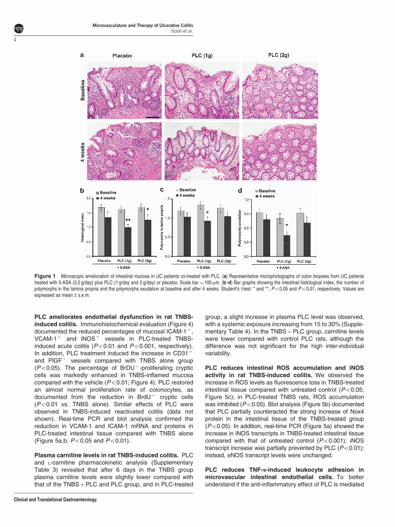
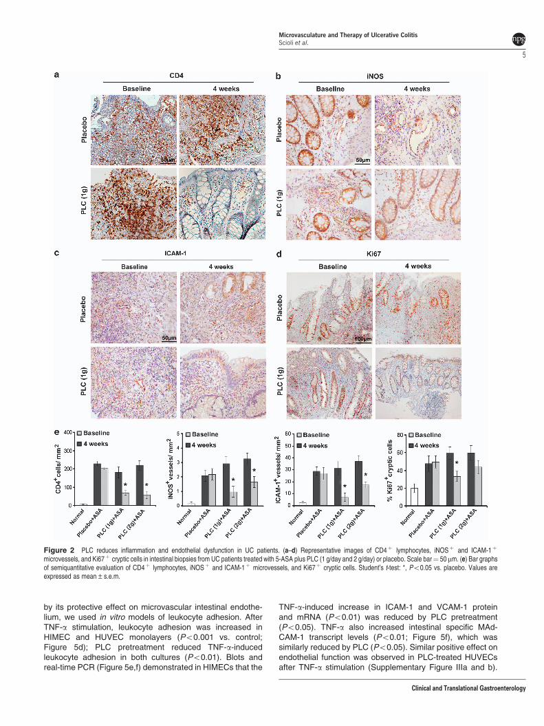
PLC co-treatment is effective and reduces mucosalinflammation and ICAM-1 expression in patients withUC. We investigated the effect of PLC co-treatment inintestinal biopsies of 23 UC patients receiving 5-ASA.Baseline values were similar in all experimental groups (notshown). Co-treatment with PLC-ameliorated clinical/endo-scopic response vs. placebo (74.2þ 5.2% vs. 49.3þ 3.1%,respectively; Po0.05) as reported;19 in particular, 76.4þ6.3% of patients receiving 1 g PLC had a clinical/endoscopicresponse (Po0.05). Representative images of biopsies areshown in Figure 1a. The histological index of mucosaldamage (Figure 1b) was reduced in PLC-treated comparedwith placebo (1g/day Po0.01; 2g/day Po0.05). The numberof polymorphs in the lamina propria was decreasedas well as intraepithelial polymorphs exudation (Po0.05;Figure 1c,d). Immunohistochemistry (Figure 2) showed thereduction in intramucosal CD4þ lymphocytes in PLC com-pared with placebo patients (Po0.05; Figure 2a,e). Anti-inflammatory effect was parallel to the reduction of iNOSþ
and ICAM-1þ microvessels (Po0.05; Figure 2b,c and e).Moreover, the percentage of proliferating Ki67þ cryptic cellsin PLC-treated patients was reduced (Po0.05) and close tothat of control (Figure 2d,e); instead, the percentage ofCD31þ vessels was unchanged (Supplementary Figure I).
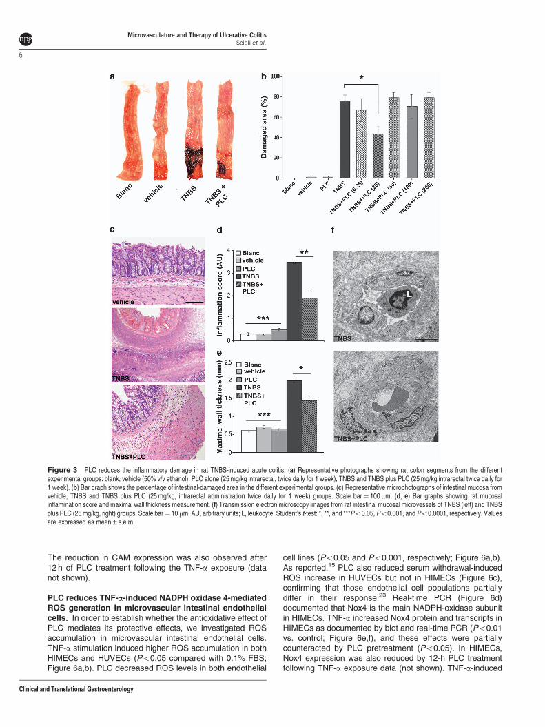
PLC reduces damaged area and inflammation in ratTNBS-induced acute colitis. To better document themicrovascular endothelial targeting of PLC, we used differentmodels of rat TNBS-induced colitis. PLC effects were inve-stigated by using different administration routes, dosagesand days of treatment (see Supplementary Figure II). InTNBS-induced acute colitis, intrarectal PLC administration(25 mg/kg twice daily, for one week) reduced the extent ofmacroscopic mucosal damage (Figure 3a,b) compared withTNBS alone (Po0.05). The positive effects of intrarectal (50and 100 mg/kg once daily, for 1 week) and oral (120mg/kgonce daily, for 1 week) administration were comparable andsimilar to that of intrarectal 5-ASA (120 mg/kg once daily, for1 week; Supplementary Figure II); instead, propionate andL-carnitine treatments were ineffective. Microscopic exam-ination confirmed the marked mucosal inflammation with thenecrosis of epithelium, the presence of granulation tissueand intestinal wall thickness in TNBS-treated rats (Po0.0001vs. control groups; Figure 3c–e) that were reduced by PLC(Po0.001 and Po0.05, respectively). Ultrastructural inves-tigation well documented the endothelial cell swelling andleukocyte recruitment in TNBS-treated inflamed intestinalmicrovessels (Figure 3f), and the reduction of inflammationand perivascular edema in PLC-treated rats. Intrarectal PLCadministration also reduced the damage area (Po0.05 at12.5 and 25 mg/kg; Supplementary Figure IId and e), as wellas intestinal wall thickness and inflammation in the TNBS-induced reactivated colitis (data not shown).
Microvasculature and Therapy of Ulcerative ColitisScioli et al.
3
Clinical and Translational Gastroenterology
PLC ameliorates endothelial dysfunction in rat TNBS-induced colitis. Immunohistochemical evaluation (Figure 4)documented the reduced percentages of mucosal ICAM-1þ ,VCAM-1þ and iNOSþ vessels in PLC-treated TNBS-induced acute colitis (Po0.01 and Po0.001, respectively).In addition, PLC treatment induced the increase in CD31þ
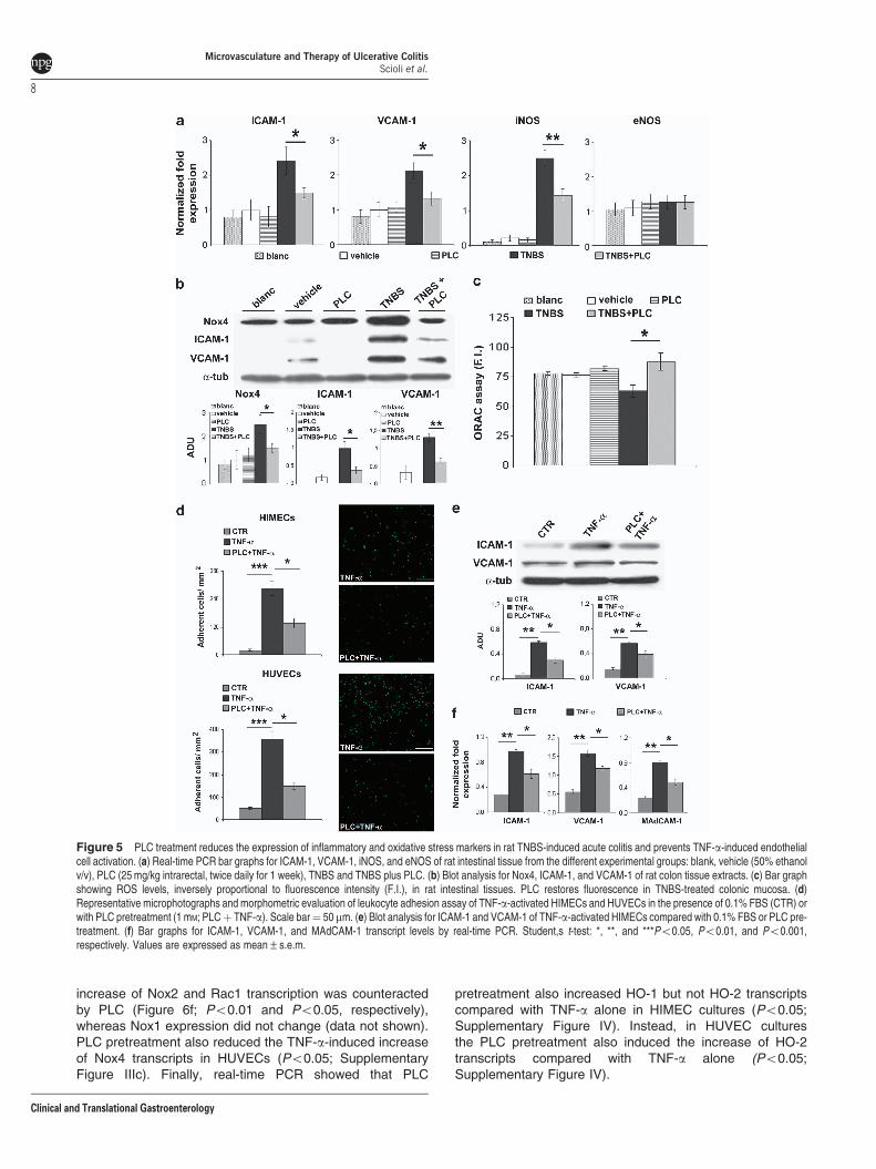
and PlGFþ vessels compared with TNBS alone group(Po0.05). The percentage of BrDUþ -proliferating crypticcells was markedly enhanced in TNBS-inflamed mucosacompared with the vehicle (Po0.01; Figure 4). PLC restoredan almost normal proliferation rate of colonocytes, asdocumented from the reduction in BrdUþ cryptic cells(Po0.01 vs. TNBS alone). Similar effects of PLC wereobserved in TNBS-induced reactivated colitis (data notshown). Real-time PCR and blot analysis confirmed thereduction in VCAM-1 and ICAM-1 mRNA and proteins inPLC-treated intestinal tissue compared with TNBS alone(Figure 5a,b; Po0.05 and Po0.01).
Plasma carnitine levels in rat TNBS-induced colitis. PLCand L-carnitine pharmacokinetic analysis (SupplementaryTable 3) revealed that after 6 days in the TNBS groupplasma carnitine levels were slightly lower compared withthat of the TNBSþPLC and PLC group, and in PLC-treated
group, a slight increase in plasma PLC level was observed,with a systemic exposure increasing from 15 to 30% (Supple-mentary Table 4). In the TNBSþPLC group, carnitine levelswere lower compared with control PLC rats, although thedifference was not significant for the high inter-individualvariability.
PLC reduces intestinal ROS accumulation and iNOSactivity in rat TNBS-induced colitis. We observed theincrease in ROS levels as fluorescence loss in TNBS-treatedintestinal tissue compared with untreated control (Po0.05;Figure 5c); in PLC-treated TNBS rats, ROS accumulationwas inhibited (Po0.05). Blot analysis (Figure 5b) documentedthat PLC partially counteracted the strong increase of Nox4protein in the intestinal tissue of the TNBS-treated group(Po0.05). In addition, real-time PCR (Figure 5a) showed theincrease in iNOS transcripts in TNBS-treated intestinal tissuecompared with that of untreated control (Po0.001); iNOStranscript increase was partially prevented by PLC (Po0.01);instead, eNOS transcript levels were unchanged.
PLC reduces TNF-a-induced leukocyte adhesion inmicrovascular intestinal endothelial cells. To betterunderstand if the anti-inflammatory effect of PLC is mediated
Figure 1 Microscopic amelioration of intestinal mucosa in UC patients co-treated with PLC. (a) Representative microphotographs of colon biopsies from UC patientstreated with 5-ASA (3.2 g/day) plus PLC (1 g/day and 2 g/day) or placebo. Scale bar¼ 100mm. (b–d) Bar graphs showing the intestinal histological index, the number ofpolymorphs in the lamina propria and the polymorphs exudation at baseline and after 4 weeks. Student’s t-test: * and **, Po0.05 and Po0.01, respectively. Values areexpressed as mean±s.e.m.
Microvasculature and Therapy of Ulcerative ColitisScioli et al.
4
Clinical and Translational Gastroenterology
by its protective effect on microvascular intestinal endothe-lium, we used in vitro models of leukocyte adhesion. AfterTNF-a stimulation, leukocyte adhesion was increased inHIMEC and HUVEC monolayers (Po0.001 vs. control;Figure 5d); PLC pretreatment reduced TNF-a-inducedleukocyte adhesion in both cultures (Po0.01). Blots andreal-time PCR (Figure 5e,f) demonstrated in HIMECs that the
TNF-a-induced increase in ICAM-1 and VCAM-1 proteinand mRNA (Po0.01) was reduced by PLC pretreatment(Po0.05). TNF-a also increased intestinal specific MAd-CAM-1 transcript levels (Po0.01; Figure 5f), which wassimilarly reduced by PLC (Po0.05). Similar positive effect onendothelial function was observed in PLC-treated HUVECsafter TNF-a stimulation (Supplementary Figure IIIa and b).
Figure 2 PLC reduces inflammation and endothelial dysfunction in UC patients. (a–d) Representative images of CD4þ lymphocytes, iNOSþ and ICAM-1þ
microvessels, and Ki67þ cryptic cells in intestinal biopsies from UC patients treated with 5-ASA plus PLC (1 g/day and 2 g/day) or placebo. Scale bar¼ 50mm. (e) Bar graphsof semiquantitative evaluation of CD4þ lymphocytes, iNOSþ and ICAM-1þ microvessels, and Ki67þ cryptic cells. Student’s t-test: *, Po0.05 vs. placebo. Values areexpressed as mean±s.e.m.
Microvasculature and Therapy of Ulcerative ColitisScioli et al.
5
Clinical and Translational Gastroenterology
The reduction in CAM expression was also observed after12 h of PLC treatment following the TNF-a exposure (datanot shown).
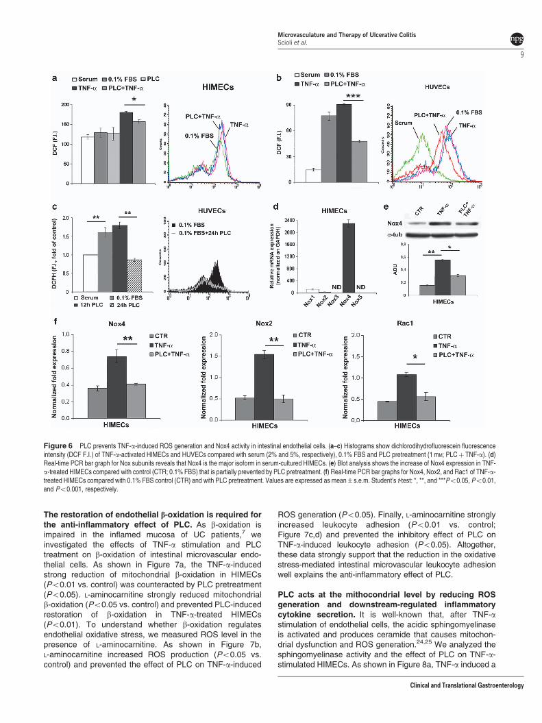
PLC reduces TNF-a-induced NADPH oxidase 4-mediatedROS generation in microvascular intestinal endothelialcells. In order to establish whether the antioxidative effect ofPLC mediates its protective effects, we investigated ROSaccumulation in microvascular intestinal endothelial cells.TNF-a stimulation induced higher ROS accumulation in bothHIMECs and HUVECs (Po0.05 compared with 0.1% FBS;Figure 6a,b). PLC decreased ROS levels in both endothelial
cell lines (Po0.05 and Po0.001, respectively; Figure 6a,b).As reported,15 PLC also reduced serum withdrawal-inducedROS increase in HUVECs but not in HIMECs (Figure 6c),confirming that those endothelial cell populations partiallydiffer in their response.23 Real-time PCR (Figure 6d)documented that Nox4 is the main NADPH-oxidase subunitin HIMECs. TNF-a increased Nox4 protein and transcripts inHIMECs as documented by blot and real-time PCR (Po0.01vs. control; Figure 6e,f), and these effects were partiallycounteracted by PLC pretreatment (Po0.05). In HIMECs,Nox4 expression was also reduced by 12-h PLC treatmentfollowing TNF-a exposure data (not shown). TNF-a-induced
Figure 3 PLC reduces the inflammatory damage in rat TNBS-induced acute colitis. (a) Representative photographs showing rat colon segments from the differentexperimental groups: blank, vehicle (50% v/v ethanol), PLC alone (25 mg/kg intrarectal, twice daily for 1 week), TNBS and TNBS plus PLC (25 mg/kg intrarectal twice daily for1 week). (b) Bar graph shows the percentage of intestinal-damaged area in the different experimental groups. (c) Representative microphotographs of intestinal mucosa fromvehicle, TNBS and TNBS plus PLC (25 mg/kg, intrarectal administration twice daily for 1 week) groups. Scale bar¼ 100mm. (d, e) Bar graphs showing rat mucosalinflammation score and maximal wall thickness measurement. (f) Transmission electron microscopy images from rat intestinal mucosal microvessels of TNBS (left) and TNBSplus PLC (25 mg/kg, right) groups. Scale bar¼ 10mm. AU, arbitrary units; L, leukocyte. Student’s t-test: *, **, and ***Po0.05, Po0.001, and Po0.0001, respectively. Valuesare expressed as mean±s.e.m.
Microvasculature and Therapy of Ulcerative ColitisScioli et al.
6
Clinical and Translational Gastroenterology
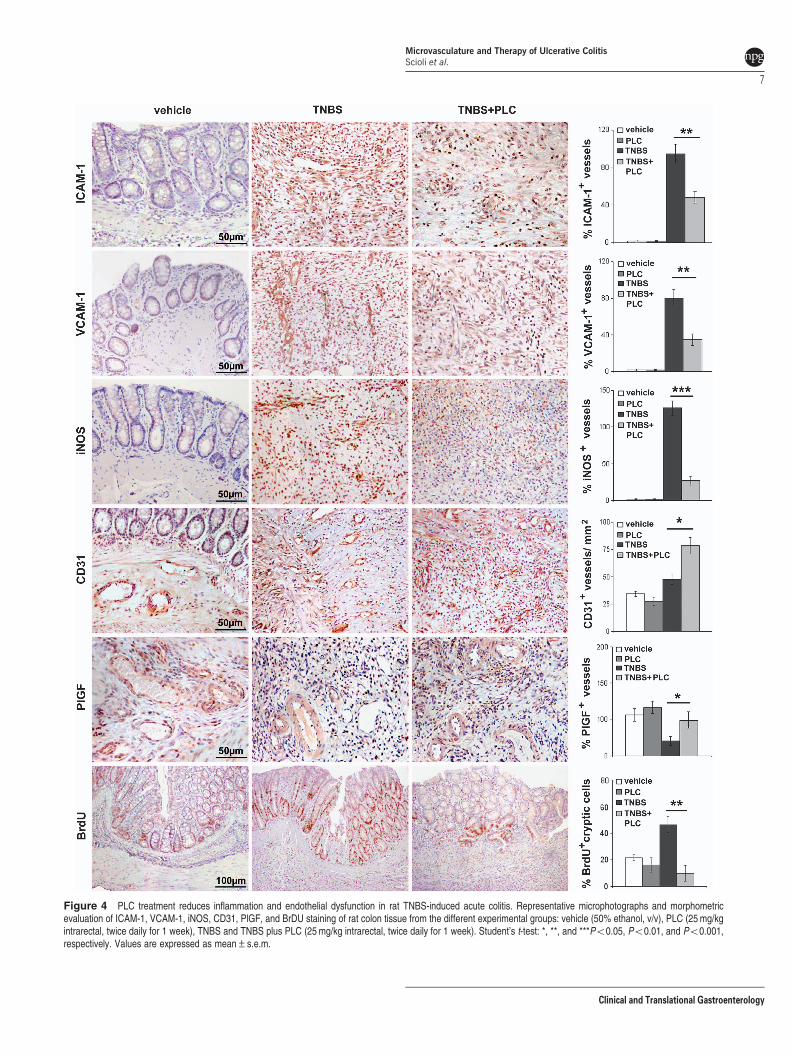
Figure 4 PLC treatment reduces inflammation and endothelial dysfunction in rat TNBS-induced acute colitis. Representative microphotographs and morphometricevaluation of ICAM-1, VCAM-1, iNOS, CD31, PlGF, and BrDU staining of rat colon tissue from the different experimental groups: vehicle (50% ethanol, v/v), PLC (25 mg/kgintrarectal, twice daily for 1 week), TNBS and TNBS plus PLC (25 mg/kg intrarectal, twice daily for 1 week). Student’s t-test: *, **, and ***Po0.05, Po0.01, and Po0.001,respectively. Values are expressed as mean±s.e.m.
Microvasculature and Therapy of Ulcerative ColitisScioli et al.
7
Clinical and Translational Gastroenterology
increase of Nox2 and Rac1 transcription was counteractedby PLC (Figure 6f; Po0.01 and Po0.05, respectively),whereas Nox1 expression did not change (data not shown).PLC pretreatment also reduced the TNF-a-induced increaseof Nox4 transcripts in HUVECs (Po0.05; SupplementaryFigure IIIc). Finally, real-time PCR showed that PLC
pretreatment also increased HO-1 but not HO-2 transcriptscompared with TNF-a alone in HIMEC cultures (Po0.05;Supplementary Figure IV). Instead, in HUVEC culturesthe PLC pretreatment also induced the increase of HO-2transcripts compared with TNF-a alone (Po0.05;Supplementary Figure IV).
Figure 5 PLC treatment reduces the expression of inflammatory and oxidative stress markers in rat TNBS-induced acute colitis and prevents TNF-a-induced endothelialcell activation. (a) Real-time PCR bar graphs for ICAM-1, VCAM-1, iNOS, and eNOS of rat intestinal tissue from the different experimental groups: blank, vehicle (50% ethanolv/v), PLC (25 mg/kg intrarectal, twice daily for 1 week), TNBS and TNBS plus PLC. (b) Blot analysis for Nox4, ICAM-1, and VCAM-1 of rat colon tissue extracts. (c) Bar graphshowing ROS levels, inversely proportional to fluorescence intensity (F.I.), in rat intestinal tissues. PLC restores fluorescence in TNBS-treated colonic mucosa. (d)Representative microphotographs and morphometric evaluation of leukocyte adhesion assay of TNF-a-activated HIMECs and HUVECs in the presence of 0.1% FBS (CTR) orwith PLC pretreatment (1 mM; PLCþ TNF-a). Scale bar¼ 50mm. (e) Blot analysis for ICAM-1 and VCAM-1 of TNF-a-activated HIMECs compared with 0.1% FBS or PLC pre-treatment. (f) Bar graphs for ICAM-1, VCAM-1, and MAdCAM-1 transcript levels by real-time PCR. Student,s t-test: *, **, and ***Po0.05, Po0.01, and Po0.001,respectively. Values are expressed as mean±s.e.m.
Microvasculature and Therapy of Ulcerative ColitisScioli et al.
8
Clinical and Translational Gastroenterology
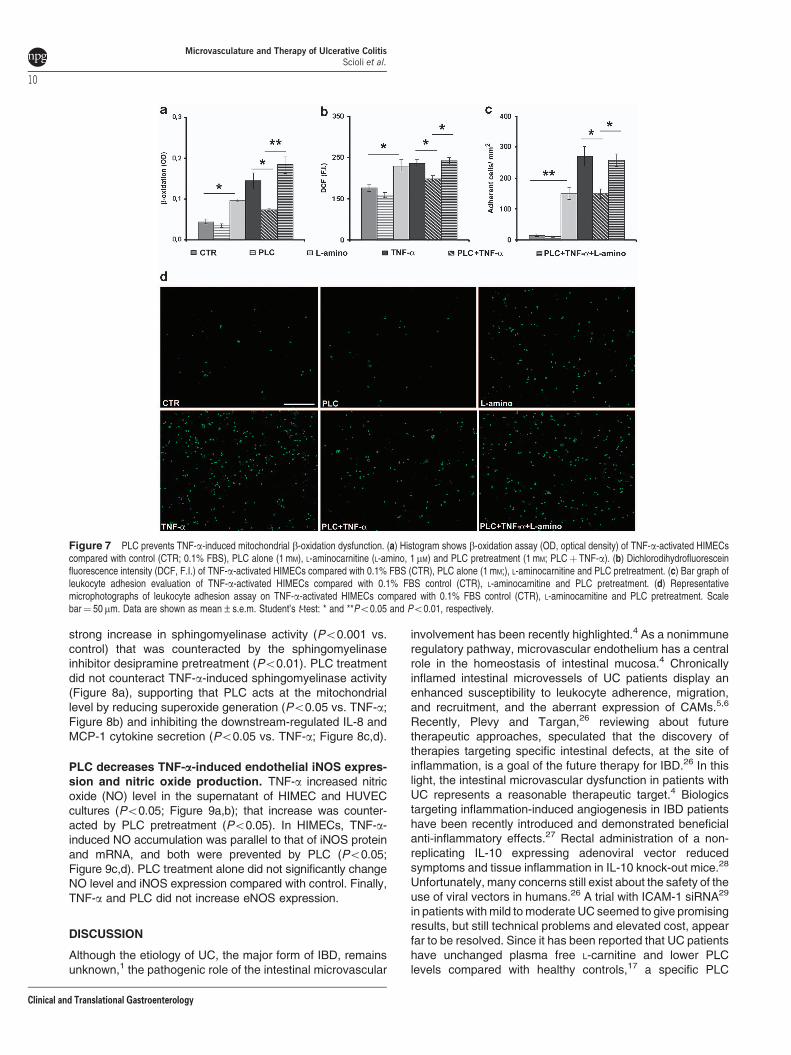
The restoration of endothelial b-oxidation is required forthe anti-inflammatory effect of PLC. As b-oxidation isimpaired in the inflamed mucosa of UC patients,7 weinvestigated the effects of TNF-a stimulation and PLCtreatment on b-oxidation of intestinal microvascular endo-thelial cells. As shown in Figure 7a, the TNF-a-inducedstrong reduction of mitochondrial b-oxidation in HIMECs(Po0.01 vs. control) was counteracted by PLC pretreatment(Po0.05). L-aminocarnitine strongly reduced mitochondrialb-oxidation (Po0.05 vs. control) and prevented PLC-inducedrestoration of b-oxidation in TNF-a-treated HIMECs(Po0.01). To understand whether b-oxidation regulatesendothelial oxidative stress, we measured ROS level in thepresence of L-aminocarnitine. As shown in Figure 7b,L-aminocarnitine increased ROS production (Po0.05 vs.control) and prevented the effect of PLC on TNF-a-induced
ROS generation (Po0.05). Finally, L-aminocarnitine stronglyincreased leukocyte adhesion (Po0.01 vs. control;Figure 7c,d) and prevented the inhibitory effect of PLC onTNF-a-induced leukocyte adhesion (Po0.05). Altogether,these data strongly support that the reduction in the oxidativestress-mediated intestinal microvascular leukocyte adhesionwell explains the anti-inflammatory effect of PLC.
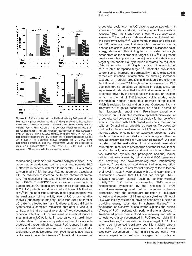
PLC acts at the mithocondrial level by reducing ROSgeneration and downstream-regulated inflammatorycytokine secretion. It is well-known that, after TNF-astimulation of endothelial cells, the acidic sphingomyelinaseis activated and produces ceramide that causes mitochon-drial dysfunction and ROS generation.24,25 We analyzed thesphingomyelinase activity and the effect of PLC on TNF-a-stimulated HIMECs. As shown in Figure 8a, TNF-a induced a
Figure 6 PLC prevents TNF-a-induced ROS generation and Nox4 activity in intestinal endothelial cells. (a–c) Histograms show dichlorodihydrofluorescein fluorescenceintensity (DCF F.I.) of TNF-a-activated HIMECs and HUVECs compared with serum (2% and 5%, respectively), 0.1% FBS and PLC pretreatment (1 mM; PLCþ TNF-a). (d)Real-time PCR bar graph for Nox subunits reveals that Nox4 is the major isoform in serum-cultured HIMECs. (e) Blot analysis shows the increase of Nox4 expression in TNF-a-treated HIMECs compared with control (CTR; 0.1% FBS) that is partially prevented by PLC pretreatment. (f) Real-time PCR bar graphs for Nox4, Nox2, and Rac1 of TNF-a-treated HIMECs compared with 0.1% FBS control (CTR) and with PLC pretreatment. Values are expressed as mean±s.e.m. Student’s t-test: *, **, and ***Po0.05, Po0.01,and Po0.001, respectively.
Microvasculature and Therapy of Ulcerative ColitisScioli et al.
9
Clinical and Translational Gastroenterology
strong increase in sphingomyelinase activity (Po0.001 vs.control) that was counteracted by the sphingomyelinaseinhibitor desipramine pretreatment (Po0.01). PLC treatmentdid not counteract TNF-a-induced sphingomyelinase activity(Figure 8a), supporting that PLC acts at the mitochondriallevel by reducing superoxide generation (Po0.05 vs. TNF-a;Figure 8b) and inhibiting the downstream-regulated IL-8 andMCP-1 cytokine secretion (Po0.05 vs. TNF-a; Figure 8c,d).
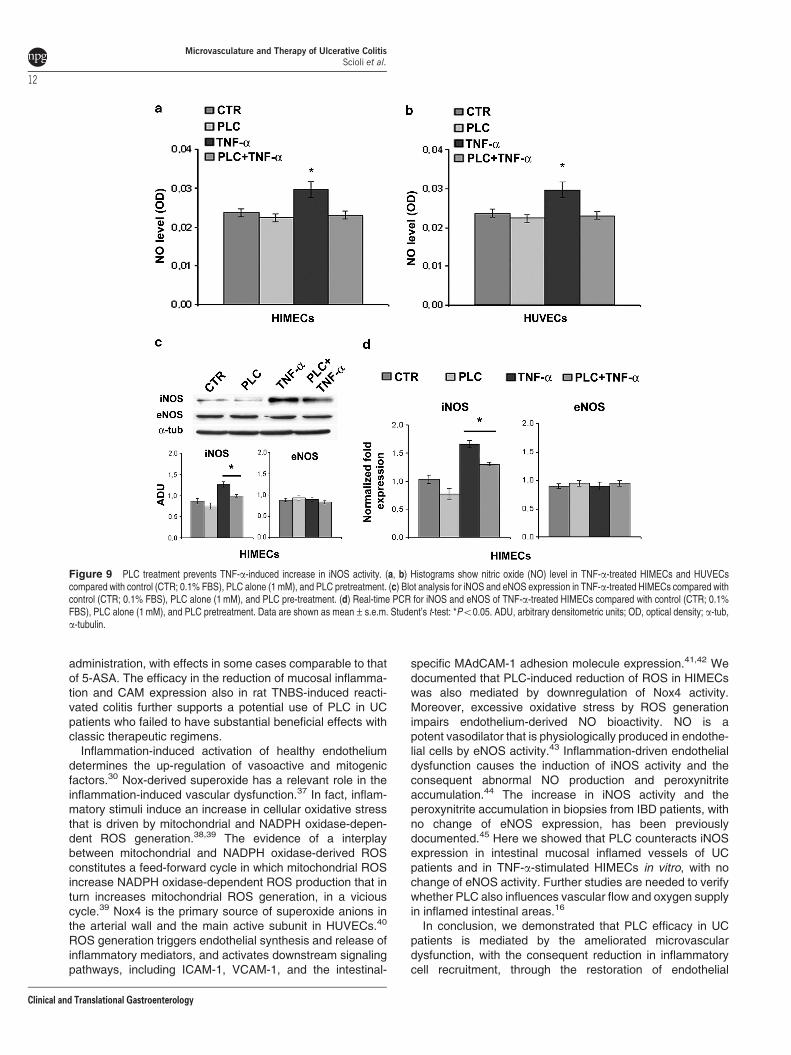
PLC decreases TNF-a-induced endothelial iNOS expres-sion and nitric oxide production. TNF-a increased nitricoxide (NO) level in the supernatant of HIMEC and HUVECcultures (Po0.05; Figure 9a,b); that increase was counter-acted by PLC pretreatment (Po0.05). In HIMECs, TNF-a-induced NO accumulation was parallel to that of iNOS proteinand mRNA, and both were prevented by PLC (Po0.05;Figure 9c,d). PLC treatment alone did not significantly changeNO level and iNOS expression compared with control. Finally,TNF-a and PLC did not increase eNOS expression.
DISCUSSION
Although the etiology of UC, the major form of IBD, remainsunknown,1 the pathogenic role of the intestinal microvascular
involvement has been recently highlighted.4 As a nonimmuneregulatory pathway, microvascular endothelium has a centralrole in the homeostasis of intestinal mucosa.4 Chronicallyinflamed intestinal microvessels of UC patients display anenhanced susceptibility to leukocyte adherence, migration,and recruitment, and the aberrant expression of CAMs.5,6
Recently, Plevy and Targan,26 reviewing about futuretherapeutic approaches, speculated that the discovery oftherapies targeting specific intestinal defects, at the site ofinflammation, is a goal of the future therapy for IBD.26 In thislight, the intestinal microvascular dysfunction in patients withUC represents a reasonable therapeutic target.4 Biologicstargeting inflammation-induced angiogenesis in IBD patientshave been recently introduced and demonstrated beneficialanti-inflammatory effects.27 Rectal administration of a non-replicating IL-10 expressing adenoviral vector reducedsymptoms and tissue inflammation in IL-10 knock-out mice.28
Unfortunately, many concerns still exist about the safety of theuse of viral vectors in humans.26 A trial with ICAM-1 siRNA29
in patients with mild to moderate UC seemed to give promisingresults, but still technical problems and elevated cost, appearfar to be resolved. Since it has been reported that UC patientshave unchanged plasma free L-carnitine and lower PLClevels compared with healthy controls,17 a specific PLC
Figure 7 PLC prevents TNF-a-induced mitochondrial b-oxidation dysfunction. (a) Histogram shows b-oxidation assay (OD, optical density) of TNF-a-activated HIMECscompared with control (CTR; 0.1% FBS), PLC alone (1 mM), L-aminocarnitine (L-amino, 1 mM) and PLC pretreatment (1 mM; PLCþ TNF-a). (b) Dichlorodihydrofluoresceinfluorescence intensity (DCF, F.I.) of TNF-a-activated HIMECs compared with 0.1% FBS (CTR), PLC alone (1 mM;), L-aminocarnitine and PLC pretreatment. (c) Bar graph ofleukocyte adhesion evaluation of TNF-a-activated HIMECs compared with 0.1% FBS control (CTR), L-aminocarnitine and PLC pretreatment. (d) Representativemicrophotographs of leukocyte adhesion assay on TNF-a-activated HIMECs compared with 0.1% FBS control (CTR), L-aminocarnitine and PLC pretreatment. Scalebar¼ 50mm. Data are shown as mean±s.e.m. Student’s t-test: * and **Po0.05 and Po0.01, respectively.
Microvasculature and Therapy of Ulcerative ColitisScioli et al.
10
Clinical and Translational Gastroenterology
sequestering in inflamed tissues could be hypothesized. In thepresent study, we documented that the co-treatment with PLCis effective in patients with mild-to-moderate UC with stableconventional 5-ASA therapy; PLC co-treatment associatedwith the reduction of intestinal acute and chronic inflamma-tion. The reduction of mucosal inflammation was parallel tothat of ICAM-1þ and iNOSþ microvessels compared with theplacebo group. Our results strengthen the clinical efficacy ofPLC in UC patients and do not contrast those of Mikhailovaet al.19 In the latter study, primary histological endpoint wasthe amelioration of the activity level of UC by comparativeanalysis, but being the majority (more than 80%) of enrolledUC patients affected from a mild disease, it was difficult tohypothesize a complete remission only after 4 weeks. Incontrast with that comparative system,19 we documented abeneficial effect of PLC co-treatment on intestinal mucosalinflammation in UC patients, in accordance with preliminaryreported data.18 The second question we tried to solve is tounderstand through which pathways PLC reduces inflamma-tion and ameliorates intestinal microvascular endothelialdysfunction. Oxidative stress from ROS accumulation has acentral role in vascular diseases.30 Intestinal microvascular
endothelial dysfunction in UC patients associates with theincrease in oxidative stress, normally absent in intestinalvessels.31 PLC has already been shown to be a superoxidescavenger11 that reduces oxidative stress in endothelial cellsand cardiomyocytes.32,33 Experimental models and biopsiesfrom UC patients showed that metabolic changes occur in thediseased colonic mucosa, with an impaired b-oxidation and anenergy shortage.8 This finding led to consider colonocytemetabolism as the therapeutic target of PLC.19 Our presentresults strongly support that the adjuvant antioxidant actiontargeting the endothelial dysfunction mediates the reductionof the inflammation, confirming the intestinal microvasculatureas a reliable therapeutic target.4–6 Endothelial dysfunctiondetermines an increase in permeability that is expected toperpetuate intestinal inflammation by allowing increasedpassage of microbial products and antigenic proteins intothe inflamed mucosa.34 Although we cannot exclude that PLCalso counteracts peroxidative damage in colonocytes, ourexperimental data show that the clinical improvement in UCpatients is driven by the ameliorated microvascular function.In fact, in the rat of TNBS-induced colitis model, acuteinflammation induces almost total necrosis of epithelium,which is replaced by granulation tissue. Consequently, it islikely that PLC targets subendothelial tissue cells, in particularthe microvascular endothelium. Preliminary experimentsperformed on PLC-treated intestinal epithelial-microvascularendothelial cell co-cultures did not display further beneficialeffects compared with PLC-treated endothelial cell mono-cultures (Orlandi et al., unpublished results). Moreover, wecould not exclude a positive effect of PLC on circulating bonemarrow-derived endothelial/hematopoietic progenitor cells,which can be locally recruited and contributing to inflamma-tion, as already reported for vascular diseases.35 We alsoreported that the restoration of mitochondrial b-oxidationcounteracts intestinal microvascular endothelial dysfunctionin vitro. In fact, inflammatory stimuli (such as inflamma-tory cytokines, hypoxia and pathogenic bacteria) inducecellular oxidative stress by mitochondrial ROS generationand activating the downstream-regulated inflammatoryresponse.36 We demonstrated that anti-inflammatory effectof PLC depends on its anti-oxidant efficacy at the mitochon-drial level. In fact, in vitro assays with L-aminocarnitine anddesipramine showed that PLC did not change TNF-a-activated upstream signals, such as sphingomyelinaseactivity.24,25 PLC action counteracted TNF-a-inducedmitochondrial dysfunction by the inhibition of ROSand downstream-regulated cellular molecule adhesionexpression, with the consequent reduction of leukocyteadhesion and secretion of inflammatory cytokines. AlthoughPLC was initially retained to have an anaplerotic function ofproviding energy substrates in ischemic tissues,12 themodulation of oxidative stress better explains the clinicalefficacy of PLC in patients with peripheral arterial disease.16
Ameliorated post-ischemic blood flow recovery and arterio-genesis were also documented in PLC-treated rabbit limbischemic tissues,15 in line with the vascular target of PLC. Thelatter also influenced positively post-injury rabbit carotidremodeling.20 PLC efficacy was macroscopically and micro-scopically documented in rat TNBS-induced colitis withvarious experimental settings, dosages, and routes of
Figure 8 PLC acts at the mitochondrial level reducing ROS generation anddownstream-regulated cytokine secretion. (a) Histogram shows sphingomyelinaseactivity assay (fluorescence units) of TNF-a-activated HIMECs compared withcontrol (CTR; 0.1% FBS), PLC alone (1 mM), desipramine pretreatment (Des, 5 mM),and PLC pretreatment (1 mM). (b) Histogram shows ethidium bromide fluorescence(DHE oxidation) of TNF-a-activated HIMECs compared with CTR, PLC alone,desipramine pretreatment, and PLC pretreatment. (c, d) Bar graphs show IL-8 andMCP-1 level of TNF-a-activated HIMECs compared with CTR, PLC alone,desipramine pretreatment, and PLC pretreatment. Values are expressed asmean±s.e.m. Student’s t-test: *, **, and ***Po0.05, Po0.01, and Po0.001,respectively. AU, arbitrary units; FI, fluorescence intensity.
Microvasculature and Therapy of Ulcerative ColitisScioli et al.
11
Clinical and Translational Gastroenterology
administration, with effects in some cases comparable to thatof 5-ASA. The efficacy in the reduction of mucosal inflamma-tion and CAM expression also in rat TNBS-induced reacti-vated colitis further supports a potential use of PLC in UCpatients who failed to have substantial beneficial effects withclassic therapeutic regimens.
Inflammation-induced activation of healthy endotheliumdetermines the up-regulation of vasoactive and mitogenicfactors.30 Nox-derived superoxide has a relevant role in theinflammation-induced vascular dysfunction.37 In fact, inflam-matory stimuli induce an increase in cellular oxidative stressthat is driven by mitochondrial and NADPH oxidase-depen-dent ROS generation.38,39 The evidence of a interplaybetween mitochondrial and NADPH oxidase-derived ROSconstitutes a feed-forward cycle in which mitochondrial ROSincrease NADPH oxidase-dependent ROS production that inturn increases mitochondrial ROS generation, in a viciouscycle.39 Nox4 is the primary source of superoxide anions inthe arterial wall and the main active subunit in HUVECs.40
ROS generation triggers endothelial synthesis and release ofinflammatory mediators, and activates downstream signalingpathways, including ICAM-1, VCAM-1, and the intestinal-
specific MAdCAM-1 adhesion molecule expression.41,42 Wedocumented that PLC-induced reduction of ROS in HIMECswas also mediated by downregulation of Nox4 activity.Moreover, excessive oxidative stress by ROS generationimpairs endothelium-derived NO bioactivity. NO is apotent vasodilator that is physiologically produced in endothe-lial cells by eNOS activity.43 Inflammation-driven endothelialdysfunction causes the induction of iNOS activity and theconsequent abnormal NO production and peroxynitriteaccumulation.44 The increase in iNOS activity and theperoxynitrite accumulation in biopsies from IBD patients, withno change of eNOS expression, has been previouslydocumented.45 Here we showed that PLC counteracts iNOSexpression in intestinal mucosal inflamed vessels of UCpatients and in TNF-a-stimulated HIMECs in vitro, with nochange of eNOS activity. Further studies are needed to verifywhether PLC also influences vascular flow and oxygen supplyin inflamed intestinal areas.16
In conclusion, we demonstrated that PLC efficacy in UCpatients is mediated by the ameliorated microvasculardysfunction, with the consequent reduction in inflammatorycell recruitment, through the restoration of endothelial
Figure 9 PLC treatment prevents TNF-a-induced increase in iNOS activity. (a, b) Histograms show nitric oxide (NO) level in TNF-a-treated HIMECs and HUVECscompared with control (CTR; 0.1% FBS), PLC alone (1 mM), and PLC pretreatment. (c) Blot analysis for iNOS and eNOS expression in TNF-a-treated HIMECs compared withcontrol (CTR; 0.1% FBS), PLC alone (1 mM), and PLC pre-treatment. (d) Real-time PCR for iNOS and eNOS of TNF-a-treated HIMECs compared with control (CTR; 0.1%FBS), PLC alone (1 mM), and PLC pretreatment. Data are shown as mean±s.e.m. Student’s t-test: *Po0.05. ADU, arbitrary densitometric units; OD, optical density; a-tub,a-tubulin.
Microvasculature and Therapy of Ulcerative ColitisScioli et al.
12
Clinical and Translational Gastroenterology
b-oxidation. Our data suggest that the microvascular targetingof PLC offers further opportunities for pharmacological strate-gies aimed to counteract the physiopathological changesinduced by oxidative stress in other tissues and organs.
CONFLICT OF INTEREST
Guarantor of the article: Augusto Orlandi, MD.Specific author contributions: Maria Giovanna Scioli:conception and design, data analysis and interpretation;Maria Antonietta Stasi: conception and design, data analysisand interpretation; Daniela Passeri: collection and assemblyof data; Elena Doldo: collection and assembly of data; GaetanaCostanza: collection and assembly of data; Roberto Camerini:conception and design, data analysis and interpretation; PaoloFociani: conception and design, data analysis andinterpretation; Gaetano Arcuri: collection and assembly of data;Katia Lombardo: collection and assembly of data; Silvia Pace:collection and assembly of data; Franco Borsini: conceptionand design, data analysis and interpretation; Augusto Orlandi:conception and design, data analysis and interpretation, writingand final approval of the manuscript.Financial support: The study was partially funded by Sigma-Tau SpA.Potential competing interests: Maria Antonietta Stasi,Roberto Camerini, Katia Lombardo, Silvia Pace and FrancoBorsini are employees of Sigma-Tau SpA. All the other authorsdeclared no conflict of interest.
Acknowledgments. We thank Drs Sabrina Cappelli, Angela Ursillo andAnna Maria Russo for their excellent technical work.
Study Highlights
WHAT IS CURRENT KNOWLEDGE
| Microvascular endothelial dysfunction characterizesulcerative colitis.
| Propionyl-L-carnitine (PLC) ameliorates propionyl-CoAbioavailability and reduces oxidative stress in ischemictissues.
WHAT IS NEW HERE
| PLC co-treatment ameliorates mucosa homeostasis byreducing inflammation and intestinal microvascularendothelial dysfunction in ulcerative colitis (UC) patients.
| In the rat TNBS-induced colitis, PLC treatment reducesmucosal inflammation and colonic damage.
| PLC reduces TNF-a-induced intestinal microvascularendothelial cell inflammation improving b-oxidation andpreventing oxidative stress.
1. Danese S, Fiocchi C. Ulcerative Colitis. N Engl J Med 2011; 365: 1713–1725.2. Loftus EV. Clinical epidemiology of inflammatory bowel disease: incidence, prevalence,
and environmental influences. Gastroenterology 2004; 126: 1504–1517.3. MacDonald TT, Monteleone I, Fantini MC et al. Regulation of homeostasis and
inflammation in the intestine. Gastroenterology 2011; 140: 1768–1775.4. Binion DG, West GA, Volk EE et al. Acquired increase in leucocyte binding by intestinal
microvascular endothelium in inflammatory bowel disease. Lancet 1998; 352: 1742–1746.
5. Ishiguro Y. Mucosal proinflammatory cytokine production correlates with endoscopicactivity of ulcerative colitis. J Gastroenterol 1999; 34: 66–74.
6. Hatoum OA. The intestinal microvasculature as a therapeutic target in inflammatory boweldisease. Ann N Y Acad Sci 2006; 1072: 78–97.
7. Roediger WE. The colonic epithelium in ulcerative colitis: an energy-deficiency disease?Lancet 1980; 2: 712–715.
8. Chapman MA, Grahn MF. Ileal and colonic epithelial metabolism in quiescent ulcerativecolitis. Gut 1994; 35: 1152–1153.
9. Bremer J. Carnitine metabolism and function. Physiol Rev 1983; 63: 1420–1479.10. Li P, Park C, Micheletti R et al. Myocyte performance during evolution of
myocardial infarction in rats: effects of propionyl-L-carnitine. Am J Physiol 1995; 268:1702–1713.
11. Vanella A, Russo A, Acquaviva R et al. L -propionyl-carnitine as superoxide scavenger,antioxidant, and DNA cleavage protector. Cell Biol Toxicol 2000; 16: 99–104.
12. Hiatt WR. Medical treatment of peripheral arterial disease and claudication. N Engl J Med2001; 344: 1608–1621.
13. Hankey GJ, Norman PE, Eikelboom JW. Medical treatment of peripheral arterial disease.JAMA 2006; 295: 547–553.
14. Touyz RM. Reactive oxygen species, vascular oxidative stress, and redoxsignaling in hypertension: what is the clinical significance? Hypertension 2004; 44:248–252.
15. Stasi MA, Scioli MG, Arcuri G et al. Propionyl-L-carnitine improves postischemicblood flow recovery and arteriogenetic revascularization and reduces endothelial NADPH-oxidase 4-mediated superoxide production. Arterioscler Thromb Vasc Biol 2010; 30:426–435.
16. Loffredo L, Marcoccia A, Pignatelli P et al. Oxidative-stress-mediated arterial dysfunction inpatients with peripheral arterial disease. Eur Heart J 2007; 28: 608–612.
17. Bene J, Komlosi K, Havasi V et al. Changes of plasma fasting carnitine ester profile inpatients with ulcerative colitis. World J Gastroenterol 2006; 12: 110–113.
18. Gasbarrini G, Mingrone G, Giancaterini A et al. Effects of propionyl-L-carnitine topicalirrigation in distal ulcerative colitis: a preliminary report. Hepatogastroenterology 2003; 50:1385–1389.
19. Mikhailova TL, Sishkova E, Poniewierka E et al. Randomised clinical trial: the efficacy andsafety of propionyl-L-carnitine therapy in patients with ulcerative colitis receiving stable oraltreatment. Aliment Pharmacol Ther 2011; 34: 1088–1097.
20. Orlandi A, Marcellini M, Pesce D et al. Propionyl-L-carnitine reduces intimal hyperplasiaafter injury in normocholesterolemic rabbit carotid artery by modulating proliferation andcaspase 3-dependent apoptosis of vascular smooth muscle cells. Atherosclerosis 2002;160: 81–89.
21. Ferlosio A, Arcuri G, Doldo E et al. Age-related increase of stem marker expressioninfluences vascular smooth muscle cell properties. Atherosclerosis 2012; 224: 51–57.
22. Campagnolo L, Costanza G, Francesconi A et al. Sortilin expression is essential for pro-nerve growth factor-induced apoptosis of rat vascular smooth muscle cells. PLoSOne2014; 9: e84969.
23. Haraldsen G, Kvale D, Lien B et al. Cytokine-regulated expression of E-selectin,intercellular adhesion molecule-1 (ICAM-1), and vascular cell adhesion molecule-1(VCAM-1) in human microvascular endothelial cells. J Immunol 1996; 156: 2558–2565.
24. Corda S, Laplace C, Vicaut E et al. Rapid reactive oxygen species production bymitochondria in endothelial cells exposed to tumor necrosis factor-alpha is mediated byceramide. Am J Respir Cell Mol Biol 2001; 24: 762–768.
25. Marino MW, Dunbar JD, Wu LW et al. Inhibition of tumor necrosis factor signal transductionin endothelial cells by dimethylaminopurine. J Biol Chem 1996; 271: 28624–28629.
26. Plevy SE, Targan SR. Future therapeutic approaches for inflammatory bowel diseases.Gastroenterology 2011; 140: 1838–1846.
27. Sands BE. Inflammatory bowel disease: past, present, and future. J Gastroenterol 2007;42: 16–25.
28. Lindsay JO, Ciesielski CJ, Scheinin T et al. Local delivery of adenoviral vectors encodingmurine interleukin 10 induces colonic interleukin 10 production and is therapeutic formurine colitis. Gut 2003; 52: 363–369.
29. van Deventer SJ, Wedel MK, Baker BF et al. A phase II dose ranging, double-blind,placebo-controlled study of alicaforsen enema in subjects with acute exacerbation ofmild to moderate left-sided ulcerative colitis. Aliment Pharmacol Ther 2006; 23:1415–1425.
30. Cai H, Harrison DG. Endothelial dysfunction in cardiovascular diseases: the role of oxidantstress. Circ Res 2000; 87: 840–844.
31. Owczarek D, Cibor D, Mach T. Asymmetric dimethylarginine (ADMA), symmetricdimethylarginine (SDMA), arginine, and 8-iso-prostaglandin F2alpha (8-iso-PGF2alpha)level in patients with inflammatory bowel diseases. Inflamm Bowel Dis 2010; 16: 52–57.
32. Van Hinsbergh VW, Scheffer MA. Effect of propionyl-L-carnitine on human endothelialcells. Cardiovasc Drugs Ther 1991; 5: 97–105.
33. Calo LA, Pagnin E, Davis PA et al. Antioxidant effect of L-carnitine and its short chainesters: relevance for the protection from oxidative stress related cardiovascular damage.Int J Cardiol 2006; 107: 54–60.
34. Stein J, Ries J, Barrett KE. Disruption of intestinal barrier function associated withexperimental colitis: possible role of mast cells. Am J Physiol 1998; 274: G203–G209.
35. Orlandi A, Bennett M. Progenitor cell-derived smooth muscle cells in vascular disease.Biochem Pharmacol 2010; 79: 1706–1713.
Microvasculature and Therapy of Ulcerative ColitisScioli et al.
13
Clinical and Translational Gastroenterology
36. Goossens V, Grooten J, De Vos K et al. Direct evidence for tumor necrosis factor-inducedmitochondrial reactive oxygen intermediates and their involvement in cytotoxicity. Proc NatlAcad Sci USA 1995; 92: 8115–8119.
37. Lassegue B, San Martın A, Griendling KK et al. Biochemistry, physiology, andpathophysiology of NADPH oxidases in the cardiovascular system. Circ Res 2012; 110:1364–1390.
38. Dikalov S. Cross talk between mitochondria and NADPH oxidases. Free Radic Biol Med2011; 51: 1289–1301.
39. Santos CX, Tanaka LY, Wosniak J et al. Mechanisms and implications of reactive oxygenspecies generation during the unfolded protein response: roles of endoplasmic reticulumoxidoreductases, mitochondrial electron transport, and NADPH oxidase. Antioxid RedoxSignal 2009; 11: 2409–2427.
40. Ago T, Kitazono T, Ooboshi H et al. Nox4 as the major catalytic component of anendothelial NAD(P)H oxidase. Circulation 2004; 109: 227–233.
41. Chen YH, Lin SJ, Chen YL et al. Anti-inflammatory effects of different drugs/agents withantioxidant property on endothelial expression of adhesion molecules. Cardiovasc HematolDisord Drug Targets 2006; 6: 279–304.
42. Ogawa H, Binion DG, Heidemann J et al. Mechanisms of MAdCAM-1 gene expression inhuman intestinal microvascular endothelial cells. Am J Physiol Cell Physiol 2005; 288:C272–C281.
43. Gunnett CA, Lund DD, McDowell AK et al. Mechanisms of inducible nitric oxide synthase-mediated vascular dysfunction. Arterioscler Thromb Vasc Biol 2005; 25: 1617–1622.
44. McCafferty DM. Peroxynitrite and inflammatory bowel disease. Gut 2000; 46: 436–439.45. Dijkstra G, Moshage H, van Dullemen HM et al. Expression of nitric oxide synthases and
formation of nitrotyrosine and reactive oxygen species in inflammatory bowel disease.J Pathol 1998; 186: 416–421.
Clinical and Translational Gastroenterology is an open-access journal published by Nature Publishing Group.
This work is licensed under a Creative Commons Attribution-NonCommercial-NoDerivs 3.0 Unported License. To view a copy ofthis license, visit http://creativecommons.org/licenses/by-nc-nd/3.0/
Supplementary Information accompanies this paper on the Clinical and Translational Gastroenterology website (http://www.nature.com/ctg)
Microvasculature and Therapy of Ulcerative ColitisScioli et al.
14
Clinical and Translational Gastroenterology
Related Documents


![carnitine deficiency[1]](https://static.cupdf.com/doc/110x72/577d20c11a28ab4e1e93ae46/carnitine-deficiency1.jpg)