Acta neuropath. (Berl.) 18, 273--285 (1971) by Springer-Verlag 1971 Originalarbeiten Original Investigations Travaux originaux Uhrastructure of the Microvasculature in Experimental Cerebral Infarction* JULIO H. GARCIA, JANICE V. Cox, and W. ROBERT IIUDGI~S t~eceived October 2, 1970/April 5, 1971 Summary. Structural evaluation of cerebral infarction in twelve squirrel monkeys was conducted for the purpose of elucidating some aspects of the pathogenesis of regional cerebral ischemia. The changes observed first were interpreted as indicative of alterations in the permeability of the vascular walls. It is suggested that during the early stages of infarction, emigration of fluid, particles and cells may occur in a transendothelial fashion. Arguments to prove the preservation of the microcirculation within the infarcted area are presented. Hypoxemia, as judged by the relative structural integrity of mitochondria, does not seem to be the most important pathogenetic factor in the development of c~rebral ischemie necrosis. Key-Words: Pathology -- Cerebral Edema -- Blood Brain-Barrier -- Itypoxia -- Proximal Middle Cerebral Artery. Introduction The experiment herein described was designed to study, by ultrastructural methods, some of the pathogenetic mechanisms that may influence the develop- ment of cerebral infarction. Several excellent models for inducing cerebral infarction in animals have been described (Sundt and Waltz, 1966) ; however, we have shown before that the use of surgical procedures requiring opening of the skull results in blood-brain- barrier alterations which are difficult to differentiate from permeability changes that occur as a consequence of cerebra] ischemia per se (Hudgins and Garcia, 1970 b). Although methods for inducing cerebral infarction in animals without opening the skull have been described (Penry and Netsky ; Hill et al.), we consider them im- practical for electron microscopic purposes, because of the unpredictability of the size and site of the lesion and because of the low percentage of infarcts obtained with them. Thus, since at this time, we are not aware of any electronmicroseopic evaluation of regional cerebralisehemia, we developed a model which appears suitable for ultra- structural studies and which closely resembles the conditions existing in human cerebral infarction of vascular occlusive etiology. The surgical method for arterial occlusion approaches the initial segment of the middle cerebral artery (MCA) through an enlarged optic foramen (Hudgins and Garcia, 1970a). It has been stated that the ultrastructural features of cerebral hypoxic lesions are probably the same, regardless of the mechanism by which they are induced * This work was conducted during the author's tenure at the Cerebrovascular Research Center of the University of Tennessee Medical Units; it was partially supported by USPHS Grants HE 11794 and NS 06826. 19 Acta neuropath. (Berl.) ]3d. 18

Welcome message from author
This document is posted to help you gain knowledge. Please leave a comment to let me know what you think about it! Share it to your friends and learn new things together.
Transcript
Acta neuropath. (Berl.) 18, 273--285 (1971) �9 by Springer-Verlag 1971
O r i g i n a l a r b e i t e n �9 O r i g i n a l I n v e s t i g a t i o n s �9 T r a v a u x o r i g i n a u x
Uhrastructure of the Microvasculature in Experimental Cerebral Infarction*
JULIO H . GARCIA, JANICE V. Cox , a n d W. ROBERT IIUDGI~S
t~eceived October 2, 1970/April 5, 1971
Summary . Structural evaluation of cerebral infarction in twelve squirrel monkeys was conducted for the purpose of elucidating some aspects of the pathogenesis of regional cerebral ischemia.
The changes observed first were interpreted as indicative of alterations in the permeability of the vascular walls. I t is suggested that during the early stages of infarction, emigration of fluid, particles and cells may occur in a transendothelial fashion. Arguments to prove the preservation of the microcirculation within the infarcted area are presented.
Hypoxemia, as judged by the relative structural integrity of mitochondria, does not seem to be the most important pathogenetic factor in the development of c~rebral ischemie necrosis.
Key-Words: Pathology - - Cerebral Edema -- Blood Brain-Barrier - - I typoxia - - Proximal Middle Cerebral Artery.
Introduction
The experiment herein described was designed to study, by ultrastructural methods, some of the pathogenetic mechanisms tha t may influence the develop- ment of cerebral infarction.
Several excellent models for inducing cerebral infarction in animals have been described (Sundt and Waltz, 1966) ; however, we have shown before that the use of surgical procedures requiring opening of the skull results in blood-brain- barrier alterations which are difficult to differentiate from permeability changes tha t occur as a consequence of cerebra] ischemia per se (Hudgins and Garcia, 1970 b).
Although methods for inducing cerebral infarction in animals without opening the skull have been described (Penry and Netsky ; Hill et al.), we consider them im- practical for electron microscopic purposes, because of the unpredictability of the size and site of the lesion and because of the low percentage of infarcts obtained with them.
Thus, since at this time, we are not aware of any electronmicroseopic evaluation of regional cerebralisehemia, we developed a model which appears suitable for ultra- structural studies and which closely resembles the conditions existing in human cerebral infarction of vascular occlusive etiology. The surgical method for arterial occlusion approaches the initial segment of the middle cerebral ar tery (MCA) through an enlarged optic foramen (Hudgins and Garcia, 1970a).
I t has been stated tha t the ultrastructural features of cerebral hypoxic lesions are probably the same, regardless of the mechanism by which they are induced
* This work was conducted during the author's tenure at the Cerebrovascular Research Center of the University of Tennessee Medical Units; it was partially supported by USPHS Grants HE 11794 and NS 06826.
19 Acta neuropath. (Berl.) ]3d. 18
274 J. H. Garcia, J. V. Cox, and W. R. Hudgins:
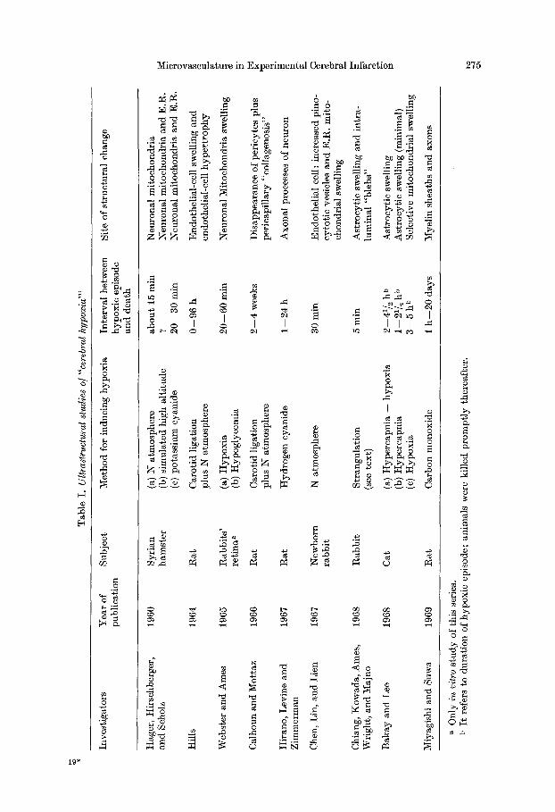
(Hirano etal.), and in order to t es t th is hypothes i s we conduc ted a survey of the ex- pe l imen t s per formed in the recent pas t . The d a t a m Table I is our summa ry of fh~se expe r imen t s in which electronmier~seopie m e t h o d s were u~ed f~r ~he s t u d y o f " c e r e b r a l hypox ia " , Two facts emerge as s ignif icant : the re seems to be con~ider- able va r i a t ion a t the site where s t ruc tu ra l damage was observed first, and the only model in which ischemia alone was ut i l ized was t h a t of Chiang etal. But , amce the r abb i t s u t i l ized for the i r exper iments were sub jec ted to s imul taneous and to t a l i n t e r rup t ion of a r te r ia l and venous cerebral circulat ions, we find such a~ experi- m e n t comparab le to the s i tua t ion preva i l ing in dea th b y s t r angu la t ion or hanging, as con t ra s t ed wi th the condi t ions to be found in regional cerebra l ischemia.
Our own s tudy of cerebra l infarct ion is expec ted to a d d new in fo rmato in a b o u t the pa~hogenes~s of th is Ie~ion, for i t has bee s cond~c~ed in a manner fha~ adheres to the following : the surgical maneuver s avo id a tmospher ic exposure and mechanica l r e t r ac t ion of the b ra in ; e lectronmicroacopic me thods have been em- p loyed in the eva lua t ion of the s t ruc tu ra l abnormal i t i e s ; the observat ions were recorded in animals k i l led sequent ia l ly over a per iod ranging f rom 21/2 h to 16 days af ter the a r te r ia l ocelusoin.
The deve lopmen t of edema and swelling in cerebra l t issues has been shown as an i m p o r t a n t cause of dea th in the immed ia t e pos t in fa rc t ion per iod by Ng and N i m m a n n i t y a ; moreover , Shaw et al. demons t r a t ed the progressive and dyna mic manner in which this phenomenon oeetrrs, thus i~dica t ing t h a t pe rhaps edema is one of the i m p o r t a n t pa thogenic factors of cerebral infarct ion. I n an a t t e m p t to u n r a v e l t h e deve lopmen t of infarc t ion edema, we d i rec ted our attention~ ini t ia l ly , to the s t ruc tu ra l fea tures of the capil lar ies and smal l arterioles.
The following is a resul t of our findings and the resul t of compar ing t hem wi th those prev ious ly r epor t ed in several exper imen ta l models of "ce rebra l hypox ia " .
Mater ia ls and Methods
The "Principles of Laboratory Animal Care" ~s promulgated by bhe NatianaI Society for Medical Research were observed in the course (ff this study,
Twelve squirrel monkeys (8aimiri sciureus) having aa average body weight of 0.75 kg were subjected to clipping of the initial 1.0 cm of the middle cerebral artery, which was approached through a surgically enlarged optic foramen (Hudgins and Garcia, 1970 b). A miniature Mayfield clip was left in the orbit. The animals were killed, according to the method described below, at intervals of 21/2, 4, 12, 18, 24, 48 h, 3 days, 1 week, and 16 days, respectively. Zero time was defined as the moment when the clip was closed around the MCA (Table II).
At the chosen time interval, a monkey was anesthetized with sodium pentobarbital (0.3 cmS/kg of body weight), given intrapleurally, Some of the animals were also administered at this time an intravenous rejection of Evans blue (0.5 rnl of a. 2.50/0 saltine solution). After (apeai•g the suture, 6he ~syffetd r was ~mtved from the orbit and an iatervai of a b o ~ t5 rain was allowed to elapse before a midline thoracotomy was performed. The descending portion of the aortic arch was clipped and the left side o f the cardiovascular system was perfused with a 6.0O/o solution of glntaraldehyde, dripping at a pressure of 100 mm I-Ig. The total volume of fixative used per animal was @proximately 375 co, and the time of fixation on each animal ranged between 45 and 90 rain. Once fixation had been performed in the manner described before, the brain was removed, sliced coronally, and sa~aples were collected for electron micro- scopic evaluation as illustrated in Fig. 1. The quality of fixation was evaluated a this point as being either poor, fair, good, or excellent, respectively (Table II). Those animals in which fixation was judged to be poor were not processed for electron microscopic evaluation, In all animals material was collected fo~" ~ltrastru ctural studie~ from both c~r~bral hemispheres. The observa-
Tab
le I
. U
ltras
truc
tura
l st
udie
s o/
"cer
ebra
l hy
poxi
a'"
Inve
stig
ator
s Y
ear
of
publ
icat
ion
Sub
ject
M
etho
d fo
r in
duci
ng h
ypox
ia
Inte
rval
bet
wee
n hy
poxi
c ep
isod
e an
d d
eath
Sit
e of
str
uctu
ral
chan
ge
ttag
er,
Hir
schb
erge
r,
1960
S
yria
n an
d Sc
holz
ha
mst
er
Hil
ls
1964
R
at
Web
ster
an
d A
mes
19
65
Rab
bit
s'
reti
na a
Cal
houn
and
Mot
taz
1966
R
at
Hir
ano,
Lev
ine
and
1967
R
at
Zim
mer
man
Che
n, L
in,
and
Lie
n 19
67
New
born
ra
bb
it
Chi
ang,
Kow
ada,
Am
es,
19
68
R
abb
it
Wri
ght,
and
Maj
no
Bak
ay a
nd
Lee
19
68
Cat
Miy
agis
hi a
nd S
uwa
1969
R
at
(a)
N a
tmos
pher
e ab
ou
t 15
min
(b
) si
mul
ated
hig
h al
titu
de
? (c
) po
tass
ium
cya
nide
2
0--
30
rai
n
Car
otid
lig
atio
n 0
--9
6 h
pl
us N
atm
osph
ere
(a)
Hyp
oxia
2
0--
60
rai
n (b
) H
ypog
lyce
mia
Car
otid
lig
atio
n 2
--4
wee
ks
plus
N a
tmos
pher
e
Hyd
roge
n cy
anid
e 1
24 h
N a
tmos
pher
e 30
rai
n
Str
angu
lati
on
5 m
in
(see
tex
t)
(a)
Hyp
erca
pnia
+
hypo
xia
2 41
/2 h
b (b
) H
yper
capn
ia
1--2
a/~
hb
(c)
Hyp
oxia
3
--5
h b
Car
bon
mon
oxid
e 1
h--
20
day
s
Neu
rona
l m
itoc
hond
ria
Neu
rona
l m
itoc
hond
ria
and
E.R
. N
euro
nal
mit
oeho
ndri
a an
d E
.R.
End
othe
lial
-cel
l sw
elli
ng a
nd
en
doth
elia
l-ce
ll h
yper
trop
hy
Neu
rona
l Mit
ocho
ndri
a sw
elli
ng
Dis
appe
aran
ce o
f pe
ricy
tes
plus
pe
rica
pill
ary
"col
lage
nosi
s"
Axo
nal
proc
esse
s of
neu
ron
End
othe
lial
cel
l : i
ncre
ased
pin
o-
cyto
tic
vesi
cles
an
d E
.R.
mit
o-
chon
dria
l sw
elli
ng
Ast
rocy
tic
swel
ling
an
d i
ntr
a-
lum
inal
"bl
ebs"
Ast
rocy
tic
swel
ling
A
stro
cyti
c sw
elli
ng (
min
imal
) S
elec
tive
mit
ocho
ndri
al s
wel
ling
Mye
lin
shea
ths
and
axo
ns
O
a 0
nly
in
vitr
o st
udy
of th
is s
erie
s.
b It
ref
ers
to d
urat
ion
of h
ypox
ic e
piso
de;
anim
als
wer
e ki
lled
pro
mpt
ly t
here
afte
r.
..j
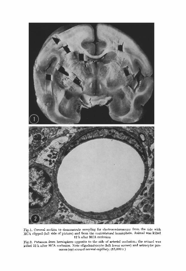
Fig. 1. Coronal section to demonstrate sampling for electronmicroscopy from the side with MCA clipped (left side of picture) and from the contralateral hemisphere. Animal was killed
12 h after MCA occlusion
:Fig.2. Putamen from hemisphere opposite to the side of arterial occlusion; the animal was killed 72 h after MCA occlusion. No~e oligodench'ocyte (left lower corner) and astrocytic pro-
cesses (ap) around normal capillary. (15,000 • )

J. H. Garcia et al. : Microvasculature in Experimental Cerebral Infarction 277
tions noted below are, in part, the result of comparing the electron microscopic differences of the non-ischemic and ischemie sides.
Processing in the electron microscopy laboratory included post-fixation in osmimn tetroxide, embedding in epoxy resins, and contrasting of ultrathin sections with ~ead ~nd uranium sMts. Eiectronmierographs were obtained in a Philips 200 ESi operating at either 60 or 80 kV.
Cerebral surfaces adjacent to those from which electron microscopy samples had been col- lected were utilized to prepare material for paraffin embedding, cutting, and staining with H.-E. cresyl violet, and iron hematoxylin (Mahon method).
Results
Examinat ion of the samples obtained from the side on which the MCA was not surgically clipped provided evidence for appropriate fixation. Absence of an- esthetic and systemic effects (such as severely decreased systemic blood pressure}, could also be assumed as there were no significant ultrastructural abnormalities in sections from the non-infarcted hemisphere. The qualify of the fixation and the degree of preservation obtained on the non-isehemie side are illustrated in Figs. 2 and 3.
Surprisingly, significant differences in the structural features could be detected by histological examination of the material obtained from the side having the MCA clipped for as short a period as four hours; vaeuolation of the white mat ter was very prominent after 12 h. A detailed description of the histological features of cerebral infarction, including the fate of the Evans blue, is included in a separate publication (Gareia). The ultrastructural abnormalities observed in nerve cells have been interpreted as demonstrating that cell-membrane-permeability changes are the first noticeable effects of ischemic injury (Gareia and Cox).
A smnmary of the intervals at which infarcts were studied, the topographical origin of the samples examined by electron mieroscope in each ease, and an outline of the infarcted a rea - -as drawn directly from a histological prepara t ion--are shown in Table I I . Notice tha t prior to 12 h, no significant and groslsy visible abnormalities were evident, although the beginning of cerebral edema could be observed by the shift of the midline.
Those alterations in the electron microseopic appearance of the microvaseu- lature, which appeared most eonspieuous and significant, will be described in chro- nologiealy order.
2 and 1/2 h after MCA clipping--Monkey 49: The predominant abnormali ty consists of marked "swelling" of astroeytie processes, particularly in the pericapil- lary areas, with relatively good preservation of the mitochondria and other cell organelles. The endothelial cells appear essentially unchanged. These abnormalities can be encountered in samples from white and gray matters.
4 h after MCA clipping--Monkey 36 : The process of astrocytie swelIing has extended from the perivascular location in a centrifugal manner, into astrocytie processes more distant from capillaries (Fig.4); pericytes also participate in the process of volume increase. The endothelial cells do not show hyperplasia of their pinocytotie vesicles, separation of their endothelial junctions, or any other signifi- cant structural abnormalities. The process of intraeellnlar fluid accumulation is fairly selective since it spares the oligodendroeytes and the endothelial cells. An occasional polymorphonuclear leukocyte (PMNL) can be encountered in the lumen of some capillaries.
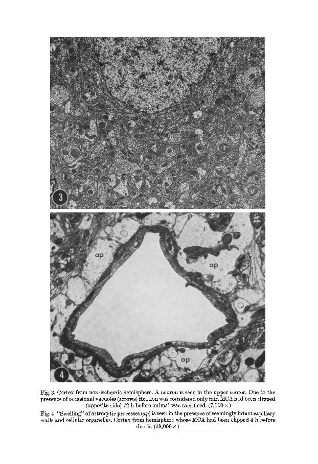
Fig. 3. Cortex from non-ischemic hemisphere. A neuron is seen in the upper center. Due to the presence of occasional vacuoles (arrows) fixation was considered only fair. MCA had been clipped
(opposite side) 72 h before animal was sacrificed. (7,500 • )
:Fig. 4. "Swelling" of astroeytic processes (ap) is seen in the presence of seemingly intact capillary walls and cellular organelles. Cortex from hemisphere whose MCA had been clipped 4 h before
death. (19,000 x )

g. I-I. Garcia et al. : Microvasculature in Experimental Cerebral Infarction 279
TABLE II
ELECTRON MICROSCOPY OF CEREBRAL INFARCTION
Monkey Duality Interval Samples Evaluated (t ) Topography of Area Number: of Between Infarcted (naked eye
Pertusian: Surgery Basal White Cortex and Death: 6anglia Matter inspection):
G* 2" I I 4 9 ahrs C. I? I F r. I I.
I 1 3 6 E 4 hrs.
4 2 F 12 hrs
35 E 18 hrs C.
c. IF~. IF,. i I I i
I I
I ! I? I Fr. I T .
I I I I
I? I Fr. I F,
I I I I
~1 I- I Fr I I
30 E 24 hrs. I.
I
29 E 48 hrs I. ",,3"Jq~,
I
37 E 3 day.~
I I 38 G 7days C. I Fr. i Fr
40 G 16d~ys I 1 ( ~ I Fr. Fr.
I I G: good; E: excellent F: fair
(T) C: caudate; P: pulnmen; I: insula; Fr: frontal; T: temporal
12 h after MCA clipping--Monkey 42 : Two features seem to prevail within the areas of more advanced isehemic damage: beginning necrosis of capillary walls with marked swelling of endothelial cells, but without visible circmnferentia[ gaps, and presence of numerous PMNL in the lumen of most capillaries with few present in the tissue (Fig. 5).
18 h after MCA clipping--Monkey 35: The phenomenon of perivaseular swelling is more pronounced, particularly in samples from the white matter area. Signs of endothelial cell necrosis and blurring of the basement laminae are more pro- minent (Fig. 7). Fragments of PMNL's are now visible also in the outer surface of the capillaries, even though endothelial gaps are not visualized.
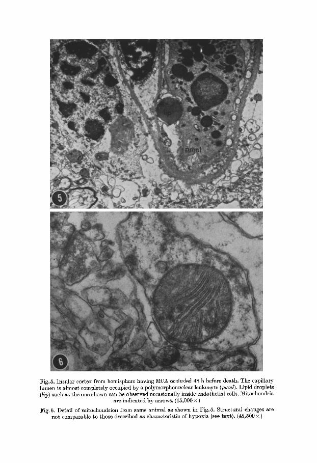
Fig.5. Insular cortex from hemisphere having MCA occluded 48 h before death. The capillary lumen is almost completely occupied by a polymorphonuclear leukocyte (pmnl). Lipid droplets (lip) such as the one shown can be observed occasionally inside endothelial cells. Mitochondria
are indicated by arrows. (15,000 • )
Fig. 6. Detail of mitochondrion from same animal as shown in Fig. 5. Structural changes are not comparable to those described as characteristic of hypoxia (see text). (48,500 • )

J. H. Garcia et al. : Microvasculature in Experimental Cerebral Infarction 281
24 h after MCA clipping--Monkey 30, 44, and 47 : Two sets of capillaries can be identified : those from areas of advanced necrosis and those from the edges of areas of infarction. In the latter, the structural abnormalities are relatively mild; in the former, extensive alterations of cytoplasmic organelles are evident, but still there is preservation of the endothelial t ight junctions. Furthermore, there is pre- servation of mitochondrial structural integrity even in those areas where the necrosis is far advanced (Fig. 6). Many relatively large lipid droplets can be seen either in the width of endothelial cells, or in the immediate vicinity of the capillary wall (Fig. 5).
48 h after MCA clipping--Monkeys 29 and 48: There are abundant examples of extensive accumulation of fluid in the extravascular spaces ; evidence of struc- tural integrity of endothelial cells and endothelial junctions can readily be found. Endothelial cells in areas of more advanced ischemic necrosis show marked mito- chondrial swelling, disappearance of pinocytotic vesicles and margination of the nuclear chromatin (Fig. 7).
3 days after MCA clipping--Monkey 37 : The extravasat ion of fluid, as judged by the degree of vacuolation, seems obvious, particularly in the white matter. Nevertheless, even in areas where edema is fairly pronounced, many of the endo- thelial cells still display remarkably normal ultrastructural features.
1 week after MCA clipping--Monkey 38: Once more it is remarkable to find, within small samples of tissue, capillaries tha t are a in state of acute necrosis and areas where the capillary architecture is either preserved or beginning to reconsti- tute. In some areas where the latter has occurred, the capillaries appear fenestrated and devoid of basement membrane.
16 days after MCA clipping--Monkey 40 : Most capillaries within area of ad- vanced necrosis show fenestrated walls and a rather wide but electron-lucent basement membrane (Fig. 8).
Discussion
Some of the pathogenic mechanisms that may infuence the development or advancement of a cerebral infarction have been evaluated. The phenomenon of flaid accumulation (edema) has been given special consideration in the preparation of this manuscript.
The surgical method for occluding the MCA in the squirrel monkey had been previously tested by us in order to eliminate the possibility that damage to the blood- brain-barrier occurred as a direct result of the surgical maneuvers and prior to the arterial clipping (Hudgins and Garcia, 1970 b). Furthermore, it had been established in our laboratory, tha t the chosen surgical technique yielded a high percentage of cerebral infarctions which had a predictable size and location (Hudgins and Garcia, 1970a).
The ultrastructural features of normal capillaries, in the central nervous system and other tissues, have been described in several animal species (Maynard et a l . ;
Fawcett) ; thus, the adequacy of the fixation methods, the absence of systemic factors tha t could alter the structure of CNS, and a baseline for comparisons was readily available in the samples obtained from the hemisphere which did not undergo clipping of the MCA.
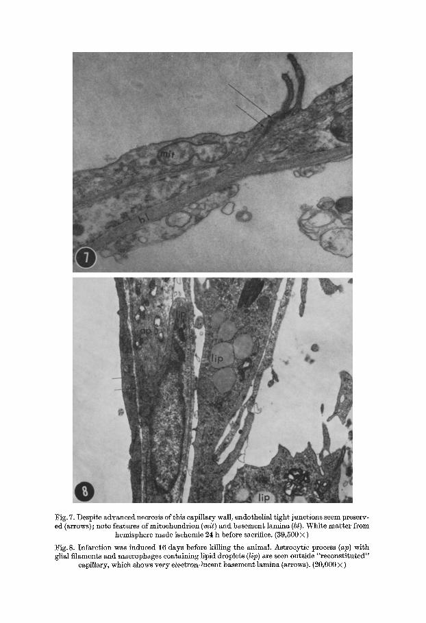
Fig. 7. Despite advanced necrosis of this capillary wall, endothelial tight junctions seem preserv- ed (arrows) ; note features of mitoehondrion (mit) and basement lamina (bl). White matter from
hemisphere made ischemic 24 h before sacrifice. (39,500 • )
Fig. 8. Infarction was induced 16 days before killing the animal. Astrocytie process (ap) with glial filaments and macrophages containing lipid droplets (lip) are seen outside "reconstituted"
c~pillary, which shows very electron-lucent basement lamina (arrows). (20,000 • )
J. H. Garcia et al.: lVIicrovasculature in Experimental Cerebral Infarction 283
The results of our experiments have been interpreted as indicating :
Within a few hours of MCA clipping, the distal capillaries show evidence--in the per ieytes- -of increased cellular volume and increased electron lueeney; this phenomenon progressively extends to the extravascular compartments (intra- and extra-cellular). I t is significant tha t no endothelial swelling or hyperp]asia is noted. This is to be compared with the results recorded after exposure of CNS to Nitrogen atmosphere alone or the combination of it plus carotid ligation; in both of these situations, endothelial swelling and hyperp]asia were emphasized (Chen et al., Hills), as prominent changes.
Increasing numbers of PMNL's can be detected inside and outside capillary walls within the ischemie area. These cells are not visualized in large numbers until 12 to 18 h post-MCA occlusion. This fact, plus the continuously increasing volume of infarcted tissues, indicates tha t in spite of occlusion of a major artery and the development of eneephalomalaeia, eh-culation is maintained within areas of cere- bral isehemia. This is in contrast with the so-called "no-reflow" phenomenon de- scribed for cerebral capillaries in rabbits killed by strangulation (Chiang e~ al.). I t is noteworthy that intraluminal blebs and capillary wall collapse were features not encountered in our material. Both have been documented in the experiments of Chiang et al.
We had assumed tha t extravasation of fluid and particles within a cerebral isehemic region could occur only after extensive mural gaps had opened in the cir- cumference of the capillaries. Such gaps would form either through breakdown and disappearance of endothelial cells or by means of dehiscenees at the site of endo- thelial junctions. Necrosis of endothelial cells was not visualized earlier than 12 h after MCA clipping and endothelial junctions were t ightly bound when evaluated in over 600 eleetronmierographs made from the 12 experimental animals. I t is, therefore, believed tha t the migration of fluid and ceils from the vascular lumen to the surrounding tissue may occur by passage through the endothelial cells. This should not constitute a biological impossibility if one reflects on the fact tha t the return of maerophages into the circulation occurs at a t ime when reeonstitution of capillary walls is completed. Calhoun and Mottaz established tha t 2 to 4 weeks after having induced cerebral necrosis, the capillaries are thickened by redupli- cation of basement laminae and adventitial eollagenosis; and, it is commonly known tha t after 2 to 4 weeks macrophages present in areas of eneephalomMaeia disappear by returning to the eireuiation. Experiments performed by others showed tha t in pancreas (Williamson and Grisham) and nerve tissue (Astr6m) passage of in- f lammatory cells can occur in a trans-endothelial way ; this phenomenon had been described originally in 1956 and designated: emperipolesis (Humble et al.). Similar observations were made recently by Dobbins et al. in studies of intestinal mueosa permeabili ty utilizing horseradish peroxidase.
Cerebral isehemic injury is a dynamic process tha t progresses in a mnltifoeal fashion as can be deduced from our observations tha t markedly necrotic capillaries exist within microns of those better preserved. This manner of multicentric ad- vancement of the necrotic process had been visualized directly by us in a separate experiment with animals of a similar type (Sundt et al.), and also had been men- tioned in previous studies of cerebral infarction by light microscopy (Meyer).
284 J .H . Garcia, J. V. Cox, and W. R. Hudgins:
U l t r a s t r uc tn r a l fea tures of autolys is could be mi s t aken for those appear ing in ear ly ischemic necrosis. Two features p rov id ing ind ica t ion for adequa t e f ixat ion in the areas of induced ischemia are to be no ted in the re la t ive ly excel lent s t ruc tu ra l p rese rva t ion of newly a r r ived leukocytes and mi tochondr ia l membranes (Figs. 5
and 6). E x p e r i m e n t s conduc ted in vitro d e m o n s t r a t e d t h a t neurona l mi tochondr ia l
swelling is specifically induced b y per iods of h y p o x i a of 20 rain or longer (Hager et al. ; W e b s t e r and Ames ; B a k a y and Lee). I t is, therefore, s ignif icant t h a t mi tochondr i a in endothe l ia l as well as o ther cells appea r so min ima l ly changed as la te as 2 days post -MCA occlusion. This could be i n t e rp re t ed as ind ica t ive of the fac t t h a t in ischemic cerebral in jury , oxygen dep r iva t ion does no t cons t i tu te the p r eva l en t fac tor responsible for the e lect ronmicroscopic changes.
Regard ing the space in which fluid m a y accumula te once i t leaves the vascu la r bed, our mierographs show a p r e d o m i n a n t l y in t r a -a s t rocy t i e "edema" , in the cortex, and a combina t ion of the l a t t e r plus ex t ra -ce l lu la r and in t ra -myel in ic edema, in the whi te ma t t e r . Some caut ion m u s t be exercised in the eva lua t ion of this pheno- menon. The work of Van H a r r e v e l d et al., indica tes t h a t some of the in t race] lu lar accumula t ion of fluid seen in e lec t ronmicrographs such as ours m a y be the resul t of a r t i fac t s inhe ren t to the methods ut i l ized in a ldehyde pcrfusion-f ixat ion. The fac t t h a t no evidence of "swel l ing" is seen in our p repa ra t ions t aken f rom the non- ischemic side (Figs. 2 and 3) would indica te t h a t the f ixat ion me thods alone should no t be held responsible for the increase in vo lume and t rans lucence d e m o n s t r a t e d
in the ischemic side of our expe r imen ta l model .
Acknowledgement. The skillful technical help of Mrs. Brigitte F15res da Cunha is gratefully acknowledged.
References
Astr5m, L. E.: Migration of lymphocytes through endothelial venules in experimental allergic neuritis. Experientia (Basel) 24, 589--590 (1968).
Bakay, L., Leel J. C. : The effect of acute hypoxia and hypercapnia on the ultrastructure of the central nervous system. Brain 91, 697--706 (1968).
Calhoun, C. L., Mottaz, J. It. : Capillary bed of the rat cerebral cortex. The fine structure in experimental cerebral infarction. Arch. Neurol. (Chic.) 15, 320--328 (1966).
Chen, tt., Lin, C. S., Lien, I. N.: Vascular permeability in experimental Kernieterus: an electron microscopic study of the blood-brain-barrier. Amer. J. Path. 51, 69--87 (1967).
Chiang, J., Kowada, M., Ames, A., Wright, R. L., Majno, G. : Cerebral ischemia. III . Vascular changes. Amcr. J. Path. 52, 455--476 (1968).
Dobbins, W. 0., Rollins, E. L.: Interstitial mucosal lymphatic permeability: an electron microscopic study of endothelial vesicles and cell junctions. J. Ultrastruct. Res. 33, 29--59 (1970).
~awcett, D. W.: Comparative observations on the fine structure of blood capillaries. In: The peripheral blood vessels, Chapt. 2, pp. 17--44 J. L. Orbison, ed.). Baltimore: Williams and Wilkins Comp. 1963.
Garcia, J. It . : Histopathology of cerebral edema in experimental cerebral infarction. In prep- aration.)
- - Cox, J. V.: Ultrastructure of experimental cerebral infarction. Nerve cell alterations. Proc. VIth Internat. Congr. ~europath. Paris, pp. 1038--1040 (1970).
ttager, H., Hirschberger, W., Scholz, W.: Electron microscopic changes in brain tissue of Syrian hamster following acute hypoxia. Aerospace Med. 81, 379--387 (1960).
Hill, N. C., Millikan, C. H., Wakim, K. G., Sayre, G. P. : Studies in cerebrovascular disease. VII. Experimental production of cerebral infarction by intracarotid injection of homo- logous blood clot preliminary report. Proc. Mayo Clin. 80, 625--633 (1955).

Microvasculature in Experimental Cerebral Infarction 285
Hills, C. P. : Uitrastructural changes in the capillary bed of the rat cerebral cortex in anoxic- isochemic brain lesions. Amer. J. Path. 44, 531--551 (1964).
Hirano, A., Levine, S., Zimmerman, H. M.: Experimental cyanide encephalopathy: electron microscopic observations of early lesions in white matter. J. ~europath. exp. Neurol. 26, 200--222 (1967).
Hudgins, W. R., Gareia, J. H.: Transorbital approach to the middle cerebral artery of the squirrel monkey: a technique for experimental cerebral infarction applicable to ultra- structural studies. Stroke 1, 107--111 (1970a).
-- -- The effect of electrocautery, atmospheric exposure, and surgical retraction on the permeability of the blood-brain-barrier. Stroke 1, 375--380 (1970b).
Humble, J. G., Jayne, W. H., Pulvertaft, R. J. V. : Biological interaction between lympho- cytes and other ceils. Brit. J. Haemat. 2, 283--294 (1956).
Maynard, E. A., Schultz, R. L., Pease, D. C. : Electron microscopy of the vascular bed of rat cerebral cortex. Amer. J. Anat. 10O, 409--433 (1957).
Meyer, J. S. : Importance of ischemic damage to small vessels in experimental cerebral infarc- tion. J. ~Neuropath. exp. Neurol. 17, 571--585 (1958).
Miyagishi, T., Suwa, N.: Electron microscopic studies on the cerebral lesions of rats in experimental carbon monoxide poisoning. Acta neuropath. (Berl.) 14, 118--125 (1969).
Ng, L. K. Y., Nimmannitya, J.: Massive cerebral infarction with severe brain swelling: a clinicopathology study. Stroke I, 158--163 (1970).
Penry, J. K., ~qetsky, M. G. : Experimental embolic occlusion of a single leptomeningeal artery. Arch. ~qeurol. (Chic.) 3, 391--398 (1960).
Shaw, C. M., Alvord, E. C., Berry, R. G. : Swelling of the brain following ischemic infarction with arterial occlusion. Arch. ~eurol. (Chic.) 1, 161--177 (1959).
Sundt, T.M., Grant, W. C., Garcia, J. I-I.: Restoration of middle cerebral artery flow in experimental infarction. J. Neurosurg. 81, 311--322 (1969).
-- Waltz, A. G.: Experimental cerebral infarction: retroorbital, extradural approach for occluding the middle cerebral artery. Proc. Mayo Clin. 41, 159--168 (1966).
Van Harreveld, A., Crowell, J., Malhotra, S. K.: A study of extracellular space in central nervous tissue by freeze-substitution. J. Cell Biol. 25, 117--137 (1965).
Webster, H., Ames III, A.: Reversible and irreversible changes in the fine structure of nervous tissue during oxygen and glucose deprivation. J. Cell Biol. 26, 884 909 (1965).
Williamson, J. K., Grisham, J. W. : Electron microscopy of leukocytic margination and emi- gration in acute inflammation in dog pancreas. Am. J. Path. 89, 239--256, 1961.
Julio H. Gareia, M.D. Department of Pathology University of Maryland, School of Medicine 32 South Greene Street Baltimore, Maryland 21201
Related Documents