Progression From Compensated Hypertrophy to Failure in the Pressure-Overloaded Human Heart Structural Deterioration and Compensatory Mechanisms Stefan Hein, MD*; Eyal Arnon, MD*; Sawa Kostin, MD; Markus Schönburg, MD; Albrecht Elsässer, MD; Victoria Polyakova, PhD; Erwin P. Bauer, MD; Wolf-Peter Klövekorn, MD; Jutta Schaper, MD Background—The progression of compensated hypertrophy to heart failure (HF) is still debated. We investigated patients with isolated valvular aortic stenosis and differing degrees of left ventricular (LV) systolic dysfunction to test the hypothesis that structural remodeling, as well as cell death, contributes to the transition to HF. Methods and Results—Structural alterations were studied in LV myectomies from 3 groups of patients (group 1: ejection fraction [EF] 50%, n12; group 2: EF 30% to 50%, n12; group 3: EF 30%, n10) undergoing aortic valve replacement. Control patients were patients with mitral valve stenosis but normal LV (n6). Myocyte hypertrophy was accompanied by increased nuclear DNA and Sc-35 (splicing factor) content. ACE and TGF- 1 were upregulated correlating with fibrosis, which increased 2.3-, 2.2-, and 3.2-fold over control in the 3 groups. Myocyte degeneration increased 10, 22, and 32 times over control. A significant correlation exists between EF and myocyte degeneration or fibrosis. Ubiquitin-related autophagic cell death was 0.5‰ in control and group 1, 1.05 in group 2, and 6.05‰ in group 3. Death by oncosis was 0‰ in control, 3‰ in group 1, and increased to 5‰ (groups 2 and 3). Apoptosis was not detectable in control and group 3, but it was present at 0.02‰ in group 1 and 0.01‰ in group 2. Cardiomyocyte mitosis was never observed. Conclusions—These structure-function correlations confirm the hypothesis that transition to HF occurs by fibrosis and myocyte degeneration partially compensated by hypertrophy involving DNA synthesis and transcription. Cell loss, mainly by autophagy and oncosis, contributes significantly to the progression of LV systolic dysfunction. (Circulation. 2003;107:984-991.) Key Words: hypertrophy heart failure structure remodeling hemodynamics T he structural basis of the progression from well- compensated hypertrophy caused by mechanical over- load to heart failure (HF) is still largely unknown. It is evident that cardiac remodeling, defined as “genome expression, molecular, cellular, and interstitial changes that are mani- fested clinically as changes in size, shape and function of the heart after injury” occurs in the chronically pressure- overloaded heart. 1 However, the correlation between morpho- logical alterations and clinical data during the different phases of transition to HF has not yet been described in the human heart. Krayenbühl and colleagues 2 in 1989 described fibrosis and myocyte enlargement in patients with aortic stenosis (AS) with normal ejection fraction (EF) but elevated left ventric- ular end-diastolic pressure (LVEDP). They studied cardiac functional recovery after aortic valve replacement (AVR) but not the progression to HF. In end-stage human HF, we reported fibrosis as well as impairment of the myocyte ultrastructure defined as degeneration. 3 Anversa et al 4 and others (review by Elsässer et al 5 ) emphasized cell death, mainly by apoptosis, as one of the key events for the occurrence of failure. Fibrosis and myocyte damage appear to be the decisive morphological alterations in the remodeling process. 6,7 The most important regulators of fibrosis are the members of the renin-angiotensin and aldosterone system. 8,9 In addition, TGF- 1 is a potent stimulator of fibrosis, as are growth factors and endocrine hormones such as norepinephrine (reviewed by Hein and Schaper 10 ). Recent studies in small rodents have shown that the development of fibrosis is prevented in the absence of TGF- 1 , indicating the crucial fibrosis-promoting role of this cytokine. 11,12 Received October 16, 2002; revision received November 14, 2002; accepted November 15, 2002. From Kerckhoff-Clinic (S.H., M.S., E.P.B., W.-P.K.) and Max-Planck-Institute (E.A., S.K., V.P., J.S.), Bad Nauheim, Germany, and the Department of Cardiology (A.E.), Albert-Ludwigs-University, Freiburg, Germany. *Drs Hein and Arnon contributed equally to this work. Correspondence to Stefan Hein, MD, Kerckhoff-Clinic, Department of Thoracic and Cardiovascular Surgery, Benekestr 4-8, D-61231 Bad Nauheim, Germany. E-mail [email protected] © 2003 American Heart Association, Inc. Circulation is available at http://www.circulationaha.org DOI: 10.1161/01.CIR.0000051865.66123.B7 984 by guest on July 12, 2015 http://circ.ahajournals.org/ Downloaded from

Welcome message from author
This document is posted to help you gain knowledge. Please leave a comment to let me know what you think about it! Share it to your friends and learn new things together.
Transcript
Progression From Compensated Hypertrophy to Failure inthe Pressure-Overloaded Human Heart
Structural Deterioration and Compensatory Mechanisms
Stefan Hein, MD*; Eyal Arnon, MD*; Sawa Kostin, MD; Markus Schönburg, MD;Albrecht Elsässer, MD; Victoria Polyakova, PhD; Erwin P. Bauer, MD;
Wolf-Peter Klövekorn, MD; Jutta Schaper, MD
Background—The progression of compensated hypertrophy to heart failure (HF) is still debated. We investigated patientswith isolated valvular aortic stenosis and differing degrees of left ventricular (LV) systolic dysfunction to test thehypothesis that structural remodeling, as well as cell death, contributes to the transition to HF.
Methods and Results—Structural alterations were studied in LV myectomies from 3 groups of patients (group 1: ejectionfraction [EF] �50%, n�12; group 2: EF 30% to 50%, n�12; group 3: EF �30%, n�10) undergoing aortic valvereplacement. Control patients were patients with mitral valve stenosis but normal LV (n�6). Myocyte hypertrophy wasaccompanied by increased nuclear DNA and Sc-35 (splicing factor) content. ACE and TGF-�1 were upregulatedcorrelating with fibrosis, which increased 2.3-, 2.2-, and 3.2-fold over control in the 3 groups. Myocyte degenerationincreased 10, 22, and 32 times over control. A significant correlation exists between EF and myocyte degeneration orfibrosis. Ubiquitin-related autophagic cell death was 0.5‰ in control and group 1, 1.05 in group 2, and 6.05‰ in group3. Death by oncosis was 0‰ in control, 3‰ in group 1, and increased to 5‰ (groups 2 and 3). Apoptosis was notdetectable in control and group 3, but it was present at 0.02‰ in group 1 and 0.01‰ in group 2. Cardiomyocyte mitosiswas never observed.
Conclusions—These structure-function correlations confirm the hypothesis that transition to HF occurs by fibrosis andmyocyte degeneration partially compensated by hypertrophy involving DNA synthesis and transcription. Cell loss,mainly by autophagy and oncosis, contributes significantly to the progression of LV systolic dysfunction. (Circulation.2003;107:984-991.)
Key Words: hypertrophy � heart failure � structure � remodeling � hemodynamics
The structural basis of the progression from well-compensated hypertrophy caused by mechanical over-
load to heart failure (HF) is still largely unknown. It is evidentthat cardiac remodeling, defined as “genome expression,molecular, cellular, and interstitial changes that are mani-fested clinically as changes in size, shape and function of theheart after injury” occurs in the chronically pressure-overloaded heart.1 However, the correlation between morpho-logical alterations and clinical data during the differentphases of transition to HF has not yet been described in thehuman heart.
Krayenbühl and colleagues2 in 1989 described fibrosis andmyocyte enlargement in patients with aortic stenosis (AS)with normal ejection fraction (EF) but elevated left ventric-ular end-diastolic pressure (LVEDP). They studied cardiacfunctional recovery after aortic valve replacement (AVR) but
not the progression to HF. In end-stage human HF, wereported fibrosis as well as impairment of the myocyteultrastructure defined as degeneration.3 Anversa et al4 andothers (review by Elsässer et al5) emphasized cell death,mainly by apoptosis, as one of the key events for theoccurrence of failure.
Fibrosis and myocyte damage appear to be the decisivemorphological alterations in the remodeling process.6,7 Themost important regulators of fibrosis are the members of therenin-angiotensin and aldosterone system.8,9 In addition,TGF-�1 is a potent stimulator of fibrosis, as are growthfactors and endocrine hormones such as norepinephrine(reviewed by Hein and Schaper10). Recent studies in smallrodents have shown that the development of fibrosis isprevented in the absence of TGF-�1, indicating the crucialfibrosis-promoting role of this cytokine.11,12
Received October 16, 2002; revision received November 14, 2002; accepted November 15, 2002.From Kerckhoff-Clinic (S.H., M.S., E.P.B., W.-P.K.) and Max-Planck-Institute (E.A., S.K., V.P., J.S.), Bad Nauheim, Germany, and the Department
of Cardiology (A.E.), Albert-Ludwigs-University, Freiburg, Germany.*Drs Hein and Arnon contributed equally to this work.Correspondence to Stefan Hein, MD, Kerckhoff-Clinic, Department of Thoracic and Cardiovascular Surgery, Benekestr 4-8, D-61231 Bad Nauheim,
Germany. E-mail [email protected]© 2003 American Heart Association, Inc.
Circulation is available at http://www.circulationaha.org DOI: 10.1161/01.CIR.0000051865.66123.B7
984 by guest on July 12, 2015http://circ.ahajournals.org/Downloaded from
Earlier, we described myocyte degeneration and loss in thetransition to end-stage HF.3 In the present study, we investi-gated patients with AS in different hemodynamic situationsranging from compensatory hypertrophy with intact systolicfunction to symptomatic HF with depressed systolic function.
We studied the relation between clinical LV parameters andmorphological alterations including fibrosis, myocyte degen-eration, and cell death. To test the assumption that compen-satory mechanisms may be active, we determined the DNAcontent and the presence of the splicing factor Sc-35.13 The
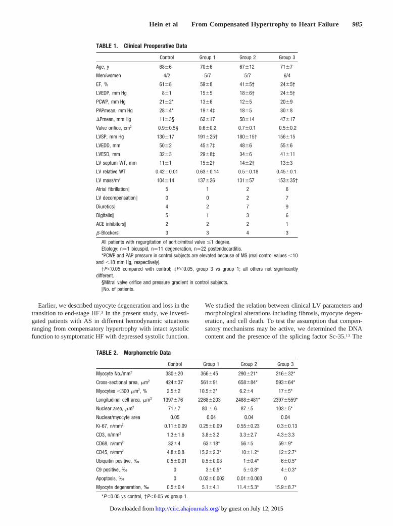
TABLE 1. Clinical Preoperative Data
Control Group 1 Group 2 Group 3
Age, y 68�6 70�6 67�12 71�7
Men/women 4/2 5/7 5/7 6/4
EF, % 61�8 59�8 41�5† 24�5†
LVEDP, mm Hg 8�1 15�5 18�6† 24�5†
PCWP, mm Hg 21�2* 13�6 12�5 20�9
PAPmean, mm Hg 28�4* 19�4‡ 18�5 30�8
�Pmean, mm Hg 11�3§ 62�17 58�14 47�17
Valve orifice, cm2 0.9�0.5§ 0.6�0.2 0.7�0.1 0.5�0.2
LVSP, mm Hg 130�17 191�25† 180�15† 156�15
LVEDD, mm 50�2 45�7‡ 48�6 55�6
LVESD, mm 32�3 29�8‡ 34�6 41�11
LV septum WT, mm 11�1 15�2† 14�2† 13�3
LV relative WT 0.42�0.01 0.63�0.14 0.5�0.18 0.45�0.1
LV mass/m2 104�14 137�26 131�57 153�35†
Atrial fibrillation� 5 1 2 6
LV decompensation� 0 0 2 7
Diuretics� 4 2 7 9
Digitalis� 5 1 3 6
ACE inhibitors� 2 2 2 1
�-Blockers� 3 3 4 3
All patients with regurgitation of aortic/mitral valve �1 degree.Etiology: n�1 bicuspid, n�11 degeneration, n�22 postendocarditis.*PCWP and PAP pressure in control subjects are elevated because of MS (real control values �10
and �18 mm Hg, respectively).†P�0.05 compared with control; ‡P�0.05, group 3 vs group 1; all others not significantly
different.§Mitral valve orifice and pressure gradient in control subjects.�No. of patients.
TABLE 2. Morphometric Data
Control Group 1 Group 2 Group 3
Myocyte No./mm2 380�20 366�45 290�21* 216�32*
Cross-sectional area, �m2 424�37 561�91 658�84* 593�64*
Myocytes �300 �m2, % 2.5�2 10.5�3* 6.2�4 17�5*
Longitudinal cell area, �m2 1397�76 2268�203 2488�481* 2397�559*
Nuclear area, �m2 71�7 80 � 6 87�5 103�5*
Nuclear/myocyte area 0.05 0.04 0.04 0.04
Ki-67, n/mm2 0.11�0.09 0.25�0.09 0.55�0.23 0.3�0.13
CD3, n/mm2 1.3�1.6 3.8�3.2 3.3�2.7 4.3�3.3
CD68, n/mm2 32�4 63�18* 56�5 59�9*
CD45, n/mm2 4.8�0.8 15.2�2.3* 10�1.2* 12�2.7*
Ubiquitin positive, ‰ 0.5�0.01 0.5�0.03 1�0.4* 6�0.5*
C9 positive, ‰ 0 3�0.5* 5�0.8* 4�0.3*
Apoptosis, ‰ 0 0.02�0.002 0.01�0.003 0
Myocyte degeneration, ‰ 0.5�0.4 5.1�4.1 11.4�5.3* 15.9�8.7*
*P�0.05 vs control, †P�0.05 vs group 1.
Hein et al From Compensated Hypertrophy to Heart Failure 985
by guest on July 12, 2015http://circ.ahajournals.org/Downloaded from

final goal of this work was 2-fold: (1) To define the mode oftransition of compensated hypertrophy to HF in the pressure-overloaded human heart with emphasis on the role of celldeath: autophagy associated with the ubiquitin-proteasomalpathway,13,14 oncosis (necrosis is cellular breakdown aftercell death has occurred15), and apoptosis,16 and (2) to deter-mine the correlation between preoperative/postoperative clin-ical data and morphological findings, which might determinethe potential for complete postoperative recovery.
MethodsPatientsThirty-four patients with isolated AS underwent clinical evaluation(Table 1) and were subdivided into 3 different groups on the basis ofEF determined by quantitative echocardiography at the time ofadmission: group 1, EF �50% (n�12); group 2, EF 50% to 30%(n�12); group 3, EF �30% (n�10). All patients underwent surgicalAVR and postoperative examination (identical n per group). Six
patients with mitral stenosis (MS) with normal EF served as controlsubjects. The institutional ethics committee approved the study, andall patients gave informed consent.
Tissue SamplingDuring open heart surgery, myectomy samples weighing �30 to 80mg were removed from the LV septum, immediately frozen in liquidnitrogen, and stored at �80°C. In MS, samples from papillarymuscles were obtained. In addition, small samples were fixed inbuffered glutaraldehyde for electron microscopy.
Electron MicroscopyThe samples were embedded in Epon, following a standard protocol.Ultrathin sections were double-stained with uranyl acetate and leadcitrate before examination in a Philips CM 10. Myocyte degenera-tion, defined as loss of contractile elements and disorganization ofultrastructural organelles, was evaluated quantitatively by two ob-servers blinded to the patient group.
Immunolabeling and Confocal MicroscopyCryosections 5 �m thick were air-dried and fixed either withparaformaldehyde or acetone. Primary antibodies were fibronectin(rabbit polyclonal, ICN), TGF-�1, ACE (Chemicon), CD3 (lympho-cytes), CD31 (endothelial marker, PECAM), CD45 (panleukocytes),CD68 (macrophages), and Ki-67 (DNA synthesis) (all Dako),vinculin, sarcomeric �-actinin, and Sc-35 splicing factor (all Sigma),ubiquitin for autophagic cell death, (Zymed Laboratories), comple-ment 9 (C9, Serotec) for oncosis, and the TUNEL method forapoptosis (Roche). The specificity of all antibodies was verified byomission of primary antibodies.
The secondary detection system was biotinylated anti-mouse oranti-rabbit IgG (Biotrend) either directly conjugated with Cy-2 orCy-3 or unconjugated followed by fluoroisothiocyanate (FITC)-linked streptavidin (Amersham). Myocyte identification was donewith TRITC-labeled phalloidin (Sigma). Nuclei were stained withTOTO-3 (Molecular Probes). Picture acquisition was performed witha Leica confocal microscope (CLSM); data were transferred to aSilicon Graphics workstation for further processing and recording(Bitplane software).
MorphometryMyocyte degeneration was evaluated in the confocal microscopefrom �-actinin–stained sections at a magnification of �400. Thetotal myocyte number was determined and degeneration calculatedas percentage. Myocyte cross-sectional and longitudinal cell areaswere determined directly in the confocal microscope from at least120 myocytes per section by delineating vinculin-stained myocytes.
Fibrosis and Numerical DensitiesFibrosis was quantified from fibronectin-stained sections from 5different fields of vision randomly chosen and expressed as percent-age of total myocardium. Capillary density was determined from 5different fields of PECAM-stained sections and calculated (asn/mm2). The same was done for ACE-positive microvessels as wellas for CD3, CD45, CD68, and Ki-67. All densities were determinedat a microscopic magnification of �250.
Cell DeathNumbers of ubiquitin-, C9-, and TUNEL-positive cells were ob-tained from the entire section. Section size and number of myocytesper mm2 were determined, and percentages of positive myocyteswere calculated. The total number of myocytes evaluated per patientvaried between 2748 and 5976.
Fluorescence Intensity by Confocal MicroscopyThe immunolabeling procedures for TGF-�1 were carried out underidentical conditions, including the microscopic magnification of�400. Confocal settings were kept constant, and quantification ofTGF-�1 was performed by measurements of fluorescence intensity
Figure 1. Degeneration and cell death. A through D, Electronmicroscopy; E through H, CLSM. A, Normal; B, slight degenera-tion with perinuclear cytoplasm; C, loss of sarcomeres rangingfrom slight (arrow) to severe (double arrow); MV, microvessel; D,one atrophied myocyte (curved arrow) contains autophagicvacuoles; the others lack sarcomeres (single arrows); F, fibro-blasts; M, macrophages. All bars are 5 �m. E and F, Apoptosis.E, Myocyte (red) nucleus is blue with TOTO and green byTUNEL in F. G, Ubiquitin labeling (green) in a myocyte (red)nuclei are blue. Note absence of nucleus in affected cell. H,Labeling of C9 (green) in a myocyte. Colors are as in G.
986 Circulation February 25, 2003
by guest on July 12, 2015http://circ.ahajournals.org/Downloaded from
by using a range of 0 to 255 values. Arbitrary units were calculatedper unit surface area (AU/mm2).
FEULGEN Staining and Quantitative EvaluationNuclear DNA was stained by the FEULGEN method. All sectionswere stained simultaneously under exactly the same protocol. Quan-titative evaluation of the nuclear DNA content (total amount timesnuclear area) and concentration (DNA/nuclear area) was done withthe CLSM at a magnification of �400. At least 400 nuclei persample were measured. The same procedure was followed for Sc-35.Nuclear area was automatically obtained from measurements ofFEULGEN staining.
StatisticsAll data are presented as mean�SEM. Differences by unpaired t test,ANOVA, Bonferroni, or Kruskal-Wallis were considered significantwhen P�0.05.
ResultsMyocyte Hypertrophy and DegenerationMyocytes showed hypertrophy. The cross-sectional and lon-gitudinal cell areas were increased in all groups comparedwith the control (MS) group (Table 2). Interestingly, thenumber of atrophied myocytes, defined as cells with across-sectional area �300 �m2, was rather large (Table 2).
Subcellular changes in myocytes by electron and confocalmicroscopy were reduction of sarcomeres, occurrence ofmyelin figures and autophagic vacuoles, numerous poly-somes, and nuclei of bizarre shape. The percentage ofmyocyte degeneration/total number of myocytes was slightlyincreased in group 1 and significantly elevated in groups 2and 3 (Figure 1 and Table 2).
Ubiquitin-related autophagic cell death and oncosis in-creased with depressed EF (Figure 1 and Table 2). In group 3,
with severe LV dysfunction, the prevalence of myocytesundergoing autophagic and oncotic death was �5‰ and 4‰,respectively, whereas classic apoptosis was detected in �1per 10 000 myocytes.
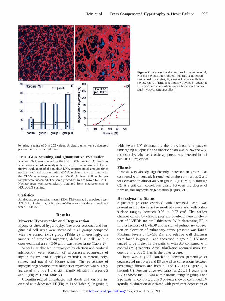
FibrosisFibrosis was already significantly increased in group 1 ascompared with control; it remained unaltered in group 2 andwas elevated to almost 40% in group 3 (Figure 2, A throughC). A significant correlation exists between the degree offibrosis and myocyte degeneration (Figure 2D).
Hemodynamic StatusSignificant pressure overload with increased LVSP waspresent in all patients as the result of severe AS, with orificesurface ranging between 0.96 to 0.22 cm2. The earliestchanges caused by chronic pressure overload were an eleva-tion of LVEDP and wall thickness. With decreasing EF, afurther increase of LVEDP and as sign of pulmonary conges-tion an elevation of pulmonary artery pressure was found.Maximal levels of LVSP, �P, and relative wall thicknesswere found in group 1 and decreased in group 3. LV masstended to be higher in the patients with AS compared withcontrol (MS) patients. Atrial fibrillation occurred more fre-quently in group 3 than in the other groups.
There was a good correlation between percentage ofdegenerated myocytes and EF as well as correlations betweenpercentage fibrosis and both EF and LVEDP (Figure 3, Athrough C). Postoperative evaluation at 2.6�1.4 years afterAVR showed that EF was within normal range in group 1 and2 patients; in contrast, group 3 patients showed continued LVsystolic dysfunction associated with persistent depression of
Figure 2. Fibronectin staining (red, nuclei blue). A,Normal myocardium shows fine septa betweenunstained myocytes; B, severe fibrosis with fewmyocytes; C, fibrosis is already severe in group 1;D, significant correlation exists between fibrosisand myocyte degeneration.
Hein et al From Compensated Hypertrophy to Heart Failure 987
by guest on July 12, 2015http://circ.ahajournals.org/Downloaded from

New York Heart Association functional class (Figure 3, Dand E).
Inflammation MarkersThere was a 3-fold increase in leukocytes (CD45) but only aslight increase in the number of lymphocytes (CD3) andmacrophages (CD68) situated in the perivascular and inter-stitial space (Table 2).
ACE, TGF-�1, and Capillary DensityThe number of CD31-positive microvessels was reduced(Figure 4C). ACE was present in the endothelium of mi-crovessels (Figure 4A) and increased in all groups whenexpressed as a percentage of capillary density (Figure 4C).TGF-�1, localized in fibroblasts and macrophages (Figure4B), was elevated in all groups (Figure 4D).
Nuclear DNA and Sc-35The content (AU times nuclear area) of DNA and SC-35 wasincreased in all groups, but the concentration (AU/nucleararea) remained unchanged (Figure 5, A through D). However,since the ratio of nucleus/cell area was decreased (Table 2),the elevation of content is insufficient for the enlargedmyocytes. The number of Ki-67–positive myocytes and ofbinucleated myocytes was unchanged in all groups (Table 2).
DiscussionIn this study, we present evidence that a close correlationexists between cardiac function and myocardial morphologyin patients with AS. With worsening of fibrosis and myocytedegeneration, LVEDP increases and later EF decreases. Thissuggests that a structure-function relation leading to HF ispresent in human pressure overload. This correlation is also
Figure 3. A through D, Good correlationexists between clinical data, degenera-tion, and fibrosis. D through E, Improve-ment after surgery. D, Recovery of EF; E,recovery of NYHA class. D and E docu-ment that adaptation to pressure over-load is present until group 2 but thatgroup 3 has reached a critical hemody-namic state with exhaustion of adapta-tional processes and incompleterecovery.
988 Circulation February 25, 2003
by guest on July 12, 2015http://circ.ahajournals.org/Downloaded from

reflected in the degree of postoperative recovery, which wasincomplete in patients group 3. This should be taken intoaccount for the decision of surgical intervention.
Fibrosis is an early morphological alteration in patientswith AS. It is a major determinant of diastolic dysfunctionand systolic pumping capacity,17 and it is one of the structural
substrates for arrhythmogenicity, thus playing a major rolefor sudden death and the progression of HF.18 This is inagreement with Krayenbühl et al,2 who performed serialobservations in the same patients and found a partial regres-sion of severe fibrosis 6 to 7 years after AVR, which isunlikely to occur in the patients of group 3 of this study.
Figure 4. ACE and TGF-�1 (green, myocytes red,nuclei blue). A, ACE is localized in numerousmicrovessels. B, TGF-�1 is present in fibroblastsand extracellular matrix. C, Number of ACE-positive microvessels is significantly elevated ingroup 1 and remains almost constant whenexpressed as percentage of total number of capil-laries. D, TGF-�1 increases in later stages ofhypertrophy.
Figure 5. DNA and Sc-35 labeling (myocytes red,nuclei green for either DNA [A] or Sc-35 [B]). Yel-low dots are lipofuscin granules. A, Typical stain-ing pattern for DNA in nuclei. B, Sc-35 localiza-tion. C, Nuclear content of Sc-35 and DNA. D,Nuclear concentration of Sc-35 and DNA.
Hein et al From Compensated Hypertrophy to Heart Failure 989
by guest on July 12, 2015http://circ.ahajournals.org/Downloaded from

Inflammation was of low grade and reflected in theincrease in leukocytes and macrophages observed. These maybe responsible for the increase in cytokine production (TGF-�1, TNF-�, interleukin family) that accounts not only for theinflammatory response but also for the formation of reactivefibrosis.
Previous studies of myocardium from patients with AShave shown that ACE and TGF-�1 were upregulated on themRNA and protein level.19 Because TGF-�1 was upregulatedin group 1, we suggest that TGF-�1 is one of the majordeterminants of fibrosis progression, thus confirming studiesby others (reviewed in Reference 20).20 ACE was elevated ingroup 1. De novo synthesized angiotensin II locally releasedby the action of ACE regulates TGF-�1 production, and it isthis fibrogenic cytokine that regulates the collagen turnoverof fibroblasts.9 The present findings corroborate the hypoth-esis that angiotensin release by the action of ACE is stimu-lated by a paracrine mechanism involving TGF-�1 as amediator.21
Myocyte DegenerationThe degree of degenerative injury of cardiomyocytes in-creased with the development of HF. The term degenerationwas chosen to emphasize involvement of all cellular or-ganelles in a chronic and most probably slow process ofdegradation that finally results in cellular atrophy, myocytedeath, and replacement fibrosis. The severity of changesexceeded those typically observed in myocardium frompatients with dilated cardiomyopathy.3 The significant corre-lation between myocyte degeneration with fibrosis and withEF suggests a mutual influence of cardiac structure andfunction.
DNA Replication, Transcription, and Lackof MitosisCompensatory mechanisms were, however, observed as well.The increase in SC-35, an RNA splicing factor, indicatesongoing transcription in all groups. Sc-35 belongs to thegroup of non-snRNP factors and is required for the first stepof splicing and spliceosome assembly.22 We would like topostulate that the presence of Sc-35 indicates that the cardio-myocytes, even when damaged, are viable and capable oftranscription and translation.
This assumption is reinforced by the DNA data presentedin this report, which confirm earlier experimental studies.23,24
The ability of the human myocyte to increase its DNAcontent avoids “dilution” of the DNA in the enlarged cellsand permits DNA repair, thus allowing for a significanthypertrophic response. However, since the nucleus/cell vol-ume ratio is disturbed, the amount of DNA is not sufficient tosustain the transcriptional levels required by the enlarged cellvolume and cellular exhaustion will be the consequence. Datapublished by other groups support the notion that DNAsynthesis takes place in the adult mammalian heart,24 and thisis strongly suggested by our own data as well.
The cell-cycle-associated nuclear nonhistone Ki-67 proteinis expressed in all active phases of the cell cycle but not inquiescent G0 cells.25 It is more abundant in DNA synthesisand mitosis but it might also be observed when DNA
synthesis is inhibited.26 The number of binucleated myocytesdid not increase with hypertrophy, and therefore we assumethat the presence of Ki-67–positive myocytes reflects DNAreplication for maintenance of the DNA content/myocytevolume ratio and for DNA repair but not mitotic nucleardivision,26 confirming results by other groups.27,28
Mitotic figures indicative of a putative regeneration pro-cess were never observed. This contrasts with another study29
but confirms earlier work in human and animal tissue.30 Sincecardiomyocyte mitosis was completely absent, the presentdata implicate that myocyte degeneration will lead to finalcell loss.
Ubiquitin-related autophagic cell death13,14 and oncosis31
appear to be more important than apoptosis, which occurredat an extremely low rate. Our data suggest that ubiquitin bindscontractile or membrane proteins destined for degradation butthat because of proteasomal insufficiency, the complexes areaccumulated and might cause nuclear fragmentation (unpub-lished). Myocytes therefore exhibit large areas with loss ofcross-striated sarcomeres. Cells will disintegrate and will bereplaced by fibrosis. Narula’s concept16 that activated caspasecauses myocyte protein degradation without nuclear DNAfragmentation might be important in that regard and will bepursued in further studies. Single-cell oncosis appears tooriginate from reduced coronary flow reserve and diffusiondisturbances caused by fibrosis and the reduction of capillarydensity.
On the basis of the data presented here, we depict inFigure 6 a model of the major adaptation to pressureoverload. It may be concluded that reduction of cardiacfunction occurs when mechanisms such as DNA repair andsynthesis as well as Sc-35 expression are partially ortotally exhausted, when myocyte death occurs and fibrosiswill have reached a certain degree. These changes willaffect an increasing number of myocytes and HF willfinally occur. A self-perpetuating process of myocytedegeneration, cell death, and replacement fibrosis will bemaintained, even when excessive afterload will return tonormal after AVR. This chronic cycle will lead to furtherimpairment of LV function and poor prognosis.
Figure 6. Schematic illustration of continuous remodeling inpressure-overload hypertrophy.
990 Circulation February 25, 2003
by guest on July 12, 2015http://circ.ahajournals.org/Downloaded from

References1. Cohn JN, Ferrari R, Sharpe N. Cardiac remodeling-concepts and clinical
implications: a consensus paper from an international forum on cardiacremodeling. J Am Coll Cardiol. 2000;35:569–582.
2. Krayenbuehl H, Hess OM, Monrad ES, et al. Left ventricular myocardialstructure in aortic valve disease before, intermediate, and late after aorticvalve replacement. Circulation. 1989;79:744–755.
3. Schaper J, Froede R, Hein S, et al. Impairment of the myocardial ultra-structure and changes of the cytoskeleton in dilated cardiomyopathy.Circulation. 1991;83:504–514.
4. Anversa PL, Kajstura J, Guerra S, et al. Myocyte death and growth in thefailing heart. Lab Invest. 1998;78:767–786.
5. Elsässer A, Suzuki K, Schaper J. Unresolved issues regarding the role ofapoptosis in the pathogenesis of ischemic injury and heart failure. J MolCell Cardiol. 2000;32:711–724.
6. Capasso JM, Palckal T, Olivetti G, et al. Left ventricular failure inducedby long-term hypertension in rats. Circ Res. 1990;66:1400–1412.
7. Weber KT. Targeting pathological remodeling: concepts of cardiopro-tection and reparation. Circulation. 2000;102:1342–1345.
8. Zhou GP, Kandala JC, Tyagi SC, et al. Effects of angiotensin II andaldosterone on collagen gene expression and protein turnover in cardiacfibroblasts. Mol Cell Biochem. 1996;154:171–178.
9. Weber KT. Angiotensin II and connective tissue: homeostasis andreciprocal regulation. Regul Pept. 1999;82:1–17.
10. Hein S, Schaper J. The extracellular matrix in normal heart and diseasedmyocardium. J Nucl Cardiol. 2001;8:188–196.
11. Pinto YM, Pinto-Sietsma SJ, Philipp T, et al. Reduction in left ventricularmessenger RNA for transforming growth factor beta(1) attenuates leftventricular fibrosis and improves survival without lowering bloodpressure in the hypertensive TGR(mRen2)27 rat. Hypertension. 2000;36:747–754.
12. Schultz J, Witt SA, Glascock BJ, et al. TGF-�1 mediates the hypertrophiccardiomyocyte growth induced by angiotensin II. J Clin Invest. 2002;109:787–796.
13. Knaapen MW, Davies MJ, De Bie M, et al. Apoptotic versus autophagiccell death in heart failure. Cardiovasc Res. 2001;51:304–312.
14. Glickman MH, Ciechanover A. The ubiquitin-proteasome proteolyticpathway: destruction for the sake of construction. Physiol Rev. 2002;82:373–428.
15. Levin S, Bucci TJ, Cohen SM, et al. The nomenclature of cell death:recommendations of an ad hoc committee of the Society of ToxicologicPathologists. Toxicol Pathol. 1999;27:484–490.
16. Narula J, Pandey P, Arbustini E, et al. Apoptosis in heart failure: releaseof cytochrome c from mitochondria and activation of caspase-3 in humancardiomyopathy. Proc Natl Acad Sci U S A. 1999;96:8144–8149.
17. Villari B, Vassalli G, Monrad ES, et al. Normalization of diastolicdysfunction in aortic stenosis late after valve replacement. Circulation.1995;91:2353–2358.
18. Assayag P, Carre F, Chevalier B, et al. Compensated cardiac hypertrophy:arrhythmogenicity and the new myocardial phenotype 1. Fibrosis Car-diovasc Res. 1997;34:439–444.
19. Fielitz J, Hein S, Mitrovic V, et al. Activation of the cardiac renin-an-giotensin system and increased myocardial collagen expression in humanaortic valve disease. J Am Coll Cardiol. 2001;37:1443–1449.
20. Lijnen PJ, Petrov VV, Fagard RH. Induction of fibrosis by transforminggrowth factor-�1. Mol Gen Metab. 2000;71:418–435.
21. Boluyt MO, Oneill L, Meredith AL, et al. Alterations in cardiac geneexpression during the transition from stable hypertrophy to heart failure:marked upregulation of genes encoding extracellular matrix components.Circ Res. 1994;75:23–32.
22. Fu XD, Maniatis T. The 35-kDa mammalian splicing factor SC35mediates specific interactions between U1 and U2 small nuclear ribonu-cleoprotein particles at the 3 splice site. Proc Natl Acad Sci U S A.1992;89:1725–1729.
23. Grove BS, Nair KG, Zak R. Biochemical correlates of cardiac hypertro-phy, III: changes in DNA content, the relative contributions of polypoidieand mitotic activity. Circ Res. 1969;25:463–471.
24. Soonpaa MH, Field LJ. Survey of studies examining mammalian cardio-myocyte DNA synthesis. Circ Res. 1998;83:15–26.
25. Schluter C, Duchrow M, Wohlenberg C, et al. The cell proliferation-associated antigen of antibody Ki-67: a very large, ubiquitous nuclearprotein with numerous repeated elements, representing a new kind of cellcycle-maintaining proteins. J Cell Biol. 1993;123:513–522.
26. van Oijen MG, Medema RH, Slootweg PJ, et al. Positivity of the prolif-eration marker Ki-67 in noncycling cells. Am J Clin Pathol. 1998;110:24–31.
27. Brodsky VY, Sarkisov DS, Arefyeva AM, et al. Polyploidy in cardiacmyocytes of normal and hypertrophic human hearts: range of values.Virchows Arch. 1994;42:429–435.
28. Adler CP, Neuburger M, Herget GW, et al. Regeneration processes inhuman myocardium after acute ischaemia-quantitative determination ofDNA, cell number and collagen content. Virchows Arch. 1997;430:149–153.
29. Leri A, Barlucchi L, Limana F, et al. Telomerase expression and activityare coupled with myocyte proliferation and preservation of telomericlength in the failing heart. Proc Natl Acad Sci U S A. 2001;98:8626–8631.
30. Huttenbach Y, Ostrowski ML, Thaller D, et al. Cell proliferation in thegrowing human heart: MIB-1 immunostaining in preterm and term infantsat autopsy. Cardiovasc Pathol. 2001;10:119–123.
31. Majno G, Joris I. Apoptosis, oncosis, and necrosis: an overview of celldeath. Am J Pathol. 1995;146:3–15.
Hein et al From Compensated Hypertrophy to Heart Failure 991
by guest on July 12, 2015http://circ.ahajournals.org/Downloaded from

Polyakova, Erwin P. Bauer, Wolf-Peter Klövekorn and Jutta SchaperStefan Hein, Eyal Arnon, Sawa Kostin, Markus Schönburg, Albrecht Elsässer, Victoria
Human Heart: Structural Deterioration and Compensatory MechanismsProgression From Compensated Hypertrophy to Failure in the Pressure-Overloaded
Print ISSN: 0009-7322. Online ISSN: 1524-4539 Copyright © 2003 American Heart Association, Inc. All rights reserved.
is published by the American Heart Association, 7272 Greenville Avenue, Dallas, TX 75231Circulation doi: 10.1161/01.CIR.0000051865.66123.B7
2003;107:984-991; originally published online February 10, 2003;Circulation.
http://circ.ahajournals.org/content/107/7/984World Wide Web at:
The online version of this article, along with updated information and services, is located on the
http://circ.ahajournals.org//subscriptions/
is online at: Circulation Information about subscribing to Subscriptions:
http://www.lww.com/reprints Information about reprints can be found online at: Reprints:
document. Permissions and Rights Question and Answer this process is available in the
click Request Permissions in the middle column of the Web page under Services. Further information aboutOffice. Once the online version of the published article for which permission is being requested is located,
can be obtained via RightsLink, a service of the Copyright Clearance Center, not the EditorialCirculationin Requests for permissions to reproduce figures, tables, or portions of articles originally publishedPermissions:
by guest on July 12, 2015http://circ.ahajournals.org/Downloaded from
Related Documents