of August 21, 2018. This information is current as Mucus Formation Goblet Cell Differentiation and Airway Promotes Th2 Polarization and Impairs Prenatal Secondhand Cigarette Smoke L. Sopori Mohan A. Wilder, Raymond J. Langley, Kevin R. Smith and Julie Peña-Philippides, Jules Rir-sima-ah, Neerad C. Mishra, Shashi P. Singh, Sravanthi Gundavarapu, Juan C. http://www.jimmunol.org/content/187/9/4542 doi: 10.4049/jimmunol.1101567 September 2011; 2011; 187:4542-4552; Prepublished online 19 J Immunol Material Supplementary 7.DC1 http://www.jimmunol.org/content/suppl/2011/09/22/jimmunol.110156 References http://www.jimmunol.org/content/187/9/4542.full#ref-list-1 , 21 of which you can access for free at: cites 74 articles This article average * 4 weeks from acceptance to publication Fast Publication! • Every submission reviewed by practicing scientists No Triage! • from submission to initial decision Rapid Reviews! 30 days* • Submit online. ? The JI Why Subscription http://jimmunol.org/subscription is online at: The Journal of Immunology Information about subscribing to Permissions http://www.aai.org/About/Publications/JI/copyright.html Submit copyright permission requests at: Email Alerts http://jimmunol.org/alerts Receive free email-alerts when new articles cite this article. Sign up at: Print ISSN: 0022-1767 Online ISSN: 1550-6606. Immunologists, Inc. All rights reserved. Copyright © 2011 by The American Association of 1451 Rockville Pike, Suite 650, Rockville, MD 20852 The American Association of Immunologists, Inc., is published twice each month by The Journal of Immunology by guest on August 21, 2018 http://www.jimmunol.org/ Downloaded from by guest on August 21, 2018 http://www.jimmunol.org/ Downloaded from

Welcome message from author
This document is posted to help you gain knowledge. Please leave a comment to let me know what you think about it! Share it to your friends and learn new things together.
Transcript

of August 21, 2018.This information is current as
Mucus FormationGoblet Cell Differentiation and AirwayPromotes Th2 Polarization and Impairs Prenatal Secondhand Cigarette Smoke
L. SoporiMohanA. Wilder, Raymond J. Langley, Kevin R. Smith and
JuliePeña-Philippides, Jules Rir-sima-ah, Neerad C. Mishra, Shashi P. Singh, Sravanthi Gundavarapu, Juan C.
http://www.jimmunol.org/content/187/9/4542doi: 10.4049/jimmunol.1101567September 2011;
2011; 187:4542-4552; Prepublished online 19J Immunol
MaterialSupplementary
7.DC1http://www.jimmunol.org/content/suppl/2011/09/22/jimmunol.110156
Referenceshttp://www.jimmunol.org/content/187/9/4542.full#ref-list-1
, 21 of which you can access for free at: cites 74 articlesThis article
average*
4 weeks from acceptance to publicationFast Publication! •
Every submission reviewed by practicing scientistsNo Triage! •
from submission to initial decisionRapid Reviews! 30 days* •
Submit online. ?The JIWhy
Subscriptionhttp://jimmunol.org/subscription
is online at: The Journal of ImmunologyInformation about subscribing to
Permissionshttp://www.aai.org/About/Publications/JI/copyright.htmlSubmit copyright permission requests at:
Email Alertshttp://jimmunol.org/alertsReceive free email-alerts when new articles cite this article. Sign up at:
Print ISSN: 0022-1767 Online ISSN: 1550-6606. Immunologists, Inc. All rights reserved.Copyright © 2011 by The American Association of1451 Rockville Pike, Suite 650, Rockville, MD 20852The American Association of Immunologists, Inc.,
is published twice each month byThe Journal of Immunology
by guest on August 21, 2018
http://ww
w.jim
munol.org/
Dow
nloaded from
by guest on August 21, 2018
http://ww
w.jim
munol.org/
Dow
nloaded from

The Journal of Immunology
Prenatal Secondhand Cigarette Smoke Promotes Th2Polarization and Impairs Goblet Cell Differentiation andAirway Mucus Formation
Shashi P. Singh,* Sravanthi Gundavarapu,* Juan C. Pena-Philippides,* Jules Rir-sima-ah,*
Neerad C. Mishra,* Julie A. Wilder,* Raymond J. Langley,* Kevin R. Smith,† and
Mohan L. Sopori*
Parental, particularly maternal, smoking increases the risk for childhood allergic asthma and infection. Similarly, in a murine
allergic asthma model, prenatal plus early postnatal exposure to secondhand cigarette smoke (SS) exacerbates airways hyperre-
activity and Th2 responses in the lung. However, the mechanism and contribution of prenatal versus early postnatal SS exposure
on allergic asthma remain unresolved. To identify the effects of prenatal and/or early postnatal SS on allergic asthma, BALB/c
dams and their offspring were exposed gestationally and/or 8–10 wk postbirth to filtered air or SS. Prenatal, but not postnatal, SS
strongly increased methacholine and allergen (Aspergillus)-induced airway resistance, Th2 cytokine levels, and atopy and acti-
vated the Th2-polarizing pathway GATA3/Lck/ERK1/2/STAT6. Either prenatal and/or early postnatal SS downregulated the Th1-
specific transcription factor T-bet and, surprisingly, despite high levels of IL-4/IL-13, dramatically blocked the allergen-induced
mucous cell metaplasia, airway mucus formation, and the expression of mucus-related genes/proteins: Muc5ac, g-aminobutyric
acid A receptors, and SAM pointed domain-containing Ets-like factor. Given that SS/nicotine exposure of normal adult mice
promotes mucus formation, the results suggested that fetal and neonatal lung are highly sensitive to cigarette smoke. Thus,
although the gestational SS promotes Th2 polarization/allergic asthma, it may also impair and/or delay the development of fetal
and neonatal lung, affecting mucociliary clearance and Th1 responses. Together, this may explain the increased susceptibility of
children from smoking parents to allergic asthma and childhood respiratory infections. The Journal of Immunology, 2011, 187:
4542–4552.
Asthma is a heterogenous disease (1), characterized byairways hyperreactivity (AHR), episodic wheezing, air-way inflammation, and mucus secretion (2). Allergic
asthma is the most common form of asthma in children. It starts inearly life and is associated with AHR, airway inflammation, andatopy (3). Th2 polarization, the hallmark of asthma, is seen inhuman (3) and animal models of allergic asthma (4, 5).Deleterious effects of tobacco smoke on human health are well
established (6, 7), and increasing epidemiological evidence sug-gests that environmental factors such as secondhand tobaccosmoke and polycyclic aromatic hydrocarbons are important con-tributors in the development of childhood asthma (8–10). Expo-
sure to cigarette smoke during fetal development and in the earlyyears of a child’s life is a strong risk factor for pulmonary dys-function, including asthma and chronic obstructive pulmonarydisease (COPD) (11). Parental smoking, particularly maternalsmoking, is strongly linked to allergic asthma and infection inchildren (12–14). Similarly, mice exposed to secondhand cigarettesmoke (SS) during early postnatal life develop exacerbated re-spiratory infections (15). However, results from epidemiologicalstudies are equivocal in identifying the developmental stage atwhich the fetus and child are vulnerable to the proasthmaticeffects of maternal smoking. Thus, a link between maternalsmoking and childhood asthma has been suggested when bothparents were asthmatic (16) or smoke exposure occurred duringearly childhood (17), during both prenatal and early postnatal life(18, 19), during pregnancy only (20–22), or during prenatal orearly postnatal life (23, 24). A possible explanation for these di-verse results is that asthma is a heterogenous disease (1) and, inhumans, it is difficult to control all of the confounders of child-hood asthma, such as genetics, birth weight, b2-adrenergic re-ceptor, breastfeeding, and air pollution.To simulate maternal exposure to cigarette smoke in animal
models of allergic asthma, we and other investigators showed thatprenatal plus early postnatal exposure to mainstream or SSexacerbates allergic asthma (5, 25, 26); however, as in humans,individual contributions of prenatal and postnatal smoke exposureon the development of early allergic asthma is not clearly re-solved. Moreover, the potential mechanism by which cigarettesmoke exposure promotes allergic asthma is not known. In thisstudy, we used an established mouse model of allergic asthma toisolate the effects of gestational and early postnatal SS exposure
*Respiratory Immunology Division, Lovelace Respiratory Research Institute, Albu-querque, NM 87108; and †Chronic Obstructive Pulmonary Disease Program, Love-lace Respiratory Research Institute, Albuquerque, NM 87108
Received for publication May 27, 2011. Accepted for publication August 20, 2011.
This work was supported in part by National Institutes of Health Grants RO1DA017003 and R01 DA04208-17, Flight Attendant Medical Research Institute grants(to S.P.S. and N.C.M.), and funds from the Lovelace Respiratory Research Institute.
Address correspondence and reprint requests to Dr. Mohan L. Sopori, Lovelace Re-spiratory Research Institute, 2425 Ridgecrest Drive SE, Albuquerque, NM 87108.E-mail address: [email protected]
The online version of this article contains supplemental material.
Abbreviations used in this article: AB-PAS, Alcian blue–periodic acid-Schiff; AHR,airways hyperreactivity; BALF, bronchoalveolar lavage fluid; COPD, chronic ob-structive pulmonary disease; FA, filtered air; GABAAR, g-aminobutyric acid A re-ceptor; i.t., intratracheally; MCh, methacholine; RL, airway resistance; RT, roomtemperature; SPDEF, SAM pointed domain-containing Ets-like factor; SS, second-hand cigarette smoke; Vs, volume density; WB, Western blot.
Copyright� 2011 by TheAmericanAssociation of Immunologists, Inc. 0022-1767/11/$16.00
www.jimmunol.org/cgi/doi/10.4049/jimmunol.1101567
by guest on August 21, 2018
http://ww
w.jim
munol.org/
Dow
nloaded from
on the development of allergic asthma. To our knowledge, weshow for the first time that gestational exposure to SS is by far theoverwhelming risk factor for exacerbated AHR, Th2 polarization,and atopy and is associated with activation of GATA3/Lck/ERK1/2/STAT6. Surprisingly, unlike the promucoid effects of cigarettesmoke/nicotine in adult humans and animals (27–29), both pre-natal and early postnatal exposure to SS suppressed the variousparameters of airway mucus response.
Materials and MethodsAnimals
Pathogen-free BALB/c mice from the Frederick Cancer Research Facility(Frederick, MD) were housed in shoebox-type plastic cages with hardwoodchip bedding and conditioned to whole-body exposure in exposurechambers (H1000; Hazleton Systems) for 2 wk before exposure to SS forbreeding (5). The chamber temperature was maintained at 26 6 2˚C, andlights were set to a 12-h on/off cycle. Food and water were provided adlibitum. The Animal Care and Use Committee of Lovelace RespiratoryResearch Institute approved all animal protocols.
Abs and reagents
All reagents, unless stated otherwise, were purchased from Sigma-Aldrich(St. Louis, MO).
Cigarette smoke generation and exposure
Mice were exposed towhole-body SS (smoke released from the burning endof a cigarette) or filtered air (FA) for 6 h/d, 7 d/wk, as described (5). Briefly,a smoking machine (AMESA Type 1300; AMESA Technologies, Geneva,Switzerland) generated two 70-cm3 puffs/min from a research cigarette(type 2R1; Tobacco and Health Research Institute, Lexington, KY), andthe smoke was captured from the lit end of the cigarettes with a plasticmanifold placed above it. This level (total particulate matter, 1.52 6 0.41mg/m3) of exposure simulates the conditions at which a pregnant womanwould be exposed to environmental tobacco smoke for 3 h/d in a smokingbar (5). Adult (324-mo-old) male and female mice were separately ac-climatized to SS or FA for 2 wk prior to mating. After ascertainingpregnancy by vaginal smear, pregnant mice were housed singly in plasticcages and continued to receive SS or FA until the pups were born. Themothers and pups continued to be exposed to either FA or SS starting fromday 1 of birth until the pups were weaned at 3 wk of age, and the pupscontinued to be exposed to FA or SS postnatally until sacrifice between 8–10 wk of age. This led to four groups of experimental animals, receivingthe following combination of prenatal/postnatal exposure: FA/FA, FA/SS,SS/FA, and SS/SS.
Sensitization with allergen
The allergen used in the study was a lyophilized culture filtrate preparationof Aspergillus fumigatus; the filtrates (kindly provided by Dr. JohnM. Routes, Department of Pediatrics, Children’s Hospital, Wisconsin Medi-cal College, Milwaukee, WI) were stored at 270˚C until use. Mice wereimmunized intratracheally (i.t.) with A. fumigatus (50 mg/0.1 mlendotoxin-free sterile saline or sterile saline alone) and subsequentlychallenged i.t. with the A. fumigatus extracts (100 mg/0.1 ml) three times at5-d intervals (5).
Total serum IgE
Total serum IgE levels were determined on diluted (1:100) serum usinga mouse-specific serum IgE ELISA kit (MD Bioproducts, St. Paul, MN),according to the manufacturer’s instructions. The sensitivity of the assaywas ,2.0 ng/ml; the cross-reactivity with IgG was ,0.01%.
Airway resistance
Forty-eight hours after the last A. fumigatus or saline challenge, airwayresistance (RL) was measured by the FlexiVent system (SCIREQ, Mon-treal, Quebec, Canada), as described (5). The peak RL response at thenebulized A. fumigatus (200 mg/ml) concentration and at each meth-acholine (MCh) concentration was used for data analysis.
Bronchoalveolar lavage fluid collection, cell differentials, andcytokine analysis
Established protocols were followed to obtain bronchoalveolar lavage fluid(BALF) from the animals (5). Briefly, mice were anesthetized and killed by
exsanguination 48 h after the last A. fumigatus challenge. Before excisionof the lungs, the trachea was surgically exposed, cannulated and, while theleft lung lobe was tied off with a silk thread, the right lobe was lavagedtwice with 1 ml sterile Ca2+/Mg2+-free PBS (pH 7.4). Aliquots were pooledfrom individual animals. Cell differentials and cytokine assays were per-formed, as described (5). Macrophage, neutrophil, lymphocyte, and eo-sinophil numbers were determined microscopically by counting $300cells/sample. Lavage cytokines were assayed using the Mouse CytokineMultiPlex ELISA kit (Biosource-Invitrogen, Camarillo, CA), according tothe manufacturer’s directions. The sensitivity of the assay was ,10 pg/ml.
Immunohistochemistry
For immunohistochemical detection of airway mucus, formaldehyde-fixedleft lung sections (5 mm) were stained with Alcian blue–periodic acid-Schiff (AB-PAS), as described previously (28). By this procedure, themucus-producing cells stained distinctive pink and were examined mi-croscopically at 403 magnification.
Analysis of mucosubstances in lung airways
The volume of mucous cells and the density of mucosubstances (Vs) in theairway epithelium were quantitated by a semiautomatic image-analysissystem (28), using the public domain National Institutes of Health Im-age program (http//rsb.info.nih.gov/nih.gov/nih-image). Morphometry wasperformed blinded, and the data were expressed as the mean6 SDVs (nl/mm2
basal lamina).
SAM pointed domain-containing Ets-like factor staining
Paraffin-embedded lung tissue (5 mm) was deparaffinized and rehydratedby placing slides on a slide warmer at 56˚C for 20 min, followed by three10-min incubations in xylene and a series of alcohol washes, followed byPBS. Ag retrieval was performed by immersing the slides in 10 mM citratebuffer at 90˚C for 15 min. Endogenous peroxidase quenching was done byplacing the slides in 3% H2O2 in methanol for 15 min at room temperature(RT), washing (five times in PBS), and blocking with 10% goat serum for2 h at RT. The slides were incubated overnight at 4˚C with a guinea piganti-mouse polyclonal SAM pointed domain-containing Ets-like factor(SPDEF) Ab (GP954; a generous gift from Dr. Jeffrey Whitsett, CincinnatiChildren’s Hospital, Cincinnati, OH) at 1:2500 dilution. Slides werewashed five times with buffer and incubated at RT for 30 min with a 1:200dilution of a goat anti-guinea pig biotinylated IgG (catalog no. BA-1000;Vector Lab, Burlingame, CA). Slides were washed in PBS and incubatedfor 30 min in the Immunoperoxidase Kit (catalog no. PK-6100; Vector),per the manufacturer’s instructions. Slides were washed five times in bufferand incubated for 2 min at RT in peroxidase substrate (catalog no. SK-4100; Vector) without nickel. Slides were then counterstained with he-matoxylin and dehydrated. Slides were examined at 403; SPDEF stainsbrown.
Quantitative PCR
Total RNA was extracted from the frozen lung samples using TRI reagent(Molecular Research Center, Cincinnati, OH) and quantified per themanufacturer’s instructions. The lung expression of the airway mucinMuc5ac, the Th1 transcription factor T-bet, the Th2 transcription factorGATA3, and the housekeeping gene GAPDH was determined usingprimer/probe sets (Applied Biosystems, Foster City, CA). The relativeexpression (test mRNA/GAPDH) was calculated (5).
Western blot analysis
Lung tissues were homogenized in radioimmunoprecipitation assay buffer(20 mM Tris, 150 mM NaCl, 20 mM b-glyceryl-phosphate, 1% Triton X-100, 10 mM NaF, 5 mM EDTA, 1 mM Na3VO4) containing proteaseinhibitors (1 mM PMSF; 1 mg/ml each aprotinin, antipain, and leupeptin)at 4˚C. Protein content and Western blot (WB) analysis were performed, asdescribed (5). NF-kB (p65), GATA3, STAT5, STAT6, and Lck were de-termined by probing the blots with anti–phospho-NF-kB-p65 (Ser529,rabbit polyclonal; Abcam, San Francisco, CA), anti–phospho-GATA3(Ser308, rabbit polyclonal; Abcam), anti–phospho-STAT5, (Tyr694/699,mouse monoclonal; Abcam), anti–phospho-STAT6 (Tyr641, rabbit poly-clonal; Cell Signaling Technology, Danvers, MA), and anti–phospho-Lck(Tyr394, rabbit polyclonal; Cell Signaling Technology). The g-amino-butyric acid A receptor (GABAAR) and T-bet expression was determinedby probing the blot with anti-GABAAR (mouse monoclonal; Millipore,Temecula, CA) and anti–T-bet Ab (mouse monoclonal; Abcam). Blotswere developed with ECL (Amersham Biosciences, U.K.) using x-rayphotograph film.
The Journal of Immunology 4543
by guest on August 21, 2018
http://ww
w.jim
munol.org/
Dow
nloaded from
Densitometry The x-ray films of WBs were scanned and quantified with aGS-800 Calibrated Densitometer using Quantity-One software (Bio-Rad,Hercules, CA).
Data presentation and statistical analysis
All data were analyzed using Graph Pad Prism software 5.03 (GraphPad,San Diego, CA). One-way ANOVAwas used to compare the mean betweengroups using the Tukey post hoc test, which compares all groups at 95%confidence intervals.Where needed, Bonferroni correction was also used formultiple comparisons. Results are presented as the mean6 SD; p values#0.05 were considered statistically significant.
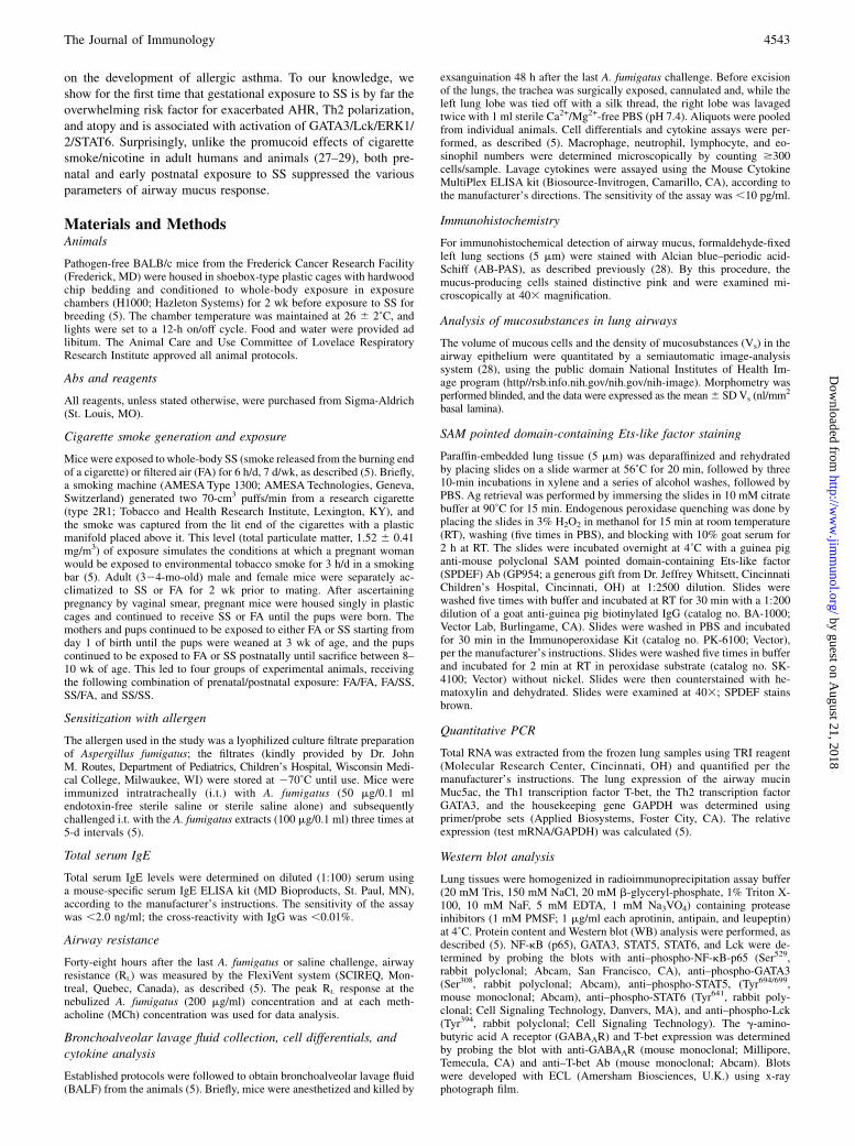
ResultsPrenatal, but not postnatal, exposure to SS increases AHR
We previously showed that prenatal plus perinatal exposure to SSincreased RL to MCh (5); however, individual contributions ofprenatal and postnatal SS exposure on AHR were not addressed.Therefore, dams were exposed to SS or FA, as described in Mate-rials and Methods; 17 d prior to sacrifice, four groups of animals(FA/FA, FA/SS, SS/FA, and SS/SS) were sensitized with A. fumi-gatus. RL was determined in response to aerosolized MCh (Fig. 1A)or a predetermined optimal concentration of aerosolized A. fumi-gatus (200 mg/ml) (Fig. 1B). Results demonstrated that, comparedwith FA/FA, both MCh and A. fumigatus caused dramatic increasesin the RL in prenatally SS-exposed animal groups (SS/SS and SS/FA); however, the RL values between FA/FA and FA/SS groups andbetween SS/SS and SS/FA groups were not significantly different,indicating that, unlike prenatal SS exposure, postnatal SS exposuredoes not affect RL significantly. MCh-induced changes in RL valuesof mice not sensitized to A. fumigatus indicated that, even in theabsence of allergen, prenatally SS-exposedmice (SS/FA and SS/SS)were significantlymore sensitive toMCh than FA/FA or FA/SSmice(Supplemental Fig. 1). Even the basal RL of prenatally SS-exposedmicewas somewhat higher (statistically significant) than that of FA/FA or FA/SS mice. These results suggested that, although prenatalSS exposure strongly exacerbates MCh- and A. fumigatus-induced
AHR, SS exposure during the early postnatal life had a relativelyminor effect on this response. Moreover, in this model, changes inAHR reached the level that allowed RLmeasurements in response tothe allergen (A. fumigatus). RL measurements in non-A. fumigatus-sensitized mice indicated that prenatal exposure to SS exacerbatedMCh-induced AHR; however, the degree of the response was con-siderably lower than in A. fumigatus-sensitized mice (SupplementalFig. 1). Thus, prenatal SS exposure primes the lung for exacerbatedAHR response.
Prenatal SS exposure induces a strong Th2 cytokine responsein the lung
The Th2 cytokines, particularly IL-13 and IL-4, play an essentialrole in allergic asthma and Th2 polarization (30, 31); however, onlyAHR responses, and not Th2 responses, are ameliorated by thephosphodiesterase4-selective inhibitor rolipram. This suggestedthat AHR and Th2 responses are regulated by different mecha-nisms (5) and, therefore, it was possible that prenatal and earlypostnatal SS exposure might affect AHR and Th2 polarizationdifferently. To ascertain this possibility, we determined the A.fumigatus-induced IL-4 and IL-13 production in four groups ofmice: FA/FA, SS/FA, FA/SS, and SS/SS. Results presented inFigure 1C and 1D indicated that, compared with control mice (FA/FA), the early postnatal exposure to smoke (FA/SS) produceda relatively small, but significant, increase in the BALF levels ofthe Th2 cytokines IL-4 (Fig. 1C) and IL-13 (Fig. 1D) in responseto A. fumigatus sensitization. However, these levels were muchlower than those present in the BALF from prenatally SS-exposedanimals (IL-4: 13.48 6 3.74 and 6.30 6 1.92, SS/FA and FA/SS,respectively; IL-13: 111.7 6 19.82 and 34.58 6 15.65 ng/ml, SS/FA and FA/SS, respectively). Compared with A. fumigatus-sensi-tized mice, the levels of IL-4 and IL-13 in nonsensitized micewere very low (Supplemental Fig. 2). These results suggested thatprenatal exposure to SS is the predominant risk factor in drivingthe lung toward Th2 polarization. In contrast, the levels of IL-4,
FIGURE 1. Prenatal, but not postnatal, exposure to SS exacerbates airway reactivity, strongly upregulates allergen-induced Th2 cytokine expression, and
increases total serum IgE levels. Lung resistance (RL: cm H2O s/ml) was measured using FlexiVent system in response to increasing doses of computer-
controlled nebulized MCh (A) and a single nebulized dose (200 mg/ml) of A. fumigatus (B). Inset in A shows baseline differences between the groups in
response to nebulized saline. Peak responses at each MCh concentration and response to A. fumigatus were used for data analysis. IL-4 (C) and IL-13 (D)
levels were determined in BALF from A. fumigatus-sensitized mice by the Multiplex ELISA kit. E, Total IgE levels in the serum of A. fumigatus-sensitized
mice were quantitated by using a mouse-specific IgE ELISA kit. All animals were sensitized with A. fumigatus, as described inMaterials and Methods prior
to RL determination. The results are representative of animal responses from two different sets of inhalation exposures. Data are expressed as mean 6 SD
(n = 5–7/group). *p # 0.05, ***p # 0.001.
4544 GESTATIONAL CIGARETTE SMOKE, MUCUS, AND ALLERGIC ASTHMA
by guest on August 21, 2018
http://ww
w.jim
munol.org/
Dow
nloaded from
and IL-13 in mice without A. fumigatus challenges, did not reachstatistical significance, indicating that the critical levels of thesecytokines are achieved only after allergic sensitization.
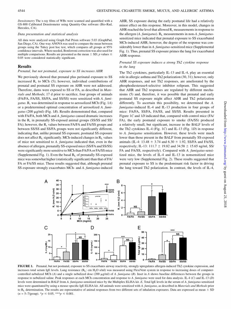
Prenatal exposure to SS increases leukocytic infiltration in thelung after A. fumigatus sensitization
Elevated numbers of eosinophils and neutrophils in the lung areassociated with allergic asthma (5). To determine whether pre-and/or postnatal exposure to SS differentially affected the leuko-cytic infiltration in the lung in response to an allergic challenge, A.fumigatus-sensitized FA/FA, FA/SS, SS/FA, and SS/SS mice werechallenged i.t. with A. fumigatus extracts. The volume of BALFrecovered was not significantly different among the groups. BALFcells were collected, cytospun, and stained to obtain the differ-ential cell count. Prior to A. fumigatus sensitization, the total num-ber of leukocytes in the BALF in both FA and SS groups was sim-ilar (6–8 6 2.6 3 104); however, after A. fumigatus challenge (+),the number of cells in FA/FA+ and FA/SS+ increased to 51–54 68.3 3 104. The number increased to 92–98 6 12.8 3 104 in SS/FA+ and SS/SS+ animals. Thus, A. fumigatus promoted leukocyticinfiltration in the lungs, and the infiltration was significantly morepronounced in the animals exposed to SS prenatally; postnatal SSdid not result in a significant difference in the total number of cellsin the BALF (Table I).The differential BALF cell count (Table I) indicated that mac-
rophages were the predominant cell population in the BALF fromFA/FA, FA/FA+, and FA/SS+, accounting for 90.86 4.4, 956 1.6,and 92.1 6 2.6%, respectively. Thus, although the A. fumigatuschallenge strongly increased the total number of BALF cells, thepercentages of macrophages did not vary significantly among thegroups; nonetheless, animals exposed postnatally to SS (FA/SS)had small, but significantly higher, numbers of neutrophils. Incontrast, prenatally SS-exposed groups (SS/FA and SS/SS)showed a dramatic decrease in the proportion of macrophages,with concomitant increases in neutrophils and eosinophils. Theseresults suggested that prenatal exposure strongly primes the lungfor neutrophilic and eosinophilic inflammation.
Prenatal SS increases total serum IgE levels
Atopy is a strong risk factor for allergic asthma in children (32). Toascertain whether prenatal and/or postnatal SS exposure affectsatopy differentially, we measured the serum levels of IgE in thefour groups of mice after A. fumigatus sensitization. Results (Fig.1E) indicated that compared with FA/FA, prenatal (SS/FA) andearly postnatal (FA/SS) exposure independently elevated totalserum IgE levels; however, the IgE level in SS/FA (prenatally SS-exposed animals) of 29.506 14.41 mg/ml was significantly higherthan the IgE level of 8.81 6 2.49 mg/ml in FA/SS (SS exposureonly during the early postnatal period). Although, the mean IgEconcentration of 39.83 6 2.75 mg/ml in SS/SS (prenatally and
postnatally SS exposure) was higher than in prenatally SS-exposedanimals, it did not reach statistical significance. These resultssuggested that although prenatal and early postnatal exposure isindependent risk factors for atopy, prenatal exposure is a sub-stantially stronger risk factor.
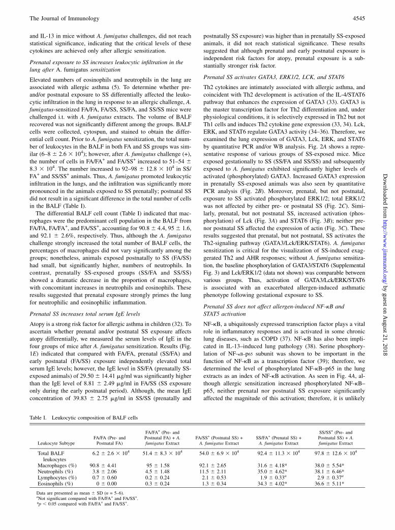
Prenatal SS activates GATA3, ERK1/2, LCK, and STAT6
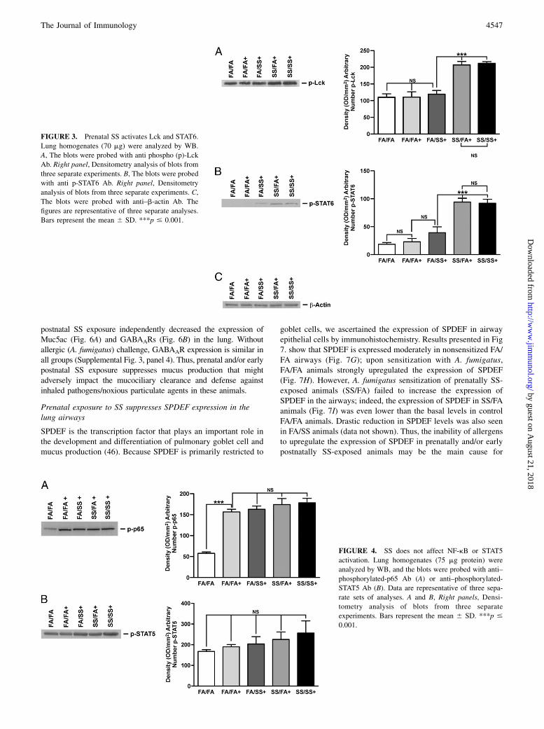
Th2 cytokines are intimately associated with allergic asthma, andcoincident with Th2 development is activation of the IL-4/STAT6pathway that enhances the expression of GATA3 (33). GATA3 isthe master transcription factor for Th2 differentiation and, underphysiological conditions, it is selectively expressed in Th2 but notTh1 cells and induces Th2 cytokine gene expression (33, 34). Lck,ERK, and STAT6 regulate GATA3 activity (34–36). Therefore, weexamined the lung expression of GATA3, Lck, ERK, and STAT6by quantitative PCR and/or WB analysis. Fig. 2A shows a repre-sentative response of various groups of SS-exposed mice. Miceexposed gestationally to SS (SS/FA and SS/SS) and subsequentlyexposed to A. fumigatus exhibited significantly higher levels ofactivated (phosphorylated) GATA3. Increased GATA3 expressionin prenatally SS-exposed animals was also seen by quantitativePCR analysis (Fig. 2B). Moreover, prenatal, but not postnatal,exposure to SS activated phosphorylated ERK1/2; total ERK1/2was not affected by either pre- or postnatal SS (Fig. 2C). Simi-larly, prenatal, but not postnatal SS, increased activation (phos-phorylation) of Lck (Fig. 3A) and STAT6 (Fig. 3B); neither pre-nor postnatal SS affected the expression of actin (Fig. 3C). Theseresults suggested that prenatal, but not postnatal, SS activates theTh2-signaling pathway (GATA3/Lck/ERK/STAT6). A. fumigatussensitization is critical for the visualization of SS-induced exag-gerated Th2 and AHR responses; without A. fumigatus sensitiza-tion, the baseline phosphorylation of GATA3/STAT6 (SupplementalFig. 3) and Lck/ERK1/2 (data not shown) was comparable betweenvarious groups. Thus, activation of GATA3/Lck/ERK/STAT6is associated with an exacerbated allergen-induced asthmaticphenotype following gestational exposure to SS.
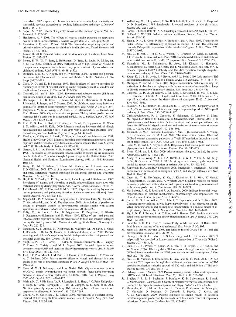
Prenatal SS does not affect allergen-induced NF-kB andSTAT5 activation
NF-kB, a ubiquitously expressed transcription factor plays a vitalrole in inflammatory responses and is activated in some chroniclung diseases, such as COPD (37). NF-kB has also been impli-cated in IL-13–induced lung pathology (38). Serine phosphory-lation of NF-kB–p65 subunit was shown to be important in thefunction of NF-kB as a transcription factor (39); therefore, wedetermined the level of phosphorylated NF-kB–p65 in the lungextracts as an index of NF-kB activation. As seen in Fig. 4A, al-though allergic sensitization increased phosphorylated NF-kB–p65, neither prenatal nor postnatal SS exposure significantlyaffected the magnitude of this activation; therefore, it is unlikely
Table I. Leukocytic composition of BALF cells
Leukocyte SubtypeFA/FA (Pre- andPostnatal FA)
FA/FA+ (Pre- andPostnatal FA) + A.fumigatus Extract
FA/SS+ (Postnatal SS) +A. fumigatus Extract
SS/FA+ (Prenatal SS) +A. fumigatus Extract
SS/SS+ (Pre- andPostnatal SS) + A.fumigatus Extract
Total BALFleukocytes
6.2 6 2.6 3 104 51.4 6 8.3 3 104 54.0 6 6.9 3 104 92.4 6 11.3 3 104 97.8 6 12.6 3 104
Macrophages (%) 90.8 6 4.41 95 6 1.58 92.1 6 2.65 31.6 6 4.18* 38.0 6 5.54*Neutrophils (%) 3.8 6 2.06 4.5 6 1.48 11.5 6 2.11 35.0 6 4.62* 38.1 6 6.46*Lymphocytes (%) 0.7 6 0.60 0.2 6 0.24 2.1 6 0.53 1.9 6 0.33a 2.9 6 0.37a
Eosinophils (%) 0 6 0.00 0.3 6 0.24 1.3 6 0.34 34.3 6 4.02* 36.6 6 5.11*
Data are presented as mean 6 SD (n = 5–6).aNot significant compared with FA/FA+ and FA/SS+.*p , 0.05 compared with FA/FA+ and FA/SS+.
The Journal of Immunology 4545
by guest on August 21, 2018
http://ww
w.jim
munol.org/
Dow
nloaded from
that exacerbated allergic asthma associated with prenatal SS ex-posure is linked to activation of NF-kB. Early production of theTh2 cytokine IL-4 is regulated by phosphorylation of the tran-scription factor STAT5 (40); however, results presented in Fig. 4Bshowed that neither pre- nor postnatal SS significantly alteredactivation (phosphorylation) of STAT5. Together, these resultssuggested that NF-kB and STAT5 are not strongly associated withthe SS-induced exacerbated Th2 responses. Prenatal and earlypostnatal SS exposure strongly suppresses the allergen-induced T-bet expression in the lung GATA3 and T-bet are two opposingtranscription factors for Th1 and Th2 development, respectively(40, 41). Results (Fig. 5) clearly showed that sensitization ofcontrol (FA/FA) animals with A. fumigatus (FA/FA+) strongly in-creased the level of T-bet in the lung; however, animals exposedto SS either prenatally (SS/FA) or postnatally (FA/SS) exhibiteda dramatic reduction in the A. fumigatus-induced T-bet proteinlevels (Fig. 5A). WB data presented in Supplemental Fig. 3 (toppanel) show that exposure to an allergen is essential to upregulateT-bet expression. Quantitative PCR data indicating that prenataland early postnatal exposure to SS downregulated the expressionof T-bet mRNA (Fig. 5B) also supported these results. Thus,prenatal and early postnatal exposure to SS suppresses theallergen-induced T-bet expression, which might impair the de-velopment of Th1 responses in the lung. To evaluate the effect ofSS exposure on the prototype Th1 cytokine, IFN-g levels wereassayed in the BALF of the experimental animals. Although thevarious groups did not exhibit significant differences in the basal(nonsensitized) levels of IFN-g (Supplemental Fig. 2C), FA/FAanimals exhibited a significant increase in the BALF levels of
IFN-g after A. fumigatus sensitization. In contrast, the IFN-glevels in animals exposed either prenatally and/or postnatally toSS failed to upregulate the BALF levels of IFN-g after the A.fumigatus challenge (Fig. 5C). Thus, SS exposure affected T-betand IFN-g similarly.
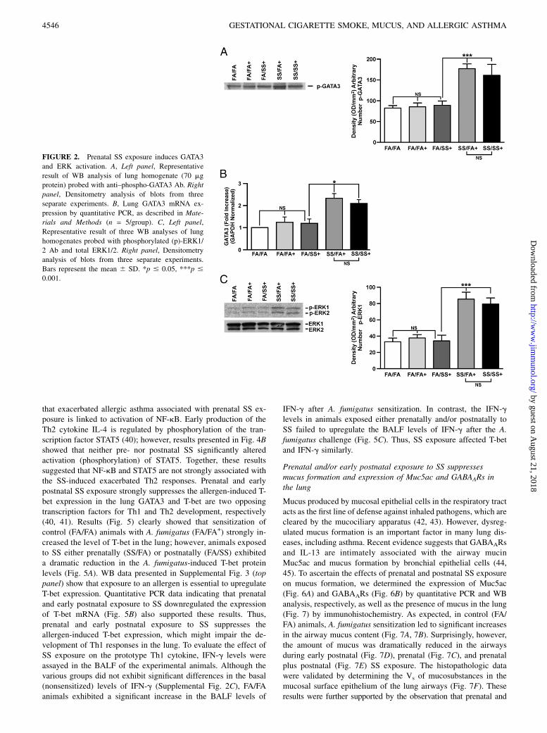
Prenatal and/or early postnatal exposure to SS suppressesmucus formation and expression of Muc5ac and GABAARs inthe lung
Mucus produced by mucosal epithelial cells in the respiratory tractacts as the first line of defense against inhaled pathogens, which arecleared by the mucociliary apparatus (42, 43). However, dysreg-ulated mucus formation is an important factor in many lung dis-eases, including asthma. Recent evidence suggests that GABAARsand IL-13 are intimately associated with the airway mucinMuc5ac and mucus formation by bronchial epithelial cells (44,45). To ascertain the effects of prenatal and postnatal SS exposureon mucus formation, we determined the expression of Muc5ac(Fig. 6A) and GABAARs (Fig. 6B) by quantitative PCR and WBanalysis, respectively, as well as the presence of mucus in the lung(Fig. 7) by immunohistochemistry. As expected, in control (FA/FA) animals, A. fumigatus sensitization led to significant increasesin the airway mucus content (Fig. 7A, 7B). Surprisingly, however,the amount of mucus was dramatically reduced in the airwaysduring early postnatal (Fig. 7D), prenatal (Fig. 7C), and prenatalplus postnatal (Fig. 7E) SS exposure. The histopathologic datawere validated by determining the Vs of mucosubstances in themucosal surface epithelium of the lung airways (Fig. 7F). Theseresults were further supported by the observation that prenatal and
FIGURE 2. Prenatal SS exposure induces GATA3
and ERK activation. A, Left panel, Representative
result of WB analysis of lung homogenate (70 mg
protein) probed with anti–phospho-GATA3 Ab. Right
panel, Densitometry analysis of blots from three
separate experiments. B, Lung GATA3 mRNA ex-
pression by quantitative PCR, as described in Mate-
rials and Methods (n = 5/group). C, Left panel,
Representative result of three WB analyses of lung
homogenates probed with phosphorylated (p)-ERK1/
2 Ab and total ERK1/2. Right panel, Densitometry
analysis of blots from three separate experiments.
Bars represent the mean 6 SD. *p # 0.05, ***p #
0.001.
4546 GESTATIONAL CIGARETTE SMOKE, MUCUS, AND ALLERGIC ASTHMA
by guest on August 21, 2018
http://ww
w.jim
munol.org/
Dow
nloaded from
postnatal SS exposure independently decreased the expression ofMuc5ac (Fig. 6A) and GABAARs (Fig. 6B) in the lung. Withoutallergic (A. fumigatus) challenge, GABAAR expression is similar inall groups (Supplemental Fig. 3, panel 4). Thus, prenatal and/or earlypostnatal SS exposure suppresses mucus production that mightadversely impact the mucociliary clearance and defense againstinhaled pathogens/noxious particulate agents in these animals.
Prenatal exposure to SS suppresses SPDEF expression in thelung airways
SPDEF is the transcription factor that plays an important role inthe development and differentiation of pulmonary goblet cell andmucus production (46). Because SPDEF is primarily restricted to
goblet cells, we ascertained the expression of SPDEF in airwayepithelial cells by immunohistochemistry. Results presented in Fig7. show that SPDEF is expressed moderately in nonsensitized FA/FA airways (Fig. 7G); upon sensitization with A. fumigatus,FA/FA animals strongly upregulated the expression of SPDEF(Fig. 7H). However, A. fumigatus sensitization of prenatally SS-exposed animals (SS/FA) failed to increase the expression ofSPDEF in the airways; indeed, the expression of SPDEF in SS/FAanimals (Fig. 7I) was even lower than the basal levels in controlFA/FA animals. Drastic reduction in SPDEF levels was also seenin FA/SS animals (data not shown). Thus, the inability of allergensto upregulate the expression of SPDEF in prenatally and/or earlypostnatally SS-exposed animals may be the main cause for
FIGURE 3. Prenatal SS activates Lck and STAT6.
Lung homogenates (70 mg) were analyzed by WB.
A, The blots were probed with anti phospho (p)-Lck
Ab. Right panel, Densitometry analysis of blots from
three separate experiments. B, The blots were probed
with anti p-STAT6 Ab. Right panel, Densitometry
analysis of blots from three separate experiments. C,
The blots were probed with anti–b-actin Ab. The
figures are representative of three separate analyses.
Bars represent the mean 6 SD. ***p # 0.001.
FIGURE 4. SS does not affect NF-kB or STAT5
activation. Lung homogenates (75 mg protein) were
analyzed by WB, and the blots were probed with anti–
phosphorylated-p65 Ab (A) or anti–phosphorylated-
STAT5 Ab (B). Data are representative of three sepa-
rate sets of analyses. A and B, Right panels, Densi-
tometry analysis of blots from three separate
experiments. Bars represent the mean 6 SD. ***p #
0.001.
The Journal of Immunology 4547
by guest on August 21, 2018
http://ww
w.jim
munol.org/
Dow
nloaded from

impaired differentiation of goblet cells and mucus production inthese animals.
DiscussionTh2 bias, atopy, and AHR are the hallmarks of allergic asthma (3–5,47) and, in animal models, prenatal plus perinatal exposure tomainstream or secondhand cigarette smoke increases AHR andTh2 lung inflammation (25); however, AHR and Th2 polarizationdo not overlap mechanistically (5). Therefore, it is possible thatprenatal and perinatal exposure to cigarette smoke affect AHR and
Th2 lung inflammation differentially. In this article, we showedthat gestational, but not early postnatal, exposure to SS primes thelung to dramatic increases in allergen-induced airway resistance(RL) as well as Th2 inflammation (increased IL-4 and IL-13 lev-els) and increased levels of serum IgE. Although the allergensensitization of gestationally SS-exposed animal caused a signifi-cant increase in the serum IgE levels, the IgE may or may not bespecific to the allergen. A mild increase in AHR has been reportedafter early postnatal exposure to SS (48), these studies used Penhvalues to quantitate AHR. However, the value of Penh in deter-
FIGURE 6. SS exposure suppresses Muc5ac and
GABAAR expression. A, Muc5ac mRNA expression in
the lung tissue analyzed by quantitative PCR (n = 5/
group). B, Representative result of three independent
WB analyses of lung homogenate (75 mg protein)
probed with anti-GABAAR Ab. Right panel, Densi-
tometry analysis of blots from three separate experi-
ments. Bars represent the mean 6 SD. ***p # 0.001.
FIGURE 5. Prenatal and/or postnatal SS suppresses T-bet and IFN-g. A, Representative results of lung homogenates (75 mg protein) analysis by WB, and
probed with anti–T-bet Ab. Right panel, Densitometry analysis of blots from three independent experiments. B, T-bet mRNA expression in the lung tissue
analyzed by quantitative PCR, as described in Materials and Methods. C, IFN-g level in BALF, as described in Materials and Methods. (n = 5/group.) Bars
represent the mean 6 SD. *p # 0.05, **p # 0.01, ***p # 0.001.
4548 GESTATIONAL CIGARETTE SMOKE, MUCUS, AND ALLERGIC ASTHMA
by guest on August 21, 2018
http://ww
w.jim
munol.org/
Dow
nloaded from

mining lung function and airway resistance has been stronglychallenged (49, 50). Therefore, there is no tangible evidence thatearly postnatal exposure to SS increases airway resistance sig-nificantly.The mechanism by which cigarette smoke exacerbates Th2
responses is largely unknown. Activation of GATA3 is intimatelyassociated with Th2 polarization and suppression of Th1 responses;on the contrary, T-bet promotes Th1 and suppresses Th2 polari-zation (51–54). We observed that although A. fumigatus sensiti-zation in normal (FA/FA) animals resulted in an upregulatedexpression of both GATA3 and T-bet, mice exposed prenatally toSS exhibited a strong upregulated expression of GATA3 and adramatic suppression of T-bet, indicating while gestational SSpromotes Th2 polarization, it simultaneously downregulates Th1activation. Effects of SS exposure on the Th1 cytokine IFN-g weresimilar to those seen with T-bet; thus, exposure to SS causesparallel changes in T-bet and IFN-g. Although prenatal and/orearly postnatal exposure to SS suppressed T-bet, effects of earlypostnatal SS on GATA3 expression were only weakly higher thancontrol. Thus, although both prenatal and early postnatal SS ex-posure downregulate T-bet and IFN-g, increased GATA3 expres-sion is primarily associated with prenatal SS exposure. GATA3 isknown to decrease T-bet expression (54); however, it is not clearwhether GATA3 is the only factor that downregulates T-bet ingestationally SS-exposed animals. Nonetheless, given that Th1responses are important in clearing infections, decreased T-betcould increase the risk for protracted lung infections and, at
least in part, explain the increased risk for infections amongchildren exposed to cigarette smoke through parental smoking(55–57).Among the factors that promote Th2 development is the IL-4–
STAT6 pathway that enhances the expression of GATA3 and Th2polarization (34). Although STAT5 has also been implicated inTh2 responses, this transcription factor primarily affects the earlyIL-4 production (40), and the A. fumigatus-induced activationSTAT5 was not significantly affected by either prenatal or post-natal SS exposures. In contrast, although prenatal exposurestrongly activated STAT6, activation of STAT6 by postnatal SSwas much weaker. Thus, activation of STAT6 may play a signifi-cant role in activation of GATA3 by prenatal SS exposure. Inaddition to STAT6, a number of other factors promote GATA3activation during Th2 polarization. Lck controls GATA3 and IL-4production, and Lck2/2 Th2 cells have lower levels of IL-4 andGATA3 and a higher expression of T-bet and IFN-g (36). More-over, activated ERK inhibits ubiquitination and degradation ofGATA3 in Th2 cells (35), thereby increasing the level of activatedGATA3. In contrast, activated Lck and ERK negatively regulate T-bet expression (35, 36). Our results showed that Lck and ERK1/2are activated in the lungs of prenatally SS-exposed animals, andthese factors are likely to contribute to the increased GATA3 anddecreased T-bet expression. The observation that early postnatalSS only weakly activated Lck/ERK1/2/GATA3 and Th2 polari-zation also supported a potential correlation between strongerupregulated activation of Lck/ERK1/2/GATA3 and stronger Th2
FIGURE 7. SS exposure suppresses the allergen-induced airway mucus and SPDEF formation. Lung sections (5 mm) were stained with AB-PAS for
mucus and SPDEF, as described in the Materials and Methods. Representative histologic photomicrographs (403) of the lung sections show the mucus-
producing cells in pink and SPDEF in brown. Mucus staining: FA/FA (A), FA/FA+ (B), FA/SS+ (C), SS/FA+ (D), and SS/SS+ (E). F, Volume density (Vs) of
AB-PAS–stained mucosubstances in the mucosal surface in the lung epithelium (n = 5/group). Bars represent the mean 6 SD. ***p # 0.001. SPDEF
staining: FA/FA (G); FA/FA+ (H), and SS/FA+ (I) (n = 3/group).
The Journal of Immunology 4549
by guest on August 21, 2018
http://ww
w.jim
munol.org/
Dow
nloaded from

polarization in gestationally SS-exposed animals. However, al-though it is likely that STAT6 and Lck/ERK strongly trigger Th2polarization in prenatally SS-exposed animals, other potentialmechanisms of GATA3 activation contributing to Th2 polarizationin these animals cannot be ruled out. For example, cigarettesmoke was shown to elevate lung inflammation through epigeneticchanges involving chromatin modifications (58), and proin-flammatory cytokines, such as IL-6 (59, 60) also activate GATA3.NF-kB is the transcription factor that regulates proinflammatory
cytokine production, and it may play an important role in IL-13–induced lung pathogenesis (38). Activated NF-kB contains NF-kB–p50 and phosphorylated NF-kB–p65 subunits (61), and ourresults indicated that although A. fumigatus sensitization causesa sharp increase in p65, neither prenatal nor early postnatal SSsignificantly altered the magnitude of this response. Thus, al-though NF-kB might be important in allergic sensitization, it isunlikely to play a critical role in SS-induced exacerbated allergicasthma.Direct methods, such as MCh or histamine, are commonly used
to assess AHR; however, this approach has several drawbacks(62). It was observed that increased AHR detected by directstimuli, such as histamine or MCh, is not specific to allergicasthma and may be associated with a number of other lung dis-eases, such as COPD, sarcoidosis, and bronchiectasis. Moreover, asignificant number of normal subjects may also show increasedAHR to these agents (reviewed in Ref. 63). In contrast, indirectstimuli (e.g., allergens) act on cell types, such as inflammatory andneuronal cells, to transmit the signal to effector cells and areconsidered a better indicator of asthma and inflammation (63).However, in general, allergen-specific AHR changes are muchweaker than MCh-induced AHR changes and, therefore, are dif-ficult to quantify. To our knowledge, allergen-induced AHR hasnot been reported in animal models, although the effects ofallergens on isolated lungs and tracheal rings have been described(64, 65). Given the magnitude of MCh-induced RL in prenatallySS-exposed animals, we determined whether the prenatal SS ex-posure was a sufficiently strong trigger for a significant increase inallergen-induced RL. Indeed, prenatal, but not postnatal, SS ex-posure strongly increased the A. fumigatus-induced RL, indicatingthat prenatal SS exposure is a strong stimulus for allergic asthmaand an excellent animal model to test the efficacy of interventionsfor allergic asthma-associated AHR.Mucus production is a cardinal feature of bronchial asthma and
is associated with goblet cell metaplasia (66). IL-13 and IL-4 playa critical role in mucus formation (45, 67); however, despite largeincreases in Th2 cytokines, including IL-13/IL-4 in gestationallySS-exposed animals, prenatal and early postnatal SS exposuredramatically reduced airway mucus formation. Although IL-13may induce lung inflammation and Muc5ac through the ERK1/2/MAPK pathway, independent of STAT6 (68, 69), STAT6 alsocontrols Th2 cytokines and goblet cell metaplasia (70). Thus,despite the presence of IL-13 and activated STAT6 and ERK1/2,prenatal and early postnatal SS exposure suppresses goblet cellformation and mucus production. This observation was counter-intuitive, because cigarette smoke is a strong promucus stimulusin humans and adult mice (71). Muc5ac is the major induciblemucin in the airways, its expression is controlled by GABAARsin airway epithelial cells (72), and the expression of both Muc5acand GABAARs was strongly downregulated by pre- and/or earlypostnatal exposure to SS. Mammalian lung development startsin the fetus and continues for a significant period after birth(i.e., several weeks to several years in mice and humans, re-spectively) (73). It is possible that SS exposure during this criticalperiod blocks or delays development of type II airway epithelial
cells. The transcription factor SPDEF plays an important role inthe growth and differentiation of goblet cells, and our resultssuggested that the lung expression of this critical transcriptionfactor is downregulated by pre- and/or early postnatal SS expo-sure. Together, these results suggested that SS affects the differ-entiation of airway epithelial cells into goblet cells. Recently, Fuet al. (72) reported more GABAAR-expressing cells in the pul-monary neuroepithelial bodies from monkeys exposed gestation-ally to nicotine, suggesting an increased potential for mucusformation. A likely explanation for this is that cigarette smoke is avery complex mixture of thousands of chemicals, and some ofthese chemicals might affect early lung development/maturation.Prenatal and early postnatal exposure to SS might at least tem-porarily impair the developmental process and make cells eitherhypo- or hyperresponsive to various growth and differentiationfactors. Indeed, prenatal nicotine was reported to adversely affectcellular communication and normal lung development (74), andearly postnatal SS exposure impaired Clara cell secretory proteinlevels (15). We have some preliminary evidence that gestationalexposure to SS affects lung development and the development oftype II cells (S.P. Singh and M.L. Sopori, unpublished observa-tions). Thus, prenatal and/or early postnatal SS exposure mayimpair/delay the development/differentiation of airway gobletcells and reduce mucus production, even in the presence of highlevels of IL-13/IL-4 and activated STAT6 and ERK1/2.The mucociliary apparatus is important in the clearance of
pathogens from the respiratory tract, and mucosal epithelial cellsact as the first line of defense against respiratory pathogens (42).Although excessive mucus production may contribute to themorbidity of some respiratory diseases, diminished mucus for-mation is likely to encourage respiratory infections. Together withsuboptimal Th1 development through decreased T-bet, loss ofmucus formation may increase the susceptibility and length ofrespiratory infections, as well as explain the increased risk forrespiratory infections among children from mothers who smokecigarettes. Overall, these studies strongly suggested that a fetus isexceptionally sensitive to cigarette smoke, which may promote thedevelopment of childhood allergic asthma and respiratory infec-tions, and every effort should be made to dissuade women frombeing exposed to cigarette smoke, including environmental to-bacco smoke, during pregnancy.
AcknowledgmentsWe thank Dr. John Routes (Medical College of Wisconsin, Milwaukee,
WI) for generous supply of Aspergillus fumigatus extracts. We also thank
Steve Randock and Wendy Piper for graphics and Paula Bradley for
editorial help.
DisclosuresThe authors have no financial conflicts of interest.
References1. Humbert, M., G. Menz, S. Ying, C. J. Corrigan, D. S. Robinson, S. R. Durham, and
A. B. Kay. 1999. The immunopathology of extrinsic (atopic) and intrinsic (non-atopic) asthma: more similarities than differences. Immunol. Today 20: 528–533.
2. Lemanske, R. F., Jr., and W. W. Busse. 2003. Asthma. J. Allergy Clin. Immunol.111(2 Suppl.) S502–S519.
3. Holt, P. G., J. Rowe, M. Kusel, F. Parsons, E. M. Hollams, A. Bosco,K. McKenna, L. Subrata, N. de Klerk, M. Serralha, et al. 2010. Toward improvedprediction of risk for atopy and asthma among preschoolers: a prospective cohortstudy. J. Allergy Clin. Immunol. 125: 653–659, 659, e1–e659, e7.
4. Cates, E. C., R. Fattouh, J. R. Johnson, A. Llop-Guevara, and M. Jordana. 2007.Modeling responses to respiratory house dust mite exposure. Contrib. Microbiol.14: 42–67.
5. Singh, S. P., N. C. Mishra, J. Rir-Sima-Ah, M. Campen, V. Kurup, S. Razani-Boroujerdi, and M. L. Sopori. 2009. Maternal exposure to secondhand cigarettesmoke primes the lung for induction of phosphodiesterase-4D5 isozyme and
4550 GESTATIONAL CIGARETTE SMOKE, MUCUS, AND ALLERGIC ASTHMA
by guest on August 21, 2018
http://ww
w.jim
munol.org/
Dow
nloaded from
exacerbated Th2 responses: rolipram attenuates the airway hyperreactivity andmuscarinic receptor expression but not lung inflammation and atopy. J. Immunol.183: 2115–2121.
6. Sopori, M. 2002. Effects of cigarette smoke on the immune system. Nat. Rev.Immunol. 2: 372–377.
7. Henderson, A. J. 2008. The effects of tobacco smoke exposure on respiratoryhealth in school-aged children. Paediatr. Respir. Rev. 9: 21–27, quiz 27–28.
8. Pinkerton, K. E., and J. P. Joad. 2000. The mammalian respiratory system andcritical windows of exposure for children’s health. Environ. Health Perspect. 108(Suppl. 3): 457–462.
9. Kumar, R. 2008. Prenatal factors and the development of asthma. Curr. Opin.Pediatr. 20: 682–687.
10. Perera, F. W., W. Y. Tang, J. Herbstman, D. Tang, L. Levin, R. Miller, andS. M. Ho. 2009. Relation of DNA methylation of 59-CpG island of ACSL3 totransplacental exposure to airborne polycyclic aromatic hydrocarbons andchildhood asthma. PLoS ONE 4: e4488–e4502.
11. DiFranza, J. R., C. A. Aligne, and M. Weitzman. 2004. Prenatal and postnatalenvironmental tobacco smoke exposure and children’s health. Pediatrics 113(4,Suppl.)1007–1015.
12. Cook, D. G., and D. P. Strachan. 1999. Health effects of passive smoking-10:Summary of effects of parental smoking on the respiratory health of children andimplications for research. Thorax 54: 357–366.
13. Cheraghi, M., and S. Salvi. 2009. Environmental tobacco smoke (ETS) andrespiratory health in children. Eur. J. Pediatr. 168: 897–905.
14. Dharmage, S. C., B. Erbas, D. Jarvis, M. Wjst, C. Raherison, D. Norback,J. Heinrich, J. Sunyer, and C. Svanes. 2009. Do childhood respiratory infectionscontinue to influence adult respiratory morbidity? Eur. Respir. J. 33: 237–244.
15. Phaybouth, V., S. Z. Wang, J. A. Hutt, J. D. McDonald, K. S. Harrod, andE. G. Barrett. 2006. Cigarette smoke suppresses Th1 cytokine production andincreases RSV expression in a neonatal model. Am. J. Physiol. Lung Cell. Mol.Physiol. 290: L222–L231.
16. Keil, T., S. Lau, S. Roll, C. Gruber, R. Nickel, B. Niggemann, U. Wahn,S. N. Willich, and M. Kulig. 2009. Maternal smoking increases risk of allergicsensitization and wheezing only in children with allergic predisposition: longi-tudinal analysis from birth to 10 years. Allergy 64: 445–451.
17. Tanaka, K., Y. Miyake, S. Sasaki, Y. Ohya, Y. Hirota; Osaka Maternal and ChildHealth Study Group. 2008. Maternal smoking and environmental tobacco smokeexposure and the risk of allergic diseases in Japanese infants: the Osaka Maternaland Child Health Study. J. Asthma 45: 833–838.
18. Gergen, P. J., J. A. Fowler, K. R. Maurer, W. W. Davis, and M. D. Overpeck.1998. The burden of environmental tobacco smoke exposure on the respiratoryhealth of children 2 months through 5 years of age in the United States: ThirdNational Health and Nutrition Examination Survey, 1988 to 1994. Pediatrics101: E8.
19. Wang, C., M. T. Salam, T. Islam, M. Wenten, W. J. Gauderman, andF. D. Gilliland. 2008. Effects of in utero and childhood tobacco smoke exposureand beta2-adrenergic receptor genotype on childhood asthma and wheezing.Pediatrics 122: e107–e114.
20. Hu, F. B., V. Persky, B. R. Flay, A. Zelli, J. Cooksey, and J. Richardson. 1997.Prevalence of asthma and wheezing in public schoolchildren: association withmaternal smoking during pregnancy. Ann. Allergy Asthma Immunol. 79: 80–84.
21. Jedrychowski, W., E. Flak, and E. Mroz. 1997. [Cigarette smoking by mothersduring pregnancy and pulmonary function of their school age children]. Pneu-monol. Alergol. Pol. 65: 605–610.
22. Xepapadaki, P., Y. Manios, T. Liarigkovinos, E. Grammatikaki, N. Douladiris,C. Kortsalioudaki, and N. G. Papadopoulos. 2009. Association of passive ex-posure of pregnant women to environmental tobacco smoke with asthmasymptoms in children. Pediatr. Allergy Immunol. 20: 423–429.
23. Kulig, M., W. Luck, S. Lau, B. Niggemann, R. Bergmann, U. Klettke,I. Guggenmoos-Holzmann, and U. Wahn. 1999. Effect of pre- and postnataltobacco smoke exposure on specific sensitization to food and inhalant allergensduring the first 3 years of life. Multicenter Allergy Study Group, Germany. Al-lergy 54: 220–228.
24. Pattenden, S., T. Antova, M. Neuberger, B. Nikiforov, M. De Sario, L. Grize,J. Heinrich, F. Hruba, N. Janssen, H. Luttmann-Gibson, et al. 2006. Parentalsmoking and children’s respiratory health: independent effects of prenatal andpostnatal exposure. Tob. Control 15: 294–301.
25. Singh, S. P., E. G. Barrett, R. Kalra, S. Razani-Boroujerdi, R. J. Langley,V. Kurup, Y. Tesfaigzi, and M. L. Sopori. 2003. Prenatal cigarette smokedecreases lung cAMP and increases airway hyperresponsiveness. Am. J. Respir.Crit. Care Med. 168: 342–347.
26. Joad, J. P., P. A. Munch, J. M. Bric, S. J. Evans, K. E. Pinkerton, C. Y. Chen, andA. C. Bonham. 2004. Passive smoke effects on cough and airways in youngguinea pigs: role of brainstem substance P. Am. J. Respir. Crit. Care Med. 169:499–504.
27. Shao, M. X., T. Nakanaga, and J. A. Nadel. 2004. Cigarette smoke inducesMUC5AC mucin overproduction via tumor necrosis factor-alpha-convertingenzyme in human airway epithelial (NCI-H292) cells. Am. J. Physiol. LungCell. Mol. Physiol. 287: L420–L427.
28. Mishra, N. C., J. Rir-Sima-Ah, R. J. Langley, S. P. Singh, J. C. Pena-Philippides,T. Koga, S. Razani-Boroujerdi, J. Hutt, M. Campen, K. C. Kim, et al. 2008.Nicotine primarily suppresses lung Th2 but not goblet cell and muscle cellresponses to allergens. J. Immunol. 180: 7655–7663.
29. Churg, A., M. Cosio, and J. L. Wright. 2008. Mechanisms of cigarette smoke-induced COPD: insights from animal models. Am. J. Physiol. Lung Cell. Mol.Physiol. 294: L612–L631.
30. Wills-Karp, M., J. Luyimbazi, X. Xu, B. Schofield, T. Y. Neben, C. L. Karp, andD. D. Donaldson. 1998. Interleukin-13: central mediator of allergic asthma.Science 282: 2258–2261.
31. Barnes, P. J. 2008. Role ofGATA-3 in allergic diseases.Curr. Mol.Med. 8: 330–334.32. Gelfand, E. W. 2009. Pediatric asthma: a different disease. Proc. Am. Thorac.
Soc. 6: 278–282.33. Zhang, D. H., L. Cohn, P. Ray, K. Bottomly, and A. Ray. 1997. Transcription
factor GATA-3 is differentially expressed in murine Th1 and Th2 cells andcontrols Th2-specific expression of the interleukin-5 gene. J. Biol. Chem. 272:21597–21603.
34. Zhu, J., B. Min, J. Hu-Li, C. J. Watson, A. Grinberg, Q. Wang, N. Killeen,J. F. Urban, Jr., L. Guo, andW. E. Paul. 2004. Conditional deletion of Gata3 showsits essential function in T(H)1-T(H)2 responses. Nat. Immunol. 5: 1157–1165.
35. Yamashita, M., R. Shinnakasu, H. Asou, M. Kimura, A. Hasegawa,K. Hashimoto, N. Hatano, M. Ogata, and T. Nakayama. 2005. Ras-ERK MAPKcascade regulates GATA3 stability and Th2 differentiation through ubiquitin-proteasome pathway. J. Biol. Chem. 280: 29409–29419.
36. Kemp, K. L., S. D. Levin, P. J. Bryce, and P. L. Stein. 2010. Lck mediates Th2differentiation through effects on T-bet and GATA-3. J. Immunol. 184: 4178–4184.
37. Murugan, V., and M. J. Peck. 2009. Signal transduction pathways linking theactivation of alveolar macrophages with the recruitment of neutrophils to lungsin chronic obstructive pulmonary disease. Exp. Lung Res. 35: 439–485.
38. Chapoval, S. P., A. Al-Garawi, J. M. Lora, I. Strickland, B. Ma, P. J. Lee,R. J. Homer, S. Ghosh, A. J. Coyle, and J. A. Elias. 2007. Inhibition of NF-kappaB activation reduces the tissue effects of transgenic IL-13. J. Immunol.179: 7030–7041.
39. Sasaki, C. Y., T. J. Barberi, P. Ghosh, and D. L. Longo. 2005. Phosphorylation ofRelA/p65 on serine 536 defines an IkappaBalpha-independent NF-kappaBpathway. J. Biol. Chem. 280: 34538–34547.
40. Christodoulopoulos, P., L. Cameron, Y. Nakamura, C. Lemiere, S. Muro,M. Dugas, L. P. Boulet, M. Laviolette, R. Olivenstein, and Q. Hamid. 2001. TH2cytokine-associated transcription factors in atopic and nonatopic asthma: evi-dence for differential signal transducer and activator of transcription 6 expres-sion. J. Allergy Clin. Immunol. 107: 586–591.
41. Jenner, R. G., M. J. Townsend, I. Jackson, K. Sun, R. D. Bouwman, R. A. Young,L. H. Glimcher, and G. M. Lord. 2009. The transcription factors T-bet andGATA-3 control alternative pathways of T-cell differentiation through a sharedset of target genes. Proc. Natl. Acad. Sci. USA 106: 17876–17881.
42. Rose, M. C., and J. A. Voynow. 2006. Respiratory tract mucin genes and mucinglycoproteins in health and disease. Physiol. Rev. 86: 245–278.
43. Evans, C. M., and J. S. Koo. 2009. Airway mucus: the good, the bad, the sticky.Pharmacol. Ther. 121: 332–348.
44. Xiang, Y. Y., S. Wang, M. Liu, J. A. Hirota, J. Li, W. Ju, Y. Fan, M. M. Kelly,B. Ye, B. Orser, et al. 2007. A GABAergic system in airway epithelium is es-sential for mucus overproduction in asthma. Nat. Med. 13: 862–867.
45. Kuperman, D. A., and R. P. Schleimer. 2008. Interleukin-4, interleukin-13, signaltransducer and activator of transcription factor 6, and allergic asthma. Curr. Mol.Med. 8: 384–392.
46. Chen, G., T. R. Korfhagen, Y. Xu, J. Kitzmiller, S. E. Wert, Y. Maeda,A. Gregorieff, H. Clevers, and J. A. Whitsett. 2009. SPDEF is required for mousepulmonary goblet cell differentiation and regulates a network of genes associatedwith mucus production. J. Clin. Invest. 119: 2914–2924.
47. Van Schoor, J., G. F. Joos, and R. A. Pauwels. 2000. Indirect bronchial hyper-responsiveness in asthma: mechanisms, pharmacology and implications forclinical research. Eur. Respir. J. 16: 514–533.
48. Barrett, E. G., J. A. Wilder, T. H. March, T. Espindola, and D. E. Bice. 2002.Cigarette smoke-induced airway hyperresponsiveness is not dependent on ele-vated immunoglobulin and eosinophilic inflammation in a mouse model of al-lergic airway disease. Am. J. Respir. Crit. Care Med. 165: 1410–1418.
49. Sly, P. D., D. J. Turner, R. A. Collins, and Z. Hantos. 2005. Penh is not a vali-dated technique for measuring airway function in mice. Am. J. Respir. Crit. CareMed. 172: 256.
50. Lundblad, L. K., C. G. Irvin, Z. Hantos, P. Sly, W. Mitzner, and J. H. Bates. 2007.Penh is not a measure of airway resistance! Eur. Respir. J. 30: 805.
51. Zhou, M., and W. Ouyang. 2003. The function role of GATA-3 in Th1 and Th2differentiation. Immunol. Res. 28: 25–37.
52. Hwang, E. S., S. J. Szabo, P. L. Schwartzberg, and L. H. Glimcher. 2005. Thelper cell fate specified by kinase-mediated interaction of T-bet with GATA-3.Science 307: 430–433.
53. Usui, T., J. C. Preiss, Y. Kanno, Z. J. Yao, J. H. Bream, J. J. O’Shea, andW. Strober. 2006. T-bet regulates Th1 responses through essential effects onGATA-3 function rather than on IFNG gene acetylation and transcription. J. Exp.Med. 203: 755–766.
54. Zhu, J., H. Yamane, J. Cote-Sierra, L. Guo, and W. E. Paul. 2006. GATA-3promotes Th2 responses through three different mechanisms: induction of Th2cytokine production, selective growth of Th2 cells and inhibition of Th1 cell-specific factors. Cell Res. 16: 3–10.
55. Dybing, E., and T. Sanner. 1999. Passive smoking, sudden infant death syndrome(SIDS) and childhood infections. Hum. Exp. Toxicol. 18: 202–205.
56. Bradley, J. P., L. B. Bacharier, J. Bonfiglio, K. B. Schechtman, R. Strunk,G. Storch, andM. Castro. 2005. Severity of respiratory syncytial virus bronchiolitisis affected by cigarette smoke exposure and atopy. Pediatrics 115: e7–e14.
57. Marseglia, G. L., M. A. Avanzini, S. Caimmi, D. Caimmi, A. Marseglia,C. Valsecchi, D. Poddighe, G. Ciprandi, F. Pagella, C. Klersy, andA. M. Castellazzi. 2009. Passive exposure to smoke results in defectiveinterferon-gamma production by adenoids in children with recurrent respiratoryinfections. J. Interferon Cytokine Res. 29: 427–432.
The Journal of Immunology 4551
by guest on August 21, 2018
http://ww
w.jim
munol.org/
Dow
nloaded from
58. Rajendrasozhan, S., H. Yao, and I. Rahman. 2009. Current perspectives on roleof chromatin modifications and deacetylases in lung inflammation in COPD.COPD 6: 291–297.
59. Levy, D. E., and J. E. Darnell, Jr. 2002. Stats: transcriptional control and bi-ological impact. Nat. Rev. Mol. Cell Biol. 3: 651–662.
60. Hodge, D. R., E. M. Hurt, and W. L. Farrar. 2005. The role of IL-6 and STAT3 ininflammation and cancer. Eur. J. Cancer 41: 2502–2512.
61. Ahn, K. S., and B. B. Aggarwal. 2005. Transcription factor NF-kappaB: a sensorfor smoke and stress signals. Ann. N. Y. Acad. Sci. 1056: 218–233.
62. Joos, G. F. 2003. Bronchial hyperresponsiveness: too complex to be useful?Curr. Opin. Pharmacol. 3: 233–238.
63. Rogers, D. F., and B. J. O’Connor. 1993. Airway hyperresponsiveness: relationto asthma and inflammation? Thorax 48: 1095–1096.
64. Larsen, G. L., H. Renz, J. E. Loader, K. L. Bradley, and E. W. Gelfand. 1992.Airway response to electrical field stimulation in sensitized inbred mice. Passivetransfer of increased responsiveness with peribronchial lymph nodes. J. Clin.Invest. 89: 747–752.
65. Witzenrath, M., B. Ahrens, S. M. Kube, A. Braun, H. G. Hoymann, A. C. Hocke,S. Rosseau, N. Suttorp, E. Hamelmann, and H. Schutte. 2006. Detection ofallergen-induced airway hyperresponsiveness in isolated mouse lungs. Am. J.Physiol. Lung Cell. Mol. Physiol. 291: L466–L472.
66. Izuhara, K., S. Ohta, H. Shiraishi, S. Suzuki, K. Taniguchi, S. Toda, T. Tanabe,M. Yasuo, K. Kubo, T. Hoshino, and H. Aizawa. 2009. The mechanism of mucusproduction in bronchial asthma. Curr. Med. Chem. 16: 2867–2875.
67. Zhu, Z., R. J. Homer, Z. Wang, Q. Chen, G. P. Geba, J. Wang, Y. Zhang, andJ. A. Elias. 1999. Pulmonary expression of interleukin-13 causes inflammation,
mucus hypersecretion, subepithelial fibrosis, physiologic abnormalities, andeotaxin production. J. Clin. Invest. 103: 779–788.
68. Lee, P. J., X. Zhang, P. Shan, B. Ma, C. G. Lee, R. J. Homer, Z. Zhu, M. Rincon,B. T. Mossman, and J. A. Elias. 2006. ERK1/2 mitogen-activated protein kinaseselectively mediates IL-13-induced lung inflammation and remodeling in vivo. J.Clin. Invest. 116: 163–173.
69. Kono, Y., T. Nishiuma, T. Okada, K. Kobayashi, Y. Funada, Y. Kotani,S. Jahangeer, S. Nakamura, and Y. Nishimura. 2010. Sphingosine kinase 1regulates mucin production via ERK phosphorylation. Pulm. Pharmacol. Ther.23: 36–42.
70. Fritz, D. K., C. Kerr, R. Fattouh, A. Llop-Guevara, W. I. Khan, M. Jordana, andC. D. Richards. 2011. A mouse model of airway disease: oncostatin M-inducedpulmonary eosinophilia, goblet cell hyperplasia, and airway hyperresponsivenessare STAT6 dependent, and interstitial pulmonary fibrosis is STAT6 independent.J. Immunol. 186: 1107–1118.
71. Yu, H., Q. Li, V. P. Kolosov, J. M. Perelman, and X. Zhou. 2011. Regulation ofcigarette smoke-induced mucin expression by neuregulin1b/ErbB3 signalling inhuman airway epithelial cells. Basic Clin. Pharmacol. Toxicol. 109: 63–72.
72. Fu, X. W., K. Wood, and E. R. Spindel. 2011. Prenatal nicotine exposureincreases GABA signaling and mucin expression in airway epithelium. Am. J.Respir. Cell Mol. Biol. 44: 222–229.
73. Shi, W., S. Bellusci, and D. Warburton. 2007. Lung development and adult lungdiseases. Chest 132: 651–656.
74. Rehan, V. K., K. Asotra, and J. S. Torday. 2009. The effects of smoking on thedeveloping lung: insights from a biologic model for lung development, ho-meostasis, and repair. Lung 187: 281–289.
4552 GESTATIONAL CIGARETTE SMOKE, MUCUS, AND ALLERGIC ASTHMA
by guest on August 21, 2018
http://ww
w.jim
munol.org/
Dow
nloaded from
Related Documents