Permeability adjustment of polyelectrolyte micro- and nanocapsules by laser irradiation B.V. Parakhonskiy a, b , T.V. Bukreeva a , G.V. Parakhonskiy a, b , A.G. Skirtach c , G.B. Sukhorukov a, d , N.G. Khlebtsov e , L.A. Feigin a , M.V. Kovalchuk a a Shubnikov Institute of Crystallography, Russian Academy of Sciences, 59 Leninskiy pr., Moscow, 119333 Russia b Moscow State University, Leninskie gory, Moscow, 119992 Russia c Max Planck Institute of Colloids and Interfaces, Potsdam/Golm, 14424 Germany d IRC/Department of Materials, Queen Mary University of London, Mile End Road, London, E1 4NS, United Kingdom e Institute of Biochemistry and Physiology of Plants and Microorganisms, 13 pr. Entuziastov, Saratov, 410049 Russia. ABSTRACT Laser radiation was used for permeability increase up to destroy of polyelectrolyte capsules. Silver and gold nanoparticles was synthesized and incorporated into capsule shells to attain the sensitivity of microcapsules to laser radiation. Lasers of different power and wavelength were used. The sensitivity of nanocomposite shell to laser radiation can be controlled by nanoparticle shape, content and distribution into the shell. Keywords: capsules, polyelectrolytes, nanoparticles, nanorods, laser irradiation, remote release, drug delivery. 1. INTRODUCTION Polymeric nano- and microcapsules are of particular interest for a number of applications due to their high potential for encapsulation of a wide variety of compounds. These items could be useful in the areas as diverse as biology, chemistry, synthesis and catalysis. For the capsules a multitude of different applications have already been proposed including microreactors, drug carriers, protective shells for cells or enzymes, carrier systems in heterogeneous Proc. of SPIE Vol. 6536 653605- 1

Welcome message from author
This document is posted to help you gain knowledge. Please leave a comment to let me know what you think about it! Share it to your friends and learn new things together.
Transcript

Permeability adjustment of polyelectrolyte micro-and nanocapsules by laser irradiation
B.V. Parakhonskiya, b, T.V. Bukreevaa, G.V. Parakhonskiya, b, A.G. Skirtachc , G.B. Sukhorukova, d, N.G. Khlebtsove, L.A. Feigina,
M.V. Kovalchuka
a Shubnikov Institute of Crystallography, Russian Academy ofSciences,
59 Leninskiy pr., Moscow, 119333 Russiab Moscow State University, Leninskie gory, Moscow, 119992 Russiac Max Planck Institute of Colloids and Interfaces, Potsdam/Golm,
14424 Germanyd IRC/Department of Materials, Queen Mary University of London,
Mile End Road, London, E1 4NS, United Kingdom e Institute of Biochemistry and Physiology of Plants and
Microorganisms,13 pr. Entuziastov, Saratov, 410049 Russia.
ABSTRACT
Laser radiation was used for permeability increase up to destroy ofpolyelectrolyte capsules. Silver and gold nanoparticles was synthesized andincorporated into capsule shells to attain the sensitivity of microcapsules tolaser radiation. Lasers of different power and wavelength were used. Thesensitivity of nanocomposite shell to laser radiation can be controlled bynanoparticle shape, content and distribution into the shell.
Keywords: capsules, polyelectrolytes, nanoparticles, nanorods, laser irradiation,remote release, drug delivery.
1. INTRODUCTION
Polymeric nano- and microcapsules are of particular interest for a number ofapplications due to their high potential for encapsulation of a wide variety ofcompounds. These items could be useful in the areas as diverse as biology,chemistry, synthesis and catalysis. For the capsules a multitude of differentapplications have already been proposed including microreactors, drug carriers,protective shells for cells or enzymes, carrier systems in heterogeneous
Proc. of SPIE Vol. 6536 653605-1

catalysis or as dye dispersants1, 2. Now, nano- and microcapsule systems have thehighest potential in the pharmaceutical industry since many differentrequirements have to be fulfilled: time, position and concentration. In somecases, for drug delivery the capsule’s activation under specific internal orexternal actions is necessary. Nowadays, research on the use of microwave orlaser radiation for these purposes is being intensively pursued; however, thetask regarding microcapsules remains unsolved. Recently, it has been proposed tosolve this problem with the use of polyelectrolyte capsules whose walls involvemetal nanoparticles3, 4.
The technique proposed a few years ago for producing polyelectrolyte shells oncolloidal particles of a different nature is one of the most promising tools forfabricating microcapsules5–7. According to this technique, multilayer shells a fewnanometers in thickness are prepared on the surface of spherical particles(templates), whose sizes vary from several hundred nanometers to tens ofmicrometers, through the sequential adsorption of oppositely chargedpolyelectrolyte macromolecules. In this case, templates are prepared from latex,SiO2, calcium carbonate, and magnesium carbonate particles; erythrocytes; andother objects. Then, cores are removed, as a rule, through dissolution8, 9
The removal of the template results in the formation of a stable hollowmicrocapsule, whose permeability can be varied at the preparation stage (throughvariation of the material and thickness of walls and the template material) andafter formation of the microcapsule (through variation of the pH or the saltcomposition of the subphase, addition of organic solvents, and heating). Thesemicrocapsules can be used as microcontainers and microreactors10–12. The advantages of polyelectrolyte capsules over other similar systems are theirmonodisperse size distribution, the simplicity of controlling their permeability,and the easiness of changing and a wide variety of wall materials. Shells ofthese microcapsules can be modified by introducing different materials,functional molecules, and nanoparticles.
Metal nanoparticles or organic dyes absorbing laser radiation can be used asshell components. Laser irradiation of the capsule can lead to its deformation ordestruction3. This method for remote release of the encapsulated material can beused for inducing the drug action in particular places of an organism, forexample, in cancer cells. In this case, the capsule walls protect theencapsulated material against the environment during delivery of the drug andcontain initiators of its release. Unlike microwave irradiation, laser light islocal in character - an important feature in medical applications.
In the present work, we have prepared the laser-sensitive polyelectrolytemicrocapsules by incorporating silver and gold nanoparticles into polyelectrolyteshells and investigated the distribution of nanoparticles in a polymer matrix. Microcapsules were prepared using different templates. They contained three typesof nanoparticles: gold nanorods, spherical gold nanoparticles and silver
Proc. of SPIE Vol. 6536 653605-2
Saratov Fall Meeting 2006: Coherent Optics of Ordered and Random Media VII,edited by Dmitry A. Zimnyakov, Nikolai G. Khlebtsov, Proc. of SPIE Vol. 6536,
653605 (2007)0277-786X/07/$18 doi: 10.1117/12.753424

nanoparticles. We compared the results of the influence of laser irradiation onthem and got a lot of information about it.
2. EXPERIMENTAL
2.1 Materials
The compounds used in this study were as follows (Aldrich, Germany): poly(sodiumstyrenesulfonate) (PSS) (molecular weight, 70 000), poly(allylaminehydrochloride) (PAA) (molecular weight, 70000), silver nitrate, EDTA trisodiumsalt, acetaldehyde, tetrachloroauric acid, sodium sulphide, sodium chloride,calcium chloride, sodium carbonate, tetrahydrofuran, and ammonium hydroxide.Water was puri ed with a Milli-Q Plus 185 (Millipore, USA) puri cation system.fi fiPolystyrene particles 10 µm in diameter were purchased from Microparticles GmbH(Berlin, Germany). For synthesis of gold nanorods, the following reagents wereused: tetrachloroauric acid (TCAA) (Aldrich, USA), cetyltrimethyilammoniumbromide (CTAB) (Serva, Germany), sodium borohydride (Serva, Germany), silvernitrate (Reachem, Russia), ascorbic acid (Reachem, Russia), and tridistilledwater. All glassware used was cleaned by 3:1 HC:NNO3 and was rinsed thoroughly inH2O before use.
2.2 Sample preparation
Calcium carbonate microspherulites were produced by mixing of calcium chlorideand sodium carbonate solutions; the procedure was described in13, 14.
Polyelectrolyte shells on CaCO3 and polystyrene cores were prepared according tothe technique described in.5. Alternate deposition of oppositely chargedmacromolecules of polyallylamine and polystyrenesulfonate on colloidal particleswas carried out even number of times (the upper layer consisted of PSS). Hollowpolyelectrolyte shells (permeable capsules) were produced by removing the cores.The CaCO3 cores were dissolved in an EDTA salt solution15. In this case, calciumwas removed from the capsules due to the formation of a stable complex with EDTA.When polystyrene particles were used as templates, the cores were dissolved intetrahydrofuran.
Au nanorods were synthesized according to the seed-mediated growth method16-18 withminor modifications concerning the concentrations of some reagents and thereaction protocol. During seed formation, fine gold nanoparticles with diametersof a few nanometers were synthesized in the presence of CTAB. These surfactant-capped particles were then used as seeds to grow rod-shaped nanoparticles. Adetailed description of the preparation protocols can be found elsewhere.18,19
2.3 Methods of investigation
Proc. of SPIE Vol. 6536 653605-3

The visual observation of capsules was performed with a light microscope “NikonE200” (Japan) and microscope of a Leica TCS SP confocal system (Leica, Germany)using a 100 immersion objective lens with a numerical aperture of 1.4. Theabsorption spectra were measured on an HP Agilent-8453 spectrophotometer (HP,UK). The effect of laser radiation on the microcapsules was studied on the setupsinstalled at the Max Planck Institute of Colloids and Interfaces (Golm, Germany)3
and at the Shubnikov Institute of Crystallography of the Russian Academy ofSciences. We used a solid-state laser (wavelength, 532 nm; power, 100 mW) and asemiconductor laser (wavelength, 830 nm; optical power, up to 90 mW; continuousmode). The transmission electron microscopy (TEM) investigation was performed bymicroscope “Phillips CM12” (FEI Company, USA).
3. RESULTS AND DISCUSSION
3.1 Metal nanoparticles and their incorporation into capsule shell
The polyelectrolyte microcapsules with shells composed of PAA and PSS areinsensitive to laser radiation. In order to the capsule walls to be sensitive tolaser radiation, it is necessary to introduce the corresponding components, intotheir composition, for example, metal nanoparticles. In this work, we used twomethods of incorporating metals into the polyelectrolyte shell of capsules: thesilver mirror reaction, and nanoparticle adsorption from a sol.
The first approach was based on the chemical reduction of the metal uponoxidation with acetaldehyde:
AgNO3 + NH4OH = AgOH + NH4NO3 AgOH + 2NH4OH = [Ag(NH3)2]OH + 2H2O
[Ag(NH3)2]OH + CH2CHO = Ag + CH3COONH4 + NH3. The technique of producing a layer of silver nanoparticles with the use of thesilver mirror reaction is described in [18]. Acetaldehyde (40 µl) was added to a1.5% suspension (2 ml) of the CaCO3 particles coated with four PAA/PSS bilayers.A tube with the prepared mixture was placed in an ultrasonic bath (ultrasoundfrequency, 35 kHz). Then, the as-prepared 5% [Ag(NH3)2]OH solution (150 µl) wasadded dropwise and the reaction mixture was kept with stirring until theparticles became black. The silver complex was prepared via addition of a 0.5 MNH4OH solution to a 0.5 M AgNO3 solution with stirring until dissolution of theAgOH precipitate. The produced particles were washed five times with the use ofcentrifugation at a rate of 2000 rpm for 1 min. Thereafter, the particles werecoated with one PAA/PSS bilayer. Under examination with the light microscope, thesilver containing capsules prepared using the silver mirror reaction (Fig. 1) areregular spherical particles uniformly colored black. This means that the capsulesurface is densely covered by a layer of reduced silver, which is retained afterdeposition of protective polyelectrolyte layers.
Proc. of SPIE Vol. 6536 653605-4

The sensitivity of capsule walls to laser radiation can be provided by theadsorption of specially synthesized nanoparticles from a sol. In the presentwork, two different types of gold nanoparticles were deposited. The first type ofgold nanoparticles was synthesized by the two-stage reaction between Na2S andHAuCl4
15. At first, aqueous solutions of chloroauric acid and sodium sulfide wereprepared and controlled amounts of the solutions were mixed to get the unstableparticles of gold sulfide. In the second step Na2S solution was injected into the
Proc. of SPIE Vol. 6536 653605-5
Fig. 2. TEM images of gold spheres (a) and nanorods (b).
Fig. 1. Photographs of hollow polyelectrolyte microcapsules formed from CaCO3 with silverin a shell
Au2S solution. At this stage , the gold atom in the Au-S bond on the surface of Au2 S was reduced by the S2- ion and the surface became gold coated. The gold-surface layer grows thicker and the Au2 S core becomes small with time. The reaction results in products of
two types, namely, gold nanoparticles (diameter, 10 nm) and nanoparticles with agold sulfide core and a gold shell (diameter, 40 nm) (Fig. 2a). Actually there is
a debate in the literature whether these nanoparticles are core-shells or not. Specifically, one group20 claims that these are the core-shells while others21 argue that
these are aggregates of nanoparticles .
The second type of nanoparticles was nanorods prepared as described in section2.2. The length or aspect ratio of nanorods can be adjusted by variation ofgrowth solution parameters as described in Refs.22, 23. Figure 3b shows TEMmicrograph of nanorods with the average width of 15-20 nm and aspect ratio about3.
In the present work, gold nanoparticles’ weight was calculated under theassumption that the material of all nanoparticles is only gold because of theamount of gold sulfide and silver is negligible. For the capsule’s shell toinclude the gold nanoparticles, they (gold nanoparticles) were deposited from themixture of the corresponding sols with a PSS solution, according to theconventional technique for depositing polyelectrolyte layers. The polyelectrolytemicrocapsules on polystyrene and CaCO3 cores with different amounts of gold
Proc. of SPIE Vol. 6536 653605-6
Fig. 3. TEM images of capsules with gold spheres (a) and nanorods (b).
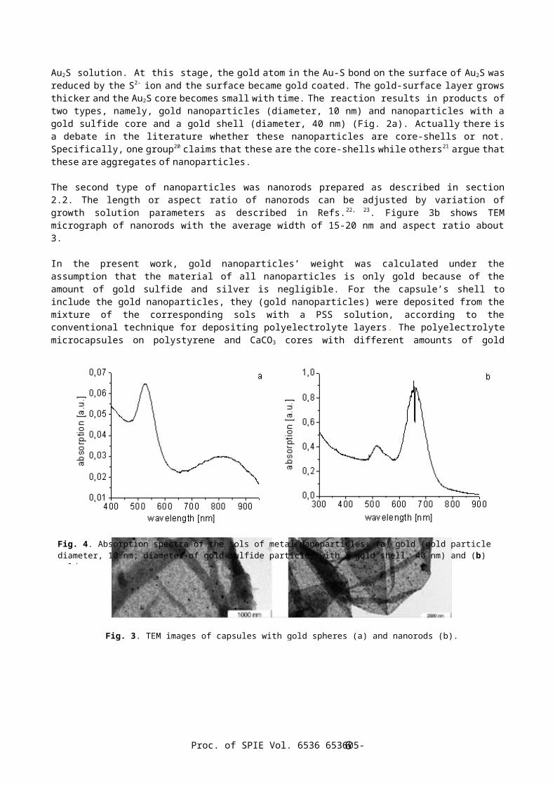
Fig. 4. Absorption spectra of the sols of metal nanoparticles: (a) gold (gold particle diameter, 10 nm; diameter of gold sulfide particles with a gold shell, 40 nm) and (b) gold nanorods

nanoparticles per capsule were obtained. The completeness of nanoparticleadsorption was controlled using UV–visible spectroscopy. The spectra of thesupernatants at all the concentrations used do not contain absorption peakscorresponding to the nanoparticles. This means that all nanoparticles werecompletely adsorbed by the capsule shells.
There are significant differences between gold distributions in capsules preparedon different templates (Fig. 4). For the polystyrene cores, the capsule’s coloris uniform; i.e. the gold nanoparticles are uniformly adsorbed over the entirecapsule surface (Fig. 4a). In the case of the CaCO3 cores, darker and lighterareas are observed on the surface (Fig. 4b). This is explained by the fact thatthe shell of these microcapsules is more loose and rough owing to the porousstructure of the CaCO3 spherolites14. Most likely, this phenomenon is responsiblefor the formation of shell regions, which metal nanoparticles are predominantlyadsorbed and form agglomerates.
A comparison of distributions of spherical nanoparticles and nanorods in thecapsule polymer matrix was made by TEM. It is shown that the nanoparticles ofboth types are uniformly distributed over capsule surface in the case ofpolystyrene cores (Fig. 5).
3.2 Effects of laser radiation
In order to analyze the influence of laser irradiation on the microcapsules whoseshells contain gold nanoparticles, we measured the absorption spectra of therelevant nanoparticles (Fig. 4). The spectrum of the spherical gold particles(Fig. 4a) exhibits two bands: the main band in the range 510–540 nm correspondingto the absorption by gold nanoparticles (~ 10 nm)and a broad peak at 830 nmattributed to the absorption by gold sulfide nanoparticles with the gold shell (~40 nm). As opposed to usual green 520-nm resonance of gold nanospheres in water,absorption spectra of nanorods have well-defined long wavelength resonance in thered and IR part of the spectrum. That peak depends on axis ratio of nanorods.(Fig. 4b) Since the wavelength of a green laser (532 nm) is close to the absorption peaksof both gold nanoparticles, it is this laser that can be used most efficientlyfor damaging the shells of the polyelectrolyte capsules. Actually, the exposureto laser radiation with a wavelength of 532 nm at a power of 100 mW leads to fulldamage of the capsules irrespective of the method used for incorporating metalnanoparticles into the capsule shell and of the type of nanoparticles.
Proc. of SPIE Vol. 6536 653605-7
However, in order to operate with biological objects, the wavelength range 700–1000 nm is most appropriate because the absorption by tissues, biologicallyactive compounds, and water in this range is minimum. In this respect, we usedthe IR laser with a controlled power of up to 90 mW and a wavelength of 830 nm.According to the UV spectroscopic data, although the laser wavelength is outsidethe main absorption peak corresponding to the nanoparticles, their absorptionfactor at this wavelength is rather large.
The capsules with shells contained silver, prepared through the silver mirrorreaction, are not subjected to damage under irradiation by an infrared laser at apower of 90 mW, even though the shell contains a large amount of metallic silver,as can be judged from the optical microscope images. Possibly, the dense layer ofsilver particles retains the capsule shape and prevents its damage.
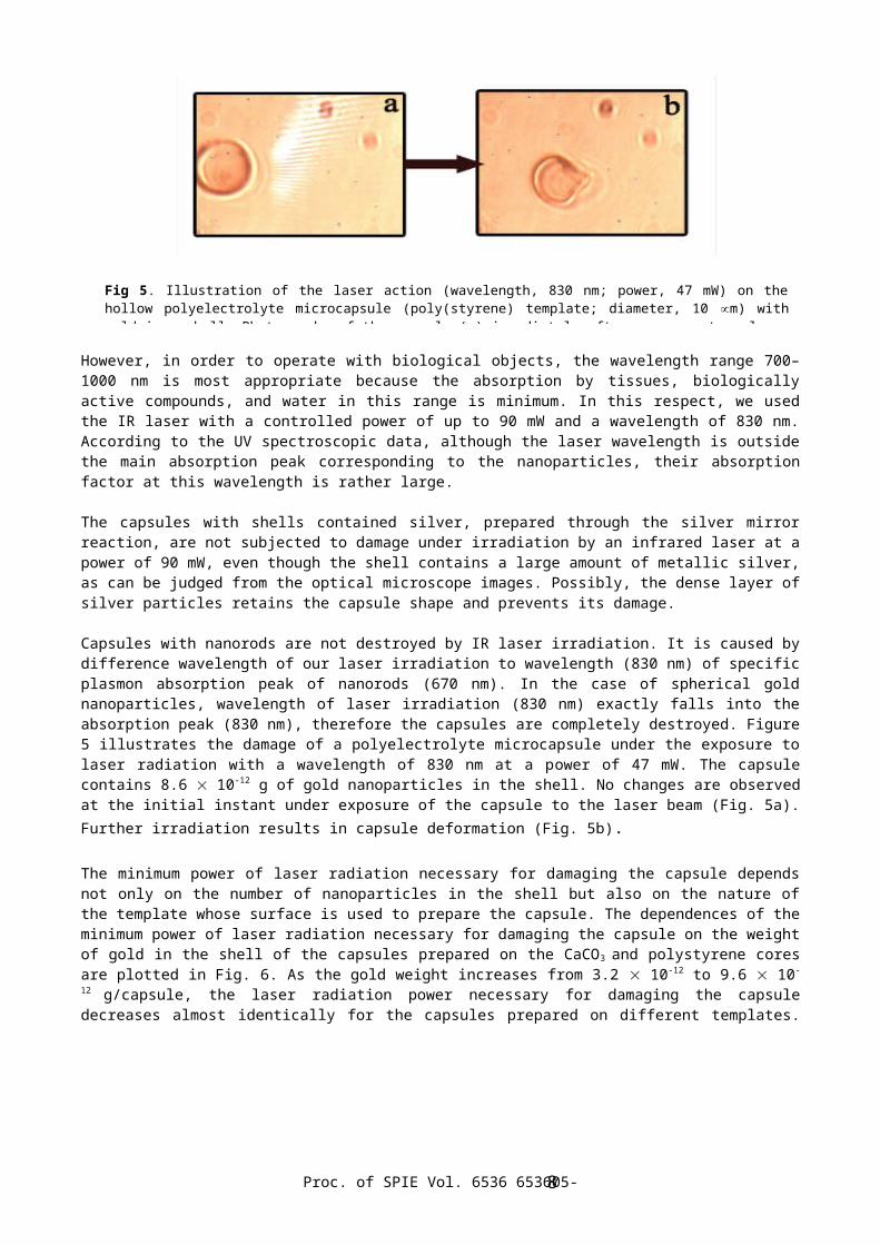
Capsules with nanorods are not destroyed by IR laser irradiation. It is caused bydifference wavelength of our laser irradiation to wavelength (830 nm) of specificplasmon absorption peak of nanorods (670 nm). In the case of spherical goldnanoparticles, wavelength of laser irradiation (830 nm) exactly falls into theabsorption peak (830 nm), therefore the capsules are completely destroyed. Figure5 illustrates the damage of a polyelectrolyte microcapsule under the exposure tolaser radiation with a wavelength of 830 nm at a power of 47 mW. The capsulecontains 8.6 10-12 g of gold nanoparticles in the shell. No changes are observedat the initial instant under exposure of the capsule to the laser beam (Fig. 5a).Further irradiation results in capsule deformation (Fig. 5b).
The minimum power of laser radiation necessary for damaging the capsule dependsnot only on the number of nanoparticles in the shell but also on the nature ofthe template whose surface is used to prepare the capsule. The dependences of theminimum power of laser radiation necessary for damaging the capsule on the weightof gold in the shell of the capsules prepared on the CaCO3 and polystyrene coresare plotted in Fig. 6. As the gold weight increases from 3.2 10-12 to 9.6 10-
12 g/capsule, the laser radiation power necessary for damaging the capsuledecreases almost identically for the capsules prepared on different templates.
Proc. of SPIE Vol. 6536 653605-8
Fig 5. Illustration of the laser action (wavelength, 830 nm; power, 47 mW) on thehollow polyelectrolyte microcapsule (poly(styrene) template; diameter, 10 m) withgold in a shell. Photographs of the capsule (a) immediately after exposure to a laser

Note that the sensitivity to laser radiation for the capsules produced on thepolystyrene cores is somewhat higher than the sensitivity of the capsulesprepared on the CaCO3 cores. Probably, this result is explained by thedifferences in the structure of the polyelectrolyte shells and in the capsulesize. With a further increase in the number of nanoparticles, the sensitivity tolaser radiation for the capsules prepared on the CaCO3 cores increases moresharply than the sensitivity of the capsules produced on the polystyrene cores.The number of nanoparticles in the shells prepared on the CaCO3 cores is veryhigh corresponding to the formation of gold agglomerates. Such a layout ischaracterized by a considerably stronger absorption as compared to that give byindividual nanoparticles.
4. CONCLUSIONS
It was revealed that the distribution of metal nanoparticles in shells ofpolyelectrolyte capsules depends on the template nature. All the proposed methodsfor incorporating silver and gold nanoparticles into the shell ensure the capsuledamage under irradiation by a green laser at a power of 100 mW, whose wavelength(532 nm) is close to the wavelength of the absorption peak of the nanoparticles.Unlike the green laser, an IR laser (with a power of up to 90 mW) suitable forapplications to biological objects damages capsules whose shells contain
Proc. of SPIE Vol. 6536 653605-9
Fig. 6. Dependences of the minimum power W of laser radiation necessary for damage of the capsule on the weight m of gold in the shell of the capsules prepared with different templates: (1) CaCO3 (particle diameter, 11.5 µm) and (2) poly(styrene) (particle diameter, 10 µm)

specially synthesized gold nanoparticles adsorbed from sol at specific amounts ofmetal per capsule. It was demonstrated that, in this case, a change in thesensitivity of capsules to laser radiation with an increase in the gold contentin the capsule walls depends substantially on the template nature. Thecomposition and structure of capsule shells play an important role in the damageof capsules or in a change in their permeability. For practically application isnecessary to reduce the power of laser, which is required to release. It can bedone by controlling of nanoparticle shape and nanoparticles distribution into theshell. Nanorods is most perspective of all types of particles, because they haveintensity peak in the long wavelength part of the spectrum (the so-calledbiologically-friendly area). Even if their concentration in shell is small,release was succeed by green laser irradiation.
ACKNOWLEDGMENTS
We would like to thank A. G. Susha (Photonics and Optoelectronics Group Physics Departmentand Center for Nanoscience (CeNS), Ludwig-Maximilian University of Munich, Munich, Germany) forsupplying the gold nanoparticles used in our experiments.
This study was supported in part by the Federal Agency for Science and Innovationof the Russian Federation (agreement no. 21/05 in the framework of the StateContract no. 02.435.11.2020), and by a grant from the Russian Foundation forBasic Research 05-02-16776a.
REFERENCES 1. K. M. Wasan, “Formulation and physiological and biopharmaceutical issues in
the development of oral lipid-based drug delivery systems,” Drug Dev. Ind. Pharm. 27, pp. 267-276, 2001.
2. A. Prokop, D. Hunkeler, A.C. Powers, R.R. Whitesell, and T.G. Wang, “Water soluble polymers for immunoisolation II: evaluation of multicomponent microencapsulation systems,” Adv. Polym. Sci. 136, pp. 53-57, 1998.
3. A. G. Skirtach, A. A. Antipov, D. G. Shchukin, and G. B. Sukhorukov, “Remote activation of capsules containing Ag nanoparticles and IR dye by laser light,”Langmuir, 20, pp. 6988-6992, 2004.
4. A. G. Skirtach, C. Dejugnat, D. Braun, A. S. Susha, A. L. Rogach, W. J. Parak,H. Molhwald, and G. B. Sukhorukov, “The role of metal nanoparticles in remote release of encapsulated materials,” Nano Lett. 5, pp. 1371-1377, 2005.
5. G. B. Sukhorukov, E. Donath, H. Lichtenfeld, M. Knippel, Knippel, A. Budde, and H. Möhwald, “Layer-by-layer self assembly of polyelectrolytes on colloidalparticles,” Colloids Surf., A 137, pp. 253-266, 1998.
6. E. Donath, G. B. Sukhorukov, F. Caruso, S. A. Davis, and H. Möhwald, “Novel hollow polymer shells by colloid-templated assembly of polyelectrolytes,” Angew. Chem., Int. Ed. 37, pp. 2202-2205, 1998.
7. G. B. Sukhorukov, E. Donath, S. A. Davis, H. Lichtenfeld, F. Caruso, V. I. Popov, and H. Möhwald, “Stepwise polyelectrolyte assembly on particle surfaces: a novel approach to colloid design,” Polym. Adv. Technol., 9, pp. 759-
Proc. of SPIE Vol. 6536 653605-10
767, 1998. 8. G. B. Sukhorukov, D. G. Shchukin, W. Dong, H. Möhwald, V.V. Lulevich, and O.I.
Vinogradova, “Comparative Analysis of Hollow and Filled Polyelectrolyte Microcapsules Templated on Melamine Formaldehyde and Carbonate Cores,” Macromol. Chem. Phys., 205, 530-535, 2004.
9. C. Dejugnat, and G. B. Sukhorukov, “pH-responsive properties of hollow polyelectrolyte microcapsules templated on various cores,” Langmuir, 20, pp. 7265-7269, 2004.
10. D. G. Shchukin and G. B. Sukhorukov, “Selective YF3 nanoparticle formation inpolyelectrolyte capsules as microcontainers for yttrium recovery from aqueous solutions,” Langmuir, 19, pp. 4427-4431, 2003.
11. A. Antipov, D. Shchukin, Y. Fedutik, I. Zanaveskina, V. Klechkovskaya, G. Sukhorukov, and H. Möhwald, “Urease-catalyzed carbonate precipitation inside the restricted volume of polyelectrolyte capsules,” Macromol. Chem. Phys., 24, pp.274-277, 2003.
12. D. G. Shchukin, W. Dong, and G. B. Sukhorukov, “Spatially confined tungstate ion polymerization in microcapsules,” Macromol. Chem. Phys., 24, pp. 462-466, 2003.
13. D. V. Volodkin, N. I. Larionova, and G. B. Sukhorukov, “Protein encapsulationvia porous CaCO3 microparticles templating,” Biomacromolecules, 5, pp. 1962-1972, 2004.
14. D. V. Volodkin, A. I. Petrov, M. Prevot, and G. B. Sukhorukov, “Matrix polyelectrolyte microcapsules: New system for macromolecule encapsulation,” Langmuir, 20, pp. 3398-3406, 2004.
15. H. S. Zhou, I. Honma, H. Komiyama, and J. W. Haus, “Controlled synthesis and quantum-size effect in gold-coated nanoparticles,” Phys. Rev. B: Condens. Matter 50, 12052-12056, 1994.
16. B. Nikoobakht and M. A. El-Sayed, “Preparation and Growth mechanism of gold nanorods (NRs) using seed-mediated growth method,” Chem. Mater., 15, pp. 1957-1962, 2003.
17. C. J. Murphy, T. K. Sau, A. M. Gole, C. J. Orendorff, J. Gao, L. Gou, S. E. Hunyadi, and T. Li, “Anisotropic metal nanoparticles: synthesis, assembly, andoptical applications,” J. Phys. Chem. B. 109, pp. 13857–13870, 2005.
18. A. V. Alekseeva, V. A. Bogatyrev, L. A. Dykman, B. N. Khlebtsov, L. A. Trachuk, A. G. Melnikov, and N. G. Khlebtsov, “Preparation and optical scattering characterization of gold nanorods and their application to a dot-immunogold assay,” Appl. Opt., 44, pp. 6285-6295, 2005
19. A. A. Antipov, G. B. Sukhorukov, Y. A. Fedutik, J. Hartmann, M. Giersig, and H. Möhwald, “Fabrication of a novel type of metallized colloids and hollow capsules,” Langmuir, 18, pp. 6687-6693, 2002.
20. S. J. Oldenburg, R. D. Averitt, S. L. Westcott, and N. J. Halas, “Nanoengineering of optical resonances,” Chem.Phys. Lett., 288, 243-247, 1998.
21. J. Z. Zhang, A. M. Schwartzberg, T. Jr. Norman, C. D. Grant, J. Liu, F. Bridges, and T. van Buuren, “Gold nanoshells improve single nanoparticle molecular sensors,” Nano Lett, 5, pp. 809-810, 2005
22. J. Pérez-Juste, I. Pastoriza-Santos, L. M. Liz-Marzán, and P. Mulvaney, “Gold
Proc. of SPIE Vol. 6536 653605-11

nanorods: Synthesis, characterization and applications,” Coordination Chem. Rev., 249, pp. 1870-1901, 2005.
23. A. V. Alekseeva, V. A. Bogatyrev, B. N. Khlebtsov, A. G. Mel'nikov, L. A. Dykman, and N. G. Khlebtsov, “Gold nanorods: synthesis and optical properties,” Colloid J. 68 (6) , pp. 661-678, 2006.
Proc. of SPIE Vol. 6536 653605-12
Related Documents


