Please cite this article in press as: C. Rodriguez-Emmenegger, et al., Polymeric nanocapsules ultra stable in complex biological media, Colloids Surf. B: Biointerfaces (2011), doi:10.1016/j.colsurfb.2010.12.013 ARTICLE IN PRESS G Model COLSUB-4376; No. of Pages 6 Colloids and Surfaces B: Biointerfaces xxx (2010) xxx–xxx Contents lists available at ScienceDirect Colloids and Surfaces B: Biointerfaces journal homepage: www.elsevier.com/locate/colsurfb Polymeric nanocapsules ultra stable in complex biological media C. Rodriguez-Emmenegger a,b,∗ , A. Jäger a , E. Jäger a , P. Stepanek a , A. Bologna Alles b , S.S. Guterres c , A.R. Pohlmann c,d , E. Brynda a a Institute of Macromolecular Chemistry, Academy of Sciences of the Czech Republic, v.v.i., Heyrovsky Sq. 2, 162 06, Prague, Czech Republic b College of Engineering, Universidad de la Republica, Julio Herrera y Reissig 565, PS 11300 Montevideo, Uruguay c Faculdade de Farmácia, Universidade Federal do Rio Grande do Sul, Av. Ipiranga, 2752, BR 90610-000, Porto Alegre, Brazil d Departamento de Química Organica, Instituto de Química, Universidade Federal do Rio Grande do Sul, Porto Alegre, Brazil article info Article history: Received 24 August 2010 Received in revised form 4 December 2010 Accepted 7 December 2010 Available online xxx Keywords: Core/shell nanocapsules Biodegradable Antifouling coating Surface initiated-ATRP Interaction with proteins abstract Non-specific protein adsorption from complex biological media, especially from blood plasma, is an urgent challenge for the application of nanoparticles as delivery systems, diagnostics, and other biomed- ical application. Nanocapsules (NC) prepared from FDA-approved degradable poly(-caprolactone) shell and Mygliol 812 ® oil in the core were coated with mono-methoxy terminated oligo(ethylene glycol) methacrylate (poly(MeOEGMA)) polymer brush layers with a well-controlled thickness at the nanome- ter scale up to 350 nm using surface initiated atom transfer radical polymerization in water or phosphate buffered saline. Incubation of uncoated NC with human serum albumin solution, fetal bovine serum, or human blood plasma resulted in fast aggregation observed by dynamic light scattering as an increase in diameter of particles present in the solutions. Conversely, these biological fluids affected only marginally the size distribution of the NC coated with a 60 nm thick poly(MeOEGMA) layer. The high suspension stability of the coated NC in complex biological fluids was related to the suppressed deposition of pro- teins from these fluids observed by surface plasmon resonance (SPR) on analogous poly(MeOEGMA) layer prepared on flat surfaces of SPR chips. © 2010 Elsevier B.V. All rights reserved. 1. Introduction A great deal of attention has been paid to the fabrication of nano-sized hollow polymeric particles due to their potential appli- cation in catalysis, separation, chromatography, diagnostics, drug delivery, and biomolecular-release systems [1–7]. Among them, self-assembled polymeric nanoparticles are ideal candidates for encapsulation of nonpolar guest molecules into their hydrophobic core [8–10]. A hydrolytically or enzymatically degradable polymer shell allows the guest molecules to be released in the surround- ing biological medium. There is only a limited number of polymers that can be used as constituent of nanoparticles designed for in vivo applications [11–13]. The material of the NC should be degradable to non-toxic low-molecular mass products that can be easily excreted from the organism, e.g. polylactide and poly- caprolactone [11–13]. Owing to their small sizes, nanoparticles are able to gain access to almost any tissue in the organism, and, if they are sufficiently small, to penetrate inside the cells [1,3,14]. In vivo biocompatibility of nanoparticles is mostly determined by ∗ Corresponding author at: Heyrovsky Sq. 2, 162 06, Prague, Czech Republic. Tel.: +420 296809234; fax: +420 296809410. E-mail addresses: [email protected], [email protected] (C. Rodriguez-Emmenegger). the biophysicochemical characteristics of their surfaces [3,5,15]. Upon contact with biological media, their surface properties are rapidly changed by coating with proteins [16–21]. Subsequent colloidal instability [20,21] of the nanoparticles (e.g. particle aggre- gation, flocculation, precipitation, etc.) or adsorption of undesirable proteins can impair the designed particle functions and initiate unfavorable biological responses. Many of the early stage biologi- cal responses are determined by the nature of the deposited protein layer. Adsorbed proteins can affect the interaction of nanoparticles with cells and their behavior in the blood stream. When admin- istrated intravenously, nanoparticles are rapidly cleared from the blood stream because they are recognized by cells of the mononu- clear phagocytic system (MPS) or by the complement system due to opsonization, i.e. the binding of antibodies and/or complement molecules [22–25]. Nearly nothing is known about interaction of nanoparticles with blood coagulation factors and platelets by which hemostasis can be perturbed. Generally, nanoparticles with sup- pressed protein adsorption are of great interest to most applications in complex biological media. Currently, nanoparticles decorated with different antifouling polymers are subject of intense research [9,26–30]. Polymer coated ultra-low fouling gold, silica, and iron oxide nanoparticles have been prepared for imaging, diagnostics, and sensing [31–35]. How- ever, their application as carriers of biologically active compounds is arguable [6,36,37]. Polymeric nanoparticles have been exten- 0927-7765/$ – see front matter © 2010 Elsevier B.V. All rights reserved. doi:10.1016/j.colsurfb.2010.12.013

Welcome message from author
This document is posted to help you gain knowledge. Please leave a comment to let me know what you think about it! Share it to your friends and learn new things together.
Transcript
G
C
P
CSa
b
c
d
a
ARRAA
KCBASI
1
ncdsecsitidbcatI
T
(
0d
ARTICLE IN PRESSModel
OLSUB-4376; No. of Pages 6
Colloids and Surfaces B: Biointerfaces xxx (2010) xxx–xxx
Contents lists available at ScienceDirect
Colloids and Surfaces B: Biointerfaces
journa l homepage: www.e lsev ier .com/ locate /co lsur fb
olymeric nanocapsules ultra stable in complex biological media
. Rodriguez-Emmeneggera,b,∗, A. Jägera, E. Jägera, P. Stepaneka, A. Bologna Allesb,.S. Guterresc, A.R. Pohlmannc,d, E. Bryndaa
Institute of Macromolecular Chemistry, Academy of Sciences of the Czech Republic, v.v.i., Heyrovsky Sq. 2, 162 06, Prague, Czech RepublicCollege of Engineering, Universidad de la Republica, Julio Herrera y Reissig 565, PS 11300 Montevideo, UruguayFaculdade de Farmácia, Universidade Federal do Rio Grande do Sul, Av. Ipiranga, 2752, BR 90610-000, Porto Alegre, BrazilDepartamento de Química Organica, Instituto de Química, Universidade Federal do Rio Grande do Sul, Porto Alegre, Brazil
r t i c l e i n f o
rticle history:eceived 24 August 2010eceived in revised form 4 December 2010ccepted 7 December 2010vailable online xxx
eywords:
a b s t r a c t
Non-specific protein adsorption from complex biological media, especially from blood plasma, is anurgent challenge for the application of nanoparticles as delivery systems, diagnostics, and other biomed-ical application. Nanocapsules (NC) prepared from FDA-approved degradable poly(�-caprolactone) shelland Mygliol 812® oil in the core were coated with mono-methoxy terminated oligo(ethylene glycol)methacrylate (poly(MeOEGMA)) polymer brush layers with a well-controlled thickness at the nanome-ter scale up to 350 nm using surface initiated atom transfer radical polymerization in water or phosphate
ore/shell nanocapsulesiodegradablentifouling coatingurface initiated-ATRPnteraction with proteins
buffered saline. Incubation of uncoated NC with human serum albumin solution, fetal bovine serum, orhuman blood plasma resulted in fast aggregation observed by dynamic light scattering as an increase indiameter of particles present in the solutions. Conversely, these biological fluids affected only marginallythe size distribution of the NC coated with a 60 nm thick poly(MeOEGMA) layer. The high suspensionstability of the coated NC in complex biological fluids was related to the suppressed deposition of pro-
serveof SP
teins from these fluids obprepared on flat surfaces
. Introduction
A great deal of attention has been paid to the fabrication ofano-sized hollow polymeric particles due to their potential appli-ation in catalysis, separation, chromatography, diagnostics, drugelivery, and biomolecular-release systems [1–7]. Among them,elf-assembled polymeric nanoparticles are ideal candidates forncapsulation of nonpolar guest molecules into their hydrophobicore [8–10]. A hydrolytically or enzymatically degradable polymerhell allows the guest molecules to be released in the surround-ng biological medium. There is only a limited number of polymershat can be used as constituent of nanoparticles designed forn vivo applications [11–13]. The material of the NC should beegradable to non-toxic low-molecular mass products that cane easily excreted from the organism, e.g. polylactide and poly-
Please cite this article in press as: C. Rodriguez-Emmenegger, et al., PolymSurf. B: Biointerfaces (2011), doi:10.1016/j.colsurfb.2010.12.013
aprolactone [11–13]. Owing to their small sizes, nanoparticles areble to gain access to almost any tissue in the organism, and, ifhey are sufficiently small, to penetrate inside the cells [1,3,14].n vivo biocompatibility of nanoparticles is mostly determined by
∗ Corresponding author at: Heyrovsky Sq. 2, 162 06, Prague, Czech Republic.el.: +420 296809234; fax: +420 296809410.
E-mail addresses: [email protected], [email protected]. Rodriguez-Emmenegger).
927-7765/$ – see front matter © 2010 Elsevier B.V. All rights reserved.oi:10.1016/j.colsurfb.2010.12.013
d by surface plasmon resonance (SPR) on analogous poly(MeOEGMA) layerR chips.
© 2010 Elsevier B.V. All rights reserved.
the biophysicochemical characteristics of their surfaces [3,5,15].Upon contact with biological media, their surface properties arerapidly changed by coating with proteins [16–21]. Subsequentcolloidal instability [20,21] of the nanoparticles (e.g. particle aggre-gation, flocculation, precipitation, etc.) or adsorption of undesirableproteins can impair the designed particle functions and initiateunfavorable biological responses. Many of the early stage biologi-cal responses are determined by the nature of the deposited proteinlayer. Adsorbed proteins can affect the interaction of nanoparticleswith cells and their behavior in the blood stream. When admin-istrated intravenously, nanoparticles are rapidly cleared from theblood stream because they are recognized by cells of the mononu-clear phagocytic system (MPS) or by the complement system dueto opsonization, i.e. the binding of antibodies and/or complementmolecules [22–25]. Nearly nothing is known about interaction ofnanoparticles with blood coagulation factors and platelets by whichhemostasis can be perturbed. Generally, nanoparticles with sup-pressed protein adsorption are of great interest to most applicationsin complex biological media.
Currently, nanoparticles decorated with different antifouling
eric nanocapsules ultra stable in complex biological media, Colloids
polymers are subject of intense research [9,26–30]. Polymer coatedultra-low fouling gold, silica, and iron oxide nanoparticles havebeen prepared for imaging, diagnostics, and sensing [31–35]. How-ever, their application as carriers of biologically active compoundsis arguable [6,36,37]. Polymeric nanoparticles have been exten-
ARTICLE IN PRESSG Model
COLSUB-4376; No. of Pages 6
2 C. Rodriguez-Emmenegger et al. / Colloids and Surfaces B: Biointerfaces xxx (2010) xxx–xxx
F EGMA
satawtsmtbsoifnc[todc
nootcmpssh
2
2
cmcadC
ig. 1. Synthesis of non-fouling nanocapsules. The surface initiated ATRP poly(MeO
ively studied as drug carriers [6,9,26,27,38]. Modifications aimingt increasing nanoparticle stability and prolonging their circula-ion in blood are of high importance for such systems [9]. Severaluthors have studied the interaction of polymeric nanoparticlesith model protein solutions [25,39–41], while fouling proper-
ies of nanocarriers in complex biological media have not beenufficiently addressed yet. There has been a common tendency toisinterpret an observed decrease in adsorption from single pro-
ein solutions as an evidence of non-fouling properties in complexiological media [42–44]. However, the reduced or even totallyuppressed non-specific adsorption from single protein solutionsf serum albumin, IgG, fibrinogen, and lysozyme observed on var-ous anti-fouling planar surfaces did not result in the suppressedouling from blood serum and plasma [16]. Anti-fouling polymericanoparticles have been mostly prepared from different PEG-ontaining polymers or by grafting PEG chains onto their surface28,45,46]. Recent studies on “PEGylated” nanoparticles indicatehat polymer architecture favoring higher surface concentrationsf PEG results in a better anti-fouling ability [46]. Generally, a highensity of attached polymers can be achieved by a grafting polymerhains from the surface.
Atom transfer radical polymerization initiated from theanoparticle surface is presented in this work as a novelrganic-solvent-free technique for coating nanoparticles in aque-us solutions with non-fouling polymer brushes. Using thisechnique nanocapsules from FDA-approved biodegradable poly(�-aprolactone) were coated under mild conditions with non-foulingono-methoxy terminated oligo(ethylene glycol) methacrylate
olymer brush layers with the thickness controlled at a nanometercale up to 350 nm (Fig. 1). The coating prevented the nanocap-ules from aggregation in albumin solution, fetal bovine serum, anduman blood plasma.
. Experimental
.1. Materials
All chemical reagents were used without further purifi-ation. Mono-methoxy terminated oligo(ethylene glycol)
Please cite this article in press as: C. Rodriguez-Emmenegger, et al., PolymSurf. B: Biointerfaces (2011), doi:10.1016/j.colsurfb.2010.12.013
ethacrylate, Mn 300 (MeOEGMA), �,� (hydroxyl) poly(�-aprolactone), Mw = 65,000 Da (PCL), Span 60®, �-bromo isobutyriccid, 98%, N-hydroxysuccinimide, 98%(NHS), N-ethyl-N′-(3-iethylaminopropyl) carbodiimide hydrochloride, 99% (EDC),uBr2 (99.999%), 2,2′-dipyridyl (99%), CuCl (99.995% trace metal
) shells resulted in particle diameters controlled within an interval of 170–900 nm.
basis), human serum albumin, 99% (HSA), fetal bovine serum,non-USA origin (FBS), pooled blood plasma (HBP), and phosphatebuffered saline (PBS) were purchased from Sigma–Aldrich. Ace-tone, Mygliol 812® oil, and cellulose membrane, 6,000–8,000 DaSpectra/Pore® were purchased from Lach-Ner, s.r.o., Sasol, andSpectrumLabs, respectively.
2.2. Preparation of PCL nanocapsules
Polymeric nanocapsules (NC) of �,� hydroxy poly(�-caprolactone) (PCL) containing Mygliol 812® in the core wereprepared by interfacial polymer deposition following solventdisplacement according to the methodology described elsewhere[47,48]. PCL, Mygliol® and Span 60® were dissolved in acetoneand subsequently injected over the aqueous phase. Acetone wasevaporated and the suspension was concentrated under reducedpressure.
Initiator decorated nanoparticles: A solution of �-bromo isobu-tyric acid, 0.15 M, N-hydroxysuccinimide, 0.05 M, and N-ethyl-N′-(3-diethylaminopropyl) carbodiimide hydrochloride, 0.2 M, inMilliQ water was kept for 7 min at 25 ◦C to activate the carboxylicgroups and then transferred to the NC suspension, 5.5 × 10⁹ mL−1
(determined by UV-turbidimetry) [47]. After stirring overnight,the solution was dialyzed for 12 h using a cellulose membraneSpectra/Pore® (6,000–8,000 Da). (All at room temperature.)
Modification of the NC surface with poly(MeOEGMA): A solution ofCuBr2 (8.1 mg, 36.4 �mol), 2,2′-dipyridyl (145 mg, 930 �mol), andMeOEGMA (5.7 g, 19 mmol) in 10 mL of water or PBS was degassedby Ar bubbling for 1 h. CuCl (37 mg, 375 �mol) was added under Aratmosphere and the Ar bubbling continued for 30 min. The solu-tion was added to 5 mL of the initiator decorated NC dispersionand the polymerization was allowed to proceed at 30 ◦C under Ar.One milliliter samples were collected after different polymerizationtimes. (All at room temperature)
Physicochemical characterization of the NCs: Size distributions ofthe NC and NC coated with poly(MeOEGMA) (NC@MeOEGMA) weredetermined by quasi elastic light scattering (QELS) at an angle of173◦ and at 25 ◦C using a Zetasizer® ZS (Nanoseries, Malvern, UK).Mean size distribution values were obtained from 3 independent
eric nanocapsules ultra stable in complex biological media, Colloids
measurements. Measurements of the �-potential in MilliQ waterwere performed with the same instrument. Ten measurementswere carried out to check the reproducibility. The measurementsof the electrophoretic mobility were converted to �-potential (mV)using the Smoluchowski approximation. Transmission electron
IN PRESSG
C
nd Surfaces B: Biointerfaces xxx (2010) xxx–xxx 3
m(ma
petsaitcfs
uswlotalrtrlf
btttuonFisopfl��metTltnsiab
AoteaTwca
ARTICLEModel
OLSUB-4376; No. of Pages 6
C. Rodriguez-Emmenegger et al. / Colloids a
icroscopy (TEM) was carried out using a microscope JEM200CXJeol, Japan). The NCs were deposited onto a copper TEM grid (300
esh) coated with a carbon film. The images were taken at ancceleration voltage of 100 kV and recorded with a digital camera.
Preparation of model planar surfaces: Silicone wafers or glasslates were coated by gold films deposited in vacuum forllipsometry or surface plasmon resonance measurements, respec-ively. PCL was spin-coated on the gold surface from chloroformolution. Poly(MeOEGMA) brushes were grafted from a self-ssembled monolayer of �-mercapto undecylbromoisobutyratenitiator attached to the gold surface using surface initiated atomransfer radical polymerization (ATRP). The polymerization wasarried out at 30 ◦C for 40 min in the same way as described aboveor the NC. Thickness of both the poly(MeOEGMA) layer and thepin-coated PCL film was 40 nm as determined by ellipsometry.
Spectroscopic ellipsometry: The measurements were performedsing Variable Angle Spectroscopic Imaging Auto-Nulling Ellip-ometer EP3-SE (Nanofilm Technologies GmbH, Germany) in theavelength range of � = 398.9–811 nm (source Xe-arc lamp, wave-
ength step ∼10 nm). The measurements were performed at anglef incidence AOI = 70◦ in air at room temperature. For assessinghe uniformity of the modifications, all samples were measuredt three points. The obtained SE spectra were fitted with multi-ayer models in the EP3-SE analysis software. The thickness andefractive indexes of polymer layers were obtained from simul-aneous fitting of the obtained ellipsometric data using Cauchyelationship model. Due to low penetration depth of light, the Auayer was modeled as bulk gold using predefined EP3-SE dispersionunction.
Surface plasmon resonance (SPR): A custom-built SPR instrumentased on the Kretschmann geometry of the attenuated total reflec-ion method and spectral interrogation of the SPR conditions fromhe Institute of Photonics and Electronics, Academy of Sciences ofhe Czech Republic was used. SPR chips were prepared by vac-um deposition of a gold layer (thickness approximately 50 nm)nto glass slide coated with an adhesion titanium layer (thick-ess approximately 2 nm). The tested solutions (HSA, 5 mg mL−1,BS, 10 and 100%) were driven by a peristaltic pump through fourndependent channels of a flow cell in which SPR responses wereimultaneously measured. The non-specific protein deposition wasbserved as a shift in the resonant wavelength, ��res. Irreversiblerotein deposition after 15 min of incubation with the biologicaluid was determined from the difference in resonant wavelength,�res, measured in PBS before and after the incubation. A shift�re = 1 nm detected by SPR was related to a deposited proteinass of 200 pg mm−2. The relationship was estimated by model
xperiments in which HSA adsorption was observed using SPR andhe deposited HSA mass was determined by FTIR GASR (see below).he same correlation was used for other proteins assuming a simi-ar refractive index. The limit of the SPR detection defined as a threeimes the sensor response of the standard deviation of the baselineoise was determined to be ��res = 0.03 nm, which roughly corre-ponded to 6 pg of deposited proteins per mm2. The protein massn a deposit was estimated by comparing ��res with that of thedsorbed HSA monolayer used for FTIR GASR calibration describedelow.
FTIR grazing angle specular reflectance (FTIR GASR) calibration:calibration curve was obtained by spreading various amounts
f HSA dissolved in water over gold surface of SPR chips, dryinghem and measuring their FTIR GASR spectra with a FTIR spectrom-ter Bruker IFS 55 equipped with Pike Technologies 80Spec GASR
Please cite this article in press as: C. Rodriguez-Emmenegger, et al., PolymSurf. B: Biointerfaces (2011), doi:10.1016/j.colsurfb.2010.12.013
ttachment. HSA deposition was measured as the ��re in SPR chips.he mass of HSA adsorbed on these SPR chips after the incubationith HSA solution, washed with PBS, and dried was determined by
omparing FTIR GASR spectra with the calibration curve mentionedbove.
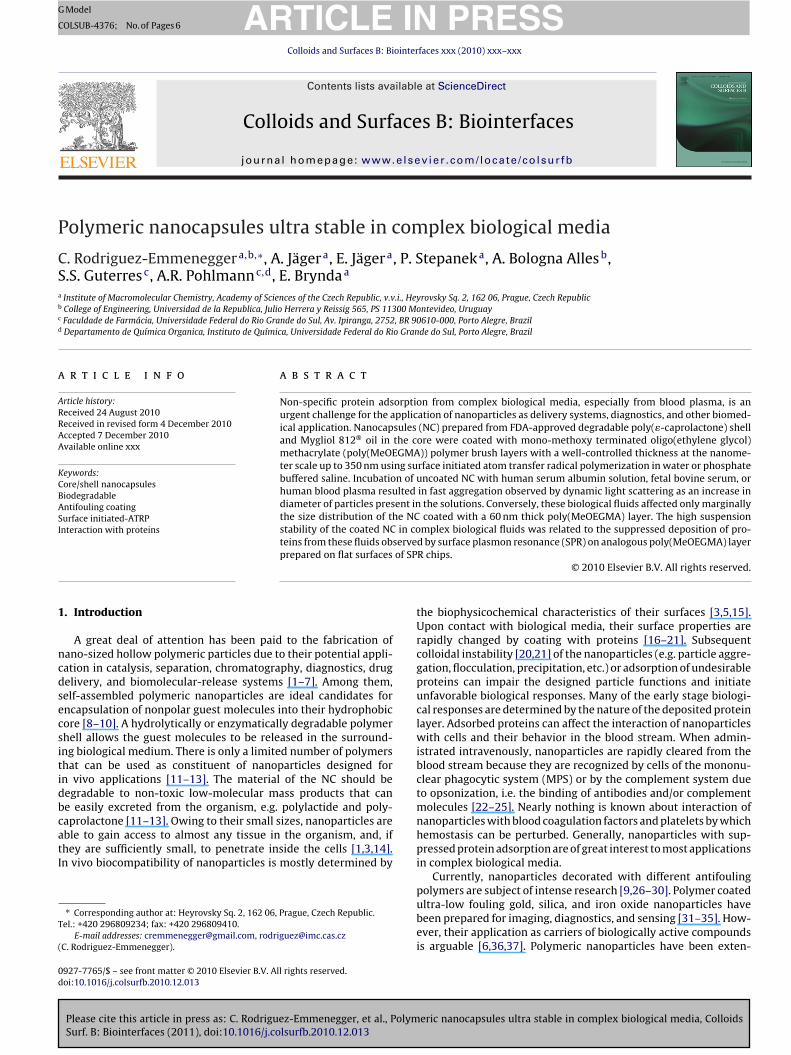
Fig. 2. Kinetics of the surface-initiated ATRP of MeOEGMA brush layers on poly(PCL)nanocapsules in water (triangles) and PBS (circles). The layer thickness was deter-mined by QELS.
3. Results and discussion
Diameters of about 170 nm determined from the particle sizedistribution measured by QELS and �-potential of −23.3 ± 0.5 mVwere characteristics of �,� (hydroxy) poly(�-caprolactone)nanocapsules prepared by interfacial polymer deposition. Thevalue of �-potential corresponds with those reported earlier [27].The nanoparticles were stable in aqueous solutions probably dueto the negative �-potential caused by charged carboxy-terminus ofPCL chains exposed at NC surface. Hydroxyl groups of PCL presentat the NC surface [49] made it possible to attach �-bromo isobutyricacid initiator activated by EDC/NHS. A shift in the �-potential to theless negative value of −13.6 ± 0.9 mV suggested that also some car-boxyl groups on the NC surface took part in the initiator attachment.The MeOEGMA was grafted from the NC in water or PBS using ATRPinitiated from �-bromoisobutyrate anchored to the NC surface. Thegrowth of polymer brushes resulted in the increase in NC diame-ter observed by QELS. The size distributions of particles present inthe samples collected at different polymerization times were deter-mined by QELS. The thickness of the polymer layer growing on theNC surface was estimated by subtracting the mean radius of 85 nmof the initial NC calculated for the initial size distribution from themean radius of the polymer coated NC calculated for the size dis-tribution measured at the selected polymerization time. A linearincrease in the polymer layer thickness with time (Fig. 2) indicatedthat the surface initiated ATRP of MeOEGMA was well-controlled.A lower rate of polymerization was observed when the solvent wasPBS as expected. ATRP uses a catalytic amount of a transition metalcomplex, which reversibly abstracts a halogen atom from a polymerchain end, thereby transforming the latter into an active propagat-ing radical from a dormant state. The high concentration of theextraneously added NaCl present in the buffer resulted in halogenexchange favoring the dormant chains capped with chlorine overthose with bromine [50,51]. Due to the higher bond energy C–Clthan C–Br the equilibrium was shifted toward the dormant chainsand consequently resulted in a drop in the rate of polymerization[50].
The observed shift in �-potential to less negative values withincreasing polymerization time could be explained by the differ-ence between the static dielectric permittivity of the hydrated
eric nanocapsules ultra stable in complex biological media, Colloids
brushes and of the ambient aqueous solution, as stated by the Coehnand Raydt phenomenological rule [52] Coated NC characterized by�-potential of −6.5 ± 0.5 mV and a mean diameter of 290 nm, i.e. NCcoated with a polymer layer thick 60 nm, were chosen for testing
ARTICLE IN PRESSG Model
COLSUB-4376; No. of Pages 6
4 C. Rodriguez-Emmenegger et al. / Colloids and Surfaces B: Biointerfaces xxx (2010) xxx–xxx
FPs
ipr�ptpn1c
wthnstopoooNl3
pp
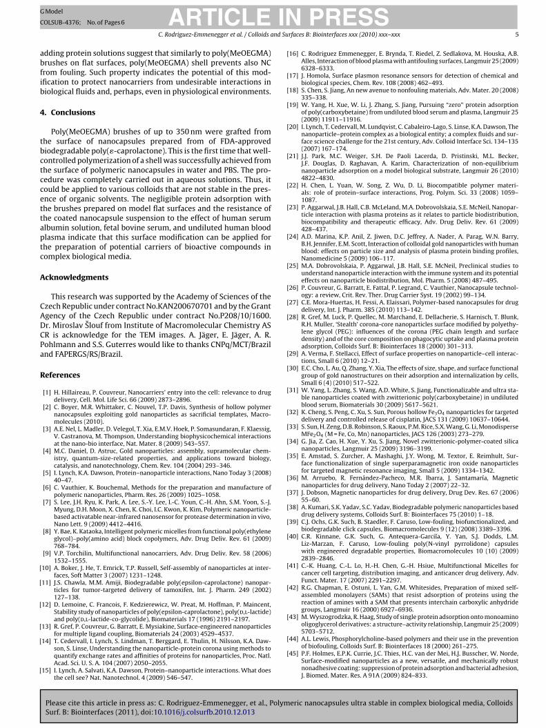
Fig. 4. Interaction of NC@MeOEGMA (a) and uncoated NC (b) with solution of humanserum albumin (HSA), fetal bovine serum (FBS), and undiluted human blood plasma
gold nanoparticles due to their aggregation mediated by adsorp-tion from single protein solutions of HSA, and other common bloodplasma proteins was described and discussed by Lacerda et al. [54].Marginal changes in the size distribution of NC@MeOEGMA after
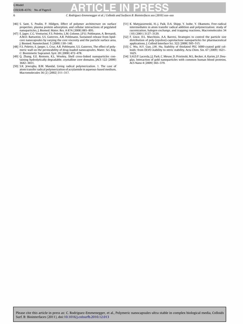
Fig. 5. Protein deposits irreversibly adsorbed from HSA solution and fetal bovine
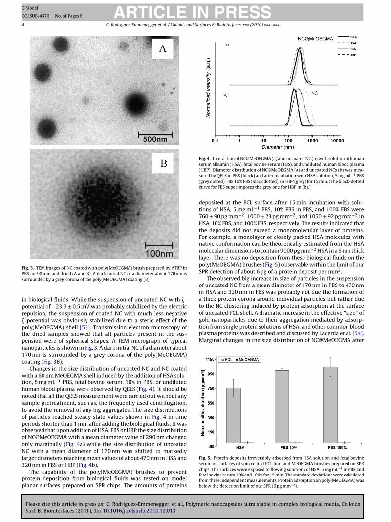
ig. 3. TEM images of NC coated with poly(MeOEGMA) brush prepared by ATRP inBS for 90 min and dried (A and B). A dark initial NC of a diameter about 170 nm isurrounded by a grey corona of the poly(MeOEGMA) coating (B).
n biological fluids. While the suspension of uncoated NC with �-otential of −23.3 ± 0.5 mV was probably stabilized by the electricepulsion, the suspension of coated NC with much less negative-potential was obviously stabilized due to a steric effect of theoly(MeOEGMA) shell [53]. Transmission electron microscopy ofhe dried samples showed that all particles present in the sus-ension were of spherical shapes. A TEM micrograph of typicalanoparticles is shown in Fig. 3. A dark initial NC of a diameter about70 nm is surrounded by a grey corona of the poly(MeOEGMA)oating (Fig. 3B).
Changes in the size distribution of uncoated NC and NC coatedith a 60 nm MeOEGMA shell induced by the addition of HSA solu-
ion, 5 mg mL−1 PBS, fetal bovine serum, 10% in PBS, or undiluteduman blood plasma were observed by QELS (Fig. 4). It should beoted that all the QELS measurement were carried out without anyample pretreatment, such as, the frequently used centrifugation,o avoid the removal of any big aggregates. The size distributionsf particles reached steady state values shown in Fig. 4 in timeeriods shorter than 1 min after adding the biological fluids. It wasbserved that upon addition of HSA, FBS or HBP the size distributionf NC@MeOEGMA with a mean diameter value of 290 nm changednly marginally (Fig. 4a) while the size distribution of uncoatedC with a mean diameter of 170 nm was shifted to markedly
arger diameters reaching mean values of about 470 nm in HSA and
Please cite this article in press as: C. Rodriguez-Emmenegger, et al., PolymSurf. B: Biointerfaces (2011), doi:10.1016/j.colsurfb.2010.12.013
20 nm in FBS or HBP (Fig. 4b).The capability of the poly(MeOEGMA) brushes to prevent
rotein deposition from biological fluids was tested on modellanar surfaces prepared on SPR chips. The amounts of proteins
(HBP). Diameter distribution of NC@MeOEGMA (a) and uncoated NCs (b) was mea-sured by QELS in PBS (black) and after incubation with HSA solution, 5 mg mL−1 PBS(grey dotted), FBS 10% PBS (black dotted), or HBP (grey) for 15 min. (The black-dottedcurve for FBS superimposes the grey one for HBP in (b).)
deposited at the PCL surface after 15 min incubation with solu-tions of HSA, 5 mg mL−1 PBS, 10% FBS in PBS, and 100% FBS were760 ± 90 pg mm−2, 1000 ± 23 pg mm−2, and 1050 ± 92 pg mm−2 inHSA, 10% FBS, and 100% FBS, respectively. The results indicated thatthe deposits did not exceed a monomolecular layer of proteins.For example, a monolayer of closely packed HSA molecules withnative conformation can be theoretically estimated from the HSAmolecular dimensions to contain 9000 pg mm−2 HSA in a 6 nm thicklayer. There was no deposition from these biological fluids on thepoly(MeOEGMA) brushes (Fig. 5) observable within the limit of ourSPR detection of about 6 pg of a protein deposit per mm2.
The observed big increase in size of particles in the suspensionof uncoated NC from a mean diameter of 170 nm in PBS to 470 nmin HSA and 320 nm in FBS was probably not due the formation ofa thick protein corona around individual particles but rather dueto the NC clustering induced by protein adsorption at the surfaceof uncoated PCL shell. A dramatic increase in the effective “size” of
eric nanocapsules ultra stable in complex biological media, Colloids
serum on surfaces of spin coated PCL film and MeOEGMA brushes prepared on SPRchips. The surfaces were exposed to flowing solutions of HSA, 5 mg mL−1 in PBS andfetal bovine serum 10% and 100% for 15 min. The standard deviations were calculatedfrom three independent measurements. Protein adsorption on poly(MeOEGMA) wasbelow the detection limit of our SPR (6 pg mm−2).
ING
C
nd Su
abfib
4
tbctccettaptc
A
CADCPa
R
[
[
[
[
[
[
[
[
[
[
[
[
[
[
[
[
[
[
[
[
[
[
[
[
[
[
[
[
[
[
[
[
[
[
ARTICLEModel
OLSUB-4376; No. of Pages 6
C. Rodriguez-Emmenegger et al. / Colloids a
dding protein solutions suggest that similarly to poly(MeOEGMA)rushes on flat surfaces, poly(MeOEGMA) shell prevents also NCrom fouling. Such property indicates the potential of this mod-fication to protect nanocarriers from undesirable interactions iniological fluids and, perhaps, even in physiological environments.
. Conclusions
Poly(MeOEGMA) brushes of up to 350 nm were grafted fromhe surface of nanocapsules prepared from of FDA-approvediodegradable poly(�-caprolactone). This is the first time that well-ontrolled polymerization of a shell was successfully achieved fromhe surface of polymeric nanocapsules in water and PBS. The pro-edure was completely carried out in aqueous solutions. Thus, itould be applied to various colloids that are not stable in the pres-nce of organic solvents. The negligible protein adsorption withhe brushes prepared on model flat surfaces and the resistance ofhe coated nanocapsule suspension to the effect of human serumlbumin solution, fetal bovine serum, and undiluted human bloodlasma indicate that this surface modification can be applied forhe preparation of potential carriers of bioactive compounds inomplex biological media.
cknowledgments
This research was supported by the Academy of Sciences of thezech Republic under contract No.KAN200670701 and by the Grantgency of the Czech Republic under contract No.P208/10/1600.r. Miroslav Slouf from Institute of Macromolecular Chemistry ASR is acknowledge for the TEM images. A. Jäger, E. Jäger, A. R.ohlmann and S.S. Guterres would like to thanks CNPq/MCT/Brazilnd FAPERGS/RS/Brazil.
eferences
[1] H. Hillaireau, P. Couvreur, Nanocarriers’ entry into the cell: relevance to drugdelivery, Cell. Mol. Life Sci. 66 (2009) 2873–2896.
[2] C. Boyer, M.R. Whittaker, C. Nouvel, T.P. Davis, Synthesis of hollow polymernanocapsules exploiting gold nanoparticles as sacrificial templates, Macro-molecules (2010).
[3] A.E. Nel, L. Madler, D. Velegol, T. Xia, E.M.V. Hoek, P. Somasundaran, F. Klaessig,V. Castranova, M. Thompson, Understanding biophysicochemical interactionsat the nano-bio interface, Nat. Mater. 8 (2009) 543–557.
[4] M.C. Daniel, D. Astruc, Gold nanoparticles: assembly, supramolecular chem-istry, quantum-size-related properties, and applications toward biology,catalysis, and nanotechnology, Chem. Rev. 104 (2004) 293–346.
[5] I. Lynch, K.A. Dawson, Protein–nanoparticle interactions, Nano Today 3 (2008)40–47.
[6] C. Vauthier, K. Bouchemal, Methods for the preparation and manufacture ofpolymeric nanoparticles, Pharm. Res. 26 (2009) 1025–1058.
[7] S. Lee, J.H. Ryu, K. Park, A. Lee, S.-Y. Lee, I.-C. Youn, C.-H. Ahn, S.M. Yoon, S.-J.Myung, D.H. Moon, X. Chen, K. Choi, I.C. Kwon, K. Kim, Polymeric nanoparticle-based activatable near-infrared nanosensor for protease determination in vivo,Nano Lett. 9 (2009) 4412–4416.
[8] Y. Bae, K. Kataoka, Intelligent polymeric micelles from functional poly(ethyleneglycol)–poly(amino acid) block copolymers, Adv. Drug Deliv. Rev. 61 (2009)768–784.
[9] V.P. Torchilin, Multifunctional nanocarriers, Adv. Drug Deliv. Rev. 58 (2006)1532–1555.
10] A. Boker, J. He, T. Emrick, T.P. Russell, Self-assembly of nanoparticles at inter-faces, Soft Matter 3 (2007) 1231–1248.
11] J.S. Chawla, M.M. Amiji, Biodegradable poly(epsilon-caprolactone) nanopar-ticles for tumor-targeted delivery of tamoxifen, Int. J. Pharm. 249 (2002)127–138.
12] D. Lemoine, C. Francois, F. Kedzierewicz, W. Preat, M. Hoffman, P. Maincent,Stability study of nanoparticles of poly(epsilon-caprolactone), poly(d,l-lactide)and poly(d,l-lactide-co-glycolide), Biomaterials 17 (1996) 2191–2197.
13] R. Gref, P. Couvreur, G. Barratt, E. Mysiakine, Surface-engineered nanoparticlesfor multiple ligand coupling, Biomaterials 24 (2003) 4529–4537.
Please cite this article in press as: C. Rodriguez-Emmenegger, et al., PolymSurf. B: Biointerfaces (2011), doi:10.1016/j.colsurfb.2010.12.013
14] T. Cedervall, I. Lynch, S. Lindman, T. Berggard, E. Thulin, H. Nilsson, K.A. Daw-son, S. Linse, Understanding the nanoparticle-protein corona using methods toquantify exchange rates and affinities of proteins for nanoparticles, Proc. Natl.Acad. Sci. U. S. A. 104 (2007) 2050–2055.
15] I. Lynch, A. Salvati, K.A. Dawson, Protein–nanoparticle interactions. What doesthe cell see? Nat. Nanotechnol. 4 (2009) 546–547.
[
[
PRESSrfaces B: Biointerfaces xxx (2010) xxx–xxx 5
16] C. Rodriguez Emmenegger, E. Brynda, T. Riedel, Z. Sedlakova, M. Houska, A.B.Alles, Interaction of blood plasma with antifouling surfaces, Langmuir 25 (2009)6328–6333.
17] J. Homola, Surface plasmon resonance sensors for detection of chemical andbiological species, Chem. Rev. 108 (2008) 462–493.
18] S. Chen, S. Jiang, An new avenue to nonfouling materials, Adv. Mater. 20 (2008)335–338.
19] W. Yang, H. Xue, W. Li, J. Zhang, S. Jiang, Pursuing “zero” protein adsorptionof poly(carboxybetaine) from undiluted blood serum and plasma, Langmuir 25(2009) 11911–11916.
20] I. Lynch, T. Cedervall, M. Lundqvist, C. Cabaleiro-Lago, S. Linse, K.A. Dawson, Thenanoparticle–protein complex as a biological entity; a complex fluids and sur-face science challenge for the 21st century, Adv. Colloid Interface Sci. 134–135(2007) 167–174.
21] J.J. Park, M.C. Weiger, S.H. De Paoli Lacerda, D. Pristinski, M.L. Becker,J.F. Douglas, D. Raghavan, A. Karim, Characterization of non-equilibriumnanoparticle adsorption on a model biological substrate, Langmuir 26 (2010)4822–4830.
22] H. Chen, L. Yuan, W. Song, Z. Wu, D. Li, Biocompatible polymer materi-als: role of protein–surface interactions, Prog. Polym. Sci. 33 (2008) 1059–1087.
23] P. Aggarwal, J.B. Hall, C.B. McLeland, M.A. Dobrovolskaia, S.E. McNeil, Nanopar-ticle interaction with plasma proteins as it relates to particle biodistribution,biocompatibility and therapeutic efficacy, Adv. Drug Deliv. Rev. 61 (2009)428–437.
24] A.D. Marina, K.P. Anil, Z. Jiwen, D.C. Jeffrey, A. Nader, A. Parag, W.N. Barry,B.H. Jennifer, E.M. Scott, Interaction of colloidal gold nanoparticles with humanblood: effects on particle size and analysis of plasma protein binding profiles,Nanomedicine 5 (2009) 106–117.
25] M.A. Dobrovolskaia, P. Aggarwal, J.B. Hall, S.E. McNeil, Preclinical studies tounderstand nanoparticle interaction with the immune system and its potentialeffects on nanoparticle biodistribution, Mol. Pharm. 5 (2008) 487–495.
26] P. Couvreur, G. Barratt, E. Fattal, P. Legrand, C. Vauthier, Nanocapsule technol-ogy: a review, Crit. Rev. Ther. Drug Carrier Syst. 19 (2002) 99–134.
27] C.E. Mora-Huertas, H. Fessi, A. Elaissari, Polymer-based nanocapsules for drugdelivery, Int. J. Pharm. 385 (2010) 113–142.
28] R. Gref, M. Luck, P. Quellec, M. Marchand, E. Dellacherie, S. Harnisch, T. Blunk,R.H. Muller, ‘Stealth’ corona-core nanoparticles surface modified by polyethy-lene glycol (PEG): influences of the corona (PEG chain length and surfacedensity) and of the core composition on phagocytic uptake and plasma proteinadsorption, Colloids Surf. B: Biointerfaces 18 (2000) 301–313.
29] A. Verma, F. Stellacci, Effect of surface properties on nanoparticle–cell interac-tions, Small 6 (2010) 12–21.
30] E.C. Cho, L. Au, Q. Zhang, Y. Xia, The effects of size, shape, and surface functionalgroup of gold nanostructures on their adsorption and internalization by cells,Small 6 (4) (2010) 517–522.
31] W. Yang, L. Zhang, S. Wang, A.D. White, S. Jiang, Functionalizable and ultra sta-ble nanoparticles coated with zwitterionic poly(carboxybetaine) in undilutedblood serum, Biomaterials 30 (2009) 5617–5621.
32] K. Cheng, S. Peng, C. Xu, S. Sun, Porous hollow Fe3O4 nanoparticles for targeteddelivery and controlled release of cisplatin, JACS 131 (2009) 10637–10644.
33] S. Sun, H. Zeng, D.B. Robinson, S. Raoux, P.M. Rice, S.X. Wang, G. Li, MonodisperseMFe2O4 (M = Fe, Co, Mn) nanoparticles, JACS 126 (2003) 273–279.
34] G. Jia, Z. Cao, H. Xue, Y. Xu, S. Jiang, Novel zwitterionic-polymer-coated silicananoparticles, Langmuir 25 (2009) 3196–3199.
35] E. Amstad, S. Zurcher, A. Mashaghi, J.Y. Wong, M. Textor, E. Reimhult, Sur-face functionalization of single superparamagnetic iron oxide nanoparticlesfor targeted magnetic resonance imaging, Small 5 (2009) 1334–1342.
36] M. Arruebo, R. Fernández-Pacheco, M.R. Ibarra, J. Santamaría, Magneticnanoparticles for drug delivery, Nano Today 2 (2007) 22–32.
37] J. Dobson, Magnetic nanoparticles for drug delivery, Drug Dev. Res. 67 (2006)55–60.
38] A. Kumari, S.K. Yadav, S.C. Yadav, Biodegradable polymeric nanoparticles baseddrug delivery systems, Colloids Surf. B: Biointerfaces 75 (2010) 1–18.
39] C.J. Ochs, G.K. Such, B. Staedler, F. Caruso, Low-fouling, biofunctionalized, andbiodegradable click capsules, Biomacromolecules 9 (12) (2008) 3389–3396.
40] C.R. Kinnane, G.K. Such, G. Antequera-Garciıa, Y. Yan, S.J. Dodds, L.M.Liz-Marzan, F. Caruso, Low-fouling poly(N-vinyl pyrrolidone) capsuleswith engineered degradable properties, Biomacromolecules 10 (10) (2009)2839–2846.
41] C.-K. Huang, C.-L. Lo, H.-H. Chen, G.-H. Hsiue, Multifunctional Micelles forcancer cell targeting, distribution imaging, and anticancer drug delivery, Adv.Funct. Mater. 17 (2007) 2291–2297.
42] R.G. Chapman, E. Ostuni, L. Yan, G.M. Whitesides, Preparation of mixed self-assembled monolayers (SAMs) that resist adsorption of proteins using thereaction of amines with a SAM that presents interchain carboxylic anhydridegroups, Langmuir 16 (2000) 6927–6936.
43] M. Wyszogrodzka, R. Haag, Study of single protein adsorption onto monoaminooligoglycerol derivatives: a structure–activity relationship, Langmuir 25 (2009)5703–5712.
eric nanocapsules ultra stable in complex biological media, Colloids
44] A.L. Lewis, Phosphorylcholine-based polymers and their use in the preventionof biofouling, Colloids Surf. B: Biointerfaces 18 (2000) 261–275.
45] P.F. Holmes, E.P.K. Currie, J.C. Thies, H.C. van der Mei, H.J. Busscher, W. Norde,Surface-modified nanoparticles as a new, versatile, and mechanically robustnonadhesive coating: suppression of protein adsorption and bacterial adhesion,J. Biomed. Mater. Res. A 91A (2009) 824–833.
ING
C
6 nd Su
[
[
[
[
[
[
[
[53] C. Wu, H.Y. Guo, J.W. Hu, Stability of thiolated PEG 5000-coated gold col-loids: from DLVO stability to steric stability, Acta Chim. Sin. 67 (2009) 1621–1625.
ARTICLEModel
OLSUB-4376; No. of Pages 6
C. Rodriguez-Emmenegger et al. / Colloids a
46] S. Sant, S. Poulin, P. Hildgen, Effect of polymer architecture on surfaceproperties, plasma protein adsorption, and cellular interactions of pegylatednanoparticles, J. Biomed. Mater. Res. A 87A (2008) 885–895.
47] E. Jager, C.G. Venturini, F.S. Poletto, L.M. Colome, J.P.U. Pohlmann, A. Bernardi,A.M.O. Battastini, S.S. Guterres, A.R. Pohlmann, Sustained release from lipid-core nanocapsules by varying the core viscosity and the particle surface area,J. Biomed. Nanotechnol. 5 (2009) 130–140.
48] F.S. Poletto, E. Jaeger, L. Cruz, A.R. Pohlmann, S.S. Guterres, The effect of poly-meric wall on the permeability of drug-loaded nanocapsules, Mater. Sci. Eng.C: Biomimetic Supramol. Syst. 28 (2008) 472–478.
Please cite this article in press as: C. Rodriguez-Emmenegger, et al., PolymSurf. B: Biointerfaces (2011), doi:10.1016/j.colsurfb.2010.12.013
49] Q. Zhang, E.E. Remsen, K.L. Wooley, Shell cross-linked nanoparticles con-taining hydrolytically degradable, crystalline core domains, JACS 122 (2000)3642–3651.
50] S.K. Jewrajka, B.M. Mandal, Living radical polymerization. 1. The case ofatom transfer radical polymerization of acrylamide in aqueous-based medium,Macromolecules 36 (2) (2002) 311–317.
[
PRESSrfaces B: Biointerfaces xxx (2010) xxx–xxx
51] K. Matyjaszewski, H.-j. Paik, D.A. Shipp, Y. Isobe, Y. Okamoto, Free-radicalintermediates in atom transfer radical addition and polymerization: study ofracemization, halogen exchange, and trapping reactions, Macromolecules 34(10) (2001) 3127–3129.
52] F. Lince, D.L. Marchisio, A.A. Barresi, Strategies to control the particle sizedistribution of poly-[epsilon]-caprolactone nanoparticles for pharmaceuticalapplications, J. Colloid Interface Sci. 322 (2008) 505–515.
eric nanocapsules ultra stable in complex biological media, Colloids
54] S.H.D.P. Lacerda, J.J. Park, C. Meuse, D. Pristinski, M.L. Becker, A. Karim, J.F. Dou-glas, Interaction of gold nanoparticles with common human blood proteins,ACS Nano 4 (2009) 365–379.
Related Documents


![Stable Non-Covalent Co(Salphen)-Based Polymeric Catalyst ......designed catalysts exhibited excellent conversion (>99%) and high selectivity (>99%) in oxidizing TMP to TMBQ [18]. It](https://static.cupdf.com/doc/110x72/5ffe48b929d65011d54956a3/stable-non-covalent-cosalphen-based-polymeric-catalyst-designed-catalysts.jpg)