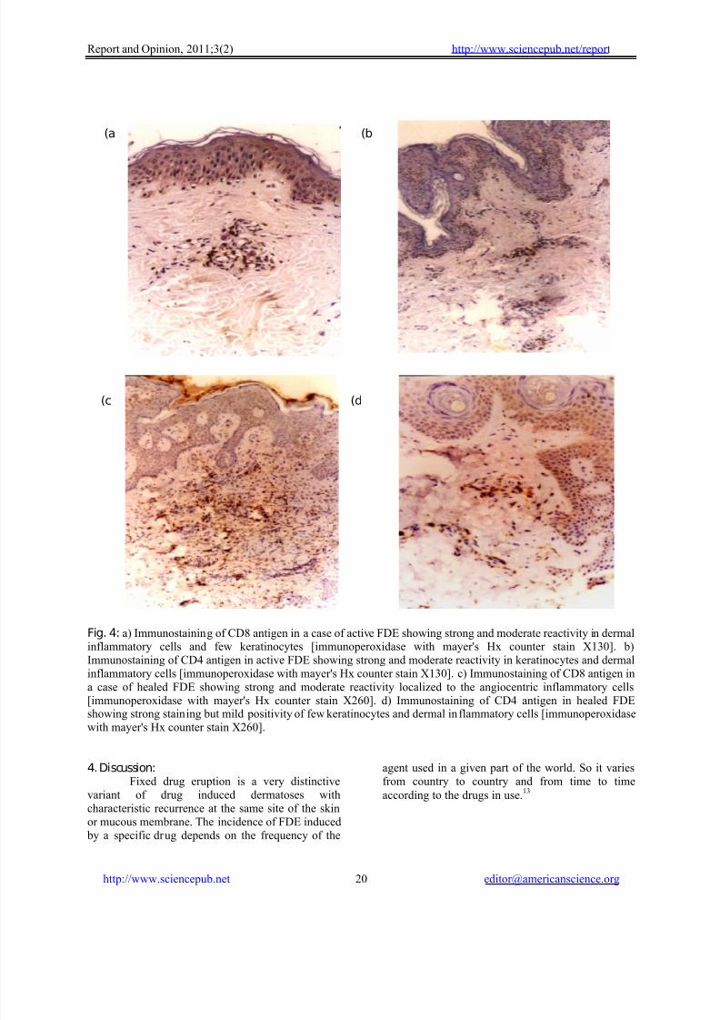
Report and Opinion, 2011;3(2) http://www.sciencepub.net/report http://www.sciencepub.net [email protected] 14 I mm unopa thologic study o f f ixe d drug e ruption Iman Abd El Fattah Seleit, Mohamed Ahmed Basha, Nansy Youssef Asaad , Ola Ahmed Amin Department of Dermatology and Andrology * an d Pathology, Faculty of Medicine Menoufiya University [email protected] Abstract: Background: Fixed drug eruption (FDE) is a common drug induced dermatosis that can be caused by a variety of drugs. Although effector and regulatory T cells play a role in progression and resolution of FDE, little in vivo data exist regarding T cell dynamics in its pathogenesis. Objectives: To through light on the immunopathogenesis of FDE through studying the par ticipation of CD8+, CD4+ T cells and HL A-DR antigen in the pathogenesis of lesions. The role of se rum Ca was studied. Patients and methods: Thirty skin biopsy specimens f rom FDE skin lesions were used (16 active lesions& 14 healed lesions). Thir ty biopsy specimens from thirty age and sex matched healthy subjects were used as a control group. Histopathological examination of hematoxylin& eosin- stained sections included analysis and scoring of histopathological parameters was done. Expression of CD4, CD8 and HLA-DR antigens was examined immunohistochemically. Blood samples were collected from patients and control subjects for assesment of serum calcium. Results: Active lesions showed interface dermatitis and dermal inflammatory infiltrate. Positive immunostainin g was observed in both epidermis and der mis for HL A-DR, CD4 and CD8 antigens. Healed lesions showed epidermal atrophy and dermal inflammatory infiltrate. Positive immunostaining was observed in both epidermis and dermis for HLA-DR, CD4 and CD8 antigens. Such results were absent in control sections. Both total and ionized Ca2+ were significantly lower in patients than control subjec ts. Conclusions: Activation of T cells residing in resting FDE lesions by ingestion of the causative drug results in epidermal injury possibly through the production of IFN-γ. [ Iman Abd El Fattah Seleit, Mohamed Ahmed Basha, Nansy Youssef Asaad, Ola Ahmed Amin. I mm unopa thologic study o f f ixed drug e ruption. Report and Opinion 2011;3(2):14-23]. (ISSN: 1553-9873). http://www.sciencepub.net . K e ywo rds : FDE, CD4+T cells, CD8+T cells, IFN-γ 1. I ntroduction Cutaneous drug eruptions are the most common adverse reactions attributed to drugs. 1 In 1889, Bourns described a patient who had ingested 20 g of antipyrine and then developed a series of sharply demarcated hyperpigmented lesions on the lips and tongue. The French term, eruption erythem ato-pigm ente e f ixe, was coined by Brocq few years later from which fixed drug eruption was derived. 2 Fixed drug eruption (FDE) is a distinct, drug-induced dermatosis characterized by relapse of lesions in the same location every time the drug is administered, 3 a phenomenon referred to as “the recall response” or “the isotopic response”. 4 Sche rer and Bi rche r 5 stated that although the pathogenesis of FDE is unknown, many factors h ad been claimed to share in lesion development including antibodies, antibody-dependent cellular cyto toxicity and serum factors. Fixed drug eruption had been classified as a type IV immunologic reaction because of a latent cytotoxic T cells in the lesions, which may become reactivated. There is also an association with HLA class II antigens, suggesting that there may be a genetic predisposition to these reactions. 6 There is a strong evidence that intraepidermal CD8 + T cells residing in FDE lesions have the key role in mediating the localized epidermal injury through the production of IFN-γ upon stimulation at an early time point (2 to 3h after challenge). 7 In addition, CD8 + T cells might also contribute to the development of FDE lesions by directly interacting with other inflammatory cells. 8 Recombinant INF-γ induced the expression of the class II antigens HLA-DR and HLA-DQ as well as ICAM-1 on human keratinocytes. Low Ca level will lead to over expression of HLA-DR, HLA- DQ and ICAM-1 on keratinocytes. Surface expression of ICAM-1 is suggested to be of major importance as recognition molecule by which T cells bind to IFN-γ exposed keratin ocy tes and suggests an integral role for this molecule in epidermal lymphocy te traffecking. 9 Shiohara 10 postulated that intraepidermal T cells may represent double-edged swords with protective and destructive capacity. The epidermotropic migration of CD4 + T cells into the epidermis protect against the tissue damaging effect of intraepidermal CD8 + T cells residing in the resting lesions. Interestingly enough, at the same time, other populations of CD4 + T cells may preserve their helper function for the CD8 + T cells. The aim of this work is to investigate the expression of keratinocytes HLA-DR in fixed drug

Welcome message from author
This document is posted to help you gain knowledge. Please leave a comment to let me know what you think about it! Share it to your friends and learn new things together.
Transcript

8/9/2019 patofisiologi fde
http://slidepdf.com/reader/full/patofisiologi-fde 1/10
8/9/2019 patofisiologi fde
http://slidepdf.com/reader/full/patofisiologi-fde 2/10

8/9/2019 patofisiologi fde
http://slidepdf.com/reader/full/patofisiologi-fde 3/10

8/9/2019 patofisiologi fde
http://slidepdf.com/reader/full/patofisiologi-fde 4/10

8/9/2019 patofisiologi fde
http://slidepdf.com/reader/full/patofisiologi-fde 5/10

8/9/2019 patofisiologi fde
http://slidepdf.com/reader/full/patofisiologi-fde 6/10
8/9/2019 patofisiologi fde
http://slidepdf.com/reader/full/patofisiologi-fde 7/10
8/9/2019 patofisiologi fde
http://slidepdf.com/reader/full/patofisiologi-fde 8/10
8/9/2019 patofisiologi fde
http://slidepdf.com/reader/full/patofisiologi-fde 9/10
8/9/2019 patofisiologi fde
http://slidepdf.com/reader/full/patofisiologi-fde 10/10
Related Documents