Pathology - Renal Block OSPE You must know features, Diagnosis and Definition of all cases. Contents: 1. ACUTE KIDNEY INJURY. 2. POLYCYSTIC KIDNEY. 3. ACUTE (POST-STREPTOCOCCAL) GLOMERULONEPHRITIS. 4. ACUTE & CHRONIC PYELONEPHRITIS. 5. HYDRONEPHROSIS. 6. Nephrotic Syndrome. 7. Nephritic Syndrome (RPGN). 8. BENIGN RENAL TUMORS. 9. MALIGNANT RENAL TUMORS. 10. WILM’S TUMOR. 11. Carcinoma of Renal Pelvis and Ureter. 12. CARCINOMA OF THE URINARY BLADDER. 13. Pathology of Renal Allograft. 1

Welcome message from author
This document is posted to help you gain knowledge. Please leave a comment to let me know what you think about it! Share it to your friends and learn new things together.
Transcript
Pathology - Renal Block OSPE
You must know features, Diagnosis and Definition of all cases.
Contents:
1. ACUTE KIDNEY INJURY. 2. POLYCYSTIC KIDNEY. 3. ACUTE (POST-STREPTOCOCCAL) GLOMERULONEPHRITIS. 4. ACUTE & CHRONIC PYELONEPHRITIS. 5. HYDRONEPHROSIS. 6. Nephrotic Syndrome. 7. Nephritic Syndrome (RPGN). 8. BENIGN RENAL TUMORS. 9. MALIGNANT RENAL TUMORS. 10. WILM’S TUMOR. 11. Carcinoma of Renal Pelvis and Ureter. 12. CARCINOMA OF THE URINARY BLADDER. 13. Pathology of Renal Allograft.
1
1- ACUTE KIDNEY INJURY.
● Acute kidney injury is Rapid loss of kidney Function dominated by oliguria or anuria (no urine flow), and recent onset of azotemia.
● Most two common causes: Acute tubular Necrosis and Progressive GlomeruloNephritis (RPGN). ● Can Result of Toxins Like Some Antibiotic and ImmunoTherapy (Aminoglycosides) OR
Myoglobinuria.
Pre-renal (Decrease effective blood flow to the
kidney)
Renal Post-renal (is a consequence of urinary tract obstruction)
● Low blood volume, low blood pressure, and heart failure.
● Renal artery stenosis, and renal vein thrombosis.
● Renal ischemia.
● Glomerulonephritis. ● Acute tubular necrosis. ● Acute interstitial
nephritis.
● Benign prostatic hyperplasia. ● Kidney/bladder stones. ● Obstructed urinary catheter. ● Bladder, ureteral or renal
malignancy.
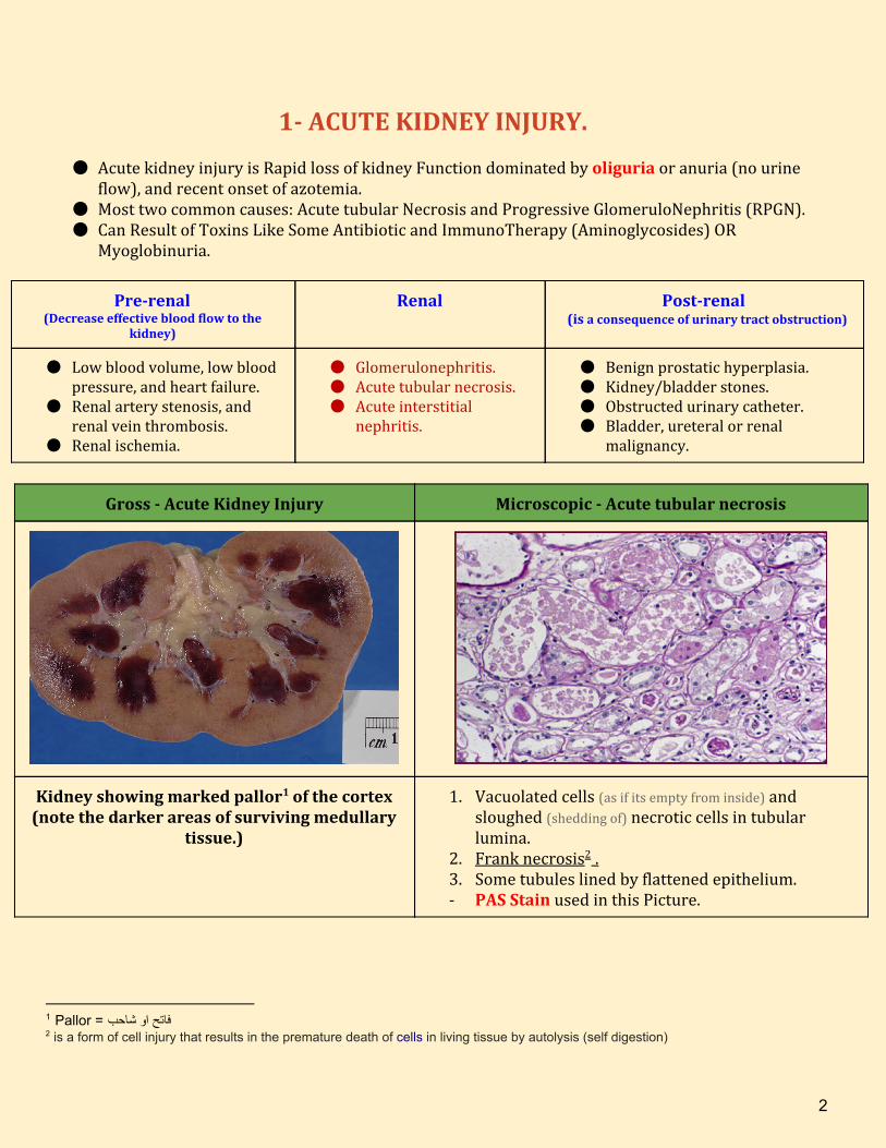
Gross - Acute Kidney Injury Microscopic - Acute tubular necrosis
Kidney showing marked pallor of the cortex 1
(note the darker areas of surviving medullary tissue.)
1. Vacuolated cells (as if its empty from inside) and sloughed (shedding of) necrotic cells in tubular lumina.
2. Frank necrosis . 2
3. Some tubules lined by flattened epithelium. - PAS Stain used in this Picture.
1 Pallor = فاتح او شاحب 2 is a form of cell injury that results in the premature death of cells in living tissue by autolysis (self digestion)
2
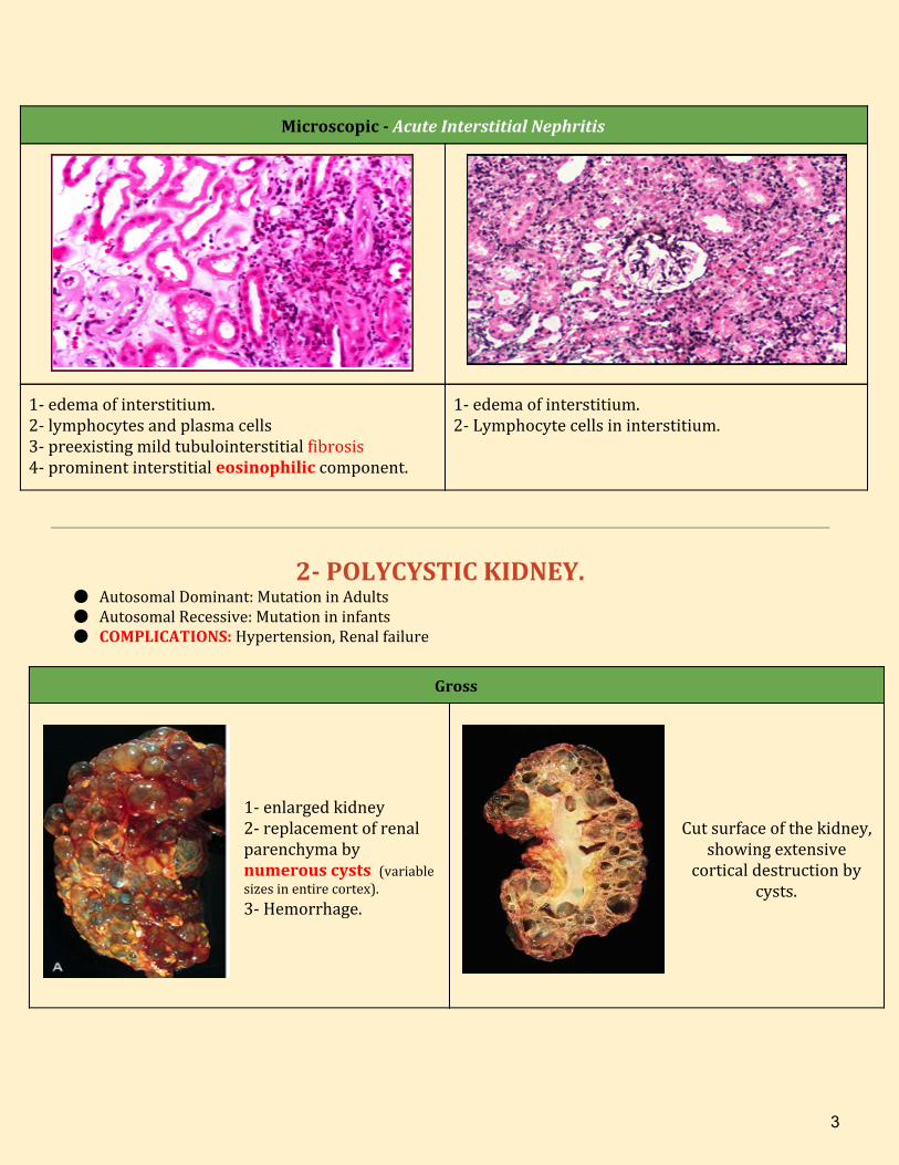
Microscopic - Acute Interstitial Nephritis
1- edema of interstitium. 2- lymphocytes and plasma cells 3- preexisting mild tubulointerstitial fibrosis 4- prominent interstitial eosinophilic component.
1- edema of interstitium. 2- Lymphocyte cells in interstitium.
2- POLYCYSTIC KIDNEY. ● Autosomal Dominant: Mutation in Adults ● Autosomal Recessive: Mutation in infants ● COMPLICATIONS: Hypertension, Renal failure
Gross
1- enlarged kidney 2- replacement of renal parenchyma by numerous cysts (variable sizes in entire cortex).
3- Hemorrhage.
Cut surface of the kidney,
showing extensive cortical destruction by
cysts.
3
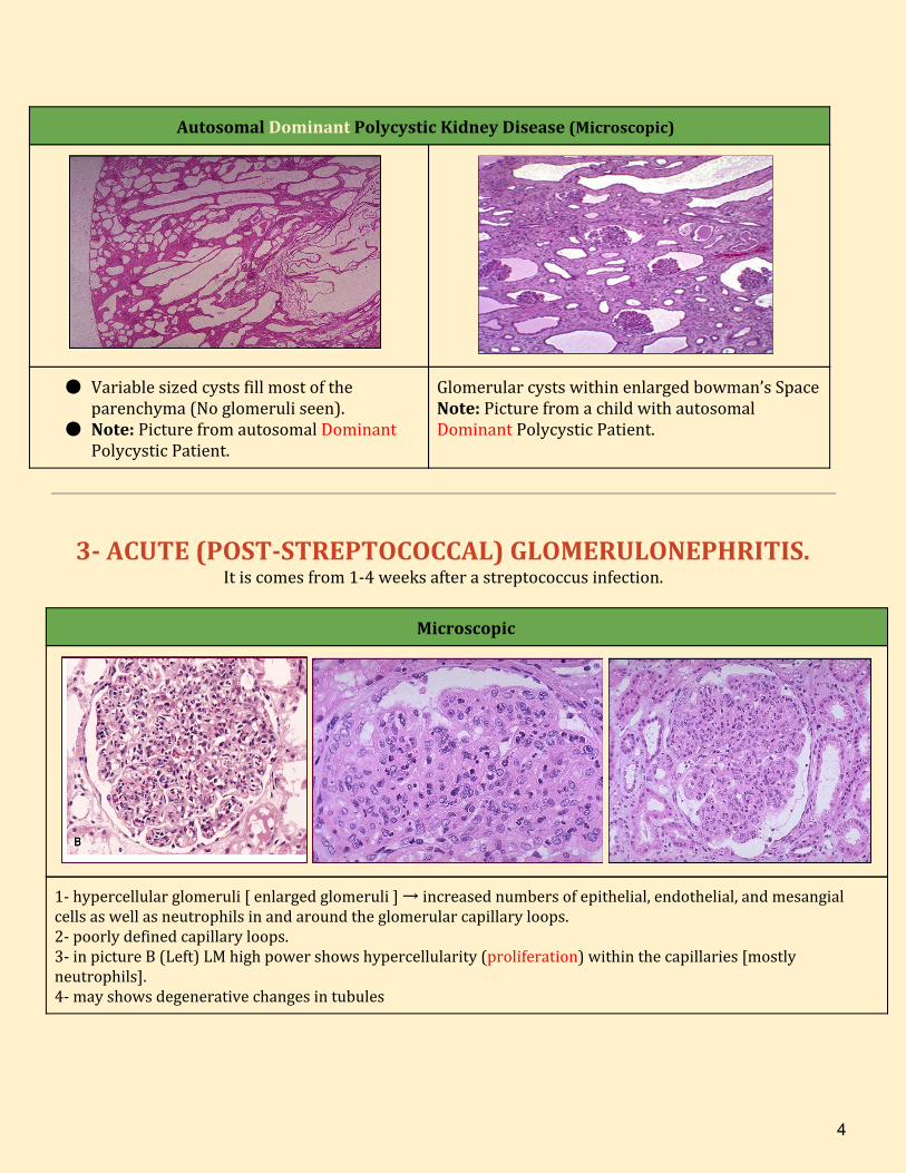
Autosomal Dominant Polycystic Kidney Disease (Microscopic)
● Variable sized cysts fill most of the parenchyma (No glomeruli seen).
● Note: Picture from autosomal Dominant Polycystic Patient.
Glomerular cysts within enlarged bowman’s Space Note: Picture from a child with autosomal Dominant Polycystic Patient.
3- ACUTE (POST-STREPTOCOCCAL) GLOMERULONEPHRITIS. It is comes from 1-4 weeks after a streptococcus infection.
Microscopic
1- hypercellular glomeruli [ enlarged glomeruli ] → increased numbers of epithelial, endothelial, and mesangial cells as well as neutrophils in and around the glomerular capillary loops. 2- poorly defined capillary loops. 3- in picture B (Left) LM high power shows hypercellularity (proliferation) within the capillaries [mostly neutrophils]. 4- may shows degenerative changes in tubules
4
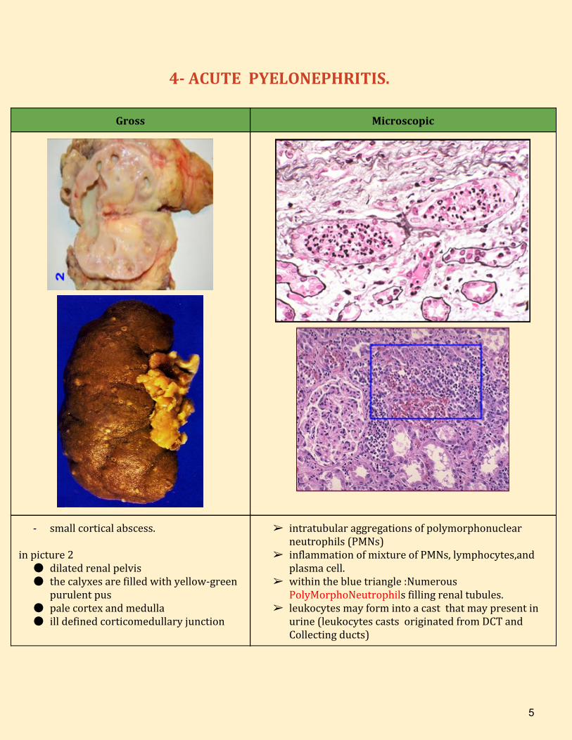
4- ACUTE PYELONEPHRITIS.
Gross Microscopic
- small cortical abscess. in picture 2
● dilated renal pelvis ● the calyxes are filled with yellow-green
purulent pus ● pale cortex and medulla ● ill defined corticomedullary junction
➢ intratubular aggregations of polymorphonuclear neutrophils (PMNs)
➢ inflammation of mixture of PMNs, lymphocytes,and plasma cell.
➢ within the blue triangle :Numerous PolyMorphoNeutrophils filling renal tubules.
➢ leukocytes may form into a cast that may present in urine (leukocytes casts originated from DCT and Collecting ducts)
5
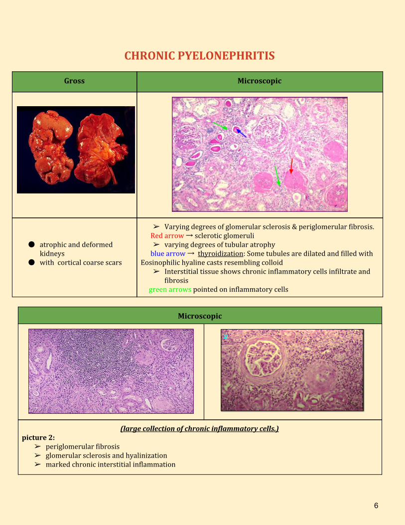
CHRONIC PYELONEPHRITIS
Gross Microscopic
● atrophic and deformed kidneys
● with cortical coarse scars
➢ Varying degrees of glomerular sclerosis & periglomerular fibrosis. Red arrow → sclerotic glomeruli
➢ varying degrees of tubular atrophy blue arrow → thyroidization: Some tubules are dilated and filled with Eosinophilic hyaline casts resembling colloid
➢ Interstitial tissue shows chronic inflammatory cells infiltrate and fibrosis
green arrows pointed on inflammatory cells
Microscopic
(large collection of chronic inflammatory cells.) picture 2:
➢ periglomerular fibrosis ➢ glomerular sclerosis and hyalinization ➢ marked chronic interstitial inflammation
6
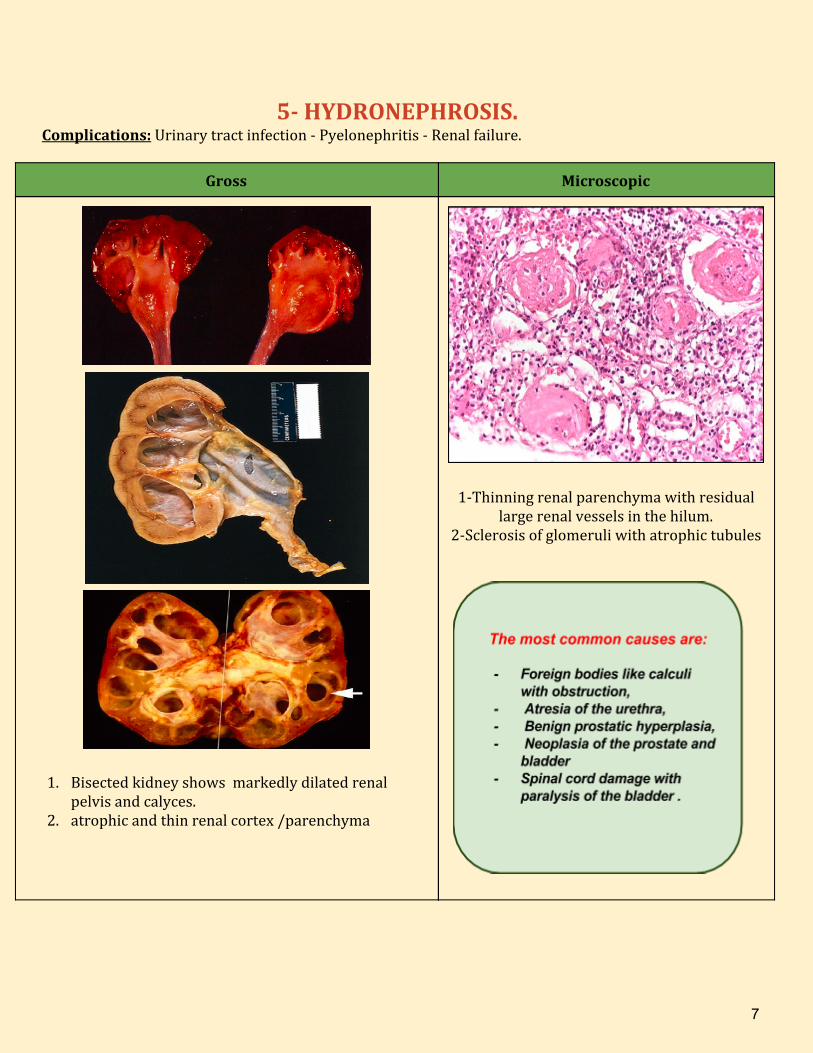
5- HYDRONEPHROSIS. Complications: Urinary tract infection - Pyelonephritis - Renal failure.
Gross Microscopic
1. Bisected kidney shows markedly dilated renal pelvis and calyces.
2. atrophic and thin renal cortex /parenchyma
1-Thinning renal parenchyma with residual large renal vessels in the hilum.
2-Sclerosis of glomeruli with atrophic tubules
7
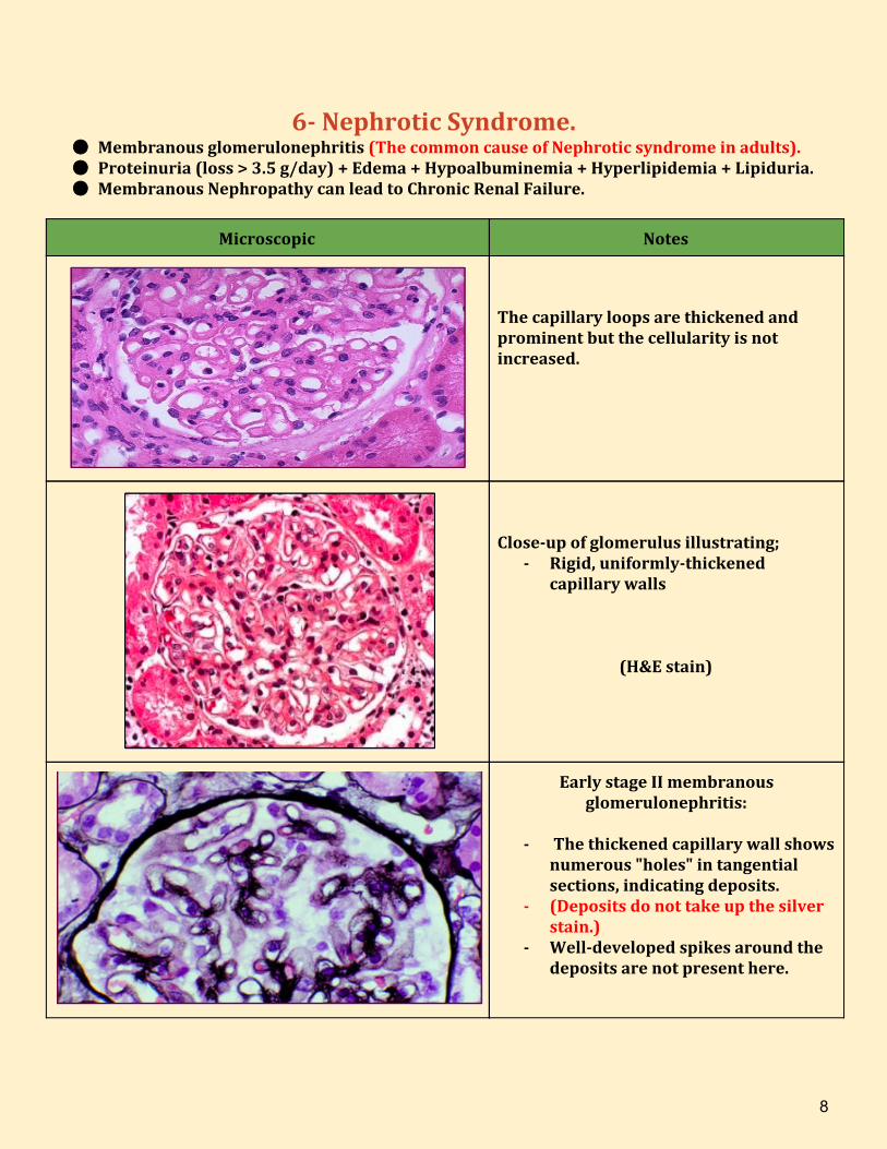
6- Nephrotic Syndrome. ● Membranous glomerulonephritis (The common cause of Nephrotic syndrome in adults). ● Proteinuria (loss > 3.5 g/day) + Edema + Hypoalbuminemia + Hyperlipidemia + Lipiduria. ● Membranous Nephropathy can lead to Chronic Renal Failure.
Microscopic Notes
The capillary loops are thickened and prominent but the cellularity is not increased.
Close-up of glomerulus illustrating;
- Rigid, uniformly-thickened capillary walls
(H&E stain)
Early stage II membranous glomerulonephritis:
- The thickened capillary wall shows
numerous "holes" in tangential sections, indicating deposits.
- (Deposits do not take up the silver stain.)
- Well-developed spikes around the deposits are not present here.
8
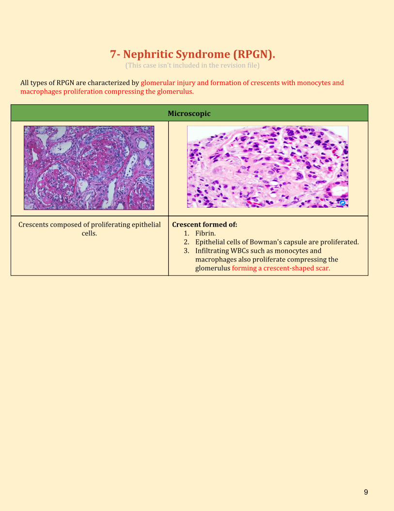
7- Nephritic Syndrome (RPGN). (This case isn’t included in the revision file)
All types of RPGN are characterized by glomerular injury and formation of crescents with monocytes and macrophages proliferation compressing the glomerulus.
Microscopic
Crescents composed of proliferating epithelial cells.
Crescent formed of: 1. Fibrin. 2. Epithelial cells of Bowman's capsule are proliferated. 3. Infiltrating WBCs such as monocytes and
macrophages also proliferate compressing the glomerulus forming a crescent-shaped scar.
9
8- BENIGN RENAL TUMORS.
Signs & Symptoms of Tumors: Fever, Malaise, Rapid loss of weight & appetite. Specific Symptoms of UT Tumors: Hematuria, Flank pain (kidney), Abdominal mass. Common histological findings in neoplasms: Polymorphism, Mitosis, Necrosis, Hyperchromatism. BENIGN RENAL TUMORS (Rare Tumors):
1. Papillary Adenoma (usually Small tumor less than 1 cm in diameter “SIZE very important”). 2. Fibroma/Hamartoma. 3. Angiomyolipoma. 4. Oncocytoma.
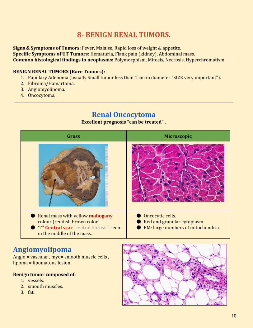
Renal Oncocytoma Excellent prognosis “can be treated” .
Gross Microscopic
● Renal mass with yellow mahogany colour (reddish brown color).
● “↗” Central scar “central fibrosis” seen in the middle of the mass.
● Oncocytic cells. ● Red and granular cytoplasm ● EM: large numbers of mitochondria.
Angiomyolipoma Angio = vascular , myo= smooth muscle cells , lipoma = lipomatous lesion. Benign tumor composed of:
1. vessels. 2. smooth muscles. 3. fat.
10
9- MALIGNANT RENAL TUMORS. ● Renal Cell Carcinoma (Also called Adenocarcinoma, Hypernephroma): Large tumor + Most
common kidney tumor + Common in male especially old male. ● Urothelial (Transitional).
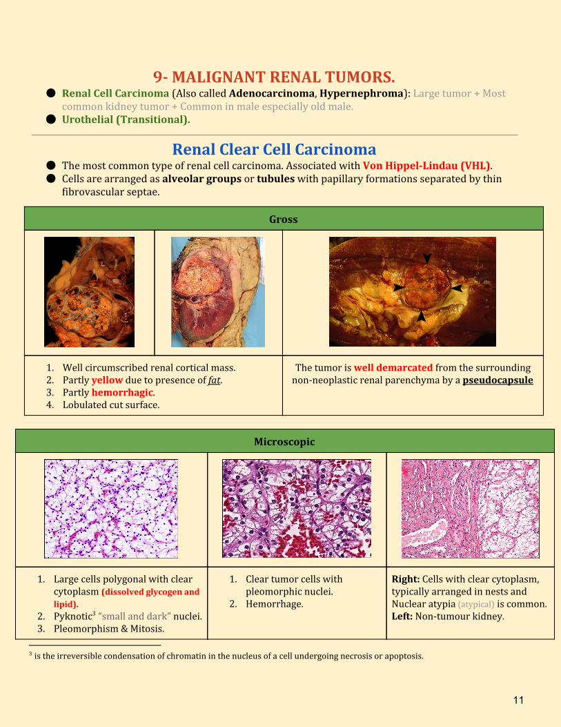
Renal Clear Cell Carcinoma ● The most common type of renal cell carcinoma. Associated with Von Hippel-Lindau (VHL). ● Cells are arranged as alveolar groups or tubules with papillary formations separated by thin
fibrovascular septae.
Gross
1. Well circumscribed renal cortical mass. 2. Partly yellow due to presence of fat. 3. Partly hemorrhagic. 4. Lobulated cut surface.
The tumor is well demarcated from the surrounding non-neoplastic renal parenchyma by a pseudocapsule
Microscopic
1. Large cells polygonal with clear cytoplasm (dissolved glycogen and lipid).
2. Pyknotic “small and dark” nuclei. 3
3. Pleomorphism & Mitosis.
1. Clear tumor cells with pleomorphic nuclei.
2. Hemorrhage.
Right: Cells with clear cytoplasm, typically arranged in nests and Nuclear atypia (atypical) is common. Left: Non-tumour kidney.
3 is the irreversible condensation of chromatin in the nucleus of a cell undergoing necrosis or apoptosis.
11
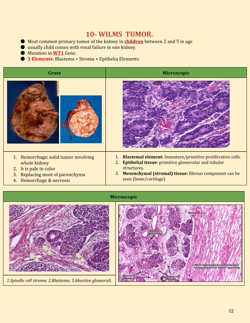
10- WILMS TUMOR. ● Most common primary tumor of the kidney in children between 2 and 5 in age ● usually child comes with renal failure in one kidney. ● Mutation in WT1 Gene. ● 3 Elements: Blastema + Stroma + Epithelia Elements.
Gross Microscopic
1. Hemorrhagic solid tumor involving whole kidney
2. It is pale in color 3. Replacing most of parenchyma 4. Hemorrhage & necrosis
1. Blastemal element: Immature/primitive proliferative cells. 2. Epithelial tissue: primitive glomerular and tubular
structures. 3. Mesenchymal (stromal) tissue: fibrous component can be
seen (bone/cartilage)
Microscopic
1.Spindle cell stroma. 2.Blastema. 3.Abortive glomeruli.
12
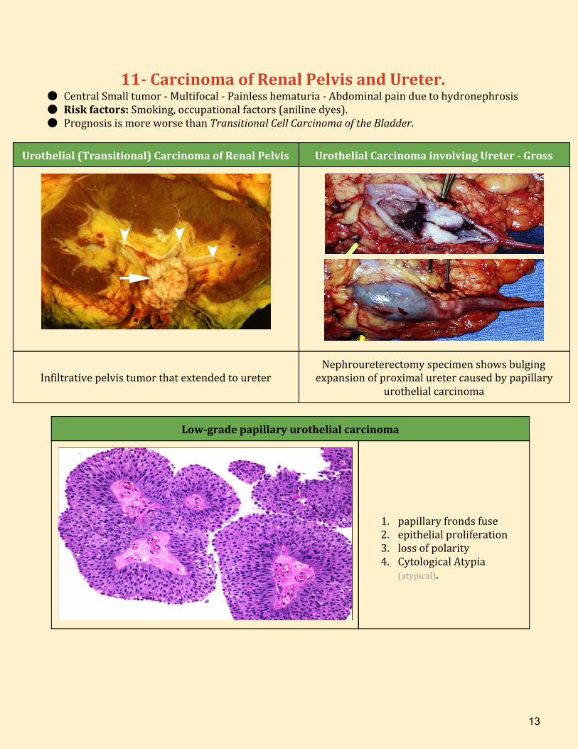
11- Carcinoma of Renal Pelvis and Ureter. ● Central Small tumor - Multifocal - Painless hematuria - Abdominal pain due to hydronephrosis ● Risk factors: Smoking, occupational factors (aniline dyes). ● Prognosis is more worse than Transitional Cell Carcinoma of the Bladder.
Urothelial (Transitional) Carcinoma of Renal Pelvis Urothelial Carcinoma involving Ureter - Gross
Infiltrative pelvis tumor that extended to ureter
Nephroureterectomy specimen shows bulging expansion of proximal ureter caused by papillary
urothelial carcinoma
Low-grade papillary urothelial carcinoma
1. papillary fronds fuse 2. epithelial proliferation 3. loss of polarity 4. Cytological Atypia
(atypical).
13
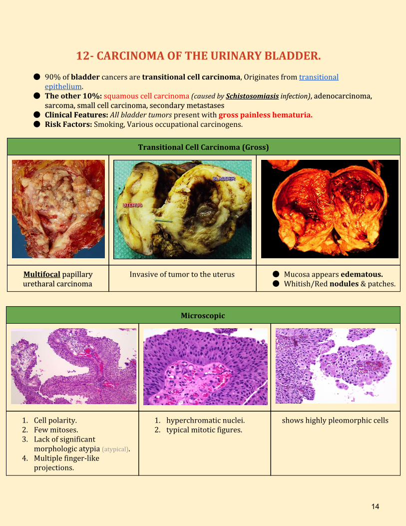
12- CARCINOMA OF THE URINARY BLADDER.
● 90% of bladder cancers are transitional cell carcinoma, Originates from transitional epithelium.
● The other 10%: squamous cell carcinoma (caused by Schistosomiasis infection), adenocarcinoma, sarcoma, small cell carcinoma, secondary metastases
● Clinical Features: All bladder tumors present with gross painless hematuria. ● Risk Factors: Smoking, Various occupational carcinogens.
Transitional Cell Carcinoma (Gross)
Multifocal papillary uretharal carcinoma
Invasive of tumor to the uterus ● Mucosa appears edematous. ● Whitish/Red nodules & patches.
Microscopic
1. Cell polarity. 2. Few mitoses. 3. Lack of significant
morphologic atypia (atypical). 4. Multiple finger-like
projections.
1. hyperchromatic nuclei. 2. typical mitotic figures.
shows highly pleomorphic cells
14
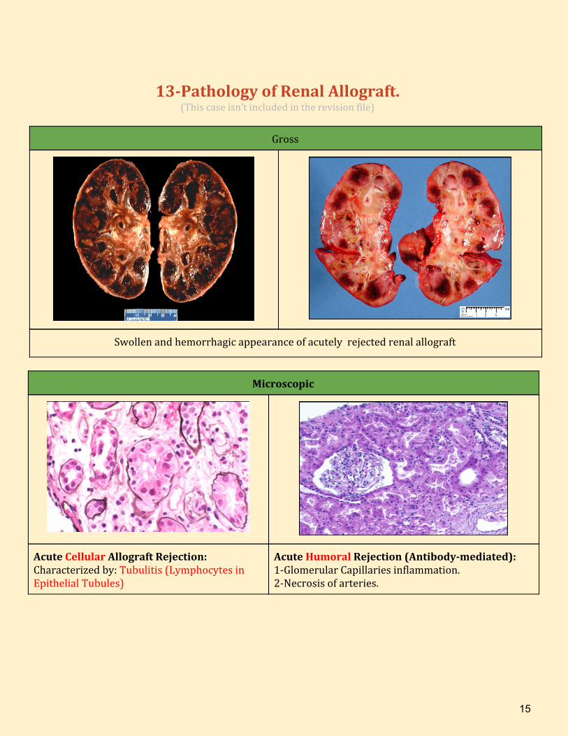
13-Pathology of Renal Allograft. (This case isn’t included in the revision file)
Gross
Swollen and hemorrhagic appearance of acutely rejected renal allograft
Microscopic
Acute Cellular Allograft Rejection: Characterized by: Tubulitis (Lymphocytes in Epithelial Tubules)
Acute Humoral Rejection (Antibody-mediated): 1-Glomerular Capillaries inflammation. 2-Necrosis of arteries.
15
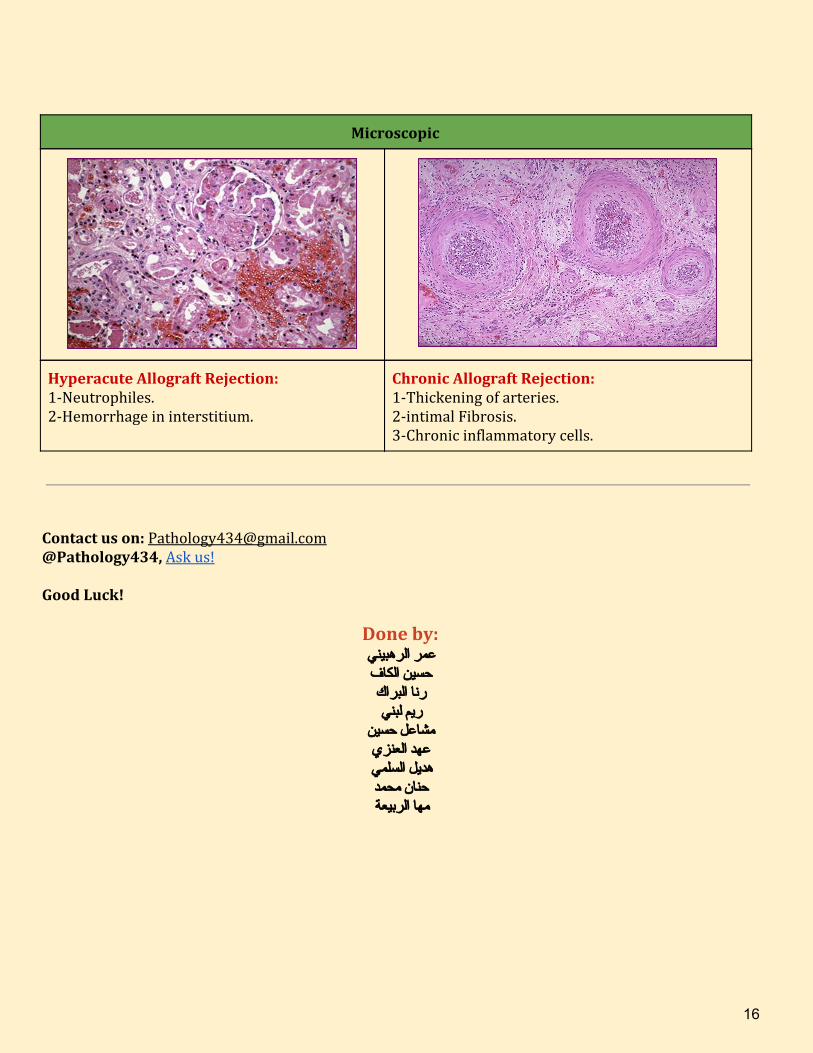
Microscopic
Hyperacute Allograft Rejection: 1-Neutrophiles. 2-Hemorrhage in interstitium.
Chronic Allograft Rejection: 1-Thickening of arteries. 2-intimal Fibrosis. 3-Chronic inflammatory cells.
Contact us on: [email protected] @Pathology434, Ask us! Good Luck!
Done by: عمر الرهبيني حسين الكاف رنا البراك ريم لبني
مشاعل حسين عهد العنزي هديل السلمي حنان محمد مها الربيعة
16
Related Documents

![[OSPE] Praktikum](https://static.cupdf.com/doc/110x72/54e1ea4e4a7959f2578b4a5b/ospe-praktikum.jpg)


