ANKLE Osteochondral defects in the ankle: why painful? C. Niek van Dijk • Mikel L. Reilingh • Maartje Zengerink • Christiaan J. A. van Bergen Received: 8 December 2009 / Accepted: 11 January 2010 / Published online: 12 February 2010 Ó The Author(s) 2010. This article is published with open access at Springerlink.com Abstract Osteochondral defects of the ankle can either heal and remain asymptomatic or progress to deep ankle pain on weight bearing and formation of subchondral bone cysts. The development of a symptomatic OD depends on various fac- tors, including the damage and insufficient repair of the sub- chondral bone plate. The ankle joint has a high congruency. During loading, compressed cartilage forces its water into the microfractured subchondral bone, leading to a localized high increased flow and pressure of fluid in the subchondral bone. This will result in local osteolysis and can explain the slow development of a subchondral cyst. The pain does not arise from the cartilage lesion, but is most probably caused by repetitive high fluid pressure during walking, which results in stimulation of the highly innervated subchondral bone underneath the cartilage defect. Understanding the natural history of osteochondral defects could lead to the develop- ment of strategies for preventing progressive joint damage. Keywords Osteochondral defect Á Cartilage Á Ankle joint Á Subchondral cyst Á Natural history Á Pain Introduction An osteochondral defect (OD) of the talus is a lesion involving the talar articular cartilage and its subchondral bone mostly caused by a single or multiple traumatic events, but idiopathic OD of the ankle do occur [8, 46, 47, 50]. The defect initially may consist only of cartilage damage caused by shearing stresses, with the subchondral bone intact, but a bone contusion following high-impact force also can cause a defect [41, 62, 65]. Ankle trauma associated with an OD often leads to subchondral bone cysts. These cysts are associated with persistent deep ankle pain thereby limiting the patients mobility. Most ODs of the talus are localized on the anterolateral or posteromedial talar dome [70]. Lateral lesions are usually shallow oval shaped and are caused by a shear mechanism. Medial lesions in contrast are usually deep, and cup-shaped, indicating a mechanism of torsional impaction and axial loading [4, 11, 12, 19, 58, 66]. Even though elaborate knowledge exists concerning ODs of the talus, its etiology and pathogenesis are still not fully understood. Increasing attention is paid to invasive and sometimes expensive surgical treatments, while research for pathogenesis of the lesions has somewhat been neglected. In order to treat ODs in all its dimensions, more should be known about their natural history. The development of an OD may have a sudden onset, but the development of a subchondral cyst is most often a slow process. Why do some ODs remain asymptomatic and inert, while others develop pain on weight bearing, demonstrate persistent bone edema on magnetic resonance imaging and result in the progressive formation of a subchondral cyst? Understanding this process might make it possible to interfere and prevent progressive damage to the joint. In this manuscript, the most important factors related to the development of ODs are analyzed. Etiology A traumatic insult is widely accepted as the most important etiologic factor of an OD of the talus. For lateral talar C. N. van Dijk (&) Á M. L. Reilingh Á M. Zengerink Á C. J. A. van Bergen Department of Orthopaedic Surgery, Academic Medical Center, University of Amsterdam, PO Box 22660, 1100 DD Amsterdam, The Netherlands e-mail: [email protected]; [email protected] 123 Knee Surg Sports Traumatol Arthrosc (2010) 18:570–580 DOI 10.1007/s00167-010-1064-x

Welcome message from author
This document is posted to help you gain knowledge. Please leave a comment to let me know what you think about it! Share it to your friends and learn new things together.
Transcript
ANKLE
Osteochondral defects in the ankle: why painful?
C. Niek van Dijk • Mikel L. Reilingh •
Maartje Zengerink • Christiaan J. A. van Bergen
Received: 8 December 2009 / Accepted: 11 January 2010 / Published online: 12 February 2010
� The Author(s) 2010. This article is published with open access at Springerlink.com
Abstract Osteochondral defects of the ankle can either heal
and remain asymptomatic or progress to deep ankle pain on
weight bearing and formation of subchondral bone cysts. The
development of a symptomatic OD depends on various fac-
tors, including the damage and insufficient repair of the sub-
chondral bone plate. The ankle joint has a high congruency.
During loading, compressed cartilage forces its water into the
microfractured subchondral bone, leading to a localized high
increased flow and pressure of fluid in the subchondral bone.
This will result in local osteolysis and can explain the slow
development of a subchondral cyst. The pain does not arise
from the cartilage lesion, but is most probably caused by
repetitive high fluid pressure during walking, which results in
stimulation of the highly innervated subchondral bone
underneath the cartilage defect. Understanding the natural
history of osteochondral defects could lead to the develop-
ment of strategies for preventing progressive joint damage.
Keywords Osteochondral defect � Cartilage �Ankle joint � Subchondral cyst � Natural history � Pain
Introduction
An osteochondral defect (OD) of the talus is a lesion
involving the talar articular cartilage and its subchondral
bone mostly caused by a single or multiple traumatic
events, but idiopathic OD of the ankle do occur [8, 46, 47,
50]. The defect initially may consist only of cartilage
damage caused by shearing stresses, with the subchondral
bone intact, but a bone contusion following high-impact
force also can cause a defect [41, 62, 65]. Ankle trauma
associated with an OD often leads to subchondral bone
cysts. These cysts are associated with persistent deep ankle
pain thereby limiting the patients mobility.
Most ODs of the talus are localized on the anterolateral
or posteromedial talar dome [70]. Lateral lesions are
usually shallow oval shaped and are caused by a shear
mechanism. Medial lesions in contrast are usually deep,
and cup-shaped, indicating a mechanism of torsional
impaction and axial loading [4, 11, 12, 19, 58, 66].
Even though elaborate knowledge exists concerning ODs
of the talus, its etiology and pathogenesis are still not fully
understood. Increasing attention is paid to invasive and
sometimes expensive surgical treatments, while research for
pathogenesis of the lesions has somewhat been neglected. In
order to treat ODs in all its dimensions, more should be
known about their natural history. The development of an
OD may have a sudden onset, but the development of a
subchondral cyst is most often a slow process.
Why do some ODs remain asymptomatic and inert,
while others develop pain on weight bearing, demonstrate
persistent bone edema on magnetic resonance imaging and
result in the progressive formation of a subchondral cyst?
Understanding this process might make it possible to
interfere and prevent progressive damage to the joint. In
this manuscript, the most important factors related to the
development of ODs are analyzed.
Etiology
A traumatic insult is widely accepted as the most important
etiologic factor of an OD of the talus. For lateral talar
C. N. van Dijk (&) � M. L. Reilingh � M. Zengerink �C. J. A. van Bergen
Department of Orthopaedic Surgery, Academic Medical Center,
University of Amsterdam, PO Box 22660, 1100 DD Amsterdam,
The Netherlands
e-mail: [email protected]; [email protected]
123
Knee Surg Sports Traumatol Arthrosc (2010) 18:570–580
DOI 10.1007/s00167-010-1064-x
defects, trauma has been described in 93–98% and for
medial defects in 61–70% [19, 66]. As not all patients
report a history of ankle injury [17], a subdivision can be
made in the etiology of nontraumatic and traumatic defects.
Ischemia, subsequent necrosis and possibly genetics are
etiologic factors in nontraumatic ODs [50]. Furthermore,
ODs in identical twins and siblings have been described [1,
16, 68]. The defect is bilateral in 10% of patients [23].
Traumatic cartilage injuries generally comprise three
categories: microdamage or blunt trauma, chondral frac-
tures and osteochondral fractures [20]. Ankle sprains have
a predominant role in traumatic ODs. When a talus twists
inside its boxlike housing during an ankle sprain, the car-
tilage lining of the talus can be damaged. This may lead to
a bruise and subsequent softening of the cartilage or even a
crack in the cartilage with subsequent delamination.
Separation in the upper layer of the cartilage occurs as a
result of shearing forces. Alternatively, separation may
occur in the subchondral bone, giving rise to a subchondral
bone lesion. Fragments may break off, and float loose in
the ankle joint, or they remain partially attached and stay in
position. The lesions can either heal and remain asymp-
tomatic or progress to deep ankle pain on weight bearing
and formation of subchondral bone cysts.
In cadaver ankles, Berndt and Harty reproduced lateral
defects by strongly inverting a dorsiflexed ankle. As the
foot was inverted on the leg, the lateral border of the
talar dome was compressed against the face of the fibula
[4]. When the lateral ligament ruptured, avulsion of the
chip began. With the use of excessive inverting force,
the talus within the mortise was rotated laterally in the
frontal plane, impacting and compressing the lateral talar
margin against the articular surface of the fibula.
A portion of the talar margin was sheared off from the
main body of the talus, which caused the lateral OD.
A medial lesion was reproduced by plantarflexing the
ankle in combination with slight anterior displacement of
the talus on the tibia, inversion and internal rotation of
the talus on the tibia.
Clinical presentation
In the acute situation, an OD of the talus often remains
unrecognized since the swelling and pain from the lateral
ligament lesion prevails. The weight-bearing anteroposte-
rior (mortise) and lateral radiographs may not reveal any
pathology, or only show an area of radiolucency. In case of
a large OD the initial radiographs may be positive. When
the symptoms of the ligament injury have resolved after
some weeks, symptoms of persistent swelling, limited
range of motion and pain on weight bearing may continue.
If symptoms have not resolved within 4–6 weeks, an
(osteo)chondral defect should be suspected. Locking and
catching are symptoms of a displaced fragment.
Chronic lesions typically present as persistent or inter-
mittent deep ankle pain during or after activity [18]. Most
patients demonstrate a normal range of motion with
absence of recognizable tenderness on palpation and
absence of swelling. However, reactive swelling or stiff-
ness may be present.
The natural history of osteochondral lesions of the talus
whether treated or not is benign. We reported the long-term
results of ODs and found only one case of radiographic
progression after 10 years in 38 cases [52]. Reports of ankle
arthrodesis following ODs of the talus are rare [18, 52].
Cartilage and bone anatomy
Cartilage consists of chondrocytes that lie groupwise in
lacunae of the extracellular matrix they produce. The car-
tilaginous matrix consists of collagen, hyaluronic acid,
proteoglycans and a small amount of glycoprotein’s
(Fig. 1). Its elasticity is based on the electrostatic connec-
tions between collagen fibers and the glycosaminoglycan
(GAG) side chains of the proteoglycans, the containment of
water by the negatively loaded GAGs of the central protein
of proteoglycans and the flexibility and the mutual sliding
of the collagen fibers.
Cartilage is avascular and is nourished by the intra-
articular fluid. The tissue fluid of the cartilage matrix, which
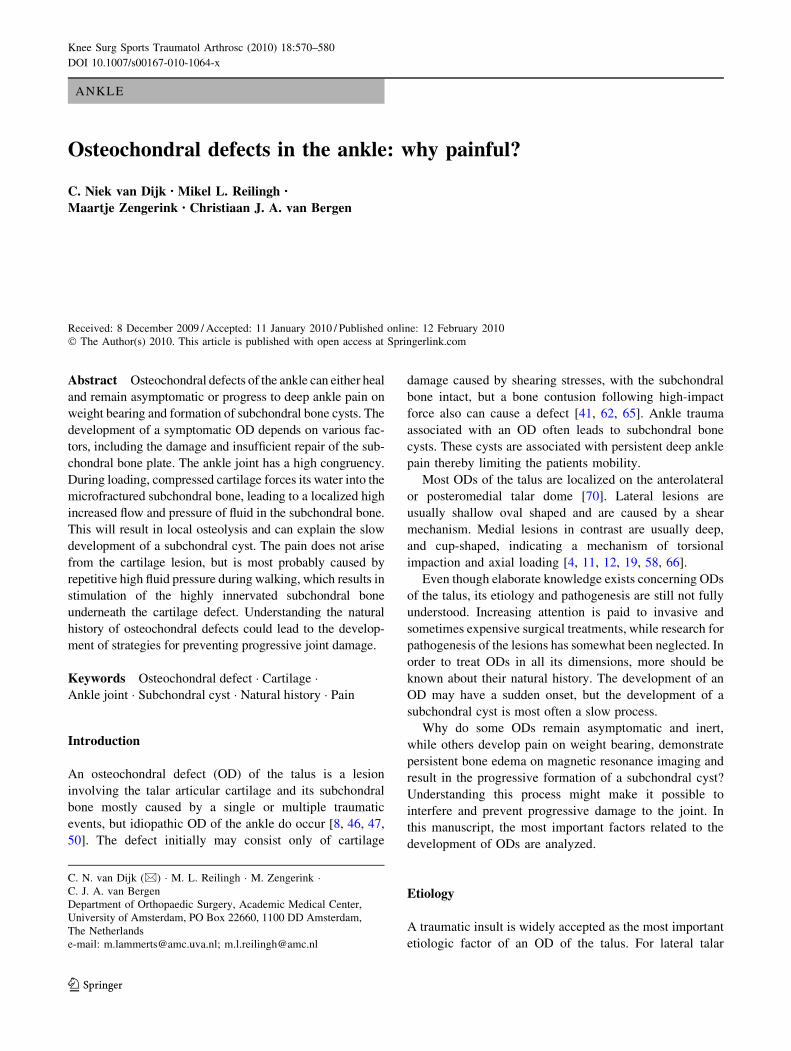
Fig. 1 Schematic diagrams showing normal anatomy of ankle carti-
lage, subchondral plate and subchondral bone area. The cartilage
consist of chondrocytes that lie groupwise in lacunae of the extracel-
lular matrix, which contains collagen fibers in an arcwise configura-
tion, hyaluronic acid, proteoglycans and 75% water (upper left). The
hollow haversian canal that runs longitudinally down the center of the
osteon in compact bone contains an arteriole, venule and lymphatic
duct for vascular and lymphatic drainage. The Volkmann canals run
perpendicular to and connect the Haversian canals (lower left)
Knee Surg Sports Traumatol Arthrosc (2010) 18:570–580 571
123
comprises about 75% of the total weight of the cartilage,
functions as a transport medium. In the healthy cartilage the
GAG side chains of the proteoglycans play an important
role for the elasticity and maintenance of the water content
of 75%. As a matter of fact, we all walk on water. Cartilage
does not contain lymph vessels or nerves and has a slow
metabolism [27]. Mineralized bone consists of both com-
pact and trabecular bone. Compact bone is found beneath
the periosteum and acts as the main weight-bearing pillar
for the skeleton. It is not a solid tissue but rather an
aggregation of osteons, the major multicellular unit of
compact bone. Each osteon is composed of groups of con-
centric calcified cylinders, each of which is made up of bone
matrix proteins that form long cylinders-shaped structures,
oriented parallel to the long axis of the bone [33].
Histopathology
Koch et al. studied the cartilage and bone morphology in
ODs of the knee [29]. They intra-operatively harvested
cylinders of the osteochondral areas as part of a cartilage-
bone transplantation in 30 patients. At the cartilage level
there was a loss of acidic GAGs from the extra-cellular
matrix and a decrease of the number of chondrocytes.
Hyaline cartilage was often replaced by fibrocartilage. The
subchondral bone plate was thinned compared to normal
osteochondral samples and had fractured areas. Parallel
with a general loss of proteoglycans from the superficial
layers of the extracellular cartilage matrix, the amount of
chondroitin sulfates and keratin sulfate was increased in
deep cartilage layers and in the subchondral bone. Koch
et al. [29] stated that all morphological features tend to
indicate that the main area of action is around the sub-
chondral bone plate.
In 2009 Uozumi et al. [63] studied the differences in the
histological findings of ODs in 12 knees. During the sur-
gery, cylinder osteochondral plugs were taken from the
center of the OD and examined with light microscopy.
They classified three types in the subchondral bone area:
(1) necrotic subchondral trabeculae, (2) viable subchondral
trabeculae, and (3) cartilage without bone trabeculae.
Uozumi et al. [63] stated that the initial change in the
subchondral area is bone necrosis or subchondral fracture;
the necrotic bone is then absorbed and replaced either by
viable subchondral trabeculae or cartilage without bone
trabeculae.
An abnormal subchondral plate is likely to be one of the
major factors in influencing the long-term outcome of
articular cartilage repair. Qiu et al. [40] studied ODs in
femoral condyles of rabbits and found that the presence of
an advanced and irregular subchondral plate was associated
with degradation of repaired articular surface.
Cause of pain in osteochondral ankle lesions
Several factors can play a role in the cause of pain in ODs.
A raise in intra-osseous pressure has been mentioned as a
cause of pain and has been associated with joint degener-
ation [2, 3, 64]. Restoration or decrease in the intra-osseous
pressure can be accomplished by medullary decompression
[28, 57].
A rise in intra-articular pressure can be a cause of pain
in degenerative joint disease. Goddard and Gosling have
found a linear correlation between experience pain in
osteoarthritis and resting intra-articular pressure of the
synovial fluid [21]. A connection of synovial hypertrophy
and raised intra-articular pressure in arthritis has been
demonstrated by Bunger et al. [10]. However, it is unlikely
that in a localized osteochondral talar defect a raise in
intra-articular pressure plays a role. These patients typi-
cally do not demonstrate relevant joint effusion.
Nerve endings can be found in the synovium and joint
capsule. Joint capsule and the soft tissue around the joint
are important triggers of nociception. The upregulation of
substance P- and CGRP-positive neurons in response to
arthritic changes suggests a mechanism involving neuro-
peptides in the maintenance of a painful degenerative joint
disease [49]. Patients with an OD of the ankle, however,
generally do not show much synovitis. The synovium of
the anterior ankle joint can be palpated since it lies directly
under the skin. These patients usually can differentiate this
secondary synovial pain from the deep ankle pain caused
by the OD. The disabling deep ankle pain on weight
bearing cannot be reproduced during physical examination.
The most probable cause of this pain is the nerve endings in
the subchondral bone that have been firstly detected in the
early nineties [33].
Within each osteon a hallow tube, known as a Haversian
canal, runs longitudinally down the center of the osteon. It
contains an arteriole, venule and lymphatic duct to provide
the vascular and lymphatic drainage of compact bone. In
addition to the longitudinally oriented Haversian canals, a
series of canals known as Volkmann’s canals run perpen-
dicularly to and interconnect the Haversian canals (Fig. 1).
Mach et al. [33] studied mouse femora and found that not
all osteons are innervated. The likelihood of an osteon
being innervated is greatest in the proximal head followed
by the distal head and then the diaphysis of the femur.
There are CGRP-immunoreactive and RT-97 (clone name
of neurofilament) immunoreactive nerve fibers, which
suggests that the mineralized bone, the bone marrow and
the periosteum are innervated by both unmyelinized and
myelinized fibers. These fibers contain A-b, A-d and
C-fibers that conduct sensory input from the periphery to
the spinal cord. In general, the areas in mineralized bone
that underwent the greatest mechanical stress and loading,
572 Knee Surg Sports Traumatol Arthrosc (2010) 18:570–580
123
that had the highest metabolic rate, and that were most
vascularized, had the highest density of sensory and sym-
pathetic fibers [33]. The fact that there is abundant inner-
vation of bone marrow possibly explains the observation
that patients with bone diseases already experience pain
before there is any radiological evidence of bone destruc-
tion or involvement of the periosteum. Macrophages, the
precursor cells of the osteoclasts, form important accessory
cells in the regulation of bone metabolism and destruction.
Chronic macrophage activation and vascular derangements
lead to low PH, local bone demineralization (acid attack),
and H?-mediated stimulation of the primary afferent
nociceptive nerve fibers [31]. Pain probably develops as a
rise in fluid pressure, and a decrease in pH excitates nerve
fibers present in bone.
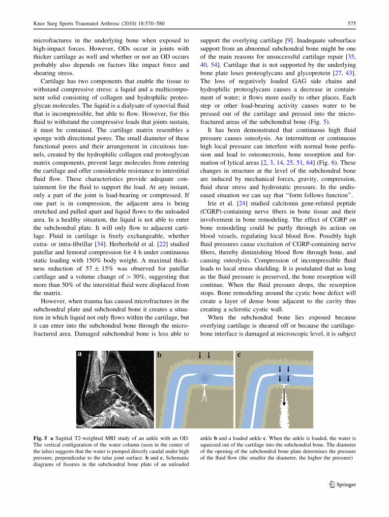
Joint congruency versus cartilage thickness
The cartilage of the talar dome is thin in comparison with
the cartilage of other articulating surfaces. The average
cartilage thickness of the talar dome is 1.11 (±0.28 mm) in
women and 1.35 (±0.22 mm) in men [59]. Shepherd and
Seedhom [55] found almost similar values. In 1891, Braune
and Fischer [5] proposed that articular cartilage is thicker
in regions of low congruence. Simon et al. [56] related joint
congruence to cartilage thickness. They calculated con-
gruence ratios for canine joints by dividing the average
length of the congruent surface by the average length of the
total articular surface. The ankle with the thinnest articular
cartilage had the highest ratio, and the knee with the
thickest cartilage had the lowest ratio. Shepherd and
Seedhom [55] conducted a similar study with human
cadaver specimens. The average thickness of the cartilage
in the ankle, hip, and knee joints were 1.2 mm (1.0–1.6),
1.6 mm (1.4–2.0) and 2.2 mm (1.7–2.6), respectively. The
thickness of the cartilage appeared to be related to the
congruence of a joint. Shepherd and Seedhom [55]
hypothesized that congruent joint surfaces, such as those in
the ankle and elbow, are covered only by thin articular
cartilage because the compressive loads are spread over a
wide area, decreasing local joint stresses and eliminating
the necessity for large cartilaginous deformations. Incon-
gruent joints are covered by thicker cartilage which more
easily deforms, thereby increasing the load-bearing area
and decreasing the stress per unit area.
Cartilage, subchondral bone and loading
Tissue changes the structure in response to the functional
demands imposed on them. Connective tissue has the
ability to alter structure in response to mechanical loading.
Adaptation is affected by different cells. Cartilage has a
much lower response to mechanical adaptation when
compared to bone. Bone remodeling is regulated by
osteocytes that respond to mechanical triggers by sending
signals that promote osteoblastic bone formation. Osteo-
clasts resorb bone at the site of microcracks that frequently
occur in the subarticular spongiosa during impact loading.
Large number of osteoclasts digesting parts of the bone
plate lie in close contact to osteoblasts that seem to be
compensating for bony instability by constantly remodel-
ing the bone stock. Loading tends to thicken the sub-
chondral bone plate in cases of overlying cartilage damage.
This results in sclerosis of the subchondral bone plate.
The load-bearing area of the ankle joint is relatively
small compared to the forces it conducts. The load on the
ankle joint during walking can be calculated. Procter and
Paul measured the load to be 3.9 times body weight at heel
rise during the stance phase of walking [39] (Fig. 2). Mow
et al. measured a load of 5.0 times body weight at heel rise
during the stance phase of walking [37]. Hence, according
to the data of Procter and Paul, the force on the talus with
every step taken by a person weighing 75 kg is 2,867 N
(3.9 9 75 kg 9 9.8 m/s2). The average tibiotalar contact
area is estimated to be 4.4 cm2 [44]. This means that the
average load on the articular cartilage during the stance
phase can be calculated to be 650 N/cm2. During running,
this load increases multiple times.
When the contact surface areas diminish in size, this will
result in an increase in load on the remaining cartilage.
This happens in malunion after an ankle fracture. Ramsey
and Hamilton [44] have shown that 1-mm lateral talar shift
reduces the contact area by 42%, while 2-mm lateral shift
reduces the contact area by 56%. Lloyd et al. [32] found
similar values . In the latter situation the average load per
Fig. 2 Schematic diagrams showing the calculation of load trans-
mission through the ankle joint during walking. Approximately
one-sixth of the load across the ankle is transmitted through the talo-
fibular facet, and the remaining load is transmitted through the
tibiotalar articulation. F = force
Knee Surg Sports Traumatol Arthrosc (2010) 18:570–580 573
123
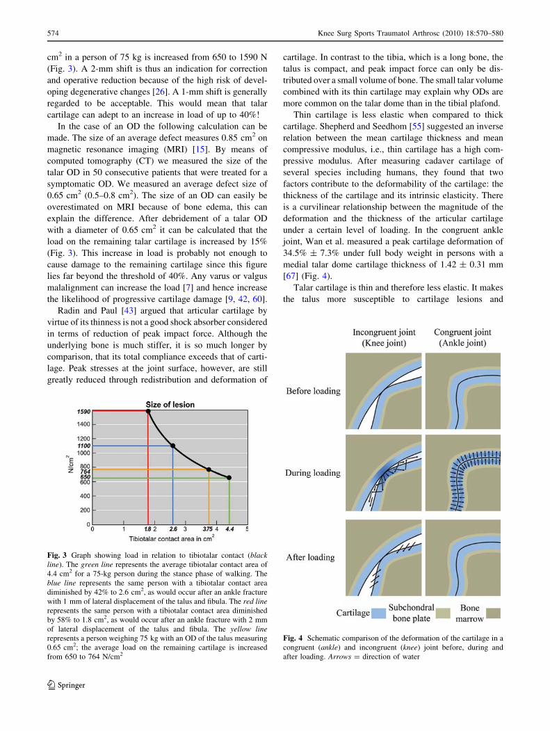
cm2 in a person of 75 kg is increased from 650 to 1590 N
(Fig. 3). A 2-mm shift is thus an indication for correction
and operative reduction because of the high risk of devel-
oping degenerative changes [26]. A 1-mm shift is generally
regarded to be acceptable. This would mean that talar
cartilage can adept to an increase in load of up to 40%!
In the case of an OD the following calculation can be
made. The size of an average defect measures 0.85 cm2 on
magnetic resonance imaging (MRI) [15]. By means of
computed tomography (CT) we measured the size of the
talar OD in 50 consecutive patients that were treated for a
symptomatic OD. We measured an average defect size of
0.65 cm2 (0.5–0.8 cm2). The size of an OD can easily be
overestimated on MRI because of bone edema, this can
explain the difference. After debridement of a talar OD
with a diameter of 0.65 cm2 it can be calculated that the
load on the remaining talar cartilage is increased by 15%
(Fig. 3). This increase in load is probably not enough to
cause damage to the remaining cartilage since this figure
lies far beyond the threshold of 40%. Any varus or valgus
malalignment can increase the load [7] and hence increase
the likelihood of progressive cartilage damage [9, 42, 60].
Radin and Paul [43] argued that articular cartilage by
virtue of its thinness is not a good shock absorber considered
in terms of reduction of peak impact force. Although the
underlying bone is much stiffer, it is so much longer by
comparison, that its total compliance exceeds that of carti-
lage. Peak stresses at the joint surface, however, are still
greatly reduced through redistribution and deformation of
cartilage. In contrast to the tibia, which is a long bone, the
talus is compact, and peak impact force can only be dis-
tributed over a small volume of bone. The small talar volume
combined with its thin cartilage may explain why ODs are
more common on the talar dome than in the tibial plafond.
Thin cartilage is less elastic when compared to thick
cartilage. Shepherd and Seedhom [55] suggested an inverse
relation between the mean cartilage thickness and mean
compressive modulus, i.e., thin cartilage has a high com-
pressive modulus. After measuring cadaver cartilage of
several species including humans, they found that two
factors contribute to the deformability of the cartilage: the
thickness of the cartilage and its intrinsic elasticity. There
is a curvilinear relationship between the magnitude of the
deformation and the thickness of the articular cartilage
under a certain level of loading. In the congruent ankle
joint, Wan et al. measured a peak cartilage deformation of
34.5% ± 7.3% under full body weight in persons with a
medial talar dome cartilage thickness of 1.42 ± 0.31 mm
[67] (Fig. 4).
Talar cartilage is thin and therefore less elastic. It makes
the talus more susceptible to cartilage lesions and
Fig. 3 Graph showing load in relation to tibiotalar contact (blackline). The green line represents the average tibiotalar contact area of
4.4 cm2 for a 75-kg person during the stance phase of walking. The
blue line represents the same person with a tibiotalar contact area
diminished by 42% to 2.6 cm2, as would occur after an ankle fracture
with 1 mm of lateral displacement of the talus and fibula. The red linerepresents the same person with a tibiotalar contact area diminished
by 58% to 1.8 cm2, as would occur after an ankle fracture with 2 mm
of lateral displacement of the talus and fibula. The yellow linerepresents a person weighing 75 kg with an OD of the talus measuring
0.65 cm2; the average load on the remaining cartilage is increased
from 650 to 764 N/cm2
Fig. 4 Schematic comparison of the deformation of the cartilage in a
congruent (ankle) and incongruent (knee) joint before, during and
after loading. Arrows = direction of water
574 Knee Surg Sports Traumatol Arthrosc (2010) 18:570–580
123
microfractures in the underlying bone when exposed to
high-impact forces. However, ODs occur in joints with
thicker cartilage as well and whether or not an OD occurs
probably also depends on factors like impact force and
shearing stress.
Cartilage has two components that enable the tissue to
withstand compressive stress: a liquid and a multicompo-
nent solid consisting of collagen and hydrophilic proteo-
glycan molecules. The liquid is a dialysate of synovial fluid
that is incompressible, but able to flow. However, for this
fluid to withstand the compressive loads that joints sustain,
it must be contained. The cartilage matrix resembles a
sponge with directional pores. The small diameter of these
functional pores and their arrangement in circuitous tun-
nels, created by the hydrophilic collagen end proteoglycan
matrix components, prevent large molecules from entering
the cartilage and offer considerable resistance to interstitial
fluid flow. These characteristics provide adequate con-
tainment for the fluid to support the load. At any instant,
only a part of the joint is load-bearing or compressed. If
one part is in compression, the adjacent area is being
stretched and pulled apart and liquid flows to the unloaded
area. In a healthy situation, the liquid is not able to enter
the subchondral plate. It will only flow to adjacent carti-
lage. Fluid in cartilage is freely exchangeable, whether
extra- or intra-fibrillar [34]. Herberhold et al. [22] studied
patellar and femoral compression for 4 h under continuous
static loading with 150% body weight. A maximal thick-
ness reduction of 57 ± 15% was observed for patellar
cartilage and a volume change of [ 30%, suggesting that
more than 50% of the interstitial fluid were displaced from
the matrix.
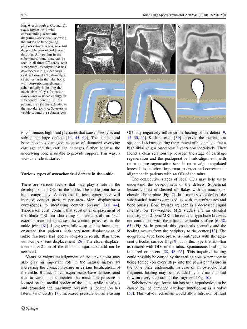
However, when trauma has caused microfractures in the
subchondral plate and subchondral bone it creates a situa-
tion in which liquid not only flows within the cartilage, but
it can enter into the subchondral bone through the micro-
fractured area. Damaged subchondral bone is less able to
support the overlying cartilage [9]. Inadequate subsurface
support from an abnormal subchondral bone might be one
of the main reasons for unsuccessful cartilage repair [35,
40, 54]. Cartilage that is not supported by the underlying
bone plate loses proteoglycans and glycoprotein [27, 43].
The loss of negatively loaded GAG side chains and
hydrophilic proteoglycans causes a decrease in contain-
ment of water; it flows more easily to other places. Each
step or other load-bearing activity causes water to be
pressed out of the cartilage and pressed into the micro-
fractured areas of the subchondral bone (Fig. 5).
It has been demonstrated that continuous high fluid
pressure causes osteolysis. An intermittent or continuous
high local pressure can interfere with normal bone perfu-
sion and lead to osteonecrosis, bone resorption and for-
mation of lytical areas [2, 3, 14, 25, 51, 64] (Fig. 6). These
changes in structure at the level of the subchondral bone
are induced by mechanical forces, gravity, compression,
fluid shear stress and hydrostatic pressure. In the undis-
eased situation we can say that ‘‘form follows function’’.
Irie et al. [24] studied calcitonin gene-related peptide
(CGRP)-containing nerve fibers in bone tissue and their
involvement in bone remodeling. The effect of CGRP on
bone remodeling could be partly through its action on
blood vessels, regulating local blood flow. Possibly high
fluid pressures cause excitation of CGRP-containing nerve
fibers, thereby diminishing blood flow through bone, and
causing osteolysis. Compression of incompressible fluid
leads to local stress shielding. It is postulated that as long
as the fluid pressure is preserved, the bone resorption will
continue. When the fluid pressure drops, the resorption
stops. Bone remodeling around the cystic bone defect will
create a layer of dense bone adjacent to the cavity thus
creating a sclerotic cystic wall.
When the subchondral bone lies exposed because
overlying cartilage is sheared off or because the cartilage-
bone interface is damaged at microscopic level, it is subject
Fig. 5 a Sagittal T2-weighted MRI study of an ankle with an OD.
The vertical configuration of the water column (seen in the center of
the talus) suggests that the water is pumped directly caudal under high
pressure, perpendicular to the talar joint surface. b and c, Schematic
diagrams of fissures in the subchondral bone plate of an unloaded
ankle b and a loaded ankle c. When the ankle is loaded, the water is
squeezed out of the cartilage into the subchondral bone. The diameter
of the opening of the subchondral bone plate determines the pressure
of the fluid flow (the smaller the diameter, the higher the pressure)
Knee Surg Sports Traumatol Arthrosc (2010) 18:570–580 575
123
to continuous high fluid pressures that cause osteolysis and
subsequent large defects [14, 45, 69]. The subchondral
bone becomes damaged because of damaged overlying
cartilage and the cartilage damages further because the
underlying bone is unable to provide support. This way, a
vicious circle is started.
Various types of osteochondral defects in the ankle
There are various factors that may play a role in the
development of ODs in the ankle. The ankle joint has a
high congruency. A decrease in joint congruence will
increase contact pressure per area. More displacement
corresponds to increasing contact pressure [32, 44].
Thordarson et al. confirm that substantial displacement of
the fibula (C2 mm shortening or lateral shift or C 5�external rotation) increases the contact pressures in the
ankle joint [61]. Long-term follow-up studies have dem-
onstrated that patients with persistent displacement of
ankle fractures had poorer long-term results than those
without persistent displacement [26]. Therefore, displace-
ment of [ 2 mm of the fibula in injuries should not be
accepted.
Varus or valgus malalignment of the ankle joint may
also play an important role in the natural history by
increasing the contact pressure in certain localizations of
the ankle. Biomechanical experiments have demonstrated
that in varus and supination the maximum pressure is
located on the medial border of the talus, while in valgus
and pronation the maximum pressure is located on het
lateral talar border [7]. Increased pressure on an existing
OD may negatively influence the healing of the defect [9,
14, 30, 42]. Koshino et al. [30] observed the medial joint
space in 146 knees during the removal of blade plate after a
high tibial valgus osteotomy 2 years postoperatively. They
found a clear relationship between the stage of cartilage
regeneration and the postoperative limb alignment, with
more mature regeneration seen in more valgus angulated
knees. It is therefore important to detect and correct mal-
alignment in patients with an OD of the talus.
The consecutive stages of local ODs may help us to
understand the development of the defects. Superficial
lesions consist of sheared off flakes with an intact sub-
chondral bone plate (Fig. 7). In a more severe defect, the
subchondral bone is damaged, as with, microfractures and
bone bruises. Bone bruises are seen as a decreased signal
intensity on T1-weighted MRI studies and an elevated
intensity on T2-bone MRI. The reticular type bone bruise is
not continuous with the adjacent articular surface [6, 38,
65] (Fig. 8). In general, this type heals normally and the
healing occurs from the periphery to the center [13]. The
geographic type bone bruise is continuous with the adja-
cent articular surface (Fig. 9). It is this type that is often
associated with ODs of the talus. Spontaneous healing is
impaired or absent [38, 48, 65]. This impaired healing
could possibly be caused by the cartilaginous water content
being forced -on every step- into the persistent fissure in
the bone plate underneath. In case of an osteochondral
fragment, healing may be precluded by intermittent fluid
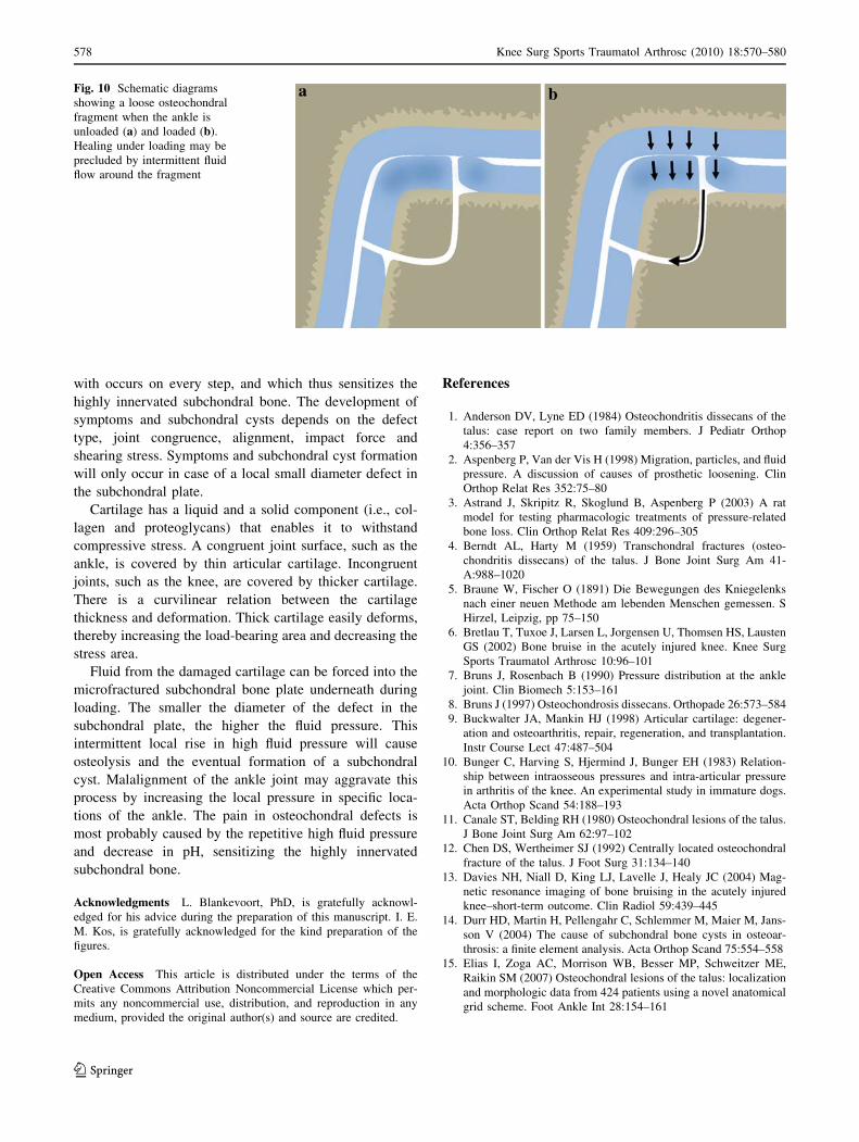
flow on every step around the fragment (Fig. 10).
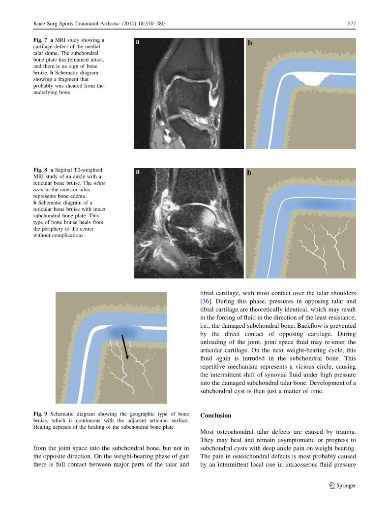
Subchondral cyst formation has been hypothesized to be
caused by the damaged cartilage functioning as a valve
[53]. This valve mechanism would allow intrusion of fluid
Fig. 6 a through c, Coronal CT
scans (upper row) with
corresponding schematic
diagrams (lower row), showing
the ankles of three young
patients (26–37 years), who had
deep ankle pain of 5–12 years
duration. An opening in the
subchondral bone plate can be
seen in all three CT scans, with
subchondral osteolysis that has
developed into a subchondral
cyst. a Coronal CT, showing a
cystic lesion in the talar body,
with corresponding diagram
schematically indicating the
mechanism of cyst formation.
Black lines = nerve endings in
subchondral bone. b, In this
patient, the cyst has extended to
the subtalar joint. c, Sclerosis is
visible around the subtalar cyst
576 Knee Surg Sports Traumatol Arthrosc (2010) 18:570–580
123
from the joint space into the subchondral bone, but not in
the opposite direction. On the weight-bearing phase of gait
there is full contact between major parts of the talar and
tibial cartilage, with most contact over the talar shoulders
[36]. During this phase, pressures in opposing talar and
tibial cartilage are theoretically identical, which may result
in the forcing of fluid in the direction of the least resistance,
i.e., the damaged subchondral bone. Backflow is prevented
by the direct contact of opposing cartilage. During
unloading of the joint, joint space fluid may re-enter the
articular cartilage. On the next weight-bearing cycle, this
fluid again is intruded in the subchondral bone. This
repetitive mechanism represents a vicious circle, causing
the intermittent shift of synovial fluid under high pressure
into the damaged subchondral talar bone. Development of a
subchondral cyst is then just a matter of time.
Conclusion
Most osteochondral talar defects are caused by trauma.
They may heal and remain asymptomatic or progress to
subchondral cysts with deep ankle pain on weight bearing.
The pain in osteochondral defects is most probably caused
by an intermittent local rise in intraosseous fluid pressure
Fig. 7 a MRI study showing a
cartilage defect of the medial
talar dome. The subchondral
bone plate has remained intact,
and there is no sign of bone
bruise. b Schematic diagram
showing a fragment that
probably was sheared from the
underlying bone
Fig. 8 a Sagittal T2-weighted
MRI study of an ankle with a
reticular bone bruise. The whitearea in the anterior talus
represents bone edema.
b Schematic diagram of a
reticular bone bruise with intact
subchondral bone plate. This
type of bone bruise heals from
the periphery to the center
without complications
Fig. 9 Schematic diagram showing the geographic type of bone
bruise, which is continuous with the adjacent articular surface.
Healing depends of the healing of the subchondral bone plate
Knee Surg Sports Traumatol Arthrosc (2010) 18:570–580 577
123
with occurs on every step, and which thus sensitizes the
highly innervated subchondral bone. The development of
symptoms and subchondral cysts depends on the defect
type, joint congruence, alignment, impact force and
shearing stress. Symptoms and subchondral cyst formation
will only occur in case of a local small diameter defect in
the subchondral plate.
Cartilage has a liquid and a solid component (i.e., col-
lagen and proteoglycans) that enables it to withstand
compressive stress. A congruent joint surface, such as the
ankle, is covered by thin articular cartilage. Incongruent
joints, such as the knee, are covered by thicker cartilage.
There is a curvilinear relation between the cartilage
thickness and deformation. Thick cartilage easily deforms,
thereby increasing the load-bearing area and decreasing the
stress area.
Fluid from the damaged cartilage can be forced into the
microfractured subchondral bone plate underneath during
loading. The smaller the diameter of the defect in the
subchondral plate, the higher the fluid pressure. This
intermittent local rise in high fluid pressure will cause
osteolysis and the eventual formation of a subchondral
cyst. Malalignment of the ankle joint may aggravate this
process by increasing the local pressure in specific loca-
tions of the ankle. The pain in osteochondral defects is
most probably caused by the repetitive high fluid pressure
and decrease in pH, sensitizing the highly innervated
subchondral bone.
Acknowledgments L. Blankevoort, PhD, is gratefully acknowl-
edged for his advice during the preparation of this manuscript. I. E.
M. Kos, is gratefully acknowledged for the kind preparation of the
figures.
Open Access This article is distributed under the terms of the
Creative Commons Attribution Noncommercial License which per-
mits any noncommercial use, distribution, and reproduction in any
medium, provided the original author(s) and source are credited.
References
1. Anderson DV, Lyne ED (1984) Osteochondritis dissecans of the
talus: case report on two family members. J Pediatr Orthop
4:356–357
2. Aspenberg P, Van der Vis H (1998) Migration, particles, and fluid
pressure. A discussion of causes of prosthetic loosening. Clin
Orthop Relat Res 352:75–80
3. Astrand J, Skripitz R, Skoglund B, Aspenberg P (2003) A rat
model for testing pharmacologic treatments of pressure-related
bone loss. Clin Orthop Relat Res 409:296–305
4. Berndt AL, Harty M (1959) Transchondral fractures (osteo-
chondritis dissecans) of the talus. J Bone Joint Surg Am 41-
A:988–1020
5. Braune W, Fischer O (1891) Die Bewegungen des Kniegelenks
nach einer neuen Methode am lebenden Menschen gemessen. S
Hirzel, Leipzig, pp 75–150
6. Bretlau T, Tuxoe J, Larsen L, Jorgensen U, Thomsen HS, Lausten
GS (2002) Bone bruise in the acutely injured knee. Knee Surg
Sports Traumatol Arthrosc 10:96–101
7. Bruns J, Rosenbach B (1990) Pressure distribution at the ankle
joint. Clin Biomech 5:153–161
8. Bruns J (1997) Osteochondrosis dissecans. Orthopade 26:573–584
9. Buckwalter JA, Mankin HJ (1998) Articular cartilage: degener-
ation and osteoarthritis, repair, regeneration, and transplantation.
Instr Course Lect 47:487–504
10. Bunger C, Harving S, Hjermind J, Bunger EH (1983) Relation-
ship between intraosseous pressures and intra-articular pressure
in arthritis of the knee. An experimental study in immature dogs.
Acta Orthop Scand 54:188–193
11. Canale ST, Belding RH (1980) Osteochondral lesions of the talus.
J Bone Joint Surg Am 62:97–102
12. Chen DS, Wertheimer SJ (1992) Centrally located osteochondral
fracture of the talus. J Foot Surg 31:134–140
13. Davies NH, Niall D, King LJ, Lavelle J, Healy JC (2004) Mag-
netic resonance imaging of bone bruising in the acutely injured
knee–short-term outcome. Clin Radiol 59:439–445
14. Durr HD, Martin H, Pellengahr C, Schlemmer M, Maier M, Jans-
son V (2004) The cause of subchondral bone cysts in osteoar-
throsis: a finite element analysis. Acta Orthop Scand 75:554–558
15. Elias I, Zoga AC, Morrison WB, Besser MP, Schweitzer ME,
Raikin SM (2007) Osteochondral lesions of the talus: localization
and morphologic data from 424 patients using a novel anatomical
grid scheme. Foot Ankle Int 28:154–161
Fig. 10 Schematic diagrams
showing a loose osteochondral
fragment when the ankle is
unloaded (a) and loaded (b).
Healing under loading may be
precluded by intermittent fluid
flow around the fragment
578 Knee Surg Sports Traumatol Arthrosc (2010) 18:570–580
123
16. Erban WK, Kolberg K (1981) Simultaneous mirror image oste-
ochondrosis dissecans in identical twins. Rofo 135:357
17. Ferkel RD, Scranton PE Jr (1993) Arthroscopy of the ankle and
foot. J Bone Joint Surg Am 75:1233–1242
18. Ferkel RD, Zanotti RM, Komenda GA, Sgaglione NA, Cheng
MS, Applegate GR et al (2008) Arthroscopic treatment of chronic
osteochondral lesions of the talus: long-term results. Am J Sports
Med 36:1750–1762
19. Flick AB, Gould N (1985) Osteochondritis dissecans of the talus
(transchondral fractures of the talus): review of the literature and
new surgical approach for medial dome lesions. Foot Ankle
5:165–185
20. Frenkel SR, Di Cesare PE (1999) Degradation and repair of
articular cartilage. Front Biosci 4:671–685
21. Goddard NJ, Gosling PT (1988) Intra-articular fluid pressure and
pain in osteoarthritis of the hip. J Bone Joint Surg Br 70:52–55
22. Herberhold C, Faber S, Stammberger T, Steinlechner M, Putz R,
Englmeier KH et al (1999) In situ measurement of articular
cartilage deformation in intact femoropatellar joints under static
loading. J Biomech 32:1287–1295
23. Hermanson E, Ferkel RD (2009) Bilateral osteochondral lesions
of the talus. Foot Ankle Int 30:723–727
24. Irie K, Hara-Irie F, Ozawa H, Yajima T (2002) Calcitonin gene-
related peptide (CGRP)-containing nerve fibers in bone tissue and
their involvement in bone remodeling. Microsc Res Tech 58:85–90
25. Johansson L, Edlund U, Fahlgren A, Aspenberg P (2009) Bone
resorption induced by fluid flow. J Biomech Eng 131:094505
26. Joy G, Patzakis MJ, Harvey JP Jr (1974) Precise evaluation of the
reduction of severe ankle fractures. J Bone Joint Surg Am
56:979–993
27. Junqueira L, Carneiro J, Kelly R (2007) Kraakbeen. In: Func-
tionele Histologie, 11th edn. Elsevier, Maarssen, pp 140–147
28. Kiaer T, Pedersen NW, Kristensen KD, Starklint H (1990) Intra-
osseous pressure and oxygen tension in avascular necrosis and
osteoarthritis of the hip. J Bone Joint Surg Br 72:1023–1030
29. Koch S, Kampen WU, Laprell H (1997) Cartilage and bone
morphology in osteochondritis dissecans. Knee Surg Sports
Traumatol Arthrosc 5:42–45
30. Koshino T, Wada S, Ara Y, Saito T (2003) Regeneration of
degenerated articular cartilage after high tibial valgus osteotomy
for medial compartmental osteoarthritis of the knee. Knee
10:229–236
31. Lassus J, Salo J, Jiranek WA, Santavirta S, Nevalainen J, Mat-
ucci-Cerinic M et al (1998) Macrophage activation results in
bone resorption. Clin Orthop Relat Res 352:7–15
32. Lloyd J, Elsayed S, Hariharan K, Tanaka H (2006) Revisiting the
concept of talar shift in ankle fractures. Foot Ankle Int 27:793–
796
33. Mach DB, Rogers SD, Sabino MC, Luger NM, Schwei MJ,
Pomonis JD et al (2002) Origins of skeletal pain: sensory and
sympathetic innervation of the mouse femur. Neuroscience
113:155–166
34. Maroudas A, Schneiderman R (1987) ‘‘Free’’ and ‘‘exchange-
able’’ or ‘‘trapped’’ and ‘‘non-exchangeable’’ water in cartilage.
J Orthop Res 5:133–138
35. Messner K (1993) Hydroxylapatite supported Dacron plugs for
repair of isolated full-thickness osteochondral defects of the
rabbit femoral condyle: mechanical and histological evaluations
from 6 to 48 weeks. J Biomed Mater Res 27:1527–1532
36. Millington S, Grabner M, Wozelka R, Hurwitz S, Crandall J
(2007) A stereophotographic study of ankle joint contact area.
J Orthop Res 25:1465–1473
37. Mow VC, Flatow EL, Ateshian GA (2000) Biomechanics. In:
Orthopaedic Basic Science: Biology and Biomechanics of the
Musculoskeletal System, 2nd edn. American Academy of
Orthopaedic Surgeons, Rosemont, pp 133–180
38. Nakamae A, Engebretsen L, Bahr R, Krosshaug T, Ochi M
(2006) Natural history of bone bruises after acute knee injury:
clinical outcome and histopathological findings. Knee Surg
Sports Traumatol Arthrosc 14:1252–1258
39. Procter P, Paul JP (1982) Ankle joint biomechanics. J Biomech
15:627–634
40. Qiu YS, Shahgaldi BF, Revell WJ, Heatley FW (2003) Obser-
vations of subchondral plate advancement during osteochondral
repair: a histomorphometric and mechanical study in the rabbit
femoral condyle. Osteoarthritis Cartilage 11:810–820
41. Quinn TM, Allen RG, Schalet BJ, Perumbuli P, Hunziker EB
(2001) Matrix and cell injury due to sub-impact loading of adult
bovine articular cartilage explants: effects of strain rate and peak
stress. J Orthop Res 19:242–249
42. Radin EL, Burr DB (1984) Hypothesis: joints can heal. Semin
Arthritis Rheum 13:293–302
43. Radin EL, Rose RM (1986) Role of subchondral bone in the
initiation and progression of cartilage damage. Clin Orthop Relat
Res 213:34–40
44. Ramsey PL, Hamilton W (1976) Changes in tibiotalar area of
contact caused by lateral talar shift. J Bone Joint Surg Am
58:356–357
45. Rangger C, Kathrein A, Freund MC, Klestil T, Kreczy A (1998)
Bone bruise of the knee: histology and cryosections in 5 cases.
Acta Orthop Scand 69:291–294
46. Ray R, Coughlin E (1947) Osteochondritis dissecans of the talus.
J Bone Joint Surg 29:697–706
47. Reilingh ML, van Bergen CJ, van Dijk CN (2009) Diagnosis and
treatment of osteochondral defects of the ankle. South Afr Orthop
J 8:44–50
48. Roemer FW, Bohndorf K (2002) Long-term osseous sequelae
after acute trauma of the knee joint evaluated by MRI. Skeletal
Radiol 31:615–623
49. Saxler G, Loer F, Skumavc M, Pfortner J, Hanesch U (2007)
Localization of SP- and CGRP-immunopositive nerve fibers in
the hip joint of patients with painful osteoarthritis and of patients
with painless failed total hip arthroplasties. Eur J Pain 11:67–74
50. Schachter AK, Chen AL, Reddy PD, Tejwani NC (2005)
Osteochondral lesions of the talus. J Am Acad Orthop Surg
13:152–158
51. Schmalzried TP, Akizuki KH, Fedenko AN, Mirra J (1997) The
role of access of joint fluid to bone in periarticular osteolysis.
A report of four cases. J Bone Joint Surg Am 79:447–452
52. Schuman L, Struijs PA, van Dijk CN (2002) Arthroscopic treat-
ment for osteochondral defects of the talus. Results at follow-up
at 2 to 11 years. J Bone Joint Surg Br 84:364–368
53. Scranton PE Jr, McDermott JE (2001) Treatment of type V
osteochondral lesions of the talus with ipsilateral knee osteo-
chondral autografts. Foot Ankle Int 22:380–384
54. Shapiro F, Koide S, Glimcher MJ (1993) Cell origin and differ-
entiation in the repair of full-thickness defects of articular carti-
lage. J Bone Joint Surg Am 75:532–553
55. Shepherd DE, Seedhom BB (1999) Thickness of human artic-
ular cartilage in joints of the lower limb. Ann Rheum Dis
58:27–34
56. Simon WH, Friedenberg S, Richardson S (1973) Joint congru-ence. A correlation of joint congruence and thickness of articular
cartilage in dogs. J Bone Joint Surg Am 55:1614–1620
57. Specchiulli F, Capocasale N, Laforgia R, Solarino GB (1987) The
surgical treatment of idiopathic osteonecrosis of the femoral
head. Ital J Orthop Traumatol 13:345–351
58. Stone JW (1996) Osteochondral lesions of the talar dome. J Am
Acad Orthop Surg 4:63–73
59. Sugimoto K, Takakura Y, Tohno Y, Kumai T, Kawate K, Kadono
K (2005) Cartilage thickness of the talar dome. Arthroscopy
21:401–404
Knee Surg Sports Traumatol Arthrosc (2010) 18:570–580 579
123
60. Tarr RR, Resnick CT, Wagner KS, Sarmiento A (1985)
Changes in tibiotalar joint contact areas following experimen-
tally induced tibial angular deformities. Clin Orthop Relat Res
199:72–80
61. Thordarson DB, Motamed S, Hedman T, Ebramzadeh E,
Bakshian S (1997) The effect of fibular malreduction on contact
pressures in an ankle fracture malunion model. J Bone Joint Surg
Am 79:1809–1815
62. Torzilli PA, Grigiene R, Borrelli J Jr, Helfet DL (1999) Effect of
impact load on articular cartilage: cell metabolism and viability,
and matrix water content. J Biomech Eng 121:433–441
63. Uozumi H, Sugita T, Aizawa T, Takahashi A, Ohnuma M, Itoi E
(2009) Histologic findings and possible causes of osteochondritis
dissecans of the knee. Am J Sports Med 37:2003–2008
64. van der Vis HM, Aspenberg P, Marti RK, Tigchelaar W, Van
Noorden CJ (1998) Fluid pressure causes bone resorption in a
rabbit model of prosthetic loosening. Clin Orthop Relat Res
350:201–208
65. Vellet AD, Marks PH, Fowler PJ, Munro TG (1991) Occult
posttraumatic osteochondral lesions of the knee: prevalence,
classification, and short-term sequelae evaluated with MR
imaging. Radiology 178:271–276
66. Verhagen RA, Struijs PA, Bossuyt PM, van Dijk CN (2003)
Systematic review of treatment strategies for osteochondral
defects of the talar dome. Foot Ankle Clin 8:233–242
67. Wan L, de Asla RJ, Rubash HE, Li G (2008) In vivo cartilage
contact deformation of human ankle joints under full body
weight. J Orthop Res 26:1081–1089
68. Woods K, Harris I (1995) Osteochondritis dissecans of the talus
in identical twins. J Bone Joint Surg Br 77:331
69. Yamamoto T, Bullough PG (2000) Spontaneous osteonecrosis of
the knee: the result of subchondral insufficiency fracture. J Bone
Joint Surg Am 82:858–866
70. Zengerink M, Struijs PA, Tol JL, van Dijk CN (2009) Treatment
of osteochondral lesions of the talus: a systematic review. Knee
Surg Sports Traumatol Arthrosc. doi:10.1007/s00167-009-0942-6
580 Knee Surg Sports Traumatol Arthrosc (2010) 18:570–580
123
Related Documents