BỘ GIÁO DỤC VÀ ĐÀO TẠO ĐẠI HỌC HUẾ TRƯỜNG ĐẠI HỌC Y DƯỢC TRƯƠNG THỊ LINH GIANG TRƯƠNG THỊ LINH GIANG NGHIÊN CỨU GIÁ TRỊ CỦA SIÊU ÂM DOPPLER TRONG TIÊN LƯỢNG TÌNH TRẠNG SỨC KHỎE CỦA THAI Ở THAI PHỤ TIỀN SẢN GIẬT CHUYÊN NGÀNH : SẢN PHỤ KHOA MÃ SỐ : 62.72.01.31 TÓM TẮT LUẬN ÁN TIẾN SĨ Y HỌC Người hướng dẫn khoa học: PGS. TS. NGUYỄN VŨ QUỐC HUY HUẾ, 2017

Welcome message from author
This document is posted to help you gain knowledge. Please leave a comment to let me know what you think about it! Share it to your friends and learn new things together.
Transcript
BỘ GIÁO DỤC VÀ ĐÀO TẠO
ĐẠI HỌC HUẾ
TRƯỜNG ĐẠI HỌC Y DƯỢC
TRƯƠNG THỊ LINH GIANG
TRƯƠNG THỊ LINH GIANG
NGHIÊN CỨU GIÁ TRỊ CỦA SIÊU ÂM DOPPLER
TRONG TIÊN LƯỢNG TÌNH TRẠNG SỨC KHỎE
CỦA THAI Ở THAI PHỤ TIỀN SẢN GIẬT
CHUYÊN NGÀNH : SẢN PHỤ KHOA
MÃ SỐ : 62.72.01.31
TÓM TẮT LUẬN ÁN TIẾN SĨ Y HỌC
Người hướng dẫn khoa học:
PGS. TS. NGUYỄN VŨ QUỐC HUY
HUẾ, 2017

Công trình được hoàn thành tại: Trường Đại học Y Dược, Đại học Huế.
Người hướng dẫn khoa học: PGS.TS. Nguyễn Vũ Quốc Huy
Trường Đại học Y Dược Huế
Phản biện 1: GS.TS. Trần Thị Lợi
Trường Đại học Y Dược, TP. Hồ Chí Minh
Phản biện 2: GS.TS. Vương Tiến Hòa
Trường Đại học Y Hà Nội
Phản biện 3: PGS.TS. Lê Trọng Khoan
Trường Đại học Y Dược Huế
Luận án sẽ được bảo vệ tại Hội đồng chấm luận án cấp Nhà nước họp
tại: Hội trường Đại học Huế
Vào hồi………….giờ………..ngày………tháng……….năm………..
Có thể tìm hiểu luận án tại thư viện: Trường Đại học Y Dược Huế
Đại học Huế.
1
ĐẶT VẤN ĐỀ
Theo dõi thai kỳ, đặc biệt là thai kỳ nguy cơ cao là nhiệm vụ quan
trọng của các nhà sản khoa nhằm đảm bảo cho trẻ ra đời khỏe mạnh,
giúp giảm tỷ lệ bệnh tật và tỷ lệ tử vong chu sinh. Theo nghiên cứu
của Marie Bolin năm 2012 ở Thụy Điển: trong những năm đầu thập
niên 40, tiền sản giật là nguyên nhân gây tử vong mẹ chiếm 34%, cho
đến những năm thập niên 1950 lệ tử vong mẹ do bệnh lý tiền sản giật
đang dần dần giảm xuống một cách đáng kể do sự phát triển của các
phương tiện khoa học hiện đại trong chẩn đoán và chăm sóc tiền sản.
Tiền sản giật (TSG) là một bệnh lý phức tạp có thể gây nên những tác
hại nguy hiểm ảnh hưởng đến sức khỏe thậm chí cả tính mạng của
sản phụ, thai nhi và trẻ sơ
Để hạn chế được những biến chứng do tiền sản giật gây ra đối với
mẹ và thai nhi, người ta đã sử dụng nhiều phương pháp thăm dò khác
nhau để đánh giá tình trạng phát triển và sức khoẻ của thai nhi ở thai
phụ có tiền sản giật nhằm phát hiện sớm biến chứng và xử trí kịp,
trong đó siêu âm Doppler thăm dò tuần hoàn mẹ - con được coi là
phương pháp thăm dò không can thiệp rất có giá trị hiện nay. Nhiều
nghiên cứu trên thế giới về siêu âm Doppler động mạch tử cung, động
mạch rốn, động mạch não giữa cho thấy Doppler có tính dự báo về tình
trạng thai suy, thai suy dưỡng ở thai phụ tiền sản giật, tuy nhiên ở Việt
Nam các nghiên cứu toàn diện trong lĩnh vực này còn chưa nhiều. Với
những lý do trên, chúng tôi thực hiện luận án: “Nghiên cứu giá trị của
siêu âm Doppler trong tiên lượng tình trạng sức khoẻ của thai ở thai
phụ tiền sản giật” góp phần giúp ích cho quyết định lâm sàng nhằm
giảm tỷ lệ mắc bệnh và tỷ lệ tử vong ở mẹ, thai, và sơ sinh với các mục
tiêu sau:
1. Nghiên cứu giá trị của một số thăm dò trên siêu âm trong tiên
lượng tình trạng thai nhi ở sản phụ bị tiền sản giật
2. So sánh hiệu quả của các chỉ số Doppler trong thăm dò đánh
giá tình trạng sức khoẻ của thai ở thai phụ tiền sản giật.
Ý NGHĨA KHOA HỌC VÀ Ý NGHĨA THỰC TIỄN
Nghiên cứu đã sử dụng phương pháp đánh giá Doppler động
mạch rốn, động mạch não giữa, động mạch tử cung đã tìm ra điểm
cắt và tại điểm cắt tìm được có thể sử dụng áp dụng trên thực hành
lâm sàng để theo dõi đánh giá sức khỏe thai, tiên lượng tình trạng
thai suy hoặc thai kém phát triển trong tử cung. Nghiên cứu này có ý
2
nghĩa khoa học và thực tiễn lớn trong việc áp dụng để theo dõi đánh
giá tình trạng sức khỏe thai ở thai phụ tiền sản giật
ĐÓNG GÓP MỚI CỦA LUẬN ÁN CẤU TRÚC LUẬN ÁN
Luân an gồm 126 trang, có cac phân: Đặt vấn đề: 2 trang; Tông
quan tai liêu: 36 trang; Đôi tương và phương pháp nghiên cưu: 14
trang; Kêt qua nghiên cưu: 38 trang; Ban luân: 32 trang; Kêt luân: 2
trang; Kiến nghị: 1 trang. Bia cua luân an, ky hiêu viêt tăt, muc luc,
danh muc bang, biêu, hinh ve, đăt vân đê, phân nôi dung, kêt luân
theo đúng quy đinh và có 132 tài liệu tham khảo chủ yếu là nước
ngoài, nghiên cứu đã có 5 công trình đã được công bố trên tạp chí có
uy tín của ngành Y trong nước, trong đó có 1 công trình cấp quốc tế .
Chương 1
TỔNG QUAN TÀI LIỆU
1.1. Bệnh lý tiền sản giật - sản giật
1.1.1. Tiêu chuẩn chẩn đoán tiền sản giật
Theo phân loại TSG - SG theo Hướng dẫn quốc gia – Bộ Y Tế (
2009) các tiêu chuẩn chẩn đoán tiền sản giật bao gồm :
- Huyết áp tối đa ≥ 140 mmHg hoặc HA tối thiểu ≥ 90mmHg.
- Hoặc huyết áp tối đa tăng ≥ 30 mmHg, HA tối thiểu tăng ≥
15mmHg, hoặc HA động mạch trung bình tăng ≥ 20mmHg (đối với
trường hợp biết trước huyết áp của sản phụ).
- Và protein niệu ≥ 0,3 g/l trong mẫu nước tiểu 24h trong mẫu
nước tiểu bất kì.
1.1.2. Phân loại tiền sản giật
Theo tài liệu mới nhất năm 2015, ACOG đã thay thế thuật ngữ
Tiền sản giật nặng bằng thuật ngữ “Tiền sản giật với các đặc điểm
nặng”, đồng thời ACOG cũng đã bỏ tiêu chuẩn protein niệu (5g/24
giờ), thiểu niệu và thai kém phát triển trong tử cung để chẩn đoán
tiền sản giật nặng bởi vì các nghiên cứu đã chứng minh không có sự
liên quan có giá trị của các tiêu chuẩn này với kết cục thai kỳ .
Có nhiều cách phân loại tiền sản giật
- Phân loại theo mức độ trầm trọng của triệu chứng bệnh gồm thể
nhẹ, trung bình và thể nặng. Hiện nay ACOG phân thành hai loại:
tiền sản giật và tiền sản giật nặng.
- Theo Hiệp hội Quốc tế Nghiên cứu về tăng huyết áp trong thai
kỳ (ISSHP) phân loại bệnh lý tăng HA trong thai kỳ gồm TSG, tăng
3
HA thai nghén, tăng HA mãn tính và tiền sản giật chồng chất trên
nền tăng HA mãn tính.
Tiền sản giật cần phải sinh trước trước 34 tuần tuổi thai gọi là tiền
sản giật sớm; từ 34 tuần - 37 tuần tuổi thai gọi là tiền sản giật trung
gian và sau 37 tuần tuổi thai là tiền sản giật muộn.
1.1.3. Sinh bệnh học: Hiện nay, ACOG (2015) nguyên nhân sinh
bệnh thuyết phục nhất là yếu tố di truyền và các rối loạn chức năng
miễn dịch trong cơ thể người mẹ. Nguyên nhân do : Yếu tố nội mô
mạch máu,sự xâm nhập của tế bào nuôi trong tiền sản giật,stress oxy
hóa rau thai trong tiền sản giật,yếu tố di truyền
1.1.4. Ảnh hưởng của tiền sản giật
Biến chứng của tiền sản giật ở mẹ:Cao huyết áp trong thai kỳ gây
nhiều biến chứng và làm tăng tỷ lệ mắc bệnh và tử vong cho mẹ và là
một trong 3 nguyên nhân gây tử vong mẹ (sau xuất huyết và nhiễm
trùng). Một trong những dấu hiệu khác chỉ ra tình trạng tiền sản giật
nặng bao gồm rối loạn chức năng tim với phù phổi, sản giật và Hội
chứng HELLP, suy tim và phù phổi cấp, Suy thận, suy giảm chức
năng gan và rối loạn đông máu và hậu quả là tử vong mẹ
1.1.4.2. Biến chứng ở con
Trên thai nhi: Thai chậm phát triển trong tử cung, suy thai,thai
chết lưu trong tử cung
Trên trẻ sơ sinh:Trẻ sơ sinh non tháng, trẻ sơ sinh nhẹ cân, tăng
huyết áp ở trẻ sơ sinh, tử vong sơ sinh ngay sau đẻ
1.2. Một số phương pháp đánh giá sức khỏe thai
1.2.1. Phương pháp soi ối: Phương pháp này hiện nay ít hoặc không
được sử dụng trên lâm sàng.
1.2.2. CTG (Cardiotocography) -Biểu đồ ghi nhịp tim thai - cơn co tử
cung: Moniorring sản khoa (thường được gọi một cách đơn giản hóa là đo
tim thai) là một phương pháp quan trọng trong việc đánh giá sức khỏe thai
nhi.
1.2.3. Trắc đồ lý sinh Manning liên quan đến tình trạng thai
1.2.4. Trắc đồ lý sinh cải biên: Để thực hiện trắc đồ lý sinh cần tốn nhiều
thời gian và nhân lực. Clark 1989 và Nageotte 1994 đề nghị trắc đồ lý sinh
cải biên gồm: (1) đo CTG phối hợp với (2) siêu âm đo lượng nước ối.
1.2.5. Phương pháp siêu âm đánh giá sự phát triển của thai
1.3. Doppler thăm dò sức khỏe thai ở thai phụ tiền sản giật
Doppler động mạch tử cung: Trong thai nghén bình thường RI
giảm dần về cuối thời kỳ thai nghén và chỉ số xung PI động mạch tử
4
cung.Tỷ lệ S/D: tỷ lệ này cũng phản ánh trở kháng của tuần hoàn
động mạch tử cung, trong thai nghén bình thường tỷ lệ này giảm đều
đặn về cuối thai kỳ.
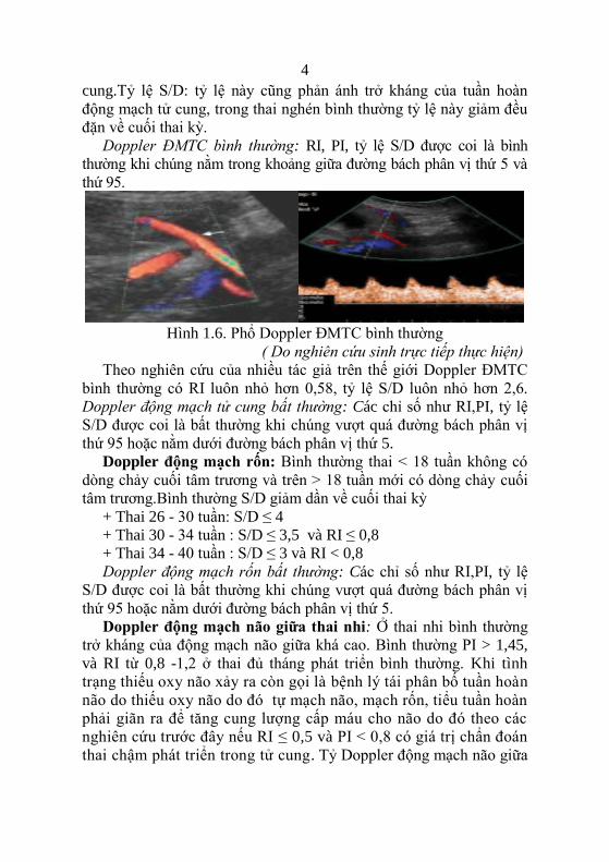
Doppler ĐMTC bình thường: RI, PI, tỷ lệ S/D được coi là bình
thường khi chúng nằm trong khoảng giữa đường bách phân vị thứ 5 và
thứ 95.
Hình 1.6. Phổ Doppler ĐMTC bình thường
( Do nghiên cứu sinh trực tiếp thực hiện)
Theo nghiên cứu của nhiều tác giả trên thế giới Doppler ĐMTC
bình thường có RI luôn nhỏ hơn 0,58, tỷ lệ S/D luôn nhỏ hơn 2,6.
Doppler động mạch tử cung bất thường: Các chỉ số như RI,PI, tỷ lệ
S/D được coi là bất thường khi chúng vượt quá đường bách phân vị
thứ 95 hoặc nằm dưới đường bách phân vị thứ 5.
Doppler động mạch rốn: Bình thường thai < 18 tuần không có
dòng chảy cuối tâm trương và trên > 18 tuần mới có dòng chảy cuối
tâm trương.Bình thường S/D giảm dần về cuối thai kỳ
+ Thai 26 - 30 tuần: S/D ≤ 4
+ Thai 30 - 34 tuần : S/D ≤ 3,5 và RI ≤ 0,8
+ Thai 34 - 40 tuần : S/D ≤ 3 và RI < 0,8
Doppler động mạch rốn bất thường: Các chỉ số như RI,PI, tỷ lệ
S/D được coi là bất thường khi chúng vượt quá đường bách phân vị
thứ 95 hoặc nằm dưới đường bách phân vị thứ 5.
Doppler động mạch não giữa thai nhi: Ở thai nhi bình thường
trở kháng của động mạch não giữa khá cao. Bình thường PI > 1,45,
và RI từ 0,8 -1,2 ở thai đủ tháng phát triển bình thường. Khi tình
trạng thiếu oxy não xảy ra còn gọi là bệnh lý tái phân bố tuần hoàn
não do thiếu oxy não do đó tự mạch não, mạch rốn, tiểu tuần hoàn
phải giãn ra để tăng cung lượng cấp máu cho não do đó theo các
nghiên cứu trước đây nếu RI ≤ 0,5 và PI < 0,8 có giá trị chẩn đoán
thai chậm phát triển trong tử cung. Tỷ Doppler động mạch não giữa
5
/động mạch rốn còn gọi là chỉ số Não / Rốn (CSNR). So sánh phổ
Doppler động mạch rốn và động mạch não giữa, bình thường chỉ số
trở kháng của động mạch não giữa luôn cao hơn động mạch rốn nên
tỷ Não / Rốn > 1. Gọi là tái phân phối tuần hoàn thai nhi khi tỷ Não /
Rốn < 1 bất thường.
1.4. Các nghiên cứu về giá trị chẩn đoán của Doppler trong tiên
lượng tình trạng thai ở thai phụ tiền sản giật.
Các nghiên cứu thăm dò Doppler ĐMTC: Năm 2014, Cru-
Martinez R, Savchev et al ở Barcelona đã nghiên cứu ứng dụng hiệu
ứng Doppler vào quý III thai kỳ có khả năng dự báo tình trạng thiếu
oxy não và tình trạng thai kém phát triển trong tử cung. Như vậy với
những thai nghén nguy cơ cao, Doppler ĐMTC có giá trị tiên đoán
rất tốt về nguy cơ biến chứng với mẹ và thai. Một số nghiên cứu khác
về vai trò của Doppler ĐMTC trong theo dõi THA và thai nghén cho
thấy tỷ lệ S/D >2,6 hoặc xuất hiện vết khuyết tiền tâm trương (dấu
hiệu NOTCH) đó là những dấu hiệu xấu về tiến triển của bệnh với độ
nhạy và độ đặc hiệu là 81% và 90%.
Các nghiên cứu thăm dò Doppler động mạch rốn: Nghiên cứu của
Nguku bệnh viện Nairobi, Kenya 110 trường hợp tăng huyết áp do
thai tỷ lệ RI Doppler động mạch rốn bình thường chiếm 72/110
(66,1%); và chỉ số RI động mạch rốn tăng chiếm 33,9%
Các nghiên cứu thăm dò Doppler động mạch não giữa: Năm 2014,
Padmaja R.Desai và cs đã nghiên cứu mối liên quan của các chỉ số
của Doppler ĐMNG với tình trạng IUGR, ông nhận thấy rằng có sự
giảm trở kháng của Doppler ĐMNG ở các trường hợp IUGR ở thai
phụ tiền sản giật. Nghiên cứu của Ebrashy (2000) trên 50 bệnh nhân
tiền sản giật nhận thấy giá trị Doppler động mạch não giữa là 0,70
±0,01. Ngưỡng của RI ĐMNG được chọn là RI < 0,69, tỷ lệ của RI <
0,69 là 17/50 (34%) .
Tỷ não – rốn trong đánh giá sức khỏe thai ở thai phụ tiền sản
giật:Tỷ chỉ số trở kháng (Rl) của động mạch não giữa/ động mạch
rốn được gọi là tỷ Não – Rau hay còn gọi là tỷ Não – Rốn, có thể coi
như dòng chảy của tái phân phối tuần hoàn tại não “ hiện tượng hiệu
ứng tiết kiệm não ”. Trong thai nghén bình thường, thành phần tâm
trương ở động mạch não thì thấp hơn tâm trương của động mạch rốn
ở bất kỳ tuối thai nào, nếu khi thai có tình trạng thiếu oxy có sự đảo
ngược tuần hoàn dòng tâm trương động mạch rốn giảm đi, dòng tâm
trương động mạch não giữa tăng lên làm cho trở kháng động mạch
6
rốn tăng lên trở kháng của động mạch não giữa giảm đi thể hiện bởi
tỷ số não rốn < 1. Năm 2013, Monika Singh và cộng sự đã tiến hành
nghiên cứu đánh giá Doppler các chỉ số RI, PI, S/D trên 50 bệnh
nhân tiền sản giật, nghiên cứu đã nhận thấy rằng, bất thường Doppler
động mạch não giữa gia tăng tỷ lệ thai kém phát triển trong tử cung
và kết cục bất lợi cho thai kỳ.
Chương 2
ĐỐI TƯỢNG VÀ PHƯƠNG PHÁP NGHIÊN CỨU
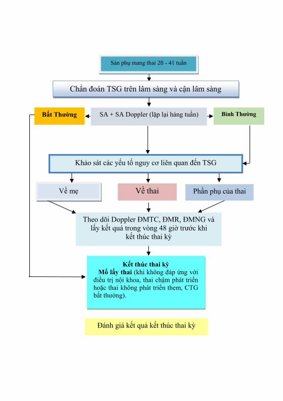
2.1. Đối tượng nghiên cứu: Thai phụ được chẩn đoán và điều trị
tiền sản giật từ tháng 03/01/2013 – 30/ 01/2016 tại Bệnh viện
Trường Đại học Y Dược Huế đồng ý tham gia nghiên cứu.
2.1.1. Tiêu chuẩn chọn bệnh: Tuổi thai từ 28 tuần trở lên (tính từ ngày
đầu tiên của kỳ kinh cuối cùng), một thai sống.
- Có các triệu chứng sau:
+ Huyết áp tâm thu ≥ 140 mmHg hoặchuyết áp tâm trương ≥ 90
mmHg
+ Protein niệu ≥ 0,5 g/l ở mẫu nước tiểu ngẫu nhiên hoặc 0,3 g/l
ở mẫu nước tiểu trong 24 giờ, có thể kèm theo phù
- Các sản phụ được khảo sát siêu âm và làm Doppler ĐMTC,
ĐMR, ĐMNG tại thời điểm trong vòng 48 giờ trước khi kết thúc thai
kỳ.
2.1.2. Tiêu chuẩn loại trừ: Đa ối, thai dị dạng, sản giật , hội chứng
HELLP, bệnh tim, bệnh thận, bệnh tăng huyết áp, bệnh Bazedow,
bệnh đái tháo đường
2.2. Phương pháp nghiên cứu
2.2.1. Phương pháp nghiên cứu tiến cứu, mô tả cắt ngang.
2.2.2. Tính cỡ mẫu:Nghiên cứu của chúng tôi là nghiên cứu về chẩn
đoán nên dựa vào hai chỉ số độ nhạy và độ đặc hiệu, đặc biệt đây là
nghiên cứu chẩn đoán lâm sàng nên chúng tôi dựa vào độ đặc hiệu để
ước tính cỡ mẫu.
Cỡ mẫu: được xác định theo công thức
1sp
dis
FP TNN
p
Trước hết, ước tính FP + TN ( tức là số dương tính giả và âm tính
thật) theo công thức
7
FP + TN =
2
2
(1 )sp spZ p p
Trong đó:
Nsp : Số cỡ mẫu cần thiết cho nghiên cứu, : Sai số cho phép =
5%, Z là hằng số của phân phối chuẩn, nếu khoảng tin cậy cho
phép =0,05 thì hằng số là Z =1,96 ,Psp: Độ tin cậy khoảng 90%
Pdis: Tỷ lệ sản phụ bị tiền sản giật trong các nghiên cứu trước đây
là 8 %, vậy 1- Pdis là 0,92.
Với tỷ lệ hiện hành của bệnh là 8%, số lượng cỡ mẫu cần thiết để
ước tính độ đặc hiệu của các phương pháp theo công thức trên ta có :
FP + TN =
2 2
2 2
(1 ) (1,96) 0,9 0,1138,3
(0,05)
sp spZ p p
1sp
dis
FP TNN
p
=
138,3150,3
0,92
Cỡ mẫu được tính theo công thức trên là 150,3, làm tròn 151 bệnh
nhân.
2.2.3. Phương tiện nghiên cứu
- Máy siêu âm hiệu Siemens Acuson X 300 với đầu dò rẻ quạt 3,5
MHz được sử dụng tại Phòng Siêu âm chẩn đoán Tiền sản Bệnh viện
Trường Đại học Y Dược Huế.
- Sử dụng các bảng phân bố bách phân vị của tỷ lệ S/D, chỉ số trở
kháng RI, chỉ số xung PI của động mạch tử cung, động mạch rốn và
động mạch não giữa theo tuổi thai ở thai phụ bình thường khỏe mạnh
của Trần Danh Cường năm 2007 .
- Phiếu nghiên cứu in sẵn, máy monitoring sản khoa hiệu Avalon
FM20, hãng PHILIPS với đầy đủ các bộ phận và giấy ghi, găng tay
bằng cao su, bình oxy với hệ thống dẫn, máy hút và ống hút nhớt,
mặt nạ và bóng hổ trợ hô hấp, dụng cụ đặt nội khí quản, đèn soi và
ống nội khí quản, các thuốc hồi sức sơ sinh.
2.2.4. Phương pháp tiến hành
2.2.4.1. Khám chẩn đoán tiền sản giật: Phân loại TSG theo bảng
phân loại của Hướng dẫn quốc gia về các dịch vụ chăm sóc sức khoẻ
sinh sản năm 2009
8
2.2.4.2. Khảo sát siêu âm thai và Doppler: Thăm dò Doppler động
mạch tử cung, động mạch rốn, động mạch não giữa xem xét hình thái
phổ và đo các chỉ số: chỉ số xung PI, chỉ số kháng RI, tỷ lệ S/D.
Chỉ số Não – Rốn: Xác định tỷ Doppler RI Động mạch não/Động
mạch rốn còn gọi là chỉ số Não/Rốn.
2.2.5. Theo dõi và đánh giá kết quả kết thúc thai kỳ
- Tuổi thai khi sinh (hoặc kết thúc thai kỳ).
- Lý do thai phụ phải kết thúc thai kỳ (nếu thai phụ kết thúc thai kỳ
chủ động):
+ Không đáp ứng với điều trị nội khoa
+ Thai không phát triển hay chậm phát triển trong tử cung.
+ CTG bất thường.
- Cách sinh.
- Đánh giá tình trạng thai:
+ Thai chậm phát triển trong tử cung (IUGR): là trẻ sinh ra có
cân nặng dưới đường bách phân vị thứ 10 của biểu đồ phát triển cân
nặng thai nhi theo tuổi thai của Phan Trường Duyệt (2005), vì đối
tượng trong mẫu nghiên cứu của chúng tôi là người Việt nam.
+ Thai suy: trong nghiên cứu này đánh giá suy thai khi có một
trong các dấu hiệu sau:
- Monitoring xuất hiện nhịp phẳng kéo dài trên 60 phút sau khi đã
loại trừ thai ngủ hoặc CTG bất thường xuất hiện nhịp giảm DIP II,
DIP biến đổi.
- Đánh giá tình trạng trẻ sau sinh:
. Tình trạng ối: số lượng, màu sắc
. Trọng lượng trẻ sau khi sinh.
. Chỉ số Apgar của trẻ.
2.3. Phương pháp xử lý số liệu
- Nhập số liệu bằng phần mềm Epi Data 3.1.
- Sử dụng phương pháp vẽ đường cong ROC, tính diện tích dưới
đường cong AUC và xác định giá trị điểm cắt tối ưu theo chỉ số
Youden bằng phần mềm Medcalc. Diện tích dưới đường cong ROC –
AUC ( Area Under the Cuver), là đại diện cho độ chính xác của
phương pháp đánh giá. Đánh giá giá trị phương pháp chẩn đoán bằng
các thông số: độ nhạy, độ đặc hiệu, giá trị tiên đoán dương tính, giá
trị tiên đoán âm tính. Sử dụng phần mềm NCSS11: sử dụng test so
sánh Mc Nemar để so sánh hiệu quả của các phương pháp khảo sát
bằng Doppler
9
- Các so sánh có ý nghĩa thống kê khi p < 0,05.
2.4. Đạo đức trong nghiên cứu:Nghiên cứu được thực hiện sau khi
đề cương được hội đồng chuyên môn ngành Sản phụ khoa thông qua.
Chương 3
KẾT QUẢ NGHIÊN CỨU
3.1. Đặc điểm mẫu nghiên cứu
3.1.1. Tuổi của thai phụ: Tuổi mẹ trung bình trong mẫu nghiên cứu
là 30,5 ± 6,4, tuổi nhỏ nhất là 20 tuổi lớn nhất là 44.
3.1.2. Số lần mang thai: Sản phụ mang thai lần đầu chiếm tỷ lệ cao
hơn 53,6%, mang thai ≥ 2 lần chiếm 46,4%.
3.1.3. Triệu chứng cơ năng:Triệu chứng phù chiếm tỷ lệ cao nhất
43,8 % sản phụ, đau đầu chiếm 23,5%, hoa mắt chóng mặt và nhìn
mờ chiếm lỷ lệ tương đương nhau là 3,9%
3.1.4. Huyết áp trung bình của mẫu nghiên cứu: Huyết áp tâm thu
trung bình của các sản phụ trong mẫu nghiên cứu là 144,58 ± 6,96
mmHg, tâm trương là 89,65 ± 7,08 mmHg, huyết áp tâm thu trung
bình ở nhóm tiền sản giật nặng là 169,39 ± 13,34 mmHg và tâm
trương trung bình là 103,60 ± 11,53mmHg.
3.1.5. Phân bố tình trạng bệnh lý tiền sản giật: Tỷ lệ tiền sản trong
nghiên cứu này là 54% và tỷ lệ tiền sản giật nặng là 46%
3.1.6. Nhóm tuổi thai: Nghiên cứu trên 153 thai phụ tiền sản giật có
tuổi thai từ 28 đến 41 tuần, tuổi thai tối thiểu là 28 tuần và tối đa là
41 tuần.
3.1.7. Phân bố tình trạng bệnh lý của thai: Ở nhóm tuổi thai 28-33
tuần, tỷ lệ thai suy và thai kém phát triển là 100%. Ở tuổi thai 34-37
tuần tỷ lệ thai suy chiếm 35,5%, thai kém phát triển 41,9%. Ở nhóm
tuổi thai > 37 tuần tỷ lệ thai suy 14,2%, thai kém phát triển 8,8%.
3.1.8. Phương thức kết thúc thai kỳ: Tỷ lệ mổ lấy thai trong nghiên
cứu của chúng tôi là 56,2%, tỷ lệ sinh đường âm đạo là 43,8%
3.2. Tình trạng trẻ sơ sinh sau sinh: Trẻ sơ sinh đa số có Apgar
phút thứ 5 >7 điểm chiếm 76,5 %, 22,1 % có Apgar từ 4 -7 điểm, chỉ
có 2 trường hợp Apgar xấu <3 điểm.
3.2.1. Tình trạng trẻ sơ sinh sau sinh 48 giờ: Tỷ lệ trẻ sơ sinh bình
thường khỏe mạnh cao nhất chiếm 86,9 %, có 11,8 % trẻ phải chăm
sóc ở đơn vị Nhi sơ sinh do thai suy và IUGR
3.2.2. Trọng lượng thai khi sinh: Đa số trẻ sinh ra có trọng lượng
10
>2500gr chiếm tỷ lệ 79,1 %, nhóm trẻ có trọng lượng thấp dưới 1500
chiếm tỷ lệ thấp nhất 2%, nhóm trẻ có trọng lượng từ 1500 -< 2000gr
chiếm tỷ lệ 5,9%, nhóm trẻ có trọng lượng từ 2000-<2500 là 13,1%.
3.3. Giá trị điểm cắt của chỉ số Doppler động mạch tử cung, động
mạch rốn, động mạch não giữa trong tiên lượng thai suy và thai
kém phát triển trong tử cung ( IUGR)
Điểm cắt chỉ số xung PI động mạch tử cung tiên lượng thai suy
ở tuổi thai 34-37 tuần là 1với độ nhạy (Se) là 72,73% và độ đặc hiệu
( Sp) là 65%.Ở nhóm tuổi thai >37 tuần là 1,15 với Se là 87,50% và
Sp là 28,87%. Diện tích dưới đường cong ROC (AUC) ở tuổi thai 34 -
37 tuần và ở tuổi thai > 37 tuần lần lượt là 0,62 và 0,51. Không có mối
tương quan rõ giữa chỉ số xung PI ĐMTC trong tiên lượng thai suy
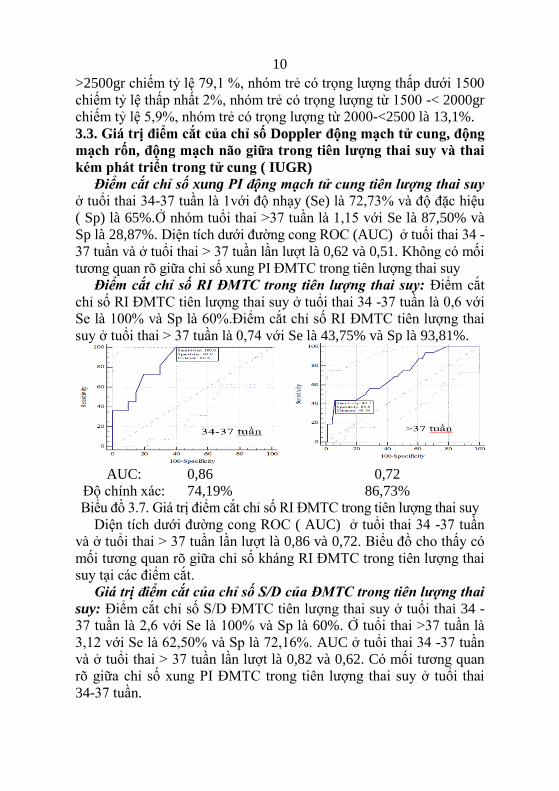
Điểm cắt chỉ số RI ĐMTC trong tiên lượng thai suy: Điểm cắt
chỉ số RI ĐMTC tiên lượng thai suy ở tuổi thai 34 -37 tuần là 0,6 với
Se là 100% và Sp là 60%.Điểm cắt chỉ số RI ĐMTC tiên lượng thai
suy ở tuổi thai > 37 tuần là 0,74 với Se là 43,75% và Sp là 93,81%.
AUC:
Độ chính xác:
0,86
74,19%
0,72
86,73%
Biểu đồ 3.7. Giá trị điểm cắt chỉ số RI ĐMTC trong tiên lượng thai suy
Diện tích dưới đường cong ROC ( AUC) ở tuổi thai 34 -37 tuần
và ở tuổi thai > 37 tuần lần lượt là 0,86 và 0,72. Biểu đồ cho thấy có
mối tương quan rõ giữa chỉ số kháng RI ĐMTC trong tiên lượng thai
suy tại các điểm cắt.
Giá trị điểm cắt của chỉ số S/D của ĐMTC trong tiên lượng thai
suy: Điểm cắt chỉ số S/D ĐMTC tiên lượng thai suy ở tuổi thai 34 -
37 tuần là 2,6 với Se là 100% và Sp là 60%. Ở tuổi thai >37 tuần là
3,12 với Se là 62,50% và Sp là 72,16%. AUC ở tuổi thai 34 -37 tuần
và ở tuổi thai > 37 tuần lần lượt là 0,82 và 0,62. Có mối tương quan
rõ giữa chỉ số xung PI ĐMTC trong tiên lượng thai suy ở tuổi thai
34-37 tuần.
11
Điểm cắt tiên lượng IUGR của các chỉ số Doppler ĐMTC
Điểm cắt chỉ số PI ĐMTC tiên lượng thai kém phát triển: ở tuổi
thai 34 -37 tuần là 1 với độ nhạy Se là 69,23% và độ đặc hiệu Sp là
66,67%., ở tuổi thai >37 tuần là 1,1 với Se là 60% và Sp là 66,69%.
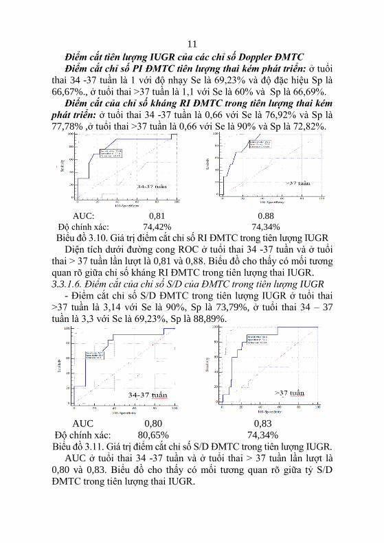
Điểm cắt của chỉ số kháng RI ĐMTC trong tiên lượng thai kém
phát triển: ở tuổi thai 34 -37 tuần là 0,66 với Se là 76,92% và Sp là
77,78% ,ở tuổi thai >37 tuần là 0,66 với Se là 90% và Sp là 72,82%.
AUC:
Độ chính xác:
0,81
74,42%
0.88
74,34%
Biểu đồ 3.10. Giá trị điểm cắt chỉ số RI ĐMTC trong tiên lượng IUGR
Diện tích dưới đường cong ROC ở tuổi thai 34 -37 tuần và ở tuổi
thai > 37 tuần lần lượt là 0,81 và 0,88. Biểu đồ cho thấy có mối tương
quan rõ giữa chỉ số kháng RI ĐMTC trong tiên lượng thai IUGR.
3.3.1.6. Điểm cắt của chỉ số S/D của ĐMTC trong tiên lượng IUGR
- Điểm cắt chỉ số S/D ĐMTC trong tiên lượng IUGR ở tuổi thai
>37 tuần là 3,14 với Se là 90%, Sp là 73,79%, ở tuổi thai 34 – 37
tuần là 3,3 với Se là 69,23%, Sp là 88,89%.
AUC
Độ chính xác:
0,80
80,65%
0,83
74,34%
Biểu đồ 3.11. Giá trị điểm cắt chỉ số S/D ĐMTC trong tiên lượng IUGR.
AUC ở tuổi thai 34 -37 tuần và ở tuổi thai > 37 tuần lần lượt là
0,80 và 0,83. Biểu đồ cho thấy có mối tương quan rõ giữa tỷ S/D
ĐMTC trong tiên lượng thai IUGR.
12
3.3.2. Giá trị tiên lượng của Doppler động mạch rốn
Điểm cắt tiên lượng thai suy của các chỉ số động mạch rốn:-
Điểm cắt chỉ số xung PI ĐMR tiên lượng thai suy ở tuổi thai 34 – 37 tuần là
1,34 với Se là 45,45% và Sp là 90%, PI ĐMR tiên lượng thai suy ở tuổi
thai >37 tuần là 0,97 với Se là 81,25% và Sp là 41,24%. AUC ở tuổi
thai 34 - 37 tuần và ở tuổi thai > 37 tuần lần lượt là 0,66 và 0,59.
Không có mối tương quan rõ giữa PI ĐMR trong tiên lượng thai suy.
Điểm cắt của RI động mạch rốn trong tiên lượng thai suy: ở
tuổi thai 34 – 37 tuần là 0,64 với Se là 90,91% và Sp là 45%. Ở tuổi
thai >37 tuần là 0,75 với Se là 50% và Sp là 86,60%. AUC ở tuổi thai
34 -37 tuần và ở tuổi thai > 37 tuần lần lượt là 0,73 và 0,67. Có mối
tương quan giữa chỉ số kháng RI ĐMR trong tiên lượng thai suy.
Điểm cắt tiên lượng suy thai của tỷ S/D động mạch rốn: Ở tuổi
thai 34 – 37 tuần là 4,24 với Se là 18,18% và Sp là 100%. Ở tuổi thai
>37 tuần là 2,24 với Se là 75% và Sp là 16,49%.
Điểm cắt của PI ĐMR tiên lượng thai kém phát triển: Ở tuổi
thai 34 – 37 tuần là 0,82 với Se là 100% và Sp là 33,33%.Ở tuổi thai
>37 tuần là 1,29 với Se là 50% và Sp là 83,5%.
Điểm cắt tiên lượng IUGR của RI động mạch rốn:- Ở tuổi thai
34–37 tuần là 0,74 với Se là 61,54% và Sp là 88,89%.Ở thai >37
tuần là 0,76 với Se là 50% và Sp là 90,29%. AUC ở tuổi thai 34 -37
tuần và ở tuổi thai > 37 tuần lần lượt là 0,79 và 0,70.
Điểm cắt của tỷ số S/D ĐMR trong tiên lượng thai kém phát
triển:- Ở tuổi thai 34 -37 tuần là 3,79 với Se là 30,77% và Sp là
94,44%. Ở tuổi thai > 37 tuần là 3,16 với Se là 60% và Sp là 66,69%.
3.3.3. Điểm cắt tiên lượng các chỉ số Doppler động mạch não giữa
Điểm cắt PI của ĐMNG trong tiên lượng thai suy: ở tuổi thai 34 -37
tuần là 1,2 với Se là 81,82% và Sp là 85%. Ở tuổi thai > 37 tuần là 1,07 với
độ nhạy Se là 37,50% và độ đặc hiệu Sp là 87,63%.
Điểm cắt các chỉ số RI ĐMNG trong tiên lượng thai suy: Điểm
cắt RI của ĐMNG trong tiên lượng thai suy ở tuổi thai 34 -37 tuần là
0,68. Điểm cắt RI của ĐMNG trong tiên lượng thai suy ở tuổi thai >37 tuần
là 0,65 với Se là 56,25% và Sp là 86,60%.
Điểm cắt các chỉ số S/D ĐMNG trong tiên lượng thai suy: ở tuổi
thai 34 -37 tuần là 3,09 với Se là 72,73% và Sp là 85,00, ở tuổi thai
>37 tuần là 3,42
13
Điểm cắt của Doppler ĐMNG trong tiên lượng IUGR:ở tuổi
thai 34 -37 tuần là 1,35 với Se là 69,23% và Sp là 77,78%, ở tuổi thai
>37 tuần là 1,06 với Se là 60% và Sp là 90,29%.
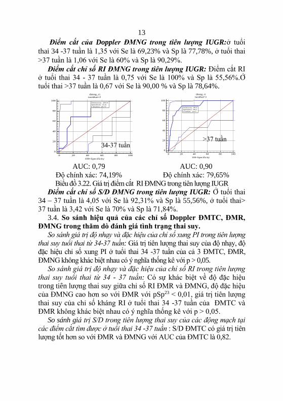
Điểm cắt chỉ số RI ĐMNG trong tiên lượng IUGR: Điểm cắt RI
ở tuổi thai 34 - 37 tuần là 0,75 với Se là 100% và Sp là 55,56%.Ở
tuổi thai >37 tuần là 0,67 với Se là 90,00 % và Sp là 78,64%.
AUC: 0,79
Độ chính xác: 74,19%
AUC: 0,90
Độ chính xác: 79,65%
Biểu đồ 3.22. Giá trị điểm cắt RI ĐMNG trong tiên lượng IUGR
Điểm cắt chỉ số S/D ĐMNG trong tiên lượng IUGR: Ở tuổi thai
34 – 37 tuần là 4,05 với Se là 92,31% và Sp là 55,56%, ở tuổi thai>
37 tuần là 3,42 với Se là 70% và Sp là 71,84%.
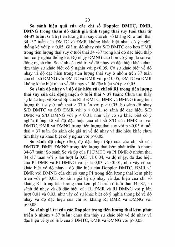
3.4. So sánh hiệu quả của các chỉ số Doppler ĐMTC, ĐMR,
ĐMNG trong thăm dò đánh giá tình trạng thai suy.
So sánh giá trị độ nhạy và đặc hiệu của chỉ số xung PI trong tiên lượng
thai suy tuổi thai từ 34-37 tuần: Giá trị tiên lượng thai suy của độ nhạy, độ
đặc hiệu chỉ số xung PI ở tuổi thai 34 -37 tuần của cả 3 ĐMTC, ĐMR,
ĐMNG không khác biệt nhau có ý nghĩa thống kê với p > 0,05.
So sánh giá trị độ nhạy và đặc hiệu của chỉ số RI trong tiên lượng
thai suy tuổi thai từ 34 - 37 tuần: Có sự khác biệt về độ đặc hiệu
trong tiên lượng thai suy giữa chỉ số RI ĐMR và ĐMNG, độ đặc hiệu
của ĐMNG cao hơn so với ĐMR với pSp23 < 0,01, giá trị tiên lượng
thai suy của chỉ số kháng RI ở tuổi thai 34 -37 tuần của ĐMTC và
ĐMR không khác biệt nhau có ý nghĩa thống kê với p > 0,05.
So sánh giá trị S/D trong tiên lượng thai suy của các động mạch tại
các điểm cắt tìm được ở tuổi thai 34 -37 tuần : S/D ĐMTC có giá trị tiên
lượng tốt hơn so với ĐMR và ĐMNG với AUC của ĐMTC là 0,82.
0
20
40
60
80
100
dmng_ri
tuoithai=2
0 20 40 60 80 100
100-Specificity
Sens
itivi
ty
Sensitivity: 100.0
Specificity: 55.6
Criterion: ≤0.75
0
20
40
60
80
100
dmng_ri
tuoithai=3
0 20 40 60 80 100
100-Specificity
Sens
itivi
ty
Sensitivity: 90.0
Specificity: 78.6
Criterion: ≤0.67
34-37 tuần >37 tuần
14
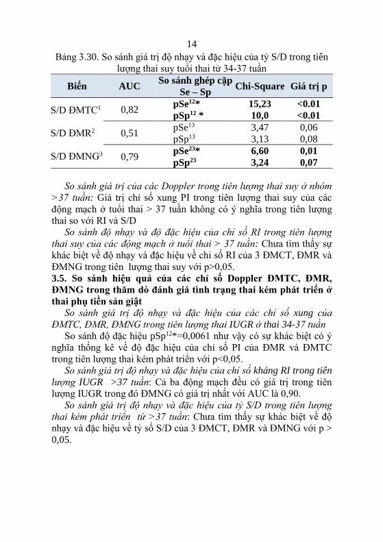
Bảng 3.30. So sánh giá trị độ nhạy và đặc hiệu của tỷ S/D trong tiên
lượng thai suy tuổi thai từ 34-37 tuần
Biến AUC So sánh ghép cặp
Se – Sp Chi-Square Giá trị p
S/D ĐMTC1
S/D ĐMR2
S/D ĐMNG3
0,82 pSe12* 15,23 <0.01
pSp12 * 10,0 <0.01
0,51 pSe13 3,47 0,06
pSp13 3,13 0,08
0,79 pSe23* 6,60 0,01
pSp23 3,24 0,07
So sánh giá trị của các Doppler trong tiên lượng thai suy ở nhóm
>37 tuần: Giá trị chỉ số xung PI trong tiên lượng thai suy của các
động mạch ở tuổi thai > 37 tuần không có ý nghĩa trong tiên lượng
thai so với RI và S/D
So sánh độ nhạy và độ đặc hiệu của chỉ số RI trong tiên lượng
thai suy của các động mạch ở tuổi thai > 37 tuần: Chưa tìm thấy sự
khác biệt về độ nhạy và đặc hiệu về chỉ số RI của 3 ĐMCT, ĐMR và
ĐMNG trong tiên lượng thai suy với p>0,05.
3.5. So sánh hiệu quả của các chỉ số Doppler ĐMTC, ĐMR,
ĐMNG trong thăm dò đánh giá tình trạng thai kém phát triển ở
thai phụ tiền sản giật
So sánh giá trị độ nhạy và đặc hiệu của các chỉ số xung của
ĐMTC, ĐMR, ĐMNG trong tiên lượng thai IUGR ở thai 34-37 tuần
So sánh độ đặc hiệu pSp12*=0,0061 như vậy có sự khác biệt có ý
nghĩa thống kê về độ đặc hiệu của chỉ số PI của ĐMR và ĐMTC
trong tiên lượng thai kém phát triển với p<0,05.
So sánh giá trị độ nhạy và đặc hiệu của chỉ số kháng RI trong tiên
lượng IUGR >37 tuần: Cả ba động mạch đều có giá trị trong tiên
lượng IUGR trong đó ĐMNG có giá trị nhất với AUC là 0,90.
So sánh giá trị độ nhạy và đặc hiệu của tỷ S/D trong tiên lượng
thai kém phát triển từ >37 tuần: Chưa tìm thấy sự khác biệt về độ
nhạy và đặc hiệu về tỷ số S/D của 3 ĐMCT, ĐMR và ĐMNG với p >
0,05.
15
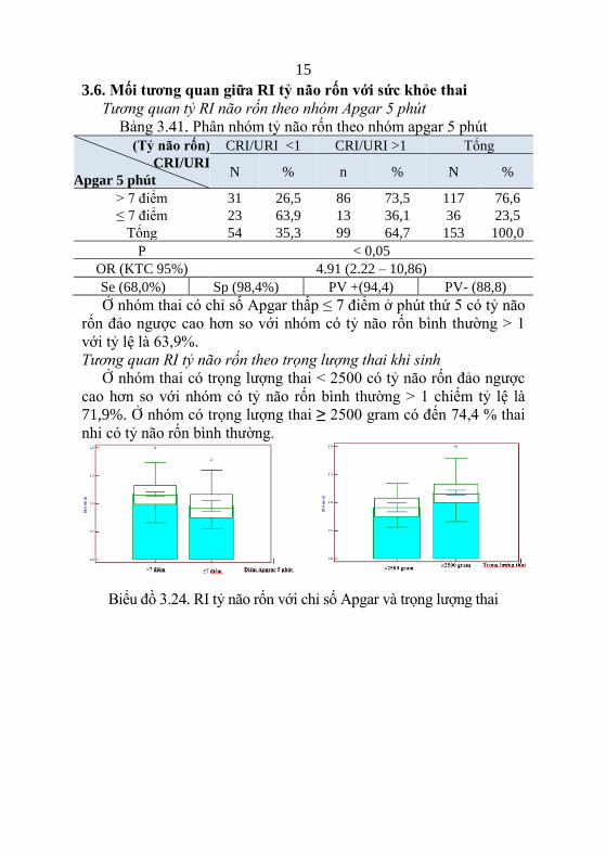
3.6. Mối tương quan giữa RI tỷ não rốn với sức khỏe thai
Tương quan tỷ RI não rốn theo nhóm Apgar 5 phút
Bảng 3.41. Phân nhóm tỷ não rốn theo nhóm apgar 5 phút
(Tỷ não rốn)
CRI/URI
Apgar 5 phút
CRI/URI <1 CRI/URI >1 Tổng
N % n % N %
> 7 điểm 31 26,5 86 73,5 117 76,6
≤ 7 điểm 23 63,9 13 36,1 36 23,5
Tổng 54 35,3 99 64,7 153 100,0
P < 0,05
OR (KTC 95%) 4.91 (2.22 – 10,86)
Se (68,0%) Sp (98,4%) PV +(94,4) PV- (88,8)
Ở nhóm thai có chỉ số Apgar thấp ≤ 7 điểm ở phút thứ 5 có tỷ não
rốn đảo ngược cao hơn so với nhóm có tỷ não rốn bình thường > 1
với tỷ lệ là 63,9%.
Tương quan RI tỷ não rốn theo trọng lượng thai khi sinh
Ở nhóm thai có trọng lượng thai < 2500 có tỷ não rốn đảo ngược
cao hơn so với nhóm có tỷ não rốn bình thường > 1 chiếm tỷ lệ là
71,9%. Ở nhóm có trọng lượng thai ≥ 2500 gram có đến 74,4 % thai
nhi có tỷ não rốn bình thường.
Biểu đồ 3.24. RI tỷ não rốn với chỉ số Apgar và trọng lượng thai
16
Chương 4
BÀN LUẬN
4.1. Đặc điểm chung
4.1.1. Tuổi mẹ: Tuổi trung bình của các sản phụ trong nghiên cứu
này là 30,5 ± 6,4 tuổi, tuổi lớn tuổi là 44 tuổi và nhỏ nhất là 20 tuổi.
Nhóm tuổi mắc bệnh nhiều nhất là 20 – 35 tuổi.
4.1.2. Phân loại tiền sản giật: TSG chiếm tỷ lệ cao hơn 53,6% và
bệnh lý TSG nặng chiếm 46,4%.Trước đây, người ta phân loại bệnh
lý tiền sản giật thành hai mức độ nhẹ và nặng. Hiện nay, người ta áp
dụng cách phân loại mới là tiền sản giật và tiền sản giật nặng. Phân
loại theo mức độ bệnh TSG và TSG nặng khá mới nên không có
nhiều nghiên cứu để so sánh.
4.2.. Đặc điểm lâm sàng: đau đầu chiếm 23,5%, phù chiếm tỷ lệ
chiếm 43,8%. Nghiên cứu của Nguyễn Thị Bích Vân cho thấy triệu
chứng lâm sàng của tiền sản giật nặng tỷ lệ cao nhất là đau đầu
chiếm 55,4%. Triệu chứng rối loạn thị giác có tỷ lệ 21,6% cao hơn
so với đau vùng thượng vị (12,2%).
4.3. Phương pháp sinh: Tỷ lệ mổ lấy thai chiếm 56,2%, tỷ lệ này
thấp hơn một số tác giả khác, theo nghiên cứu của Phạm Thị Mai
Anh tỷ lệ mổ lấy thai chiếm tới 90%.
4.3.1. Tình trạng sơ sinh sau khi đẻ: Theo kết quả nghiên cứu của
chúng tôi trong 153 trẻ, có 151 trẻ sơ sinh sống trong đó 36 trẻ sinh
ra có Apgar thấp, trong đó 2 trẻ tử vong 34 trẻ có tình trạng suy thai ở
phút thứ 5, và sau 5 phút thì vẫn cần hồi sức tích cực và hô hấp hỗ trợ
(22,1%).
4.3.2. Chỉ số Apgar: Có 36 trẻ sơ sinh có chỉ số Apgar ở phút thứ
năm < 7 điểm chiếm tỷ lệ 23,5 %.
4.4. Giá trị các chỉ số Doppler với tình trạng thai
Mối tương quan giữa RI ĐMTC tình trạng thai: Kết quả
nghiên cứu phản ánh mối tương quan giữa RI ĐMTC với suy thai.
Chúng tôi tìm được điểm cắt 0,6 ở nhóm tuổi thai 34 – 37 tuần với độ
nhạy, độ đặc hiệu, giá trị tiên đoán dương tính, giá trị tiên đoán âm
tính lần lượt là 100%, 65%, 57,9%, 100%. Còn ở nhóm trên 37 tuần
điểm cắt tiên lượng thai suy là 0,74. Như vậy điểm cắt 0,6 ở tuổi thai
34 -37 tuần của chúng tôi có thể ứng dụng trên lâm sàng. Theo
nghiên cứu mới nhất năm 2016 của tác giả Mozibur Rahman Laskar
giá trị RI ĐMTC ở nhóm tiền sản giật cao hơn so với nhóm thai kỳ
17
bình thường. Độ nhạy của tăng RI ĐMTC là 84,7 trong nghiên cứu
của Mozibur (2016), theo nghiên cứu của Colemann et al. là 83%.
Kết quả nghiên cứu của chúng tôi cũng tương đồng với các nghiên
cứu của tác giả khác.
Về mối tương quan giữa tỷ lệ S/D ĐMTC và tình trạng thai
Ở tuổi thai 34 -37 điểm cắt tiên lượng thai suy và thai kém phát
triển lần lượt là 2,6 và 3,3 với độ nhạy, độ đặc hiệu, giá trị tiên đoán
dương tính, giá trị tiên đoán âm tính là 100%, 65%, 57,9%, 100% và
độ nhạy, độ đặc hiệu, giá trị tiên đoán dương tính, giá trị tiên đoán âm
tính là 69,2%, 88,9%, 81,85%, 80%, còn ở nhóm tuổi thai trên 37 tuần
điểm cắt S/D tiên lượng tình trạng thai suy và thai kém phát triển lần
lượt là 3,12 và 3,14. Kết quả điểm cắt của chúng tôi cao hơn so với kết
quả nghiên cứu của Phạm Thị Mai Anh giữa tỷ lệ S/D của ĐMTC
trong tiên lượng thai IUGR là 2,6. Ở Việt Nam Vũ Hoàng Yến (2007),
tìm được điểm cắt của tỷ lệ S/D ở ĐMTC trong tiên lượng thai IUGR
và suy thai là 2,7. Tại điểm cắt này giá trị tiên lượng thai IUGR với độ
nhạy, độ đặc hiệu là 84,1%, 71,4% và giá trị tiên lượng thai suy là
92,3% và 75% . Như vậy điểm cắt 2,6 của tỷ số S/D ĐMTC ở tuổi thai
34-37 tuần trong nghiên cứu của chúng tôi có giá trị trong tiên lượng
thai suy. Kết quả nghiên cứu mới nhất năm 2016 của Vijakumar năm
2016 trên 80 bệnh nhân tiền sản giật cho thấy giá trị tiên đoán IUGR
của S/D ĐMTC có độ nhạy 56,7%, độ đặc hiệu 88,3%, giá trị tiên
đoán dương tính 77,5%.
Giá trị của chỉ số Doppler động mạch rốn trong tiên lượng thai
Giá trị của chỉ số xung động mạch rốn trong tiên lượng thai
suy: Điểm cắt PI tiên lượng thai suy ở nhóm tuổi thai 34 -37 tuần tại
1,34, và ở nhóm tuổi thai > 37 tuần là 0,97 kết quả điểm cắt trong
nghiên cứu của chúng tôi cao hơn so với Nguyễn Thị Bích Vân tại
điểm cắt 1,2 giá trị của chỉ số xung có vai trò tiên lượng thai suy cao
nhất ở tuổi thai 33 - 37 tuần tương ứng với độ nhạy 84,6, độ đặc hiệu
73,3%, giá trị tiên đoán dương tính 57,9%, giá trị tiên đoán âm tính
91,7%. Kết quả này cao hơn so với các tác giả trong nước Tạ Thị
Xuân Lan lựa chọn điểm cắt là 1,2. Đinh Thị Thuý Hằng là 1,1
nhưng tương đồng với các tác giả nước ngoài.
Giá trị của chỉ số xung ĐMR trong tiên lượng thai chậm phát
triển: Kết quả nghiên cứu của chúng tôi ở tuổi thai 34 - 37 tuần, tại
điểm cắt 0,82 tương ứng với độ nhạy 100 %, độ đặc hiệu 33,3%, giá trị
tiên đoán dương tính 52 %, giá trị tiên đoán âm tính 100 %. Ở tuổi thai
18
> 37 tuần, tại điểm cắt 1,29 tương ứng với độ nhạy 50 % độ đặc hiệu
83,5 %. Kết quả nghiên cứu của Nguyễn Thị Bích Vân ở tuổi thai 34 -
37 tuần, tại điểm cắt 1,ở tuổi thai > 37 tuần, tại điểm cắt 1,0 tương ứng
với độ nhạy 62,5%, độ đặc hiệu 65% , còn nghiên cứu của chúng tôi
điểm cắt của chỉ số PI ĐMR ở tuổi thai 34-37 tuần trong tiên lượng
thai kém phát triển là 0,82 với độ nhạy 100% nhưng độ đặc hiệu chỉ
33,33% là từ đây chúng tôi rút ra nhận xét là PI ĐMR có khả năng phát
hiện thai IUGR khá cao nhưng khả năng loại trừ thai không IUGR lại
không cao.
Giá trị của chỉ số kháng ( RI) động mạch rốn trong tiên lượng
thai suy: Nghiên cứu của chúng tôi đã cho thấy giá trị của RI ĐMR
tuổi thai 34 - 37 tuần > 0,64 và ở tuổi thai > 37 tuần > 0,75 có giá trị
sàng lọc thai suy cao nhất vì tại các điểm cắt này giá trị tiên lượng thai
suy có độ nhạy cao từ 84,6 - 87,5 %. Nghiên cứu của Nguyễn Thị
Bích Vân với RI > 0,73 thì khả năng chẩn đoán thai suy ở tuổi thai 34
- 37. Ở tuổi thai trên 37 tuần, nghiên cứu của chúng tôi đã tìm ra
điểm cắt chỉ số kháng (RI) là 0,75 với độ nhạy 100%, độ đặc hiệu
86,6% cao, trong khi nghiên cứu Nguyễn Thị Bích Vân đã chọn điểm
cắt 0,71 vì ở điểm cắt này giá trị tiên lượng thai suy của chỉ số kháng
có độ nhạy và độ đặc hiệu đạt giá trị cao nhất, tuy nhiên giá trị của độ
nhạy chỉ 50%, mặc đù độ đặc hiệu đạt tới 81,3 %. Nghiên cứu mới
nhất năm 2016 của tác giả Vịjaykumar Mane cho thấy rằng giá trị
Doppler động mạch rốn tiên đoán thai suy với độ nhạy cao 91,7%, độ
đặc hiệu là 70,9%, giá trị tiên đoán dương tính 73,8%.
Giá trị của chỉ số kháng động mạch rốn trong tiên lượng thai
chậm phát triển: Nghiên cứu của V.A.A Lakshmi et al năm 2015 cho
thấy rằng chỉ số kháng động mạch rốn trong tiên lượng thai kém phát
triển với độ nhạy là 84,9%, độ đặc hiệu 72,3%. Kết quả nghiên cứu
Nguyễn Thị Bích Vân cho thấy ở tuổi thai 34-37 tuần, chỉ số kháng
tại điểm cắt 0,65 tương ứng với Se 82,8%, độ đặc hiệu 28,6%.
Giá trị chẩn đoán thai suy và IUGR của S/D động mạch rốn
Theo nghiên cứu của tác giả Đinh Thị Thúy Hằng cho thấy với tại
điểm cắt S/D < 2,6 giá trị chẩn đoán thai chậm phát triển trong tử
cung có độ nhạy hay khả năng phát hiện thai IUGR là 100%, độ đặc
hiệu chỉ đạt 58%. Đối với thai suy, giá trị có khả năng phát hiện đạt
được > 95% và khả năng loại trừ thai không suy cao nhất là 45%. Khi
giá trị tỷ số S/D > 2,6, khả năng phát hiện IUGR có độ nhạy cao nhất
là 95% và độ đặc hiệu thấp nhất đạt 78%, khả năng phát hiện thai suy
19
khi S/D >2,6 đạt cao nhất là 88% , tác giả đã chọn điểm cắt tại 2,6 để
chẩn đoán thai suy và IUGR với độ nhạy là 98% .Theo nghiên cứu
của các tác giả Lê Thị Thu Hà, Tạ Thị Xuân Lan giá trị chẩn đoán
thai chậm phát triển của S/D có độ nhạy từ 79 – 97,6%. Nghiên cứu
của các tác giả Divon, Maulik giá trị chẩn đoán của tỷ lệ S/D có độ
nhạy nằm trong khoảng từ 49 % đến 79%.
Giá trị của chỉ số xung PI ĐMNG trong tiên lượng thai
Để chẩn đoán tình trạng thai suy của PI ĐMNG điểm cắt 1,2 ở
tuổi thai 34 - 37 tuần, tương ứng với độ nhạy 81,8%, độ đặc hiệu
85%, giá trị tiên đoán dương tính 75 %, giá trị tiên đoán âm tính 89,5.
Ở tuổi thai > 37 tuần, tại điểm cắt 1,07. Để chẩn đoán tình trạng thai
IUGR kết quả nghiên cứu của chúng tôi ghi nhận được điểm cắt PI
cao hơn so với điểm cắt để dự báo thai suy, tại điểm cắt 1,35 tương
ứng với tuổi thai 34-37 tuần với độ nhạy 69,2 %, độ đặc hiệu 77,8 %,
giá trị tiên đoán đoán dương tính 69,2%, giá trị tiên đoán âm tính
77,8. ở tuổi thai >37 tuần cho thấy tại điểm cắt 1.06 tương ứng với độ
nhạy 60 %, độ đặc hiệu 90,3 %, giá trị tiên đoán dương tính 37,5 %,
giá trị tiên đoán âm tính 95,9 %. Nghiên cứu của Nguyễn Thị Bích
Vân cho thâý để chẩn đoán tình trạng thai suy của PI ĐMNG, ở tuổi
thai 34 - 37 tuần ở điểm cắt 1,3, ở tuổi thai > 37 tuần, tại điểm cắt
1,2. Nghiên cứu của chúng tôi cũng phù hợp với nghiên cứu của
Mozibur Rahman Laskar et al năm 2016 chỉ số xung của động mạch
não giữa (MCA PI )có độ đặc hiệu cao 90% trong tiên lượng thai suy
và thai kém phát triển .
Giá trị của điểm cắt RI ĐMNG với tiên lượng thai suy: Giá trị
dự báo thai suy của chỉ số kháng RI ĐMNG là 0,68 có độ nhạy
81,82%, độ đặc hiệu 85%. Như vậy RI ĐMNG có thể có giá trị trong
chẩn đoán thai suy ở thai phụ tiền sản giật (p≤ 0,01). Kết quả nghiên
cứu của Nguyễn Thị Bích Vân chẩn đoán thai suy ở tuổi thai 34 - 37
tuần, tại điểm cắt RI ĐMNG 0,7 . Ở tuổi thai > 37 tuần, tại điểm cắt
0,7 tương ứng với độ nhạy 75%, độ đặc hiệu 59,4%, giá trị tiên đoán
dương tính 18,8%, giá trị tiên đoán âm tính 95%.
Giá trị của điểm cắt RI ĐMNG với tiên lượng thai IUGR: Tác
giả Nguyễn Thị Bích Vân cho thấy giá trị chẩn đoán thai IURG ở
tuổi thai 34-37 tuần, tại điểm cắt 0,72 tương ứng với Se 41,4%, Sp
64,29%, giá trị tiên đoán dương tính 58,6%, giá trị tiên đoán âm tính
35,7% và ở tuổi thai >37 tuần, tại điểm cắt 0,72 tương ứng với Se
43,8%, Sp là 40%.
20
So sánh hiệu quả của các chỉ số Doppler ĐMTC, ĐMR,
ĐMNG trong thăm dò đánh giá tình trạng thai suy tuổi thai từ
34-37 tuần: Giá trị tiên lượng thai suy của chỉ số kháng RI ở tuổi thai
34 -37 tuần của ĐMTC và ĐMR không khác biệt nhau có ý nghĩa
thống kê với p > 0,05. Giá trị độ nhạy của S/D ĐMTC cao hơn ĐMR
trong tiên lượng thai suy ở tuổi thai 34 -37 trong khi độ đặc hiệu thấp
hơn có ý nghĩa thống kê. Độ nhạy ĐMNG cao hơn có ý nghĩa so với
động mạch rốn. So sánh các giá trị về độ nhạy và đặc hiệu khác chưa
tìm thấy sự khác biệt có ý nghĩa với p>0,05. Có sự khác biệt về độ
nhạy và độ đặc hiệu trong tiên lượng thai suy ở nhóm trên 37 tuần
của chỉ số ĐMNG với ĐMTC và ĐMR với p < 0,05, ĐMTC và ĐMR
không khác biệt nhau về độ nhạy và độ đặc hiệu với p > 0,05.
So sánh độ nhạy và độ đặc hiệu của chỉ số RI trong tiên lượng
thai suy của các động mạch ở tuổi thai > 37 tuần: Chưa tìm thấy
sự khác biệt về Se và Sp của RI 3 ĐMTC, ĐMR và ĐMNG trong tiên
lượng thai suy ở tuổi thai > 37 tuần với p > 0,05. So sánh độ nhạy
S/D ĐMTC và S/D ĐMR với p < 0,01, so sánh độ đặc hiệu S/D
ĐMR và S/D ĐMNG với p < 0,01, như vậy có sự khác biệt có ý
nghĩa thống kê về độ đặc hiệu của chỉ số S/D của ĐMR so với
ĐMTC, ĐMR và ĐMNG trong tiên lượng thai suy với p <0,05 ở tuổi
thai > 37 tuần. So sánh các giá trị về độ nhạy và đặc hiệu khác chưa
tìm thấy sự khác biệt có ý nghĩa với p>0.05.
So sánh độ nhạy (Se), độ đặc hiệu (Sp) của các chỉ số của
ĐMTCP, ĐMR, ĐMNG trong tiên lượng thai kém phát triển ở nhóm
34-37 tuần: So sánh Se và Sp của PI ĐMTC và PI ĐMR ở nhóm thai
34 -37 tuần với p lần lượt là 0,03 và 0,04, và độ nhạy, độ đặc hiệu
của PI ĐMR và PI ĐMNG với p là 0,03 và <0,01, như vậy có sự
khác biệt về độ nhạy , độ đặc hiệu của Doppler ĐMTC, ĐMR và
ĐMR với ĐMNG của chỉ số xung PI trong tiên lượng thai kém phát
triển với p< 0,05. So sánh giá trị độ nhạy và đặc hiệu của chỉ số
kháng RI trong tiên lượng thai kém phát triển ở tuổi thai 34 -37, so
sánh độ nhạy và độ đặc hiệu của RI ĐMR và RI ĐMNG với p lần
lượt 0,01 và 0,03, như vậy có sự khác biệt có ý nghĩa thống kê về độ
nhạy và độ đặc hiệu của chỉ số kháng RI ĐMR và ĐMNG với
p<0,05.
So sánh giá trị của các Doppler trong tiên lượng thai kém phát
triển ở nhóm > 37 tuần: chưa tìm thấy sự khác biệt về độ nhạy và
đặc hiệu về tỷ số S/D của 3 ĐMTC, ĐMR và ĐMNG với p>0,05.
21
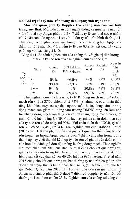
4.6. Giá trị của tỷ não- rốn trong tiên lượng tình trạng thai
Mối liên quan giữa tỷ Doppler trở kháng não rốn với tình
trạng suy thai: Mối liên quan có ý nghĩa thống kê giữa tỷ lệ não rốn
< 1 với thai suy Apgar phút thứ 5 < 7 điểm, tỷ lệ suy thai cao ở nhóm
có tỷ não rốn đảo ngược <1 so với nhóm tỷ não rốn bình thường >1.
Thật vậy, trong nghiên cứu của chúng tôi có 36 trường hợp Apgar < 7
điểm thì tỷ lệ não rốn < 1 chiếm tỷ lệ cao 63,9 %, kết quả này cũng
phù hợp với các tác giả khác
Bảng 4.11: So sánh nghiên cứu của chúng tôi với giá trị tiên lượng
thai của tỷ não rốn của các nghiên cứu trên thế giới
Tỷ
não –
rốn
Giá trị Chúng
tôi
B.N Lakkhar
K.V.Rajgopal
Rozeta
et al
Padmini
C.P
Nguyễn
Thị
Bích
Vân
Se 68 % 66,6% 98% 88% 84,8%
Sp 98,4% 73,9% 66% 91% 70,6%
PV + 94,4% 40% 30,8% 78% 58,3%
PV - 88,8% 89,4% 99,7% 73% 70,6%
Theo nghiên cứu của Ebrashy, tỷ lệ RI động mạch não giữa/động
mạch rốn < 1 là 37/50 chiếm tỷ lệ 74% . Shahinaj R et al nhận thấy
rằng khi thiếu oxy, có sự đảo ngược tuần hoàn, dòng tâm trương
động mạch rốn giảm đi, dòng tâm trương ĐMNG tăng lên làm cho
trở kháng động mạch rốn tăng lên và trở kháng động mạch não giữa
giảm đi thể hiện bằng CSNR < 1, lúc này giá trị chẩn đoán thai suy
của tỷ não rốn có độ nhạy tới 90% . Với chẩn đoán thai IUGR, tỷ não
rốn < 1 có Se 54,4%, Sp là 61,4%. Nghiên cứu của Orabona R et al
(2015) trên 168 sản phụ bị tiền sản giật kết quả cho thấy rằng tỷ não
rốn trong tiên lượng Apgar của trẻ dưới 7 điểm cũng như trọng lượng
thai thấp hay chết thai thì kết hợp tỷ não rốn có giá trị tiên đoán chính
xác hơn khi đánh giá đơn độc riêng lẻ từng động mạch. Theo nghiên
cứu mới nhất năm 2016 của Rani S. et al cũng cho kết quả tương tự,
giá trị tỷ não rốn trong tiên lượng thai thai suy, thai kém phát triển
liên quan kết cục thai kỳ với độ đặc hiệu là 98% . Adiga P. et al năm
2015 cũng cho kết quả tương tự, bất thường tỷ não rốn có giá trị tiên
đoán tình trạng thai ở bệnh nhân tiền sản giật . Nghiên cứu của tác
giả Robert Qirko năm 2015 trên 106 sản phụ tiền sản giật nhóm trẻ
Apgar sau sinh ở phút thứ 5 dưới 7 điểm có doppler tỷ não rốn bất
thường < 1 cao hơn chiếm 23 %. Nghiên cứu của chúng tôi cũng cho
22
kết quả tương tự các nghiên cứu mới nhất năm 2016 của các tác giả
Padmini C. et al, nghiên cứu của K.S.Vedaraju, Suresh S
Kanakannavar.
Mối liên quan giữa tỷ số Doppler não rốn với thai kém phát
triển trong tử cung
Trong IUGR có hiện tượng phân phối lại tuần hoàn não gọi là
hiệu ứng tiết kiệm cho não được cho rằng để bù trừ lại tình trạng
thiếu oxy. Nghiên cứu chúng tôi thấy có mối liên quan giữa tỷ lệ não
rốn với IUGR, tình trạng thai IUGR cao ở nhóm tỷ lệ não rốn <1
chiếm 71,9%. Nghiên cứu mới nhất 2016, Mozibur Rahman Laskar
cũng đã nhận thấy rằng giá trị tỷ não rốn có độ nhạy cao nhất
(88,88%) trong tiên lượng sức khỏe thai. Nghiên cứu của tác giả
V.A.A Lakshmi et al năm 2015 tiến hành trên 100 phụ nữ mang thai
bị tiền sản giật tuổi thai từ 28 – 40 tuần, kết quả cho thấy tỷ lệ tử
vong trẻ cao chiếm 31,5 % ở nhóm có tỷ não rốn < 1 . Như vậy kết
quả trong nghiên cứu của chúng tôi cho thấy khả năng phát hiện thai
suy cũng như thai kém phát triển trong tử cung khi tỷ Não – rốn < 1
là rất cao và có thể sử dụng trong lâm sàng để xác định tình trạng thai
suy trước khi có quyết định chấm dứt thai kỳ.
KẾT LUẬN
Từ kết quả nghiên cứu trên 153 thai phụ tiền sản giật tuổi thai từ
28 - 41 tuần tại Khoa Sản Bệnh viện Trường Đại học Y Dược Huế
chúng tôi có một số kết luận sau:
1. Giá trị của một số thăm dò Doppler ĐMTC, ĐMR, ĐMNG
trong tiên lượng tình trạng thai nhi ở sản phụ bị tiền sản giật
- Các nhà lâm sàng có thể sử dụng các điểm cắt ở trong luận án
này để tiên lượng tình trạng sức khỏe thai góp phần nâng cao hiệu
quả theo dõi và xử trí .Tìm được giá trị điểm cắt tiên lượng thai suy
và IUGR của RI ĐMTC ở tuổi thai 34 -37 tuần là 0,6, giá trị điểm cắt
2,6 của tỷ số S/D ĐMTC ở tuổi thai 34 - 37 tuần trong tiên lượng thai
suy với Se 100% và Sp là 60%. Giá trị điểm cắt RI ĐMR trong tiên
lượng thai suy ở tuổi thai 34 – 37 tuần tại điểm cắt là 0,64 với Se là
90,9%, ở tuổi thai >37 tuần là 0,75 với Se là 100%, điểm cắt RI
ĐMR trong tiên lượng IUGR ở tuổi thai 34 -37 là 0,74 và ở tuổi thai
> 37 tuần ở điểm cắt 0,76. Giá trị của Doppler điểm cắt RI của
ĐMNG tiên lượng thai suy ở thai 34 -37 tuần là 0,68 với Se là 81,8%,
23
Sp là 85,0%, điểm cắt RI của ĐMNG trong tiên lượng thai suy ở tuổi
thai >37 tuần là 0,65.
- Diện tích dưới đường cong ROC = 0,81 ở tuổi thai 34 -37 tuần
và ở tuổi thai > 37 tuần là 0,88 cho thấy có mối tương quan rõ giữa
chỉ số kháng RI ĐMTC trong tiên lượng thai suy. PI và RI ĐMNG
rất có giá trị trong tiên lượng thai suy ở tuổi thai 34 -37 tuần với diện
tích dưới đường cong ROC = 0,85 và 0,82 ở tuổi thai 34 -37 tuần.
Độ đặc hiệu trong tiên lượng thai suy của RI ĐMNG cao hơn so với
ĐMR với p < 0,01.
2. So sánh hiệu quả của các chỉ số Doppler trong thăm dò đánh
giá tình trạng sức khoẻ của thai ở thai phụ tiền sản giật.
Doppler ĐMTC là phương pháp tốt có thể tiên lượng thai chậm
phát triển trong tử cung. Doppler ĐMNG đơn độc có giá trị thấp
trong tiên đoán tình trạng thai suy nhưng có giá trị cao tiên đoán thai
chậm phát triển trong tử cung. Kết hợp Doppler ĐMR – ĐMNG rất
có giá trị trong tiên lượng sức khỏe thai , giá trị tỷ não rốn RI
ĐMNG/ĐMR < 1 bất thường có mối liên quan với tình trạng thai suy
và IUGR ở bệnh nhân tiền sản giật.
KIẾN NGHỊ
1. Sử dụng thăm dò bằng hiệu ứng Doppler trong sản khoa là cần
thiết vì đây là thăm dò không can thiệp hoàn toàn có khả năng thực
hiện được và rất có hiệu quả. Có giá trị chẩn đoán trong thai nghén
bệnh lý như thai chậm phát triển trong tử cung, suy thai, cao huyết áp
và thai nghén, tiền sử thai nghén bệnh lý. Có khả năng phát hiện sớm
thai nghén bệnh lý khi các chỉ số và tỷ số Doppler bệnh lý. Có giá trị
tiên lượng rất tốt trong đánh giá tình trạng sức khoẻ của thai do đó
nên khuyến cáo tiến hành thường quy ở các tuyến y tế cơ sở.
2. Siêu âm Doppler động mạch tử cung, động mạch não giữa và
động mạch rốn ở bệnh nhân tiền sản giật có khả năng đánh giá tình
trạng thiếu oxy não thai nhi, tình trạng thai kém phát triển trong tử
cung và suy thai. Do đó, ứng dụng hiệu ứng Doppler trong thăm dò
sức khỏe thai nói chung ở bệnh nhân bị tiền sản giật nói riêng có thể
giúp làm giảm tỷ lệ bệnh tật và tử vong ở thai nhi và trẻ sơ sinh và kỹ
thuật này được xem như là một phương pháp quan trọng cần thiết cần
được ứng dụng rộng rãi ở Việt nam và thế giới.
24
DANH MỤC CÁC CÔNG TRÌNH NGHIÊN CỨU CÔNG BỐ
CỦA TÁC GIẢ CÓ LIÊN QUAN ĐẾN ĐỀ TÀI LUẬN ÁN
1. Trương Thị Linh Giang, Nguyễn Vũ Quốc Huy (2014), “Ứng
dụng Doppler trong đánh giá sức khỏe thai”, Tạp chí Y Dược Học,
số 22+23, 9-18.
2. Trương Thị Linh Giang và cs (2015), “Nghiên cứu giá trị siêu
âm Doppler động mạch não giữa để dự báo thai suy ở thai phụ tiền
sản giật”, Tạp chí Y Dược Học, số 27, 61-65.
3. Trương Thị Linh Giang, Nguyễn Vũ Quốc Huy, Trương Quang
Vinh, Võ Văn Đức (2016), “Nghiên cứu giá trị siêu âm Doppler
động mạch tử cung ở thai phụ tiền sản giật”, Tạp chí Y Dược Học,
số 31, 57-64.
4. Trương Thị Linh Giang, Nguyễn Vũ Quốc Huy (2016), “Giá trị
tỷ não rốn trong tiên lương sức khỏe thai kỳ ở thai phụ tiền sản
giật”, Tạp chí Phụ Sản, Tập 14(03), 16-20.
5. Trương Thị Linh Giang, Nguyễn Vũ Quốc Huy (2016),
“Correlation between uterine artery Doppler sonography and
severity of pre-ecclampsia & fetal distress”; Poster presentation, 15th
World Congress in Fetal Medicine, 26–30th, June, Palma-de-
Mallorca, Barcelona, Spain.
Bất Thường Bình Thường
Đánh giá kết quả kết thúc thai kỳ
Sản phụ mang thai 28 - 41 tuần
Chẩn đoán TSG trên lâm sàng và cận lâm sàng
Khảo sát các yếu tố nguy cơ liên quan đến TSG
Về mẹ Về thai Phần phụ của thai
Theo dõi Doppler ĐMTC, ĐMR, ĐMNG và
lấy kết quả trong vòng 48 giờ trước khi
kết thúc thai kỳ
SA + SA Doppler (lặp lại hàng tuần)
Kết thúc thai kỳ
Mổ lấy thai (khi không đáp ứng với
điều trị nội khoa, thai chậm phát triển
hoặc thai không phát triển them, CTG
bất thường).
MINISTRY OF EDUCATION AND TRAINING
HUE UNIVERSITY
HUE UNIVERSITY OF MEDICINE AND PHARMACY
TRUONG THI LINH GIANG
VALUE OF DOPPLER ULTRASONOGRAPHY IN
PREDICTING FETAL WELL-BEING IN PREGNANT
WOMEN WITH PREECCLAMPSIA
SPECIALTY: OBSTETRICS AND GYNECOLOGY
Code : 62.72.01.31
SUMMARY OF MEDICAL DOCTORAL DISSERTATION
Scientific Supervisor:
A/Prof. Dr. NGUYEN VU QUOC HUY
HUE, 2017

1
INTRODUCTION
Gestational monitoring, especially in high-risk pregnancies, is an
important task of obstetricians to ensure healthy birth as well as to
reduce the perinatal morbidity and mortality rates. According to the
study of Marie Bolin in Sweden in 2012, in the early 1940s,
preeclampsia accounted for 34% of maternal death. In the 1950s, the
rate of maternal death caused by preeclampsia has gradually declined
considerably due to modern technological advances in prenatal care
and diagnosis. Preeclampsia is a complicated disease which can
cause serious harm to the health and even life of pregnant women,
fetuses and newborns. In order to limit the complications caused by preeclampsia in
mother and fetus, various methods of exploration were used to
evaluate fetal development and health in pregnant women with
preeclampsia, which enabled early detection and management of its
complications in a timely manner. Among these methods, Doppler
assessment of maternal-fetal circulation is now considered a very
valuable non-invasive tool. Many studies of Doppler assessment of
uterine artery (UTA), umbilical artery (UMA), middle cerebral artery
(MCA) showed that Doppler has predictive value of fetal distress,
fetal growth restriction in pregnant women with preeclampsia.
However, in Vietnam, there have not been many comprehensive
studies on this topic. With the above-mentioned reasons, we
conducted the study: “Value of Doppler ultrasonography in
predicting fetal well-being in pregnant women with preecclampsia”
to facilitate clinical decision to reduce maternal, fetal and neonatal
morbidity and mortality rates with the following objectives:
1. To study the value of some ultrasound explorations in
predicting fetal well-being in pregnant women with preeclampsia.
2. To compare the effectiveness of different Doppler indices in
predicting fetal well-being in pregnant women with preeclampsia.
SCIENTIFIC CONTRIBUTIONS AND PRACTICAL
IMPLICATIONS
The study used Doppler assessment of UMA, MCA, and UTA and
determined the cutoff points which were applicable in clinical
practice for monitoring and evaluating of fetal well-being and
2
prediction of fetal distress and intrauterine growth restriction
(IUGR). This study was of great scientific contributions and practical
values in the monitoring of fetal well-being in pregnant women with
preeclampsia.
NEW CONTRIBUTIONS OF THE THESIS
The study found the cut-off values of PI, RI, S/D ratios of the
UTA, UMA and MCA to predict fetal distress, IUGR in preeclampsia
to help clinicians determine the most appropriate management to
reduce perinatal morbidity and mortality rates.
The study also compared the effectiveness of different Doppler
indices in predicting fetal well-being.
THESIS LAYOUT
The thesis consists of 126 pages including: introduction (2 pages),
literature review (36 pages), objects and methods (14 pages), results
(38 pages), discussion (32 pages), conclusion (2 pages) and
recommendation (1 page). The cover of thesis, list of abbreviations,
lists of tables, diagrams, illustrations, introduction, main contents and
conclusion were written following current regulations. There were
132 references, most of which were in foreign languages. There were
5 related publications in prestigious domestic medical journals, and 1
international publication.
Chapter 1
LITERATURE REVIEW
1.1. Preeclampsia and eclampsia
1.1.1. Diagnostic criteria of preeclampsia
According to preeclampsia – eclampsia classification in the
National Guideline of the Ministry of Health (2009), the diagnostic
criteria of preeclampsia are as followed:
- A systolic blood pressure (SBP) ≥ 140 mmHg or a diastolic
blood pressure (DBP) ≥ 90mmHg.
- An increase of SBP ≥ 30 mmHg, of DPB ≥ 15mmHg or mean
arterial BP ≥ 20mmHg (in case of known baseline BP)
- And proteinuria ≥ 0,3 g/l in a 24-hour urine collection.
1.1.2. Classification of preeclampsia
According to the latest document in 2015, ACOG replaced the
3
term "severe pre-eclampsia" with the term "pre-eclampsia with
severe features". ACOG also eliminated the criteria of proteinuria
(5g/24h), oliguria and IUGR in the diagnosis of severe pre-
eclampsia because the studies failed to demonstrate the association of
these criteria with pregnancy outcomes.
There are multiple ways to classify pre-eclampsia
- Classification by severity of symptoms as mild, moderate and
severe preeclampsia. ACOG currently classifies preeclampsia into
two types: preeclampsia and preeclampsia with severe features.
- According to the International Society for the Study of
Hypertension in pregnancy (ISSHP), hypertension in pregnancy is
classified as including preeclampsia, gestational hypertension,
chronic hypertension and chronic hypertension with superimposed
preeclampsia.
Preeclampsia requiring delivery prior to 34 weeks, from 34 to 37
weeks and after 37 weeks of gestation is called early preeclampsia,
intermediate preeclampsia and late preeclampsia, respectively.
1.1.3. Pathogenesis
According to ACOG (2015), maternal genetic factors and immune
dysfunction are the most persuasive explications of the disease.
Pathogenesis involves vascular endothelial factors, trophoblast
invasion, placental oxidation stress and genetic factors.
1.1.4. Consequences of preeclampsia
1.1.4.1. Maternal complications
Hypertension in pregnancy causes many complications and
increases maternal morbidity and mortality rates and is one of the
three leading causes of maternal death (after hemorrhage and
infection). Other signs of severe pre-eclampsia includes cardiac
dysfunction with pulmonary edema, eclampsia and HELLP
syndrome, heart failure and acute pulmonary edema, renal failure,
hepatic impairment and coagulation abnormalities and eventually
maternal death.
1.1.4.2. Fetal and neonatal complications
In fetuses: IUGR, fetal distress and stillbirth.
In neonates: preterm neonates, low birth weight, neonatal
hypertension, neonatal deaths.
1.2. Methods for assessment of fetal well-being
4
1.2.1. Amnioscopy: this method is not commonly used in clinical
practice nowadays.
1.2.2. CTG (Cardiotocography): the recording of fetal heartbeat and
uterine contraction, also known as obstetrical monitoring or
simplified as fetal heart rate monitoring, is an important method of
evaluation of fetal well-being.
1.2.3. Fetal biophysical profile of Manning
1.2.4. Modified fetal biophysical profile: A complete fetal biophysical
profile examination is time- and manpower- consuming. Clark 1989 and
Nageotte 1994 proposed a modified fetal biophysical profile including: (1)
CTG in combination with (2) amniotic fluid volume assessment using
ultrasonography.
1.2.5. Ultrasonography assessment of fetal development
1.3. Doppler assessement of fetal well-being in preeclampsia
Doppler ultrasound of the UTA: In normal pregnancy, RI and PI of
UTA gradually decrease toward the end of gestation. S/D ratio, reflecting
the resistance of UTA circulation, also declines in the end of gestation.
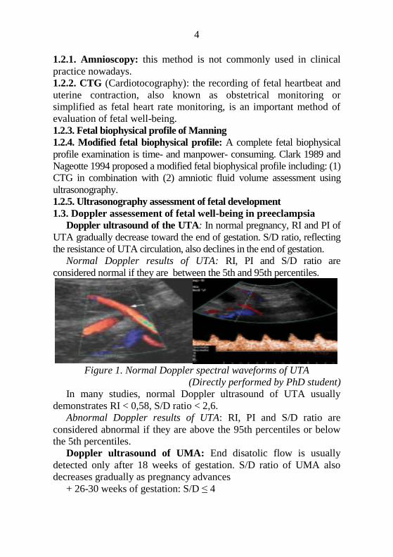
Normal Doppler results of UTA: RI, PI and S/D ratio are
considered normal if they are between the 5th and 95th percentiles.
Figure 1. Normal Doppler spectral waveforms of UTA
(Directly performed by PhD student)
In many studies, normal Doppler ultrasound of UTA usually
demonstrates RI < 0,58, S/D ratio < 2,6.
Abnormal Doppler results of UTA: RI, PI and S/D ratio are
considered abnormal if they are above the 95th percentiles or below
the 5th percentiles.
Doppler ultrasound of UMA: End disatolic flow is usually
detected only after 18 weeks of gestation. S/D ratio of UMA also
decreases gradually as pregnancy advances
+ 26-30 weeks of gestation: S/D ≤ 4
5
+ 30-34 weeks of gestation: S/D ≤ 3,5 and RI ≤ 0,8
+ 34-40 weeks of gestation: S/D ≤ 3 and RI < 0,8
Abnormal Doppler results of UMA: RI, PI and S/D ratio are
considered abnormal if they are above the 95th percentiles or below
the 5th percentiles.
Doppler ultrasound of MCA: The resistance of MCA in normal
fetuses is quite high. In normally developped fetuses, PI>1,45 and RI
ranges from 0,8 to 1,2. In case of cerebral hypoxia, circulation
redistribution occurs in which cerebral, umbilical, and pulmonary
vessels dilate to increase blood supply to the brain. Therefore, RI≤
0,5 and PI < 0,8 had diagnostic value of IUGR in previous studies.
The ratio between Doppler resistance indices of MCA/UMA is
also called cerebral/umbilical ratio. In normal fetuses, the
resistance indices of MCA are always higher than UMA, hence
MCA/UMA ratio>1. When MCA/UMA ratio<1, fetal circulation
redistribution occurs.
1.4. Studies on diagnostic value of Doppler ultrasound in the
prediction of fetal well-being in pregnant women with
preeclampsia
Studies on Doppler ultrasound of UTA: In 2014, Cru- Martinez R,
Savchev et al. in Barcelona studied the Doppler assessment in the
third trimester of pregnancy and concluded that Doppler ultrasound
can predict cerebral hypoxia and IUGR. Thus, for high-risk
pregnancies, the Doppler of UTA has a very good predictive value
for the risk of maternal and fetal complications. Several other studies
on the role of Doppler ultrasoound in hypertension in pregnancy
showed that S/D ratio of> 2.6 or the presence of early diastolic notch
(NOTCH sign) were adverse signs of disease outcomes with
sensitivity and specificity of 81% and 90%, respectively.
Studies on Doppler ultrasound of UMA: In a study of Nguku,
Nairobi Hospital, Kenya, 110 cases of hypertension in pregnancy,
there were 72/110 (66.1%) cases with normal RI and 33.9% of cases
with increased RI of UMA.
Studies on Doppler ultrasound of MCA: In 2014, Padmaja
R.Desai et al. studied the association of Doppler ultrasound indices of
MCA with IUGR, he noted that there was a decrease in the resistance
of MCA in case of IUGR in pre-eclampsia. Ebrashy's study (2000) of
6
50 pre-eclampsia patients found that the mean RI of MCA was 0.70 ±
0.01. The threshold for RI of MCA was RI <0.69. The proportion of
patients having RI <0.69 was 17/50 (34%).
Cerebral/Umbilical ratio in the assessment of fetal well-being
in PE: The ratio of the resistance (RI) of MCA/UMA is called the
cerebral/placental ratio or cerebral/umbilical ratio. The reversal of the
normal ratio can be seen as a circulatory redistribution to the brain or
"fetal brain sparing effect". In normal pregnancy, the diastolic flow
of the cerebral artery is lower than that of the umbilical artery in any
phases of gestation. If there is hypoxia, there will be a redistribution
of circulation in which the diastolic flow of MCA increases while
that of UMA decreases, causing the increase in MCA RI and
decrease in UMA RI, hence a reversed cerebral/umbilical ratio (<1).
In 2013, Monika Singh et al used Doppler to calculate the RI, PI, S/D
ratios in 50 PE patients, the study found that the abnormalities in
Doppler indices of MCA signified an increase in the incidence of
IUGR and adverse outcomes in pregnancy.
Chapter 2
SUBJECTS AND RESEARCH METHODS
2.1. Research objects: Pregnant women diagnosed with
preeclampsia and treated from 03/01/2013 to 30/ 01/2016 at Hue
University Hospital who agreed to participate in the research.
2.1.1. Inclusion criteria: Having minimum gestational age of 28
weeks (calculated from the first day of the last menstrual period) with
one living fetus.
- Having the following signs and symptoms
+ SBP ≥ 140 mmHg or DBP ≥ 90 mmHg
+ Proteinuria ≥ 0.5 g/l on a random urine specimen or 0.3 g/l on
a 24 hour urine collection, with or without edema
- Patients evaluated by ultrasonography and Doppler ultrasonography
to assess UTA, UMA, and MCA at 48 hours prior to the termination of
pregnancy.
2.1.2. Exclusion criteria: Polyhydramnios, fetal malformations,
eclampsia, HELLP syndrome, congenital heart diseases, kidney
diseases, hypertension, Bazedow’s disease, diabetes mellitus
7
2.2. Research methods
2.2.1. A prospective, descriptive cross-sectional study
2.2.2. Sample size calculation: Since we studied the characteristics
of a diagnostic tool, calculation of sample size would be based on
sensitivity (Se) and specificity (Sp). Furthemore, this is a clinical
study, thus, we used Sp to estimate the required sample size.
Sample size was calculated using the following formula
1sp
dis
FP TNN
p
Firstly, we estimated FP + TN (false positive and true negative
value) using the formula:
FP + TN =
2
2
(1 )sp spZ p p
In which:
Nsp : Sample size required for the study, = 5%, Z is the
normal distribution coefficient, if the significant level =0.05, Z
=1.96 ,Psp: Power = 90%
Pdis: prevelance of pregnant women diagnosed with preeclampsia
in previous studies = 8 %, thus 1- Pdis = 0.92.
With current disease prevelance of 8%, using the above formula
for calculation of sample size for Sp estimation, we had:
FP + TN =
2 2
2 2
(1 ) (1,96) 0,9 0,1138,3
(0,05)
sp spZ p p
1sp
dis
FP TNN
p
=
138,3150,3
0,92
The required sample size was 150.3, rounded as 151 patients.
2.2.3. Study tools
- Ultrasound machine (Siemens Acuson X 300) with 3.5 MHz
curvilinear probe was used in the Prenatal Ultrasound Examination Unit
at Hue University of Medicine and Pharmacy Hospital.
- Tables of percentile distributions of S/D ratios, resistance ratio
(RI), and pulsatile ratio (PI) of UTA, UMA, MCA in different
8
gestational ages in healthy normal pregnancies of Tran Danh Cuong
(2007).
- Preprinted questionnaires, Avalon FM20 cardiotocography
monitoring machine manufactured by PHILIPS with all functional of
parts and recording papers, rubber gloves, oxygen tanks and wires,
suction machine and tubes, bag valve mask, endotracheal intubation
equipment, endoscope and endotracheal tube, and drugs used in
neonatal resuscitation.
2.2.4. Research steps
2.2.4.1. Diagnosis of preeclampsia: using classification of
preeclampsia in the National Guidelines for Reproductive Health
Services 2009.
2.2.4.2. Doppler assessment of fetuses: using Doppler ultrasound to
evaluate UTA, UMA and MCA to assess waveforms and measure
indices including pulsatile index (PI), resistance index (RI) and S/D
ratio.
Cerebral/Umbilical ratio: the ratio of RI of MCA and UMA.
2.2.5. Monitor and evaluate the outcomes of pregnancy
termination
- Fetal age at birth (or at termination of pregnancy)
- Reasons for termination of pregnancy (if actively terminated)
+ No response to medical therapy
+ Intrauterine growth restriction or ceasation
+ Abnormal CTG recordings
- Types of delivery
- Assessment of fetal condition
+ Intrauterine growth restriction (IUGR): Birth weight below
the 10th percentiles of the fetal weight development chart of Phan
Truong Duyet (2005). We used this chart because our patients were
all Vietnamese.
+ Fetal distress: diagnosed if there were the following signs:
reduced variability during more than 60 minutes (after excluding fetal
sleeping) or presence of DIP II or variable DIP.
- Postpartum assessment of fetal condition
+ Amniotic fluid: volume, color
+ Birth weight
+ Apgar score
9
2.3. Data analysis
- Data was entered using Epi Data 3.1 software.
- We used the ROC curve plot method and calculated the area
under the ROC curve and determined the optimum cut-off value
acoording to the Youden index using the Medcalc software. AUC
(Area Under the Cuver) represented the accuracy of the diagnostic
method. Additional evaluation of diagnostic value by sensitivity,
specificity, positive predictive value, negative predictive value. Using
NCSS11 software: Mc Nemar test to compare the effectiveness of
different indices obtained by Doppler ultrasound.
- Comparisons were statistically significant when p <0.05.
2.4. Research ethics: The study was conducted after the protocol
was approved by the board of Obstetrics and Gynecology.
Chapter 3
RESULTS
3.1. Characteristics of sample
3.1.1. Maternal age: Mean maternal age was 30.5 ± 6.4 (20-44).
3.1.2. Number of pregnancies: The majority of cases were
diagnosed in their first pregnancies (53.6%). Other patients had ≥ 2
pregnancies (46.4%).
3.1.3. Symptoms: Edema was the leading symptoms (43.8%),
followed by headache (23.5%), dizziness and blurred vision with
comparable rates (3.9%).
3.1.4. Mean blood pressure: Mean SBP was 144.58 ± 6.96 mmHg,
mean DBP was 89.65 ± 7.08 mmHg. In preeclampsia women with
severe features, mean SBP was 169.39 ± 13.34 mmHg and mean
DBP was 103.60 ± 11.53 mmHg.
3.1.5. Severity of preeclampsia: there were 54% of cases with
preeclampsia and 46% with severe preeclampsia.
3.1.6. Gestational age: there were more than 153 preeclampsia cases
with gestational age ranging from 28 to 41 weeks.
3.1.7. Fetal conditions: in the group with gestational age of 28-33
weeks, the rates of fetal distress and IUGR were 100%. In the 34 to
37-week gestational age group, there were 35.5% cases with fetal
distress and 41.9% cases with IUGR. In the >37 week-gestational age
10
group, fetal distress presented in 14.2% and IUGR in 8.8% of cases.
3.1.8. Mode of delivery: Cesarean birth was the mode of delivery in
56.2% of cases while with vaginal delivery accounted for 43.8% of
cases.
3.2. Neonatal condition after birth
The majority of newborns had Apgar score at 5 minutes > 7 (
76.5%), 22.1% had Apgar score of 4 -7 and only 2 cases had Apgar
<3.
3.2.1. Neonatal condition 48 hours after birth: The majority of
newborns were healthy (86.9%) and there were only 11.8% of
newborns admitted to neonatal units due to fetal distress and IUGR
3.2.2. Birth weight: the majority of baby weighed > 2,500gr at birth,
accounting for 79.1%. Only the minority of cases weighed <1,500gr
at birth (2%). The proportion of babies weighed 1,500-2,000gr and
2,000-2,500gr were 5.9% and 13.1%, respectively.
3.3. Cut-off values of Doppler ultrasound indices of umbilical
artery, uterine artery and middle cerebral artery in the
prediction of fetal distress and intrauterine growth restriction
Cut-off values of PI of UTA in the prediction of fetal distress
were 1 at gestational age of 34-37 weeks with Se of 72.73% and Sp
of 65% and 1.15 at gestational age of >37 weeks with Se of 87.50%
and Sp of 28.87%. AUC at gestational age of 34 -37 weeks and > 37
weeks were 0.62 and 0.51, respectively. There was no clear correlation
between PI of UTA with fetal distress.
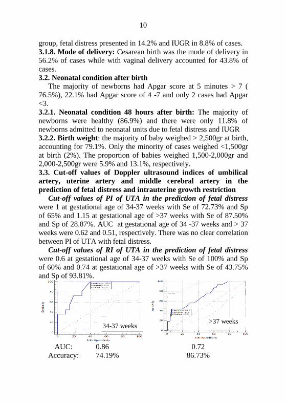
Cut-off values of RI of UTA in the prediction of fetal distress
were 0.6 at gestational age of 34-37 weeks with Se of 100% and Sp
of 60% and 0.74 at gestational age of >37 weeks with Se of 43.75%
and Sp of 93.81%.
AUC:
Accuracy:
0.86
74.19%
0.72
86.73%
>37 weeks 34-37 weeks
11
Diagram 3.1. Cut-off value of RI of UTA in the prediction of fetal distress
AUC at gestational age of 34 -37 weeks and > 37 weeks were 0.86
and 0.72, respectively. The diagram demonstrated a clear correlation
between RI of UTA with fetal distress at cut-off values.
Cut-off values of S/D ratio of UTA in the prediction of fetal
distress were 2,6 at gestational age of 34-37 weeks with Se of 100%
and Sp of 60% and 3.12 at gestational age of >37 weeks with Se of
62.5% and Sp of 72.16%. AUC at gestational age of 34 -37 weeks
and > 37 weeks were 0.82 and 0.62, respectively. There was a clear
correlation between S/D ratio of UTA with fetal distress at gestational
age of >37 weeks
Cut-off values in the prediction of IUGR of Doppler ultrasound
indices of uterine artery
Cut-off values of PI of UTA in the prediction of IUGR were 1 at
gestational age of 34-37 weeks with Se of 69.23% and Sp of 66.67%
and 1.1 at gestational age of >37 weeks with Se of 60% and Sp of
66.69%.
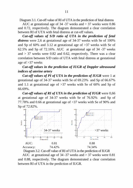
Cut-off values of RI of UTA in the prediction of IUGR were 0,66
at gestational age of 34-37 weeks with Se of 76.92% and Sp of
77.78% and 0.66 at gestational age of >37 weeks with Se of 90% and
Sp of 72.82%.
AUC:
Accuracy:
0.81
74.42%
0.88
74.34%
Diagram 3.2. Cut-off value of RI of UTA in the prediction of IUGR
AUC at gestational age of 34 -37 weeks and > 37 weeks were 0.81
and 0.88, respectively. The diagram demonstrated a clear correlation
between RI of UTA in the prediction of IUGR.
34-37 weeks >37 weeks
12
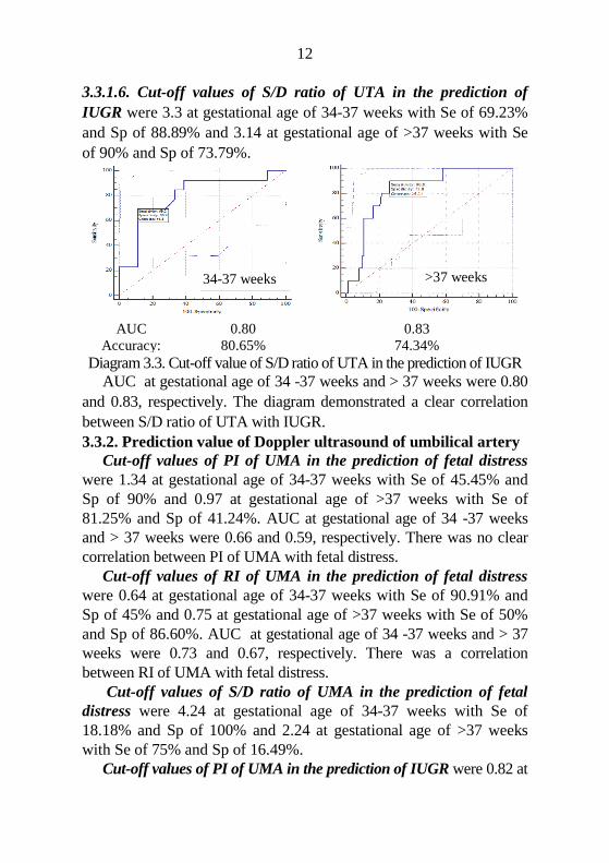
3.3.1.6. Cut-off values of S/D ratio of UTA in the prediction of
IUGR were 3.3 at gestational age of 34-37 weeks with Se of 69.23%
and Sp of 88.89% and 3.14 at gestational age of >37 weeks with Se
of 90% and Sp of 73.79%.
AUC
Accuracy:
0.80
80.65%
0.83
74.34%
Diagram 3.3. Cut-off value of S/D ratio of UTA in the prediction of IUGR
AUC at gestational age of 34 -37 weeks and > 37 weeks were 0.80
and 0.83, respectively. The diagram demonstrated a clear correlation
between S/D ratio of UTA with IUGR.
3.3.2. Prediction value of Doppler ultrasound of umbilical artery
Cut-off values of PI of UMA in the prediction of fetal distress
were 1.34 at gestational age of 34-37 weeks with Se of 45.45% and
Sp of 90% and 0.97 at gestational age of >37 weeks with Se of
81.25% and Sp of 41.24%. AUC at gestational age of 34 -37 weeks
and > 37 weeks were 0.66 and 0.59, respectively. There was no clear
correlation between PI of UMA with fetal distress.
Cut-off values of RI of UMA in the prediction of fetal distress
were 0.64 at gestational age of 34-37 weeks with Se of 90.91% and
Sp of 45% and 0.75 at gestational age of >37 weeks with Se of 50%
and Sp of 86.60%. AUC at gestational age of 34 -37 weeks and > 37
weeks were 0.73 and 0.67, respectively. There was a correlation
between RI of UMA with fetal distress.
Cut-off values of S/D ratio of UMA in the prediction of fetal
distress were 4.24 at gestational age of 34-37 weeks with Se of
18.18% and Sp of 100% and 2.24 at gestational age of >37 weeks
with Se of 75% and Sp of 16.49%.
Cut-off values of PI of UMA in the prediction of IUGR were 0.82 at
34-37 weeks >37 weeks
13
gestational age of 34-37 weeks with Se of 100% and Sp of 33.33% and
1.29 at gestational age of >37 weeks with Se of 50% and Sp of 83.5%.
Cut-off values of RI of UMA in the prediction of IUGR were 0.74 at gestational age of 34-37 weeks with Se of 61.54% and Sp of 88.89% and 0,76 at gestational age of >37 weeks with Se of 61.54% and Sp of 90.29%. AUC at gestational age of 34 -37 weeks and > 37 weeks were 0.79 and 0.70, respectively.
Cut-off values of S/D ratio of UMA in the prediction of IUGR
were 3.79 at gestational age of 34-37 weeks with Se of 30.77% and Sp of 94.44% and 3.16 at gestational age of >37 weeks with Se of 60% and Sp of 66.69%. 3.3.3 Cut-off values of middle cerebral artery doppler indices
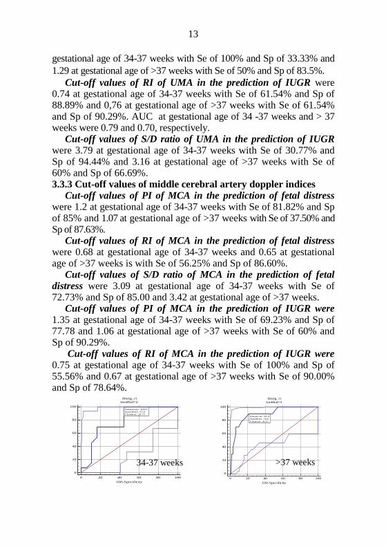
Cut-off values of PI of MCA in the prediction of fetal distress were 1.2 at gestational age of 34-37 weeks with Se of 81.82% and Sp of 85% and 1.07 at gestational age of >37 weeks with Se of 37.50% and Sp of 87.63%.
Cut-off values of RI of MCA in the prediction of fetal distress were 0.68 at gestational age of 34-37 weeks and 0.65 at gestational age of >37 weeks is with Se of 56.25% and Sp of 86.60%.
Cut-off values of S/D ratio of MCA in the prediction of fetal
distress were 3.09 at gestational age of 34-37 weeks with Se of 72.73% and Sp of 85.00 and 3.42 at gestational age of >37 weeks.
Cut-off values of PI of MCA in the prediction of IUGR were
1.35 at gestational age of 34-37 weeks with Se of 69.23% and Sp of 77.78 and 1.06 at gestational age of >37 weeks with Se of 60% and Sp of 90.29%.
Cut-off values of RI of MCA in the prediction of IUGR were 0.75 at gestational age of 34-37 weeks with Se of 100% and Sp of 55.56% and 0.67 at gestational age of >37 weeks with Se of 90.00% and Sp of 78.64%.
0
20
40
60
80
100
dmng_ri
tuoithai=2
0 20 40 60 80 100
100-Specificity
Sens
itivi
ty
Sensitivity: 100.0
Specificity: 55.6
Criterion: ≤0.75
0
20
40
60
80
100
dmng_ri
tuoithai=3
0 20 40 60 80 100
100-Specificity
Sens
itivi
ty
Sensitivity: 90.0
Specificity: 78.6
Criterion: ≤0.67
34-37 weeks >37 weeks
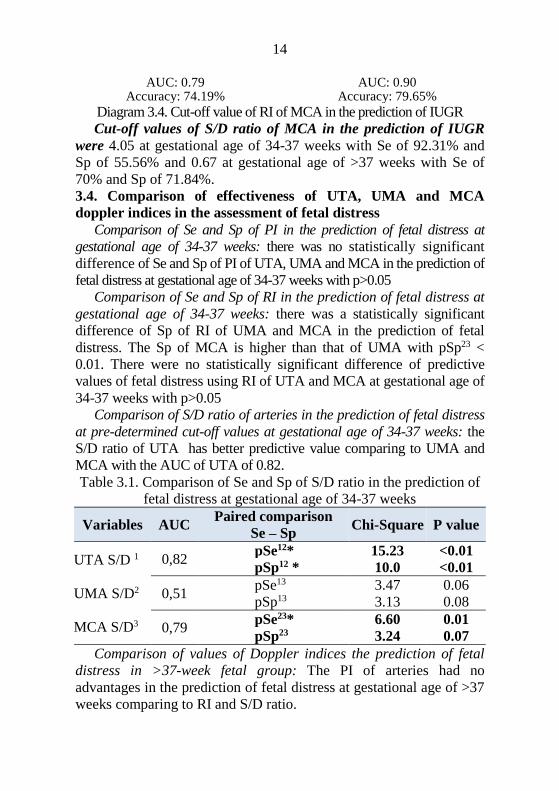
14
AUC: 0.79
Accuracy: 74.19% AUC: 0.90
Accuracy: 79.65%
Diagram 3.4. Cut-off value of RI of MCA in the prediction of IUGR
Cut-off values of S/D ratio of MCA in the prediction of IUGR
were 4.05 at gestational age of 34-37 weeks with Se of 92.31% and
Sp of 55.56% and 0.67 at gestational age of >37 weeks with Se of
70% and Sp of 71.84%.
3.4. Comparison of effectiveness of UTA, UMA and MCA
doppler indices in the assessment of fetal distress
Comparison of Se and Sp of PI in the prediction of fetal distress at
gestational age of 34-37 weeks: there was no statistically significant
difference of Se and Sp of PI of UTA, UMA and MCA in the prediction of
fetal distress at gestational age of 34-37 weeks with p>0.05
Comparison of Se and Sp of RI in the prediction of fetal distress at
gestational age of 34-37 weeks: there was a statistically significant
difference of Sp of RI of UMA and MCA in the prediction of fetal
distress. The Sp of MCA is higher than that of UMA with pSp23 <
0.01. There were no statistically significant difference of predictive
values of fetal distress using RI of UTA and MCA at gestational age of
34-37 weeks with p>0.05
Comparison of S/D ratio of arteries in the prediction of fetal distress
at pre-determined cut-off values at gestational age of 34-37 weeks: the
S/D ratio of UTA has better predictive value comparing to UMA and
MCA with the AUC of UTA of 0.82.
Table 3.1. Comparison of Se and Sp of S/D ratio in the prediction of
fetal distress at gestational age of 34-37 weeks
Variables AUC Paired comparison
Se – Sp Chi-Square P value
UTA S/D 1
UMA S/D2
MCA S/D3
0,82 pSe12* 15.23 <0.01
pSp12 * 10.0 <0.01
0,51 pSe13 3.47 0.06
pSp13 3.13 0.08
0,79 pSe23* 6.60 0.01
pSp23 3.24 0.07
Comparison of values of Doppler indices the prediction of fetal
distress in >37-week fetal group: The PI of arteries had no
advantages in the prediction of fetal distress at gestational age of >37
weeks comparing to RI and S/D ratio.
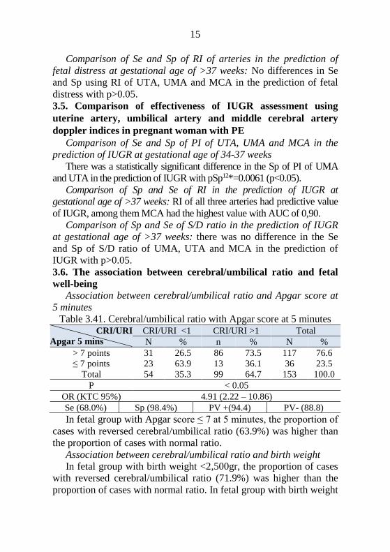
15
Comparison of Se and Sp of RI of arteries in the prediction of
fetal distress at gestational age of >37 weeks: No differences in Se
and Sp using RI of UTA, UMA and MCA in the prediction of fetal
distress with p>0.05.
3.5. Comparison of effectiveness of IUGR assessment using
uterine artery, umbilical artery and middle cerebral artery
doppler indices in pregnant woman with PE
Comparison of Se and Sp of PI of UTA, UMA and MCA in the
prediction of IUGR at gestational age of 34-37 weeks
There was a statistically significant difference in the Sp of PI of UMA
and UTA in the prediction of IUGR with pSp12*=0.0061 (p<0.05).
Comparison of Sp and Se of RI in the prediction of IUGR at
gestational age of >37 weeks: RI of all three arteries had predictive value
of IUGR, among them MCA had the highest value with AUC of 0,90.
Comparison of Sp and Se of S/D ratio in the prediction of IUGR
at gestational age of >37 weeks: there was no difference in the Se
and Sp of S/D ratio of UMA, UTA and MCA in the prediction of
IUGR with p>0.05.
3.6. The association between cerebral/umbilical ratio and fetal
well-being
Association between cerebral/umbilical ratio and Apgar score at
5 minutes
Table 3.41. Cerebral/umbilical ratio with Apgar score at 5 minutes CRI/URI
Apgar 5 mins
CRI/URI <1 CRI/URI >1 Total
N % n % N %
> 7 points 31 26.5 86 73.5 117 76.6
≤ 7 points 23 63.9 13 36.1 36 23.5
Total 54 35.3 99 64.7 153 100.0
P < 0.05
OR (KTC 95%) 4.91 (2.22 – 10.86)
Se (68.0%) Sp (98.4%) PV +(94.4) PV- (88.8)
In fetal group with Apgar score ≤ 7 at 5 minutes, the proportion of
cases with reversed cerebral/umbilical ratio (63.9%) was higher than
the proportion of cases with normal ratio.
Association between cerebral/umbilical ratio and birth weight
In fetal group with birth weight <2,500gr, the proportion of cases
with reversed cerebral/umbilical ratio (71.9%) was higher than the
proportion of cases with normal ratio. In fetal group with birth weight
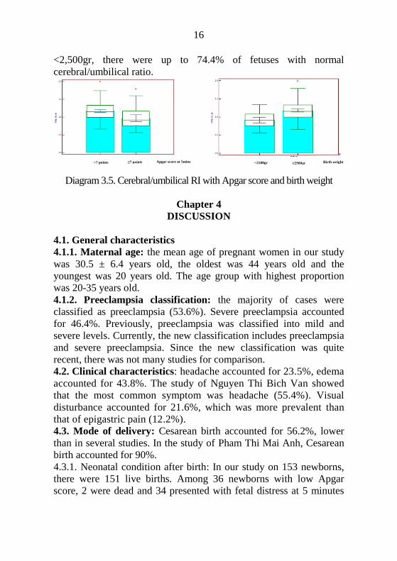
16
<2,500gr, there were up to 74.4% of fetuses with normal
cerebral/umbilical ratio.
Diagram 3.5. Cerebral/umbilical RI with Apgar score and birth weight
Chapter 4
DISCUSSION
4.1. General characteristics
4.1.1. Maternal age: the mean age of pregnant women in our study
was 30.5 ± 6.4 years old, the oldest was 44 years old and the
youngest was 20 years old. The age group with highest proportion
was 20-35 years old.
4.1.2. Preeclampsia classification: the majority of cases were
classified as preeclampsia (53.6%). Severe preeclampsia accounted
for 46.4%. Previously, preeclampsia was classified into mild and
severe levels. Currently, the new classification includes preeclampsia
and severe preeclampsia. Since the new classification was quite
recent, there was not many studies for comparison.
4.2. Clinical characteristics: headache accounted for 23.5%, edema
accounted for 43.8%. The study of Nguyen Thi Bich Van showed
that the most common symptom was headache (55.4%). Visual
disturbance accounted for 21.6%, which was more prevalent than
that of epigastric pain (12.2%).
4.3. Mode of delivery: Cesarean birth accounted for 56.2%, lower
than in several studies. In the study of Pham Thi Mai Anh, Cesarean
birth accounted for 90%.
4.3.1. Neonatal condition after birth: In our study on 153 newborns,
there were 151 live births. Among 36 newborns with low Apgar
score, 2 were dead and 34 presented with fetal distress at 5 minutes
Apgar score at 5mins ≤7 points >7 points Birth weight <2500gr ≥2500gr

17
and still required intensive care and respiratory support after 5
minutes (22.1%).
4.3.2. Apgar score: 36 newborns had 5-minute Apgar score below
7, accounting for 23.5%.
4.4. The value of Doppler indices in the prediction of fetal well-being
Correlation between uterine artery RI and fetal status: the
study results demonstrated the correlation between RI of UTA and
fetal distress. We found the cut-off of 0,6 at gestational age of 34-37
weeks with the Sp, Se, PPV, NPV of 100%, 65%, 57.9%, 100%
respectively. At gestational age above 37 weeks, the cut-off value for
fetal distress prediction was 0.74. Therefore, the cut-off of 0.6 at
gestational age of 34-37 weeks can be used clinically. According to
the newest study in 2016 of Mozibur Rahman Laskar, RI of UTA in
preeclampsia group was higher than that of normal gestation group.
The Se of increased RI of UTA was 84.7% in the study of Mozibur
(2016) and 83% in the study of Coleman et al. Our study results were
similar to others.
The correlation between S/D ratio and fetal well-being
At gestational age of 34-37 weeks, cut-off values for fetal distress
and IUGR prediction were 2.6 and 3.3, respectively with Sp, Se, PPV
and NPV of 100%, 65%, 57.9%, 100%, respectively for fetal distress
and 69.2%, 88.9%, 81.85%, 80%, respectively for IUGR. At gestational
age above 37 weeks, S/D ratio cut-off values for fetal distress and IUGR
prognosis were 3.12 and 3.14, respectively. The cut-off value for S/D
ratio of UTA in our study was 2.6, which was higher than that in the
study of Phạm Thị Mai Anh. In Vietnam, Vũ Hoàng Yến (2007) found
the cut-off value for S/D ratio of UTA in the prediction of IUGR and
fetal distress was 2.7. At this cut-off value, IUGR prediction had the Se,
Sp of 84.1% and 71.4%, fetal distress prediction Se, Sp of 92.3% and
75%. Hence, our cut-off value of 2.6 for S/D ratio of UTA at gestational
age of 34-37 weeks in our study had the prediction value for fetal
distress. The newest study in 2016 of Vijakumar with 80 PE patients
showed that IUGR prediction value of S/D ratio of UTA had the Se of
56.7%, Sp of 88.3% and PPV of 77.5%.
The value of umbilical artery doppler indices in fetal prognosis
The value of UMA PI in the prediction of fetal distress: Cut-
off values for the prediction of fetal distress using UMA PI at
gestational age of 34-37 weeks was 1.34, and 0.97 at gestational age
18
above 37 weeks. Our cut-off values were higher than that in the study
of Nguyen Thi Bich Van (1.2). PI had the highest predictive value at
gestational age of 33-37 weeks, with Se, Sp, PPV and NPV of 84.6%,
73.3%, 57.9% and 91.7% respectively. Our study results were higher
than those of other domestic studies. Ta Thi Xuan Lan chose the cut-
off value of 1.2 while Dinh Thi Thuy Hang chose 1.1, which were
similar to studies in other countries.
The value of UMA PI in the prediction of IUGR: In our study,
at gestational age of 34-37 weeks, the cut-off value of 0,82 had the
Se, Sp, PPV and NPV of 100%, 33.3%, 52% and 100%, respectively.
At gestational age above 37 weeks, the cut-off value of 1.29 had the Se
of 50% and Sp of 83.5%. The study of Nguyen Thi Bich Van using the
cut-off value of 1 at gestational age of 34-37 weeks and 1.0 at
gestational age above 37 weeks had the Se of 62.5% and Sp of 65%. In
our study, UMA PI cut-off value at gestational age of 34-37 weeks was
0.82 with Se of 100% but Sp of only 33.33%. Consequently, we
concluded that umbilical artery PI can effectively detect IUGR but
cannot exclude IUGR effectively.
The value of UMA RI in the prediction of fetal distress: Our
study showed that UMA RI above 0.64 at 34-37 weeks of gestation
and above 9.75 after 37 weeks of gestation had the highest predictive
value for fetal distress with Se of 84.6 – 87.5 %. The study of Nguyen
Thi Bich Van with RI>0.73 showed the capability of detecting fetal
distress at gestational age of 34-37 weeks. At gestational age above
37 weeks, our study found the RI cut-off value of 0.75 with the Se
and Sp of 100% and 86,6%, while Nguyen Thi Bich Van’s study
chose the cut-off of 0.71 because of the highest Se and Sp achieved.
However, the Se was only 50% although the Sp was 81.3%. The
most recent study in 2016 of Vịjaykumar Mane showed that UMA
doppler could predict fetal distress with high Se of 91.7%, Sp of
70.9% and PPV of 73.8%.
The value of UMA RI in the prediction of IUGR: The study of
V.A.A Lakshmi et al. in 2015 showed that the UMA RI in the prediction
of IUGR had the Se of 84.9% and Sp of 72.3%. The study of Nguyen
Thi Bich Van showed that at gestational age of 34-37 weeks, RI cut-off
value of 0.65 had the Se of 82.8% and Sp of 28.6%.
The diagnostic value of UMA S/D ratio in the prediction of
19
fetal distress and IUGR
Study result of Dinh Thi Thuy Hang showed that at S/D ratio cut-
off value of <2.6, the prediction of IUGR had Se of 100%, Sp of only
58%. For fetal distress, these indices had PPV of >95% and NPV of
45%. When S/D ratio >2.6, the detection of IUGR had the highest Se
of 95% and lowest Sp of 78%, the highest PPV of fetal distress was
88%. The author chose the cut-off value of 2.6 for the prediction of
fetal distress and IUGR with Se of 98%. According to Le Thi Thu
Ha, Ta Thi Xuan Lan, the prediction of IUGR using S/D ratio had the
Se of 79 – 97.6%. The study of Divon and Maulik showed that the
prediction of S/D ratio had the Se ranging from 49% to 79%.
The value of MCA PI in fetal prognosis
The MCA PI cut-off value of 1.2 for the prediction of fetal
distress at gestational age of 34-37 weeks had the Se, Sp, PPV and
NPV of 81.8%, 85%, 75 % and 89.5% respectively. At gestational
age above 37 weeks, the cut-off value was 1.07. For the prediction of
IUGR, our study noted a higher cut-off value of PI than in the
diagnosis of fetal distress. The cut-off of 1.35 at gestational age of
34-37 weeks had the Se, Sp, PPV and NPV of 69.2 %, 77.8 %, 69.2%
and 77.8 respectively. The cut-off of 1.06 at gestational age above 37
weeks had the Se, Sp, PPV and NPV of 60 %, 90.3 %, 37.5 % and
95.9 % respectively. The study of Nguyễn Thị Bích Vân proposed
the cut-off values of MCA PI at gestational age of 34-37 weeks of 1.3
and above 37 weeks of 1.2. Our study result was similar to the study
of Mozibur Rahman Laskar et al. in 2016, in which, the MCA PI has
high Se of 90% in the prediction of fetal distress and IUGR.
The value of MCA RI in the prediction of fetal distress:
prediction of fetal distress with the cut-off value of MCA RI of 0.68
had the Se 81.82% of and Sp of 85%. As the result, MCA RI may
have the diagnostic value for fetal distress in PE patients (p≤ 0.01). In
the study of Nguyễn Thị Bích Vân, fetal distress was diagnosed at
gestational age of 34-37 weeks with MCA RI cut-off value of 0.7. At
gestational age above 37 weeks, the cut-off value of 0.7 had the Se, Sp,
PPV and NPV of 75%, 59.4%, 18.8% and 95% respectively.
The value of MCA RI in the prediction of IUGR: Nguyen Thi
Bich Van showed that at gestational age of 34-37 weeks, with the
cut-off value of 0.72, the Se, Sp, PPV and NPV were 41.4%, 64.29%,
20
58.6% and 35.7% respectively, and at gestational age above 37
weeks, with cut-off of 0.72, the Se and Sp were 43.8% and 40%.
Comparison of the effectiveness of fetal distress prediction using Doppler indices of UTA, UMA, MCA at gestational age of 34-37 weeks: There was no statistically significant difference in fetal distress prediction using RI of UTA and UMA at gestational age of 34-37 weeks with p>0.05. The Se of UTA S/D ratio in fetal distress prediction at gestational age of 34-37 weeks was higher than that of UMA while the Sp was lower with statistical significance. The Se of MCA was higher than that of UMA with statistical significance. No statistically significant difference were found when comparing other Se and Sp values with p>0.05. There was a statistically significant difference in the Se and Sp of fetal distress prediction at gestational age above 37 weeks using MCA indices comparing to UTA and UMA indices with p<0.05. No statistically significant difference in the Se and Sp of MCA and UMA with p > 0.05.
Comparison of Se and Sp of RI in the prediction of fetal
distress using Doppler indices at gestational age above 37 weeks No statistical difference in Se and Sp in fetal distress prediction using RI of MCA, UMA, UTA at gestational age above 37 weeks with p > 0.05. There was statistically significant difference between Se of S/D ratio of UTA and UMA, p < 0.01. There was a statistically significant difference between Sp of S/D ratio of UMA and MCA, p<0.01. Therefore, there was a statistically significant difference in Sp of S/D ratio of UMA comparing to that of UTA and MCA in fetal distress prediction at gestational age >37 weeks with p<0.05. No statistically significant difference found in the comparison of other Se and Sp values with p>0.05.
Comparison of Se, Sp of UMA, UTA and MCA Doppler
indices in the prediction of IUGR at gestational age of 34-37 weeks In comparison of the Se and Sp of UMA and UTA PI at gestational age of 34-37 weeks, p values were 0.03 and 0.04 respectively. When we compared the Se and Sp of UMA and MCA PI, p values were 0.03 and <0.01 respectively. Hence, there was a statistically significant difference of the Se and Sp in IUGR prediction using PI of UTA, UMA and MCA with p<0.05. While comparing the Se and Sp of RI in the prediction of IUGR at gestational age of 34-37 weeks, and the Se and Sp of UMA and MCA
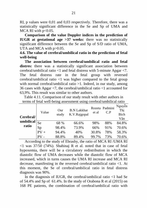
21
RI, p values were 0,01 and 0,03 respectively. Therefore, there was a statistically significant difference in the Se and Sp of UMA and MCA RI with p<0.05.
Comparison of the value Doppler indices in the prediction of
IUGR at gestational age >37 weeks: there was no statistically
significant difference between the Se and Sp of S/D ratio of UMA,
UTA and MCA with p>0.05.
4.6. The value of cerebral/umbilical ratio in the prediction of fetal
well-being
The association between cerebral/umbilical ratio and fetal
distres: there was a statistically significant association between
cerebral/umbilical ratio <1 and fetal distress with 5-minute Apgar <7.
The fetal distress rate in the fetal group with reversed
cerebral/umbilical ratio <1 was higher compared to the fetal group
with normal cerebral/umbilical ratio >1. Indeed, in our study, among
36 cases with Apgar <7, the cerebral/umbilical ratio <1 accounted for
63,9%. This result was similar to other authors.
Table 4.11. Comparison of our study result with other authors in
terms of fetal well-being assessment using cerebral/umbilical ratio
Cerebral/
umbilical
ratio
Value Our
study
B.N Lakkhar
K.V.Rajgopal
Rozeta
et al
Padmini
C.P
Nguyễn
Thị
Bích
Vân
Se 68 % 66.6% 98% 88% 84.8%
Sp 98.4% 73.9% 66% 91% 70.6%
PV + 94.4% 40% 30.8% 78% 58.3%
PV - 88.8% 89.4% 99.7% 73% 70.6%
According to the study of Ebrashy, the ratio of MCA RI /UMA RI
<1 was 37/50 (74%). Shahinaj R et al. noted that in case of fetal
hypoxemia, there will be a circulatory redistribution in which the
diastolic flow of UMA decreases while the diastolic flow of MCA
increased, which in turns causes the UMA RI increase and MCA RI
decrease, manifesting in the reversed cerebral/umbilical ratio <1. At
this moment, the Se of cerebral/umbilical ratio in fetal distress
diagnosis was 90%.
In the diagnosis of IUGR, the cerebral/umbilical ratio <1 had Se
of 54.4% and Sp of 61.4%. In the study of Orabona R et al (2015) on
168 PE patients, the combination of cerebral/umbilical ratio with

22
Apgar score below 7 points or low fetal weight or fetal death had
higher predictive value compared to separated use of Doppler
indices. The most recent study in 2016 of Rani S. et al showed
similar results in which the cerebral/umbilical ratio in the prediction
of fetal distress, IUGR correlated with gestational outcomes with Sp
of 98%. Adiga P. et al reported similar results in 2015, abnormal
cerebral/umbilical ratio had predictive value for fetal well-being in
PE patients. In the study of Robert Qirko in 2015 on 106 PE patients
with newborn Apgar score at 5 minutes below 7 points, the abnormal
cerebral/umbilical ratio <1 accounted for more than 23% of cases.
Our study also showed similar results to other recent studies in 2016
of Padmini C. et al, K.S.Vedaraju, and Suresh S Kanakannavar.
The association between cerebral/umbilical ratio and IUGR
In IUGR, there is a redistribution of cerebral circulation called
fetal brain sparing effect in order to compensate the hypoxia state.
Our study showed an association between cerebral/umbilical ratio
and IUGR with high rates of IUGR in the group with reversed
cerebral/umbilical ratio <1, accounting for 71.9%. In the newest
study in 2016, Mozibur Rahman Laskar noted that the
cerebral/umbilical ratio had the highest Se (88.88%) in fetal well-
being prediction. In 2015, V.A.A Lakshmi et al studied 100 PE
pregnant women at gestational age 28-40 weeks, the result showed
high fetal death rate (31.5%) in fetal group with cerebral/umbilical
ratio <1. Our study proved the high predictive value of
cerebral/umbilical ratio <1 of IUGR or fetal distress and this ratio can
be used clinically to identify fetal distress before the decision of
pregnancy termination.
CONCLUSION
From the study result on 153 preeclampsia pregnant women at
gestational age 28-41 weeks at the Obstetric department of Hue
University Hospital, we had the following conclusions:
1. Value of Doppler assessment of uterine artery, umbilical
artery and middle cerebral artery in the prediction of fetal well-
being in preeclampsia pregnant women
- Clinicians can use cut-off values in this thesis for the prediction
23
of fetal well-being in order to improve the effectiveness of
observation and management. Cut-off value of UTA RI for IUGR
and fetal distress prediction at gestational age of 34-37 weeks was
0.6. The UTA S/D ratio cut-off value of 2.6 for fetal distress
prediction at gestational age of 34-37 weeks had the sensitivity of
100% and specificity of 60%. Fetal distress prediction using UMA RI
at gestational age of 34-37 weeks with cut-off value of 0.64 had the
sensitivity of 90.9%, at gestational age above 37 weeks with cut-off
value of 9.75 had the sensitivity of 100%. Cut-off values for UMA RI
for IUGR prediction at gestational age of 34-37 weeks was 0.74 and
at gestational age above 37 weeks was 0.76. Fetal distress prediction
using MCA RI at gestational age of 34-37 weeks with cut-off value
of 0.68 had the sensitivity of 81.8% and specificity of 85.0%, cut-off
value of MCA RI for fetal distress prediction at gestational age >37
weeks was 0.65.
- The area under the ROC curve of 0.81 at gestational age of 34-
37 weeks and of 0.88 at gestational age >37 weeks showed a clear
correlation between MCA RI and fetal distress. MCA PI and RI were
highly valuable in fetal distress prediction with area under the ROC
curve of 0.85 at gestational age of 34-37 weeks and 0.82 at
gestational age >37 weeks. Fetal distress prediction using MCA RI
had higher specificity than UMA with p < 0.01.
2. Comparison of the effectiveness of Doppler indices in fetal
well-being assessment in preeclampsia pregnant women.
UTA Doppler was the best method for IUGR prognosis. MCA
Dopper indices alone had low predictive value for fetal distress but
high predictive value in predicting IUGR. The combination of MCA
and UMA Doppler had high value in fetal well-being prediction.
There was a correlation between cerebral/umbilical ratio below 1 and
fetal distress and IUGR in PE patients.
RECOMMENDATIONS
1. Using Doppler assessment in obstetric is necessary because this
is a non-invasive, feasible and highly effective procedure. Doppler
assessment also has diagnostic value in pathologic pregnancy such as
IUGR, fetal distress, hypertension and pregnancy and history of
24
pathologic pregnancy. Early detection of pathologic pregnancy can
be suggested by abnormal doppler indices. Doppler assessment also
has high predictive value in fetal well-being evaluation. Hence, this
technique should be routinely performed at local health services.
2. Doppler ultrasound of UTA, UMA and MCA in preeclampsia
patients can evaluate the cerebral hypoxia status of the fetus, IUGR
and fetal distress. Therefore, the application of doppler ultrasound in
fetal well-being assessment as well as in preeclampsia patients can
help limit the fetal and neonatal morbidity and mortality rates. This
technique is considered as an important and necessary method which
should be widely applied in Vietnam and worldwide.
LIST OF RELATED PUBLICATED SCIENTIFIC PAPERS
1. Truong Thi Linh Giang, Nguyen Vu Quoc Huy (2014), "Doppler
application in fetal health assessment", Journal of Medicine and
Pharmacy, 22 + 23, 9-18.
2. Truong Thi Linh Giang, et al. (2015), "The value of middle
cerebral artery Doppler ultrasonography in the prediction of
preterm birth in pregnant women with preeclampsia", Journal of
Medicine and Pharmacy, 27, 61-65. .
3. Truong Thi Linh Giang, Nguyen Vu Quoc Huy, Truong Quang
Vinh, Vo Van Duc (2016), "The value of Uterine artery Doppler
ultrasonography in pregnant women with preeclampsia", Journal of
Medicine and Pharmacy, 31, 57-64.
4. Truong Thi Linh Giang, Nguyen Vu Quoc Huy (2016), "The
value of the middle cerebral to umbilical artery Doppler ratio in the
prediction of pregnancy outcomes in pregnant women with
preeclampsia", Journal of Obstetrics and Gynecology, Volume 14
(03), 16-20. .
5. Truong Thi Linh Giang, Nguyen Vu Quoc Huy (2016),
"Correlation between uterine artery Doppler sonography and
severity of pre-ecclampsia & fetal distress"; Poster presentation,
15th World Congress in Fetal Medicine, 26-30th, June, Palma-de-
Mallorca, Barcelona, Spain.
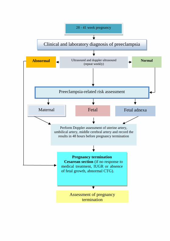
Abnormal Normal
Assessment of pregnancy
termination
28 - 41 week pregnancy
Clinical and laboratory diagnosis of preeclampsia
Preeclampsia-related risk assessment
Maternal Fetal Fetal adnexa
Perform Doppler assessment of uterine artery,
umbilical artery, middle cerebral artery and record the
results in 48 hours before pregnancy termination
Ultrasound and doppler ultrasound
(repeat weekly)
Pregnancy termination
Cesarean section (if no response to
medical treatment, IUGR or absence
of fetal growth, abnormal CTG).
Related Documents


