GENERAL AND COMPARATIVE ENDOCRINOLOGY 88, 351-363 (1992) Neurotensin, Substance P, GastrinKhoiecystokinin, and Bombesin in the Intestine of the Tilapia (Oreochromis mossambicus) and the Goldfish (Carassius auratus): lmmunochemical Detection and Effects on Electrophysiological Characteristics AMANDA KILIAAN,*,' SUSANNE HOLMGREN,? ANN-CATHRINE JbNSSON,? KLAASDEKKER.' ANDJACKGROOT* *Department of Experimental Zoology, University of Amsterdam, Kruislaan 320, 1098 SM, Amsterdam, The Netherlands: and fDepartment of Zoophysiology, University of Giiteborg, Box 25059, S-400 31 Gcteborg, Sweden Accepted June 2, 1992 The distribution of neurotensin-, substance P-, gastrin/cholecystokinin/caeruiein- and bombesin-like immunoreactivities has been studied in the gut of the tilapia (Oreochromis mossambicus) and the goldfish (Carassius auratus) using immunohistochemistry and radio- immunoassay; the electrophysiological effects of these peptides on the intestinal epithelium were also examined with the Ussing-type chamber technique. Neurotensin- and gastrin/ cholecystokinin/caerulein-like immunoreactivities were present in endocrine cells in both species. Substance P- and bombesin-like immunoreactive endocrine cells were present in the intestine of the tilapia. Neurotensin-like immunoreactivity was observed in varicose fibers and nerve cell bodies in the muscle layers and myenteric plexus of both species, whereas nerve fibers showing substance P-like immunoreactivity were found in the goldfish only. Using radioimmunoassays, neurotensin- and gastrin/cholecystokinin/caerulein-like im- munoreactive materials were detected in intestinal extracts of both species. The amounts of substance P- and bombesin-like material were below detection level. The ion selectivity of the intestinal epithelium of both species was modulated by exogenously applied neurotensin. This effect was blocked by tetrodotoxin in the tilapia but not in the goldfish. In the tilapia, neurotensin may act via stimulation of a CAMP-dependent increase of the Cl- conductance of the tight junctions, whereas in the goldfish, neurotensin induced, via an unknown mes- senger, a transient decrease of the cation selectivity without a decrease in the resistance. Substance P, cholecystokinin, and bombesin were without effect on the electrophysiological characteristics of the epithelium. o 192 Academic press. 1~. The enteric nervous system (ENS) has been defined as the third division of the au- tonomic nervous system because it can function independent of the central nervous system and has complete reflex pathways (Langley, 1921; Furness and Costa, 1980). The ENS consists of two interconnected networks: the myenteric and submucosal plexuses. In mammals, both plexuses con- tain nerve cell bodies that innervate the mu- cosal layer, whereas in fish few or no gan- ’ To whom correspondence should be addressed. glionic cells occur in the submucosal plexus and the mucosa is innervated predomi- nantly by nerve fibers of the myenteric plexus (Kirtisinghe, 1940; Nicol, 1952). The ENS and the endocrine/paracrine systems probably play important roles in fish, as in mammals, in coordinating various intestinal processes such as motility, blood flow, and secretion/absorption, but the precise func- tions of the several neurotransmitters and hormones found in the gut are not yet un- derstood (Cooke, 1989; Nilsson and Holm- gren, 1989). 351 0016~6480/92 $4.00 Copyright 0 1992 by Academic Press, Inc. All rights of reproduction in any form reserved.

Welcome message from author
This document is posted to help you gain knowledge. Please leave a comment to let me know what you think about it! Share it to your friends and learn new things together.
Transcript

GENERAL AND COMPARATIVE ENDOCRINOLOGY 88, 351-363 (1992)
Neurotensin, Substance P, GastrinKhoiecystokinin, and Bombesin in the Intestine of the Tilapia (Oreochromis mossambicus) and the
Goldfish (Carassius auratus): lmmunochemical Detection and Effects on Electrophysiological Characteristics
AMANDA KILIAAN,*,' SUSANNE HOLMGREN,? ANN-CATHRINE JbNSSON,? KLAASDEKKER.' ANDJACKGROOT*
*Department of Experimental Zoology, University of Amsterdam, Kruislaan 320, 1098 SM, Amsterdam, The Netherlands: and fDepartment of Zoophysiology, University of Giiteborg, Box 25059,
S-400 31 Gcteborg, Sweden
Accepted June 2, 1992
The distribution of neurotensin-, substance P-, gastrin/cholecystokinin/caeruiein- and bombesin-like immunoreactivities has been studied in the gut of the tilapia (Oreochromis mossambicus) and the goldfish (Carassius auratus) using immunohistochemistry and radio- immunoassay; the electrophysiological effects of these peptides on the intestinal epithelium were also examined with the Ussing-type chamber technique. Neurotensin- and gastrin/ cholecystokinin/caerulein-like immunoreactivities were present in endocrine cells in both species. Substance P- and bombesin-like immunoreactive endocrine cells were present in the intestine of the tilapia. Neurotensin-like immunoreactivity was observed in varicose fibers and nerve cell bodies in the muscle layers and myenteric plexus of both species, whereas nerve fibers showing substance P-like immunoreactivity were found in the goldfish only. Using radioimmunoassays, neurotensin- and gastrin/cholecystokinin/caerulein-like im- munoreactive materials were detected in intestinal extracts of both species. The amounts of substance P- and bombesin-like material were below detection level. The ion selectivity of the intestinal epithelium of both species was modulated by exogenously applied neurotensin. This effect was blocked by tetrodotoxin in the tilapia but not in the goldfish. In the tilapia, neurotensin may act via stimulation of a CAMP-dependent increase of the Cl- conductance of the tight junctions, whereas in the goldfish, neurotensin induced, via an unknown mes- senger, a transient decrease of the cation selectivity without a decrease in the resistance. Substance P, cholecystokinin, and bombesin were without effect on the electrophysiological characteristics of the epithelium. o 192 Academic press. 1~.
The enteric nervous system (ENS) has been defined as the third division of the au- tonomic nervous system because it can function independent of the central nervous system and has complete reflex pathways (Langley, 1921; Furness and Costa, 1980). The ENS consists of two interconnected networks: the myenteric and submucosal plexuses. In mammals, both plexuses con- tain nerve cell bodies that innervate the mu- cosal layer, whereas in fish few or no gan-
’ To whom correspondence should be addressed.
glionic cells occur in the submucosal plexus and the mucosa is innervated predomi- nantly by nerve fibers of the myenteric plexus (Kirtisinghe, 1940; Nicol, 1952). The ENS and the endocrine/paracrine systems probably play important roles in fish, as in mammals, in coordinating various intestinal processes such as motility, blood flow, and secretion/absorption, but the precise func- tions of the several neurotransmitters and hormones found in the gut are not yet un- derstood (Cooke, 1989; Nilsson and Holm- gren, 1989).
351 0016~6480/92 $4.00 Copyright 0 1992 by Academic Press, Inc. All rights of reproduction in any form reserved.

352 KILIAAN I3 AL.
Previous work on the gut of the goldfish and tilapia (Kiliaan et al., 1989a,b. 1990) demonstrated the presence of vasoactive intestinal polypeptide (VIP)-, galanin-, neu- ropeptide Y (NPY)-, calcitonin gene related peptide (CGRP)-, and enkephalin-like pep- tides and serotonin. Little is known of the role neuropeptides and gut hormones play in fish intestinal ion transport but there is evidence that VIP and serotonin may be en- dogenous messengers to regulate the epi- thelial ion selectivity (Kiliaan et al., 1989a, 1990). A number of neuropeptides exert their action via CAMP and it has been shown in teleost intestinal epithelia that CAMP can modulate the ion selectivity of the tight junctions by increasing their Cl permeability (Bakker and Groot, 1984, 1989; Groot, 1985).
Immunohistochemistry (IHC), radioim- munoassays (RIA), and the Ussing-type chamber techniques have been applied to assess the presence and possible electro- physiological effects of bombesin (BOM), cholecystokinin (CCK), caerulein, gastrin (G), substance P (SP), and neurotensin (NT) on the intestinal epithelia of two tele- ost fishes.
METHODS
Goldfish (Carassius auratus), 18 to 20 cm long, and tilapia (Oreochromis mossambicus), 20 to 25 cm long, were used for the experiments. The fish were fed daily in the morning (Tetramin fish food) except for the day they were killed. They were kept in well-aerated fresh water at 18 and 27”, respectively, and killed by pithing both the brain and spinal cord after cervical cord transection. The gut was rapidly removed and divided into four segments: I, the proximal one-fifth of the intestine (including the intestinal bulb of the goldfish); II, the following 35% of the intestine; III (mid intes- tine), the next 35% of the gut; and IV, the distal por- tion of the last 10% of the intestine.
Immunohistochemistry
The tissue pieces were fixed for 18 hr at 4” in a mixture of 2% formaldehyde and 15% picric acid in 0.1 M phosphate buffer (pH 7.3), rinsed 3 x 15 min in 80% ethanol, dehydrated, cleared in 100% xylene for 30 min, and rehydrated.
Whole Mounts
Whole mounts were prepared according to the method of Costa et ul. (1980). Muscle layers were sep- arated from mucosal layers and small pieces of the longitudinal muscle layers (LM) and circular muscle layers (CM) were incubated for 18 hr at lx” with the primary antibody (Table l), rinsed 4 x 10 min in phos- phate-buffered saline (PBS, pH 7.3), and incubated for 1 hr with a secondary antibody (anti-rabbit IgG) tagged with fluorescein-isothiocyanate (FITC, DAKO). After rinsing in PBS, the pieces were mounted in bicarbon- ate buffered glycerol (0.35 M. 1: I, pH 8.0.
Sections
The fixed and xylene-treated tissues were rinsed overnight (20% sucrose in 0.1 M Na-cacodylate buffer, pH 7.3,4”) and snap frozen in liquid nitrogen. Sections (10 urn thick) were cut on a cryostat, placed on chrome-alum gelatin-coated slides, and incubated with antisera (Table 1) for the whole mounts.
The tissues were examined with a Leitz Ortholux fluorescence microscope. A Leitz Orthomat automatic camera and Kodak TriX film were used for the pho- tography.
Specificity of positive reaction was tested by over- night incubation (4”) with antisera preincubated with the original hapten (lo-’ M).
Radioimmunoassay (RIA) Tilapia and goldfish intestines were rinsed with ice-
cold saline, divided into the four segments (see above), and then separated into muscle and mucosal layers. The tissues were weighed, immediately boiled in 10 vol of water for 5-10 min, and homogenized with an Ultraturrax. Homogenates were centrifuged (2OOOg, 15 min) and the supematants stored. The pellets were resuspended and rehomogenized in 0.5 M acetic acid and centrifuged. Both water and acetic acid superna- tants were stored at or below - 20” until assayed. Lev- els of G/CCK-, SP-, BOM-, and NT-like immunoreac- tivities (IR) were estimated by RIA following pub- lished methods (Dockray, 1980; Jensen et al., 1987; Jonsson et al., 1987; Holmgren and Jiinsson, 1988). The levels of BOM- and NT-like IRS were measured in acid extracts only and SP- and GICCK-like IRS were measured in both water and acid extractions. The acidic samples were neutralized with &CO, before being assayed.
Bombesin. The RIA employed the antiserum L90, diluted 1: 10,000, and G07, dihrted 1:25OQ (Tabje 11, as described in detail elsewhere (Ho-n and Jiinsson, 1988). Incubations were performed in a 0.02 M Verona1 buffer (pH 8.4) containing 0.75% bovumin (L90) and a 0.2 M phosphate buffer (pH 7.9) containing 0.1% bovine serum aIbumin (BSA, fraction V) and

NEUROPEPTIDES IN FISH INTESTINE 353
TABLE 1 LIST OF ANTISERA(ALL RAISED IN RABBIT)
Working dilution
Antigen Raised against Code IHC RIA SourceiRef.
Caerulein Gastrin 17 G/CCK
BOM
SP
NT
SY sy human C-term CCK32-39 Porcine CCK39 CCKlO-20 C-term CCK4 C-term CCK4 C-term CCK8 sy BOM l-14 sy BOM l-14 sy SP l-11 sy SP l-11 C-term NT
G31 G17 CCKB CCK39
GO3 Ll12 L48 L90 GO7 GlO Gil NTAb
1:lOO 1:lOo 1:lOO 1:lOO 1:lOO
1:lOO
1:lOO
1:lOO 1:lOO 1:lOO
1:20,000
1:50,000 l:lO,OOO 1:2,500 1:250,000 1: 150,000 1:750,000
Jonsson Milab Diagnostica Diagnostica Milab Jonsson/l Dockray/ Do&ray/3 Dockray/ Jonssonl5 Jiinssonl6 JGnsson/6 Dockray/pers.
Note. IHC, immunohistochemistry; RIA, radioimmunoassay. Ref. 1, Jonsson et al., 1987; Ref. 2, Dockray et al., 1981; Ref. 3, Dockray et al., 1980; Ref. 4, Holmgren et al. 1982; Ref. 5, Holmgren and Jonsson, 1988; Ref. 6, Jensen et al., 1987.
0.1% NaCl (G07). For both assays synthetic bombesin (Peninsula St. Helens, UK) was used as the standard, and iodinated Tyr4-bombesin (2000 Ci/mmol) was used as marker.
Gastrinlcholecystokinin. Immunoreactive material was measured using either antiserum L48, 1:50,000 (Table 1), in a 0.02 M veronal buffer (pH 8.4) contain- ing 0.75% bovumin (Dockray, 1980) or antiserum G03, 1:20,000 (Table 1), in a 0.05 M phosphate buffer containing 0.05% BSA, pH 7.3 (Jonsson et al., 1987). For both assays, iodinated nonsulfated CCK8 was used as the label (2000 Ci/mmol) and nonsulfated CCKI (Research Plus, Bayonne, NJ) was used as the standard.
Substance P. The antisera GlO and Gll (diluted 1: 250,000 and 1:150,000, respectively, Table 1) were used in a 0.05 M Verona1 buffer containing 0.8% NaCl and 0.1% BSA (pH 8.6) for GlO and 1.0% NaCl and 0.1% BSA (pH 8.4) for assay with Gll (Jensen et al., 1987). Synthetic SP (Sigma, St. Louis, MO) was used to construct the standard curve and ‘251-SP (2200 Ci/ mmol. NEN, Du Pont Scandinavia A.B., Stockholm, Sweden and 2000 Ci/mmol, Amersham Inter. plc., Amersham, UK) was used as the label.
Neurotensin. The RIA was performed using the an- tiserum NTAb (C-terminal specific; G. J. Dockray, personal communication), diluted 1:75O,ooO, and incu- bated with samples in a 0.035 M phosphate buffer (pH 7.6) containing 0.05% BSA. Synthetic NT (Diagnos- tika, Falkenberg, Sweden) was used as the standard and ‘251-NT (2200 Ci/mmol, Amersham Inter. plc., and 2200 Ci/mmol, NEN Du Pont Scandinavia) was used as the tracer.
All incubations were performed for 48 hr at 4”. An- tibody-free and bound tracers were separated by add- ing dextran-coated charcoal-slurry containing plasma. The level of radioactivity was measured in pellet and supematant after centrifugation (4 min, 3OOOg).
Electrophysiological Experiments
The epithelia of goldfish (segment II) and tilapia in- testines (segment I) were stripped from the underlying muscle layers. The epithelium was mounted in a mod- ified Ussing chamber (Groot et al., 1979) which was perfused with a Ringer’s solution with the following composition (in mM): NaCl, 117.5; KCl, 5.7; NaHCO,, 25.0; NaH,PO,, 1.2; CaC12, 2.5; MgSO,, 1.2; mannitol, 27.8 (pH 7.4). The transepithelial poten- tial difference (with respect to the mucosal solution) and transepithetial resistance were measured as de- scribed by Bakker and Groot (1984) for 30 min before the neuropeptide/hormone was added to the serosal side, and measurements were performed for a further 15 min. Changes in ion selectivity of the tight junctions caused by the addition of the peptides were studied by their effect on the dilution potential (the transepithelial potential induced by replacement of half of the NaCl with mannitol) (Kiliaan et nl., 1989a). Because ion se- lectivity of tight junctions is related to intracellular CAMP, this is a convenient way of screening for neu- rotransmitter- and hormone-coupling to the adenylate cyclase system in the fish enterocyte (Bakker and Groot, 1989). Significance of the changes was tested by the Student t test. P values CO.05 were considered significant.
354 KILIAAN El‘ AL..
RESULTS
Immunoreactive material was apparent in endocrine cells and neurons of the gut wall of the tilapia and goldfish (Table 2). In both species, the immunostaining of nerves and endocrine cells were more pronounced in proximal than in distal parts of the intes- tine. The variety of endocrine cells was larger in the tilapia than in the goldfish.
Neurotensin
Immunohistochemistry. In both species, nerve fibers and cell bodies immunoreac- tive for NT were present in the myenteric plexus and in the longitudinal muscle layers but no immunoreactive nerve fibers were visible in the circular muscle layer, the mu- cosa, or the submucosa (Table 2, Figs. 1 A- 1D). NT-like immunoreactive endocrine cells occurred in the anterior intestine only (Table 2). These endocrine cells were
mainly found in the basal half of the mu- cosal folds of the goldfish (Fig. 1C) and at the tip of the folds in the tilapia.
Radioimmunoassuy. lmmunoreactive material was present in the 0.5 M acetic acid extracts of the mucosa and muscle lay- ers of all parts of the intestine (Table 3). All the extracts diluted in parallel with the stan- dard curve (synthetic NT), indicating a close relationship of the immunoreactive material with NT. The highest concentra- tions were detected in the muscle layers. More NT-like material was found in the ti- lapia than was found in the goldfish intes- tine.
Electrophysiology. Addition of NT to the serosal side of the proximal part of the tila- pia intestine, stripped free from muscle Iay- ers and mounted in an Ussing chamber, re- duced the transepithelial resistance and the dilution potential (Table 4). This effect was concentration dependent; the threshold concentration was 10e9 M, the maximal re-
TABLE 2 THE PRESENCE OF NEUROPEP~IDES IN NERVE FIBERS (F), ENDOCRINE CELLS (EC), AND NERVE CELL
BODIES (CB) THROUGHOUT THE INTESTINE (PART 1, PROXIMAL, TO PART IV, DISTAL) OF THE GOLDFISH AND TILAPIA
Part: ____-
Mucosa
Submuc. CM MP
LM
Mucosa
Submuc. CM MP
LM
Goldfish Tilapia __-. .--__
I II III IV I II III IV -
Neurotensin EC 2 3 - - 2 _ - - F _ - - - - - -. - F -------- F -------- CB 3 3 2 2 2 2 I 1 F 332222-- F 2 2 2 I 2 2 2 2
Substance P EC - - - - 2 3 1 1 F _------- F ,,------ F II------ C-j - - - - - - _ _ F II------ F II------
_--
EC F F F CB F F
EC F F F CB F F
Goldfish Tilapia
1 II III IV I II III IV
GKCWcaerulein 2 3 1 1 3 3 1 I
Bombesin - 3 3 2 2
_ - _ - - -. - -
Note. 1 indicates very low amounts of visible imrnunoreactive material, 2 indicates mode+@ amounts, 3 indicates hi density, and - indicates an absence of visible immunoreactive m&e&& LM, longiWi& mu&e layer; MP, myenteric plexus; CM, circular muscle layer; Submuc., submucosa.

NEUROPEPTIDES IN FISH INTESTINE 355
FIG. 1. (A-D) NT-like IR. (A) Group of nerve cell bodies and nerve fibers in the myenteric plexus of the goldfish. x747. (B) Nerve cells and nerve fibers in the myenteric plexus of the tilapia intestine. x448. (C) Nerve cells in the myenteric plexus and endocrine cells in the base of the intestinal epithe- lium of the goldfish. x332. (D) Varicose nerve fibers in the myenteric plexus of the goldfish. ~415. (E) SP-like immunoreactive nerve fiber with many varicosities in the longitudinal muscle layer of the goldfish intestine. x423. LM, longitudinal muscle layer; CM, circular muscle layer; EP, epithelium.

356 KII.IAAN ET AL.
TABLE 3 NT-LIKE IR MATERIAL EXTRACTED WITH ACETIC
ACID FROM THE INTESTINE OF TILAPIA
AND GOLDFISH
Goldfish Tilapia
Mucosa 1 II III IV
Muscle I 11 III IV
3.53 t 0.78 (6) 6.60 i 2.90 (5) 4.05 2 0.86 (6) 6.67 t 1.11 (5) 4.96 2 1.99 (6) 14.06 2 3.30 (4) 2.62 k 0.58 (6) 7.50 k 1.46 (4)
10.77 i 2.57 (6) 23.87 k 4.39 (5) 11.04 k 1.26 (6) 22.53 ” 5.42 (5) 8.08 +- 2.39 (4) 18.16 t 6.77 (4)
14.54 2 2.30 (4) 26.83 t 3.30 (4)
Note. Values are expressed as means t SEM pmoYg tissue. The number of animals is given in pa- rentheses. I, proximal intestine; IV, distal part of in- testine.
duction of the potential was found at 10m6 M neurotensin (1.1 + 0.2 mV, II = 5) and the half-maximal effect was 8 x lo-’ M. This effect was not sensitive to tetrodo- toxin (TTX, low6 A4) added 5 min before or in the presence of NT. Addition of cloni- dine (10e6 M) or adrenaline (10e6 M) after NT stimulation restored the dilution poten- tial (Fig. 2), suggesting that the effect of NT can be counteracted by the q-cat- echolaminergic action (Groot, 1985).
Addition of NT to the serosal side of seg-
TABLE 4 EFFECT OF NEUROTENSIN (10d6 M) ON
TRANSEPITHELIAL POTENTIAL (Y,), RESISTANCE (R,) AND ON THE DILUTION POTENTIAL (Yy, DIL) IN
ISOLATED TILAPIA AND GOLDFISH EPITHELIUM OF THE FIRST PART OF THE INTESTINE
Goldfish
\Ir, (mV) controf A induced by NT
0.2 k 0.1 (16) +0.4 -+ 0.2 (16)
P <: 0.03 R, (0) control 31.5 2 1.8 (16) A induced by NT + 1.7 2 0.1 (16)
P < 0.001 qt dil (mV) control -9.6 2 0.4 (16) A induced by NT 2.1 * 0.03 (16)
P < 0.001
Tilapia ___--~.
0.1 r 0.1 (5) +0.05 zt 0.05 (5)
P<O.o4 25.8 + 1.7 (5)
-1.5 2 0.2 (5) P<O.o03
-7.1 t 0.2 (5) 1.1 + 0.1 (5) P<O.ool
iVore. + indicates increase of Y, OT R1 and - indicates decrease of 0, or Rt. P values indicate difference from zero.
FIG. 2. Recording of a typical experiment showing the negative shift in the transepithelial potential across a tilapia intestinal epithelium, induced by mucusal re- placement of 56 mM NaCl by 112 mM mannitol (mu- cosal dilution). In the presence of TTX, NT reduces the potential difference: this effect is reversed by the addition of clonidine.
ment II of the goldfish intestine (see Meth- ods) caused an increase of the transepithe- lial resistance and a transient reduction of the transepithelial potential which could be prevented by TTX (Table 4; Fig. 3). Since this response resembled the action of car- bachol (Groot, 1985), the effect of atropine was tested. Although the addition of atro- pine blocked the effect of carbachol, it did not affect the action of NT (Fig. 3), indicat- ing that cholinergic receptors are not in- volved in the NT effect. Similar results were obtained with stripped epithelium from segment IV of the goldfish intestine. This part of the tilapia intestine could not be stripped and was not studied in the Ussing- type chambers.
mucosal dilution
FIG. 3. Typical experiment showing the transient effect of NT on the diIution potential, induced as in Fig. 2, across the goldfish intestinal epithelium in the presence of atropine (solid line) and in the presence of TTX (dashed line). The presence of atropine had no effect on the NT response. TTX strongly reduced the effect of NT.

NEUROPEPTIDES IN FISH INTESTINE 357
Substance P
Zmmunohistochemistry. Some varicose nerve fibers showing SP-like IR were found running parallel to the smooth muscle fibers in segments I and II of the goldfish intestine (Fig. 1E). A few nerve fibers were present in the submucosal layer but not in close vi- cinity to the epithelium. No immunoreac- tive nerve cell bodies were seen. Endocrine cells were only found in the tilapia (Fig. 4A). Each endocrine cell was large, had a basally located nucleus, and a long thin pro- cess that extended toward the lumen.
Radioimmunoassay. The amount of SP- like material in both the water and the ace- tic acid extracts of the tilapia and the gold- fish intestine was below the detection level of the assay (5-10 fmol, Jensen et al., 1987) for both antisera used.
Electrophysiology. Addition of substance P to the serosal side of the intestinal epithe- lium had no measurable effect on transepi- thelial potential, resistance, or dilution po- tential.
Bombesin
Zmmunohistochemistry. BOM-like IR was only seen in the tilapia. The immuno- reactive material was restricted to endo- crine cells which were present in large num- bers mainly in the upper half of the mucosal folds of the first and second parts of the intestine. These cells were large, pear- shaped, and elongated with processes ex- tending toward the lumen (Fig. 4B).
Radioimmunoassay. BOM-like IR were not detected in the acid extracts of the in- testines of the tilapia or goldfish.
Electrophysiology. BOM did not affect electrogenic ion transport or transepithelial resistance of the intestinal epithelia of ei- ther species.
GastrinlCholecystokininlCaerulein
Zmmunohistochemistry. Only endocrine cells showed IR to G/CCWcaerulein. The
IR cells were most abundant in the proxi- mal part of the intestine, mainly located in the upper half of the folds. The cells in the tilapia intestine were elongated and had thin processes that reached toward the lu- men (Fig. 4C), whereas in the goldfish these processes extended from both basal and lu- minal sides (Figs. 4D and 4E). The exten- sions were often broadened at the end and sometimes branched at the basal side (Fig. 4F). The antisera G31, G03, L112, L48, CCK39, and CCK32-39 (CCKS), which are all directed to the C-terminal region, showed the same regional differences in the distribution of endocrine cells. Antisera against CCKlO-20 and gastrin 17 were also negative.
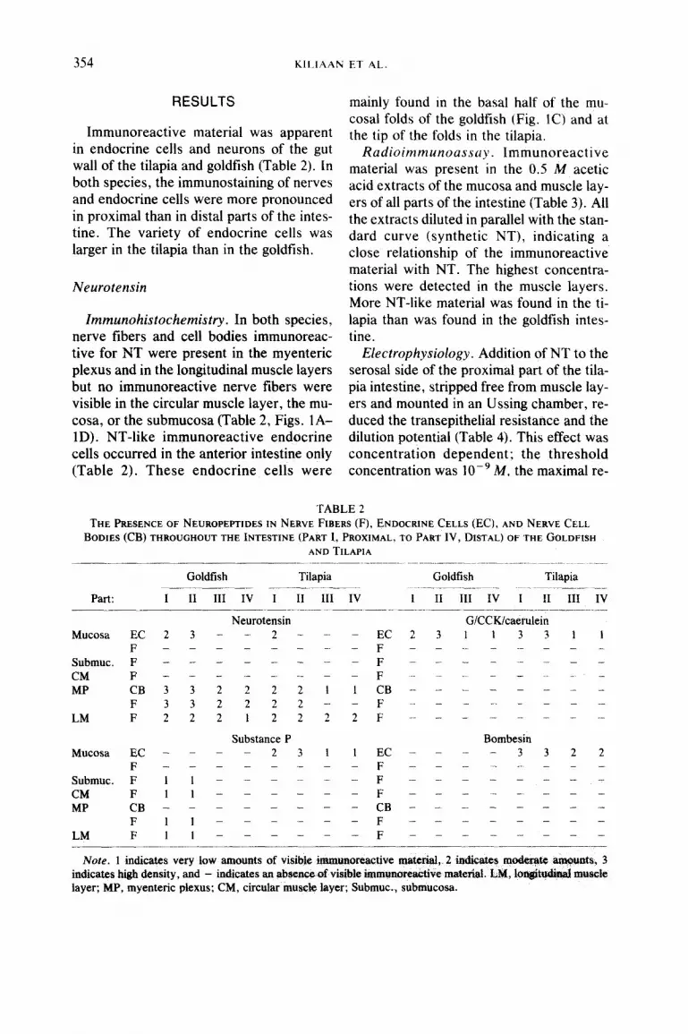
Radioimmunoassay. Antisera raised against CCK4 and CCK8, respectively, gave G/CCK/caerulein-like IR in all parts and all layers of the intestine of both the tilapia and the goldfish (Table 5). All the extracts diluted in parallel with the stan- dard curve (CCK8), indicating a close rela- tionship of the immunoreactive material with gastrin/CCK. The amounts of G/CCK/ caerulein-like immunoreactive material were comparable in the two species. Table 6 shows that the measured amounts were always larger using the antiserum raised against CCK4 (G03) than when using L48 raised against CCK8.
Electrophysiology. CCK added to the se- rosa1 side of the intestinal epithelia of tila- pia and goldfish was without measurable electrophysiological effect.
DISCUSSION
The presence in the gut and the pharma- cological actions on ion transport of certain neuropeptides/gut hormones have been ex- amined.
The regional distribution of NT-like IR in the anterior intestine of both fish species agrees with previous observations in the ide (Burkhardt-Holm and Holmgren, 1988) and in the barb (Rombout and Reinecke, 1984).

358 KII.tAAN E’t AL.
FIG. 4. (A) SP-like immunoreactive large endocrine cells with processes extended toward the lumen in the tilapia intestine. x415. (B) BOM-like immunoreactive endocrine cells scattered in the intestinal epithelium of the tilapia. x 183. (C-F) GiCCKicaerulein-like immunoreactive material in the goldfish and tilapia intestine. (C) Endocrine cell with thin apical process in the tilapia intestine (CCK39). x270. (D, E) Endocrine cells with processes toward the lumen and basal membrane in the goldfish intestine (L112). D, x498; E, x598. (F) Endocrine cell with two small extensions into the lamina propria of the tilapia intestine (CCK39). x282. LU. lumen; EP, epithelium.
Similarly the concentrations of NT-like im- levels found in the duodenum of Cottus and munoreactive material found in the acetic Raniceps (Reinecke et al., 1980). In the acid extracts of the goldfish intestine in the present study, the highest concer&ations present RIAs were comparable with the were found in the muscle layer extracts of

NEUROPEPTIDES IN FISH INTESTINE 359
TABLE 5 G/CCK-LIKE IR MATERIAL (ANTISERUM G03)
EXTRACTEDWITH~.~ M ACETIC ACID(HAC)AND H,O FROM THE INTESTINE OF THE TILAPIA
AND GOLDFISH
Mucosa I II III IV
Muscle I II III IV
Mucosa I II III IV
Muscle I II III IV
Goldfish Tilapia -
KzO
201.1 2 29.4 (5) 374.7 2 66.2 (6) 81.1 + 12.5 (6) 97.4 +- 7.2 (6)
257.8 2 35.4 (6) 68.3 ? 9.9(6) 67.1 2 19.9 (4) 91.9 k 22.3 (4)
HAc
161.2 ? 71.7 (3)” 131.4 2 27.5 (5) 325.0 ” 62.8 (3)b 164.4 f 42.6(4)
45.2 k 6.4 (5) 34.5 2 8.4 (5) 66.2 t 18.0 (4) 24.9 “_ 4.3 (4)
54.9 k 14.2 (6) 49.9 * 19.3 (4) 11.9 2 1.1 (6) 12.2 f 2.8 (4) 7.1 -+ 1.0 (6) 29.4 2 5.2 (4) 6.3 + 1.1 (6) 44.1 2 5.7 (3)
17.5 f 2.3 (6) 34.6 k 7.7 (6) 23.9 +- 3.8 (4) 28.3 f 1.5 (3)d
23.1 + 2.2 (5) 20.0 2 3.9(4) 28.4 ” 10.4 (4) 27.9 2 9.0 (3)'
Note. Values are expressed as means 2 SEM pmol/g tissue. The number of animals tested is given in parentheses. I, proximal intestine; IV, distal part of intestine. Ranges: “159.0-379.7, b205.2-417.6; ‘34.3- 54.0;d25.5-30.8;'14.&45.1.
both fish, which is in agreement with the immunocytochemical results. A dissimilar- ity between the immunohistochemistry (IHC) and RIA results is that NT-like IR could be demonstrated in the mucosa of the distal part of the goldfish and tilapia intes- tine with the RIA, whereas no IR could be observed using IHC. We have no explana- tion for this difference.
The location of the immunoreactive en- docrine cells in the basal half of the villi in the goldfish intestine is the same as that reported for Cottus scorpius and Raniceps raninus (Reinecke et al., 1980) but differs from that in tilapia; it is unknown whether these differences have a functional signifi- cance .
The reduction of the transepithelial ion selectivity in both tilapia and goldfish after exogenous addition of NT suggests that NT is involved in the control of transepi- thelial ion transport. The fact that NT re- duced the mucosal as well as the serosal dilution potential supports the idea that NT in both species affected the ion selectivity of the paracellular pathway, because the decrease of mucosal and serosal dilution potentials indicates that only one barrier between the mucosal and serosal bath was modulated.
The insensitivity to TTX of the effect of NT on the ionselectivity in the tilapia sug- gests that NT may have a direct effect on the enterocytes. However, TTX insensitiv- ity does not rule out the possibility that NT exerts its effect via the stimulation of re- lease of messengers from varicosities un- derneath the epithelium. The small, al- though significant, decrease of the trans- epithelial resistance and the decline of the dilution potential brought about by NT are similar but smaller than the effects of sero- tonin or 8-Br-CAMP (Groot, 1985; Kiliaan et al., 1989a). The restoration of the NT- induced decrease of the dilution potential with clonidine and adrenaline parallels the effect of clonidine and adrenaline on the se- rotonin-induced decrease of the dilution po- tential and indicates an action via I+- adrenoceptors (Groot, 1985). These tind- ings suggest that NT also may act via stimulation of adenylate cyclase in entero- cytes of the tilapia.
The TTX sensitivity of the effect of NT on the ionselectivity in the goldfish indi- cates an indirect action of NT. Moreover, NT induced an increase of the transepithe- lial resistance in this tissue, suggesting that the endogenous neurotransmitter(s) re- leased by NT does not act via stimulation of adenylate cyclase. Although the transient response and the increase of the transepi- thelial resistance are similar to the re- sponses to carbachol (Groot, 1985), the in- sensitivity to atropine indicates that cholin-
360 KILIAAN ET‘ AL.
'I ABLE 6 COMPARISON OF RIA WITH THE ANTISERA GO3 AND L48 ON WATER AND Acw EXTRACTS OF PAKT I OF
THE INTESTINE OF THF. GOLDFISH AND THE TILAPIA
Goldfish Tilapia
Part1 Hz0 HAc i,O HAc
GO3 Mucosa 201.1 5 29.4 (6) 54.9 t 14.2 (6) 275.1 ?I 63.9 (3) 49.9 k 19.3 (4) Muscle 257.8 2 35.4 (6) 17.5 z 2.3 (6) 45.2 + 6.4 (4) 23.1 t 2.2 (5)
L48 Mucosa 77.5 r 5.6 (6) 8.9 + 0.8 (6) 94.8 t 6.0 (9) 23.0 t 5.7 (7) Muscle 27.3 z!z 8.6 (6) 7.0 rt 1.0 (5) 13.8 -+ 5.3 (5) 4.4 + 0.7 (3jb
Note. Values are expressed as means ? SEM pmol/g tissue. The number of animals tested is given in parentheses.
a Range, 159.0-379.7. ’ Range, 3.2-5.6.
ergic receptors are not involved in the neurotensin pathway.
The increase of the transepithelial resis- tance may be caused by a reduction in the conductance for Naf in the tight junctions or by a decrease in the diameter of the lat- eral intercellular space via a change in the transport activity of the epithelial cells. Both changes could account for the decline in dilution potential but we cannot differen- tiate between these possibilities in the present experiments.
In mammals, where NT is primarily con- fined to endocrine cells in the mucosa, NT released from these enteroendocrine cells may stimulate anion secretion indirectly via activation of NT receptors in submucosal nerves (Kachur et al., 1982; Brown and Treder, 1982; Walsh, 1987). However, the effect of NT on cultured intestinal cells sug- gests that NT may act directly on epitheiial cells (Roebroek et al., 1992).
The presence of SP-like immunoreactive endocrine cells in tilapia agrees with the re- ported presence in another cichlid, P. pul- cher (Langer et al., 1979). The negative RIAs are in accordance with the scarcity of immunoreactive material revealed in the immunohistochemical studies. No electro- physiological elect of exogenous SP was found in the intestinal epithelium of either goldfish or tilapia, suggesting that the SP-
like peptide present in intestinal endocrine cells of tilapia is not involved in the control of electrogenic ion movement through the mucosa. Similarly, the electrophysiological studies suggest that the BOM-like material in endocrine cells of tilapia is not involved in the regulation of electrogenic properties.
The absence of BUM-liie IR in the gold- fish intestine contrasts with the abundance of immunoreactive nerve fibers described in the carp (&em&g and H-en, 1988) and in the ide (Burkhardt-Helm and Holm- gren, 1988) in studies that used the same antiserum, again showing the large differ- ences that may occur between related spe- cies. The discrepancy between the IHC and RIA found in the present study is not unique to the tilapia (cf. Holmgren et al., 1982; Holmgren and Ji)nsson, 1988). The amino acid sequence of fish bomb&n may differ partly from the amphibian sequence, so that the dilute antiserum used in the RIA procedure is unable to recognize the fish peptide properly, in contrast to the more concentrated antiserum used for noncom- petitive immunohistochemical methods. Thus even weakly immunoreractive material may give a positive result in IHC.
Gastrin and CCK and caerulein are closely reh&zd +nd share the me C-termi-
zistochemical t3epmce. Ih44iw& i-no-
studies of G/CCK in fish

NEUROPEPTIDES IN FISH INTESTINE 361
have been performed with antisera raised against the C-terminal tetra- or pentapep- tide of caerulein, CCK, and gastrin, which is the sequence they have in common. With antisera raised against CCKlO-20 and gas- trin 17 we found no positive reaction, sug- gesting that the midportion antigenic deter- minants in mammalian CCK differ from those in fish. Thus we were unable to dis- criminate between CCK and gastrin and can neither confirm nor contradict the sug- gestion that CCK and gastrin do not occur as separate entities in fish and amphibians but may occur as a single peptide that may be related to an ancestral molecule of the gastrin family (Larsson and Rehfeld, 1977; Buchan et al., 1980).
In all parts and layers of the intestines, the water extracts gave high yields in RIA when compared to the acetic acid extracts. This indicates that most of the gastrim CCK-like material has a shorter (CCK8) rather than longer sequence (CCK33) but both long and short forms of G/CCK/ caerulein are present in fish species (Vigna et al., 1985; Aldman et al., 1989). The re- action with GO3 always gave higher values than with L48 (Table 6). L48 has been re- ported to have a much lower cross- reactivity in RIA with CCK4 and gastrin 5 (GS; Dockray, 1980). It is thus possible that there is a large proportion of CCK4 and GS in the extracts that GO3 recognizes but L48 does not.
In the present study no electrogenic ef- fects of CCK on the intestinal epithelium could be found.
The present work, using three ap- proaches to determine the role of peptides in the gut, underlines the difficulty of draw- ing conclusions from one type of experi- ment only. The very low amount or the ab- sence of BOM- and SP-like IR correlates with the outcome of the RIA in the goldfish, whereas the presence of SP and BOM im- munohistochemically visualized in endo- crine cells of the tilapia did not show up in the RIA.
The presence of CCK or a similar peptide in endocrine cells correlates with the RIA, but with the RIA the peptide was also de- tected in the muscular layer where no nerve cells or fibers could be visualized with im- munohistochemical staining methods.
The electrophysiological experiments suggest that if the enterocytes are under control of SP, BOM, CCK, or a similar pep- tide, the effect is electroneutral and does not change the transepithelial resistance.
Although NT-like IR could only be de- tected in the proximal part of the mucosa in both fish using IHC, RIA indicates that it is present in all parts and the electrophysio- logical experiments also suggest that it has a role in the anterior as well as in the pos- terior part.
In tilapia intestine, the NT-like endocrine cells may influence adjacent epithelial cells directly via an adenylatecyclase coupled receptor. In the goldfish, NT may evoke a decrease in the ion selectivity by action on NT receptors in nerve cells underlying the epithelial cells.
In both tilapia and goldfish intestine, a decrease in the number of immunoreactive endocrine cells and in nerve fiber density toward the rectum has been demonstrated for the studied peptides. A teleological ex- planation for this could be that the first part of the intestine plays an important role in detection of the luminal contents and nutri- ent digestion and absorption and requires a variety of neuropeptides for a refined regu- lation.
ACKNOWLEDGMENTS
We thank Professor G. .I. Dockray (University of Liverpool, UK) for the gifts of antisera, Professor B. L. Roberts (University of Amsterdam) for reading and commenting on the manuscript, and Ms. I. Holmqvist (University of Giiteborg) and Mr. S. van Mechelen (University of Amsterdam) for the photog- raphy. This study was supported by a grant from the Netherlands Organisation for Scientific Research (NWO) and the Swedish Natural Science Research Council.

362
REFERENCES
Aldman, G., Jonsson, A-C., Jensen, J., and Holm- gren, S. (1989). Gastrin/CCK-like peptides in the spiny dogfish, Squalus acanthias: Concentrations and actions in the gut. Camp. Biochem. Phy.siol. C 92, 103-108.
Bakker, R., and Groot, J. A. (1984). CAMP-mediated effects of ouabain and theophylline on paracellu- lar ionselectivity. Am. J. Physiol. 246, 213-217.
Bakker, R., and Groot, J. A. (1989). Further evidence for the regulation of the tight junctions ionselec- tivity by CAMP in goldfish intestinal mucosa. J. Memhr. Biol. 11, 25-35.
Bjenning, C., and Holmgren, S. (1988). Neuropeptides in the fish gut: An immunohistochemical study of evolutionary patterns. Histochemistry 88, lS5- 163.
Brown, D. R., and Treder, B. G. (1982). Neurohor- monal regulation of ion transport in the porcine distal jejunum: Actions of neurotensin and its nat- ural homologs. J. Pharmacol. Exp. Ther. 249, 348-3.57.
Buchan, A. M. J., Polak, J. M., and Pearse, A. G. E. (1980). Gut hormones in Salamandra salaman- dra. Cell Tissue Res. 211, 331-343.
Burkhardt-Helm, P., and Holmgren, S. (1988). A com- parison of regulatory neuropeptides in the intes- tine of two stomachless teleosts (Poecilia reticu- lata and Leuciscus idus melanotus) under feeding and starving conditions. Cell Tissue Res. 255, 245-254.
Cooke, H. J. (1989). Role of the “little brain” in the gut in water and electrolyte homeostasis. Fed. Proc. Fed. Am. Sot. Exp. Biol. 3, 127-138.
Costa, M., Buffa, R., Fumess, J. B., and Solcia, E. (1980). Immunohistochemical localization of poly- peptides in peripheral autonomic nerves using whole mount preparations. Histochemistry 65, 157-169.
Dockray, G. J. (1980). Cholecystokinins in rat cere- bral cortex: Identification, purification and char- acterization by immunochemical methods. Bruin Res. 188, 155-165.
Dockray, G. J., Hutchinson, J. B., Gregory. R. A.. Tracy, H. J., and Zhu, W-Y. (1980). Cholecysto- kinin octapeptide: putative neurotransmitter in the gut. In “Advanced Physiolocal Science” (T. Gati, L. G. Szoller, and G. Ungvary, Eds.), Vol. 12, pp. 321-328. Pergamon Press, Oxford.
Dockray, G. J., Williams, R. G., and Zhu, W-Y. (1981). Development of region-specific antisera for the C-terminal tetra-peptide of gastrim cholecystokinin and their use in studies of immu- noreactive form of cholecystokinin in rat brain. Neurochem. Int. 3, 281-288.
Furness. J. 8.. and Costa. M. (1980). Types of nerves in the enteric nervous system. NPrIroycIerlcc 5, I-10.
Groot, J. A. (1985). Modulation of transepithelial Cl permeability. In “Transport Processes Iono- and Osmoregulation” (R. Gilles and M. Giiles- Baillien. Eds.). pp. 206-217. Springer-Verlag, Berlin.
Groot, J. A.. Albus, H.. and Siegenbeek van Heuke- lam, J. (1979). A mechanistic explanation of the effect of potassium on goldfish intestinal trans- port. PJluegers Arch. 379, 1-9.
Holmgren, S., and Jonsson, A-C. (1988). Occurrance and effects on motility of bombesin-related pep- tides in the gastrointestinal tract of the Atlantic cod, Gadus morhua. Camp. Biochem. Physiol. C 89, 249-256.
Holmgren, S., Vaillant. C.. and Dimaline, R. (1982). VIP, substance P-, gastrin/CCK-, bombesin-, so- matostatin- and glucagon-like immunoreactivities in the gut of the rainbow trout, Snlmo gairdneri. Cell Tissue Res. 223, 141-153.
Jensen, J., Holmgren, S., and Jonsson. A-C. (1987). Substance P-like immunoreactivity and the effects of tachykinins in the intestine of the Atlantic cod, Gadus morhua. J. Auton. Nerv. Syst. 20, 25-33.
Jonsson, A-C., Holmgren, S.. and Holstein, B. (1987). GastriniCCK-like immunoreactivity in endocrine cells and nerves in the gastrointestinal tract of the cod. Gadus morhua. and the effect of peptides of the gastrin/CCK family on cod gastrointestinal smooth muscle. Gen. Camp. Endocrinol. 66, 190- 202.
Kachur, J. F., Miller, R. J., Field, M., and Rivier, J. (1982). Neurohormonal control of ileal electrolyte transport. II. Neurotensin and substance P. J. Pharmacol. Exp. Ther. 2M, 456-463.
Kiliaan, A. J., Joosten, H. W. J., Bakker, R., Dekker, K., and Groot, J. A. (1989a). Serotonergic neu- rons in the intestine of two teleosts, Carassius auratus and Oreochromis mossambicus, and the effect of serotonin on transepithelial ion- selectivity and muscle tension. Neuroscience 31, 817-824.
Kiliaan, A. J., Holmgren, S., and Groot, J. A. (1989b). Comparison of 5-HT-like and neuropep- tide-like immunoreactivity in goldfish and tilapia intestine. Z. Gastroenterol. 27, 295.
Kiliaan, A. J., Scholten, G., Klaren, P., Flik, G., van OS, C. H., and Groat, J. A. (1990). Vasoactive intestinal peptide in the epithelial function of tila- pia intestine. 2. Gastroenterol. 28, 426.
Kirtisinghe, P. (1940). The myenteric nerve-plexus in some lower chordates. Q. J. Microsc. Sci. 81, 521-539.
Langer, M., Noorden, S., van Polak, J. M., and Pearse. A. G. E. (1979). Peptide hormone-likeim-
NEUROPEPTIDES IN FISH INTESTINE 363
munoreactivity in the gastrointestinal tract and endocrine pancreas of eleven teleost species. Cell Tissue Res. 199, 493-508.
Langley, J. N. (1921). “The Autonomic Nervous Sys- tem,” Part I. Heffer, Cambridge.
Larsson, L-I., and Rehfeld, J. F. (1977). Evidence for a common evolutionary origin of gastrin and cholecystokinin. Nature 269, 335-338.
Nicol, J. A. C. (1952). Autonomic nervous systems in lower chordates. Bio/. Rev. 27, l-49.
Nilsson, S., and Holmgren, S. (1989). Novel neuro- transmitters in the autonomic nervous system of nonmammalian vertebrates. Pharmacol. Ther. 41, 257-287.
Reinecke, M., Carraway, R. E., Falkmer, S., Feurle, G. E., and Forssmann, W. G. (1980). Occurrence of neurotensin-immunoreactive cells in the diges- tive tract of lower vertebrates and deuterostomian invertebrates. Cell Tissue Res. 212, 173-183.
Roebroek, J. G. H., Bajnath, R. B., and Groot, J. A. (1992). Neurotensin-induced changes in intracel- lular potential of the human colon carcinoma cell line HT 29~1. 19A. Z. Gastroenterof. 30, 232.
Rombout, J. H. W. M., and Reinecke, M. (1984). An immunocytochemical and electron-microscopical study of endocrine cells in the gut and pancreas of a stomachless teleost fish, Barbus conchonius (cyprinidae). Cell Tissue Res. 227, 577-593.
Vigna, S. R., Fischer, B. L., Morgan, J. L. M., and Rosenquist, G. L. (1985). Distribution and molec- ular heterogeneity of cholecystokinin-like immu- noreactive peptides in the brain and gut of the rainbow trout, Salmo gairdneri. Comp. Biochem. Physiol. C 82, 143-146.
Walsh, J. (1987). Gastrointestinal hormones. In “Physiology of the Gastrointestinal Tract” (L. R. Johnson, Ed.), pp. 181-253. Raven Press, New York.
Related Documents