6/5/17 1 NEURO- OPHTHALMOLOGICAL EMERGENCIES PRESENTING IN PRIMARY CARE OPTOMETRY Jacqueline Theis, OD, FAAO Optometry’s Meeting 2017 Washington DC • Training • BS Biochemistry – Bucknell University • OD –UC Berkeley School of Optometry • Residency – Binocular Vision, Neuro-Optometry, Vision Therapy – UC Berkeley School of Optometry • Current • Director, Clinic Chief– UC Berkeley Sports Vision Clinic • Clinical Care and Research for Vision Problems in Concussion • Clinical Instructor – UC Berkeley School of Optometry • Clinical Research Funding – UC Regents, NIH SBIR • Neuro-Optometrist – Kaiser Permanente Background Financial Disclosures • C. Light Technologies • Clinical Research Consultant Goals/General Outline • Outline the signs, symptoms, and optometric management of neuro-ophthalmological emergencies • Identify questions providers should add to their patient history • Describe the epidemiology, ocular and systemic manifestations, diagnosis and optometric management of: • Giant Cell Arteritis • Horner’s Syndrome • Intracranial (Posterior-communicating artery) Aneurysms • Myasthenia Gravis • Intracranial Space Occupying Lesions • Cavernous Sinus Lesions • Pituitary Apoplexy “Neuro-ophthalmological emergencies constitute vision or life-threatening conditions if diagnosis and treatment are not promptly undertaken. Even with immediate therapy, these clinical entities carry a high rate of morbidity.” -Lemos J, Eggenberger E. Neuro-ophthalmological emergencies. Neurohospitalist . 2015. 5(4): 223-233. Symptoms of Neuro-Ophthalmological Emergencies • Vision Loss • Diplopia • Eye Pain • Headache

Welcome message from author
This document is posted to help you gain knowledge. Please leave a comment to let me know what you think about it! Share it to your friends and learn new things together.
Transcript

6/5/17
1
NEURO-OPHTHALMOLOGICAL EMERGENCIES PRESENTING IN PRIMARY CARE OPTOMETRY
Jacqueline Theis, OD, FAAO Optometry’s Meeting 2017 Washington DC
• Training • BS Biochemistry – Bucknell University • OD –UC Berkeley School of Optometry • Residency – Binocular Vision, Neuro-Optometry, Vision
Therapy – UC Berkeley School of Optometry • Current
• Director, Clinic Chief– UC Berkeley Sports Vision Clinic • Clinical Care and Research for Vision Problems in
Concussion • Clinical Instructor – UC Berkeley School of Optometry • Clinical Research Funding – UC Regents, NIH SBIR • Neuro-Optometrist – Kaiser Permanente
Background
Financial Disclosures • C. Light Technologies
• Clinical Research Consultant
Goals/General Outline • Outline the signs, symptoms, and optometric management of
neuro-ophthalmological emergencies • Identify questions providers should add to their patient
history • Describe the epidemiology, ocular and systemic
manifestations, diagnosis and optometric management of: • Giant Cell Arteritis • Horner’s Syndrome • Intracranial (Posterior-communicating artery) Aneurysms • Myasthenia Gravis • Intracranial Space Occupying Lesions
• Cavernous Sinus Lesions • Pituitary Apoplexy
“Neuro-ophthalmological emergencies constitute vision or life-threatening conditions if diagnosis and treatment are not promptly undertaken. Even with immediate therapy, these clinical entities carry a high rate of morbidity.” -Lemos J, Eggenberger E. Neuro-ophthalmological emergencies. Neurohospitalist. 2015. 5(4): 223-233.
Symptoms of Neuro-Ophthalmological Emergencies
• Vision Loss • Diplopia • Eye Pain • Headache

6/5/17
2
Signs of Neuro-Ophthalmological Emergencies
• Optic nerve edema or pallor • Extraocular/intraocular abnormality
• Multiple cranial nerve palsies • Pupil-involving CN III Palsy
• Anisocoria • Ptosis
• PEARL for Concern: If you have more than one of the following • Pupil abnormality • Eyelid abnormality • EOM abnormality
Karmel M, Eggenberger E. Deciphering Diplopia. Eye Net. Nov/Dec 2009. PP31-34
Patient History in a Neuro-Ophthalmological Emergency HPI CC: Diplopia CC: Vision Loss Location Monocular or Binocular? Extent Gaze dependent?
-Left vs. Right, Up vs. Down, Distance vs. Near
Central or peripheral visual field? Left vs. right visual field? Quadrant/location?
Onset When did it start? Sudden or gradual? What were you doing?
Frequency Is it getting better, worse or staying the same since it started?
Duration How long does it last? (seconds, minutes, hours or days) Intermittent or constant
Timing Is it worse at he beginning or end of the day?
What makes it better?
Covering an eye? Blinking?
Has it happened before?
History of childhood strabismus, previous eye surgery?
Patient History in a Neuro-Ophthalmological Emergency • Pain • Vision Changes (CNII) • Headaches • Tingling – limbs, fingers, toes • Numbness/weakness – facial (CNV/VII), extremities • Nausea/vomiting • Imbalance/vertigo • Photophobia • Hearing loss, tinnitus • Jaw claudication, neck stiffness, temporal artery pain • Difficulties swallowing/breathing • Recent Weight Loss/Gain • Recent Fever • Hx of (Head) Trauma • Hx of systemic disease àVasculopathy (HTN, DM, HyperChol),
Cancer, MS, thyroid, etc • MEDICATIONS (including topical, OTC, and discontinued)
Monocular vs. Binocular Diplopia
1) Do you see double with both eyes open?
Yes No
2) Cover the right eye, do you still see double?
3) Cover the left eye, do you still see double?
Yes
Yes
Monocular
No
No
Binocular
Examination • Distance Visual Acuity
• Pinhole Acuity • Color Vision
• Ishihara, AOHRR, Red Cap • Pupils
• Size in light/dark • Reaction to light/dark • Near response • RAPD?
• Visual Fields • Confrontation • Amsler • Automated
• Eyelids (MRD1/MRD2) • Optic Nerve Evaluation • Fundus Evaluation • Cranial Nerve Evaluation
1) Does visual acuity improve with a pinhole?
2) Pupils – Presence of RAPD?
YES Refraction/SLE • Uncorrected Refractive Error • Lens/Corneal Opacity • Vitreous abnormality
YES Dilated Fundus Exam/OCT • Pre-optic chiasm optic
neuropathy
NO
NO
3) Visual Field Defect? Altitudinal defect
Central Scotoma
Peripheral Constriction, Enlarged blind spot
Unilateral
Bilateral
Dilated Fundus Exam/OCT • AION
Dilated Fundus Exam/OCT • Papilledema MRI/MRV, Lumbar puncture
Dilated Fundus Exam/OCT • Optic Neuritis MRI Brain/orbits + contrast
Bitemporal Defect
Dilated Fundus Exam/OCT • Optic disc pallor • Chiasm lesion – pituitary
adenoma MRI Brain contrast
Homonymous quad/hemianopia
Dilated Fundus Exam/OCT • Normal MRI Brain contrast Graves JS, Galetta SL. Acute Visual
Loss and Other Neuro-Ophthalmologic Emergencies: Management. Neurol Clin 30 (2012): 75-99
Acute Vision Loss

6/5/17
3
Giant Cell Arteritis Epidemiology GCA AAION
• Most common systemic vasculitis affecting adults >50yo • Rare in people <50yo • Average age of onset is 74-76yo • For each decade after 50,
incidence increases from • 2.0 (50-60yo) • 11.8 (61-70yo) • 31.3 (71-80yo) per 100,000 persons/
year
• Women affected 2-3x more than men • More common in whites, Nordic/
Northern European ancestry, and other northern latitudes
• Annual incidence of AAION from GCA is 1.3 per 100,000 in patients >50yo
• Vision loss from AAION and CRAO from GCA is severe • 73% present with VA worse
than 20/200 • 15% of eyes have an
improvement, likely from eccentric fixation
Hoffman GS. Giant Cell Arteritis. Ann Intern Med. 2016;165(9):ITC65-80
Chen JJ, Leavitt JA, Fang C, Crowson CS, Matteson EL, Warrington KJ. Evaluating the incidence of arteritic ischemic optic neuropathy and other causes of vision loss from giant cell arteritis. Ophthalmology 2016: 123:1999-2003
Pathogenesis • • Infectious? immune
trigger in a genetically predisposed subject
• T-cell mediated granulomatous inflammation of medium- and large-vessels • Aorta • External carotid
artery • Posterior ciliary
artery à AAION • Temporal arteries
•
Ciccia F, Rizzo A, Ferrante A, Guggino G, Croci S, Cavazza A, Salvarani C, Triolo G. New Insights into the pathogenesis of giant cell arteritis. Autoimmunity reviews. 2017 May. Epub ahead of print. Accessed May 21, 2017.
Pathogenesis
Image from: Weyand CM, Goronzy JJ. Arterial wall injury in giant cell arteritis. Arthr & Rheum. 1999;42(5):844-853
Persistent vessel wall inflammationà vascular damage à stenosis, occlusions, and aneurysms
Ciccia F, Rizzo A, Ferrante A, Guggino G, Croci S, Cavazza A, Salvarani C, Triolo G. New Insights into the pathogenesis of giant cell arteritis. Autoimmunity reviews. 2017 May. Epub ahead of print. Accessed May 21, 2017.
Why is it an emergency? • Blindness
• Vision loss ~10% of patients • Stroke • Aortic aneurysm or dissection
• The sooner GCA is diagnosed and treated, the lower the incidence of visual loss • However, patients presenting
with poor vision have little chance of recovery despite immediate steroid treatment
• The main goal of treatment is to prevent vision loss in the fellow eye • Usually occurs within days in
50% of cases of untreated GCA
Hayreh SS, Podhajsky PA, Zimmerman B. Ocular manifestations of giant cell arteritis. Am J Ophthalmol. 1998;125(4):509-20
Chen JJ, Leavitt JA, Fang C, Crowson CS, Matteson EL, Warrington KJ. Evaluating the incidence of arteritic ischemic optic neuropathy and other causes of vision loss from giant cell arteritis. Ophthalmology 2016: 123:1999-2003
Ocular Manifestations Symptoms
• Sudden visual loss • Most frequent symptom ~50% of
cases
• Transient visual loss ~30% • Often followed by permanent
visual loss
• Diplopia ~6% • Eye pain ~8%
Signs • AAION (6.9%) – 1.3 per
100,000 populuation • (+) RAPD • Pallid ON edema • (+/-) retinal cotton wool spots • Accounts for 85% of cases of
permanent vision loss • CRAO (1.6%) • CilioRAO (0.4%) • Posterior ION • Ocular Ischemic Syndrome Chen JJ, Leavitt JA, Fang C, Crowson CS, Matteson EL, Warrington KJ. Evaluating the incidence of arteritic ischemic optic neuropathy and other causes of vision loss from giant cell arteritis. Ophthalmology 2016: 123:1999-2003
Hayreh SS, Podhajsky PA, Zimmerman B. Ocular manifestations of giant cell arteritis. Am J Ophthalmol. 1998;125(4):509-20
6/5/17
4
Systemic Manifestations Symptoms
• Jaw Claudication (48%) • Neck pain (17%) • Headache (57%) • Scalp tenderness (20%) • Weight loss (40%) • Anorexia (31%) • Myalgias (28%) • Malaise (37%)
Signs
• Temporal artery tortuosity, prominence, and/or tenderness
20% of cases with permanent vision loss from GCA may present without
systemic symptoms of GCA Chen JJ, Leavitt JA, Fang C, Crowson CS, Matteson EL, Warrington KJ. Evaluating the incidence of arteritic ischemic optic neuropathy and other causes of vision loss from giant cell arteritis. Ophthalmology 2016: 123:1999-2003
Hayreh SS, Podhajsky PA, Zimmerman B. Occult giant cell arteritis: ocular manifestations. Am J Ophthalmol. 1998:125L4):521-526.
Diagnosis • Serological Studies
• Elevated ESR (85.7% sensitivity) • Normal in 9.2-14.3%
• Elevated C-Reactive Protein (97.5% sensitivity) • Normal in 1.7% • ESR/CRP can also be elevated in
infection and cancer • Abnormal CBC w/ differential (sensitivity
<60%) • Thrombocytosis • Normocytic anemia • Leukocytosis
Fluorescein angiography Differentiate AAION from NAION
Choroidal hypoperfusion Delayed choroidal filling
Temporal Artery Biopsy Gold Standard
BUT only 49-85% of patients with GCA have a (+) TAB
May need sequential if high clinical suspicion
Lemos J, Eggenberger E. Neuro-Ophthalmological emergencies. Neurohospitalist. 2015;5(4):223-233. Information and image from: Hoffman GS. Giant Cell Arteritis. Ann Intern Med. 2016;165(9):ITC65-80
Differential Diagnosis • Common or migraine headache • Atherosclerosis of large vessels
• NA-AION
• Takayasu arteritis • Other forms of vasculitis • Polymyalgia rheumatica
• 30-50% of patients with GCA also have PMR • PMR is 2-3x more common than GCA
Hoffman GS. Giant Cell Arteritis. Ann Intern Med. 2016;165(9):ITC65-80
Treatment • Steroids – IMMEDIATELY once AAION
is suspected • Oral (60mg po) vs. IV ASAP • Oral taper over months to year
• Low dose Aspirin? • To reduce risk of ischemic events if
no contraindications • Consult PCP/internist
• Monitor for steroid-related complications – hypertension, diabetes, osteoporosis, infection, etc.
• Smoking cessation • Follow up – 2-4weeks
• Only 4% of patients will improve visual loss with steroids
• 4% of patients lose vision within the first 5 days, even on steroid treatment
Hayreh SS, Biousse V. Treatment of acute visual loss in giant cell arteritis: should we prescribe high-dose intravenous steroids or just oral steroids? J Neuroophthalmol. 2012;32(3):278-287. Hayreh SS, Zimmerman B. Visual deterioration in giant cell arteritis patients while on high doses of corticosteroid therapy. Ophthalmology. 2003;110(6):1204-1215 Hayreh SS, Zimmerman B, Kardon RH. Visual improvement with corticosteroid therapy in giant cell arteritis. Report of a large study and review of literature. Acta Ophthalmol Scand. 2002;80(4):355-67 Jivraj I, Tamhankar M. The treatment of giant cell arteritis. Curr Treat Options Neurol. 2017;19(2):1-18.
Future • Glucocorticoids are very effective at high doses
• Relapses occur in up to 50% of patients when doses are tapered • Are not ideal for chronic management
• Adjunct immunosuppressants • Methotrexate
• Reduce risk of relapses • Positive effects may take >6 months to emerge
• Anti-TNFα (Infliximab, Adalimumab, Etanercept) • No additional benefit above prednisone monoterhapy • Increased risk of infection
• Anti-IL-6 (Tocilizumab) • May provide additional benefit to prednisone by inducing and maintaining remission for up
to 52 weeks. Quick onset
• Other targeted anti-inflammatory therapies • Abatacept – reduced risk of relapse • Ustekinumab – glucocorticoid-sparing response • Rituximab
Roberts J, Clifford A. Update on the management of giant cell arteritis. Ther Adv Chronic Disease. 2017. 8(4-5):69-79 Jivraj I, Tamhankar M. The treatment of giant cell arteritis. Curr Treat Options Neurol. 2017;19(2):1-18.
Horner’s Syndrome

6/5/17
5
Pupil Pathway Review: Sympathetic • Efferent (3 neuron chain)
1. Hypothalamusàciliospinal center of Bulge at C8-T2 (spinal cord)
2. Through sympathetic chain (adjacent lung apex)à superior cervical ganglion (located at level of carotid bifurcation)
3. Postganglionic neuron travels with the internal carotid artery until cavernous sinus, then follows CN6àCNV1à long ciliary nervesà iris dilator à MYDRIASIS
Image from: Reede DL, Garcon E, Smoker WRK, Kardon R. Horner’s syndrome: clinical and radiographic evaluation. Neuroimaging Clin N Am. 2008;18:369-385
Abnormal: Pupil is larger in the Dark Miotic pupil is abnormal
Gross JR, McClelland CM, Lee MS. An approach to anisocoria. Curr Opin Ophthalmol. 2016;27:486-492
Pharmacologic Miosis: Prostaglandins, opioids, clonidine, organo-phosphate insecticides
Pathophysiology • CAD: Intimal wall disruption à
intrusion of bloodà intramural hematoma
Image from: Gross JR, McClelland CM, Lee MS. An approach to anisocoria. Curr Opin Ophthalmol. 2016;27:486-492
Image from: Caplan LR. Dissections of brain-supplying arteries. Nature Clin Pract Neurol. 2008:4(1):34-42
Epidemiology of Carotid Artery Dissection
Epidemiology
• Incidence of 2.6-5 per 100,000 population
• Peak incidence – 5th decade
• Mild male predominance • Responsible for 25% of
strokes in young adults (<45yo)
• Traumatic • Spontaneous • Underlying vasculopathy
• Fibromuscular dysplasia • Connective tissue disorder
• Marfans • Ehler’s Danlos
Etiology
Robertson JJ, Koyfman A. Cervical artery dissections: a review. J Emerg Med 2016;52(5):508-518
Ocular Manifestations of Horner’s Syndrome in Carotid Artery Dissection • Painful third-order Horner Syndrome
~60% • Ptosis ~1-2mm
• “Inverse ptosis” – when lower lid is slightly elevated
• Miosis (anisocoria of 1-1.5mm) • Dilation Lag
• WITHOUT facial anhidrosis or partial
• Less common ocular signs of CAD • Transient monocular vision loss • NAION • PION • CRAO • Ocular ischemic syndrome • Ocular motor nerve palsies
Image from: Walton KA, Buono LM. Horner syndrome. Curr Opin Ophthalmol. 2003;14:357-363
Systemic Manifestations • Ipsilateral headache ~70% • Ipsilateral neck pain (10-30%) • Ipsilateral ear pain (10-30%) • Less common
• Lower cranial nerves affected • Pulsatile tinnitus • Vertigo • Dysgeusia (foul taste in the mouth)
• Diseases that affect the brainstem, spinal cord, chest or neck can present with purely ocular symptoms • Ophthalmologic signs/symptoms can proceed ocular or cerebral
infarction in 33% of patients 6-14 days
Silbert PL, Mokri B, Schievink WI. Headache and neck pain in spontaneous internal carotid and vertebral artery dissections. Neurology. 1995;45(8):1517-1522 Lemos J, Eggenberger E. Neuro-Ophthalmological Emergencies. Neurohospitalist. 2015. 5(4):223-233 Biousse V, Touboul PJ, D’Anglejan-Chatillon J, Levy C, Schaison M, Bousser M.Ophthalmologic Manifestations of Internal Carotid Artery Dissection. Amer J Oph. 1998:9394(98):565-577

6/5/17
6
Diagnosis Horner’s Syndrome
Pharmacologic diagnosis/localization • Cocaine 10%
• (+) = Failure of pupillary dilation after one hour (ie anisocoria >1mm remains)
• Apraclonidine 1% • (+) = mydriasis of affected pupil >1mm
• Hydroxyamphetamine • Localizes between central (1st)/
preganglionic (2nd) and postganglionic (3rd) order lesion
• If mydriasisà 1st/2nd order • (-) mydriasis (ie anisocoria>1mm
remains)à 3rd order • Caveats
• Cant perform cocaine and hydroxyamphetamine test on the same day (within 24-48 hours)
• Hydroxyamphetamine test may yield a false-negative, and is not commercially available
Carotid Artery Dissection
• MRI/MRA • 85% sensitivity • 95% specificity for dissection
• CTA of head/neck or carotid doppler ultrasound
• CT of chest • CBC with differential
Walton KA, Buono LM. Horner syndrome. Curr Opin Ophthalmol. 2003;14:357-363 Levy C, Laissy JP, Raveau V, et al. Carotid and vertebral artery dissections: three-dimensional time-of-flight MR angiography and MR imaging versus conventional angiography. Radiology. 1994;190(1):97-103.
Intracranial Aneurysms
Epidemiology • 9-36% of CNIII palsies
are caused by an intracranial aneurysm
• Posterior communicating artery (PCA) aneurysms present with a CNIII palsy 30-60% of the time
• 40% of aneurysms are located at the level of the PCA, ophthalmic artery, and cavernous sinus
Lemos J, Eggenberger E. Neuro-Ophthalmological Emergencies. Neurohospitalist. 2015. 5(4):223-233
Image From: Park HK, Rha HK, Lee KJ, Chough CK, Joo W. Microsurgical anatomy of the oculomotor nerve. Clin Anat. 2017;30(1):21-31.
Risk factors for Aneurysmal Rupture • Increasing age (peak 6th decade) • Female gender • Smoking • Hypertension • Heavy alcohol consumption • (+) Family history of intracranial aneurysm or subarachnoid
hemorrhage • Aneurysm size>10mm • Genetic disorders– polycystic kidney disease • Aneurysm location
• PCA and basilar tip have higher rupture risk at 5 years
Etminan N, Rinkel GJ. Unruptured intracranial aneurysms: development, rupture, and prevention. Nat Rev Neurol. 2016;12(12):699-713,
Lemos J, Eggenberger E. Neuro-Ophthalmological Emergencies. Neurohospitalist. 2015. 5(4):223-233
Ocular Manifestations • Pain • Mid-dilated pupil
• Poor or absent light reaction • Complete or partial external CNIII palsy
Image From: Ahmad K, Wright M, Lueck CJ. Ptosis. Practical Neurology. 2011;11:332-340
Rule of the Pupil • Complete CN III
• Pupil-sparingà likely ischemic
• Pupil-involvingà likely compressive
• Incomplete CN III • Pupil-sparing à???
• 14% will have aneurysm, just present to office in an early phase before the pupillary fibers are involved
• Pupil-Involvingà compressive
CN3
Parasympathetic Pupil Fibers
Internal vasculature
Aneurysm
Compressive lesions: • Most common – aneurysm • Less common – tumor, trauma, congenital, uncal herniation, cavernous
sinus mass, pituitary apoplexy, orbital disease, varicella zoster virus, ischemia, leukemia
6/5/17
7
Topographical localization of CNIII Palsy • Brainstem
• Hemiparesis • Hemisensory loss • Other cranial neuropathies
• Subarachnoid space • Meningeal signs • Stiff neck • Severe headache • Other cranial neuropathies
• Cavernous sinus • CN – 4, 5, or 6 involvement • Horner syndrome
• Orbit • Proptosis • Chemosis • Optic neuropathy
Lee AG, Hayman LA, Brazis PW. The evaluation of isolated third nerve palsy revisited. An update on the evolving role of magnetic resonance, computed tomography, and catheter angiography. Surv Ophthalmol. 2002;47(2):137-57
Diagnosis and Management Diagnosis
• Digital subtraction angiography (DSA) • 1-2% morbidity risk • Gold stadad
• MRA/CTA • Noninvasive • Can detect 95% of aneurysms • Aneurysm needs to be >5mm
• MRI/CT/LP • ESR/CRP/CBC w differential
Management
• 70-100% of surviving patients make a complete or partial recovery of the oculomotor deficit • Usually starting with resolution of
ptosis • Pupillary and EOM abnormalities
may persist • Sometimes aberrant
regeneration
Lee AG, Hayman LA, Brazis PW. The evaluation of isolated third nerve palsy revisited. An update on the evolving role of magnetic resonance, computed tomography, and catheter angiography. Surv Ophthalmol. 2002;47(2):137-57
Patel K, Guilfoyle MR, Bulters DO, Kirollos RW, Antoun NM, Higgins JNP, Kirkpatrick PJ, Trivedi RA. Recovery of oculomotor nerve palsy secondary to posterior communicating artery aneurysms. Brit J Neurosurg. 2014;28(4):483-487
Myasthenia Gravis Epidemiology • Incidence – 0.4-5 cases/100,000 per year • Prevalence – 0.5-30 cases/100,000 per year • Age
• Juvenile MG • 10-15% of Caucasians (28) • 50% of Chinese cases (29)
• Early-Onset MG (EOMG) <50yo • Female predominance (60-70%)
• Between 50-60yo • No gender difference
• Late-Onset MG (LOMG) >60yo • Male predominance
VaphiadesM,Bha5M,LesserR.OcularMyastheniaGravis:AReview.CurrOpinOphthalmol.2012.23(6):537-542
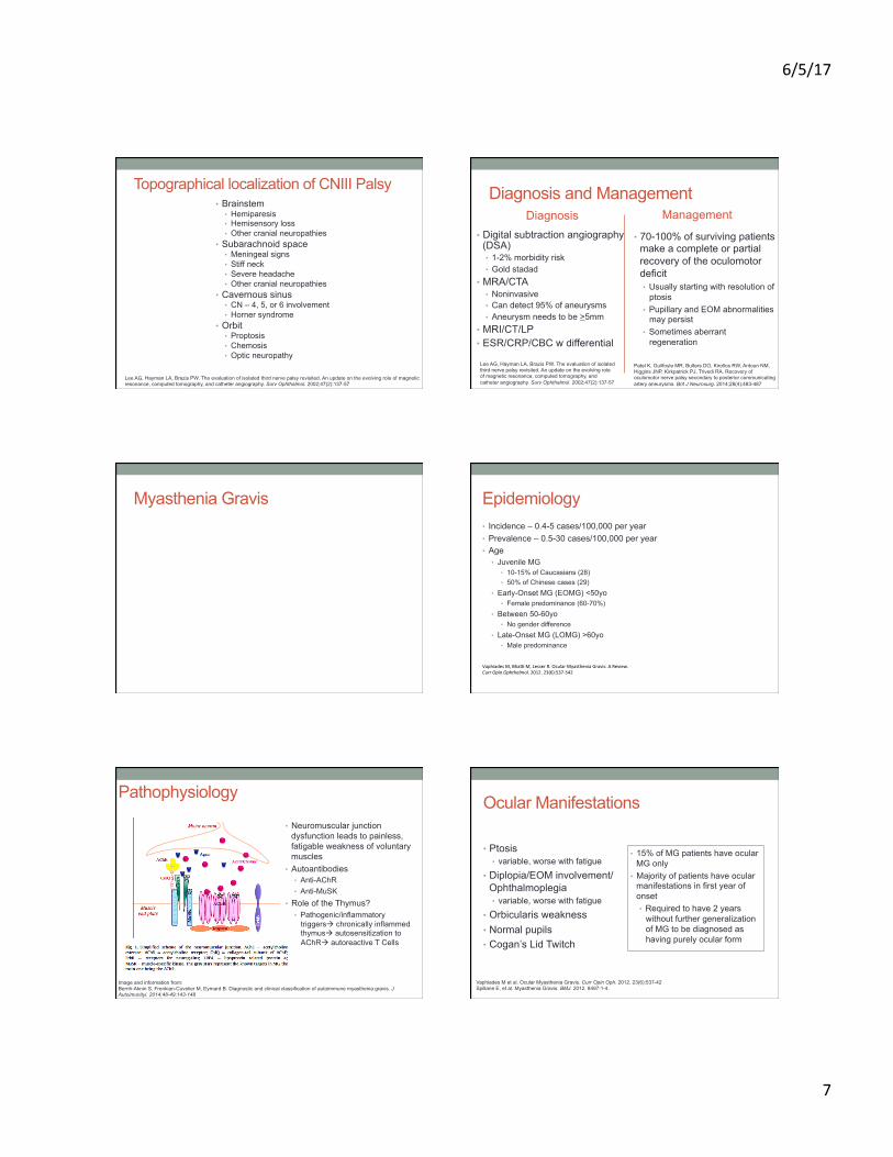
Pathophysiology
• Neuromuscular junction dysfunction leads to painless, fatigable weakness of voluntary muscles
• Autoantibodies • Anti-AChR • Anti-MuSK
• Role of the Thymus? • Pathogenic/inflammatory
triggersà chronically inflammed thymusà autosensitization to AChRà autoreactive T Cells
Image and information from: Berrih-Aknin S, Frenkian-Cuvelier M, Eymard B. Diagnostic and clinical classification of autoimmune myasthenia gravis. J Autoimunityi. 2014;48-49:143-148
Ocular Manifestations
• 15% of MG patients have ocular MG only
• Majority of patients have ocular manifestations in first year of onset • Required to have 2 years
without further generalization of MG to be diagnosed as having purely ocular form
• Ptosis • variable, worse with fatigue
• Diplopia/EOM involvement/Ophthalmoplegia • variable, worse with fatigue
• Orbicularis weakness • Normal pupils • Cogan’s Lid Twitch
Vaphiades M et al. Ocular Myasthenia Gravis. Curr Opin Oph. 2012. 23(6):537-42 Spillane E, et al. Myasthenia Gravis. BMJ. 2012. 8497:1-4.
6/5/17
8
Systemic manifestations • 85% of MG patients • Skeletal Muscle Fatigue
• Facial muscles • Proximal limb muscles • Muscles for swallowing/breathing
• Other autoimmune disease • Thyroid disorders (Hashimoto’s or Basedow diseases)
• Thymus problems • Thymic follicular hyperplasia ~70% • Thymoma (thymic epithelial cell tumor) ~10-15%
Why is it life threatening? • Myasthenic crisis
• Respiratory failure due to muscle weakness • Severe weakness of repiratory muscles, upper airway muscles, or both • Usually due to poor control of generalized disease
Other triggers for crisis • Concomitant use of certain antibiotic (aminoglycosides, quinolones,
antimalarials), muscle relaxants, anti-convulsants antipsychotics, botox, Beta-blockers (including topicals), and iodinated radiocontrast agents
• Systemic infection involving respiratory tract, aspiration, and surgery • Emotional stress, hot environment, sudden elevation of body temperature (9) • Hyperthyroidism
• Require immediate ventilatory assistance • Pre-immunotherapy era
• Myasthenic crisis had significant mortality rate (up to 75%), but has fallen to <5% in recent years
• 20-30% life-time prevalence of myasthenic crisis in patients with MG • Usually occurs during the course of first symptomatic presentation in the
young and later in the course of disease in the elderly • White patients more likely to respond poorly to treatment then black patients • Pregnancy is known to aggravate MG
Chaudhuri A, Behan PO. Myasthenic Crisis. Q J Med. 2009;102:97-107 Juel VC. Myasthenia gravis: management of myasthenic crisis and perioperative care. Semin Neurol. 2004;24:75-81.
Diagnostic Management • ODDiagnosNcTests–InOffice
• PtosisàFaNgue,Rest,IceTests
• MDDiagnosNcTests(Neurologist/Neuro-Ophthalmologist)• TensilonTest(EdrophoniumChloride),95%sensiNvity
• SideEffects:Bradycardia,arrhythmia,hypotension
• SerologicTesNngforAutoanNbodies,ThyroidfuncNon• 5%ofpaNentswithmyastheniagravisalsohavedysthyroidism
• CT/MRIofThorax(rule/outThymicabnormaliNes)• NeurophysiologicalSpecificTests
• RNS–RepeNNveNerveSNmulaNon• SFEMG–SingleFiberElectromyography
Benetar M, Kaminski H. Evidence report: the medical treatment of ocular myasthenia (an evidence-based review). Neurology 2007. 68(24):2144-2149 Benatar M. A systematic review of diagnostic studies in myasthenia gravis. Neuromusc Disord. 2006. 16(7):459-467
Serological Testing • GeneralizedMyasthenia–96%sensiNvity,highPPV
• 87%haveautoanNbodies• 85%-AChR• 40-70%MuSK
• OcularMyasthenia–44%sensiNvity,lowNPV• 50-80%haveautoanNbodies
• (+)AutoanNbodyassayhighlysuggestsMG• BUT(-)AutoanNbodyassay=inconclusive
• SeronegaNve–(-)AChR,(-)MuSK• Highprevalenceofcranialandbulbarmuscleinvolvement• Repeatedserologicaltests
• 15%seroconversionrateover1yearperiod• PaNentswithOMGmaypersistentlytestseronegaNve
Benetar M, Kaminski H. Evidence report: the medical treatment of ocular myasthenia (an evidence-based review). Neurology 2007. 68(24):2144-2149 Benatar M. A systematic review of diagnostic studies in myasthenia gravis. Neuromusc Disord. 2006. 16(7):459-467
Medical Treatment Long-Ac(ngAcetylcholinesterase
inhibitor(Pyridos(gmine)
• Improvesptosis,lesseffecNveinresoluNonofEOMinvolvement/diplopia
• Doesnotaffectthecourseofthedisease
ImmunomodulatoryTherapy
• OralcorNcosteroids• EffecNvelycontrolsdiplopiaANDptosissymptoms
• LowersriskofprogressionfromOMGàGMG
• Other:Azathioprine,MycophenolateMofeNl,Methotrexate,Tacrolimus,Cyclosporine,Rituximab
• Thymectomy• IVIg• PlasmaExchange
Controversial• (-)PotenNallongtermsideeffects• (-)Diseaseisnotlife-threatening• (+)Ocularsymptomscoulddecrease
qualityoflife
Intracranial Space Occupying Lesions • Tumor • Inflammation • Infection • Ischemic infarct • Increased intracranial pressure
• Mass • Pseudotumor/IIH
6/5/17
9
Swollen Optic Nerve(s)
Papilledema
• “Disc swelling from elevated intracranial pressure”
Differential Diagnosis
• Congenital Anomaly • Optic disc drusen
• Infection • Inflammation • Ischemia
Until lumbar puncture – better to diagnose “optic nerve head edema”
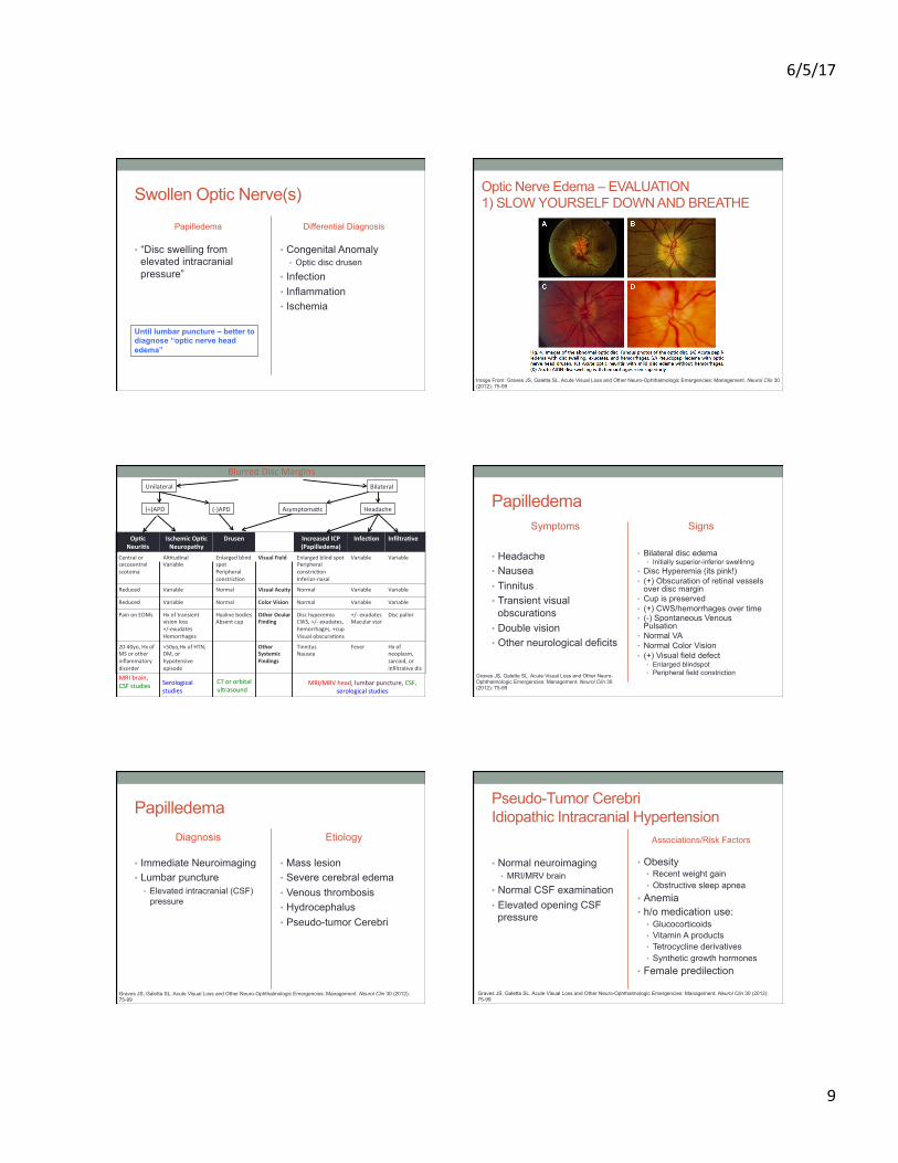
Optic Nerve Edema – EVALUATION 1) SLOW YOURSELF DOWN AND BREATHE
Image From: Graves JS, Galetta SL. Acute Visual Loss and Other Neuro-Ophthalmologic Emergencies: Management. Neurol Clin 30 (2012): 75-99
BlurredDiscMarginsBilateralUnilateral
(+)APD (-)APD AsymptomaNc Headache
Op(cNeuri(s
IschemicOp(cNeuropathy
Drusen IncreasedICP(Papilledema)
Infec(on Infiltra(ve
Centralorcecocentralscotoma
AlNtudinalVariable
EnlargedblindspotPeripheralconstricNon
VisualField EnlargedblindspotPeripheralconstricNonInferior-nasal
Variable Variable
Reduced Variable Normal VisualAcuity Normal Variable Variable
Reduced Variable Normal ColorVision Normal Variable Variable
PainonEOMs Hxoftransientvisionloss+/-exudatesHemorrhages
HyalinebodiesAbsentcup
OtherOcularFinding
DischyperemiaCWS,+/-exudates,hemorrhages,+cupVisualobscuraNons
+/-exudatesMacularstar
Discpallor
20-40yo,HxofMSorotherinflammatorydisorder
>50yo,HxofHTN,DM,orhypotensiveepisode
OtherSystemicFindings
TinnitusNausea
Fever Hxofneoplasm,sarcoid,orinfiltraNvedis
MRI/MRVhead,lumbarpuncture,CSF,serologicalstudies
MRIbrain,CSFstudies Serological
studiesCTororbitalultrasound
Papilledema Symptoms
• Headache • Nausea • Tinnitus • Transient visual
obscurations • Double vision • Other neurological deficits
Signs
• Bilateral disc edema • Initially superior-inferior swellinng
• Disc Hyperemia (its pink!) • (+) Obscuration of retinal vessels
over disc margin • Cup is preserved • (+) CWS/hemorrhages over time • (-) Spontaneous Venous
Pulsation • Normal VA • Normal Color Vision • (+) Visual field defect
• Enlarged blindspot • Peripheral field constriction Graves JS, Galetta SL. Acute Visual Loss and Other Neuro-
Ophthalmologic Emergencies: Management. Neurol Clin 30 (2012): 75-99
Papilledema Diagnosis
• Immediate Neuroimaging • Lumbar puncture
• Elevated intracranial (CSF) pressure
Etiology
• Mass lesion • Severe cerebral edema • Venous thrombosis • Hydrocephalus • Pseudo-tumor Cerebri
Graves JS, Galetta SL. Acute Visual Loss and Other Neuro-Ophthalmologic Emergencies: Management. Neurol Clin 30 (2012): 75-99
Pseudo-Tumor Cerebri Idiopathic Intracranial Hypertension
• Normal neuroimaging • MRI/MRV brain
• Normal CSF examination • Elevated opening CSF
pressure
Associations/Risk Factors
• Obesity • Recent weight gain • Obstructive sleep apnea
• Anemia • h/o medication use:
• Glucocorticoids • Vitamin A products • Tetrocycline derivatives • Synthetic growth hormones
• Female predilection
Graves JS, Galetta SL. Acute Visual Loss and Other Neuro-Ophthalmologic Emergencies: Management. Neurol Clin 30 (2012): 75-99

6/5/17
10
IIH - Management • Acetazolamide
• May improve papilledema, visual complaints, headache • OR other diuretics/CAIs
• Weight loss • Baseline automated VFs after treatment initiated
• Progressive or severe vision loss may need more aggressive therapy • Ventriculoperitoneal shunt • Optic nerve fenestration
Banta JT, Farris BK. Pseudotumor cerebri and optic nerve sheath decompression. Ophthalmology 2000;107:1907–12. Liu GT, Glaser JS, Schatz NJ. High-dose methylprednisolone and acetazolamide for visual loss in pseudotumor cerebri. Am J Ophthalmol 1994;118:88–96
Optic Disc Pallor If its progressing… IMAGE
Longstanding compression of optic nerve, chiasm or tract by a tumor or aneurysm can cause progressive optic disc pallor
Cavernous Sinus Lesions
Image fro: Nadarajah J, Madhusudhan KS, Yadav AK, Chandrashekhara SH, Kumar A, Gupta AK. MR imaging of cavernous sinus lesions: pictorial review. J Neuroradiol. 2015;42(6):305-319.
Cavernous Sinus Syndromes
Complete
• Ophthalmoplegia (Diplopia)
• Ptosis • Mydriasis • Hypesthesia of V1/V2
(facial numbness) • Orbital Pain
Partial
• Depends on the location of the lesion
Etiology • Chronic Causes
• Meningiomas • Metastases of head and neck cancers • Slow-growing carotid aneurysm
• Acute Causes • Cavernous sinus thrombosis
• Extension of facial/sinus infection • Carotid cavernous fistula
• Painful red eye + chemosis + pulsatile exophthalmos • Inflammatory reaction (Tolosa-Hunt syndrome) • Pituitary apoplexy
Cavernous Sinus Thrombosis Ocular Manifestations
• Most common 80-100% presentation • Proptosis • Chemosis • Ptosis • CN III, IV, and/or VI palsies
• Less common 50-80% presentation • Periorbital edema • Optic disc edema • Venous engorgement
• Least common <50% • Decreased visual acuity (due to ION,
CRAO, CRVO, or corneal ulceration) • Sluggish/dilated pupils • Periorbital and corneal sensory loss
(CNV)
Systemic Manifetations
• Most common 80-100% presentation • Acute onset fever
• Less common 50-80% presentation • Headache • Lethargy • Altered sensorium
• Least common <50% • Meningismus • Seizures • Hemiparesis
Lemos J, Eggenberger E. Neuro-ophthalmological emergencies. Neurohospitalist. 2015. 5(4): 223-233.
6/5/17
11
Differential Diagnosis
Orbital Cellulitis
• Painful ophthalmoplegia • Proptosis • Chemosis • Fever • Decreased vision • UNILATERAL
Direct High-Flow Carotid Cavernous Fistula
• Periorbital edema • Ophthalmoplegia • Increased IOP • Decreased vision • (+) supraorbital bruit • Arterialized conjunctival
vessels
Lemos J, Eggenberger E. Neuro-ophthalmological emergencies. Neurohospitalist. 2015. 5(4): 223-233.
Pituitary Apoplexy
Epidemiology • Between 2-12% of patients with adenoma experience
apoplexy • Diagnosis of pituitary tumor unknown at time of apoplexy
in ¾ of cases • Presentation for 0.6-9.0% of surgically managed pituitary
adenomas • Male predominance ~60% • Peak incidence in 5th decade
Briet C, Salenave S, Bonneville JF, Laws ER, Chanson P. Pituitary Apoplexy. Endocrine Reviews. 2015;36(6):622-645 Glezer A, Bronstein MD. Pituitary apoplexy: pathophysiology, diagnosis, and management. Arch Endocrinol Metab. 2015;59(3):259-64.
Pathophysiology • Hemorrhage or infarction of a pituitary tumor causes a
sudden enlargement of the gland due to ischemia and/or necrosis
• 2/3 are spontaneous • 1/3 precipitating factor:
• Hypotension • Surgery
• Malignant hypertension • Anticoagulant treatment • Dopaminergic agonist treatment
Biousse V, Newman NJ, Oyesiku NM. Precipitating factors in pituitary apoplexy. J Neurol Neurosurg Psychiatry. 2001; 71(4):542-545 Glezer A, Bronstein MD. Pituitary apoplexy: pathophysiology, diagnosis, and management. Arch Endocrinol Metab. 2015;59(3):259-64. Semple PL, Webb MK, de Villiers JC, Laws ER. Pituitary apoplexy. Neurosurgery. 2005;56(1):65-72. discussion 3.
Ocular Manifestations • Visual Field Loss
• Bitemporal • Junctional scotoma
• Unilateral and/or bilateral ophthalmolplegia ~50% • CNIII>CNVI>CNIV
Junctional Scotoma
Bitemporal Loss
Systemic manifestations • Sudden severe headache
~80% • Retroorbital • Bifrontal • Diffuse • Assoc with vomiting/nausea
• Neck stiffness • Brain stem/hypothalamus
compression • Reduced consciousness • Thermoregulatory dysfunction • Cardiorespiratory dysfunction
• Pituitary dysfunction • Thyrotropic deficiency
• Hypothyroidism • Corticotropic deficiency
• Hyponatremia • Hypotension
• Hypercortisolism
Briet C, Salenave S, Bonneville JF, Laws ER, Chanson P. Pituitary Apoplexy. Endocrine Reviews. 2015;36(6):622-645 Glezer A, Bronstein MD. Pituitary apoplexy: pathophysiology, diagnosis, and management. Arch Endocrinol Metab. 2015;59(3):259-64.
Symptoms evolve from hours to 2 days after onset of apoplexy
6/5/17
12
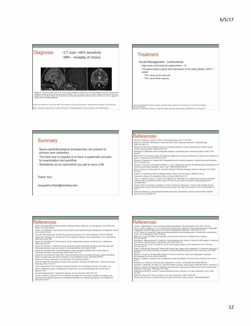
Diagnosis • CT scan ~46% sensitivity • MRI – modality of choice
Image From: Mukhi SV, Lincoln CM. MRI in the evaluation of acute visual syndrome. Top Magn Reson Imaging. 2015;24:309-324 Briet C, Salenave S, Bonneville JF, Laws ER, Chanson P. Pituitary Apoplexy. Endocrine Reviews. 2015;36(6):622-645
Treatment • Acute Management - controversial
• High-dose corticosteroid replacement – IV • Transphenoidal surgical decompression of the sella (ideally within 1
week) • 76% visual acuity improves • 79% visual fields improve
Glezer A, Bronstein MD. Pituitary apoplexy: pathophysiology, diagnosis, and management. Arch Endocrinol Metab. 2015;59(3):259-64. Semple PL, Webb MK, de Villiers JC, Laws ER. Pituitary apoplexy. Neurosurgery. 2005;56(1):65-72. discussion 3.
Summary
• Neuro-ophthalmological emergencies can present to primary care optometry
• The best way to prepare is to have a systematic process for examination and workflow
• Sometimes as an optometrist you get to save a life
Thank You! [email protected]
References • Ahmad K, Wright M, Lueck CJ. Ptosis. Practical Neurology. 2011;11:332-340 • Banta JT, Farris BK. Pseudotumor cerebri and optic nerve sheath decompression. Ophthalmology
2000;107:1907–12. • Benetar M, Kaminski H. Evidence report: the medical treatment of ocular myasthenia (an evidence-based
review). Neurology 2007. 68(24):2144-2149 • Benatar M. A systematic review of diagnostic studies in myasthenia gravis. Neuromusc Disord. 2006. 16(7):
459-467 • Berrih-Aknin S, Frenkian-Cuvelier M, Eymard B. Diagnostic and clinical classification of autoimmune myasthenia
gravis. J Autoimunityi. 2014;48-49:143-148 • Biousse V, Newman NJ, Oyesiku NM. Precipitating factors in pituitary apoplexy. J Neurol Neurosurg Psychiatry.
2001; 71(4):542-545 • Biousse V, Touboul PJ, D’Anglejan-Chatillon J, Levy C, Schaison M, Bousser M.Ophthalmologic Manifestations of
Internal Carotid Artery Dissection. Amer J Oph. 1998:9394(98):565-577 • Briet C, Salenave S, Bonneville JF, Laws ER, Chanson P. Pituitary Apoplexy. Endocrine Reviews. 2015;36(6):
622-645 • Caplan LR. Dissections of brain-supplying arteries. Nature Clin Pract Neurol. 2008:4(1):34-42 • Chaudhuri A, Behan PO. Myasthenic Crisis. Q J Med. 2009;102:97-107 • Chen JJ, Leavitt JA, Fang C, Crowson CS, Matteson EL, Warrington KJ. Evaluating the incidence of arteritic
ischemic optic neuropathy and other causes of vision loss from giant cell arteritis. Ophthalmology 2016: 123:1999-2003
• Ciccia F, Rizzo A, Ferrante A, Guggino G, Croci S, Cavazza A, Salvarani C, Triolo G. New Insights into the pathogenesis of giant cell arteritis. Autoimmunity reviews. 2017 May. Epub ahead of print. Accessed May 21, 2017.
• Etminan N, Rinkel GJ. Unruptured intracranial aneurysms: development, rupture, and prevention. Nat Rev Neurol. 2016;12(12):699-713
• Glezer A, Bronstein MD. Pituitary apoplexy: pathophysiology, diagnosis, and management. Arch Endocrinol Metab. 2015;59(3):259-64
• Graves JS, Galetta SL. Acute Visual Loss and Other Neuro-Ophthalmologic Emergencies: Management. Neurol Clin 30 (2012): 75-99
• Gross JR, McClelland CM, Lee MS. An approach to anisocoria. Curr Opin Ophthalmol. 2016;27:486-492 • Hayreh SS, Podhajsky PA, Zimmerman B. Occult giant cell arteritis: ocular manifestations. Am J Ophthalmol.
1998:125L4):521-526. • Hayreh SS, Podhajsky PA, Zimmerman B. Ocular manifestations of giant cell arteritis. Am J Ophthalmol.
1998;125(4):509-20 • Hayreh SS, Biousse V. Treatment of acute visual loss in giant cell arteritis: should we prescribe high-dose
intravenous steroids or just oral steroids? J Neuroophthalmol. 2012;32(3):278-287. • Hayreh SS, Zimmerman B. Visual deterioration in giant cell arteritis patients while on high doses of
corticosteroid therapy. Ophthalmology. 2003;110(6):1204-1215 • Hayreh SS, Zimmerman B, Kardon RH. Visual improvement with corticosteroid therapy in giant cell arteritis.
Report of a large study and review of literature. Acta Ophthalmol Scand. 2002;80(4):355-67 • Hoffman GS. Giant Cell Arteritis. Ann Intern Med. 2016;165(9):ITC65-80 • Jacks AS, Miller NR. Spontaneous retinal venous pulsation: aetiology and significance. J Neurol Neurosurg
Psychiatry. 2003. 74(1):7-9 • Jivraj I, Tamhankar M. The treatment of giant cell arteritis. Curr Treat Options Neurol. 2017;19(2):1-18. • Juel VC. Myasthenia gravis: management of myasthenic crisis and perioperative care. Semin Neurol.
2004;24:75-81 • Karmel M, Eggenberger E. Deciphering Diplopia. Eye Net. Nov/Dec 2009. PP31-34 • Lee AG, Hayman LA, Brazis PW. The evaluation of isolated third nerve palsy revisited. An update on the
evolving role of magnetic resonance, computed tomography, and catheter angiography. Surv Ophthalmol. 2002;47(2):137-57
•
References
• Lemos J, Eggenberger E. Neuro-ophthalmological emergencies. Neurohospitalist. 2015. 5(4): 223-233. • Levy C, Laissy JP, Raveau V, et al. Carotid and vertebral artery dissections: three-dimensional time-of-flight MR
angiography and MR imaging versus conventional angiography. Radiology. 1994;190(1):97-103. • Liu GT, Glaser JS, Schatz NJ. High-dose methylprednisolone and acetazolamide for visual loss in pseudotumor
cerebri. Am J Ophthalmol 1994;118:88–96 • Mukhi SV, Lincoln CM. MRI in the evaluation of acute visual syndrome. Top Magn Reson Imaging.
2015;24:309-324 • Nadarajah J, Madhusudhan KS, Yadav AK, Chandrashekhara SH, Kumar A, Gupta AK. MR imaging of cavernous
sinus lesions: pictorial review. J Neuroradiol. 2015;42(6):305-319. • Park HK, Rha HK, Lee KJ, Chough CK, Joo W. Microsurgical anatomy of the oculomotor nerve. Clin Anat.
2017;30(1):21-31. • Patel K, Guilfoyle MR, Bulters DO, Kirollos RW, Antoun NM, Higgins JNP, Kirkpatrick PJ, Trivedi RA. Recovery of
oculomotor nerve palsy secondary to posterior communicating artery aneurysms. Brit J Neurosurg. 2014;28(4):483-487
• Reede DL, Garcon E, Smoker WRK, Kardon R. Horner’s syndrome: clinical and radiographic evaluation. Neuroimaging Clin N Am. 2008;18:369-385
• Roberts J, Clifford A. Update on the management of giant cell arteritis. Ther Adv Chronic Disease. 2017. 8(4-5):69-79
• Robertson JJ, Koyfman A. Cervical artery dissections: a review. J Emerg Med 2016;52(5):508-518 • Semple PL, Webb MK, de Villiers JC, Laws ER. Pituitary apoplexy. Neurosurgery. 2005;56(1):65-72. discussion 3. • Silbert PL, Mokri B, Schievink WI. Headache and neck pain in spontaneous internal carotid and vertebral artery
dissections. Neurology. 1995;45(8):1517-1522 • Vaphiades M, Bhatti M, Lesser R. Ocular Myasthenia Gravis: A Review. Curr Opin Ophthalmol. 2012. 23(6):
537-542 • Walton KA, Buono LM. Horner syndrome. Curr Opin Ophthalmol. 2003;14:357-363 • Weyand CM, Goronzy JJ. Arterial wall injury in giant cell arteritis. Arthr & Rheum. 1999;42(5):844-853 •
References
Related Documents