Transactions of the American Ophthalmological Society Volume CXV One Hundred and Fifty-Third Annual Meeting The Homestead Hot Springs, Virginia 2017 Published for the American Ophthalmological Society San Francisco, California 2017

Welcome message from author
This document is posted to help you gain knowledge. Please leave a comment to let me know what you think about it! Share it to your friends and learn new things together.
Transcript

Transactions of the American Ophthalmological Society
Volume CXV
One Hundred and Fifty-Third Annual Meeting
The Homestead
Hot Springs, Virginia 2017
Published for the
American Ophthalmological Society
San Francisco, California 2017
ii
TABLE OF CONTENTS THE AMERICAN OPHTHALMOLOGICAL SOCIETY 2017
OFFICERS AND COUNCIL viPRESIDENTS OF THE SOCIETY viiRECIPIENTS OF THE LUCIEN HOWE MEDAL xFREDERICK H. VERHOEFF LECTURERS xiiBLODI LECTURERS xiiiMEMBERS xivEDITORIAL BOARD xvii
NECROLOGY IN MEMORIUM Necrology 1
MINUTES OF THE PROCEEDINGS INTRODUCTION Proceedings 1PAPERS: FRIDAY, MAY 20 1EXECUTIVE SESSION 1
REPORT OF THE EXECUTIVE VICE-PRESIDENT 1REPORT OF THE CHAIR OF THE COUNCIL 2REPORT OF THE AUDIT COMMITTEE 2REPORT OF THE COMMITTEE ON THESES 3REPORT OF THE EDITOR 3REPORT OF THE COMMITTEE ON PROGRAMS 4REPORT OF THE COMMITTEE ON MEMBERSHIP 4REPORT OF THE ARCHIVIST / PHOTOGRAPHER 5REPORT OF THE REPRESENTATIVE TO THE COUNCIL OF THE AMERICAN ACADEMY OF OPHTHALMOLOGY
7
REPORT OF THE REPRESENTATIVE TO THE AMERICAN COLLEGE OF SURGEONS 8REPORT OF THE REPRESENTATIVES TO THE AMERICAN ORTHOPTIC COUNCIL 9
PAPERS: SATURDAY, MAY 21 12BANQUET 12REPORT OF THE COMMITTEE ON ATHLETICS 15PAPERS: SUNDAY, MAY 22 18MEMBERS IN ATTENDANCE 19 PAPER ABSTRACTS 21 IMPACT OF RESIDENT AND FELLOW TRAINEES ON PATIENT ENCOUNTER LENGTH IN AN ACADEMIC OPHTHALMOLOGY CENTER MICHAEL CHIANG, ISAAC GOLDSTEIN, SARAH READ-BROWN, MICHELLE HRIBAR GENETIC ANALYSIS OF 1000 CONSECUTIVELY ASCERTAINED FAMILIES WITH INHERITED RETINAL DISEASE EDWIN STONE, JEANEEN ANDORF, ADAM DELUCA, SCOTT WHITMORE, JOSEPH GIACALONE, LUAN STREB, TERRY BRAUN, ROBERT MULLINS, TODD SCHEETZ, VAL SHEFFIELD, BUDD TUCKER CONJUNCTIVAL TUMORS IN CHILDREN IN 806 CASES. FEATURES DIFFERENTIATING BENIGN FROM MALIGNANT TUMORS CAROL L. SHIELDS, KAREEM SIOUFI, ADEL E. ALSET, EMIL ANTHONY T. SAY, JERRY A. SHIELDS LIMBAL AND CONJUNCTIVAL TRANSPLANTATION FROM THE SAME LIVING-RELATED BONE MARROW DONOR TO PATIENTS WITH OCULAR GRAFT VERSUS HOST DISEASE MASSIMO BUSIN, GIUSEPPE GIANNACCARE, LAURA SAPIGNI, EMILIO CAMPOS RANIBIZUMAB FOR THE PREVENTION OF RADIATION COMPLICATIONS IN PATIENTS TREATED WITH PROTON BEAM IRRADIATION FOR CHOROIDAL MELANOMA IVANA KIM, ANNE MARIE LANE, PURVA JAIN, CAROLINE AWH, EVANGELOS GRAGOUDAS

iii
MISSED DIAGNOSIS OF PARAPROTEINEMIC KERATOPATHY IN PATIENTS WITH MONOCLONAL GAMMOPATHY OF UNDETERMINED SIGNIFICANCE WALTER LISCH JOANNA WASIELICA-POSLEDNIK, TERO KIVELÄ, UWE PLEYER, CHRISTINA LISCH, JAYNE S. WEISS ASSOCIATION OF PSEUDOEXFOLIATION WITH SYSTEMIC VASCULAR DISEASES IN A SOUTH INDIAN POPULATION- THE ARAVIND PSEUDOEXFOLIATION (APEX) STUDY ARAVIND HARIPRIYA, ASHOK VARDHAN, BANUSHREE RATUKONDLA, PRADEEP RAMULU, ALAN ROBIN TEMPORAL PROFILE OF RETINOPATHY OF PREMATURITY IN EXTREMELY PREMATURE INFANTS R. MICHAEL SIATKOWSKI, VINCENT VENINCASA, VICTORIA BUGG, DVORAK JUSTIN, KAI DING, FAIZAH BHATTI THERAPEUTIC BIOMARKERS IN LACRIMAL GLAND ADENOID CYSTIC CARCINOMA: INSIGHTS FROM THE TUMOR'S RESPONSE TO INTRA-ARTERIAL CYTOREDUCTIVE CHEMOTHERAPY DANIEL PELAEZ, NEDA NIKPOOR, WENSI TAO, RAVI DODDAPANENI, DAVID TSE MUSHROOM SHAPED INTRAOCULAR LESIONS OTHER THAN MELANOMAS JERRY A. SHIELDS, CAROL L. SHIELDS QUALITY METRICS AND RETINAL DETACHMENT SURGERY: TIME TO UNPLANNED RETURN TO THE OPERATING ROOM SOPHIE BAKRI, ALEXANDER GROSINGER, BENJAMIN NICHOLSON, ANDREW BARKMEIER, RAYMOND IEZZI IS DISCRIMINANT SCORE ASSOCIATED WITH GEP CLASS IN DECISIONDX-UM TEST IMPORTANT PROGNOSTICALLY? JAMES J. AUGSBURGER, ZELIA M. CORREA COMPARATIVE EFFECTIVENESS AND COST-EFFECTIVENESS ANALYSES OF THE PROSTAMIDES AND TIMOLOL FOR OPEN ANGLE GLAUCOMA GARY BROWN, MELISSA BROWN, HEIDI LIESKE EN FACE OCT AND OCT ANGIOGRAPHY OF PERIVENULAR ISCHEMIA ASSOCIATED WITH CENTRAL RETINAL VEIN OCCLUSION DAVID SARRAF, KHALIL FALAVARJANI, NOPASAK PHASUKKIJWATANA, EMMETT CUNNINGHAM JR., ANANDA KALEVAR, ROSA DOLZ MARCO, IRENA TSUI, RICHARD ROSEN, K. BAILEY FREUND, LEE JAMPOL, SRINIVAS SADDA THE PROGNOSTIC VALUE OF CHROMOSOME STATUS IN UVEAL MELANOMA IS ENHANCED BY ADDING AJCC STAGE MARTINE JAGER, MEHMET DOGRUSOZ, METTE BAGGER, GREGORIUS LUYTEN, JENS KIILGAARD GLAUCOMA IN HIGH MYOPIA AND PARAPAPILLARY DELTA ZONE JOST JONAS, KYOKO OHNO-MATSUI SPECTRAL DOMAIN OPTICAL COHERENCE TOMOGRAPHY ANGIOGRAPHY IN CHILDREN WITH AMBLYOPIA STACY PINELES, MARCELA LONNGI, IRINA TSUI, FEDERICO VELEZ, DAVID SARRAF ACCURACY OF IMAGING MODALITIES IN DIFFERENTIATING PSEUDOPAPILLEDEMA FROM TRUE OPTIC DISK EDEMA (ODE) IN CHILDREN MELINDA CHANG, VELEZ FEDERICO, DEMER JOSEPH, BONELLI LAURA, QUIROS PETER, SADUN ALFREDO, PINELES STACY, ANTHONY ARNOLD POSTER ABSTRACTS 30THE ROLE OF OXYGEN-INDUCED VASO-OBLIERATION IN THE DEVELOPMENT OF PARAVENTRICULAR LEUKOMALACIA AND RETINOPATHY OF PREMATURITY KENNETH WRIGHT, LINGKUN KONG INTRACRANIAL PRESSURE, INTRAOCULAR PRESSURE, TRANSLAMINAR PRESSURE GRADIENT, AND PAPILLEDEMA SEVERITY TIMOTHY MCCULLEY, JESSICA CHANG CHAOS THEORY, THE ARRHENIUS EQUATION, AND THE FDA JOHN D. BULLOCK
iv
MATERNAL PREECLAMPSIA AND INFANT RISK OF RETINOPATHY OF PREMATURITY JULIA P. SHULMAN, CINDY WENG, JACOB WILKES, TOM GREENE, M. ELIZABETH HARTNETT ASSESSMENT OF A NOVEL TELE-EDUCATION SYSTEM TO ENHANCE RETINOPATHY OF PREMATURITY (ROP) TRAINING BY INTERNATIONAL OPHTHALMOLOGISTS-IN-TRAINING IN MEXICO SAMIR PATEL, MARIA ANA MARTINEZ-CASTELLANOS, DAVID BERRONES-MEDINA, RYAN SWAN, MICHAEL RYAN, KARYN JONAS, SUSAN OSTMO, J. PETER CAMPBELL, MICHAEL CHIANG, RV PAUL CHAN SOMATOTYPE AND THE RISK OF HYDROXYCHLOROQUINE RETINOPATHY DAVID BROWNING, CHONG LEE CLINICAL AND ANATOMIC OUTCOMES OF CONCURRENT PHACOVITRECTOMY SURGERY FOR A VARIETY OF INDICATIONSWILLIAM F. MIELER, IVY ZHU, ELMER Y. TU COMPARISON OF SINGLE NUCLEOTIDE POLYMORPHISM PROFILES AMONG DIFFERENT PHENOTYPES OF NEOVASCULAR AGE-RELATED MACULAR DEGENERATION CLEMENT CHAN, PREMA ABRAHAM, ANDRE HAFNER, LORAH PERLEE INTRAOPERATIVE USE OF MICROSCOPE-INTEGRATED OPTICAL COHERENCE TOMOGRAPHY FOR SUBRETINAL GENE THERAPY NINEL GREGORI, BYRON LAM, JANET DAVIS CLINICAL AND GENETIC PROFILE AND MANAGEMENT OUTCOMES OF UNILATERAL PRIMARY CONGENITAL GLAUCOMA IN SAUDI ARABIA SULTAN ALZUHAIRY, LEEN ABU SAFIEH, RAJIV KHANDEKAR, DEEPAK EDWARD LACRIMAL GLAND ABSCESS IN A CHILD; A RARE ASSOCIATION WITH IGG4-RELATED DISEASE EDWARD RAAB, HAMIDEH MOAYEDPARDAZI, STEVEN NAIDS, ALAN FRIEDMAN, MURRAY MELTZER
THESES BIOCHEMICAL MEASUREMENTS OF FREE OPSIN IN MACULAR DEGENERATION EYES: EXAMINING THE 11-CIS RETINAL DEFICIENCY HYPOTHESIS OF DELAYED DARK ADAPTATION (AN AMERICAN OPHTHALMOLOGICAL SOCIETY THESIS) ANNE HANNEKEN, MD, THOMAS NEIKIRK, JENNIFER JOHNSON, PHD, MASAHIRO KONO, PHD CONTINUOUS CURVILINEAR CAPSULORHEXIS TRAINING AND NON-RHEXIS RELATED VITREOUS LOSS: THE SPECIFICITY OF VIRTUAL REALITY SIMULATOR SURGICAL TRAINING (AN AMERICAN OPHTHALMOLOGICAL SOCIETY THESIS) COLIN A. MCCANNEL, MD ENHANCED DETECTION OF SUB-RETINAL PIGMENT EPITHELIAL CELL LAYER DEPOSITS IN HUMAN AND MURINE TISSUE: IMAGING ZINC AS A BIOMARKER FOR AGE-RELATED MACULAR DEGENERATION (AN AMERICAN OPHTHALMOLOGICAL SOCIETY THESIS) FREDERIK J.G.M. VAN KUIJK, MD, PHD, SCOTT W. MCPHERSON, PHD, HEIDI ROEHRICH, DIPL-BIOL EVALUATION OF VISUAL FIELD AND IMAGING OUTCOMES FOR GLAUCOMA CLINICAL TRIALS (AN AMERICAN OPHTHALOMOLOGICAL SOCIETY THESIS) DAVID F. GARWAY-HEATH, BSC, MB BS, MD, FRCOPHTH, ANA QUARTILHO, MSC, PHILIP PRAH, MSC, DAVID P. CRABB, PHD, QIAN CHENG, BSC, HAOGANG ZHU, MSC, PHD THE RELATIONSHIP BETWEEN OCULAR ITCH, OCULAR PAIN, AND DRY EYE SYMPTOMS (AN AMERICAN OPHTHALMOLOGICAL SOCIETY THESIS) ANAT GALOR, MD, MSPH, LESLIE SMALL, OD, WILLIAM FEUER, MS, ROY C. LEVITT, MD, KONSTANTINOS D. SARANTOPOULOS, MD, PHD, AND GIL YOSIPOVITCH, MD PREDICTORS OF INTRAOCULAR PRESSURE AFTER PHACOEMULSIFICATION IN PRIMARY OPEN-ANGLE GLAUCOMA EYES WITH WIDE VERSUS NARROWER ANGLES (AN AMERICAN OPHTHALMOLOGICAL SOCIETY THESIS) SHAN C. LIN, MD, MARISSE MASIS, MD, TRAVIS C. PORCO, PHD, MPH, AND LOUIS R. PASQUALE, MD

v
THE EFFECTS OF PHACOEMULSIFICATION AND INTRAOCULAR LENS IMPLANTATION ON ANATOMICAL AND FUNCTIONAL PARAMETERS IN PATIENTS WITH PRIMARY ANGLE CLOSURE: A PROSPECTIVE STUDY (AN AMERICAN OPHTHALMOLOGICAL SOCIETY THESIS) CARLO ENRICO TRAVERSO, MD, AND CARLO ALBERTO CUTOLO, MD AUTOIMMUNE RETINOPATHY: CURRENT CONCEPTS AND PRACTICES (AN AMERICAN OPHTHALMOLOGICAL SOCIETY THESIS) H. NIDA SEN, MD, MHSC, LANDON GRANGE, MD, MARIB AKANDA, BS, AND AUSTIN FOX, MD
OFFICERS AND COUNCIL OF THE
AMERICAN OPHTHALMOLOGICAL SOCIETY Elected at the Annual Meeting
May 18-21, 2017
PRESIDENT DR DAVID J. WILSON, MD, PORTLAND, OR
EXECUTIVE VICE PRESIDENT
DR HANS E. GROSSNIKLAUS, ATLANTA, GEORGIA
EDITOR OF THE TRANSACTIONS DR EMILY Y. CHEW, BETHESDA, MARYLAND
COUNCIL
DR WOODFORD S. VAN METER, LEXINGTON, KENTUCKY DR MARCO A. ZARBIN, NEWARK, NEW JERSEY
TIMOTHY W. OLSEN, ATLANTA, GEORGIA DR EDWARD G. BUCKLEY, DURHAN, NORTH CAROLINA
DR JULIA A. HALLER, MD GLENCOE, MD
vi
vii
PRESIDENTS OF THE SOCIETY 1864-1868 DR EDWARD DELAFIELD, New York 1869-1873 DR HENRY W. WILLIAMS, Boston 1874-1878 DR C. R. AGNEW, New York 1879-1884 DR HENRY D. NOYES, New York 1885-1889 DR WILLIAM F. NORRIS, Philadelphia 1890-1893 DR HASKET DERBY, Boston 1894-1898 DR GEORGE C. HARLAN, Philadelphia 1899-1902 DR O. F. WADSWORTH, Boston 1903-1905 DR CHARLES S. BULL, New York
1906 DR ARTHUR MATHEWSON, Washington, DC 1907 DR CHARLES J. KIPP, Newark 1908 DR SAMUEL D. RISLEY, Philadelphia 1909 DR S. B. ST JOHN, Hartford 1910 DR SAMUEL THEOBALD, Baltimore 1911 DR EMIL GRUENING, New York 1912 DR EDWARD JACKSON, Denver 1913 DR MYLES STANDISH, Boston 1914 DR ROBERT SATTLER, Cincinnati 1915 DR M. H. POST, St Louis 1916 DR GEORGE E. DE SCHWEINITZ, Philadelphia 1917 DR PETER A. CALLAN, New York 1918 DR WILLIAM H. WILDER, Chicago 1919 DR LUCIEN HOWE, Buffalo 1920 DR HIRAM WOODS, Baltimore 1921 DR JOHN E. WEEKS, New York 1922 DR WILLIAM M. SWEET, Philadelphia 1923 DR WILLIAM H. WILMER, Washington, DC 1924 DR ALEXANDER DUANE, New York 1925 DR CASSIUS D. WESTCOTT, Chicago 1926 DR DAVID HARROWER, Worcester 1927 DR WILLIAM ZENTMAYER, Philadelphia 1928 DR WALTER E. LAMBERT, New York 1929 DR WALTER R. PARKER, Detroit 1930 DR WILLIAM CAMPBELL POSEY, Philadelphia 1931 DR ARNOLD KNAPP, New York 1932 DR EDWARD C. ELLETT, Memphis 1933 DR THOMAS B. HOLLOWAY, Philadelphia 1934 DR W. GORDON M. BYERS, Montreal 1935 DR WALTER B. LANCASTER, Boston 1936 DR LOUIS S. GREENE, Washington, DC 1937 DR HARRY FRIEDENWALD, Baltimore 1938 DR F. H. VERHOEFF, Boston 1939 DR FREDERICK T. TOOKE, Montreal 1940 DR E. V. L. BROWN, Chicago 1941 DR F. PHINIZY CALHOUN, Atlanta 1942 DR ALLEN GREENWOOD, Boston 1943 DR HUNTER H. MCGUIRE, Winchester, Virginia 1944 DR JOHN GREEN, St Louis 1945 DR S. JUDD BEACH, Portland, Maine 1946 DR EUGENE M. BLAKE, New Haven 1947 DR JOHN W. BURKE, Washington, DC 1948 DR HENRY C. HADEN, Houston 1949 DR BERNARD SAMUELS, New York
Presidents of the Society
viii
1950 DR PARKER HEATH, Boston 1951 DR JOHN H. DUNNINGTON, New York 1952 DR LAWRENCE T. POST, St Louis 1953 DR CONRAD BERENS, New York 1954 DR WILLIAM L. BENEDICT, Rochester, Minnesota 1955 DR EVERETT L. GOAR, Houston 1956 DR ALAN C. WOODS, Baltimore 1957 DR FREDERICK C. CORDES, San Francisco 1958 DR WALTER S. ATKINSON, Watertown, NY 1959 DR DERRICK VAIL, Chicago 1960 DR ALGERNON B. REESE, New York 1961 DR EDWIN B. DUNPHY, Boston 1962 DR FRANCIS HEED ADLER, Philadelphia 1963 DR PAUL A. CHANDLER, Boston 1964 DR MAYNARD C. WHEELER, New York 1965 DR FRANK B. WALSH, Baltimore 1966 DR WILFRED E. FRY, Philadelphia 1967 DR PHILLIP M. LEWIS, Memphis 1968 DR GORDON C. BRUCE, New York 1969 DR JAMES N. GREEAR, Reno 1970 DR C. WILBUR RUCKER, Rochester, Minnesota 1971 DR DOHRMANN K. PISCHEL, San Francisco 1972 DR TRYGVE GUNDERSEN, Boston 1973 DR ARTHUR GERARD DEVOE, New York 1974 DR WILLIAM P. MCGUIRE, Winchester, Virginia 1975 DR M. ELLIOTT RANDOLPH, Baltimore 1976 DR JOSEPH A. C. WADSWORTH, Durham 1977 DR DAVID O. HARRINGTON, San Francisco 1978 DR SAMUEL D. MCPHERSON, JR., Durham 1979 DR F. PHINIZY CALHOUN, JR., Atlanta 1980 DR JOHN WOODWORTH HENDERSON, Ann Arbor 1981 DR WILLIAM F. HUGHES, Chicago 1982 DR ROBERT W. HOLLENHORST, Rochester, Minnesota 1983 DR CLEMENT MCCULLOCH, Toronto 1984 DR ROBERT N. SHAFFER, San Francisco 1985 DR DUPONT GUERRY III, Richmond 1986 DR A. EDWARD MAUMENEE, Baltimore 1987 DR FRANK W. NEWELL, Chicago 1988 DR EDWARD W. D. NORTON, Miami 1989 DR DAVID SHOCH, Chicago 1990 DR ROBERT E. KENNEDY, Rochester, New York 1991 DR FREDERICK C. BLODI, lowa City 1992 DR THOMAS P. KEARNS, Rochester, Minnesota 1993 DR BRADLEY R. STRAATSMA, Los Angeles 1994 DR ROBERT B. WELCH, Annapolis, Maryland 1995 DR BRUCE E. SPIVEY, Chicago 1996 DR STANLEY TRUHLSEN, Omaha 1997 DR WILLIAM H. SPENCER, San Francisco 1998 DR W. RICHARD GREEN, Baltimore 1999 DR WILLIAM S. TASMAN, Wyndmoor, Pennsylvania
Presidents of the Society
ix
2000 DR W. BANKS ANDERSON, JR., Durham 2001 DR PAUL R. LICHTER, Ann Arbor 2002 DR ROBERT C. DREWS, Clayton, Missouri 2003 DR MARILYN T. MILLER, Chicago, Illinois 2004 DR FRONCIE A. GUTMAN, Cleveland, Ohio 2005 DR J. BROOKS CRAWFORD, San Francisco, California 2006 DR DANIEL M. ALBERT, Madison, Wisconsin 2007 DR JOHN G. CLARKSON, Miami, Florida 2008 DR DAN B. JONES, Houston, Texas 2009 DR SUSAN H. DAY, San Francisco, California 2010 DR CHARLES P. WILKINSON, Baltimore, Maryland 2011 DR LEE M. JAMPOL, Chicago, Illinois 2012 DR DOUGLAS D. KOCH, Houston, Texas 2013 DR RICHARD K. PARRISH, II, Miami, Florida 2014 DR HANS E. GROSSNIKLAUS, Atlanta, Georgia 2015 DR RICHARD P. MILLS, Seattle, Washington 2016 DR MARILYN B. METS, Chicago, Illinois 2017 DR GEORGE B. BARTLEY, Jacksonville, Florida 2018 DR DAVID J. WILSON, Portland, Oregon

RECIPIENTS OF THE LUCIEN HOWE MEDAL 1922 DR CARL KOLLER, New York 1923 DR ALEXANDER DUANE, New York 1924 DR ERNEST FUCHS, Vienna, Austria 1925 NO AWARD 1926 DR EDWARD JACKSON, Denver 1927 MR PRIESTLY SMITH, Birmingham, England 1928 NO AWARD 1929 DR THEODOR AXENFELD, Freiburg, Germany 1930 NO AWARD 1931 NO AWARD 1932 DR F. H. VERHOEFF, Boston 1933 NO AWARD 1934 DR GEORGE E. DE SCHWEINITZ, Philadelphia 1935 NO AWARD 1936 SIR JOHN HERBERT PARSONS, London, England 1937 DR ARNOLD KNAPP, New York 1938 NO AWARD 1939 NO AWARD 1940 NO AWARD 1941 NO AWARD 1942 DR E. V. L. BROWN, Chicago 1943 NO AWARD 1944 NO AWARD 1945 DR WALTER B. LANCASTER, Boston 1946 SIR STEWART DUKE-ELDER, London, England 1947 DR LAWRENCE T. POST, St Louis 1948 DR WILLIAM ZENTMAYER, Philadelphia 1949 DR PHILLIPS THYGESON, San Jose, California 1950 DR ALGERNON B. REESE, New York 1951 DR JONAS S. FRIEDENWALD, Baltimore 1952 DR FRANCIS H. ADLER, Philadelphia 1953 DR ALAN C. WOODS, Baltimore 1954 DR JOHN H. DUNNINGTON, New York 1955 DR ARTHUR J. BEDELL, Albany 1956 DR BERNARD SAMUELS, New York 1957 DR GEORGIANA DVORAK-THEOBALD, Oak Park, Illinois 1958 MISS IDA MANN, Nedlands, Western Australia 1959 DR LUDWIG VON SALLMANN, Bethesda, Maryland 1960 DR DERRICK T. VAIL, Chicago 1961 DR FREDERICK C. CORDES, San Francisco 1962 DR FRANK B. WALSH, Baltimore 1963 DR EDWIN B. DUNPHY, Boston 1964 DR WILLIAM L. BENEDICT, Rochester, Minnesota 1965 DR DAVID G. COGAN, Boston 1966 DR DOHRMANN K. PISCHEL, San Francisco 1967 DR PAUL A. CHANDLER, Boston 1968 DR WALTER MORTON GRANT, Boston 1969 DR A. EDWARD MAUMENEE, Baltimore 1970 DR PETER C. KRONFELD, Chicago 1971 DR C. WILBUR RUCKER, Rochester, Minnesota 1972 DR WALTER S. ATKINSON, Watertown, New York 1973 DR GORDON M. BRUCE, Fort Lee, New Jersey 1974 DR IRVING H. LEOPOLD, New York
x

Recipients of the Howe Medal
1975 DR MICHAEL J. HOGAN, San Francisco 1976 DR EDWARD W. D. NORTON, Miami 1977 DR KENNETH C. SWAN, Portland, Oregon 1978 DR S. RODMAN IRVINE, Newport Beach, California 1979 DR FRANK W. NEWELL, Chicago 1980 DR FREDERICK C. BLODI, lowa City 1981 DR DAVID O. HARRINGTON, San Francisco 1982 DR ARTHUR GERARD DEVOE, New York 1983 DR J. DONALD M. GASS, Miami 1984 DR HAROLD G. SCHEIE, Philadelphia 1985 DR ROBERT N. SHAFFER, San Francisco 1986 DR ROBERT W. HOLLENHORST, Rochester, Minnesota 1987 DR DUPONT GUERRY III, Richmond, Virginia 1988 DR THOMAS D. DUANE, Philadelphia 1989 DR MARSHALL M. PARKS, Washington, DC 1990 DR DAVID SHOCH, Chicago 1991 DR ARNALL PATZ, Baltimore 1992 DR BRADLEY R. STRAATSMA, Los Angeles 1993 DR BRUCE E. SPIVEY, San Francisco 1994 DR THOMAS P. KEARNS, Rochester, Minnesota 1995 DR WILLIAM H. SPENCER, San Francisco 1996 DR ROBERT MACHEMER, Durham 1997 DR W. RICHARD GREEN, Baltimore 1998 DR ALAN B. SCOTT, San Francisco 1999 DR LORENZ E. ZIMMERMAN, Washington, DC 2000 DR WILLIAM S. TASMAN, Philadelphia 2001 DR STANLEY M. TRUHLSEN, Omaha 2002 DR CROWELL BEARD, San Jose, California 2003 DR ALFRED SOMMER, Baltimore, Maryland 2004 DR ARTHUR JAMPOLSKY, Belvedere, California 2005 DR STEPHEN J. RYAN, Los Angeles, California 2006 DR MATTHEW D. DAVIS, Madison, Wisconsin 2007 DR DANIEL M. ALBERT, Madison, Wisconsin 2008 DR PAUL R. LICHTER, Ann Arbor, Michigan 2009 DR DENIS O’DAY, Nashville, Tennessee 2010 DR MARILYN T. MILLER, Chicago, Illinois 2011 DR ROBERT R. WALLER, Memphis, Tennessee 2012 DR HUGH R. TAYLOR, Carlton, Australia 2013 DR. DAN B. JONES, Bellaire,Texas 2014 DR MORTON F. GOLDBERG, Baltimore, Maryland 2015 DR JOHN G. CLARKSON, Miami, Florida 2016 DR SUSAN H. DAY, Chicago, Illinois 2017 Dr GEORGE L. SPAETH, Philadelphia, Pennsylvania
xi

FREDERICK H. VERHOEFF LECTURERS
1961 DR ARTHUR J. BEDELL 1964 SIR STEWART DUKE-EDLER 1969 DR DAVID G. COGAN 1971 DR LORENZ E. ZIMMERMAN 1973 DR IRVING H. LEOPOLD 1975 DR ARTHUR GERARD DEVOE 1977 PROFESSOR JULES FRANCOIS 1979 DR SAIICHI MISHIMA 1983 DR RICHARD W. YOUNG 1989 DR FREDERICK C. BLODI 1992 DR FRANCIS I. COLLINS 1993 DR JORAM PIATIGORSKY 1997 DR GEOFFREY ARDEN 2002 DR PAUL SIEVING 2003 DR THADDEUS P. DRYJA 2010 DR ADRIAN GLASSER 2013 DR ALFRED SOMMER 2014 DR TIMOTHY STOUT
xii
FREDERICK BLODI LECTURERS
2015 (Inaugural)
THE AGE-RELATED MACULAR DEGENERATION COMPLEX: LINKING EPIDEMIOLOGY AND HISTOPATHOLOGY USING THE MINNESOTA GRADING SYSTEM (THE INAUGURAL FREDERICK C. BLODI LECTURE) DR TIMOTHY W. OLSEN
2016 ZONULES AND MOLECULES: THE UNDERLYING PATHOPHYSIOLOGY OF ECTOPIA LENTIS DR ELIAS I. TRABOULSI
2017 RETINAL GANGLION CELL RESCUE IN GLAUCOMA DR JOSEPH CAPRIOLI,
xiii
xiv
ACTIVE MEMBERS 2017 Abbott, Richard (2003) Adelman, Ron (2011) Akpek, Esen (2015) Alfonso, Eduardo (2008) Allingham Rand (2008) Archer, Steven (2007) Arnold, Anthony (2013) Asbell, Penny (1999) Augsburger, James (1988) Azar, Dimitri (2006) Bakri, Sophie (2013) Bartley, George (1994) Bateman , Bronwyn (1992) Baudouin, Christophe (2012) Bilyk, Jurij (2017) Black, Bradley (2006) Blomquist, Preston (2006) Bobrow, James (1998) Brodsky, Michael (2007) Brown, Gary (1999) Browning, David (2010) Buckley, Edward (2007) Budenz, Donald (2008) Busin, Massimo (2015) Cameron, Douglas (2014) Cantor, Louis (2001) Caprioli, Joseph (1994) Chan, Chi-Chao (2003) Chan, Clement (2014) Chan, Robison (2015) Char, Devron (1988) Chen, Teresa (2009) Chew, Emily (2005) Chiang, Michael (2013) Chodosh , James (2006) Chow, Alan (2010) Cioffi, George (2005) Clarkson, John (1992) Coats, David (2005) Cohen, Elisabeth (2009) Coleman, Anne (2007) Dana, Reza (2007) Danias, John (2015) Davis, Janet (2012) Day, Susan (1995) Donahue, Sean (2005) Douglas, Raymond (2014) Dua, Harminder (2014) Dupps, William (2016) Durrie, Daniel (2006) Eagle, Ralph (1988) Edward, Deepak (2011) Elman, Michael (1996) Elner, Susan (2002) Elner, Victor (2002) Erie, Jay (2000) Esmaeli, Bita (2012) Feldon, Steven (2004)
Ferris, Frederick (1996) Fingert, John (2016) Fish, Gary (2008) Flaxel, Christina (2013) Fountain , Tamara (2014) Francis, Peter (2011) Fraunfelder, Frederick (2008) Friedman, Alan (1984) Galor, Anat (2017) Gardner, Thomas (1995) Garway-Heath, David (2017) Gelender, Henry (2006) Goldbaum, Michael (2005) Goldberg, Robert (2011) Golnik, Karl (2013) Good, William (2001) Gottsch, John (1996) Gragoudas, Evangelos (1998) Grand, Gilbert (2003) Gross, Ronald (1999) Grossniklaus, Hans (1998) Haller, Julia (1996) Han, Dennis (2004) Hanneken, Anne (2017) Harris, Gerald (1993) Hartnett, Mary (2010) Hersh, Peter (2005) Holland, Edward (1996) Holland, Gary (2007) Holz, Eric (2009) Horton, Jonathan (1997) Huang, Andrew (2007) Huang, David (2013) Humayun, Mark (2001) Ing, Malcolm (1981) Jabs, Douglas (1995) Jager, Martine (2016) Jampel, Henry (2001) Johnson, Mark (2005) Johnson, David (2006) Jonas, Jost (2015) Kaiser, Peter (2009) Katz, Jay (2013) Kaushal, Shalesh (2006) Kerr, Natalie (2011) Khan, Arif (2015) Kikkawa, Don (2010) Kim, Ivana (2016) Kim, Judy (2012) Kinoshita, Shigeru (2012) Kinyoun, James (2008) Koch, Douglas (1996) Kokame, Gregg (2014) Krueger, Ronald (2012) Kuppermann, Baruch (2014) Lai, Timothy (2015) Lakhanpal, Vinod (1993) Lawrence, Mary (2004) Levin, Leonard (2007)
Levin, Alex (2014) Lin, Shan (2017) Lisch, Walter (2016) Liu, Don (2005) Ludwig, Irene (1999) Lueder, Gregg (2012) Macsai, Marian (2008) Manche, Edward (2011) Mannis, Mark (2000) Mansberger, Steven (2013) McCannel, Colin (2017) McCulley, James (1990) McCulley, Timothy (2013) McDonald, Marguerite (2005) McLeod, Stephen (2006) Merriam, John (1996) Mets, Marilyn (1999) Mian, Shahzad (2015) Mieler, William (1997) Miller, Joan (2008) Miller, Joseph (2010) Mindel, Joel (1986) Mitchell, Paul (1998) Morrison, John (2006) Nelson, Daniel (1995) Netland, Peter (2009) Newman, Steve (2007) Nguyen, Quan Dong (2016) Nirankari, Verinder (1992) Nischal, Kanwal (2016) Nork, Michael (2000) Olsen, Timothy (2008) Packer, Samuel (2005) Parke, David (2007) Parrish, Richard (1996) Parsa, Cameron (2013) Parver, Leonard (2000) Pasquale , Louis (2013) Paysse, Evelyn (2004) Pepose, Jay (2011) Pulido, Jose (1996) Puro, Donald (2002) Raab, Edward (1982) Rapuano, Christopher (2003) Ravin, James (2008) Reynolds, James (2007) Ritch, Robert (1994) Rogers, Gary (2003) Runge, Paul (2000) Sadun, Alfredo (1998) Sarraf, David (2014) Schaefer, Daniel (2007) Schanzlin, David (1999) Schein, Oliver (2000) Schubert , Hermann (2005) Schuman, Joel (2008) Schwab, Ivan (1999) Schwartz, Daniel (2003) Scott, Alan (1981)
xv
Sears, Jonathan (2013) Sebag, Jerry (2005) Sen, Nida (2017) Sherwood, Mark (2006) Shields, Jerry (1981) Shields, Carol (2000) Shtein, Roni (2016) Siatkowski, Michael (2011) Silkiss, Rona (2016) Simon, John (2007) Slakter, Jason (2015) Small, Kent (1998) Spaeth, George (1975) Spencer, Rand (2006) Stager, David (2013) Stamper, Robert (1984) Stein, Joshua (2013) Steinert, Roger (1997) Stone, Edwin (2003) Stout, Timothy (2006) Summers, Gail (1996) Tan, Donald (2015) Terry, Mark (2007) Thompson, John (2003) Traboulsi, Elias (2004) Traverso, Carlo (2017) Tsai, James (2008) Tsang, Stephen (2014) Tse, David (2005) Tychsen, Lawrence (2007) Van Kuijk, Fredericus (2017) Van Meter, Woodford (1996) Volpe, Nicholas (2015) Wallace, David (2007) Walton, David (1979) Weakley, David (1999) Weinreb, Robert (2001) Weiss, Jayne (2007) Wiggs, Janey (2014) Wilensky, Jacob (1991) Wilson, David (2002) Wilson, Steven (2002) Wilson, Edward (2004) Wilson, Roy (2014) Wright, Kenneth (1999) Yannuzzi, Lawrence (1986) Yeatts, Patrick (2005) Young, Terri (2004) Zacks, David (2009) Zarbin, Marco (2003) EMERITUS MEMBERS 2017 Aaberg, Thomas (1986) Albert, Daniel (1979) Alvarado, Jorge (1989) Anderson, Douglas (1981) Anderson Jr., W. Banks (1968) Asbury, Taylor (1966) Baum, Jules (1982) Berler, David (1989)
Biglan, Albert (1990) Blair, Norman (2000) Blankenship, George (1986) Bourne, William (1983) Bronson, Nathaniel (1972) Brubaker, Richard (1982) Bullock, John (1983) Burton, Thomas (1982) Caldwell, Delmar (1997) Carr, Ronald (1974) Cibis, Gerhard (1994) Coleman, D. Jackson (1986) Crawford, J. Brooks (1980) Darrell, Richard (1981) Davis, Matthew (1973) Dayton, Glenn (1977) Donshik, Peter (1994) Ellis, Phillip (1971) Farris, R. Linsy (1985) Federman, Jay (1982) Feman, Stephen (1994) Ferry, Andrew (1973) Flach, Allan (1998) Flanagan, Joseph (1980) Flynn, John (1983) Forbes, Max (1974) Forster, Richard (1992) Foster, C. Stephen (1986) France, Thomas (1984) Frank, Robert (1998) Fraunfelder, Frederick (1976) Freeman, H. MacKenzie (1978) Gaasterland, Douglas (1986) Glew, William (1979) Godfrey, William (1987) Goldberg, Morton (1978) Gutman, Froncie (1979) Guyton, David (1986) Hagler, William (1980) Hamilton, Ralph (1966) Heckenlively, John (1987) Helveston, Eugene (1980) Hiatt, Roger (1973) Hull, David (1990) Hyndiuk, Robert (1981) Iliff, W. Jackson (1985) Irvine, Alexander (1980) Jaeger, Edward (1980) Jakobiec, Frederick (1984) Jampol, Lee (1987) Jampolsky, Arthur (1970) Jarrett, William (1981) Jones, Dan (1980) Kass, Michael (1989) Kaufman, Paul (1990) Kelley, James (1983) Kenyon, Kenneth (1985) Klein, Barbara (1993) Klein, Ronald (1992)
Knox, David (1973) Kolker, Allan (1977) Kreiger, Allan (1991) Laibson, Peter (1976) Landers III, Maurice (1978) Laties, Alan (1974) Lawwill, Theodore (1982) Lemp, Michael (1989) L'Esperance Jr., Francis (1968) Lewis, Richard Alan (1989) Lichter, Paul (1976) Liesegang, Thomas (1988) Lindstrom, Richard (1990) Little, Hunter (1976) Luxenberg, Malcolm (1979) Manchester, Jr., P. Thomas (1970) Maumenee Hussels, Irene (1981) Mazow, Malcolm (1987) McMeel, J. Wallace (1971) Meredith, Travis (1993) Metz, Henry (1983) Meyer, Roger (1986) Meyers, Sanford (1994) Miller, Marilyn (1991) Mills, Richard (1998) Minckler, Donald (1986) Miranda, Manuel (1979) Okun, Edward (1972) O'Neill, John (1998) Owens, William (1953) Payne, John (1980) Pico, Guillermo (1957) Pollard, Zane (1997) Rao, Narsing (1990) Regan, Ellen (1957) Rich, Larry (1999) Richards, Richard (1966) Robb, Richard (1974) Robertson, Dennis (1978) Robin, Alan (1989) Schocket, Stanley (1986) Schultz, Richard (1971) Sears, Marvin (1973) Sergott, Robert (1991) Shields, M. Bruce (1983) Sieving, Paul (1993) Sommer, Alfred (1983) Spencer, William (1972) Spivey, Bruce (1976)

xvi
Srinivasan, B. Dobli (1982) Stager, David (1996) Stark, Walter (1980) Straatsma, Bradley (1968) Sugar, Alan (1989) Taylor, Daniel (1972) Taylor, Hugh (1989) Thompson, H. Stanley (1977) Tornambe, Paul (1997) Townsend, William (1991)
Truhlsen, Stanley (1965) Tso, Mark (1987) Van Buskirk, E. Michael (1988) Van Newkirk, Mylan (1997) Veronneau-Troutman, Suzanne (1978) Vine, Andrew (2000) Waller, Robert (1982) Waltman, Stephen (1984) Watzke, Robert (1968) Welch, Robert (1970)
Wilhelmus, Kirk (2000) Wilkinson, CP (1981) Wilson, R. Sloan (1983) Wolff, Stewart (1972) Wong, Vernon (1972) Wood, Thomas (1984) Woog, John (2007) Yanoff, Myron (1975) Younge, Brian (1984)
Active Members: 226 Emeritus Members: 145 Total Membership: 371
EDITORIAL BOARD OF THE
AMERICAN OPHTHALMOLOGICAL SOCIETY
DIMITRI AZAR – CHAIR, COMMITTEE ON THESES HENRY JAMPEL – MEMBER, COMMITTEE ON THESES JANET DAVIS – MEMBER, COMMITTEE ON THESES
EMILY CHEW – EDITOR, TAOS HANS E. GROSSNIKLAUS – EXECUTIVE VICE PRESIDENT
GEORGE BARTLEY – PRESIDENT ANNE COLEMAN – COUNCIL CHAIR
WOODFORD S. VAN METER – COUNCIL MEMBER MARCO ZARBIN – COUNCIL MEMBER TIMOTHY OLSEN – COUNCIL MEMBER
EDWARD BUCKLEY – COUNCIL MEMBER
xvii
Trans Am Ophthalmol Soc / 115 / 2017 Necrology 1
NECROLOGY
In Memorium
ELIOT L. BERSON, MD, ELECTED 1990 BRIAN J. CURTIN, MD, ELECTED 1969 * ROBERT C. DREWS, MD, ELECTED 1979 ABBOT G. SPAULDING, MD, ELECTED 1978 WILLIAM TASMAN, MD, ELECTED 1970 RICHARD TROUTMAN MD, ELECTED 1962 GUNTER K. VON NOORDEN, MD, ELECTED 1969
* Obituary unavailable
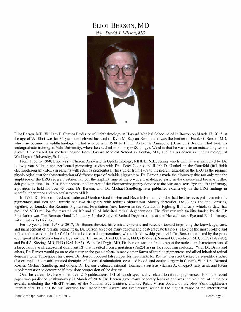
ELIOT BERSON, MD By David J. Wilson, MD
Eliot Berson, MD, William F. Chatlos Professor of Ophthalmology at Harvard Medical School, died in Boston on March 17, 2017, at the age of 79. Eliot was for 35 years the beloved husband of Kyra M. Kaplan Berson, and was the brother of Frank G. Berson, MD, who also became an ophthalmologist. Eliot was born in 1938 to Dr. H. Arthur & Annabelle (Bernstein) Berson. Eliot took his undergraduate training at Yale University, where he excelled in his major (Zoology). Word is that he was also an outstanding tennis player. He obtained his medical degree from Harvard Medical School in Boston, MA, and his residency in Ophthalmology at Washington University, St. Louis.
From 1966 to 1968, Eliot was a Clinical Associate in Ophthalmology, NINDB, NIH, during which time he was mentored by Dr. Ludwig von Sallman and performed pioneering studies with Drs. Peter Gouras and Ralph D. Gunkel on the Ganzfeld (full-field) electroretinogram (ERG) in patients with retinitis pigmentosa. His studies from 1968 to the present established the ERG as the premier physiological test for characterization of different types of retinitis pigmentosa. Dr. Berson’s made the discovery that not only was the amplitude of the ERG severely subnormal, but the implicit time of the b-wave was delayed early in the disease and became further delayed with time. In 1970, Eliot became the Director of the Electroretinography Service at the Massachusetts Eye and Ear Infirmary, a position he held for over 45 years. Dr. Berson, with Dr. Michael Sandberg, later published extensively on the ERG findings in specific inheritance and molecular types of RP.
In 1971, Dr. Berson introduced Lulie and Gordon Gund to Ben and Beverly Berman. Gordon had lost his eyesight from retinitis pigmentosa and Ben and Beverly had two daughters with retinitis pigmentosa. Shortly thereafter, the Gunds and the Bermans, together, co-founded the Retinitis Pigmentosa Foundation (now known as the Foundation Fighting Blindness), which, to date, has provided $700 million for research on RP and allied inherited retinal degenerations. The first research facility funded by the RP Foundation was The Berman-Gund Laboratory for the Study of Retinal Degenerations at the Massachusetts Eye and Ear Infirmary, with Eliot as its Director.
For 49 years, from 1968 to 2017, Dr. Berson devoted his clinical expertise and research toward improving the knowledge, care, and management of retinitis pigmentosa. Dr. Berson accepted many fellows and post-graduate trainees. Three of the most prolific and influential researchers in the field of inherited retinal degenerations, who took fellowship years with Dr. Berson are, listed by the years each spent at the Massachusetts Eye and Ear Infirmary, David G. Birch, PhD, (1979-82), Samuel G. Jacobson, MD, PhD, (1982-83), and Paul A. Sieving, MD, PhD (1984-1985). With Ted Dryja, MD, Dr. Berson was the first to report the molecular characterization of a large family with autosomal dominant RP that resulted from a mutation (Pro23His) in the rhodopsin molecule. With Dr. Dryja and others, Dr. Berson would go on to characterize the gene defects in many other forms of retinitis pigmentosa and allied inherited retinal degenerations. Throughout his career, Dr. Berson opposed false hopes for treatments for RP that were not backed by scientific studies (for example, the unsubstantiated therapies of electrical stimulation, ozonated blood, and ocular surgery in Cuban). With Drs. Bernard Rosner, Michael Sandberg, and others, Dr. Berson evaluated rational treatments such as vitamin A, omega-3 fatty acid, and lutein supplementation to determine if they slow progression of the disease.
Over his career, Dr. Berson had over 275 publications, 181 of which specifically related to retinitis pigmentsoa. His most recent paper was published posthumously in March of 2018. Dr. Berson gave many honorary lectures and was the recipient of numerous awards, including the MERIT Award of the National Eye Institute, and the Pisart Vision Award of the New York Lighthouse International. In 1990, he was awarded the Franceschetti Award and Lectureship, which is the highest award of the International
Trans Am Ophthalmol Soc / 115 / 2017 Necrology 2
Necrology
Trans Am Ophthalmol Soc / 115 / 2017 Necrology 3
Society for Genetic Eye Disease. In 1991, he was given the Taylor Smith Award by the New England Ophthalmological Society. In 1992, he received the Friedenwald Award from the Association for Research in Vision and Ophthalmology. For his lifetime of achievements, the Foundation Fighting Blindness in 1999 honored Dr. Berson with its highest award, the Llura Liggett Gund Award. In 2006, Dr. Berson was awarded the Ludwig von Sallmann Prize of the International Congress for Eye Research.
Throughout his years of clinical practice, Dr. Berson’s interactions with his patients was that of a compassionate, caring physician who sought to provide the optimum care to his patients with retinitis pigmentosa and to provide them with hope for future cures and treatments for their disease. Perhaps the greatest honor that Dr. Berson received was the respect, admiration, and gratitude of his many patients for his kindness, enthusiasm, and dedication to the search for meaningful treatments for inherited retinal degenerations.

ROBERT C. DREWS, MD By James Bobrow, MD
Robert C. Drews, M.D., died peacefully on May 9, 2017, surrounded by his family. The flags on the buildings of Washington University were placed at half-mast for three days in honor of his contributions both to ophthalmology and to the University. Robert graduated from both Washington University College of Arts and Sciences and the Washington University School of Medicine. He completed his residency under the watchful eye of Bernard Becker, M.D., and was chosen Chief Resident in 1958. After service in the United States Navy at the U.S. Naval Hospital, Great Lakes, he returned to Saint Louis to join his father in private practice. He retained his academic affiliation to the University, rising to the rank of Professor of Clinical Ophthalmology and Visual Sciences. Dr. Drews was a member of Washington University’s Board of Trustees from 1988-1992, chaired the university’s Alumni Board of Governors and the Medical Alumni Annual Fund and other giving programs. He received Washington University’s Distinguished Alumni Award in 1988 at Founders Day and the School of Medicine’s Second Century Award in 2001.
He was committed to the Saint Louis Society for the Blind and Visually Impaired, serving on the Board, as its President, and on the Advisory Board for a total of 53 years. Its clinic, which serves the metropolitan area without regard to ability to pay, bears the title “The Leslie and Robert Drews Low Vision Clinic.”
He became a member of the American Ophthalmological Society in 1979 and served as President (2002) and member of the Council. The professional component of his legacy spans six decades. It includes membership in 32 societies and 38 Boards, of which he served as president of 11, 22 international medals and other honors, and 11 named lectureships. He wrote 485 articles, chapters, letters and other publications and served as editor or reviewer for multiple publications, both in the United States and around the world. In the early 1970s, he learned Spanish sufficiently fluently to deliver lectures to his colleagues in South America, Europe, and Asia. He also translated several important texts from Spanish to English and English to Spanish. He was instrumental in initiating a summary session given in Spanish of the major presentations each year at the American Academy of Ophthalmology. He gave over 1000 presentations at more than 600 meetings. He accommodated over 1500 ophthalmologists in his operating rooms during his very active and prolific surgical career, teaching techniques of ophthalmic surgery to two generations of surgeons.
The common theme that united his endeavors was his selfless pursuit of knowledge and understanding. While engaged in more than a quarter of a million patient visits over more than 30 years, he undertook projects because he sensed that they contained questions that needed to be answered. Throughout his career, he remained true to his commitment to bring state-of-the-art techniques
Trans Am Ophthalmol Soc / 115 / 2017 Necrology 4

ABBOT G. SPAULDING, MD By JAMES J. AUGSBURGER, MD
Abbot G. Spaulding was born in Chicago on August 24, 1933. He attended elementary school in Springfield, Illinois, and high school in Prairie duChien, Wisconsin. Following high school, he attended St. Louis University, majoring in English and minoring in Philosophy and Chemistry. He obtained his BA degree cum laude in 1955. His studied medicine at the St. Louis University School of Medicine, from which he obtained his MD degree in 1959. He interned at Presbyterian-St. Luke’s Hospital in Chicago 1959-1960 and then entered the US Navy in 1960. He was appointed a general medical officer and was assigned to a naval transport ship, the General Edwin D. Patrick. After his tour of duty ended, he obtained a residency in ophthalmology (1962-1965) at the University of Cincinnati College of Medicine (Donald J. Lyle, MD, Professor, Taylor Asbury, MD, Program Director). After completing his residency in 1965, he took 6 months of fellowship training in Ophthalmic Pathology at the Armed Forces Institute of Pathology in Washington, DC (Lorenz E. Zimmerman, Director). He returned to Cincinnati after this fellowship and started a solo practice of general ophthalmology in the Anderson suburb of the city. He also accepted appointment to a part-time faculty position in the University of Cincinnati’s Department of Ophthalmology, where his principal roles were ophthalmic pathologist and director of the ophthalmic pathology laboratory. His private practice grew and developed into a five-ophthalmologist group over the years. This group subsequently joined a number of other ophthalmology practices in the city to form a multisubspecialty group known as Tri State Eye Care. Dr. Spaulding retired from full-time clinical practice in 1999; however, he continued as ophthalmic pathologist at the University of Cincinnati on a part-time basis until 2014, serving under Chairmen Taylor Asbury (1966-1977), Joel G. Sacks (1977-1994), William V. Good (1995-1997), and James J. Augsburger (1999-2014). Originally appointed as Clinical Instructor of Ophthalmology, he was promoted to Assistant Clinical Professor in 1968, Associate Clinical Professor in 1970,, and Clinical Professor in 1976. In 1996, Dr. Spaulding was named the inaugural Mary Knight Asbury Chair of Ophthalmic Pathology at the University of Cincinnati College of Medicine. He continued to hold this endowed chair position until 2006. Dr. Spaulding was named Emeritus Professor of Ophthalmology at the University of Cincinnati in 2014.
In addition to his membership in the American Ophthalmological Society (new member in 1978), Dr. Spaulding was a member of the American College of Surgeons (which he served as a member of the credentials review committee), American Medical Association, Ohio State Medical Association, Cincinnati Academy of Medicine, American Academy of Ophthalmology (which he served as long-serving Councilor), Ohio Ophthalmological Society (which he served as board member and president), Cincinnati Society of Ophthalmology (which he served as long-term Secretary-Treasurer), American Association of Ophthalmic Pathologists (charter member), AFIP Ophthalmic Pathology Alumni Association, and (before joining the AOS) American Eye Study Club.
Dr. Spaulding died in Cincinnati on June 6, 2016.
Trans Am Ophthalmol Soc / 115 / 2017 Necrology 5

WILLIAM TASMAN, MD By JULIA A. HALLER, MD
William Tasman, Howe Medalist, avid and engaged member and past president of the AOS, and one of the legendary leaders of modern American medicine, died peacefully surrounded by his family on March 28, 2017, after breaking his hip in a fall, and subsequent congestive heart failure. Bill had deep roots in the Quaker soil of Philadelphia. He was a loyal and influential alumnus of the Germantown Friends Academy, and Haverford College, where he lettered on the single wing football team. He graduated from Temple University School of Medicine and interned at Philadelphia General Hospital, before serving his country as a captain in the U.S. Air Force in Wiesbaden, Germany. Returning from military service, he followed his father’s distinguished path into Ophthalmology, and joined the residency class at the Wills Eye Hospital in Philadelphia in 1959. He was named Chief Resident at Wills, and went on to complete a retina fellowship at Massachusetts Eye and Ear Infirmary in Boston.
1962 was a banner year for Dr. Tasman: he completed his retina fellowship, joined the staff of the Wills Eye Hospital, and married Alice Lea Mast, an Art History graduate of Barnard College and associate director of public relations at the Philadelphia Art Museum. Alice Lea and Bill formed a true Team of Destiny in every aspect of their lives, and the AOS benefited immeasurably from their many contributions organizationally, as well as on the dance floor, the ski slope, and the tennis courts! Bill served as editor of the Transactions from 1992-1996, and as President from 1998-1999.
Dr. Tasman’s stature in the retina field is legendary. He literally rewrote the book on pediatric retina disease, while making seminal contributions to the fields of retinal surgery, treatment of diabetic disease, and trauma, among many others. He was a founding member of the Wills Retina Service, the Retina Society and Club Jules Gonin.
Dr. Tasman’s history at the Wills Eye Hospital is one of remarkable and distinguished service. He became Ophthalmologist-in-Chief of Wills and Professor and Chairman of the Department of Ophthalmology of Jefferson Medical College of Thomas Jefferson University in 1985. In 2007, after 22 years at the helm of the nation’s oldest eye hospital and director of the country’s first ophthalmology residency, he moved to the position of Professor and Emeritus Chairman of the department of Ophthalmology of Jefferson Medical College, and Ophthalmologist-in Chief Emeritus of Wills Eye. Universally revered and beloved, “Dr. T” continued until the last months of his life to be a mentor and role model for generations of Wills medical students, residents, and fellows, to whose projects, papers, life decisions, and skits he always lent his time, wisdom, humor, and skill!
In addition to his AOS presidency, Dr. Tasman’s extraordinary service, dedication, and leadership skills were recognized by his election as chairman of the American Board of Ophthalmology, president of the American Academy of Ophthalmology, and president of the Retina Society. He has received many honors including the Zentmayer Award, the Heed Award, the Jules Stein Lifetime Achievement Award, the gold medal from the Kingdom of Saudi Arabia, membership in Academia Internationalis Ophthalmologica, the AAO’s Honor Award, Senior Honor Award, Distinguished Service award and Lifetime Achievement Award, among many others. He served on numerous editorial boards and authored over 220 articles in the peer-reviewed literature, 38 book chapters, 4 annual
Trans Am Ophthalmol Soc / 115 / 2017 Necrology 6

Necrology
Trans Am Ophthalmol Soc / 115 / 2017 Necrology 7
Retina reviews, 44 commentaries, and nine ophthalmology books. Dr. William Tasman is one of those rare individuals who truly left everything he touched better than he found it. He is survived by
his wife and true soulmate Alice Lea and their three children, James, Graham and Alice, and the six grandchildren who adored sailing and snorkeling and telling jokes with their beloved grandfather, whether on the Eastern shore of Maryland or in the Florida Keys, or on global family adventures. A towering figure both literally and metaphorically in modern American medicine and a larger than life figure in his worldwide community, the memory and contributions of Dr. William Tasman will forever be treasured by all of us privileged to know him.

RICHARD TROUTMAN MD By IVAN R. SCHWAB MD, FACS
In every generation, there are a few iconic individuals who change the course of a profession, and the rest of the world is better for it. Richard Troutman was one such individual. We mourn his loss and celebrate his achievements.
Richard Troutman MD, DSC died on 5 April 2017 at his home in Bal Harbour FL. He was 94. His achievements were legend, and he was venerated. He was a pioneer in the use of the operating microscope, microsurgical techniques, and instruments in ophthalmology at a time (the early 1950s) when bare eyes or loupes were all that was used. His microsurgical genius extended to instrument design and the refinement of techniques. Because the operating microscope is so commonplace in our profession now, it is difficult to imagine the resistance he encountered and his perseverance to see the microscope used and improved. Microsurgical techniques have been one of the ten or fifteen greatest and most influential steps in our profession in the last 150 years.
He is survived by his wife, Suzanne Veronneau-Troutman MD with whom he established the Microsurgical Research Foundation. This non-profit organization is devoted to improving the understanding and application of microscopic tools. He is also survived by two children and a grandson.
Although the promulgation of the operating microscope was his greatest achievement, it was not his only one. He was professor and chair of Ophthalmology at SUNY Downstate from 1955 to 1983. Dr. Troutman, his wife Suzanne, and others established the Richard C. Troutman, MD Distinguished Chair in Ophthalmology and Ophthalmic Microsurgery at Downstate in 2002. During his time at SUNY Downstate, he was known as a gifted and skilled surgeon, a curious scientist, a consummate teacher, and above all, a devoted clinician who was dedicated to his patients and students. He was a pioneer in refractive surgery and was among the first North American surgeons trained in lamellar refractive surgery by Dr. Jose Barraquer in Bogota, Columbia. Later in his career, he, Dr. Barraquer and Dr. Swinger would found the International Society for Refractive Surgery and later preside as its president.
He was devoted to the improvement of sight with a global reach serving on the Board of Directors (1962-1981) for Sight Restoration in New York City. He was a strong advocate for The Pan American Association of Ophthalmology and promoted its growth throughout his life.
Richard was born in Columbus, Ohio on May 16, 1922 when his father was training in Vienna by preceptorship to be an EENT specialist. Richard’s prize-winning career began early as he won Best Baby in the State of Ohio for which his mother received a $25.00 gold piece. He also received an engraved Silver Loving Cup and a gold Medal attesting to this award. He attended Catholic schools and at age 13, enrolled in the Culver Military Academy. Remarkably, his senior thesis was on glaucoma. When he graduated in 1939, he was commissioned as a second lieutenant in the infantry. He attended Ohio State for premed and medical school, graduating in 1945. In medical school, where he was mentored by Jack Frost (another AOS member), he won the Eli Alcorn Prize in Ophthalmology. He had been assigned to the Navy in 1941 when the war broke out but during WWII was assigned to finish medical school. He would later fulfill his military commitment as chief of EENT between 1946-48 before his residency in 1951. Because of this experience he became interested in the poor cosmetic result of enucleations. He worked with Davis Durham to create a mesh covered “integrated” implant. This would be a sign of his future as a compassionate innovator and a curious clinical investigator.
Dr. Troutman became a member of the AOS in 1962 and his wife, Suzanne, became a member in 1978 making them the first AOS couple. They were married for 50 years. His AOS thesis was entitled Artiphakia and Aniseikonia, published in 1962.
Richard was more than an ophthalmic genius, though, as his range of interests and abilities was broad. He was a kind and generous man who was loved and respected by the residents and fellows he trained. He held yearly reunions at the AAO for them and their fellows. He was an avid dancer and eager participant whenever the band would start to play. He would jump up to be one of the first on the dance floor with his wife, Suzanne Veronneau-Troutman. He was an avid sailor and owned a boat he enjoyed piloting. The surprise in Dr. Troutman’s personality was his desire for speed in his fast cars and fast electric carts! These carts would be
Trans Am Ophthalmol Soc / 115 / 2017 Necrology 8
Necrology
Trans Am Ophthalmol Soc / 115 / 2017 Necrology 9
reserved at the AAO meetings in his latter years so he could speed about the meeting at maximum speed from venue to venue. While his iconic world legacy will be as the father of microsurgery with the introduction and promotion of the operating
microscope, instruments, and sutures to ophthalmology, he will be remembered most by his friends and family as a loving and devoted husband and father, and a close, enduring and generous friend. We will all miss him. He had a life well lived.
Trans Am Ophthalmol Soc / 115 / 2017 Necrology 10
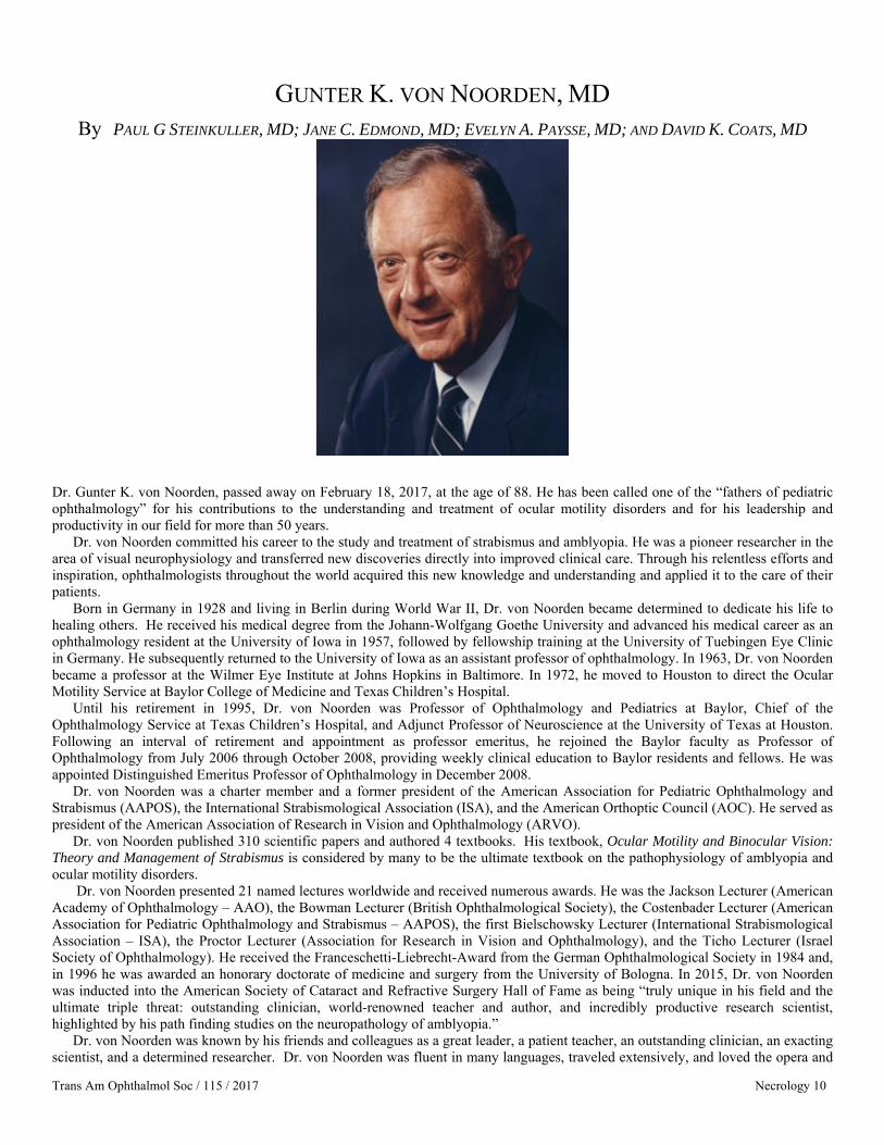
GUNTER K. VON NOORDEN, MD By PAUL G STEINKULLER, MD; JANE C. EDMOND, MD; EVELYN A. PAYSSE, MD; AND DAVID K. COATS, MD
Dr. Gunter K. von Noorden, passed away on February 18, 2017, at the age of 88. He has been called one of the “fathers of pediatric ophthalmology” for his contributions to the understanding and treatment of ocular motility disorders and for his leadership and productivity in our field for more than 50 years.
Dr. von Noorden committed his career to the study and treatment of strabismus and amblyopia. He was a pioneer researcher in the area of visual neurophysiology and transferred new discoveries directly into improved clinical care. Through his relentless efforts and inspiration, ophthalmologists throughout the world acquired this new knowledge and understanding and applied it to the care of their patients.
Born in Germany in 1928 and living in Berlin during World War II, Dr. von Noorden became determined to dedicate his life to healing others. He received his medical degree from the Johann-Wolfgang Goethe University and advanced his medical career as an ophthalmology resident at the University of Iowa in 1957, followed by fellowship training at the University of Tuebingen Eye Clinic in Germany. He subsequently returned to the University of Iowa as an assistant professor of ophthalmology. In 1963, Dr. von Noorden became a professor at the Wilmer Eye Institute at Johns Hopkins in Baltimore. In 1972, he moved to Houston to direct the Ocular Motility Service at Baylor College of Medicine and Texas Children’s Hospital.
Until his retirement in 1995, Dr. von Noorden was Professor of Ophthalmology and Pediatrics at Baylor, Chief of the Ophthalmology Service at Texas Children’s Hospital, and Adjunct Professor of Neuroscience at the University of Texas at Houston. Following an interval of retirement and appointment as professor emeritus, he rejoined the Baylor faculty as Professor of Ophthalmology from July 2006 through October 2008, providing weekly clinical education to Baylor residents and fellows. He was appointed Distinguished Emeritus Professor of Ophthalmology in December 2008.
Dr. von Noorden was a charter member and a former president of the American Association for Pediatric Ophthalmology and Strabismus (AAPOS), the International Strabismological Association (ISA), and the American Orthoptic Council (AOC). He served as president of the American Association of Research in Vision and Ophthalmology (ARVO).
Dr. von Noorden published 310 scientific papers and authored 4 textbooks. His textbook, Ocular Motility and Binocular Vision: Theory and Management of Strabismus is considered by many to be the ultimate textbook on the pathophysiology of amblyopia and ocular motility disorders.
Dr. von Noorden presented 21 named lectures worldwide and received numerous awards. He was the Jackson Lecturer (American Academy of Ophthalmology – AAO), the Bowman Lecturer (British Ophthalmological Society), the Costenbader Lecturer (American Association for Pediatric Ophthalmology and Strabismus – AAPOS), the first Bielschowsky Lecturer (International Strabismological Association – ISA), the Proctor Lecturer (Association for Research in Vision and Ophthalmology), and the Ticho Lecturer (Israel Society of Ophthalmology). He received the Franceschetti-Liebrecht-Award from the German Ophthalmological Society in 1984 and, in 1996 he was awarded an honorary doctorate of medicine and surgery from the University of Bologna. In 2015, Dr. von Noorden was inducted into the American Society of Cataract and Refractive Surgery Hall of Fame as being “truly unique in his field and the ultimate triple threat: outstanding clinician, world-renowned teacher and author, and incredibly productive research scientist, highlighted by his path finding studies on the neuropathology of amblyopia.”
Dr. von Noorden was known by his friends and colleagues as a great leader, a patient teacher, an outstanding clinician, an exacting scientist, and a determined researcher. Dr. von Noorden was fluent in many languages, traveled extensively, and loved the opera and

Necrology
Trans Am Ophthalmol Soc / 115 / 2017 Necrology 11
symphony. He was an avid swimmer and tennis player, and maintained a fit physique throughout his life. He was a lover of good food and great wine. Dr. von Noorden was knowledgeable in many areas outside of medicine; a true Renaissance man. He enjoyed life to the fullest; a man who committed his energy and time to fulfilling his many missions, ambitions and passions while enjoying every step of his journey. Dr. von Noorden was a dedicated husband for more than five decades. He is survived by his wife, Betty and their daughter, Sonya.

Trans Am Ophthalmol Soc / 115 / 2017 Proceedings 1
MINUTES OF THE PROCEEDINGS One Hundred and Fifty-Third Annual Meeting
May 18-21, 2017 The ONE HUNDRED AND FIFTY-THIRD ANNUAL MEETING of the American Ophthalmological Society (AOS) was held at The Homestead, Hot Springs, Virginia. On May 19, 2016, Friday, President George Bartley, MD called the opening session to order. The program began with the following AOS-Knapp symposium.
SYMPOSIUM: OPTIC NERVE REGENERATION AND RECONNECTION: CURRENT STATUS, CHALLENGES AND AUDACIOUS FUTURE GOALS
1. Introduction David T. Tse, MD 2. RGC Types Differ In Function And In Vulnerability To Disease Joshua R. Sanes, PhD 3. Progress And Challenges In RGC Protection Harry Quigley, MD 4. Challenges In Axon Pathfinding And Target Recognition Kevin K. Park, PhD 5. Progress And Challenges In Axon Regeneration Zhigang He, PhD
SCIENTIFIC SESSION, FRIDAY, MAY 18, 2017 1. Impact Of Resident And Fellow Trainees On Patient Encounter Length In An Academic Ophthalmology
Center. Michael Chiang, Isaac Goldstein, Sarah Read-Brown, Michelle Hribar 2. Genetic Analysis Of 1000 Consecutively Ascertained Families With Inherited Retinal Disease. Edwin Stone,
Jeaneen Andorf, Adam DeLuca, Scott Whitmore, Joseph Giacalone, Luan Streb, Terry Braun, Robert Mullins, Todd Scheetz, Val Sheffield, Budd Tucker
3. Conjunctival Tumors In Children In 806 Cases. Features Differentiating Benign From Malignant Tumors. Carol L. Shields, Kareem Sioufi, Adel E. Alset, Emil Anthony T. Say, Jerry A. Shields
4. Limbal And Conjunctival Transplantation From The Same Livingrelated Bone Marrow Donor To Patients With Ocular Graft Versus Host Disease. Massimo Busin, Giuseppe Giannaccare, Laura Sapigni, Emilio Campos
5. Ranibizumab For The Prevention Of Radiation Complications In Patients Treated With Proton Beam Irradiation For Choroidal Melanoma. Ivana Kim, Anne Marie Lane, Purva Jain, Caroline Awh, Evangelos Gragoudas
EXECUTIVE SESSION, SATURDAY, MAY 20, 2017 George Bartley, MD: Good morning everyone I’d like to call the order of this Executive Session of the 153nd Meeting of the American Ophthalmological Society. The Executive Vice President, Dr. Hans Grossniklaus will now give his report:
REPORT OF THE EXECUTIVE VICE-PRESIDENT 2017 Hans E. Grossniklaus MD As of March 31, 2016, the balance of the AOS is $8,993,132, which is up from last year. We continue to use approximatley $150 thousand a year to subsidize the annual meeting. We have 225 active members and 145 emeritus members. We support the Heed retreat in part, which is a fall educational of academically inclined ophthalmology residents, and these residents are told about the AOS. We have 8 council/Knapp and 3 CES travel grants available for students, residents, fellows and ophthalmologists to attend the annual meetings as guests. We support the Blodi and Verhoeff lectures and 4 non-AOS symposium speakers for the annual meeting.
Our infrastructure is being improved and modernized. The new AOS website is functioning well. The website includes online abstract submission, membership nominating, and soon online thesis uploading areas. Michael Chiang MD is the chair of our newly formed communications committee, which includes Anne Coleman, Paul Chan, and Tamara Fountain. The committee is already working on incorporating social media, including Facebook, Twitter and Instagram into the AOS, including a talk and lunch and learn session at our next annual meeting. These changes constitute implimentation of the strategic plan we developed last year.
The membership accepted several bylaws changes which were mainly structural in nature. We are in discussions about how to move forward regarding the nature of the theses and TAOS, which will be further discussed at the fall council meeting. We have 22 persons proposed to submit applications for membership, which may be an all time high. We will continue to pursue maintaining an active, vital AOS.

Minutes of the Proceedings
Trans Am Ophthalmol Soc / 115 / 2017 Proceedings 2
REPORT FROM THE COUNCIL CHAIR Anne Coleman, MD, PhD The 2017 153rd AOS Annual Meeting took place at the Homestead, a location we hadn’t visited since 2004 and was a huge success. This year we welcomed many new members with the New Member Luncheon followed by the New Member Spotlight presentations and reception. The following day, members enjoyed a very insightful Knapp Symposium, organized by David Tse, MD, focusing on “Optic Nerve Regeneration and Reconnection: Current Status, Challenges and Audacious Future Goals”. In addition to these academic events, members enjoyed the annual Artistic Performances assembled by Susan Day, of very musically talented AOS members. Members enjoyed further education about retinal ganglion cell rescue in glaucoma from the Frederick C. Blodi Lecture presented by Joseph Caprioli, MD. Several members participated in the Golf and/or Tennis tournaments, and skeet shooting! We later attended the Saturday evening Banquet where the Howe Medal was presented to George Spaeth, a most deserved honor! We are already looking forward to next year’s annual meeting which will be held in the beautiful Dana Point May 17-20, 2018. Make sure to register for this always educational and enjoyable experience.
REPORT OF THE AOS AUDIT COMMITTEE Jay C. Erie, MD Dr. Erie is the Chair of the AOS Audit Committee this year with additional members M. Edward Wilson, Jr, MD, and Hans E. Grossniklaus (EVP). The Audit Committee met on June 28, 2017 with additional guests including Rianne Suico and Chris Pritchard of Moss Adams Accountants, Alice Paw as Finance Manager, AAO, and Michael Roll as Director of Finance, AAO. Attending SF AMS Management staff included Timothy Losch and Amber J. Mendes, AOS, Client Services Manager. The Committee reviewed the Fiscal Year 2016 Audited Financial Statements. Ms. Paw provided an overview and noted that there were changes to the presentation of audited financials in order to align with reporting standards. The changes included a consolidated statement of financial positions and activities presented at a summarized level, detailed statements were now included as supplemental schedules, and because financials are reported on a comparative basis, footnotes where amounts were reported included current and prior year balances. Ms. Paw reported that the total net assets increased $197,000 from the prior year. Dr. Erie excused the SF AMS and Academy Finance staff and the Committee met in executive session with representatives from Moss Adams. They did not encounter anything with respect to the financial condition of the organization that would be considered unusual or warrant further investigation. The Committee accepted the auditor’s report as presented and Dr. Erie adjourned the meeting of the Audit Committee without any having determined any irregularities.
2017 AOS PRESIDENT GEORGE B. BARTLEY, MD

Minutes of the Proceedings
Trans Am Ophthalmol Soc / 115 / 2017 Proceedings 3
REPORT OF THE COMMITTEE ON THESES Dimitri T. Azar, MD Chair and reporting member, Committee Members include: Janet Davis, M.D. and Henry Jampel, M.D. The AOS Thesis Committee reviewed 13 theses since the 2016 AOS meeting. Nine theses required minor revisions and the revisions are expected to be returned within one month. There were four theses that will require major revisions and the authors will have until January 2018 to resubmit the revised theses.
REPORT OF THE EDITOR: Emily Y. Chew, MD It is a great privilege and honor to serve as your editor of the Transactions of the American Ophthalmological Society (TAOS). I will indeed be your last editor of the TAOS as we transition this year to have the AOS theses published as a supplement in the American Journal of Ophthalmology (AJO). This was an important step as we now will have an impact factor associated with the publication of the AOS theses.
A task force was convened last year to explore the role of the thesis for membership in the AOS. This issue was raised following a retreat that was conducted to modernize our organization. The thesis has been a tradition that distinguishes the AOS from other societies in our profession and the AOS membership voted overwhelmingly to maintain this as a requirement for membership. A number of members have commented that they have received negative comments as well as refusals to join the AOS because of the thesis requirement. These are candidates who routinely publish their research in various peer-reviewed journals, including prestigious journals in their field. Because of the lack of impact factor with the TAOS, potential candidates were either not willing to put an effort into the thesis or reluctant to present their best work. The AOS membership also voted to relinquish the TAOS and find other methods of publishing the theses. For now, we believe we have resolved this issue by partnering with the American Journal of Ophthalmology. The Committee on Theses will continue to function and the final production of the theses will be by the managing editor of the AJO.
This brings an end to the TAOS which has served the membership well but it is important to now move on in the 21st century. We hope this change will attract more members that are vital to our organization. It has indeed been an honor and a privilege to serve as your last editor.
2017 AOS PRESIDENT PRESIDENT GEORGE B. BARTLEY, MD & HIS WIFE LYNN BARTLEY
Minutes of the Proceedings
Trans Am Ophthalmol Soc / 115 / 2017 Proceedings 4
REPORT OF THE COMMITTEE ON PROGRAMS David T. Tse, MD The 2017 AOS Knapp Symposium entitled Optic Nerve Regeneration and Reconnection: Current Status, Challenges and Audacious Future Goals presents a broad perspective on the challenges of RGC rescue, optic nerve neuronal regeneration, and the possibilities of eventual connectivity to mediate functional recovery. The symposium theme meets the regenerative objective of restoring vision loss and is aligned with central goals of the NEI Audacious Goals Initiative (AGI) - regenerate neurons and neural connections in the eye and visual system.
The 2017 Frederick C. Blodi Lecture entitled Retinal Ganglion Cell Rescue in Glaucoma was presented by Joseph Caprioli, MD. Introduction was given by George Spaeth, MD.
SYMPOSIUM: FREDERICK C. BLODI LECTURE 1. Introduction. George L. Spaeth, MD 2. Retinal Ganglion Cell Rescue In Glaucoma. Joseph Caprioli, MD The 2017 AOS Saturday Symposium theme on “Quality of Care: Improvement Based on Evidence” featured Paul Lee,
MD on evidence-based care; David W. Parke, II, MD on registries; and Michael F. Chiang, MD on current practice and the future. These important developments will impact all members of the AOS, regardless of specialty and practice setting.
SYMPOSIUM: QUALITY OF CARE: IMPROVEMENT BASED ON EVIDENCE 1. Introduction. Anne L. Coleman, MD 2. Quality Of Care And Evidence-Based Care. Paul P. Lee, MD, JD 3. Quality Of Care And Registries. David W. Parke, II, MD 4. Quality Of Care In Current Practice And The Future For the general program there were 36 abstracts received, of which 19 were accepted as podium presentations (2 were
AOS theses). Of the remaining 17, 11 were poster presentations. New this year was that only the presenting author is required to disclose financial interests with commercial companies
in medicine that are relevant to the presentation. This change was in response to the new ACCME guidelines, which were adopted by the American Academy of Ophthalmology - the joint provider of CME for the meeting. Since CME credits were offered for the Friday and Saturday poster sessions during the morning breaks, a moderator was assigned to each guided session.
The general program also featured 3 new members, 4 PhDs, and 2 non-members at the podium. The 2017 AOS scientific program was awarded the full 12 hours of CME credits. This is the second year that the CME auditor approved everything upon first review and the second year AOS was granted CME hours for poster sessions.
Also new this year was that all symposia speakers and abstract presenters were asked to use the AOS logo template for the title page and financial disclosure, and then transition into own slide deck without AOS logo, if desired.
I want to thank members of the Committee on Programs, Eduardo Alfonso, Preston Blomquist and Ivan Schwab, and Council Chair Anne Coleman for their tremendous help in organizing the 153rd AOS Annual Meeting.
REPORT OF THE COMMITTEE ON MEMBERSHIP William F Mieler, MD This past year, the AOS invited 9 candidates to apply for membership. Currently, we have 22 candidates whose applications will be reviewed later this year, and will then be considered for membership.
Over the past 12 years, 129 candidates have been welcomed into the membership of the American Ophthalmological Society (AOS). This is an average of 10.75 candidates/year. The highest number was 18 candidates in 2007, with a low of 5 candidates in 2010. During the timeframe of 2009-12, new candidate numbers were rather low at 6, 5, 8, and 7 candidates. In the time frame of 2013-16, the numbers have improved to 15, 13, 10, and 8.
The Committee on Membership (and the AOS in general) has been actively encouraging members to nominate colleagues for membership into the AOS. We all strive to have the AOS organization remain at the forefront of scientific advances in ophthalmic clinical, translational, and basic science research.

Minutes of the Proceedings
Trans Am Ophthalmol Soc / 115 / 2017 Proceedings 5
2017 AOS COUNCIL (FROM LEFT): WOODFORD S. VAN METER, MD, MARCO A. ZARBIN, MD, HANS E.
GROSSNIKLAUS, MD, EMILY Y. CHEW, MD, TIMOTHY W. OLSEN, MD, PRESIDENT GEORGE B. BARTLEY, MD, EDWARD G. BUCKLEY, MD AND ANNE LOUISE COLEMAN, MD
REPORT OF THE ARCHIVIST PHOTOGRAPHER RALPH C. EAGLE, JR, MD I took more than 1050 high resolution digital photographs at the One Hundred Fifty Second Annual Meeting of the American Ophthalmological Society held at the Broadmoor Hotel in Colorado Springs, Colorado on May 19-22, 2016. The photos were taken using a Nikon D810 digital camera. Eight photos were included as color illustrations in the frontmatter of the 2016 on-line volume CXIV (v.114) of the TRANSACTIONS OF THE AMERICAN OPHTHALMOLOGICAL SOCIETY. These included photos of 2016 AOS President Marilyn B. Mets, MD, President Mets and her husband Laurens J. Mets and group photos of The 2016 AOS Council and eleven new members who attended the meeting. 2016 Lucien Howe Medalist Susan H. Day, MD and 2016 Frederick C. Blodi Lecturer Elias Traboulsi, MD were shown in additional figures. A photo of new member Tamara R. Fountain signing the AOS Membership Book and a group photo of the participants in Knapp Symposium on Innovative Paradigm Shifts in Ophthalmology also was included. A photoshow comprising selected digital images in PDF format from the 2016 meeting can be downloaded from the meeting photos section of the Members-Only section of the AOS website. The digital archives of the AOS now comprise more than 11650 high-resolution digital photographs and 1400 digital images prepared from scanned transparencies. Additional slides will be scanned in the future. The images are stored on redundant digital
Minutes of the Proceedings
Trans Am Ophthalmol Soc / 115 / 2017 Proceedings 6
hard drives and flash drives and on CD’s and DVD’s in some instances. A backup hard drive containing all the images will be stored in the AOS office in San Francisco.
REPORT OF THE COMMITTEE ON EMERITI Thomas D. France, MD The Annual Emeritus luncheon will be held on May 19, 2017 in the Dominion Room of the hotel. All Emeritus members, both old and new, and their guests, are invited! We have invited the hotel’s naturalist to tell us more about the flora and fauna around the hotel.
I regret to inform you of the passing of the following AOS members since our last Annual Meeting: Eliot L. Berson, MD Brookline, MA Member since 1990 Brian J. Curtin, MD Rye, NY Member since 1969 Robert C. Drews, MD Olivette, MO Member since 1979 Barrett G. Haik, MD Memphis, TN Member since 1991 Abbot G. Spaulding, MD Cincinnati, OH Member since 1978 William S. Tasman, MD Wyndmoor, PA Member since 1970 Richard C. Troutman, MD Bal Harbor, FL Member since 1962 Gunter K. von Noorden, MD Houston, TX Member since 1969
COUNCIL APPOINTMENTS FOR 2017-2018 AOS Council – Julia A. Haller to join Woodford S. Van Meter, Chair Marco A. Zarbin Timothy W. Olsen and Edward G. Buckley AOS President – David J. Wilson Executive Vice President – Hans E. Grossniklaus to continue Editor – Emily Y. Chew to continue Member, Committee on Theses – J. Douglas Cameron to join Henry Jampel, Chair and Janet L. Davis Member, Committee on Programs – Jayne S. Weiss to join Eduardo Alfonso, Chair Preston Blomquist and Ivan R. Schwab Member, Committee on Membership – Anthony C. Arnold to join Mary Elizabeth Hartnett, Chair R. Michael Siatkowski
Christopher J. Rapuano Chairs, Committee on New Members – Evelyn A. Paysse & David Coats to continue Member, Committee on Prizes – Alfred Sommer to join Dan B. Jones, Chair and Susan Day Chair, Committee on Emeriti – Thomas D. France to continue Committee on Athletics – Frederick W. Fraunfelder to continue Chair, Audit Committee – Anne L. Coleman to join M. Edward Wilson, Jr., Chair and
Hans E. Grossniklaus Investment Committee – David J. Wilson, Woodford S. Van Meter, and Hans E. Grossniklaus Archivist/Photographer – Ralph C. Eagle, Jr. to continue Representative to AAO Council – Marco A. Zarbin to continue, alternate Sophie J. Bakri Representative to the International Council of Ophthalmology – Marilyn T. Miller to continue Representative to the American College of Surgeons – Robert A. Goldberg to continue; alternate George L. Spaeth
to continue Representative to the Pan American Association of Ophthalmology – Eduardo C. Alfonso to continue

Minutes of the Proceedings
Trans Am Ophthalmol Soc / 115 / 2017 Proceedings 7
Representatives to the American Orthoptic Council – Marilyn B. Mets to join Steven Archer and James D. Reynolds Representative to JCAHPO – William F. Mieler to continue Parliamentarian – Edward L. Raab to continue
REPORT OF THE REPRESENTATIVE TO THE COUNCIL OF THE AMERICAN ACADEMY OF OPHTHALMOLOGY Marco Zarbin, MD, PhD
1. Lifetime Certification: The Academy and the ABO have a joint liaison committee that meets regularly to discuss issues related to CME, quality improvement initiatives such as the IRIS registry and MOC. The Academy has regularly provided feedback to the ABO from the committees that develop Academy related MOC study resources as well as from the general membership and the Council. This interaction has provided a forum for the Academy to more quickly learn about changes to the MOC process and new ABO initiatives. The Academy can then determine how best support its members in the MOC process. Two components of MOC have been particularly unpopular: Demonstration of Ophthalmic Cognitive Knowledge (DOCK) Examination and the Practice Improvement Modules (PIMs). Recently the ABO announced 3 key and important changes to its MOC process:
a. Effective immediately, Periodic Ophthalmic Review Tests (PORTs) are no longer required activities for recertification.
b. The Demonstration of Ophthalmic Cognitive Knowledge (DOCK) Examination will be phased out as soon as practicable.
c. Over the next few months but by the end of 2018 at the latest, the Practice Improvement Modules (PIMs) will be replaced by a simpler tool to measure practice improvement that will satisfy both MIPS and the ABMS requirements for MOC.
d. The Board’s stated goal is to replace the traditional high-stakes examination, which has been required to comply with American Board of Medical Specialties (ABMS) standards for MOC Part III, with a new longitudinal assessment program, Quarterly Questions. Quarterly Questions is currently available to all diplomates in pilot format. (Note: Diplomates who are on track to recertify in 2017 should complete the DOCK exam, but will have the option to take the test remotely from their home or office.) For the complete announcement visit the Diplomate Digest published by the ABO.
e. Legally the ABO cannot require” that lifetime certificate holders participate in MOC. The ABO strongly encourages all of its diplomates to voluntarily participate in MOC and provides 2 options for non time-limited certificate holders to do so. One of these options is a “fast track” process completed within a three-year timeframe. Additionally, The ABO publicly reports whether diplomates are participating in MOC, regardless of certification year. See the public ABO website Verify a Physician for more information. No medical specialty boards have eliminated MOC, but some have suspended or placed their MOC “on hold” while modifying their requirements. Even doing this places those boards out of compliance with ABMS requirements.
f. In response to diplomate input the ABMS announced in September that it is launching a major initiative, called “Continuing Board Certification: Vision for the Future.” This will be a collaborative process, bringing together multiple partners to envision a system of continuing board certification that is meaningful, relevant and of value, while remaining responsive to the patients, hospitals and others who expect that physician specialists are maintaining their knowledge and skills to provide quality specialty care.
2. Medicare’s New Physician Payment System: This is the first year of the Quality Payment Program, Medicare’s value-based payment system for physicians. This includes the Merit-Based Incentive Payment System, the fee-for-service pathway through which most ophthalmologists participate. Recent surveys show that ophthalmologists are less likely than our counterparts in specialty medicine to forgo Quality Payment Program participation, and more likely to seek a bonus in the program’s first year of reporting. Also, because of Medicare’s pick-your-pace initiative, the Academy believes that it is exceedingly possible for all ophthalmologists to avoid penalties in 2019 for this year’s reporting. Our “Zero Penalties in 2019” campaign provides Academy members with resources to easily avoid a penalty. Visit www.aao.org/zeropenalty2019 for more information.
Minutes of the Proceedings
Trans Am Ophthalmol Soc / 115 / 2017 Proceedings 8
3. Independent Payment Advisory Board Repeal: The Academy and other surgical groups have been working to repeal the Independent Payment Advisory Board, one of the Affordable Care Act’s key provisions. The IPAB is a rate-setting body that would identify Medicare savings that would go into effect if spending exceeds a certain target growth rate. There is strong, bipartisan opposition to IPAB in Congress, with legislation under consideration that would repeal the board in both the House and Senate.
4. Specialists Guide to Quality Reporting in MIPS: Four MIPS categories: quality measures (60%), advancing care information (25%), clinical practice improvement activities (15%) and resource use (0%). MIPS is a budget neutral program. Final MIPS score ≥3 will avoid negative payment adjustments (achieved by submitting 1 quality measure or 1 improvement activity. Final MIPS >3 and <70 may be eligible for a small positive incentive payment of up to 4%. Final MIPS score of ≥70 will be eligible for incentive payment of at least 0.5% from funding authorized for MIPS participants with “exceptional performance”. The additional bonus for exceptional performance is capped at 10%. Email questions to Rebecca Hancock ([email protected]), MIPS ([email protected]), IRIS Registry ([email protected]).
5. AAO-SSA Big Data Project: a. Between 77-81% of persons in most states are within 30 minute drive of an ophthalmologist. b. Approximately 50% of Medicare enrollees who underwent co-managed cataract surgery resided in small
rural or isolated rural communities. c. Majority of procedures performed by optometrists: eyelash epilation, punctal procedure, foreign body
removal. d. 17% of laser trabeculoplasties in Oklahoma were done by optometrists; 36% of eyes required additional
LTP (vs. 15% done by ophthalmologists).
REPORT OF THE REPRESENTATIVE TO THE AMERICAN COLLEGE OF SURGEONS Edward L. Raab, MD, JD An ACS Specialty Society Governor acts as liaison between the organization represented by the Governor and the Board of Regents of the College, for consideration of problems of mutual concern and facilitation of intercommunication between Fellows and the Regents. In addition to the Governors representing specialty societies across the range of surgical fields, others represent geographic regions of the United States and Canada and a number of other countries.
Our Society’s representative Dr. Edward L. Raab continues to serve on the Board of Governors Patient Education Committee, and on the Ophthalmic Surgery Advisory Council, which provides input to the ACS Board of Regents on matters concerning Ophthalmologist Fellows of the College. He is the Advisory Council’s liaison to the Program Committee, and again for the 2017 Annual Clinical Congress of the College has organized a Panel Session entitled “Avoidance an Management of complications Associated with Aesthetic and Reconstructive Surgery of the Face”. The Advisory Council for Plastic Surgery is the invited co-sponsor. Each Council is furnishing two speakers. Those for Ophthalmology will discuss complications of blepharoplasty and ocular and orbital complications of endoscopic sinus surgery. Dr. James Gigantelli, a present candidate for membership in our Society, will present the latter discussion. The Plastic Surgery participants will address complications of facial reconstruction and of aesthetic surgery of the face. As this program is pertinent to several other surgical specialists as well, a substantial audience is anticipated.
Dr. Gigantelli also will present a Luncheon with the Experts on emergency treatment of eye injuries for non-ophthalmologist ACS members at the 2018 Annual Clinical Congress.
This year marks the end of Dr. Raab’s second and final term as Governor and Society representative to the College. Our Council will be requested to provide a successor for at least the next three years.
The American College of Surgeons has continued its initiative of providing more frequent updating of Governors with information to be shared with their constituents, by means of informational webinars and the circulation of weekly “NewsScopes” discussing various current issues. The Executive Committee of the Board of Governors has developed materials for the Governors to use for informing their regional chapters and their individual constituencies of what the College recognizes as its principal concerns and its responses and initiatives. As examples, the Division of Health Policy provides resources on the ACS website that help Fellows avoid Medicare penalties, and is working to create a policy to insure the future of surgical training and its funding; additionally, it provides information to Fellows about MACRA and the problems associated with electronic medical records.
Advocacy Summits have been held annually in preparation for participants to visit Capitol Hill to express member concerns to federal legislators. Increased funding for emergency medical services to trauma victims continues to be prominent in the College’s advocacy efforts.
The College has noted that many graduating surgery residents do not feel that they are well prepared for practice.

Minutes of the Proceedings
Trans Am Ophthalmol Soc / 115 / 2017 Proceedings 9
Decline in the number of cases during residency because of pre-emption for the training of post-residency fellows, and a decrease in opportunities for progressive assumption of responsibility are important factors in this result. Ongoing attention is directed to this difficult situation.
Your representative has observed that our specialty is a minor presence in the ACS. However, the College is a major voice on critical issues for the future of medical care that are relevant to Ophthalmology. Although probably we consider the American Academy of Ophthalmology our primary representative on the national scene, our Society should maintain a prominent voice in this vigorous organization.
REPORT OF THE REPRESENTATIVES TO THE AMERICAN ORTHOPTIC COUNCIL James D. Reynolds, MD The American Orthoptic Council [AOC] accredits orthoptic teaching programs, examines candidates for certification, sets standards for required continuing education of Certified Orthoptists [CO], and promotes and oversees the knowledgeable and ethical practice of orthoptics.
The Council is comprised of ophthalmologists specializing in pediatric ophthalmology and strabismus, and of certified orthoptists. Our Society’s current representatives to the AOC are Drs. James Reynolds, Steven Archer, and Marilyn Mets. All three are actively involved in Orthoptic teaching programs.
During the past year, Dr. Raab retired from the AOC after 35 years of service. Dr. Mets is his replacement. She sits on the Ethics and Program Committee.
Dr. Reynolds served as a member of the Accreditation Committee and as Chair of the Editorial Committee. He is also the current editor of the American Orthoptic Journal.
Dr. Archer, who replaced Society member Dr. Natalie Kerr at the end of 2015, served on the Program, Accreditation, American Orthoptic Journal, Nominating and Long Range Planning Committees.
There are thirteen AOC accredited Orthoptic Programs. The Council continues to review its accreditation renewal process and its criteria for requiring site visits. Confidential surveys of orthoptic students are an important element in the Council’s oversight of quality in its teaching programs.
The Council’s Annual Meeting and Certifying examinations for 2017 will be held at the Vanderbilt Eye Institute in Nashville August 24 - 27. Videos of motility disorders will be included in one section of the exam. Seventeen candidates will be sitting for the written exam, followed by 18 for the oral exams in August for those who pass the written test and one candidate who only needed to take the oral exam this year.
Candidates are from AOC accredited programs as well as foreign-trained orthoptists who have been approved to sit for the AOC certifying exams. All three of our Society’s representatives take part in this vital certification process. Candidates must initially pass a previously administered written examination, now given in electronic format and available at Pearson Vue testing sites in the US and abroad. The Examination Committee is particularly sensitive to accommodating those with handicaps or candidates with other special needs for both the written and the oral/practical exams.
The AOC and the American Association of Certified Orthoptists [AACO] conducted a workshop titled “Controversies in pediatric ophthalmology and orthoptics” at the
Annual Meeting of the American Association for Pediatric Ophthalmology and Strabismus [AAPOS] meeting in Nashville, April 2017.
This year’s AAO/AOC/AACO Sunday Symposium, titled “Ophthalmoplegia—when the eyes won’t move” continues the Council’s participation in major meetings and will be presented at the AAO in New Orleans in 2017.
The new Board of Directors of the American Orthoptic Journal continues to explore the future direction of the Journal, the possible reduction of publishing costs and ways to increase circulation. A new publisher has been selected for the 2018 issue. The commitment to scholarship remains firm. The Journal’s Medline recognition has enhanced the standing of this peer-reviewed publication, which celebrated the 75th anniversary of the American Association of Certified Orthoptists with an expanded issue in 2015.
The Council is facing several challenges. As it is not an officially designated credentialing body, its certificate does not allow the clinical activities of a Certified Orthoptists to contribute to “meaningful use”. For this and other reasons, the AOC and the AACO are continuing efforts to obtain official recognition. As a related concern, trademarking the Council logo and the designation of “Certified Orthoptist” [CO] has so far been found to not yet be feasible given this lack of official status.

Minutes of the Proceedings
Trans Am Ophthalmol Soc / 115 / 2017 Proceedings 10
REPORT OF REPRESENTATIVE TO THE INTERNATIONAL COUNCIL OF OPHTHALMOLOGY Marilyn Miller, MD
Past Activities On February 2016, the AOS sponsored a symposium on retinoblastoma at the woe in Guadalajara, Mexico that
was well received. Drs. Edward Wilson, Emma Villasenor Fierro and Marilyn Miller were chairpersons of the symposium
Future Activities The WOC invited the AOS to present another symposium at the June meeting in Barce lona, Spain. The accepted
symposium is entitled "Economic Topics in Global Ophthalmology". Drs. Timothy Olsen, High Taylor and Marilyn Mets organized the symposium.
Recommendation The AOS continue its support of the ICO and consider other activities that highlight our organization and its
global commitment.
REPORT OF THE REPRESENTATIVE TO THE PAN AMERICAN ASSOCIATION OF OPHTHALMOLOGY Eduardo Alfonso, MD
1. Pan-American Council of University Professors (PACUPO) Eduardo Mayorga MD (Argentina) chairs PACUPO. The purpose of this program is to unite and standardize university training programs throughout Latin American through exchange programs and other means. Towards this end, Dr. Mayorga, as Chair of the PAAO eLearning Committee, created “Campus PAAO” to offer PAAO webinars and over the years many PACUPO members have given more than 150 educational courses free of charge. Dr. Mayorga has also created asynchronous courses students to take for a modest fee. The complete list of archived webinars and the schedule of upcoming webinars and asynchronous courses are available on the PAAO’s website www.paao.org.
2. Fellowships Committee. Lihteh Wu MD (Costa Rica) chairs the Fellowships Committee. Scholarships are funded from a variety of sources. In 2017 $100,000 in scholarships and other awards were given. In addition to using its Pan-American Foundation unrestricted resources, funding for these programs is provided by personal donations to the Pan-American Foundation, from donations from industry partners and private or family foundations, such as the Retina Research Foundation, the Tim & Judith Sear Foundation and the David E.I. Pyott Foundation.
3. Visiting Professors Committee
José Antonio Roca MD (Peru) chairs the Visiting Professors Committee. The Visiting Professors Program sends Visiting Professors to present the “Pan-American Lecture” at national ophthalmological meetings in the Americas. In 2017 Over $16,000 in travel awards were given out to seven national society meetings.
4. 2018 Meetings & Educational Activities
Continuing education and prevention of blindness programs are the primary mission for the PAAO. Dr. Mayorga launched a series of very successful free webinars in English, Spanish and Portuguese that has generated much interest in a variety of subspecialties. Approximately two webinars are presented per month. Our face-to-face educational activities include: • 15th Leadership Development Course “Curso de Liderazgo”
Jointly with the American Academy of Ophthalmology (AAO) January 12-14, 2018 San Francisco, California
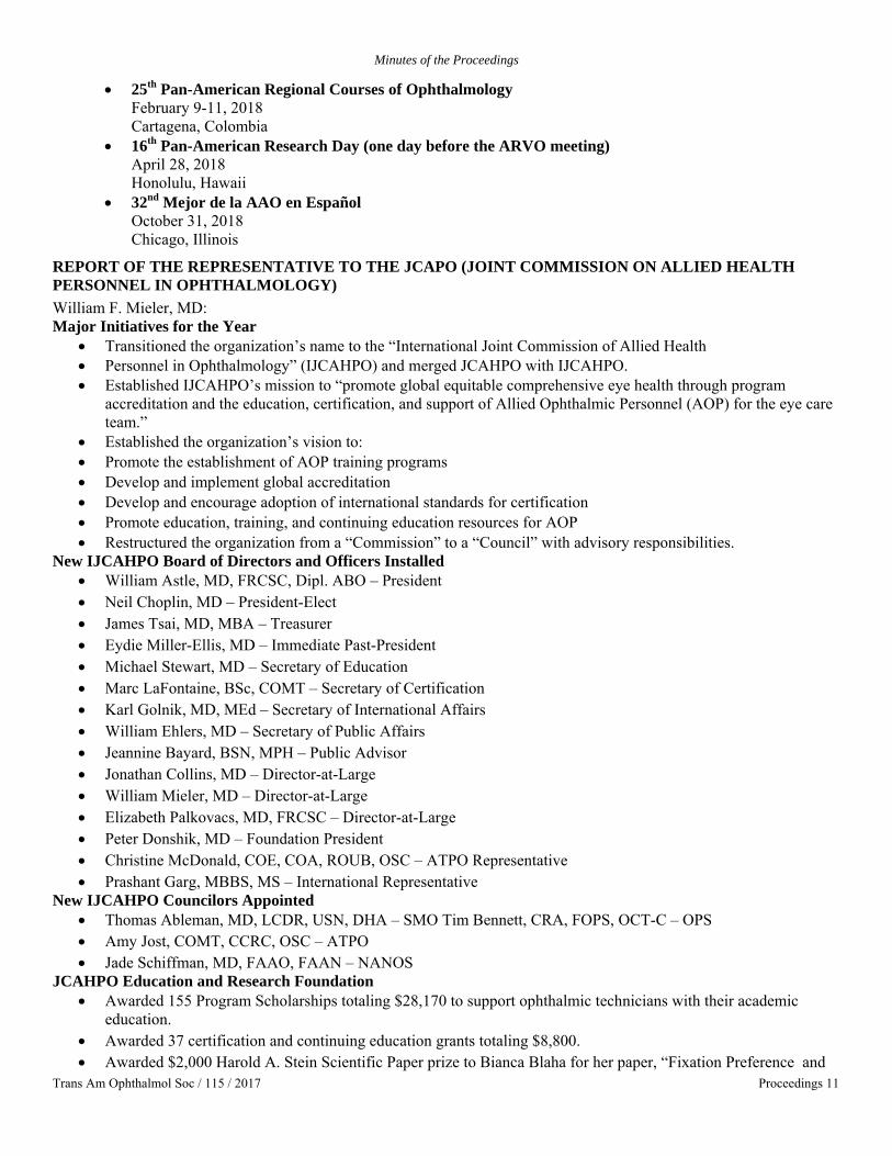
Minutes of the Proceedings
Trans Am Ophthalmol Soc / 115 / 2017 Proceedings 11
• 25th Pan-American Regional Courses of Ophthalmology February 9-11, 2018 Cartagena, Colombia
• 16th Pan-American Research Day (one day before the ARVO meeting) April 28, 2018 Honolulu, Hawaii
• 32nd Mejor de la AAO en Español October 31, 2018 Chicago, Illinois
REPORT OF THE REPRESENTATIVE TO THE JCAPO (JOINT COMMISSION ON ALLIED HEALTH PERSONNEL IN OPHTHALMOLOGY) William F. Mieler, MD: Major Initiatives for the Year
• Transitioned the organization’s name to the “International Joint Commission of Allied Health • Personnel in Ophthalmology” (IJCAHPO) and merged JCAHPO with IJCAHPO. • Established IJCAHPO’s mission to “promote global equitable comprehensive eye health through program
accreditation and the education, certification, and support of Allied Ophthalmic Personnel (AOP) for the eye care team.”
• Established the organization’s vision to: • Promote the establishment of AOP training programs • Develop and implement global accreditation • Develop and encourage adoption of international standards for certification • Promote education, training, and continuing education resources for AOP • Restructured the organization from a “Commission” to a “Council” with advisory responsibilities.
New IJCAHPO Board of Directors and Officers Installed • William Astle, MD, FRCSC, Dipl. ABO – President • Neil Choplin, MD – President-Elect • James Tsai, MD, MBA – Treasurer • Eydie Miller-Ellis, MD – Immediate Past-President • Michael Stewart, MD – Secretary of Education • Marc LaFontaine, BSc, COMT – Secretary of Certification • Karl Golnik, MD, MEd – Secretary of International Affairs • William Ehlers, MD – Secretary of Public Affairs • Jeannine Bayard, BSN, MPH – Public Advisor • Jonathan Collins, MD – Director-at-Large • William Mieler, MD – Director-at-Large • Elizabeth Palkovacs, MD, FRCSC – Director-at-Large • Peter Donshik, MD – Foundation President • Christine McDonald, COE, COA, ROUB, OSC – ATPO Representative • Prashant Garg, MBBS, MS – International Representative
New IJCAHPO Councilors Appointed • Thomas Ableman, MD, LCDR, USN, DHA – SMO Tim Bennett, CRA, FOPS, OCT-C – OPS • Amy Jost, COMT, CCRC, OSC – ATPO • Jade Schiffman, MD, FAAO, FAAN – NANOS
JCAHPO Education and Research Foundation • Awarded 155 Program Scholarships totaling $28,170 to support ophthalmic technicians with their academic
education. • Awarded 37 certification and continuing education grants totaling $8,800. • Awarded $2,000 Harold A. Stein Scientific Paper prize to Bianca Blaha for her paper, “Fixation Preference and
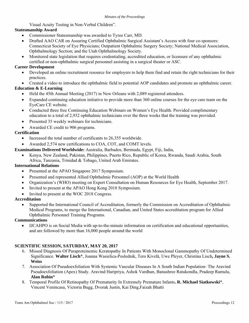
Minutes of the Proceedings
Trans Am Ophthalmol Soc / 115 / 2017 Proceedings 12
Visual Acuity Testing in Non-Verbal Children”. Statesmanship Award
• Commissioner Statesmanship was awarded to Tyree Carr, MD. • Drafted AAO CAR on Assuring Certified Ophthalmic Surgical Assistant’s Access with four co-sponsors:
Connecticut Society of Eye Physicians; Outpatient Ophthalmic Surgery Society; National Medical Association, Ophthalmology Section; and the Utah Ophthalmology Society.
• Monitored state legislation that requires credentialing, accredited education, or licensure of any ophthalmic certified or non-ophthalmic surgical personnel assisting in a surgical theater or ASC.
Career Development • Developed an online recruitment resource for employers to help them find and retain the right technicians for their
practices. • Created a video to introduce the ophthalmic field to potential AOP candidates and promote an ophthalmic career.
Education & E-Learning • Held the 45th Annual Meeting (2017) in New Orleans with 2,089 registered attendees. • Expanded continuing education initiative to provide more than 360 online courses for the eye care team on the
EyeCare CE website. • Conducted three free Continuing Education Webinars on Women’s Eye Health. Provided complimentary
education to a total of 2,932 ophthalmic technicians over the three weeks that the training was provided. • Presented 35 weekly webinars for technicians. • Awarded CE credit to 906 programs.
Certification • Increased the total number of certificants to 26,355 worldwide. • Awarded 2,574 new certifications to COA, COT, and COMT levels.
Examinations Delivered Worldwide: Australia, Barbados, Bermuda, Egypt, Fiji, India, • Kenya, New Zealand, Pakistan, Philippines, Puerto Rico, Republic of Korea, Rwanda, Saudi Arabia, South
Africa, Tanzania, Trinidad & Tobago, United Arab Emirates. International Relations
• Presented at the APAO Singapore 2017 Symposium. • Presented and represented Allied Ophthalmic Personnel (AOP) at the World Health • Organization’s (WHO) meeting on Expert Consultation on Human Resources for Eye Health, September 2017. • Invited to present at the APAO Hong Kong 2018 Symposium. • Invited to present at the WOC 2018 Congress.
Accreditation • Supported the International Council of Accreditation, formerly the Commission on Accreditation of Ophthalmic
Medical Programs, to merge the International, Canadian, and United States accreditation program for Allied Ophthalmic Personnel Training Programs.
Communications • IJCAHPO is on Social Media with up-to-the-minute information on certification and educational opportunities,
and are followed by more than 16,000 people around the world
SCIENTIFIC SESSION, SATURDAY, MAY 20, 2017 6. Missed Diagnosis Of Paraproteinemic Keratopathy In Patients With Monoclonal Gammopathy Of Undetermined
Significance. Walter Lisch*, Joanna Wasielica-Poslednik, Tero Kivelä, Uwe Pleyer, Christina Lisch, Jayne S. Weiss
7. Association Of Pseudoexfoliation With Systemic Vascular Diseases In A South Indian Population- The Aravind Pseudoexfoliation (Apex) Study. Aravind Haripriya, Ashok Vardhan, Banushree Ratukondla, Pradeep Ramulu, Alan Robin*
8. Temporal Profile Of Retinopathy Of Prematurity In Extremely Premature Infants. R. Michael Siatkowski*, Vincent Venincasa, Victoria Bugg, Dvorak Justin, Kai Ding,Faizah Bhatti
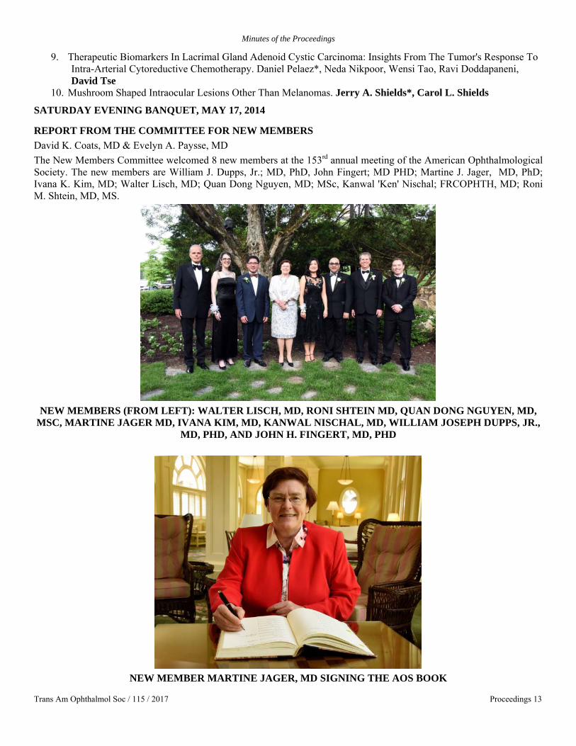
Minutes of the Proceedings
Trans Am Ophthalmol Soc / 115 / 2017 Proceedings 13
9. Therapeutic Biomarkers In Lacrimal Gland Adenoid Cystic Carcinoma: Insights From The Tumor's Response To Intra-Arterial Cytoreductive Chemotherapy. Daniel Pelaez*, Neda Nikpoor, Wensi Tao, Ravi Doddapaneni, David Tse
10. Mushroom Shaped Intraocular Lesions Other Than Melanomas. Jerry A. Shields*, Carol L. Shields
SATURDAY EVENING BANQUET, MAY 17, 2014
REPORT FROM THE COMMITTEE FOR NEW MEMBERS David K. Coats, MD & Evelyn A. Paysse, MD The New Members Committee welcomed 8 new members at the 153rd annual meeting of the American Ophthalmological Society. The new members are William J. Dupps, Jr.; MD, PhD, John Fingert; MD PHD; Martine J. Jager, MD, PhD; Ivana K. Kim, MD; Walter Lisch, MD; Quan Dong Nguyen, MD; MSc, Kanwal 'Ken' Nischal; FRCOPHTH, MD; Roni M. Shtein, MD, MS.
NEW MEMBERS (FROM LEFT): WALTER LISCH, MD, RONI SHTEIN MD, QUAN DONG NGUYEN, MD,
MSC, MARTINE JAGER MD, IVANA KIM, MD, KANWAL NISCHAL, MD, WILLIAM JOSEPH DUPPS, JR., MD, PHD, AND JOHN H. FINGERT, MD, PHD
NEW MEMBER MARTINE JAGER, MD SIGNING THE AOS BOOK
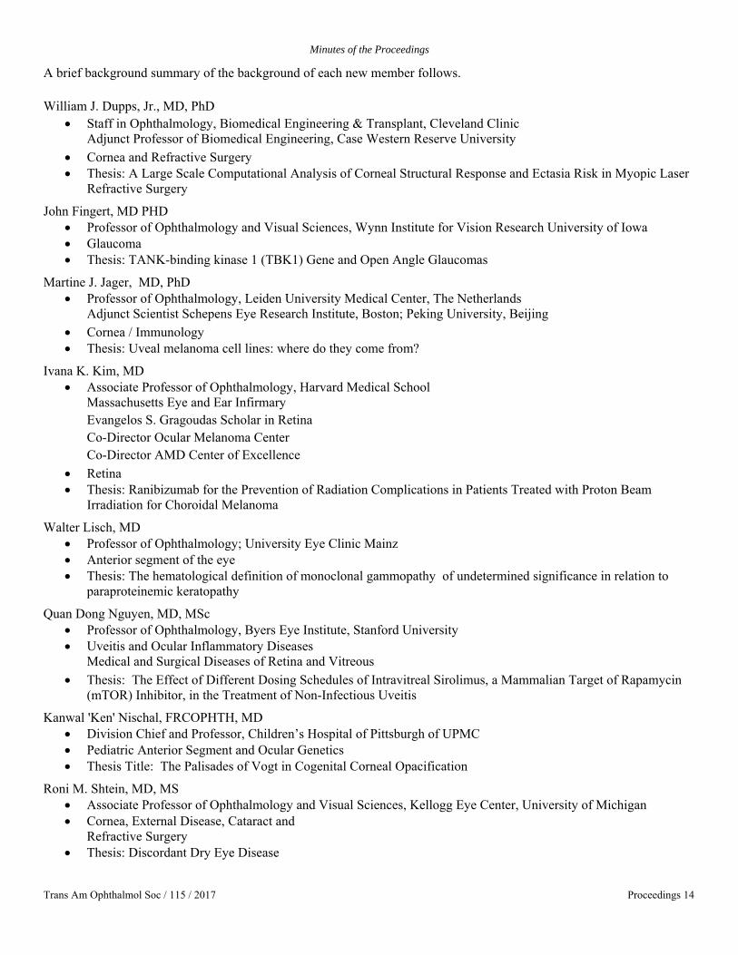
Minutes of the Proceedings
Trans Am Ophthalmol Soc / 115 / 2017 Proceedings 14
A brief background summary of the background of each new member follows. William J. Dupps, Jr., MD, PhD
• Staff in Ophthalmology, Biomedical Engineering & Transplant, Cleveland Clinic Adjunct Professor of Biomedical Engineering, Case Western Reserve University
• Cornea and Refractive Surgery • Thesis: A Large Scale Computational Analysis of Corneal Structural Response and Ectasia Risk in Myopic Laser
Refractive Surgery
John Fingert, MD PHD • Professor of Ophthalmology and Visual Sciences, Wynn Institute for Vision Research University of Iowa • Glaucoma • Thesis: TANK-binding kinase 1 (TBK1) Gene and Open Angle Glaucomas
Martine J. Jager, MD, PhD • Professor of Ophthalmology, Leiden University Medical Center, The Netherlands
Adjunct Scientist Schepens Eye Research Institute, Boston; Peking University, Beijing • Cornea / Immunology • Thesis: Uveal melanoma cell lines: where do they come from?
Ivana K. Kim, MD • Associate Professor of Ophthalmology, Harvard Medical School
Massachusetts Eye and Ear Infirmary Evangelos S. Gragoudas Scholar in Retina Co-Director Ocular Melanoma Center Co-Director AMD Center of Excellence
• Retina • Thesis: Ranibizumab for the Prevention of Radiation Complications in Patients Treated with Proton Beam
Irradiation for Choroidal Melanoma
Walter Lisch, MD • Professor of Ophthalmology; University Eye Clinic Mainz • Anterior segment of the eye • Thesis: The hematological definition of monoclonal gammopathy of undetermined significance in relation to
paraproteinemic keratopathy
Quan Dong Nguyen, MD, MSc • Professor of Ophthalmology, Byers Eye Institute, Stanford University • Uveitis and Ocular Inflammatory Diseases
Medical and Surgical Diseases of Retina and Vitreous • Thesis: The Effect of Different Dosing Schedules of Intravitreal Sirolimus, a Mammalian Target of Rapamycin
(mTOR) Inhibitor, in the Treatment of Non-Infectious Uveitis
Kanwal 'Ken' Nischal, FRCOPHTH, MD • Division Chief and Professor, Children’s Hospital of Pittsburgh of UPMC • Pediatric Anterior Segment and Ocular Genetics • Thesis Title: The Palisades of Vogt in Cogenital Corneal Opacification
Roni M. Shtein, MD, MS • Associate Professor of Ophthalmology and Visual Sciences, Kellogg Eye Center, University of Michigan • Cornea, External Disease, Cataract and
Refractive Surgery • Thesis: Discordant Dry Eye Disease

Minutes of the Proceedings
Trans Am Ophthalmol Soc / 115 / 2017 Proceedings 15
KNAPP SYMPOSIUM PARTICIPANTS (FROM LEFT): KEVIN K. PARK, PHD, HARRY QUIGLEY, MD,
JOSHUE R. SANES, PHD, DAVID T. TSE, MD AND ZHIGANG HE, PHD
REPORT FROM THE ATHLETIC COMMITTEE: Woodford Van Meter, MD: The AOS homecoming to the Homestead Resort in Hot Springs, VA after a thirteen year hiatus was celebrated with a full complement of athletic events and spectacular weather. The athletic events are somewhat unique to the AOS, where members young and old, new and emeritus, can compete in a spirited and friendly athletic competition and get to know one another in a casual and informal setting outside meeting halls and classrooms. The collection of over twenty silver trophies, many of them sterling and some of them hand crafted, were initiated during friendly contests at the Homestead and now reside in the AAO museum in San Francisco. “Virtual trophies” today are awarded to the winners of the different contests, but the winners’ names today go with the list of names of previous winners on the trophies that connect us all to an illustrious past.
Men’s and ladies golf was played under full sunshine on the Cascades Golf Course, designed by William Flynn (Shinnecock Hills, the Country Club Brookline, Merion) in 1923 and has been rated at times the #1 course in Virginia.
Men’s and ladies tennis was played on the Homestead clay courts, in the shadow of tradition, where AOS members have contested tennis fifty-four times over the last century.
Social tennis was enjoyed by all, except by the AOS ladies, who for the first time in 153 years failed to muster a single female for mixed doubles. Both skeet shooting and fly fishing were held in the grand Homestead tradition on the same facilities that date back to the inception of the trophies nearly 100 years ago.
An increasingly popular activity, the Susan Day Artistic Soiree, followed the AOS Talent Show on Friday and generated three teams of outstanding talent. After a spirited performance that showcased the breadth and depth of AOS talent, an impartial and completely unbiased set of judges awarded the Athletes Team the Golden Voice Award.
TROPHIES IN GOLF Men
Mishima-Michels Trophy Low Gross Woody Van Meter Canada-McCullough Cup Low Net David Wallace Truhlsen Trophy Senior Low Net Malcolm Mazow Knapp Trophies Random Draw Team Tony Arnold Peter Netland
Women Ellsworth Trophy Low Gross Tamara Fountain

Minutes of the Proceedings
Trans Am Ophthalmol Soc / 115 / 2017 Proceedings 16
Homestead-Callaway Cup Low Net Martine Jager Dubious Awards from the Athletics Director
Long Drive - Men Paul Lichter Closest to the Pin – Men David Wallace Long Drive – Senior Men Joe Flanagan Closest to the Pin – Senior Men Dan Durrie Closest to the Pin – Ladies #1 Erin Stahl Closest to the Pin – Ladies #2 Martine Jager Worst Sand Shot Ever Peter Netland
TROPHIES IN TENNIS Men
EVLBrown Bowl Doubles Winners David Sarraf Jim Tsai Brown-Wilson Tray Doubles Runners-up Bill Coles Woody Van Meter Michels-Wilkinson Trophy Most games won- Senior Sloan Wilson
Ladies Perera Bowl Doubles Winners Deena Laties Erin Stahl Hughes Bowl Doubles Runners up Tamara Fountain Alice Wilkinson
Mixed Doubles Wong McDonald Bowl Mixed Doubles Winners No Winner* Wilson Trophy Mixed Doubles Runners-up No winner* *No ladies showed up to play
TROPHY IN SKEET SHOOTING Beetham – Bullock Trophy Most targets hit Tim Stout Ken Wright
DUBIOUS AWARDS FROM THE ATHLETICS DIRECTOR AOS Sportsmanship Award Tennis - Men Sloan Wilson AOS Sportsmanship Award Tennis - Ladies Alice Wilkinson AOS Worst Sportsmanship Ever Award The Ladies of AOS (who failed to show for mixed doubles)
ARTISTIC SOIREE Golden Voice Award The Athletes
2018 MEETING AT DANA POINT, CA Make plans now to attend the 2018 AOS meeting at the luxurious St. Regis Monarch Beach Resort at Dana Point, CA. The AOS met here seven years ago and the facilities are exceptional.
The meeting will offer the usual AOS activities, plus ocean and southern California weather. Golf, tennis, sailing, whale and dolphin watching, and beach access are available onsite at the St. Regis. Golf will be played on the adjacent Monarch Beach Links Golf Course, rated the #1 public course in Orange County. The 18-hole golf course is conveniently located next to the hotel property, and has recently gone through a complete renovation of both the golf course and the pro shop. The golf course is in immaculate condition with beautiful scenery and has two holes on the ocean, offering multiple tee boxes to accommodate golfers of all talents. Tennis will be offered at the private Tennis Club at Monarch Beach, across the street from the St. Regis. The Monarch Beach Tennis Club has 8 championship hard courts, plus ball machines, hitting walls and a fully stocked tennis pro shop. Whale and dolphin watching, sailing, and ocean and beach activities are available. Combine these activities with southern California weather and an ocean setting, and this meeting should be fabulous. Hope to see you there!

Minutes of the Proceedings
Trans Am Ophthalmol Soc / 115 / 2017 Proceedings 17
2017 FREDERICK C. BLODI LECTURER JOSEPH CAPRIOLI, MD
REPORT FROM THE COMMITTEE ON PRIZES C. P. Wilkinson, MD: The Lucien Howe Medal is the highest honor that the American Ophthalmological Society (AOS) can bestow. Susan Day, MD, Dan Jones, MD, and I were this year’s members of the Committee on Prizes, on which I served as Chair. We each reviewed the list of members of AOS while acknowledging that non-members were also eligible. We solicited and reviewed additional nominations from the membership and then voted on two occasions: first, we produced a proverbial “short list” and then selected (unanimously) an awardee, George L. Spaeth, MD.
Dr. Spaeth, a genuine Renaissance man, has enjoyed a career on many fronts. A native of Philadelphia and the son of an ophthalmologist, he was introduced to medicine at an early age but entered Yale with greater interests in music and history and graduated Summa Cum Laude in history while earning Phi Beta Kappa recognition. At Harvard Medical School, his MD degree was awarded Cum Laude, and he became a member of AOA. Following an internship at the University of Michigan, he began his residency at Wills and then joined the clinical faculty at the National Eye Institute. This latter move was serendipitous, as most slots for clinical fellows were full, with Dick Green in pathology, Vernon Wong in Immunology, and Ron Carr (all future AOS members) in attendance. Fortunately for ophthalmology, and presumable for George, a position on the glaucoma service was available, and as they say, “the rest is history”. He then returned to Philadelphia and joined his father in practice for three years before returning to Wills and heading the Glaucoma Service. In 2000, he became the Louis J. Esposito Glaucoma Research Professor at Wills. His academic achievements began when he was a resident when in 1966 when he and G.W. published a classic paper on homocystinuria, and he subsequently has published over 400 manuscripts in peer-reviewed journals, approximately 100 book chapters, and 18 books, several of which have been translated into foreign languages. Most of these have been on glaucoma topics, but many have included ethical issues and quality-of-life subjects. He has trained over 34 fellows who now practice in many countries on six continents. He was a founding member of the American Glaucoma Society (AGS) and served as its first president. He has received numerous awards, including medals from many international glaucoma societies, the Weisenfeld Award from ARVO (2012), and the Outstanding Educator Award form the AGS (2017). But his interests and participation are not limited to medical topics, as he is an award-winning gardener, a poet, fiction writer, photographer, composer, pianist, and organist. He is an Emeritus member of the Board of Directors of both the Pennsylvania Ballet and Philadelphia Bach Festival.
Clearly this gentleman is, as noted, a genuine Renaissance Man, one whose award was clearly a delight to the audience at the 2017 AOS meeting at the Homestead.

Minutes of the Proceedings
Trans Am Ophthalmol Soc / 115 / 2017 Proceedings 18
2017 LUCIEN HOWE MEDALIST GEORGE L. SPAETH, MD
SCIENTIFIC SESSION, SUNDAY, MAY 21, 2017
11. Quality Metrics And Retinal Detachment Surgery: Time To Unplanned Return To The Operating Room. Sophie Bakri*, Alexander Grosinger, Benjamin Nicholson, Andrew Barkmeier, Raymond Iezzi
12. Is Discriminant Score Associated With GEP Class In Decisiondx-UM Test Important Prognostically? James J. Augsburger*, Zelia M. Correa
13. Comparative Effectiveness And Cost-Effectiveness Analyses Of The Prostamides And Timolol For Open Angle Glaucoma. Gary Brown*, Melissa Brown, Heidi Lieske
14. En Face OCT And OCT Angiography Of Perivenular Ischemia Associated With Central Retinal Vein Occlusion. David Sarraf*, Khalil Falavarjani, Nopasak Phasukkijwatana, Emmett Cunningham Jr., Ananda Kalevar, Rosa Dolz Marco, Irena Tsui, Richard Rosen, K. Bailey Freund, Lee Jampol, SriniVas Sadda
15. The Prognostic Value Of Chromosome Status In Uveal Melanoma Is Enhanced By Adding Ajcc Stage. Martine Jager*, Mehmet Dogrusoz, Mette Bagger, Gregorius Luyten, Jens Kiilgaard
Minutes of the Proceedings
Trans Am Ophthalmol Soc / 115 / 2017 Proceedings 19
16. Glaucoma In High Myopia And Parapapillary Delta Zone. Jost Jonas*, Kyoko Ohno-Matsui 17. Spectral Domain Optical Coherence Tomography Angiography In Children With Amblyopia. Stacy Pineles*,
Marcela Lonngi, Irina Tsui, Federico Velez, David Sarraf 18. Evaluation Of The Runge Near Card For Standardized Visual Acuity Assessment. Matthew Cooke, Patricia
Winter, McKenney Kaitlin, Krissa Packard, Vesper Williams, Dorsey Eleanor, Aniko Szabo, Alexis Visotcky, William Wirostko, David Weinberg, Judy Kim, Edward Barnett, Dennis Han*
19. Accuracy Of Imaging Modalities In Differentiating Pseudopapilledema From True Optic Disk Edema (ODE) In Children. Melinda Chang, Federico Velez, Joseph Demer, Laura Bonelli, Peter Quiros, Alfredo Sadun, Stacy Pineles, Anthony Arnold*
Members registered for the 2017 meeting. 15 professional guests are at the end of the list.
Anthony Arnold Active Member Penny Asbell Active Member James Augsburger Active Member Dimitri Azar Active Member Sophie Bakri Active Member George Bartley Active Member Albert Biglan Emeritus Member Preston Blomquist Active Member Gary Brown Active Member David Browning Active Member Edward Buckley Active Member Donald Budenz Active Member John Bullock Emeritus Member Massimo Busin Active Member J. Douglas Cameron Active Member Louis Cantor Active Member Joseph Caprioli Active Member Robison Chan Active Member Clement Chan Active Member Emily Chew Active Member Michael Chiang Active Member James Chodosh Active Member George Cioffi Active Member John Clarkson Active Member David Coats Active Member D. Jackson Coleman Emeritus Member Anne Coleman Active Member Janet Davis Active Member Susan Day Active Member William Dupps Active Member Daniel Durrie Active Member Ralph Eagle Active Member Deepak Edward Active Member John Fingert Active Member Joseph Flanagan Emeritus Member Christina Flaxel Active Member C Stephen Foster Emeritus Member Tamara Fountain Active Member Thomas France Emeritus Member Alan Friedman Active Member Evangelos Gragoudas Active Member Ronald Gross Active Member Hans Grossniklaus Active Member Froncie Gutman Emeritus Member Dennis Han Active Member Mary Hartnett Active Member
Peter Hersh Active Member Eric Holz Active Member Mark Humayun Active Member Malcolm Ing Active Member Douglas Jabs Active Member Martine Jager Active Member Lee Jampol Emeritus Member William Jarrett Emeritus Member David Johnson Active Member Jost Jonas Active Member Ivana Kim Active Member James Kinyoun Active Member Peter Laibson Emeritus Member Vinod Lakhanpal Active Member Alan Laties Emeritus Member Mary Lawrence Active Member Paul Lichter Emeritus Member Walter Lisch Active Member Irene Ludwig Active Member Marian Macsai Active Member Edward Manche Active Member Malcolm Mazow Emeritus Member Timothy McCulley Active Member Marguerite McDonald Active Member Marilyn Mets Active Member William Mieler Active Member Joan Miller Active Member Richard Mills Active Member Paul Mitchell Active Member Peter Netland Active Member Steve Newman Active Member Quan Nguyen Active Member Verinder Nirankari Active Member Kanwal Nischal Active Member T. Michael Nork Active Member Timothy Olsen Active Member David Parke Active Member Richard Parrish Active Member Cameron Parsa Active Member Leonard Parver Active Member Louis Pasquale Active Member Jose Pulido Active Member Donald Puro Active Member Edward Raab Active Member Christopher Rapuano Active Member James Reynolds Active Member Alan Robin Active Member Paul Runge Active Member

Minutes of the Proceedings
Trans Am Ophthalmol Soc / 115 / 2017 Proceedings 20
David Sarraf Active Member Daniel Schaefer Active Member David Schanzlin Active Member Oliver Schein Active Member Hermann Schubert Active Member Ivan Schwab Active Member Mark Sherwood Active Member Jerry Shields Active Member Carol Shields Active Member Roni Shtein Active Member R. Michael Siatkowski Active Member Rona Silkiss Active Member Alfred Sommer Emeritus Member George Spaeth Active Member Rand Spencer Active Member Bruce Spivey Emeritus Member David Stager Active Member Joshua Stein Active Member Edwin Stone Active Member J. Timothy Stout Active Member Mark Terry Active Member James Tsai Active Member David Tse Active Member Lawrence Tychsen Active Member Woodford Van Meter Active Member David Wallace Active Member Robert Waller Emeritus Member Charles Wilkinson Emeritus Member R. Sloan Wilson Emeritus Member Steven Wilson Active Member David Wilson Active Member M. Roy Wilson Active Member Kenneth Wright Active Member R. Patrick Yeatts Active Member Marco Zarbin Active Member
Professional Guests Gary Brown Professional Guest R. Mark Hatfield Professional Guest Zhigang He Professional Guest Paul Lee Professional Guest W. Barry Lee Professional Guest Walter Lisch Professional Guest Sonia Mehta Professional Guest Kyung Park Professional Guest Daniel Pelaez Professional Guest Stacy Pineles Professional Guest Harry Quigley Professional Guest Joshua Sanes Professional Guest Julia Shulman Professional Guest Michael Stewart Professional Guest Bryan Winn Professional Guest

Trans Am Ophthalmol Soc / 115 / 2017 Proceedings 21
PAPER ABSTRACTS

Trans Am Ophthalmol Soc / 115 / 2017 Proceedings 22
IMPACT OF RESIDENT AND FELLOW TRAINEES ON PATIENT ENCOUNTER LENGTH IN AN ACADEMIC OPHTHALMOLOGY CENTER Michael Chiang*, Isaac Goldstein, Sarah Read-Brown, Michelle Hribar Purpose: Although presence of resident and fellow trainees is a characteristic feature of academic medical centers, little research has examined their impact on workflow. Meanwhile, physician reimbursement models are increasingly based on metrics such as quality and efficiency of care. The purpose of this study is to examine the impact of trainees on patient encounter length at an academic ophthalmology center.
Methods: The EHR enterprise reporting system (Epic; Verona, WI) was used to collect data on all outpatient encounters in 2014 (n=49,644) by 33 faculty providers with stable practices at an academic center (OHSU Casey Eye Institute). Patient check-in time, check-out time, and trainee presence during encounters were derived from the EHR using previously-published methods. We used t-tests and developed a linear mixed model to analyze the relationship between encounter length and presence of trainees.
Results: Encounters where trainees were involved were significantly longer (102.0 ± 56.5 minutes, n=15,628) than those without trainees (81.9 ± 45.7 minutes, n=34,016) (p<2.2E-16). During clinic sessions where trainees were present, the specific encounters without trainees involved were significantly longer (85.2 ± 47.3 minutes, n=15,792) than encounters from clinic sessions where no trainees were present (79.1 ± 44.2 minutes, n=18,224) (P<2.2E-16). Linear mixed model analysis showed that encounter length was increased 11.8 minutes by presence of a fellow, 14.9 minutes by presence of a resident, and 25.5 minutes by presence of both (p<.0001 vs. sessions and encounters without trainees), and that presence of a trainee in the clinic session increased encounter length by 1.8 minutes in specific encounters where the trainee was not involved (p=.003).
Conclusion: Presence of resident and fellow trainees in a clinic session is associated with increased length of outpatient ophthalmology encounters, both in encounters where the trainee is involved and even in encounters where the trainee is not involved. This has important implications for clinical efficiency, teaching models, and reimbursement models.
GENETIC ANALYSIS OF 1000 CONSECUTIVELY ASCERTAINED FAMILIES WITH INHERITED RETINAL DISEASE Edwin Stone*, Jeaneen Andorf, Adam DeLuca, Scott Whitmore, Joseph Giacalone, Luan Streb, Terry Braun, Robert Mullins, Todd Scheetz, Val Sheffield, Budd Tucker
Purpose: To devise and evaluate a strategy for genetic testing of patients with inherited retinal diseases that maximizes sensitivity and statistical significance while minimizing cost. A secondary purpose was to identify the fraction of disease caused by each gene to aid in the design of a comprehensive strategy for treating every individual affected with one of these disorders.
Methods: 1000 consecutive families diagnosed with an inherited retinal disease by a single physician (EMS) were studied. Patients were divided into 62 different clinical categories based on their medical history and examination findings. A focused genetic test consisting of conventional DNA sequencing and/or allele-specific testing was devised for each of the 62 clinical categories. When the focused test was negative, whole exome and/or whole genome sequencing was performed.
Results: 65% of the families were diagnosed with a photoreceptor disorder and 28% with a macular dystrophy. The remaining 7% were distributed among heritable tumors, vitreoretinopathies, foveal hypoplasia, retinoschisis and choroidopathies. Disease-causing genotypes were identified in 762 families (76.2%) and were distributed across 104 different genes. The most common disease-causing gene was ABCA4 (176 families) while 31 of the genes caused disease in only a single family. Clinically focused conventional testing can detect mutations in 57.9% of the families for an average research cost of $450 per family while exome and genome sequencing can detect mutations in an additional 18.1% and 0.2% of families for $1200 and $2500 respectively.
Conclusion: A clinically focused, sequential testing strategy is more sensitive and less expensive than a strategy based entirely on next generation sequencing. The narrower pretest hypothesis associated with the sequential strategy also results in a much lower false discovery rate than strategies that evaluate dozens or hundreds of genes in every patient. Very low false discovery rates will be essential for clinical trials of gene replacement therapy.
CONJUNCTIVAL TUMORS IN CHILDREN IN 806 CASES. FEATURES DIFFERENTIATING BENIGN FROM MALIGNANT TUMORS. Carol L. Shields*, Kareem Sioufi, Adel E. Alset, Emil Anthony T. Say, Jerry A. Shields Purpose: To evaluate the clinical features of conjunctival tumors in children with comparison of benign versus (vs) malignant counterparts.
Methods: Retrospective case series.
Paper Abstracts
Trans Am Ophthalmol Soc / 115 / 2017 Proceedings 23
Results: Of 806 eyes with a conjunctival tumor, the lesion was melanocytic (n=553, 69%) or non-melanocytic (n=253, 31%). Of 553 melanocytic lesions, the most common tumor diagnosis was nevus (89%), primary acquired melanosis (5%), and melanoma (3%). Of 253 non-melanocytic lesions, the leading diagnosis was benign reactive lymphoid hyperplasia (BRLH) (15%), dermoid, (12%), dermolipoma (10%), lymphangioma (5%), capillary hemangioma (5%), and conjunctivitis with nodule simulating tumor (12%). Overall, the tumor was benign in 97% and malignant in 3%, with malignancy including melanoma (2.2%) and lymphoma (1.1%). The mean age at detection of benign vs malignant tumor was 11 vs 14 years (p=0.0052). The relative frequency of any malignancy (per all conjunctival tumors) by age bracket (0-5, 5-10, 10-15, and 15-21 years) was 1%, 2%, 3%, and 7%. Regarding tumor per race (Caucasian vs African American vs Asian vs Hispanic), the findings included conjunctival nevus (83% vs 6% vs 8% vs 3%), melanoma (89% vs 11% vs 0% vs 0%), BRLH (74% vs 18% vs 0% vs 8%), and lymphoma (56% vs 44% vs 0% vs 0%). A comparison (nevus vs melanoma) found significant (p<0.05) differences with melanoma in older age bracket (RR=4.80), with greater tumor thickness (RR=1.14), greater base, (RR=4.92), tumor hemorrhage (RR=25.30) and lacking intrinsic cysts (RR=5.06). These features predictive of conjunctival melanoma in children can be remembered by the mnemonic CATCH Melanoma representing Children Age older, Thickness/Base greater, Cyst lacking, Hemorrhage for Melanoma. A comparison (BRLH vs lymphoma) revealed lymphoma with significantly larger basal dimension (RR=5.16) and location as diffuse, inferior, or superior vs nasal (RR=16.5, 12.38, 8.25, respectively).
Conclusion: Conjunctival tumors in children are generally benign (97%) and the top five tumors included conjunctival nevus, BRLH, primary acquired melanosis, dermoid, and dermolipoma. Clinical features can assist in differentiating benign from malignant counterparts.
LIMBAL AND CONJUNCTIVAL TRANSPLANTATION FROM THE SAME LIVING-RELATED BONE MARROW DONOR TO PATIENTS WITH OCULAR GRAFT VERSUS HOST DISEASE Massimo Busin*, Giuseppe Giannaccare, Laura Sapigni, Emilio Campos Purpose: To investigate the outcomes of conjunctival and limbal transplantation from the same living-related bone marrow donor to eyes with severe ocular graft versus host disease (GVHD).
Methods: A 10 x 5 mm graft, including bulbar conjunctiva and limbus, was transplanted from the same living-related bone marrow donor into eyes with severe GVHD. Postoperative treatment included only topical steroids and tetracycline ointment for 3 months. No systemic immunosuppressive therapy was given. Ocular surface disease index (OSDI), visual acuity and Schirmer test type I were evaluated preoperatively as well as 1, 3, 6 and 12 months postoperatively. In addition, 1 year after surgery conjunctival and corneal cells from the eye of a female patient with donor/recipient sex-mismatch were submitted to fluorescence in situ hybridization (FISH) analysis with X and Y chromosome probes.
Results: The donor tissue was grafted successfully and remained vital for the whole follow-up time in all 4 eyes of 2 patients who had undergone CLT. Also the eyes of the living-related donors healed without complications. OSDI score was above 60 in both patients before CLT and improved to 16 and 50 respectively, 6 months after CLT. Preoperative vision was 20/400 or less in all eyes and improved to 20/200 or better in all but one eye, which had advanced corneal keratinization. In one eye vision improved from 20/400 before CLT to 20/25 six months after CLT. In all eyes Schirmer test type I was below 1 mm preoperatively and improved to 5 mm or more 6 months postoperatively. In the eye tested, FISH analysis demonstrated the presence of cells with donor chromosomes as late as 1 year after CLT.
Conclusion: Conjunctival and limbal tissue transplanted from the same living-related bone marrow donor into eyes with severe GVHD survives in the recipient environment for at least 1 year in the absence of immunologic rejection. Vision in eyes without advanced corneal keratinization is substantially increased after CLT. Restoring normal tear secretion, CLT improves ocular surface conditions and may possibly allow successful corneal surgery, when required.
RANIBIZUMAB FOR THE PREVENTION OF RADIATION COMPLICATIONS IN PATIENTS TREATED WITH PROTON BEAM IRRADIATION FOR CHOROIDAL MELANOMA Ivana Kim* , Anne Marie Lane, Purva Jain, Caroline Awh, Evangelos Gragoudas Purpose: To investigate the safety and potential efficacy of ranibizumab for prevention of radiation complications in patients treated with proton irradiation for choroidal melanoma
Methods: Forty patients with tumors located within 2 disc diameters of the optic nerve and/or macula were enrolled in this open-label study. Participants received ranibizumab 0.5 mg or 1.0 mg at tumor localization and every 2 months thereafter for the study duration of 24 months. The incidence of adverse events, visual acuity, and other measures of ocular morbidity related to radiation complications were assessed. Historical controls with similar follow-up meeting the eligibility criteria for tumor size, location, and baseline visual acuity were assembled for comparison.
Paper Abstracts
Trans Am Ophthalmol Soc / 115 / 2017 Proceedings 24
Results: Fifteen patients with large tumors and 25 patients with small/medium tumors were enrolled. Thirty patients completed the month 24 visit. No serious ocular or systemic adverse events related to ranibizumab were observed. At 24 months, the proportion of patients with visual acuity ≥ 20/200 was 30/31 (97%) in the study group versus 92/205 (45%) in historical controls (P < .001). 24/31 (77%) versus 46/205 (22%) of controls had visual acuity ≥ 20/40 at 24 months (P <.001). Clinical evidence of radiation maculopathy at month 24 was seen in 8/24 (33%) patients with small/medium tumors versus 42/62 (68%) of controls (P = .004). Three patients with large tumors developed metastases.
Conclusion: In this small pilot study, prophylactic ranibizumab appears generally safe in patients treated with proton irradiation for choroidal melanoma. High rates of visual acuity retention were observed through 2 years.
MISSED DIAGNOSIS OF PARAPROTEINEMIC KERATOPATHY IN PATIENTS WITH MONOCLONAL GAMMOPATHY OF UNDETERMINED SIGNIFICANCE Walter Lisch*, Joanna Wasielica-Poslednik, Tero Kivelä, Uwe Pleyer, Christina Lisch, Jayne S. Weiss Purpose: To distinguish distinct bilateral corneal opacities of monoclonal gammopathies in cases initially misdiagnosed as hereditary or inflammatory diseases.
Methods: Slit-lamp examination of thirteen patients with completely distinct bilateral opacity patterns of the cornea. After weeks, months,and years of the first ophthalmological diagnosis a serum protein electrophoresis (SPE) was performed in twelve patients in order to rule out a monoclonal gammopathy of undetermined significance (MGUS). In one patient a penetrating keratoplasty and a DNA analysis had been performed prior to making the diagnosis of MGUS. In another patient with known biclonal gammopathy of undetermined significance a blood copper analysis was performed because of brownish discoloration at the level of Descemets mebrane.
Results: Initial ophthalmological diagnosis of all twelve patients had to be revised as distinct forms of paraproteinemic keratopathy (PPK) after SPE was performed: patient 1 cystinosis, SPE one year later: punctiform crystalline-like PPK; patient 2 Schnyder Corneal Dystrophy (CD), SPE few weeks later: comma-shaped crystalline PPK; patient 3 Lattice CD 1, SPE two years later: lattice-like PPK; patient 4 Granular CD, SPE one year later: peripheral granular-like PPK; patient 5 Peripheral keratitis, SPE one year later: peripheral inflammatory band-like PPK; patient 6 Reis-Bücklers CD, SPE three months later: geographic-like PPK; patient 7 Cornea farinata, SPE one month later: posterior flake-like PPK; patient 8 Stromal CD, SPE six years later: stromal flake-like PPK; light microscopy showed irregular extracellular deposits staining red with Masson trichrome. TGFBI-and decorin-genes were negative; patient 9 Peripheral keratitis/scleritis, SPE five years later: peripheral inflammatory PPK; patient 10 Lattice CD 1, SPE seven months later:lattice-like PPK; patient 11 Peripheral keratitis/scleritis, SPE two years later: peripheral inflammatory band-like PPK; patient 12 Stromal keratitis,SPE three years later: stromal punctiform-like PPK; patient 13 showed a syndrome that we propose to call Lewis syndrome which includes biclonal GUS and hypercupremia (1326yg/dL; normal range 76-152)+ discoid brownish discoloration at the level of Descemets membrane.
Conclusion: The ophthalmologist has the important responsibility to distinguish PPK vs distinct heritable and inflammatory corneal entities. Patients with distinct forms of bilateral corneal opacity and without any hint of inheritance should be analyzed by serum protein electrophoresis in order to rule out the chameleon-like PPK. The Lewis syndrome is to be differentiated vs Wilson`s disease.
ASSOCIATION OF PSEUDOEXFOLIATION WITH SYSTEMIC VASCULAR DISEASES IN A SOUTH INDIAN POPULATION- THE ARAVIND PSEUDOEXFOLIATION (APEX) STUDY Aravind Haripriya, Ashok Vardhan, Banushree Ratukondla, Pradeep Ramulu, Alan Robin* Purpose: To determine whether specific vascular risk factors and cardiac abnormalities are more common among PEX patients than non-PEX controls.
Methods: Design: Cross-sectional analysis of patients recruited into the APEX (Aravind Pseudoexfoliation) study (begun in 2011). Setting: Multi-centered study done at four tertiary Aravind Eye Hospitals in Tamil Nadu, India. Participants: Patients > 40 years with or without PEX, requiring cataract surgery were eligible. We enrolled 930 PEX and 476 non-PEX subjects and performed detailed ocular examinations of each subject including grading of specific ocular features reflecting PEX. We also evaluated for multiple systemic potential cardiovascular diseases and their risk factors. Main Outcome(s) and Measures: Blood pressure and prevalence of hypertension and diabetes. We also evaluated ECG findings, cholesterol levels, and homocysteine levels in subjects with and without PEX.
Results: The mean ages of PEX and non PEX patients were 64.8±6.8 and 59.9±7.3 years (p<0.001), respectively. PEX patients were more often male than non-PEX patients (54.7% vs 45.3%; p<0.001). In multivariable analyses adjusting for both age and sex, higher systolic blood pressure values were noted for PEX patients as compared to non-PEX patients, (Δ=+3.97mmHg; p=0.001, [95%CI 1.7 - 6.2]). Also, PEX patients were more likely to demonstrate an ECG abnormality than non-PEX patients (OR 1.64; [95% CI 1.04 - 2.60]). PEX material at pupil margin and pupillary ruff hypotrophy were the two physical findings specifically found to be associated

Paper Abstracts
Trans Am Ophthalmol Soc / 115 / 2017 Proceedings 25
with higher systolic BP and a greater likelihood of ECG abnormality (p<0.05 for both). PEX was not observed to be associated with a higher risk of diabetes, hypercholesterolemia or hyperhomocysteinemia (p >0.1 for all).
Conclusion: In South Indian patients requiring cataract surgery, PEX is associated with higher systolic BP and more frequent ECG abnormalities. Association of specific ocular PEX findings as markers for systemic comorbidities should be further investigated in the future studies. PEX patients may benefit from greater attention to BP and prevention of cardiovascular disease.
TEMPORAL PROFILE OF RETINOPATHY OF PREMATURITY IN EXTREMELY PREMATURE INFANTS R. Michael Siatkowski*, Vincent Venincasa, Victoria Bugg, Dvorak Justin, Kai Ding, Faizah Bhatti Purpose: Although benchmark studies on retinopathy of prematurity (ROP) (CRYO-ROP, ET-ROP, eROP, STOP-ROP) included extremely premature infants, none produced a detailed analysis of differences in the clinical course of ROP between extremely premature (gestational age <= 28 weeks) vs premature (gestational age >28-37 weeks) infants. The purpose of this study was to compare the temporal profile of ROP in premature vs extremely premature infants to determine whether a change in diagnostic examination criteria is required for this population.
Methods: This was a retrospective review of 301 patients/586 eyes (62/122 premature, 236/464 extremely premature) born from 2010-2015. ROP and various systemic data were collected from birth until spontaneous regression, treatment, or death.
Results: Extremely premature infants were diagnosed with ROP earlier than premature infants after adjusting for gestational age (33.6 vs 36.0 weeks, p < 0.0001), took 3.7 weeks (53%) longer to achieve regression (p < 0.0001), and were 3 times more likely to require treatment (29.7% vs 9.9%, p < 0.0001). Birthweight was not independent of gestation age as a risk factor in this population. Rate of weight gain was greater in extremely premature infants compared to premature infants after adjustment for birth weight, and independent of whether treatment was necessary.
Conclusion: Although extremely premature infants develop ROP earlier, have a longer temporal profile of disease, and are more likely to require treatment than premature infant, current AAP/AAO/AAPOS/AACO guidelines are sufficient to detect referral-warranted ROP in this population.
THERAPEUTIC BIOMARKERS IN LACRIMAL GLAND ADENOID CYSTIC CARCINOMA: INSIGHTS FROM THE TUMOR'S RESPONSE TO INTRA-ARTERIAL CYTOREDUCTIVE CHEMOTHERAPY Daniel Pelaez*, Neda Nikpoor, Wensi Tao, Ravi Doddapaneni, David Tse Purpose: To characterize the molecular response of lacrimal gland adenoid cystic carcinoma tissue to intra-arterial cytoreductive chemotherapy (IACC) in order to identify potential therapeutic biomarkers.
Methods: Patient pre-chemotherapy tumor biopsy samples and corresponding (paired) post-IACC resection specimen were identified and paraffin sectioned for microdissection (n=6). Full-length proteins were extracted and quantified. Lysates were assayed through unbiased proteomic screening. Concurrently, cell cultures from patient samples collected pre-, and post-chemotherapy were used to validate molecular targets and drug screening of these targets for cellular viability or cellular proliferation.
Results: Proteomic assay revealed several markers with statistically significant tendencies in all specimens. Apoptotic markers are upregulated and correlate well with TUNEL, Parp, and Caspase-3 immunohistochemistry. Similarly, markers indicative of a stem cell phenotype were also upregulated following chemotherapy. Bioinformatic analysis revealed the FGF signaling pathway as upregulated following chemotherapy. The FGF receptor 1 (FGFR1) was the most significantly upregulated protein across all samples following IACC. Immunohistochemistry confirmed this upregulation in post-chemo samples and in the cell cultures from patients collected after chemotherapy. Drug screening in LGACC cell lines showed that, while supplementation with an FGFR1&2 selective inhibitor reduced cellular proliferation in all cultures, post-chemo cultures were exquisitely sensitive to this inhibition. The use of other inhibitors did not affect cell growth or viability. Combinatorial drug studies has confirmed a synergistic effect between with the use of cisplatin and AZD4547.
Conclusion: This study validates the use of tissue response in the elucidation of novel therapeutic targets to augment the effects of current management of this lethal orbital tumor. Identification of mechanisms used to subvert chemotherapeutic toxicity, or the phenotype of chemoresistant cells, can help elaborate more effective treatment regimens. We advance the inhibition of FGFR as a novel adjuvant therapy to intra-arterial chemotherapy for LGACC. We are exploring this strategy in a pre-clinical xenograft model for this disease.

Paper Abstracts
Trans Am Ophthalmol Soc / 115 / 2017 Proceedings 26
MUSHROOM SHAPED INTRAOCULAR LESIONS OTHER THAN MELANOMAS Jerry A. Shields*, Carol L. Shields
Purpose: It is generally believed that a mushroom shaped intraocular lesion is highly suggestive, if not pathognomonic, of choroidal melanoma. The purpose of this presentation is to demonstrated conditions that can also assume a mushroom shape, suggesting a choroidal melanoma.
Methods: The files of an ocular oncology service were reviewed and a literature search done for lesions that can also assume a mushroom shape with ultrasonography.
Results: We identified 13 conditions other than melanoma that occasionally assumed a mushroom configuration. This included adenocarcinoma of pigment epithelium, choroidal metastasis, schwannoma, melanocytoma, choroidal hemangioma, recurrent retinoblastoma, macular degeneration, mycotic abscess, retinal vasoproliferative tumor, solitary fibrous tumor, fibrovascular proliferation, and dislocated lens.
Conclusion: Severe conditions other that melanoma can have a mushroom shape and should not be confused with melanoma.
QUALITY METRICS AND RETINAL DETACHMENT SURGERY: TIME TO UNPLANNED RETURN TO THE OPERATING ROOM Sophie Bakri*, Alexander Grosinger, Benjamin Nicholson, Andrew Barkmeier, Raymond Iezzi Purpose: To assess 45-day return to the operating room (ROR) as a quality metric in retinal detachment (RD) surgery.
Methods: Eyes with a new clinical diagnosis of untreated rhegmatogenous RD between January 2012 and June 2014 were identified using the institutional electronic medical record. Charts were reviewed to identify all subjects who returned to the operating room and the reason for their return. The primary outcome variable was 45-day ROR, and data was also collected on any ROR that occurred outside this 45-day period. Numerous clinical characteristics were analyzed for associations with ROR. For all ROR events, the charts were reviewed to identify possible causes for ROR.
Results: There were 307 previously untreated RDs identified; 220 were uncomplicated RDs (i.e. excluded eyes with proliferative vitreoretinopathy [PVR] and trauma-related detachment). The uncomplicated RD group had a 45-day ROR of 12/220 (5.5%). The ROR rate over the entire follow-up period was 23/220 (10.5%). The mean final visual acuity in 45 day-ROR eyes was 1.19±1.13 (~20/310) versus 0.34±0.38 (~20/43) in those with no ROR (P=0.026). The mean time to ROR in the uncomplicated group was 71.8 days (SD 71.3, range 5-312 days). The "all RD" group (including PVR and trauma) had a 45-day ROR rate of 21/307 (6.8%). The ROR rate over the entire follow-up period was 41/307 (13.3%). The multivariate analysis to assess for risk factors for 45 day return showed associations with a history of an open globe injury (P=0.0050) and more clock hours of RD (P=0.043). Among these 45 day RORs, 11/21 (52%) had pre-existing extenuating circumstances such as trauma.
Conclusion: The 45-day ROR metric captured about half of all ROR events. Even after excluding trauma and PVR-related RDs, one third of all 45 day RORs were related to pre-existing extenuating circumstances including acute retinal necrosis and macular hole. ROR was associated with significantly worse visual outcomes, both in the 45 day ROR groups and the all-time ROR groups. A risk-adjusted quality metric with longer followup should be sought for the assessment of retinal detachment outcomes.
IS DISCRIMINANT SCORE ASSOCIATED WITH GEP CLASS IN DECISIONDX-UM TEST IMPORTANT PROGNOSTICALLY? James J. Augsburger*, Zelia M. Correa Purpose: When the result of prognostic gene expression profile testing of a uveal melanoma using the DecisionDx-UM test is reported, the GEP class assigned to the tumor is associated with a discriminant score. The higher the absolute value of this discriminant score, the greater the alleged strength of the assignment of the tumor to the reported GEP class. We designed a study to determine whether this discriminant score holds any prognostic significance above that conveyed by the GEP class assignment.
Methods: The authors identified cases of posterior uveal melanoma evaluated by fine needle aspiration biopsy prior to or at the time of initial treatment of the intraocular tumor (9/2007 through 8/2015) and divided them initially into GEP Class 1 and GEP Class 2 subgroups. Within each subgroup, they subdivided the cases into four quartiles determined by the discriminant scores associated with the GEP Class assignment. They computed and compared actuarial event rate curves for death from metastatic melanoma for the patients in those four subdivisions of the two GEP Class subgroups.
Results: The total study group consisted of 560 cases. Of these, 391 (69.8%) were GEP Class 1 and 169 (30.2%) were GEP Class 2. The mean largest basal diameter and thickness of the tumors in the GEP Class 2 subgoup (13.5 mm, 6.6 mm) were substantially larger than these dimensions of the tumors in the GEP Class 1 subgroup (11.1 mm, 5.1 mm). The GEP Class 2 group included a substantially

Paper Abstracts
Trans Am Ophthalmol Soc / 115 / 2017 Proceedings 27
greater proportion of posterior tumors involving the ciliary body (42.6%) than did the GEP Class 1 subgroup (19.2%). The cumulative actuarial probability of death from metastatic melanoma at 6 years was substantially higher in the Class 2 subgroup (0.51) than in the Class 1 subgroup (0.12). The cumulative actuarial survival curves of the discriminant score subdivisions of cases in the Class 2 subgroup were not significantly different from one another; however, the lowest quartile discriminant score subdivision of the GEP Class 1 subgroup exhibited a substantially higher cumulative 6-yr probability of melanoma-related death (0.25) than any of the other three quartile subdivisions.
Conclusion: GEP Class 1 uveal melanomas associated with a low discriminant score may have a higher probability of metastasis and metastatic death than GEP Class 1 tumors associated with a higher discriminant score.
COMPARATIVE EFFECTIVENESS AND COST-EFFECTIVENESS ANALYSES OF THE PROSTAMIDES AND TIMOLOL FOR OPEN ANGLE GLAUCOMA Gary Brown*, Melissa Brown, Heidi Lieske
Purpose: To perform patient, preference-based, comparative effectiveness and cost-utility (cost-effectiveness) analyses evaluating topical bimatoprost 0.01%, latanoprost 0.005%, travoprost 0.004%, tafluprost 0.0015% and timolol 0.5% for open-angle glaucoma (OAG) therapy.
Methods: Comparative effectiveness and incremental and average cost-utility analyses were performed assuming a twenty-year (mean life expectancy) model, bilateral therapy, 2015 U.S. real dollars, and societal and third party insurer cost perspectives. A Value-Based Medicine (standardized) approached was utilized, including: 1) patient-derived, time tradeoff utilities, 2) average national ophthalmic and non-ophthalmic Medicare Fee Schedule costs, and 3) other societal (caregiver, transportation, activities-of-daily-living, employment, etc.) costs. Drug data were obtained from FDA submission applications, published meta-analyses and other clinical trials. Murdoch and Jay data provided the natural history of untreated OAG. Comparative effectiveness drug outcomes of QALY gain and quality-of-life gain integrated: 1) intraocular pressure reduction, 2) visual field loss lessening, 3) vision acuity maintenance and 4) adverse event incidences and disutilities.
Results: Bimatoprost conferred a mean 22.8% patient quality-of-life gain for the average OAG patient, while travoprost conferred a 21.8 quality-of-life gain, tafluprost a 21.1% gain, latanoprost a 19.0% gain, and timolol a 15.7% gain. The incremental cost-utility ratio of bimatoprost referent to travoprost was $2,767/QALY. Bimatoprost dominated latanoprost, tafluprost and timolol, delivering greater patient value for lesser cost. The annual, financial return-on-investment to society---predominantly patients---for the direct ophthalmic costs expended ranged from 12.2% for tafluprost to 19.7% for latanoprost. All drugs were very cost-effective.
Conclusion: Topical bimatoprost delivers greater patient value and is more cost-effective than other prostamides and timolol for OAG therapy. Nonetheless, each drug studied is very cost-effective referent to interventions across medicine, and all increase the wealth of the nation. The model herein can also evaluate other glaucoma therapies, a relevant issue as healthcare interventions compete for scarce financial resources.
EN FACE OCT AND OCT ANGIOGRAPHY OF PERIVENULAR ISCHEMIA ASSOCIATED WITH CENTRAL RETINAL VEIN OCCLUSION David Sarraf* , Khalil Falavarjani, Nopasak Phasukkijwatana, Emmett Cunningham Jr., Ananda Kalevar, Rosa Dolz Marco, Irena Tsui, Richard Rosen, K. Bailey Freund, Lee Jampol, SriniVas Sadda Purpose: OCT angiography and en face OCT to assess the spectrum of perivenular ischemia at the level of the deep retinal capillary plexus in eyes with retinal vein occlusion.
Methods: Eyes with recent retinal vascular occlusion illustrating deep retinal capillary ischemia and paracentral acute middle maculopathy (PAMM) in a perivenular fern-like pattern with en face OCT were evaluated in this study. Multimodal retinal imaging including en face OCT and OCT angiography with segmentation of the inner nuclear layer was performed in all patients. Color fundus photography and fluorescein angiography (FA) images were used to create a vascular overlay of the retinal veins versus the retinal arteries to map the distribution of PAMM with en face OCT analysis.
Results: Multimodal retinal imaging was performed in 10 eyes with acute retinal vascular obstruction. While 7 eyes demonstrated obvious funduscopic findings of retinal vein obstruction (5 with central and 2 with hemicentral retinal vein occlusion), 3 eyes were unremarkable. OCT angiography and en face OCT analysis demonstrated a spectrum of ischemia at the level of the inner nuclear layer (i.e. PAMM) in a remarkable perivenular fern-like pattern with sparing of the periarteriolar area in all cases.
Conclusion: OCT angiography and en face OCT may illustrate a remarkable perivenular pattern of ischemia involving the inner nuclear layer in eyes with retinal vascular obstruction even in the absence of significant funduscopic findings. Perivenular ischemia of the deep retinal capillary plexus demonstrates a wide spectrum of presentation, identified best with en face OCT, and developing as a
Paper Abstracts
Trans Am Ophthalmol Soc / 115 / 2017 Proceedings 28
result of high outflow pressure reducing flow through the deep capillary plexus with ischemia greatest at the venular end of the capillary unit.
THE PROGNOSTIC VALUE OF CHROMOSOME STATUS IN UVEAL MELANOMA IS ENHANCED BY ADDING AJCC STAGE Martine Jager*, Mehmet Dogrusoz, Mette Bagger, Gregorius Luyten, Jens Kiilgaard Purpose: The chance to develop metastases in a patient with a uveal melanoma can be determined using mRNA expression or chromosome status, with chromosome 3 loss/a class 2 RNA pattern giving a high chance of metastases. We studied whether adding information on tumor size (AJCC staging) enhances this prognostic information.
Methods: We retrospectively studied a cohort of 522 patients, who had been treated for UM in two different centers, between 1999 and 2015. 156 patients underwent brachytherapy. The mean follow-up time was 47.7 months. Death due to UM metastases was chosen as the primary endpoint.
Results: When we considered only patients with a uveal melanoma with a normal chromosome 3 and 8q status, no patient with a small tumor (stage I) died due to metastases, while those with a stage II and stage III showed an increased but comparable incidence of UM deaths (p=0.13). Among tumors with either monosomy 3 or chromosome 8q gain, patients with a stage II or a stage III tumor showed a higher incidence of UM death than stage I cases (p=0.03). In tumors with monosomy 3 plus chromosome 8q gain, the 5-year incidence of death was 30% in stage I (95% CI 0-54%), 45% in stage II (95% CI 32-55%) and 77% in stage III (95% CI 64-85%), (p<0.001).
Conclusion: We conclude that molecular information and the tumor size should be used together to provide good prognostic information to patients with uveal melanoma.
GLAUCOMA IN HIGH MYOPIA AND PARAPAPILLARY DELTA ZONE Jost Jonas* , Kyoko Ohno-Matsui Purpose: To examine the prevalence of glaucomatous optic neuropathy (GON) in a medium myopic to highly myopic group of patients and to assess associated factors.
Methods: The retrospective observational hospital-based study included patients who had attended the Tokyo High Myopia Clinics within January 2012 and December 2012 and for whom fundus photographs were available. GON was defined based on the appearance of the optic nerve head on the fundus photographs.
Results: The study included 519 eyes (262 individuals) with a mean age of 62.0±14.3 years (range:13-89 years) and mean axial length of 29.5±2.2 mm (range:23.2-35.3mm). GON was present in 164 (28.1%; 95% confidence intervals (CI):24.4,31.7%) eyes. GON prevalence increased from 12.2% (95%CI:1.7,22.7) in eyes with an axial length of <26.5mm to 28.5% (24.4,32.5), 32.6% (27.9,37.2), 35.6% (30.5,41.1), and 42.1% (35.5,48.8) in eyes with an axial length of ≥26.5mm, ≥28mm, ≥29mm and ≥ 29mm, respectively. In multivariate analysis, higher GON prevalence was associated (Nagelkerke r2: 0.28) with larger parapapillary delta zone diameter (P<0.001; odds ratio (OR):1.86;95%CI:1.33,2.61), longer axial length (P<0.001;OR:1.45;95%CI:1.26,1.67) and older age (P=0.01;OR:1.03;95%CI:1.01,1.05). If parapapillary delta zone width was replaced by vertical disc diameter, higher GON prevalence was associated (r2:0.24) with larger vertical optic disc diameter (P=0.04;OR:1.70;95%CI:1.03,2.81), after adjusting for longer axial length (P<0.001;OR:1.44;95%CI:1.26,1.64) and older age (P<0.001;OR:1.04;95%CI:1.02,1.06).
Conclusion: Axial elongation associated increase in GON prevalence (mean: 28.1% in a medium to highly myopic study population) was associated with parapapillary delta zone as surrogate for an elongated peripapillary scleral flange and with larger optic disc size.
SPECTRAL DOMAIN OPTICAL COHERENCE TOMOGRAPHY ANGIOGRAPHY IN CHILDREN WITH AMBLYOPIA Stacy Pineles*, Marcela Lonngi, Irina Tsui, Federico Velez, David Sarraf Purpose: Amblyopic patients have thicker choroid and increased retinal outer segment layer thickness on optical coherency tomography (OCT). The purpose of this study was to compare blood flow in the retinal capillary layers in amblyopic children versus controls using non-invasive OCT-angiography (OCT-A).
Methods: Prospective study of children with amblyopia and normal controls. Parameters studied included macular vessel density (MVD), foveal avascular zone (FAZ) area in the superficial retinal capillary plexus (SCP) and deep retinal capillary plexus (DCP), and foveal thickness. T-tests and a linear regression (LR) to accounting for age and refractive error were utilized.

Paper Abstracts
Trans Am Ophthalmol Soc / 115 / 2017 Proceedings 29
Results: 11 amblyopes and 46 controls were included. Mean age was 10.1 years (range 5-17). Mean MVD of the SCP was 49.5% for the amblyopes and 51.1% for controls (LR, p=0.049). MVD of the DCP was 54.43% and 59.06%, respectively (LR, p=0.013). FAZ at the SCP and DCP was 0.26 mm2 and 0.35 mm2 vs 0.27 mm2 and 0.34 mm2 in amblyopes and controls, respectively (p=0.565 and 0.848). Foveal thickness was similar in the groups (p=0.787).
Conclusion: OCT-A shows a statistically significant lower retinal vessel density in patients with amblyopia. The clinical significance of this finding should be explored in future studies. In our population of amblyopes, there was no difference in the FAZ nor foveal thickness compared to controls.
EVALUATION OF THE RUNGE NEAR CARD FOR STANDARDIZED VISUAL ACUITY ASSESSMENT Matthew Cooke, Patricia Winter, McKenney Kaitlin, Krissa Packard, Vesper Williams, Dorsey Eleanor, Aniko Szabo, Alexis Visotcky, William Wirostko, David Weinberg, Judy Kim, Edward Barnett, Dennis Han* Purpose: To evaluate the Runge visual acuity near card for clinical use by comparing it with standard protocol visual acuity testing using the ETDRS visual acuity chart.
Methods: Volunteer subjects prospectively underwent protocol-refracted VA testing with three different acuity charts: the back-illuminated ETDRS chart at 4 meters, a projected Snellen chart at 20 feet, and the Runge Near Card at 16 inches. Analysis was stratified by good (logMAR <0.6; 20/80 equivalent or better) or poor (logMAR ≥0.6; 20/100 equivalent or worse) visual acuity on the ETDRS chart.
Results: Of 144 subjects enrolled, ETDRS acuity measurements were obtainable in 138. In these, the mean (+/-S.D.) logMAR visual acuities [Snellen equivalent] as measured by the ETDRS chart, Runge card, and Snellen chart, respectively, were 0.69+/-.51 [20/98, n=138], 0.66+/-.50 [20/91, n=138], and 0.67+/-.62 [20/94, n=137], (p=N.S.) The Runge card showed similar overall agreement with ETDRS testing as with the Snellen chart. Lin's concordance correlation coefficients (CCCs) for the group overall were: between Runge card and ETDRS, 0.92; between Snellen and ETDRS, 0.91; and between Runge card and Snellen, 0.87. (p=N.S.) The Runge card showed slightly better agreement with ETDRS in subjects with poor acuity (CCC=.79, n=71) compared to those with good acuity (CCC=.70, n=67, p=0.23). The Runge card agreed with ETDRS better than did the Snellen in subjects with poor acuity (CCC=.79 vs .63, respectively, p=.001), but not in those with good visual acuity (CCC=.70 vs .87, respectively, p=.005).
Conclusion: The Runge near card agreed well with both the ETDRS and Snellen charts. Across all VA levels, its agreement with ETDRS was more uniform than was the Snellen, which best agreed with ETDRS at good VA levels. This study supports the Runge card for clinical use. It may be particularly useful in urgent care or resource-poor settings where ease and portability of administration are important considerations.
ACCURACY OF IMAGING MODALITIES IN DIFFERENTIATING PSEUDOPAPILLEDEMA FROM TRUE OPTIC DISK EDEMA (ODE) IN CHILDREN Melinda Chang, Velez Federico, Demer Joseph, Bonelli Laura, Quiros Peter, Sadun Alfredo, Pineles Stacy, Anthony Arnold* Purpose: Differentiation between pseudopapilledema and true optic disk edema (ODE) in children is challenging because drusen, the most common cause of pseudopapilledema, are often buried and non-calcified at this age. The optimal method for differentiating pseudopapilledema from ODE in children is unknown.
Methods: We prospectively recruited children (5 to 18 years old) diagnosed with pseudopapilledema or ODE. All patients underwent imaging with: b-scan ultrasonography, fundus photography, autofluorescence (AF), fluorescein angiography (FA), optical coherence tomography (OCT) of the retinal nerve fiber layer (RNFL), spectral-domain OCT (SD-OCT) of the optic nerve, and enhanced-depth imaging OCT (EDI-OCT) of the optic nerve. Image interpretations by three masked neuro-ophthalmologists were compared to the clinical diagnosis to compute the sensitivity and specificity of each imaging modality for detecting ODE.
Results: Twenty-one eyes (17 with pseudopapilledema and 4 with ODE) of 11 patients were included. Consistency of image interpretation by intraclass correlation coefficient ranged from -0.21 (ultrasonography) to 0.83 (FA). FA had the highest sensitivity (100%) and specificity (100%) for detection of ODE. Fundus photography had 75% sensitivity and 71% specificity. The other imaging modalities had low sensitivity (0 to 50%) but moderate specificity (75 to 88%).
Conclusion: FA was the best imaging modality for differentiating pseudopapilledema from ODE in children. The other imaging techniques, except fundus photography, had low sensitivity for identifying ODE, due to irregularities in the images suggestive of drusen rather than ODE. While EDI-OCT shows improved identification of buried drusen, the ability of OCT to identify mild ODE remains limited.

Trans Am Ophthalmol Soc / 115 / 2017 Proceedings 30
POSTER ABSTRACTS

Trans Am Ophthalmol Soc / 115 / 2017 Proceedings 31
THE ROLE OF OXYGEN-INDUCED VASO-OBLIERATION IN THE DEVELOPMENT OF PARAVENTRICULAR LEUKOMALACIA AND RETINOPATHY OF PREMATURITY Kenneth Wright* , Lingkun Kong Purpose: Two major complications that affect the outcomes of prematurity during perinatal period are retinopathy of prematurity (ROP), and periventricular leukomalacia (PVL). The goal of this study is to test our hypothesis that PVL and ROP share a common pathogenesis of hyperoxia down regulating VEGF, thus inducing vascular involution of immature vessels that leads to tissue ischemia.
Methods: Animal model of oxygen induced retinopathy in mice were generated by exposing 7 days old (P7) FVB mice to 95% O2 for 5 days then returning to room air (RA). Animals were examined at P12, P19 and P30; 2) In vivo 3D-micro MRI images and angiography of mice of high quality and detail were obtained using lyposomal sc-Gd (19m MGD) as a nanoparticle MRI contrast agent with a Bruker Biospec 9.4T 20 cm bore MRI system; 3) Histopathology study was done on both ocular and brain tissues; and 4) VEGF levels in the serum and CSF were measured.
Results: After exposure to hyperoxia for 5 days, 3D in vivo MRI angiography showed a 50% to 60% global reduction of both cerebral and ocular vessels as compared to controls raised in room air (RA). Changes occurred to the cerebral small vessels and Circle of Willis. One week after the animals were returned to room air, there were significant neovascularization in both brain and eyes with the majority of these new vessels showing discontinuation (leakage) of contrast agent into surrounding tissues. The density of both cerebral and ocular blood vessels were still about 30% less than that in RA treated animals. MRI T2 imaging showed edema of the cortex especially surrounding the ventricles, hippocampus and corpus callosum. Histology showed new blood vessels and hemorrhage in the ventricle. Post oxygen exposure both serum and CSF showed a significant decrease in VEGF levels, then 7 days after being returned to room air VEGF levels increased.
Conclusion: Hyperoxia resulted in down regulation of VEGF causing global oxygen induced vascular involution in both eyes and brain leading to tissue ischemia. PVL has a similar root cause as ROP that is oxygen induced vascular involution of immature vessels. PVL can be considered "ROP of the brain".
INTRACRANIAL PRESSURE, INTRAOCULAR PRESSURE, TRANSLAMINAR PRESSURE GRADIENT, AND PAPILLEDEMA SEVERITY Timothy McCulley*, Jessica Chang Purpose: This study is designed to assess the correlation between intracranial pressure (ICP) and papilledema severity in patients with idiopathic intracranial hypertension (IIH). We also assess the influence of intraocular pressure (IOP) as a component of TPG on papilledema severity.
Methods: In this university-based retrospective study, the electronic medical record database was used to identify 261 consecutive patients assigned the diagnosis of IIH within the neuro-ophthalmology clinic of the Wilmer Eye Institute between 2011 and 2015. Twenty-nine patients (4 males, 25 females, mean age 34 years, range 16-73) met inclusion criteria. These were having undergone lumbar puncture (LP), optical coherence tomography (OCT) of the peripapillary retinal nerve fiber layer (RNFL), and measurement of IOP, all within a three month period. Translaminar pressure gradient (TPG, the difference between ICP and IOP) and ICP were plotted against average RNFL thickness, and the Pearson correlation coefficient was calculated. Linear regression was then performed to assess whether IOP contributes to RNFL thickness. Further analysis evaluated whether asymmetric IOP correlates with asymmetric RNFL thickness.
Results: Using single variable linear regression, there was a correlation between LP opening pressure (ICP) and OCT RNFL average thickness (R=0.4, p= 0.03). TPG (ICP minus IOP) correlated less closely (R=0.30, p=0.11). On multivariate regression analysis, neither ICP nor TLG were found to significantly correlate with average OCT RNFL thickness. Among patients with asymmetry in IOP between right and left eyes, there was no trend to suggest a corresponding asymmetry in OCT RNFL thickness.
Conclusion: Our data confirms a correlation between ICP and papilledema severity. This correlation was less than perfect (R=0.4), suggesting that disc edema is not a function of ICP alone, and that other factors contribute. Our data suggests that IOP in the clinical setting has no significant influence on papilledema severity: TPG correlated less well than ICP alone.
CHAOS THEORY, THE ARRHENIUS EQUATION, AND THE FDA John D. Bullock* Purpose: Chaos theory is the branch of mathematics that deals with complex systems whose behavior is highly sensitive to slight changes in conditions. The Arrhenius Equation (TAE) relates the rate of a chemical reaction to temperature. Based on TAE, the FDA suggested that shelf-lives of contact lens solutions (104 weeks) are halved with every 10oC rise in storage temperature. The purpose of this study was to determine the validity of the FDA formula.

Poster Abstracts
Trans Am Ophthalmol Soc / 115 / 2017 Proceedings 32
Methods: Prior experimental data from studies involving the worldwide ReNu with MoistureLoc (RML)-related Fusarium keratitis event of 2004-2006 were compared with the shelf-lives (SL [in weeks]), determined by the FDA's simple halving principle: SL=104x(½)[STARTsuperscript]{[T-23]/10}[ENDsuperscript], where T=actual storage temperature and 23[degree_sign]C=room temperature (RT).
Results: 42[degree_sign]C is near the "tipping point" of RML's thermal stability. For 56[degree_sign]C, the FDA formula predicted a RML shelf-life of 10.6 weeks. A simulated RML solution (alexidine [0.00045%], in phosphate buffered saline) stored at 56[degree_sign]C/1-7 days in a plastic ReNu bottle failed to inhibit Fusarium (P=0.003), with ~96% alexidine absorption by the ReNu bottle. However, challenged Fusarium cultures were inhibited by RML heated (56[degree_sign]C/4 weeks) in a glass container; those alexidine concentrations were similar to levels in a RT-stored ReNu bottle (P=0.7272). After boiling (~100[degree_sign]C/10 minutes) in a glass tube, RML did not lose fungistatic efficacy (P=1.00). With continuous RT storage, RML retained anti-Fusarium capability for ≥36 months past the stated "expiration date." RML's thermally-induced anti-Fusarium failure follows a nonlinear, chaotic-like mathematical step function [with Boolean values of 1 ("anti-Fusarium") and 0 ("not-anti-Fusarium")] rather than the predictable, deterministic, continuous exponential decay function of Arrhenius.
Conclusion: The FDA's shelf-life formula was inconsistent (both over- and under-estimation) with actual RML storage data. Therefore, formulators of contact lens solutions should perform stability testing at actually encountered storage temperatures rather than those based on a simplistic formula inapplicable to complex, real-world conditions.
MATERNAL PREECLAMPSIA AND INFANT RISK OF RETINOPATHY OF PREMATURITY Julia P. Shulman*, Cindy Weng, Jacob Wilkes, Tom Greene, M. Elizabeth Hartnett Purpose: Maternal preeclampsia causes morbidity to infants and mothers in 4-18% of births worldwide. Controversy exists as to the effect of preeclampsia on infant retinopathy of prematurity (ROP), a leading cause of childhood blindness. Some studies report preeclampsia associated with increased ROP, but others report a seemingly protective effect. To gain understanding into the association of preeclampsia and ROP, we evaluated unrestricted and restricted birth cohorts from a population of mothers and infants in Utah over a 10-year period.
Methods: The Utah Institutional review board approved retrospective analysis of all live births recorded in Intermountain Healthcare's electronic medical record system, including 21 hospitals and one tertiary children's hospital. Generalized estimating equations for logistic regressions with covariate adjustment were applied to relate ROP to preeclampsia in an unrestricted birth cohort of full and preterm infant births and in a restricted subcohort of preterm, very low-birth weight (P-VLBW) infants born less than 31 weeks gestation and less than 1500 grams.
Results: The unrestricted cohort included 290,992 infants, of whom 2015 were P-VLBW. Preeclampsia was associated with increased ROP (adjusted odds ratio [aOR] 2.46; 95% CI 2.17 - 2.79), severe ROP (aOR 5.21; 95% CI 3.44 - 7.91), infant death (aOR 1.66; 95% CI 1.16 - 2.38), and of having a P-VLBW infant (aOR 7.74; 95% CI 6.92 - 8.67) in the unrestricted cohort, but was inversely associated with all ROP (aOR of 0.79; 95% CI 0.68 - 0.92), severe ROP (aOR of 0.62; 95% CI 0.36 - 1.06) and infant death (aOR = 0.19; 95% CI 0.11-0.32) in the P-VLBW subcohort.
Conclusion: Our results strongly suggest an adverse total effect of preeclampsia with ROP and severe ROP. The association of reduced risk of ROP in the restricted, preterm-VLBW subcohort may reflect a bias, because prematurity is an outcome of preeclampsia.
ASSESSMENT OF A NOVEL TELE-EDUCATION SYSTEM TO ENHANCE RETINOPATHY OF PREMATURITY (ROP) TRAINING BY INTERNATIONAL OPHTHALMOLOGISTS-IN-TRAINING IN MEXICO Samir Patel, Maria Ana Martinez-Castellanos, David Berrones-Medina, Ryan Swan, Michael Ryan, Karyn Jonas, Susan Ostmo, J. Peter Campbell, Michael Chiang, RV Paul Chan* Purpose: To evaluate a tele-education system developed to improve diagnostic competency in retinopathy of prematurity (ROP) by international ophthalmologists-in-training.
Methods: This is a prospective, randomized, cohort study. 58 international residents and fellows particpated. 29/58 (50%) trainees were randomized to the educational intervention (pretest, ROP tutorial, ROP educational chapters, and posttest), and 29/58 (50%) trainees were randomized to a control group (pretest and posttest only). A secure web-based educational system was developed using a repository of over 2,500 unique image sets of ROP. For each image set used, a reference standard ROP diagnosis was established. Trainees were presented with image-based clinical cases of ROP during a pretest, posttest, and training chapters. Accuracy of ROP diagnosis was determined using sensitivity and specificity calculations from the pretest and posttest results. The unweighted kappa

Poster Abstracts
Trans Am Ophthalmol Soc / 115 / 2017 Proceedings 33
statistic was used to analyze the intra-grader agreement for ROP diagnosis by the ophthalmologists-in-training during the pretest and posttest for both groups.
Results: Trainees completing the tele-education system had statistically significant improvements (P < 0.01) in the accuracy of ROP diagnosis for plus disease, zone, stage, category, and aggressive posterior ROP (AP-ROP). Compared to the control group, trainees who completed the ROP tele-education system performed better on the posttest for accurately diagnosing plus disease (67% vs. 48%, P = 0.04) and the presence of ROP (96% vs. 91%, P < 0.01). The specificity for diagnosing AP-ROP (94% vs. 78%, P < 0.01), type-2 ROP or worse (92% vs. 84%, P = 0.04) and treatment-requiring ROP (89% vs. 79%, P < 0.01) was better for the trainees completing the tele-education system as compared to the control group. Intra-grader agreement improved for identification of plus disease, zone, stage, and category of ROP after completion of the educational intervention.
Conclusion: A tele-education system for ROP education is effective in improving diagnostic accuracy of ROP by international ophthalmologists-in-training.
SOMATOTYPE AND THE RISK OF HYDROXYCHLOROQUINE RETINOPATHY David Browning*, Chong Lee Purpose: To determine the relative importance of actual body weight (ABW) and ideal body weight (IBW) as risk factors for hydroxychloroquine retinopathy (HR) and whether dosing by ABW, IBW, or the lesser of the two predicts HR best.
Methods: A retrospective chart review was performed of patients screened for HR in whom both height and weight were documented. Retinopathy was diagnosed based on 10-2 visual fields, spectral domain optical coherence tomography, multifocal electroretinograms, or fundus autofluorescence. Daily dose and duration of treatment were extracted.
Results: The charts of 740 patients were reviewed to yield 469 in which both height and weight were recorded. Thirty-six (7.7%) had HR. Median weight was 161 lbs IQR 155-187 and 143 lbs IQR 119-180 for the patients without and with HR, respectively (P=0.0173). Body mass index (BMI) was 27.6 IQR 24.1-32.4 and 24.1 IQR 20.9-31.9 for the patients without and with HR, respectively (P=0.0241). The percentage of asthenic patients among patients without and with HR was 27% and 53%, respectively (P=0.0018). The percentage of short, obese patients was 9.9% and 8.3% in those without and with HR, respectively (P=1.000). Three patients with retinopathy (11.1%) were both short and obese. Under 2016 AAO guidelines, in one of the three, dosing would have been described as safe, but under guidelines based on the lesser of ABW and IBW, none would have. In a logistic regression model, the strongest risk factor for HR was adjusted daily dose and the next strongest was duration of treatment. Neither ABW nor IBW were additionally predictive.
Conclusion: Previous AAO guidelines erroneously direct attention to short, obese patients as having unusual risk for HR. In fact, short, asthenic patients are at the highest risk. Daily dosing based on the lesser of actual and ideal body weight is safest.
CLINICAL AND ANATOMIC OUTCOMES OF CONCURRENT PHACOVITRECTOMY SURGERY FOR A VARIETY OF INDICATIONS William F. Mieler*, Ivy Zhu, Elmer Y. Tu Purpose: To report the clinical and anatomic outcomes of concurrent phacovitrectomy surgery for epiretinal membrane (ERM), vitreomacular adhesion (VMA), macular hole (MH), retinal detachment (RD), proliferative diabetic retinopathy (PDR), or other indications at post-operative months 1, 3, and 6.
Methods: Fifty-two patients (n = 52 eyes) participated in a retrospective cross-sectional analysis of concurrent phacovitrectomy surgeries performed at the University of Illinois at Chicago for ERM, VMA, MH, RD, or other indications (non-clearing vitreous hemorrhage of non-diabetic origin or silicone oil removal) by the same surgeons (Mieler, Tu). Cases of VMA were further subdivided by etiology (i.e. idiopathic versus proliferative diabetic retinopathy). The following data was gathered from clinical documentation: pre-operative indication for surgery and best-corrected visual acuity (BCVA); intraoperative and short-term complications; status of post-operative retinal anatomy and BCVA at months 1, 3, and 6. A paired two-sample, one-tailed t-test was performed to determine statistical significance for improvements in BCVA. A chi-squared test was performed to determine the statistical significance in improvements of retinal anatomy.
Results: Average age was 59.6 + 12.1 years with 53.7% (28/52) males; 38.4% (20/52) of cases were performed for VMA (3 idiopathic, 17 PDR), 23.0% for ERM, 15.3% for other indications, 15.4% for RD, and 9.6% for MH. Pre-operative BCVA as measured by logMAR was 1.46 + 1.09. There were no intraoperative complications which compromised the intended goals of surgery, though 7 repeat operations were performed with one patient requiring two repeat operations for refractory PDR. Post-operative BCVA was 1.28 + 1.14, 0.95 + 0.95, and 1.08 + 1.10 at post-operative months 1, 3, and 6, respectively. At post-operative month 3 and 6, 2
Poster Abstracts
Trans Am Ophthalmol Soc / 115 / 2017 Proceedings 34
(3.8%) and 9 (17.3%) patients had been lost to follow up. Of the remaining patients, the improvement in BCVA after 6 months was 0.36 in logMAR analogous to a two-line improvement, though this was only of borderline statistical significance (p = 0.05). Further stratification by pre-operative indication grouping ERM, MH, and idiopathic VMA together found a 0.27 improvement in BCVA after 6 months that was statistically significant (p=0.002). In cases of VMA due to PDR an improvement in BCVA of 0.29 as measured by logMAR was found, but was not statistically significant (p = 0.25). Abnormalities in retinal anatomy (cystoid macular edema (CME), failure of hole closure, retinal pigment mottling, chorioretinal scarring, vitreous hemorrhage) were seen in 63.4%, 60.0%, and 72.1% at post-operative months 1, 3, and 6, respectively. Changes in retinal anatomy between months 1 and 6 were not statistically significant (p = 0.95).
Conclusion: In our study of concurrent phacovitrectomy surgery, subgroup analysis of the BCVA after 6 months in cases of ERM, MH, and idiopathic VMA, did improve significantly. However, the cases of PDR did not show a statistically significant improvement in BCVA. These findings highlight the difficult and often intractable nature of PDR surgical cases. No patients suffered any intraoperative or short-term complications, though 7 repeat operations were required with one patient undergoing two repeat pars plana vitrectomies for refractory PDR. Overall, final visual function improved in cases of ERM, MH, or idiopathic VMA, though in a few cases, recovery was limited by mild persistent CME, macular pigment mottling, or recurrent opening of the MH. Visual potential in cases of VMA secondary to PDR was guarded. Limitations of this study include the loss to follow-up in a minority of patients.
COMPARISON OF SINGLE NUCLEOTIDE POLYMORPHISM PROFILES AMONG DIFFERENT PHENOTYPES OF NEOVASCULAR AGE-RELATED MACULAR DEGENERATION Clement Chan*, Prema Abraham, Andre Hafner, Lorah Perlee Purpose: Primary outcome measure was comparison of single nucleotide polymorphism (SNP) profiles among patients with different phenotypes of AMD, including bilateral geographic atrophy (GA), vascularized pigment epithelial detachment (vPED) or type-1 neovascular AMD (nAMD), and type-2 nAMD to determine if certain genetic variants are more associated with specific phenotypes of advanced AMD. Secondary measure was comparison of SNP profiles of Good with Poor Responders to anti-VEGF treatment for Type-1 or Type-2 nAMD.
Methods: Case control study of Caucasian subjects ≥50 years were phenotyped based on clinical data and assigned to one of three cohorts: GA, Type-1 nAMD, Type-2 nAMD. Buccal mucosal swabs from each subject were genotyped with a panel of 12 AMD associated SNPs using matrix-assisted laser desorption ionization-time of flight mass spectrometry system, at Sequenom Center for Molecular Medicine. SNP frequencies and allelic odds ratios (ORs) were analyzed for significance using Fisher's exact test. Good responders were defined as ≥ 10 letters improvement or ≥ 50% reduction central subfield thickness.
Results: Genotyping was performed on 37 eyes (E) with GA, 63E with Type-1 nAMD, and 57E with Type-2 nAMD. Higher frequencies of C3 risk variant rs2230199 (Arg102Gly), CFH SNPs rs12144939 and rs2274700 were observed more in GA than Type-1 E (p=0.007-0.04). No significant differences in SNP profiles were noted between Good and Poor Responders when assessing entire cohort, or when stratified according to Type-1 or Type-2 AMD, or according to individual anti-VEGF drug (bevacizumab, ranibizumab, or aflibercept).
Conclusion: Genetic variants in complement pathway have been implicated in pathogenesis of inflammatory and immune responses. Higher frequencies of genetic variants linked to inflammation found in GA may contribute to its pathogenesis. Lack of differences in SNP profiles between Good and Poor Responders could be due to multiple reasons including sample size issues. More studies are needed to confirm these findings including multiplicity testing.
INTRAOPERATIVE USE OF MICROSCOPE-INTEGRATED OPTICAL COHERENCE TOMOGRAPHY FOR SUBRETINAL GENE THERAPY Ninel Gregori, Byron Lam, Janet Davis* Purpose: Report the novel use of microscope integrated optical coherence tomography as an adjunct to delivery of subretinal gene therapy
Methods: A Lumera 700 operating microscope outfitted with Rescan 700 technology (Carl Zeiss-Meditech, Dublin CA) was used to perform subretinal injections of a Rab-escort protein 1 encoded in an adenovirus-associated virus 2 vector (AAV2-Rep1) in 5 of 6 patients with choroideremia enrolled in an investigational trial.
Results: Intraoperative scanning confirmed elevation of the retina during the initial injection of balanced salt solution and then confirmed subretinal injection of the vector by imaging bleb elevation. Inadvertent suprachoroidal injection was averted in 2 of 5
Poster Abstracts
Trans Am Ophthalmol Soc / 115 / 2017 Proceedings 35
cases. Thin foveal tissue and preexisting macular holes were monitored during injection for hole formation or enlargement. There were no cases of subretinal or suprachoroidal hemorrhage. Coverage of the pre-determined target zone was achieved in all cases.
Conclusion: Intraoperative confirmation of subretinal injection is useful in retinal degenerations with highly altered retina, RPE, and choroid, such as choroideremia. In other degenerations, mapping the surface area and dome height of the bleb after vector injection would help confirm administration of the correct dose. Use of this technique would likely make gene therapy accessible to more surgeons and clinical centers.
CLINICAL AND GENETIC PROFILE AND MANAGEMENT OUTCOMES OF UNILATERAL PRIMARY CONGENITAL GLAUCOMA IN SAUDI ARABIA. Sultan Alzuhairy, Leen Abu Safieh, Rajiv Khandekar, Deepak Edward* Purpose: Unilateral primary congenital glaucoma (UPCG) is a rare variant of congenital glaucoma. We describe the clinical presentation, genotype and outcomes of the management of UPCG
Methods: The study included a retrospective review of non syndromic UPCG. Patient demographics, uncorrected visual acuity (UCVA), intraocular pressure (IOP), axial length (AL) of the eye, and corneal diameter(CD) were noted. Genotyping included screening for CYP1B1 pathogenic mutations. Comparisons of ocular parameters at presentation and at last follow up visits was performed. UCVA improvement by one or more lines was considered as 'good' visual outcome.
Results: Of the 500 children with PCG in a registry, 13 (4.3%) (6 male;7female) had UPCG. At presentation, the median IOP was 30 mmHg, CD in the affected eye was 13 mm and median C/D ratio was 0.8. Four eyes had no corneal haze, 7 eyes had mild haze and 2 eyes had moderate haze. Haab's striae were noted in 5 (38%) eyes. The median AL of the UPCG eye was 23 mm. The p.G61E homozygous CYP1B1 common mutation was found in 12 (92.3%) eyes and there was no correlation with the phenotype. The median duration of follow up was 6.8 years. At last follow up, good UCVA was reported for 10 patients and poor UCVA for 3 patients. The median IOP at last follow up was 15 mmHg (range, 9 mmHg to 26 mmHg). Three eyes had mild corneal haze and 10 eyes had none. The AL at last follow-up in 6 of the 13 eyes showed no change and the CD in all affected eyes showed no change.
Conclusion: The clinical phenotype and genotype of UPCG was similar to that of bilateral PCG in this population. Management outcomes seemed better for UPCG than reported for bilateral congenital glaucoma in the region.
LACRIMAL GLAND ABSCESS IN A CHILD; A RARE ASSOCIATION WITH IGG4-RELATED DISEASE Edward Raab*, Hamideh Moayedpardazi, Steven Naids, Alan Friedman, Murray Meltzer Purpose: To increase awareness of lacrimal gland inflammation in children as a possible manifestation of IgG4-related disease.
Methods: Single case report, including clinical history, pertinent imaging studies, and biopsy findings. Emphasis on clues from the history and appearance of the lesion at surgery.
Results: High index of suspicion by one of the authors led to appropriate evaluation of surgical specimen.
Conclusion: This case adds to the recent awareness of the continuing evolution of knowledge of IgG4-related diseases and the need that it be considered as a rare manifestation of orbital inflammatory conditions.
Related Documents