Neuro-ophthalmology Update Neuro-ophthalmological approach to facial nerve palsy Joana Portelinha ⇑ , Maria Picoto Passarinho, João Marques Costa Abstract Facial nerve palsy is associated with significant morbidity and can have different etiologies. The most common causes are Bell’s palsy, Ramsay–Hunt syndrome and trauma, including surgical trauma. Incidence varies between 17 and 35 cases per 100,000. Initial evaluation should include accurate clinical history, followed by a comprehensive investigation of the head and neck, including oph- thalmological, otological, oral and neurological examination, to exclude secondary causes. Routine laboratory testing and diag- nostic imaging is not indicated in patients with new-onset Bell’s palsy, but should be performed in patients with risk factors, atypical cases or in any case without resolution within 4 months. Many factors are involved in determining the appropriate treat- ment of these patients: the underlying cause, expected duration of nerve dysfunction, anatomical manifestations, severity of symp- toms and objective clinical findings. Systemic steroids should be offered to patients with new-onset Bell’s palsy to increase the chance of facial nerve recovery and reduce synkinesis. Ophthalmologists play a pivotal role in the multidisciplinary team involved in the evaluation and rehabilitation of these patients. In the acute phase, the main priority should be to ensure adequate corneal protection. Treatment depends on the degree of nerve lesion and on the risk of the corneal damage based on the amount of lag- ophthalmos, the quality of Bell’s phenomenon, the presence or absence of corneal sensitivity and the degree of lid retraction. The main therapy is intensive lubrication. Other treatments include: taping the eyelid overnight, botulinum toxin injection, tarsorrha- phy, eyelid weight implants, scleral contact lenses and palpebral spring. Once the cornea is protected, longer term planning for eyelid and facial rehabilitation may take place. Spontaneous complete recovery of Bell’s palsy occurs in up to 70% of cases. Long- term complications include aberrant regeneration with synkinesis. FNP after acoustic neuroma surgery remains the most common indication for FN rehabilitation. Keywords: Facial nerve palsy, Bell’s palsy, Lagophthalmos, Acoustic neuroma, Neuro-ophthalmology Ó 2014 Saudi Ophthalmological Society, King Saud University. Production and hosting by Elsevier B.V. All rights reserved. http://dx.doi.org/10.1016/j.sjopt.2014.09.009 Introduction Facial nerve palsy (FNP) can have many different causes. It spans across all races and ages and has significant functional, psychological and social consequences. Appropriate management is complicated by the wide spectrum of clinical presentation and disease severity. This article reviews the anatomy, the main causes and discusses acute management as well as the long-term options for long-standing FNP. The ophthalmologist plays a pivotal role in the multi-disciplinary team involved in the evaluation and rehabilitation of these patients. Anatomy The facial nerve (FN) may become dysfunctional anywhere along its course. The knowledge of its anatomy and origin of its branches may help the clinicians to localize the lesion. Peer review under responsibility of Saudi Ophthalmological Society, King Saud University Production and hosting by Elsevier Access this article online: www.saudiophthaljournal.com www.sciencedirect.com Received 4 August 2014; accepted 9 September 2014; available online 28 September 2014. Hospital de Egas Moniz, Centro Hospitalar Lisboa Ocidental, Lisbon, Portugal ⇑ Corresponding author at: Rua da Junqueira n° 126, 1349-019 Lisbon, Portugal. Tel.: +351 964109571. e-mail addresses: [email protected] (J. Portelinha), [email protected] (M.P. Passarinho), [email protected] (J.M. Costa). Saudi Journal of Ophthalmology (2015) 29, 39–47

Welcome message from author
This document is posted to help you gain knowledge. Please leave a comment to let me know what you think about it! Share it to your friends and learn new things together.
Transcript
Saudi Journal of Ophthalmology (2015) 29, 39–47
Neuro-ophthalmology Update
Neuro-ophthalmological approach to facial nerve palsy
Peer review under responsibilityof Saudi Ophthalmological Society,King Saud University Production and hosting by Elsevier
Access this article onlinwww.saudiophthaljournwww.sciencedirect.com
Received 4 August 2014; accepted 9 September 2014; available online 28 September 2014.
Hospital de Egas Moniz, Centro Hospitalar Lisboa Ocidental, Lisbon, Portugal
⇑ Corresponding author at: Rua da Junqueira n� 126, 1349-019 Lisbon, Portugal. Tel.: +351 964109571.e-mail addresses: [email protected] (J. Portelinha), [email protected] (M.P. Passarinho), [email protected] (J.M. Costa).
Joana Portelinha ⇑, Maria Picoto Passarinho, João Marques Costa
Abstract
Facial nerve palsy is associated with significant morbidity and can have different etiologies. The most common causes are Bell’spalsy, Ramsay–Hunt syndrome and trauma, including surgical trauma. Incidence varies between 17 and 35 cases per 100,000. Initialevaluation should include accurate clinical history, followed by a comprehensive investigation of the head and neck, including oph-thalmological, otological, oral and neurological examination, to exclude secondary causes. Routine laboratory testing and diag-nostic imaging is not indicated in patients with new-onset Bell’s palsy, but should be performed in patients with risk factors,atypical cases or in any case without resolution within 4 months. Many factors are involved in determining the appropriate treat-ment of these patients: the underlying cause, expected duration of nerve dysfunction, anatomical manifestations, severity of symp-toms and objective clinical findings. Systemic steroids should be offered to patients with new-onset Bell’s palsy to increase thechance of facial nerve recovery and reduce synkinesis. Ophthalmologists play a pivotal role in the multidisciplinary team involvedin the evaluation and rehabilitation of these patients. In the acute phase, the main priority should be to ensure adequate cornealprotection. Treatment depends on the degree of nerve lesion and on the risk of the corneal damage based on the amount of lag-ophthalmos, the quality of Bell’s phenomenon, the presence or absence of corneal sensitivity and the degree of lid retraction. Themain therapy is intensive lubrication. Other treatments include: taping the eyelid overnight, botulinum toxin injection, tarsorrha-phy, eyelid weight implants, scleral contact lenses and palpebral spring. Once the cornea is protected, longer term planning foreyelid and facial rehabilitation may take place. Spontaneous complete recovery of Bell’s palsy occurs in up to 70% of cases. Long-term complications include aberrant regeneration with synkinesis. FNP after acoustic neuroma surgery remains the most commonindication for FN rehabilitation.
Keywords: Facial nerve palsy, Bell’s palsy, Lagophthalmos, Acoustic neuroma, Neuro-ophthalmology
� 2014 Saudi Ophthalmological Society, King Saud University. Production and hosting by Elsevier B.V. All rights reserved.http://dx.doi.org/10.1016/j.sjopt.2014.09.009
Introduction
Facial nerve palsy (FNP) can have many different causes. Itspans across all races and ages and has significant functional,psychological and social consequences.
Appropriate management is complicated by the widespectrum of clinical presentation and disease severity. Thisarticle reviews the anatomy, the main causes and discussesacute management as well as the long-term options for
long-standing FNP. The ophthalmologist plays a pivotal rolein the multi-disciplinary team involved in the evaluation andrehabilitation of these patients.
Anatomy
The facial nerve (FN) may become dysfunctional anywherealong its course. The knowledge of its anatomy and origin ofits branches may help the clinicians to localize the lesion.
e:al.com
40 J. Portelinha et al.
It is both a motor and sensory nerve with 3 nuclei:
(1) The main motor nucleus controls the muscles of facialexpression. It lies deep in the lower part of the pons.Voluntary facial movements originate in the precentralgyrus. White matter tracts pass through the internalcapsule and cerebral peduncles along with other corti-cobulbar fibers. The portion of the nucleus that sup-plies the muscles of the upper part of the facereceives corticonuclear fibers from both cerebral hemi-spheres and that of the lower half of the face receivesfibres only from the contralateral cortex. Therefore,with a lesion involving the upper motor neurons, onlythe contralateral lower part of the face will be para-lyzed (central palsy). However, with a lesion of the mainmotor nucleus or FN itself (lower motor neuron lesion),all the affected ipsilateral side will be paralyzed (periph-eral palsy). Another separate involuntary pathway exists(extrapyramidal pathways) controlling mimetic or emo-tional changes in facial expression and is largelyresponsible for involuntary blinking.
(2) The parasympathetic nuclei are the superior salivatorynucleus, which sends fibers for salivary secretion andthe lacrimal nucleus, which supplies the lacrimal gland.It receives afferent fibers from the hypothalamus foremotional responses and from the trigeminal sensorynuclei for reflex lacrimation secondary to irritation ofthe cornea and conjunctiva.
(3) The sensory nucleus receives taste fibers from the ante-rior two-thirds of the tongue.
The FN has both a motor root and a sensory/parasympa-thetic root (the intermediate nerve). They emerge betweenthe pons and the medulla oblongata. They pass laterally inthe posterior cranial fossa and in the cerebellopontine angle,with the vestibulocochlear nerve, and enter the internalacoustic meatus of the temporal bone, where it traversesthe fallopian canal. The fallopian canal has 3 portions: the lab-yrinthine, the tympanic and the mastoidal. The geniculateganglion is located between the labyrinthine and the tym-panic portion. It is an important anatomical landmark sincethe great superficial petrosal nerve (responsible for lacrimalsecretion) and the small petrosal nerve (which carries secre-tory fibers to the parotid gland) emerge from it. Therefore,FN lesions above the geniculate ganglion classically causemore severe ophthalmic symptoms because lacrimal secre-tion and orbicularis closure are involved. On the other hand,the nerve to the stapedius muscle and the chorda tympani(responsible for taste sensation from the anterior two-thirdsof the tongue and salivary secretion) branch out at the mas-toid segment. The main branch of the FN exists throughthe stylomastoid foramen. It runs through the parotid glandto innervate the facial musculature through five terminalbranches: temporal, zygomatic, buccal, mandibular andcervical.
Epidemiology
Incidence of FNP varies between 17 and 35 cases per100,000.1,2 There is no sexual preponderance.1
The incidence in neonates varies from 0.6 to 1.8 per 1000live births, 91% due to forceps delivery.1
Bell’s palsy (BP) is the most common disorder and affects11–40 persons per 100,000 each year, with peak incidencebetween 15 and 50 years old.3 In pregnancy, especially dur-ing the third trimester and early postpartum, there is a 3-times greater incidence.1,3
Ramsay–Hunt syndrome (RHS), one main cause of FNP,presents in only 0.2% of all Varicella Zoster Virus (VZV) cases.4
Etiology (Table 1)
Etiology of FNP varies according to the published series.As reported by Rahman (2007), the most common causesinclude BP (51%), trauma (22%) and RHS (7%).1 Peitersen(2002) found 38 different etiologies of peripheral FNP(1701 cases of BP, 116 RHS, 76 diabetic, 46 pregnant and169 neonates).5 According to Hohman (2014), BP accountedfor 38% of cases, acoustic neuroma resections 10%, cancer7%, iatrogenic injuries 7%, RHS 7%, benign lesions 5%, con-genital palsy 5%, Lyme disease 4%, and other causes 17%.6
One analysis of 40 pediatric patients with peripheral FNPfound 65% of BP, 37.5% infection, 2.5% tumor lesion and2.5% suspected chemotherapy toxicity.7
FNP can occur with supranuclear, nuclear or infranuclearlesions and may be grouped into idiopathic (1), infectious(2), traumatic (3) and neoplastic (4) (Table 1).8,9
Supranuclear lesions may be caused by a lesion in themotor cortex, the subcortex or corticobulbar tracts. Com-monly, the etiology is vascular, but may be demyelinatingor tumoral.
Lower motor neuron lesions can be categorizedanatomically10:
(1) Nuclear: Tumoral, inflammatory or ischemic pathology.It is usually associated with ipsilateral 6th nerve palsyand may also affect the descending corticospinal tractscausing contralateral limb weakness (Millard–Gublersyndrome).
(2) Cerebellopontine angle: Its contents include the CN Vsuperiorly, the CN IX and X inferiorly and the CN VIIand VIII in between. One of the first signs of this syn-drome is the loss of corneal reflex on the ipsilateralside. Usually caused by an acoustic neuroma, it can alsobe caused by meningiomas, metastases, cholesteato-mas or aneurysms. It is suspected in the case of impair-ment associated with CN VIII (deafness, vertigo,hyperacusis, tinnitus).
(3) Facial canal: The proximal part of the canal is particu-larly prone to ischemia and compression. BP, fracturesof the temporal bone, malignant otitis externa or sup-purative otitis media, RHS and neoplastic processescan affect the FN here.
(4) Parotid: A parotid mass with FNP is in general malig-nant. Other etiologies include inflammatory parotitisfrom infection or granulomatous conditions(sarcoidosis).
Pathophysiology
Idiopathic
BP is an acute paralysis of one side of the face of unknownetiology, which remains a diagnosis of exclusion.11
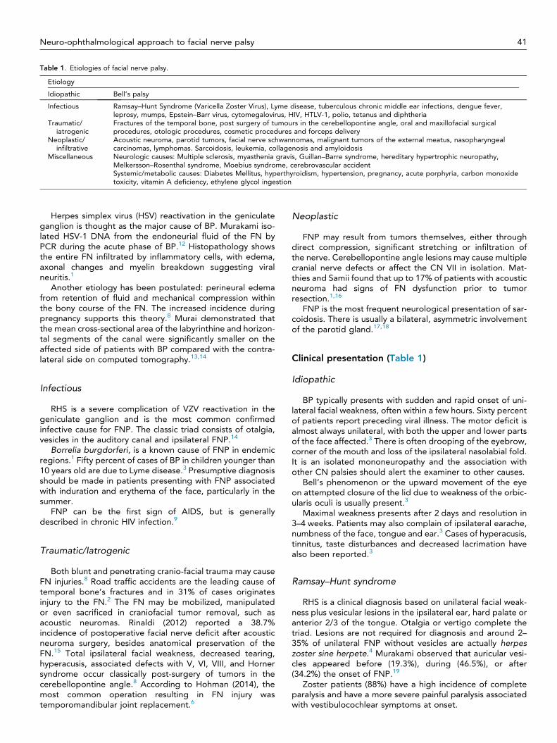
Table 1. Etiologies of facial nerve palsy.
Etiology
Idiopathic Bell’s palsy
Infectious Ramsay–Hunt Syndrome (Varicella Zoster Virus), Lyme disease, tuberculous chronic middle ear infections, dengue fever,leprosy, mumps, Epstein–Barr virus, cytomegalovirus, HIV, HTLV-1, polio, tetanus and diphtheria
Traumatic/iatrogenic
Fractures of the temporal bone, post surgery of tumours in the cerebellopontine angle, oral and maxillofacial surgicalprocedures, otologic procedures, cosmetic procedures and forceps delivery
Neoplastic/infiltrative
Acoustic neuroma, parotid tumors, facial nerve schwannomas, malignant tumors of the external meatus, nasopharyngealcarcinomas, lymphomas. Sarcoidosis, leukemia, collagenosis and amyloidosis
Miscellaneous Neurologic causes: Multiple sclerosis, myasthenia gravis, Guillan–Barre syndrome, hereditary hypertrophic neuropathy,Melkersson–Rosenthal syndrome, Moebius syndrome, cerebrovascular accidentSystemic/metabolic causes: Diabetes Mellitus, hyperthyroidism, hypertension, pregnancy, acute porphyria, carbon monoxidetoxicity, vitamin A deficiency, ethylene glycol ingestion
Neuro-ophthalmological approach to facial nerve palsy 41
Herpes simplex virus (HSV) reactivation in the geniculateganglion is thought as the major cause of BP. Murakami iso-lated HSV-1 DNA from the endoneurial fluid of the FN byPCR during the acute phase of BP.12 Histopathology showsthe entire FN infiltrated by inflammatory cells, with edema,axonal changes and myelin breakdown suggesting viralneuritis.1
Another etiology has been postulated: perineural edemafrom retention of fluid and mechanical compression withinthe bony course of the FN. The increased incidence duringpregnancy supports this theory.8 Murai demonstrated thatthe mean cross-sectional area of the labyrinthine and horizon-tal segments of the canal were significantly smaller on theaffected side of patients with BP compared with the contra-lateral side on computed tomography.13,14
Infectious
RHS is a severe complication of VZV reactivation in thegeniculate ganglion and is the most common confirmedinfective cause for FNP. The classic triad consists of otalgia,vesicles in the auditory canal and ipsilateral FNP.14
Borrelia burgdorferi, is a known cause of FNP in endemicregions.1 Fifty percent of cases of BP in children younger than10 years old are due to Lyme disease.3 Presumptive diagnosisshould be made in patients presenting with FNP associatedwith induration and erythema of the face, particularly in thesummer.
FNP can be the first sign of AIDS, but is generallydescribed in chronic HIV infection.9
Traumatic/Iatrogenic
Both blunt and penetrating cranio-facial trauma may causeFN injuries.8 Road traffic accidents are the leading cause oftemporal bone’s fractures and in 31% of cases originatesinjury to the FN.2 The FN may be mobilized, manipulatedor even sacrificed in craniofacial tumor removal, such asacoustic neuromas. Rinaldi (2012) reported a 38.7%incidence of postoperative facial nerve deficit after acousticneuroma surgery, besides anatomical preservation of theFN.15 Total ipsilateral facial weakness, decreased tearing,hyperacusis, associated defects with V, VI, VIII, and Hornersyndrome occur classically post-surgery of tumors in thecerebellopontine angle.8 According to Hohman (2014), themost common operation resulting in FN injury wastemporomandibular joint replacement.6
Neoplastic
FNP may result from tumors themselves, either throughdirect compression, significant stretching or infiltration ofthe nerve. Cerebellopontine angle lesions may cause multiplecranial nerve defects or affect the CN VII in isolation. Mat-thies and Samii found that up to 17% of patients with acousticneuroma had signs of FN dysfunction prior to tumorresection.1,16
FNP is the most frequent neurological presentation of sar-coidosis. There is usually a bilateral, asymmetric involvementof the parotid gland.17,18
Clinical presentation (Table 1)
Idiopathic
BP typically presents with sudden and rapid onset of uni-lateral facial weakness, often within a few hours. Sixty percentof patients report preceding viral illness. The motor deficit isalmost always unilateral, with both the upper and lower partsof the face affected.3 There is often drooping of the eyebrow,corner of the mouth and loss of the ipsilateral nasolabial fold.It is an isolated mononeuropathy and the association withother CN palsies should alert the examiner to other causes.
Bell’s phenomenon or the upward movement of the eyeon attempted closure of the lid due to weakness of the orbic-ularis oculi is usually present.3
Maximal weakness presents after 2 days and resolution in3–4 weeks. Patients may also complain of ipsilateral earache,numbness of the face, tongue and ear.3 Cases of hyperacusis,tinnitus, taste disturbances and decreased lacrimation havealso been reported.3
Ramsay–Hunt syndrome
RHS is a clinical diagnosis based on unilateral facial weak-ness plus vesicular lesions in the ipsilateral ear, hard palate oranterior 2/3 of the tongue. Otalgia or vertigo complete thetriad. Lesions are not required for diagnosis and around 2–35% of unilateral FNP without vesicles are actually herpeszoster sine herpete.4 Murakami observed that auricular vesi-cles appeared before (19.3%), during (46.5%), or after(34.2%) the onset of FNP.19
Zoster patients (88%) have a high incidence of completeparalysis and have a more severe painful paralysis associatedwith vestibulocochlear symptoms at onset.
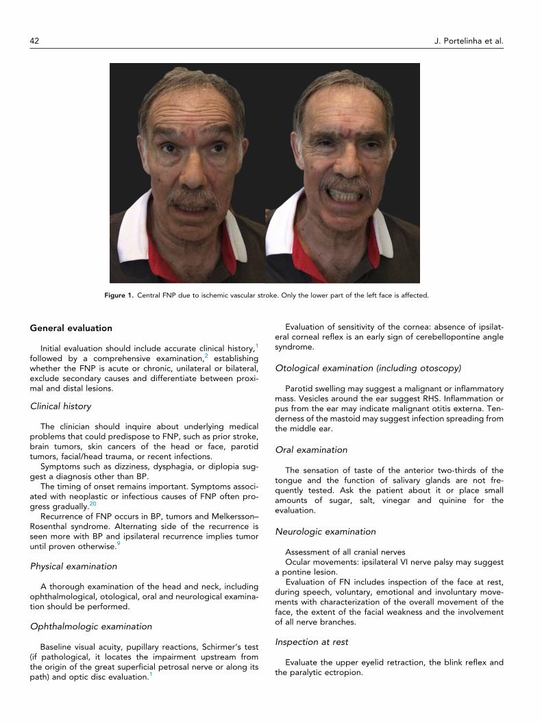
Figure 1. Central FNP due to ischemic vascular stroke. Only the lower part of the left face is affected.
42 J. Portelinha et al.
General evaluation
Initial evaluation should include accurate clinical history,1
followed by a comprehensive examination,2 establishingwhether the FNP is acute or chronic, unilateral or bilateral,exclude secondary causes and differentiate between proxi-mal and distal lesions.
Clinical history
The clinician should inquire about underlying medicalproblems that could predispose to FNP, such as prior stroke,brain tumors, skin cancers of the head or face, parotidtumors, facial/head trauma, or recent infections.
Symptoms such as dizziness, dysphagia, or diplopia sug-gest a diagnosis other than BP.
The timing of onset remains important. Symptoms associ-ated with neoplastic or infectious causes of FNP often pro-gress gradually.20
Recurrence of FNP occurs in BP, tumors and Melkersson–Rosenthal syndrome. Alternating side of the recurrence isseen more with BP and ipsilateral recurrence implies tumoruntil proven otherwise.9
Physical examination
A thorough examination of the head and neck, includingophthalmological, otological, oral and neurological examina-tion should be performed.
Ophthalmologic examination
Baseline visual acuity, pupillary reactions, Schirmer’s test(if pathological, it locates the impairment upstream fromthe origin of the great superficial petrosal nerve or along itspath) and optic disc evaluation.1
Evaluation of sensitivity of the cornea: absence of ipsilat-eral corneal reflex is an early sign of cerebellopontine anglesyndrome.
Otological examination (including otoscopy)
Parotid swelling may suggest a malignant or inflammatorymass. Vesicles around the ear suggest RHS. Inflammation orpus from the ear may indicate malignant otitis externa. Ten-derness of the mastoid may suggest infection spreading fromthe middle ear.
Oral examination
The sensation of taste of the anterior two-thirds of thetongue and the function of salivary glands are not fre-quently tested. Ask the patient about it or place smallamounts of sugar, salt, vinegar and quinine for theevaluation.
Neurologic examination
Assessment of all cranial nervesOcular movements: ipsilateral VI nerve palsy may suggest
a pontine lesion.Evaluation of FN includes inspection of the face at rest,
during speech, voluntary, emotional and involuntary move-ments with characterization of the overall movement of theface, the extent of the facial weakness and the involvementof all nerve branches.
Inspection at rest
Evaluate the upper eyelid retraction, the blink reflex andthe paralytic ectropion.
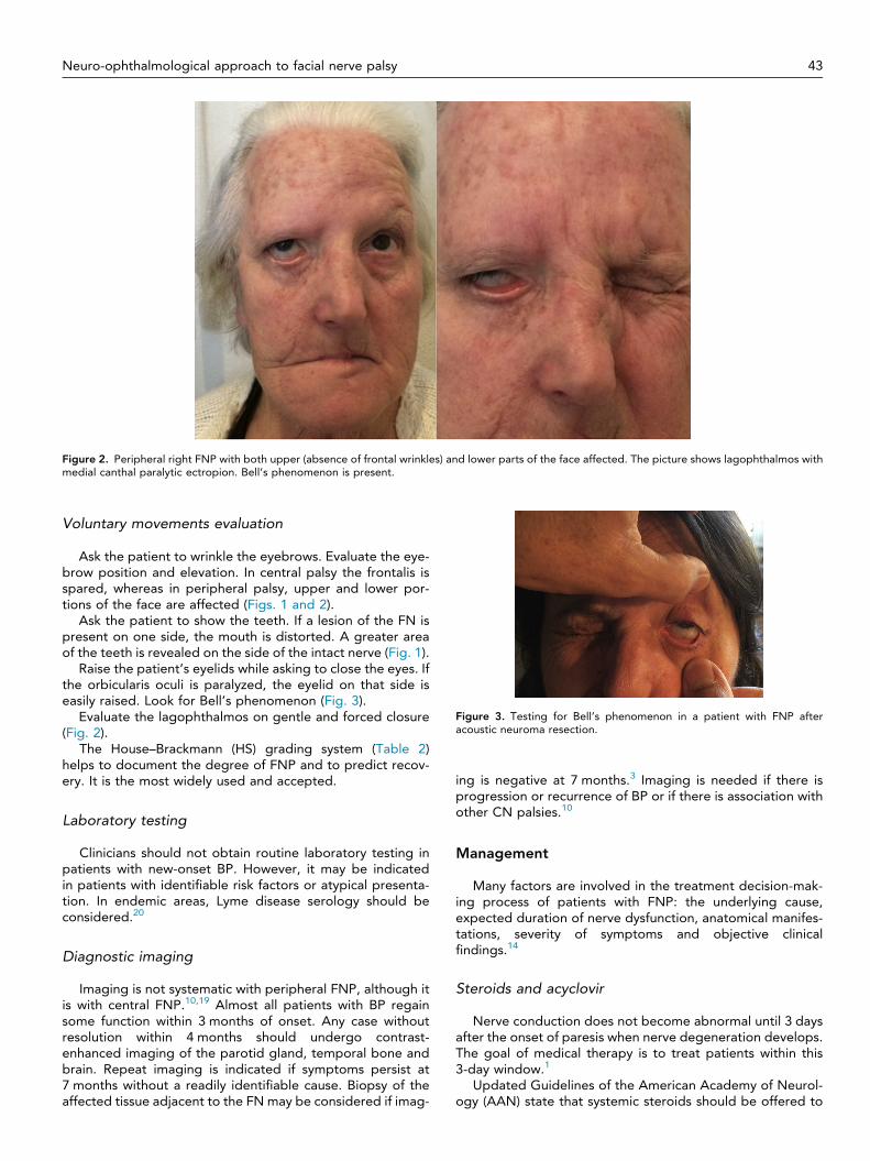
Figure 2. Peripheral right FNP with both upper (absence of frontal wrinkles) and lower parts of the face affected. The picture shows lagophthalmos withmedial canthal paralytic ectropion. Bell’s phenomenon is present.
Figure 3. Testing for Bell’s phenomenon in a patient with FNP afteracoustic neuroma resection.
Neuro-ophthalmological approach to facial nerve palsy 43
Voluntary movements evaluation
Ask the patient to wrinkle the eyebrows. Evaluate the eye-brow position and elevation. In central palsy the frontalis isspared, whereas in peripheral palsy, upper and lower por-tions of the face are affected (Figs. 1 and 2).
Ask the patient to show the teeth. If a lesion of the FN ispresent on one side, the mouth is distorted. A greater areaof the teeth is revealed on the side of the intact nerve (Fig. 1).
Raise the patient’s eyelids while asking to close the eyes. Ifthe orbicularis oculi is paralyzed, the eyelid on that side iseasily raised. Look for Bell’s phenomenon (Fig. 3).
Evaluate the lagophthalmos on gentle and forced closure(Fig. 2).
The House–Brackmann (HS) grading system (Table 2)helps to document the degree of FNP and to predict recov-ery. It is the most widely used and accepted.
Laboratory testing
Clinicians should not obtain routine laboratory testing inpatients with new-onset BP. However, it may be indicatedin patients with identifiable risk factors or atypical presenta-tion. In endemic areas, Lyme disease serology should beconsidered.20
Diagnostic imaging
Imaging is not systematic with peripheral FNP, although itis with central FNP.10,19 Almost all patients with BP regainsome function within 3 months of onset. Any case withoutresolution within 4 months should undergo contrast-enhanced imaging of the parotid gland, temporal bone andbrain. Repeat imaging is indicated if symptoms persist at7 months without a readily identifiable cause. Biopsy of theaffected tissue adjacent to the FN may be considered if imag-
ing is negative at 7 months.3 Imaging is needed if there isprogression or recurrence of BP or if there is association withother CN palsies.10
Management
Many factors are involved in the treatment decision-mak-ing process of patients with FNP: the underlying cause,expected duration of nerve dysfunction, anatomical manifes-tations, severity of symptoms and objective clinicalfindings.14
Steroids and acyclovir
Nerve conduction does not become abnormal until 3 daysafter the onset of paresis when nerve degeneration develops.The goal of medical therapy is to treat patients within this3-day window.1
Updated Guidelines of the American Academy of Neurol-ogy (AAN) state that systemic steroids should be offered to

Table 2. House–Brackmann grading system.
Grade Description Characteristics
I Normal Normal facial functionII Mild dysfunction Gross: slight weakness noticeable on close inspection, may have very slight synkinesis. At rest: normal symmetry
and tone. Motion: forehead- moderate to good function, eye-complete closure with minimum effort, mouth-slight asymmetry
III Moderate dysfunction Gross: obvious but not disfiguring difference between the two sides; contracture and/or hemifacial spasm. Atrest: normal asymmetry and tone. Motion: forehead- slight to moderate movement; eye- complete closure witheffort; mouth- slightly weak with maximum effort
IV Moderately severedysfunction
Gross: obvious weakness and/or disfiguring asymmetry. At rest: normal asymmetry and tone. Motion: forehead-none; eye- incomplete closure; mouth: asymmetric with maximum effort
V Severe dysfunction Gross: only barely perceptible motion. At rest: asymmetry. Motion: forehead-none; eye- incomplete closure;mouth- slight movement
VI Total paralysis No movement
44 J. Portelinha et al.
patients with new-onset BP to increase the chance of FNrecovery (Level A recommendation).21 A recent Cochranereview found that only 23% of patients treated with cortico-steroids had incomplete recovery of facial motor function at6 months, compared to 33% of patients treated with pla-cebo.22 In addition, patients receiving corticosteroids had asignificant reduction in motor synkinesis.3,22
HSV infection is believed to be the main cause of BP; itwould then be reasonable to include acyclovir in its treat-ment.1 However, a Cochrane systematic review concludedthat antivirals provide no significant benefit over placebo ingenerating complete recovery from BP.23 According to theAAN, patients with new-onset BP, might be offered antivirals(in addition to steroids) (Level C), because of the possibility ofmodest increase in recovery.21
In RHS, a Cochrane review of the sole randomized con-trolled trial comparing combined treatment to corticoste-roids alone showed no significant difference in outcomes.24
The largest RHS treatment study was a retrospective analysisof 80 cases. Patients treated with acyclovir–prednisone within72-h of symptoms onset had a complete recovery rate of 75%vs 30% of patients treated after 7 days. Early administrationalso reduced nerve degeneration.4,25 Acyclovir 800 mg5 times daily or 1 g valacyclovir TID for 7–10 days plus pred-nisone 1 mg/kg for 5 days and taper was used in publishedtrials.4
Surgery for FN decompression
Surgical decompression remains highly controversial dueto its risks and should be considered in refractory cases.3
There is some evidence in favor of surgical decompressionwith documented loss of >90% of axonal fibers on the elec-troneurography prior to day 14 of weakness onset.1,26 Hatoet al. decompressed the tympanic and mastoid segmentsof the canal with concurrent placement of basic fibroblastgrowth factor impregnated biodegradable gelatin hydrogelaround the nerve. The rate of complete recovery was 75%compared to 44.8% by the conventional decompressionmethod and 23.3% on the steroids alone.14
Corneal exposure and lagophthalmos
On FNP patients, the cornea is especially at risk becauseof improper lid closure due to lagophthalmos and paralyticectropion and due to decreased tear production anddistribution of the tear film. Some works also demonstratedincreased meibomian gland dysfunction.27,28
Treatment directed at protecting the cornea depends onthe degree of nerve lesion and of the risk of corneal damagebased on the amount of lagophthalmos, the quality of Bell’sphenomenon and the presence or absence of paralytic ectro-pion.8,9 Furthermore, corneal sensation testing should beperformed, as patients with both V and VII palsies are atincreased risk of developing corneal decompensation.29 Neu-rotrophic corneal epithelium is more prone to injury, healspoorly and patients may be unaware of the corneal damage.
The goals of therapy are to protect the cornea and torestore the blink response using minimal intervention andmaintaining good visual acuity.1
Temporary treatment
If recovery is expected, less invasive techniques should beemployed.
When there is low corneal risk and good prognosis forrecovery, intensive lubricants and taping the lid with a stifftape overnight will usually be enough.8 Preservative-freeteardrops during the day and a more viscous ointment over-night may be used. Moisture chambers act as barriers toevaporation.9
Scleral contact lenses were recently described as a validalternative to tarsorrhaphy for patients with corneal exposureand anesthesia, providing effective protection in an estheti-cally acceptable way and optimizing visual function.30,31
Botulinum toxin injection induces ptosis by temporarilyparalyzing the levator palpebrae superioris and thus protect-ing the cornea. It is an excellent low-risk temporary (effectsustained for a mean of 46 days) alternative for postoperativehigh grade FNP, when the FN is anatomically intact.32 In 16 of21 patients further surgical intervention was avoided and cor-neal healing was obtained. Significant improvement in cor-neal symptoms and decreased use of artificial tears wasalso reported.14 However, it affects patients’ vision and mayprovide less than adequate protection as the levator functionreturns.8,9
Temporary tarsorrhaphy can be achieved with a simplesuture or cyanoacrylate glue. The classic central tarsorrhaphyis cosmetically and visually poor, but gives good protection.Lateral tarsorrhaphy may not adequately close the eye, par-ticularly if there is significant lower lid ectropion.8
External eyelid weights have similar design to those forimplantation and are fixed to the pretarsal skin surface withdouble-sided hypoallergenic adhesive tape.9
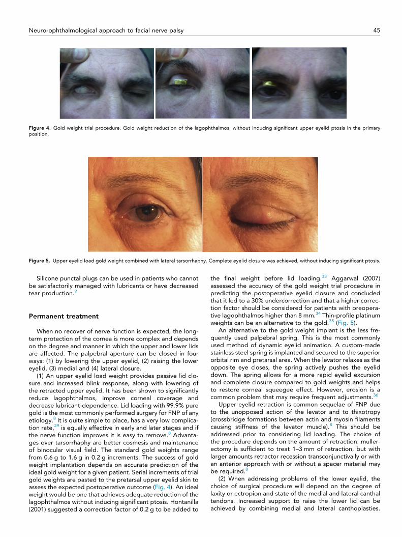
Figure 4. Gold weight trial procedure. Gold weight reduction of the lagophthalmos, without inducing significant upper eyelid ptosis in the primaryposition.
Figure 5. Upper eyelid load gold weight combined with lateral tarsorrhaphy. Complete eyelid closure was achieved, without inducing significant ptosis.
Neuro-ophthalmological approach to facial nerve palsy 45
Silicone punctal plugs can be used in patients who cannotbe satisfactorily managed with lubricants or have decreasedtear production.9
Permanent treatment
When no recover of nerve function is expected, the long-term protection of the cornea is more complex and dependson the degree and manner in which the upper and lower lidsare affected. The palpebral aperture can be closed in fourways: (1) by lowering the upper eyelid, (2) raising the lowereyelid, (3) medial and (4) lateral closure.
(1) An upper eyelid load weight provides passive lid clo-sure and increased blink response, along with lowering ofthe retracted upper eyelid. It has been shown to significantlyreduce lagophthalmos, improve corneal coverage anddecrease lubricant-dependence. Lid loading with 99.9% puregold is the most commonly performed surgery for FNP of anyetiology.8 It is quite simple to place, has a very low complica-tion rate,29 is equally effective in early and later stages and ifthe nerve function improves it is easy to remove.8 Advanta-ges over tarsorrhaphy are better cosmesis and maintenanceof binocular visual field. The standard gold weights rangefrom 0.6 g to 1.6 g in 0.2 g increments. The success of goldweight implantation depends on accurate prediction of theideal gold weight for a given patient. Serial increments of trialgold weights are pasted to the pretarsal upper eyelid skin toassess the expected postoperative outcome (Fig. 4). An idealweight would be one that achieves adequate reduction of thelagophthalmos without inducing significant ptosis. Hontanilla(2001) suggested a correction factor of 0.2 g to be added to
the final weight before lid loading.33 Aggarwal (2007)assessed the accuracy of the gold weight trial procedure inpredicting the postoperative eyelid closure and concludedthat it led to a 30% undercorrection and that a higher correc-tion factor should be considered for patients with preopera-tive lagophthalmos higher than 8 mm.34 Thin-profile platinumweights can be an alternative to the gold.35 (Fig. 5).
An alternative to the gold weight implant is the less fre-quently used palpebral spring. This is the most commonlyused method of dynamic eyelid animation. A custom-madestainless steel spring is implanted and secured to the superiororbital rim and pretarsal area. When the levator relaxes as theopposite eye closes, the spring actively pushes the eyeliddown. The spring allows for a more rapid eyelid excursionand complete closure compared to gold weights and helpsto restore corneal squeegee effect. However, erosion is acommon problem that may require frequent adjustments.36
Upper eyelid retraction is common sequelae of FNP dueto the unopposed action of the levator and to thixotropy(crossbridge formations between actin and myosin filamentscausing stiffness of the levator muscle).8 This should beaddressed prior to considering lid loading. The choice ofthe procedure depends on the amount of retraction: muller-ectomy is sufficient to treat 1–3 mm of retraction, but withlarger amounts retractor recession transconjunctivally or withan anterior approach with or without a spacer material maybe required.8
(2) When addressing problems of the lower eyelid, thechoice of surgical procedure will depend on the degree oflaxity or ectropion and state of the medial and lateral canthaltendons. Increased support to raise the lower lid can beachieved by combining medial and lateral canthoplasties.
46 J. Portelinha et al.
When there is significant lower lid retraction, this can be com-bined with insertion of a spacer. In cases of marked tissueatrophy, an autogenous fascial sling can be threaded ham-mock-like through the entire length of the lid, anchored byfixation to the medial canthal tendon and lateral orbitalperiosteum.8.
(3) The medial palpebral aperture closure depends on thelaxity of the medial canthal tendon. A punctual ectropion canbe treated with a medial canthoplasty or a medial tarsal strip.Where there is significant tendon laxity, a deep periostealRoyce-Johnston suture or medial wedge excision could beused.
(4) Permanent lateral tarsorraphy has largely been super-seded by the lateral canthal sling.8. It has the advantage thatit can be augmented by inserting the strip higher on the rimto assist in tear drainage or combining it with a small lateraltarsorraphy if the horizontal aperture needs to beshortened.8
Dynamic correction of paralytic lagophthalmos frequentlyinvolves transfer of the temporalis muscle, which is effectiveand can provide strong eyelid closure over and for anextended period of time. Reanimation of paralyzed musclesusing adjacent motor nerves has also been attempted: Hay-ashi performed an hypoglossal-facial nerve anastomosis withexcellent success rates37; Corrales used hypoglossal-facialnerve anastomosis to treat patients with central FNP withcomparable success with those for peripheral dysfunction38
and the masseter nerve can also be utilized.13
The use of permanent tarsorrhaphy has decreased sinceother rehabilitation procedures arise. It is most suited tocases with corneal sensory deficits. Despite being cosmeti-cally and visually poor, for patients in whom medical therapyis difficult and lacrimal gland function is lost, it remains animportant treatment option.
Other modalities of therapy
No evidence supports significant benefit from physicaltherapy or acupuncture for BP. Hyperbaric oxygen therapymight be effective on moderate to severe BP.39–41
Prognosis
Spontaneous, complete recovery of BP occurs in up to70% of cases. Usually remission begins within 3–4 weeks, withcomplete recovery within 6 months. Peitersen found that, in2570 cases of peripheral FNP, 85% of patients’ functionwas returned within 3 weeks and in the remaining 15% after3–5 months. In 71% normal mimical function was obtained,
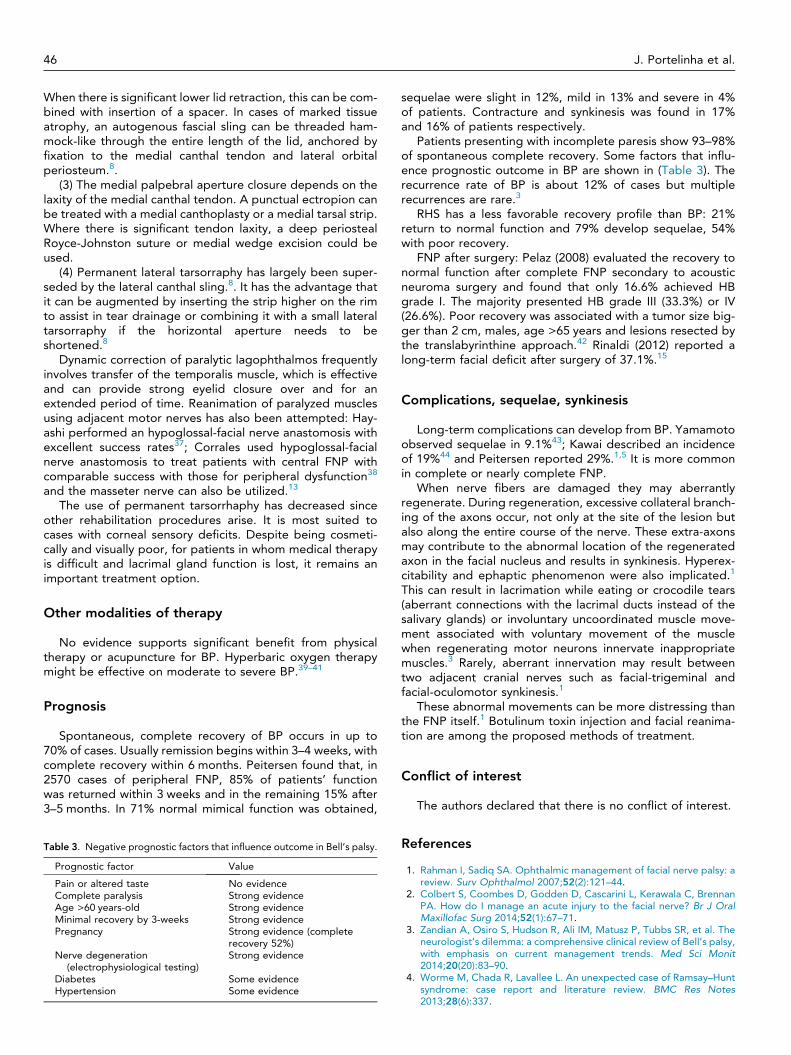
Table 3. Negative prognostic factors that influence outcome in Bell’s palsy.
Prognostic factor Value
Pain or altered taste No evidenceComplete paralysis Strong evidenceAge >60 years-old Strong evidenceMinimal recovery by 3-weeks Strong evidencePregnancy Strong evidence (complete
recovery 52%)Nerve degeneration
(electrophysiological testing)Strong evidence
Diabetes Some evidenceHypertension Some evidence
sequelae were slight in 12%, mild in 13% and severe in 4%of patients. Contracture and synkinesis was found in 17%and 16% of patients respectively.
Patients presenting with incomplete paresis show 93–98%of spontaneous complete recovery. Some factors that influ-ence prognostic outcome in BP are shown in (Table 3). Therecurrence rate of BP is about 12% of cases but multiplerecurrences are rare.3
RHS has a less favorable recovery profile than BP: 21%return to normal function and 79% develop sequelae, 54%with poor recovery.
FNP after surgery: Pelaz (2008) evaluated the recovery tonormal function after complete FNP secondary to acousticneuroma surgery and found that only 16.6% achieved HBgrade I. The majority presented HB grade III (33.3%) or IV(26.6%). Poor recovery was associated with a tumor size big-ger than 2 cm, males, age >65 years and lesions resected bythe translabyrinthine approach.42 Rinaldi (2012) reported along-term facial deficit after surgery of 37.1%.15
Complications, sequelae, synkinesis
Long-term complications can develop from BP. Yamamotoobserved sequelae in 9.1%43; Kawai described an incidenceof 19%44 and Peitersen reported 29%.1,5 It is more commonin complete or nearly complete FNP.
When nerve fibers are damaged they may aberrantlyregenerate. During regeneration, excessive collateral branch-ing of the axons occur, not only at the site of the lesion butalso along the entire course of the nerve. These extra-axonsmay contribute to the abnormal location of the regeneratedaxon in the facial nucleus and results in synkinesis. Hyperex-citability and ephaptic phenomenon were also implicated.1
This can result in lacrimation while eating or crocodile tears(aberrant connections with the lacrimal ducts instead of thesalivary glands) or involuntary uncoordinated muscle move-ment associated with voluntary movement of the musclewhen regenerating motor neurons innervate inappropriatemuscles.3 Rarely, aberrant innervation may result betweentwo adjacent cranial nerves such as facial-trigeminal andfacial-oculomotor synkinesis.1
These abnormal movements can be more distressing thanthe FNP itself.1 Botulinum toxin injection and facial reanima-tion are among the proposed methods of treatment.
Conflict of interest
The authors declared that there is no conflict of interest.
References
1. Rahman I, Sadiq SA. Ophthalmic management of facial nerve palsy: areview. Surv Ophthalmol 2007;52(2):121–44.
2. Colbert S, Coombes D, Godden D, Cascarini L, Kerawala C, BrennanPA. How do I manage an acute injury to the facial nerve? Br J OralMaxillofac Surg 2014;52(1):67–71.
3. Zandian A, Osiro S, Hudson R, Ali IM, Matusz P, Tubbs SR, et al. Theneurologist’s dilemma: a comprehensive clinical review of Bell’s palsy,with emphasis on current management trends. Med Sci Monit2014;20(20):83–90.
4. Worme M, Chada R, Lavallee L. An unexpected case of Ramsay–Huntsyndrome: case report and literature review. BMC Res Notes2013;28(6):337.
Neuro-ophthalmological approach to facial nerve palsy 47
5. Peitersen E. Bell’s palsy: the spontaneous course of 2500 peripheralfacial nerve palsies of different etiologies. Acta Otolaryngol Suppl2002;549:4–30.
6. Hohman MH, Hadlock TA. Etiology, diagnosis, and management offacial palsy: 2000 patients at a facial nerve center. Laryngoscope2014;124(7):E283–93.
7. Ozkale Y, Erol I, Saygı S, Yılmaz I. Overview of pediatricperipheral facial nerve paralysis: analysis of 40 patients. J ChildNeurol 2014, 0883073814530497.
8. Lee V, Currie Z, Collin JR. Ophthalmic management of facial nervepalsy. Eye (Lond) 2004;18(12):1225–34.
9. Mavrikakis I. Facial nerve palsy: anatomy, etiology, evaluation, andmanagement. Orbit 2008;27(6):466–74.
10. Toulgoat F, Sarrazin JL, Benoudiba F, Pereon Y, Auffray-Calvier E,Daumas-Duport B, et al. Facial nerve: from anatomy to pathology.Diagn Interv Imaging 2013;94(10):1033–42.
11. McAllister K, Walker D, Donnan PT, Swan I. Surgical interventions forthe early management of Bell’s palsy. Cochrane Database Syst Rev2013;10:CD007468.
12. Murakami S, Mizobuchi M, Nakashiro Y, Doi T, Hato N, Yanagihara N.Bell palsy and herpes simplex virus: identification of viral DNA inendoneurial fluid and muscle. Ann Intern Med 1996;124(1 Pt):27–30.
13. Murai A, Kariya S, Tamura K, Doi A, Kozakura K, Okano M, et al. Thefacial nerve canal in patients with Bell’s palsy: an investigation byhigh-resolution computed tomography with multiplanarreconstruction. Eur Arch Otorhinolaryngol 2013;270(7):2035–8.
14. Kim C, Lelli Jr GJ. Current considerations in the management of facialnerve palsy. Curr Opin Ophthalmol 2013;24(5):478–83.
15. Rinaldi V, Casale M, Bressi F, Potena M, Vesperini E, De Franco A,et al. Facial nerve outcome after vestibular schwannoma surgery: ourexperience. J Neurol Surg B Skull Base 2012;73(1):21–7.
16. Matthies C, Samii M. Management of 1000 vestibular schwannomas(acoustic neuromas): clinical presentation. Neurosurgery1997;40(1):1–9, Discussion 9–10.
17. Sharma SK, Soneja M, Sharma A, Sharma MC, Hari S. Raremanifestations of sarcoidosis in modern era of new diagnostictools. Indian J Med Res 2012;135(5):621–9.
18. James DG. All that palsies is not Bell’s. J R Soc Med1996;89(4):184–7.
19. Murakami S, Hato N, Horiuchi J, Miyamoto Y, Aono H, Honda N,et al. Clinical features and prognosis of facial palsy and hearing loss inpatients with Ramsay–Hunt syndrome. Nihon Jibiinkoka Gakkai Kaiho1996;99(12):1772–9.
20. Baugh RF, Basura GJ, Ishii LE, Schwartz SR, Drumheller CM,Burkholder R, et al. Clinical practice guideline: Bell’s palsy.Otolaryngol Head Neck Surg 2013;149(3 Suppl):S1–S27.
21. Gronseth GS, Paduga R. American Academy of Neurology. Evidence-based guideline update: steroids and antivirals for Bell palsy: reportof the Guideline Development Subcommittee of the AmericanAcademy of Neurology. Neurology 2012;79(22):2209–13.
22. Salinas RA, Alvarez G, Daly F, Ferreira J. Corticosteroids for Bell’spalsy (idiopathic facial paralysis). Cochrane Database Syst Rev2010;3:CD001942.
23. Lockhart P, Daly F, Pitkethly M, Comerford N, Sullivan F. Antiviraltreatment for Bell’s palsy (idiopathic facial paralysis). CochraneDatabase Syst Rev 2009;4:CD001869.
24. Uscategui T, Dorée C, Chamberlain IJ, Burton MJ. Antiviral therapyfor Ramsay–Hunt syndrome (herpes zoster oticus with facial palsy) inadults. Cochrane Database Syst Rev 2008;4:CD006851.
25. Murakami S, Hato N, Horiuchi J, Honda N, Gyo K, Yanagihara N.Treatment of Ramsay–Hunt syndrome with acyclovir-prednisone:significance of early diagnosis and treatment. Ann Neurol1997;41(3):353–7.
26. Gantz BJ, Rubinstein JT, Gidley P, Woodworth GG. Surgicalmanagement of Bell’s palsy. Laryngoscope 1999;109(8):1177–88.
27. Call CB, Wise RJ, Hansen MR, Carter KD, Allen RC. In vivoexamination of meibomian gland morphology in patients with facialnerve palsy using infrared meibography. Ophthal Plast Reconstr Surg2012;28(6):396–400.
28. Shah CT, Blount AL, Nguyen EV, Hassan AS. Cranial nerve sevenpalsy and its influence on meibomian gland function. Ophthal PlastReconstr Surg 2012;28(3):166–8.
29. Seiff SR, Carter SR. Facial nerve paralysis. Int Ophthalmol Clin2002;42(2):103–12.
30. Gire A, Kwok A, Marx DP. PROSE treatment for lagophthalmos andexposure keratopathy. Ophthal Plast Reconstr Surg2013;29(2):e38–40.
31. Weyns M, Koppen C, Tassignon MJ. Scleral contact lenses as analternative to tarsorrhaphy for the long-term management ofcombined exposure and neurotrophic keratopathy. Cornea2013;32(3):359–61.
32. Prell J, Rampp S, Rachinger J, Scheller C, Alfieri A, Marquardt L, et al.Botulinum toxin for temporary corneal protection after surgery forvestibular schwannoma. J Neurosurg 2011;114(2):426–31.
33. Hontanilla B. Weight measurement of upper eyelid gold implants forlagophthalmos in facial paralysis. Plast Reconstr Surg2001;108(6):1539–43.
34. Aggarwal E, Naik MN, Honavar SG. Effectiveness of the gold weighttrial procedure in predicting the ideal weight for lid loading in facialpalsy: a prospective study. Am J Ophthalmol 2007;143(6):1009–12.
35. Rudman KL, Rhee JS. Habilitation of facial nerve dysfunction afterresection of a vestibular schwannoma. Otolaryngol Clin North Am2012;45(2):513–30.
36. Demirci H, Frueh BR. Palpebral spring in the management oflagophthalmos and exposure keratopathy secondary to facial nervepalsy. Ophthal Plast Reconstr Surg 2009;25(4):270–5.
37. Hayashi A, Nishida M, Seno H, Inoue M, Iwata H, Shirasawa T, et al.Hemihypoglossal nerve transfer for acute facial paralysis. JNeurosurg 2013;118(1):160–6.
38. Corrales CE, Gurgel RK, Jackler RK. Rehabilitation of central facialparalysis with hypoglossal-facial anastomosis. Otol Neurotol2012;33(8):1439–44.
39. Holland NJ, Bernstein JM, Hamilton JW. Hyperbaric oxygen therapyfor Bell’s palsy. Cochrane Database Syst Rev 2012;2:CD007288.
40. Teixeira LJ, Valbuza JS, Prado GF. Physical therapy for Bell’s palsy(idiopathic facial paralysis). Cochrane Database Syst Rev2011;12:CD006283.
41. Chen N, Zhou M, He L, Zhou D, Li N. Acupuncture for Bell’s palsy.Cochrane Database Syst Rev 2010;8:CD002914.
42. Coca Pelaz A, Fernández Lisa C, Gómez JR, Rodrigo JP, Llorente JL,Suárez C. Complete facial palsy following surgery for acoustic nerveneurinoma: evolution and associated ophthalmologicalcomplications. Acta Otorrinolaringol Esp 2008;59(5):223–7.
43. Yamamoto E, Nishimura H, Hirono Y. Occurrence of sequelae in Bell’spalsy. Acta Otolaryngol Suppl 1988;446:93–6.
44. Kawai M. Sequelae and transient sequelae-like symptoms in Bell’spalsy. Nihon Jibiinkoka Gakkai Kaiho 1989;92(1):88–92.
Related Documents