Neural correlates of taste perception in congenital olfactory impairment Léa Gagnon a,b,n , Martin Vestergaard c , Kristoffer Madsen c , Helena G Karstensen d , Hartwig Siebner c,e , Niels Tommerup d , Ron Kupers a,b , Maurice Ptito a,b,c a Harland Sanders Chair, School of Optometry, University of Montreal, Quebec, Canada b Brain Research and Integrative Neuroscience Laboratory, Department of Neuroscience and Pharmacology, Panum Institute, University of Copenhagen, Denmark c Danish Research Centre for Magnetic Resonance, Copenhagen University Hospital, Hvidovre, Denmark d Wilhelm Johannsen Centre for Functional Genome Research, Department of Cellular and Molecular Medicine, University of Copenhagen, Denmark e Department of Neurology, Bispebjerg Hospital, University of Copenhagen, Denmark article info Article history: Received 15 March 2014 Received in revised form 25 June 2014 Accepted 16 July 2014 Available online 29 July 2014 Keywords: Taste Smell Congenital olfactory impairment fMRI Orbitofrontal cortex abstract Olfaction and gustation contribute both to the appreciation of food flavours. Although acquired loss of smell has profound consequences on the pleasure of eating, food habits and body weight, less is known about the impact of congenital olfactory impairment on gustatory processing. Here we examined taste identification accuracy and its neural correlates using functional magnetic resonance imaging (fMRI) in 12 congenitally olfactory impaired individuals and 8 normosmic controls. Results showed that taste identification was worse in congenitally olfactory impaired compared to control subjects. The fMRI results demonstrated that olfactory impaired individuals had reduced activation in medial orbitofrontal cortex (mOFC) relative to normosmic subjects while tasting. In addition, olfactory performance as measured with the Sniffin' Sticks correlated positively with taste-induced blood-oxygen-level dependent (BOLD) signal increases in bilateral mOFC and anterior insula. Our data provide a neurological underpinning for the reduced taste perception in congenitally olfactory impaired individuals. & 2014 Elsevier Ltd. All rights reserved. 1. Introduction Our appreciation of fine foods or drinks largely comes from a rich diversity of flavours perceived mainly by our sense of smell, together with taste (e.g. sweet, salty) and trigeminal (e.g. tem- perature, texture) sensations (Auvray & Spence, 2008; Lundstrom, Boesveldt, & Albrecht, 2011). The loss of olfaction has therefore strong repercussions on flavour perception but it remains unclear how this is manifested at the cerebral level. Studies have demon- strated that the olfactory and gustatory systems largely overlap. Following activation of taste receptors, taste information travels from the VII, IX and X cranial nerves to reach first the nucleus of the solitary tract of the brainstem, followed by the ventral poster- ior medial nucleus of the thalamus and then converges towards the primary and secondary taste cortices in the insula/operculum and orbitofrontal cortex (OFC), respectively (Sewards, 2004). Smell information, on the other hand, is conveyed by the olfactory nerve (I) and synapses first in the olfactory bulb before reaching the piriform and entorhinal cortices (primary olfactory cortices), fol- lowed by various higher order olfactory areas such as the amygdala, cingulate cortex, insula and orbitofrontal cortex. According to the traditional view, taste-odour integration occurs in the orbitofrontal cortex (Rolls, 2001, 2008; De Araujo, Rolls, Kringelbach, McGlone, & Phillips, 2003) and insula (Small & Prescott, 2005; Verhagen & Engelen, 2006). However, recent evidence from rodent studies challenges the classical view (Small, Veldhuizen, & Green, 2013) as taste neurons were also recorded within the posterior piriform cortex (Maier, Wachowiak, & Katz, 2012). This close anatomical relationship between olfactory and gustatory systems suggests that odour impairments may affect the central processing of taste. Odour impairments are common and affect nearly 5% of the population (Karstensen & Tommerup, 2011). Acquired anosmia resulting from traumatic brain injury, infection of the upper respiratory tract or other diseases leads to a decreased appetite and lower interest in eating, changes in body weight, disturbances in affective behaviour (e.g. depression), and a reduced quality of life (Ferris et al., 1985; Mattes & Cowart, 1994; Van Toller, 1999; Miwa et al., 2001; Temmel et al., 2002; Aschenbrenner et al., 2008). In addition, these patients display decreases in gustatory (Gudziol, Rahneberg, & Burkert, 2007) and trigeminal (Gudziol, Schubert, & Hummel, 2001; Frasnelli, Schuster, & Hummel, 2010) Contents lists available at ScienceDirect journal homepage: www.elsevier.com/locate/neuropsychologia Neuropsychologia http://dx.doi.org/10.1016/j.neuropsychologia.2014.07.018 0028-3932/& 2014 Elsevier Ltd. All rights reserved. n Corresponding author at: Brain Research and Integrative Neuroscience Laboratory, Department of Neuroscience and Pharmacology, Panum Institute 33-3, University of Copenhagen, Blegdamsvej 3B, 2200 Copenhagen, Denmark. E-mail address: [email protected] (L. Gagnon). Neuropsychologia 62 (2014) 297–305

Welcome message from author
This document is posted to help you gain knowledge. Please leave a comment to let me know what you think about it! Share it to your friends and learn new things together.
Transcript

Neural correlates of taste perception in congenitalolfactory impairment
Léa Gagnon a,b,n, Martin Vestergaard c, Kristoffer Madsen c, Helena G Karstensen d,Hartwig Siebner c,e, Niels Tommerup d, Ron Kupers a,b, Maurice Ptito a,b,c
a Harland Sanders Chair, School of Optometry, University of Montreal, Quebec, Canadab Brain Research and Integrative Neuroscience Laboratory, Department of Neuroscience and Pharmacology, Panum Institute,University of Copenhagen, Denmarkc Danish Research Centre for Magnetic Resonance, Copenhagen University Hospital, Hvidovre, Denmarkd Wilhelm Johannsen Centre for Functional Genome Research, Department of Cellular and Molecular Medicine, University of Copenhagen, Denmarke Department of Neurology, Bispebjerg Hospital, University of Copenhagen, Denmark
a r t i c l e i n f o
Article history:Received 15 March 2014Received in revised form25 June 2014Accepted 16 July 2014Available online 29 July 2014
Keywords:TasteSmellCongenital olfactory impairmentfMRIOrbitofrontal cortex
a b s t r a c t
Olfaction and gustation contribute both to the appreciation of food flavours. Although acquired loss ofsmell has profound consequences on the pleasure of eating, food habits and body weight, less is knownabout the impact of congenital olfactory impairment on gustatory processing. Here we examined tasteidentification accuracy and its neural correlates using functional magnetic resonance imaging (fMRI) in12 congenitally olfactory impaired individuals and 8 normosmic controls. Results showed that tasteidentification was worse in congenitally olfactory impaired compared to control subjects. The fMRIresults demonstrated that olfactory impaired individuals had reduced activation in medial orbitofrontalcortex (mOFC) relative to normosmic subjects while tasting. In addition, olfactory performance asmeasured with the Sniffin' Sticks correlated positively with taste-induced blood-oxygen-level dependent(BOLD) signal increases in bilateral mOFC and anterior insula. Our data provide a neurologicalunderpinning for the reduced taste perception in congenitally olfactory impaired individuals.
& 2014 Elsevier Ltd. All rights reserved.
1. Introduction
Our appreciation of fine foods or drinks largely comes from arich diversity of flavours perceived mainly by our sense of smell,together with taste (e.g. sweet, salty) and trigeminal (e.g. tem-perature, texture) sensations (Auvray & Spence, 2008; Lundstrom,Boesveldt, & Albrecht, 2011). The loss of olfaction has thereforestrong repercussions on flavour perception but it remains unclearhow this is manifested at the cerebral level. Studies have demon-strated that the olfactory and gustatory systems largely overlap.Following activation of taste receptors, taste information travelsfrom the VII, IX and X cranial nerves to reach first the nucleus ofthe solitary tract of the brainstem, followed by the ventral poster-ior medial nucleus of the thalamus and then converges towardsthe primary and secondary taste cortices in the insula/operculumand orbitofrontal cortex (OFC), respectively (Sewards, 2004). Smellinformation, on the other hand, is conveyed by the olfactory nerve(I) and synapses first in the olfactory bulb before reaching the
piriform and entorhinal cortices (primary olfactory cortices), fol-lowed by various higher order olfactory areas such as the amygdala,cingulate cortex, insula and orbitofrontal cortex. According to thetraditional view, taste-odour integration occurs in the orbitofrontalcortex (Rolls, 2001, 2008; De Araujo, Rolls, Kringelbach, McGlone,& Phillips, 2003) and insula (Small & Prescott, 2005; Verhagen& Engelen, 2006). However, recent evidence from rodent studieschallenges the classical view (Small, Veldhuizen, & Green, 2013) astaste neurons were also recorded within the posterior piriformcortex (Maier, Wachowiak, & Katz, 2012). This close anatomicalrelationship between olfactory and gustatory systems suggests thatodour impairments may affect the central processing of taste.
Odour impairments are common and affect nearly 5% of thepopulation (Karstensen & Tommerup, 2011). Acquired anosmiaresulting from traumatic brain injury, infection of the upperrespiratory tract or other diseases leads to a decreased appetiteand lower interest in eating, changes in body weight, disturbancesin affective behaviour (e.g. depression), and a reduced quality oflife (Ferris et al., 1985; Mattes & Cowart, 1994; Van Toller, 1999;Miwa et al., 2001; Temmel et al., 2002; Aschenbrenner et al.,2008). In addition, these patients display decreases in gustatory(Gudziol, Rahneberg, & Burkert, 2007) and trigeminal (Gudziol,Schubert, & Hummel, 2001; Frasnelli, Schuster, & Hummel, 2010)
Contents lists available at ScienceDirect
journal homepage: www.elsevier.com/locate/neuropsychologia
Neuropsychologia
http://dx.doi.org/10.1016/j.neuropsychologia.2014.07.0180028-3932/& 2014 Elsevier Ltd. All rights reserved.
n Corresponding author at: Brain Research and Integrative NeuroscienceLaboratory, Department of Neuroscience and Pharmacology, Panum Institute 33-3,University of Copenhagen, Blegdamsvej 3B, 2200 Copenhagen, Denmark.
E-mail address: [email protected] (L. Gagnon).
Neuropsychologia 62 (2014) 297–305

sensitivity. At the cortical level, brain imaging studies on trigem-inal processing using event-related potentials (Frasnelli, Schuster,& Hummel, 2007a) and functional magnetic resonance imaging(fMRI; Iannilli, Gerber, Frasnelli, & Hummel, 2007) in patients withacquired anosmia compared to controls revealed lower activity inchemosensory brain areas, such as the right somatosensory cortexand left insula (Iannilli et al., 2007).
In contrast to acquired anosmia, the consequences of congeni-tal absence of smell on eating habits and flavour processinghave received little attention. The few published studies failedto observe differences in eating patterns or electrogustatory andtrigeminal sensitivity in individuals with isolated congenitalanosmia (Frasnelli, Schuster, & Hummel, 2007b; Croy, Negoias,Novakova, Landis, & Hummel, 2012) or Kallmann syndrome(Hasan, Reddy, & Barsony, 2007). In sharp contrast, Levy andcolleagues (2013) found that nearly half of their congenitallyanosmic patients had lower taste detection and taste identificationthresholds compared to normosmics, indicating that life-longolfactory deprivation can have a negative effect on taste function.
Here, we investigated taste perception in a cohort of otherwisehealthy participants with isolated congenital olfactory impairment(COI). All participants had close relatives also affected with iso-lated COI, indicating a genetic pre-disposition of the disorder.Their symptoms could not be ascribed to Kallmann syndrome orother known genetic syndromes, in which olfactory impairment ispart of a larger clinical picture with various other symptoms. COIpatients are particularly interesting because of their life-longabsence of odour perception that may have changed the matura-tion of brain pathways and triggered crossmodal neuroplasticrearrangements. The goal of the present fMRI study was to testwhether COI tasting impairments are related to altered BOLDresponses in gustatory- and olfactory-processing brain areas. Ourmain region of interest (ROI) was the medial orbitofrontral cortex(mOFC), as this region is the classical area where taste and smellinformation are combined into a flavour percept (De Araujoet al., 2003). The anterior insula and the piriform cortices (Small& Prescott, 2005; Maier et al., 2012; Small et al., 2013) wereconsidered as secondary ROIs.
2. Methods
2.1. Participants
Twenty-five right-handed (Edinburgh handedness inventory; Oldfield, 1971)subjects participated in the fMRI experiment. Within the affected group ofindividuals (COI), 3 were from a Danish family and the remaining were Faroeseand mixed Danish and Faroese origin. Gender and age-matched normosmic controlsubjects were recruited from the Faroese family cohort and from the Faroesecommunity living in the Copenhagen area through advertising.
All subjects with COI reported to have a life-long inability to smell. Subjectsliving in the Faroe Islands or Denmark were examined by an otolaryngologist at theNational hospital of the Faroe Islands or at the Vejle hospital in Denmark. Theexamination procedure included laryngoscopy of the nose and throat, a clinicalinterview with questions related to their history of olfactory dysfunction, headtrauma, employment and pubertal development. Two subjects were excludedbased on either childhood head trauma or chronic nasal infection. For theremaining sample, no other associated neuropathies could be related to the lossof smell. Demographic data of the subjects are given in Table 1a.
A trained radiologist assessed the T1-weighted, T2-weighted and FLAIR imagesfor pathologies in the brain, nasal cavity, sinuses or nasal mucosa. This inspectionled to the removal of one COI participant. Another COI subject was also removedfrom the fMRI analysis because of technical scanning problems. One controlparticipant that showed presbyosmia related to age (62 years old) was excludedfrom the analysis to avoid heterogeneity within the otherwise normosmic controlgroup (NC; n¼8; 4 females).
The groups were matched in terms of age, sex, body mass index (BMI),education and cognitive function (MoCA©) (Table 1b). Experiments were performedat the Danish Research Centre for Magnetic Resonance, Copenhagen UniversityHospital Hvidovre in Denmark. The research ethics committee of the Faroe Islands
[200812] and the capital region of Denmark approved the study [H-A-2009-063,28963] and all subjects gave informed and written consent prior examination.
2.2. Olfactory Assessment
Examination of olfactory ability was done at the Danish Research Center forMagnetic Resonance, at Hvidovre Hospital, Denmark. We used the Sniffin' Sticksthreshold-discrimination-identification (TDI) score (Hummel, Sekinger, Wolf, Pauli,& Kobal, 1997; Hummel, Kobal, Gudziol, & Mackay-Sim, 2007; Table 1a) and historyof odour perception, head trauma as well as medical and psychiatric illnessobtained from the semi-structured interviews to exclude psychopathology andclassify the participants into congenitally olfactory impaired (COI; TDIo30.3;Hummel et al., 2007) and normosmic control (NC; TDI430.3) groups. Allparticipants were also tested for phenylthiocarbamide (PTC) sensitivity using tastestrips (Fisher scientific).
2.3. MRI data acquisition
Subjects were scanned using a 3-T Siemens Magnetom Verio MR scanner(Siemens, Erlangen, Germany) equipped with a 32-channel head coil (Invivo,FL, USA).
We collected single shot gradient echo-planar images (EPI) covering the whole-brain with BOLD contrast in an oblique orientation to the commissural plane (TR/TEof 2,15 s/ 26 ms, 781 flip angle, 64�64 matrix, FoV of 192�192 mm2, 42 slices withno gap, 3 mm thickness, 3�3�3 mm3 voxels). In each of the two functional sessions,197 dynamic scans were acquired. Head motion was restricted with comfortablepadding around the participant's head.
2.4. Stimuli and stimulation equipment
Three different tastants: sweet (sucrose; 0.028 M), salty (sodium chloride;0.16 M), bitter (quinine hydrochloride; 0.024 mM) and solvent (deionised water)were prepared for the fMRI sessions. During scanning, tastants were manuallydelivered at a rate of 3 mL / 3 s, using a homemade gustometer consisting of amouthpiece attached to syringes (60 mL) through separate tubing (length of 1.7 m;diameter of 3 mm). A 3 mL volume of water was administered after each tastant torinse the subject's mouth. All liquids were swallowed during scanning.
2.5. Experimental fMRI procedure
Participants underwent two fMRI runs with 25 stimulus presentations per run,resulting in a total of 50 stimulus presentations. In both runs, every tastant (sweet,salty and bitter) was administered 5 times whereas the control condition (water,solvent) was repeated 10 times, all presented in a pseudo-randomized fashion. Asillustrated in Fig. 1, the 3-s stimuli were separated by an inter-stimulus intervalvarying between 16 and 23 s. A visual warning cue (“Ready”) presented on a retroprojection screen indicated that the delivery of the tastant was imminent. At thesame time, the experimenter received an auditory cue (MR Confon system) thatinformed him of the nature of the tastant. During stimulus delivery, he heard acount-down (“3-2-1-stop”) and manually pushed the plunger of a syringe at arelatively constant flow of 3 mL / 3 s. Following tastant delivery, a second visual cue(“Answer”) required the participant to indicate, in a forced-choice paradigm, thenature of the stimulus (“Sweet, Salty, Bitter, Water”; projected on the default screendisplay) using a computer-mouse key. The average time between the end ofstimulus delivery and the response cue was 3 s, with a jitter from 0 to 6 s.Participants were asked to keep the fluid in their mouth until the swallow cue(“Swallow”, appeared 3 s). Immediately after, 3 mL of water was administered torinse the mouth (“Water”, appeared 3 s), which was followed by the second“Swallow” cue signalling participants to swallow again. The mean total durationof each run was 488 s. Participants were instructed to swallow only when theswallow cue appeared, as swallowing can elicit important head movement.Respiratory motion was recorded throughout scanning with a respiration beltsampled at 50 Hz.
Following the fMRI session, participants were asked to recall the 3 tastants andrate their intensity and pleasantness on a 5-point rating scale with “1” as notperceptible or not pleasant and “5” as very strong or very pleasant.
2.6. Statistical analysis of the behavioural data
Based on the subjects' responses from the taste identification task, wecalculated the number of hits, misses, false alarms and correct rejections. Thesemeasures were combined to estimate the sensitivity index d-prime following thesignal detection theory method (Swets, 1961). A high d-prime indicates a goodidentification accuracy and a readily detected signal compared to noise.All variables were first assessed for normality using the Shapiro–Wilk test. To testfor group differences in taste perception, we conducted 3 ANCOVAs with group(COI and NC) as independent variable, and the 3 tastes d-primes as dependant
L. Gagnon et al. / Neuropsychologia 62 (2014) 297–305298
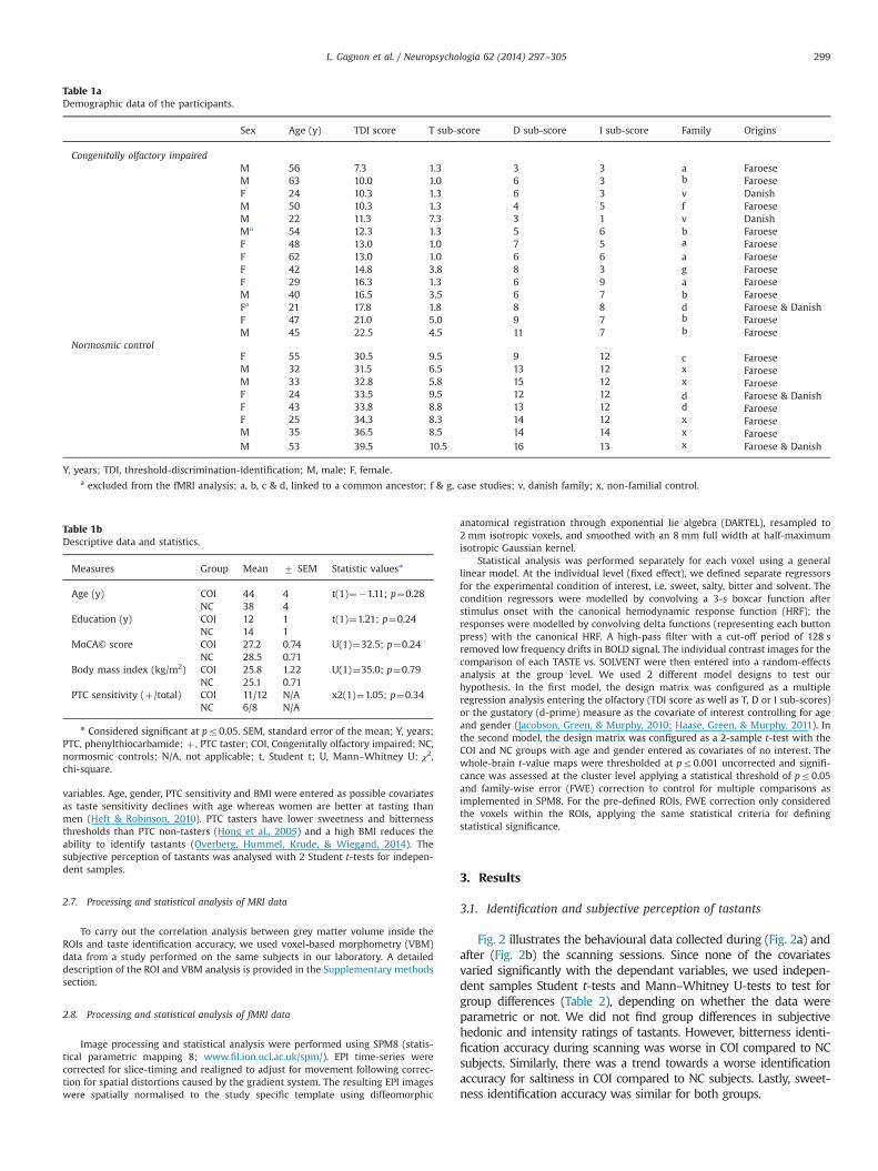
variables. Age, gender, PTC sensitivity and BMI were entered as possible covariatesas taste sensitivity declines with age whereas women are better at tasting thanmen (Heft & Robinson, 2010). PTC tasters have lower sweetness and bitternessthresholds than PTC non-tasters (Hong et al., 2005) and a high BMI reduces theability to identify tastants (Overberg, Hummel, Krude, & Wiegand, 2014). Thesubjective perception of tastants was analysed with 2 Student t-tests for indepen-dent samples.
2.7. Processing and statistical analysis of MRI data
To carry out the correlation analysis between grey matter volume inside theROIs and taste identification accuracy, we used voxel-based morphometry (VBM)data from a study performed on the same subjects in our laboratory. A detaileddescription of the ROI and VBM analysis is provided in the Supplementary methodssection.
2.8. Processing and statistical analysis of fMRI data
Image processing and statistical analysis were performed using SPM8 (statis-tical parametric mapping 8; www.fil.ion.ucl.ac.uk/spm/). EPI time-series werecorrected for slice-timing and realigned to adjust for movement following correc-tion for spatial distortions caused by the gradient system. The resulting EPI imageswere spatially normalised to the study specific template using diffeomorphic
anatomical registration through exponential lie algebra (DARTEL), resampled to2 mm isotropic voxels, and smoothed with an 8 mm full width at half-maximumisotropic Gaussian kernel.
Statistical analysis was performed separately for each voxel using a generallinear model. At the individual level (fixed effect), we defined separate regressorsfor the experimental condition of interest, i.e. sweet, salty, bitter and solvent. Thecondition regressors were modelled by convolving a 3-s boxcar function afterstimulus onset with the canonical hemodynamic response function (HRF); theresponses were modelled by convolving delta functions (representing each buttonpress) with the canonical HRF. A high-pass filter with a cut-off period of 128 sremoved low frequency drifts in BOLD signal. The individual contrast images for thecomparison of each TASTE vs. SOLVENT were then entered into a random-effectsanalysis at the group level. We used 2 different model designs to test ourhypothesis. In the first model, the design matrix was configured as a multipleregression analysis entering the olfactory (TDI score as well as T, D or I sub-scores)or the gustatory (d-prime) measure as the covariate of interest controlling for ageand gender (Jacobson, Green, & Murphy, 2010; Haase, Green, & Murphy, 2011). Inthe second model, the design matrix was configured as a 2-sample t-test with theCOI and NC groups with age and gender entered as covariates of no interest. Thewhole-brain t-value maps were thresholded at pr0.001 uncorrected and signifi-cance was assessed at the cluster level applying a statistical threshold of pr0.05and family-wise error (FWE) correction to control for multiple comparisons asimplemented in SPM8. For the pre-defined ROIs, FWE correction only consideredthe voxels within the ROIs, applying the same statistical criteria for definingstatistical significance.
3. Results
3.1. Identification and subjective perception of tastants
Fig. 2 illustrates the behavioural data collected during (Fig. 2a) andafter (Fig. 2b) the scanning sessions. Since none of the covariatesvaried significantly with the dependant variables, we used indepen-dent samples Student t-tests and Mann–Whitney U-tests to test forgroup differences (Table 2), depending on whether the data wereparametric or not. We did not find group differences in subjectivehedonic and intensity ratings of tastants. However, bitterness identi-fication accuracy during scanning was worse in COI compared to NCsubjects. Similarly, there was a trend towards a worse identificationaccuracy for saltiness in COI compared to NC subjects. Lastly, sweet-ness identification accuracy was similar for both groups.
Table 1bDescriptive data and statistics.
Measures Group Mean 7 SEM Statistic valuesn
Age (y) COI 44 4 t(1)¼�1.11; p¼0.28NC 38 4
Education (y) COI 12 1 t(1)¼1.21; p¼0.24NC 14 1
MoCA© score COI 27.2 0.74 U(1)¼32.5; p¼0.24NC 28.5 0.71
Body mass index (kg/m2) COI 25.8 1.22 U(1)¼35.0; p¼0.79NC 25.1 0.71
PTC sensitivity (þ/total) COI 11/12 N/A x2(1)¼1.05; p¼0.34NC 6/8 N/A
n Considered significant at pr0.05. SEM, standard error of the mean; Y, years;PTC, phenylthiocarbamide; þ , PTC taster; COI, Congenitally olfactory impaired; NC,normosmic controls; N/A, not applicable; t, Student t; U, Mann–Whitney U; χ2,chi-square.
Table 1aDemographic data of the participants.
Sex Age (y) TDI score T sub-score D sub-score I sub-score Family Origins
Congenitally olfactory impairedM 56 7.3 1.3 3 3 a FaroeseM 63 10.0 1.0 6 3 b FaroeseF 24 10.3 1.3 6 3 v DanishM 50 10.3 1.3 4 5 f FaroeseM 22 11.3 7.3 3 1 v DanishMa 54 12.3 1.3 5 6 b FaroeseF 48 13.0 1.0 7 5 a FaroeseF 62 13.0 1.0 6 6 a FaroeseF 42 14.8 3.8 8 3 g FaroeseF 29 16.3 1.3 6 9 a FaroeseM 40 16.5 3.5 6 7 b FaroeseFa 21 17.8 1.8 8 8 d Faroese & DanishF 47 21.0 5.0 9 7 b FaroeseM 45 22.5 4.5 11 7 b Faroese
Normosmic controlF 55 30.5 9.5 9 12 c FaroeseM 32 31.5 6.5 13 12 x FaroeseM 33 32.8 5.8 15 12 x FaroeseF 24 33.5 9.5 12 12 d Faroese & DanishF 43 33.8 8.8 13 12 d FaroeseF 25 34.3 8.3 14 12 x FaroeseM 35 36.5 8.5 14 14 x FaroeseM 53 39.5 10.5 16 13 x Faroese & Danish
Y, years; TDI, threshold-discrimination-identification; M, male; F, female.a excluded from the fMRI analysis; a, b, c & d, linked to a common ancestor; f & g, case studies; v, danish family; x, non-familial control.
L. Gagnon et al. / Neuropsychologia 62 (2014) 297–305 299

3.2. Imaging results
Based on our behavioural results, we focused our fMRI analysison the brain activity for bitterness, the tastant that COI subjectshad the most difficulty to identify. Post-hoc analyses for sweet andsalty are presented in the Supplementary materials section.
Fig. 3 illustrates the lower BOLD response in COI compared to NCsubjects in bitterness-induced brain responses. The ROI analysis revea-led that only the left mOFC was significantly less activated in COIcompared to NC subjects when tasting bitterness (pFWE-cluster¼0.04).
Fig. 4 shows the positive correlations between the threshold (T)sub-score (Fig. 4a) or total TDI score (Fig. 4b) and BOLD signal(Fig. 4b) and BOLD signal changes for bitterness. The whole brainanalysis reveals 3 significant clusters in the left frontal lobe thatvaried with the olfactory threshold, namely in the medial orbito-frontal cortex, medial superior frontal gyrus and pars triangularis ofthe inferior frontal gyrus (Table 3). None of the other olfactorymeasures significantly co-varied with changes in BOLD signal forbitterness. In line with this, our ROI analysis further revealed thatthe T sub-score, but not the discrimination (D) and identification(I) sub-scores, varied statistically with BOLD signal changes forbitterness. More specifically, BOLD signal changes correlated posi-tively with the olfactory threshold in mOFC (Fig. 4a; pFWE-
cluster¼0.01) and anterior insula (Fig. 4a; pFWE-cluster¼0.04). Whenusing the total TDI score, there was a significant linear relationshipwith the regional BOLD response in the mOFC (Fig. 4b; pFWE-
cluster¼0.05). Finally, both the whole-brain and the ROI analysis
reveal that the ability to identify bitterness did not vary significantlywith the BOLD signal while tasting.
Fig. 5 illustrates the strong positive correlation between mOFCgrey matter volume (as measured by VBM) and bitterness identi-fication. Whole brain analysis revealed two significant clusters inbilateral rectus gyri. In line with this, the ROI analysis furtherconfirmed that the bilateral mOFC was the only area showing asignificant correlation between bitter identification and greymatter volume (Fig. 5). A summary of all the whole-brain andROI analysis is provided in Tables 3 and 4, respectively.
4. Discussion
Compared to normosmic controls, congenitally olfactory impairedsubjects exhibit bitterness identification impairment combined with areduced activation of the deprived higher-order olfactory cortices.Moreover, the grey matter volume within the mOFC correlatespositively with bitterness identification accuracy, demonstrating afunctional and structural underpinning of this taste impairment.
Our results are consistent with previous studies showingsimilar taste deficits in isolated congenitally (Levy, Degnan, Sethi,& Henkin, 2013) and acquired (Gudziol et al., 2007; Landis et al.,2010) anosmic patients. Among the three tastants used in ourexperiment, bitterness was the most difficult to identify byolfactory impaired subjects. This is in accordance with Gudziol'sstudy (2007) where bitterness was also the most difficult taste to
Fig. 1. Mixed event-related design illustrating one stimulus presentation. Participants were asked to fixate a cross and to follow the (Danish) written cues. A default screenidentifies the nature of the four computer mouse keys (two for each hand) used to identify the sweet, salty, bitter or neutral tastes. The signal “Ready” warns the participantof the taste delivery. After receiving the tastant, the subject keeps the liquid in his mouth while he/she responds. Then, he/she swallows the liquid according to theinstruction on the screen, rinses his mouth with deionised water and swallows a second time before the next trial.
Fig. 2. Bar charts showing the mean7standard error of the mean (SEM) for taste identification accuracy (A) and subjective perception of tastants (B) in congenitallyolfactory impaired (COI) and normosmic control (NC) subjects. The asterisk shows significant results at pr0.05. COI have significantly worse bitter identification accuracythan NC subjects but similar identification accuracy for sweet and salty. The groups did not differ for taste hedonic and intensity ratings.
L. Gagnon et al. / Neuropsychologia 62 (2014) 297–305300

identify by acquired anosmia subjects, followed by salty and acid.Interestingly, the bitterness impairment in COI subjects was notrelated to PTC sensitivity, as a similar number of PTC tasters andnon-tasters were present in both populations. Bitterness is tradi-tionally considered as the taste signalling the potential presence ofpoisonous substances, such as alkaloids, and therefore threat anddanger (Hladik, Pasquet, & Simmen, 2002). This view is interestingas anosmic subjects are also impaired in recognising variousenvironmental hazards involving foods. For example, they “scorchfood” and eat “spoiled foods” more often than normosmic subjects(Croy et al., 2012). The failure to identify bitterness might increasethe ingestion of scorched food and/or unfamiliar poisonous foods(such as exotic berries) and, in this way, contribute to increase thefrequency of food accidents.
The fMRI data further revealed that COI subjects less stronglyrecruit their mOFC compared to NC when tasting bitterness.Interestingly, we also found a positive correlation between odourperformance and BOLD signal changes for bitterness in regions
that integrate taste and smell information, namely the mOFC andanterior insula. Taken together, our data indicate that olfactionmight have an influence on taste perception since congenitalolfactory impairment leads to a lower signal in flavour processingbrain areas during tasting.
4.1. Medial orbitofrontal cortex
The lower BOLD signal observed in the mOFC of our COIparticipants is in accordance with structural and metabolicchanges within this region following the loss of smell perception.These changes include grey matter volume reductions (Bitter,Bruderle, Gudziol, Burmeister, & Guntinas-Lichius, 2010; Bitter,Gudziol et al., 2010), increased grey matter thickness (Frasnelli,Fark, Lehmann, Gerber, & Hummel, 2013), hypometabolism(Varney, Pinkston, & Wu, 2001) and hypoperfusion (Eftekhari etal., 2006; Atighechi et al., 2009). Two previous studies reported apositive correlation between olfactory performance and greymatter thickness in the mOFC in healthy individuals (Frasnelli etal., 2010; Seubert, Freiherr, Frasnelli, Hummel, & Lundstrom, 2012).Our results additionally show that grey matter volume withinmOFC varies positively with bitterness identification.
The mOFC is a chemosensory area involved in the hedonicperception of flavours (Rolls, Grabenhorst, Margot, da Silva, &Velazco, 2008; Grabenhorst & Rolls, 2008), olfactory-taste learning(Rolls, 2011), processing of palatable odours (Small, Gerber, Mak, &Hummel, 2005a) and sensory-specific satiation (Rolls, 2008;Pritchard et al., 2008). The cluster observed in our study has beenreported as pivotal structure in decision-making related to thehedonic valence of odours (Rolls, Grabenhorst, & Parris, 2010),i.e. comparing whether an odour is more or less pleasant. It ispreferentially activated during extinction learning after an olfac-tory conditioning task (Gottfried & Dolan, 2004). Extinctionhappens when a response (e.g. eating) previously reinforced (e.g.hunger) is no longer effective. Extinction can be facilitated bysensory-specific satiation also known as “boredom with flavour”,a phenomenon that happens after repeated exposure to the samefood. Boredom with flavour describes the decline in the perceivedpleasantness of a food during its consumption. Moreover, con-genitally anosmic subjects maintain their hedonic evaluation atmore stable levels than normosmics (Novakova, Bojanwski,Havlicek, & Croy, 2012). Our results suggest that the lower BOLDsignal observed in the mOFC of congenitally olfactory impaired
Table 2Behavioural data and statistics.
Measures Group Mean 7 SEM Statistic valuesn
Taste identification d-primeSweet COI 2.43 0.31 t(1)¼1.26, p¼0.23
NC 3.00 0.30Salty COI 2.95 0.31 U(1)¼29.5, p¼0.16
NC 3.59 0.16Bitter COI 2.41 0.18 t(1)¼2.14, p¼0.05n
NC 3.06 0.25Subjective ratings
Sweet Hedonic COI 3.00 0.41 t(1)¼1.52, p¼0.15NC 3.75 0.25
Intensity COI 2.56 0.24 U(1)¼32.0, p¼0.74NC 2.75 0.31
Salty Hedonic COI 1.67 0.29 U(1)¼31.5, p¼0.67NC 1.88 0.35
Intensity COI 4.00 0.37 U(1)¼31.5, p¼0.67NC 4.25 0.37
Bitter Hedonic COI 1.11 0.11 U(1)¼21.0, p¼0.17NC 1.88 0.40
Intensity COI 3.88 0.35 U(1)¼21.5, p¼0.17NC 4.56 0.20
n Considered significant at pr0.05. SEM, standard error of the mean;COI, congenitally olfactory impaired; NC, normosmic control; t, Student t; U,Mann–Whitney U.
Fig. 3. T-statistical map and bar chart showing areas and contrast estimates (7 90% confidence interval) where BOLD signal increases are significantly lower in congenitallyolfactory impaired (COI) compared to normosmic controls (NC) when tasting bitterness. The coordinate over the slice refers to the Montreal Neurological Institute (MNI)space. The threshold is set at p¼0.001, uncorrected, and activation maps are displayed on the group template. The right hemisphere corresponds to the right of the image.
L. Gagnon et al. / Neuropsychologia 62 (2014) 297–305 301

might provide a neurological basis for reduced sensory-specificsatiation.
4.2. Anterior Insula
The anterior insula is another region where we observed a positivecorrelation between the bitterness-induced BOLD signal changes andthe smell performance, such that subjects that easily detected anodour activate more strongly this area when tasting bitterness.
The anterior insula is considered as the primary taste cortex(Ogawa et al., 2005; Veldhuizen et al., 2011) and plays an important
role in taste intensity and pleasantness coding (Grabenhorst & Rolls,2008; Bender, Veldhuizen, Meltzer, Gitelman, & Small, 2009; Ohla,Toepel, le Coutre, & Hudry, 2010; Cerf-Ducastel, Haase, & Murphy,2012), interoception and self-awareness (Bud Craig, 2009).This region also processes visceral, tactile, pain (Hummel, Iannilli,Frasnelli, Boyle, & Gerber, 2009) and olfactory information (Cerf-Ducastel & Murphy, 2001; De Araujo et al., 2003) and is consideredas a flavour-processing node (Small et al., 2013). Our data supportthe results of a previous study reporting a lower insular activity in agroup of congenitally and acquired anosmic subjects comparedto normosmic participants during trigeminal processing (Iannilli
Fig. 4. Scatter plots and T-statistical maps showing areas where BOLD signal increases when tasting bitterness strongly correlate with the ability to smell. A: The olfactorydetection threshold (T sub-score) varies positively with the BOLD signal (mean eigenvalues) in bilateral mOFC and anterior insula. B: The total Sniffin' Sticks TDI score variespositively with the BOLD signal in bilateral mOFC. The coordinates over the slices refer to the Montreal Neurological Institute (MNI) space. The visualisation threshold is set atp¼0.001, uncorrected, and activation maps are displayed on the group template. The right hemisphere corresponds to the right of the image. Filled circles, congenitallyolfactory impaired (COI); Open circles, normosmic controls (NC). T, threshold; TDI, threshold-discrimination-identification; R, Pearson correlation coefficient.
L. Gagnon et al. / Neuropsychologia 62 (2014) 297–305302
et al., 2007). Interestingly, similarly to our COI cohort, Small,Bernasconi, Bernasconi, Sziklas, & Jones-Gotman (2005b) also foundelevated taste recognition and detection thresholds in a case-reportstudy of a patient without left insula and a mild atrophy of the rightinsula (together with mild atrophy of left orbitofrontal cortex andsevere bilateral atrophy of piriform cortex). Furthermore,Veldhuizen, Douglas, Aschenbrenner, Gitelman, & Small (2011)found that the anterior insula was more activated when subjectsdrank an unexpected sweet stimulus, i.e. when the taste expecta-tion was breached. Expectations about foods tastes are ecologicallyacquired by visual and mainly smell information prior to ingestion.Anosmic patients who are restricted to visual food cues can beeasily impaired in creating proper expectations, and therefore willbe more prone to judgement errors (Croy et al., 2012). It is possiblethat for subjects impaired in smell detection, the lack of reliabletaste expectations dampens tasting-induced insular activations.
4.3. Piriform cortex
This region was not recruited by either group of subjects duringtasting. The piriform cortex is the main primary olfactory area anda recent study has recently found that it processes taste informa-tion in normosmic and anosmic rats (Maier et al., 2012). The lackof piriform activity in our study could be explained by the durationof the olfactory impairment together with the small size of thisregion and/or lack of statistical power. However, our data are morein favour of the classical hierarchical view of taste-odour conver-gence that happens in the orbitofrontal cortex (Small et al., 2013).
Furthermore, we show that the plasticity of a chemical senselargely differs from that of audition and vision, also known as thedistal senses. Whereas the deprived “visual” cortex of congenitallyblind subjects processes stimuli from other modalities like touch,hearing or olfaction (Kupers & Ptito, 2013), our data do not lend tosupport the hypothesis that the deprived “olfactory” piriformcortex of congenitally olfactory impaired subjects becomesinvaded by taste. In the case of blindness (or deafness), cortico-cortical and thalamo-cortical connections to and from the visual(or auditory) cortex are modified in order to allow cross-modalplasticity to take place (reviews in Merabet & Pascual-Leone, 2010;Kupers & Ptito, 2013). Further studies are needed to confirm ifsimilar adaptive structural changes, such as weaker connectionsbetween primary olfactory areas and the mOFC, also take place inthe brain of isolated congenitally anosmic subjects.
5. Conclusion
In conclusion, while olfaction amplifies taste perception andtaste processing, life-long olfactory impairment might have detri-mental effect on this modality. This is supported by behaviouraland functional imaging results. In sharp contrast with congenitalblindness or congenital deafness, crossmodal plasticity does notseem to take place in congenital olfactory impairment.
Table 3Summary of the whole-brain analysis.
Brain Region BA MNIcoordinates
clustersize
peakp
peakT
x y z
fMRIPositive correlation with T-score
Medial orbitofrontal L 11 �6 32 �18 393 0.01 6.78Medial superior frontalgyrus
L 10 �6 64 4 691 0.00 6.28
Inferior frontal gyrus L 45 �50 22 6 594 0.00 5.53VBMPositive correlation with bitter d-prime
Rectus gyrus R 11 10 39 �22 991 0.05 4.97L 11 �6 24 �27 1066 0.03 7.34
Peak p reflects the probability of the cluster using the FWE correction for multipletests. The cluster size is expressed in voxels. MNI, Montreal Neurological Institute;R, right; L, left; BA, Brodman area.
Fig. 5. Scatter plot and T-statistical map showing areas where VBM values correlate with bitter identification accuracy. The bitter identification d-prime varies positivelywith the volume of grey matter within bilateral mOFC. The coordinate over the slice refers to the Montreal Neurological Institute (MNI) space. The visualisation threshold isset at p¼0.001, uncorrected, and VBM map is displayed on the group template. The right hemisphere corresponds to the right of the image. Filled circles, congenitallyolfactory impaired (COI); Open circles, normosmic controls (NC). R, Pearson correlation coefficient.
Table 4Summary of the ROI analysis.
Brain Region cluster size peak p peak T Local maximum*
x y z
fMRICOIoNC
Medial orbitofrontal L 76 0.04 5.10 �6 32 �18Positive correlation with T-score
Medial orbitofrontal L 207 0.01 6.78 �6 32 �18Anterior insula L 44 0.04 4.88 �42 14 �6
Positive correlation with TDI-scoreMedial orbitofrontal L 54 0.05 5.08 �6 32 �18
VBMPositive correlation with bitter d-prime
Medial orbitofrontal R 821 0.00 4.97 9 40 �22L 941 0.00 7.34 �6 24 �27
n MNI coordinates of the local maximum. Peak p reflects the probability of thecluster using the FWE correction for multiple tests. The cluster size is expressed invoxels. COI, congenitally olfactory impaired; NC, normosmic controls; R, right;L, left.
L. Gagnon et al. / Neuropsychologia 62 (2014) 297–305 303

Author contributions
Conceived and designed the experiments: LG, KM, HS, NT, RK,MP. Performed the experiments: LG, HGK, MV. Analysed the data:LG, MV, KM. Wrote the paper: LG, MV, RK, MP.
Acknowledgements
The authors are indebted to the subjects who participated tothe experiment. The authors would like to acknowledge Dr. BjarkiEllefsen and Dr. Bjarki Djurhuus for recruiting and examining theparticipants, and Dr. Anne-Mette Leffers and Dr. Camilla Klausenfor evaluating the MR images. This work is financially supportedby the Danish National Research Foundation (Grant No. DNRF48),the Lundbeck Foundation (Grant No. R32-A2947), Chevron Texacoand the Faroese Research Council (Grant No. 0313).
Appendix A. Supporting information
Supplementary data associated with this paper can be found inthe online version at http://dx.doi.org/10.1016/j.neuropsychologia.2014.07.018.
References
Aschenbrenner, K., Hummel, C., Teszmer, K., Krone, F., Ishimaru, T., Seo, H. S., &Hummel, T. (2008). The influence of olfactory loss on dietary behaviors.Laryngoscope, 118, 135–144.
Atighechi, S., Salari, H., Baradarantar, M. H., Jafari, R., Karimi, G., & Mirjali, M. (2009).A comparative study of brain perfusion single-photon emission computedtomography and magnetic resonance imaging in patients with post-traumaticanosmia. American Journal of Rhinology Allergy, 23, 409–412.
Auvray, M., & Spence, C. (2008). The multisensory perception of flavor. Conscious-ness and Cognition, 17, 1016–1031.
Bender, G., Veldhuizen, M. G., Meltzer, J. A., Gitelman, D. R., & Small, D. M. (2009).Neural correlates of evaluative compared with passive tasting. European Journalof Neuroscience, 30, 327–338.
Bitter, T., Bruderle, J., Gudziol, H., Burmeister, H. P., Gaser, C., & Guntinas-Lichius, O.(2010). Gray and white matter reduction in hyposmic subjects–A voxel-basedmorphometry study. Brain Research, 1347, 42–47.
Bitter, T., Gudziol, H., Burmeister, H. P., Mentzel, H. J., Guntinas-Lichius, O., & Gaser,C. (2010). Anosmia leads to a loss of gray matter in cortical brain areas. ChemicalSenses, 35, 407–415.
Bud Craig, A. D. (2009). How do you feel–now? The anterior insula and humanawareness. Nature Review Neuroscience, 10, 59–70.
Cerf-Ducastel, B., Haase, L., & Murphy, C. (2012). Effect of magnitude estimation ofpleasantness and intensity on fMRI activation to taste. Chemosens Percept, 5,100–109.
Cerf-Ducastel, B., & Murphy, C. (2001). fMRI activation in response to odorantsorally delivered in aqueous solutions. Chemical Senses, 26, 625–637.
Croy, I., Negoias, S., Novakova, L., Landis, B. N., & Hummel, T. (2012). Learning aboutthe functions of the olfactory system from people without a sense of smell. PLoSONE, 7, e33365.
De Araujo, I. E., Rolls, E. T., Kringelbach, M. L., McGlone, F., & Phillips, N. (2003).Taste-olfactory convergence, and the representation of the pleasantness offlavour, in the human brain. Europen Journal of Neuroscience, 18, 2059–2068.
Eftekhari, M., Assadi, M., Kazemi, M., Saghari, M., Mojtahedi, A., Fard-Esfahani, A.,Sichani, B. F., & Beiki, D. (2006). Brain perfusion single photon emissioncomputed tomography findings in patients with posttraumatic anosmia andcomparison with radiological imaging. American Journal of Rhinology, 20,577–581.
Ferris, A., Schlitzer, J., Schierberl, M., Catalanotto, F., Gent, J., Peterson, M.,Bartoshuk, L., Cain, W., Goodspeed, R., Leonard, G., & Donaldson, J. (1985).Anosmia and nutritional status. Nutrition Research, 5, 149–156.
Frasnelli, J., Fark, T., Lehmann, J., Gerber, J., & Hummel, T. (2013). Brain structure ischanged in congenital anosmia. Neuroimage.
Frasnelli, J., Lundstrom, J. N., Boyle, J. A., Djordjevic, J., Zatorre, R. J., & Jones-Gotman,M. (2010). Neuroanatomical correlates of olfactory performance. ExperimentalBrain Research, 201, 1–11.
Frasnelli, J., Schuster, B., & Hummel, T. (2007a). Subjects with congenital anosmiahave larger peripheral but similar central trigeminal responses. Cerebral Cortex,17, 370–377.
Frasnelli, J., Schuster, B., & Hummel, T. (2007b). Interactions between olfaction andthe trigeminal system: what can be learned from olfactory loss. Cerebral Cortex,17, 2268–2275.
Frasnelli, J., Schuster, B., & Hummel, T. (2010). Olfactory dysfunction affectsthresholds to trigeminal chemosensory sensations. Neuroscience Letters, 468,259–263.
Gottfried, J. A., & Dolan, R. J. (2004). Human orbitofrontal cortex mediatesextinction learning while accessing conditioned representations of value.Nature Neuroscience, 7, 1144–1152.
Grabenhorst, F., & Rolls, E. T. (2008). Selective attention to affective value alters howthe brain processes taste stimuli. European Journal of Neuroscience, 27, 723–729.
Gudziol, H., Rahneberg, K., & Burkert, S. (2007). Anosmics are more poorly able totaste than normal persons. Laryngorhinootologie, 86, 640–643.
Gudziol, H., Schubert, M., & Hummel, T. (2001). Decreased trigeminal sensitivity inanosmia. ORL Journal for Otorhinolaryngology and its Related Specialties, 63,72–75.
Haase, L., Green, E., & Murphy, C. (2011). Males and females show differential brainactivation to taste when hungry and sated in gustatory and reward areas.Appetite, 57, 421–434.
Hasan, K. S., Reddy, S. S., & Barsony, N. (2007). Taste perception in kallmannsyndrome, a model of congenital anosmia. Endocrine Practise, 13, 716–720.
Heft, M. W., & Robinson, M. E. (2010). Age differences in orofacial sensorythresholds. Journal of Dental Research, 89, 1102–1105.
Hladik, C. M., Pasquet, P., & Simmen, B. (2002). New perspectives on taste andprimate evolution: the dichotomy in gustatory coding for perception ofbeneficent versus noxious substances as supported by correlations amonghuman thresholds. American Journal of Physical Anthropology, 117, 342–348.
Hong, J. H., Chung, J. W., Kim, Y. K., Chung, S. C., Lee, S. W., & Kho, H. S. (2005). Therelationship between PTC taster status and taste thresholds in young adults.Oral Surgery, Oral Medicine, Oral Pathology, Oral Radiology, and Endodontics, 99,711–715.
Hummel, T., Iannilli, E., Frasnelli, J., Boyle, J., & Gerber, J. (2009). Central processingof trigeminal activation in humans. Annals of the New York Academy of Sciences,1170, 190–195.
Hummel, T., Kobal, G., Gudziol, H., & Mackay-Sim, A. (2007). Normative data for the“Sniffin’ Sticks” including tests of odor identification, odor discrimination, andolfactory thresholds: an upgrade based on a group of more than 3000 subjects.European Archives of Otorhinolaryngology, 264, 237–243.
Hummel, T., Sekinger, B., Wolf, S. R., Pauli, E., & Kobal, G. (1997). 'Sniffin' sticks':olfactory performance assessed by the combined testing of odor identification,odor discrimination and olfactory threshold. Chemical Senses, 22, 39–52.
Iannilli, E., Gerber, J., Frasnelli, J., & Hummel, T. (2007). Intranasal trigeminalfunction in subjects with and without an intact sense of smell. Brain Research,1139, 235–244.
Jacobson, A., Green, E., & Murphy, C. (2010). Age-related functional changes ingustatory and reward processing regions: an fMRI study. Neuroimage, 53,602–610.
Karstensen, H., & Tommerup, N. (2011). Isolated and syndromic forms of congenitalanosmia. Clinical Genetics, 81, 210–215.
Kupers, R., & Ptito, M. (2013). Compensatory plasticity and cross-modal reorganiza-tion following early visual deprivation. Neuroscience and Biobehavioral Reviews.
Landis, B. N., Scheibe, M., Weber, C., Berger, R., Bramerson, A., Bende, M., Nordin, S.,& Hummel, T. (2010). Chemosensory interaction: acquired olfactory impair-ment is associated with decreased taste function. Journal of Neurology, 257,1303–1308.
Levy, L. M., Degnan, A. J., Sethi, I., & Henkin, R. I. (2013). Anatomic olfactorystructural abnormalities in congenital smell loss: magnetic resonance imagingevaluation of olfactory bulb, groove, sulcal, and hippocampal morphology.Journal of Computer Assisted Tomography, 37, 650–657.
Lundstrom, J. N., Boesveldt, S., & Albrecht, J. (2011). Central processing of thechemical senses: an overview. ACS Chemical Neuroscience, 2, 5–16.
Maier, J. X., Wachowiak, M., & Katz, D. B. (2012). Chemosensory convergence onprimary olfactory cortex. Journal of Neuroscience, 32, 17037–17047.
Mattes, R. D., & Cowart, B. J. (1994). Dietary assessment of patients withchemosensory disorders. Journal of the American Dietetic Association, 94, 50–56.
Merabet, L. B., & Pascual-Leone, A. (2010). Neural reorganization following sensoryloss: the opportunity of change. Nature Reviews Neuroscience, 11, 44–52.
Miwa, T., Furukawa, M., Tsukatani, T., Costanzo, R. M., DiNardo, L. J., & Reiter, E. R.(2001). Impact of olfactory impairment on quality of life and disability. Archivesof Otolaryngology Head and Neck Surgery, 127, 497–503.
Novakova, L., Bojanowski, V., Havlicek, J., & Croy, I. (2012). Differential patterns offood appreciation during consumption of a simple food in congenitally anosmicindividuals: an explorative study. PLoS ONE, 7, e33921.
Ogawa, H., Wakita, M., Hasegawa, K., Kobayakawa, T., Sakai, N., Hirai, T., Yamashita,Y., & Saito, S. (2005). Functional MRI detection of activation in the primarygustatory cortices in humans. Chemical Senses, 30, 583–592.
Ohla, K., Toepel, U., le Coutre, J., & Hudry, J. (2010). Electrical neuroimaging revealsintensity-dependent activation of human cortical gustatory and somatosensoryareas by electric taste. Biological Psychology, 85, 446–455.
Oldfield, R. C. (1971). The assessment and analysis of handedness: the Edinburghinventory. Neuropsychologia, 9, 97–113.
Overberg, J., Hummel, T., Krude, H., & Wiegand, S. (2014). Differences in tastesensitivity between obese and non-obese children and adolescents. Archives ofDisease in Childhood, 97, 1048–1052.
Pritchard, T. C., Nedderman, E. N., Edwards, E. M., Petticoffer, A. C., Schwartz, G. J., &Scott, T. R. (2008). Satiety-responsive neurons in the medial orbitofrontal cortexof the macaque. Behavioral Neuroscience, 122, 174–182.
L. Gagnon et al. / Neuropsychologia 62 (2014) 297–305304
Rolls, E. T. (2001). The rules of formation of the olfactory representations found inthe orbitofrontal cortex olfactory areas in primates. Chemical Senses, 26,595–604.
Rolls, E. T. (2008). Functions of the orbitofrontal and pregenual cingulate cortex intaste, olfaction, appetite and emotion. Acta Physiologica Hungarica, 95, 131–164.
Rolls, E. T. (2011). Chemosensory learning in the cortex. Frontiers in SystemsNeuroscience, 5, 13.
Rolls, E. T., Grabenhorst, F., Margot, C., da Silva, M. A., & Velazco, M. I. (2008).Selective attention to affective value alters how the brain processes olfactorystimuli. Journal of Cognitive Neuroscience, 20, 1815–1826.
Rolls, E. T., Grabenhorst, F., & Parris, B. A. (2010). Neural systems underlyingdecisions about affective odors. Journal of Cognitive Neuroscience, 22,1069–1082.
Seubert, J., Freiherr, J., Frasnelli, J., Hummel, T., & Lundstrom, J. N. (2012).Orbitofrontal cortex and olfactory bulb volume predict distinct aspects ofolfactory performance in healthy subjects. Cerebral Cortex, 23, 2448–2456.
Sewards, T. V. (2004). Dual separate pathways for sensory and hedonic aspects oftaste. Brain Research Bulletin, 62, 271–283.
Small, D. M., Bernasconi, N., Bernasconi, A., Sziklas, V., & Jones-Gotman, M. (2005b).Gustatory agnosia. Neurology, 64, 311–317.
Small, D. M., Gerber, J. C., Mak, Y. E., & Hummel, T. (2005a). Differential neuralresponses evoked by orthonasal versus retronasal odorant perception inhumans. Neuron, 47, 593–605.
Small, D. M., & Prescott, J. (2005). Odor/taste integration and the perception offlavor. Experimental Brain Research, 166, 345–357.
Small, D. M., Veldhuizen, M. G., & Green, B. (2013). Sensory neuroscience: tasteresponses in primary olfactory cortex. Current Biology, 23, R157–159.
Swets, J. A. (1961). Is there a sensory threshold - when effects of observers responsecriterion are isolated, a sensory limitation is not evident. Science, 134, 168.
Temmel, A. F., Quint, C., Schickinger-Fischer, B., Klimek, L., Stoller, E., & Hummel, T.(2002). Characteristics of olfactory disorders in relation to major causes ofolfactory loss. Archives of Otolaryngology Head Neck and Surgery, 128, 635–641.
Van Toller, S. V. (1999). Assessing the impact of anosmia: review of a question-naire's findings. Chemical Senses., 24, 705–712.
Varney, N. R., Pinkston, J. B., & Wu, J. C. (2001). Quantitative PET findings in patientswith posttraumatic anosmia. Journal of Head Trauma Rehabilitation, 16,253–259.
Veldhuizen, M. G., Albrecht, J., Zelano, C., Boesveldt, S., Breslin, P., & Lundstrom, J. N.(2011). Identification of human gustatory cortex by activation likelihoodestimation. Human Brain Mapping.
Veldhuizen, M. G., Douglas, D., Aschenbrenner, K., Gitelman, D. R., & Small, D. M.(2011). The anterior insular cortex represents breaches of taste identityexpectation. Journal of Neuroscience, 31, 14735–14744.
Verhagen, J. V., & Engelen, L. (2006). The neurocognitive bases of human multi-modal food perception: sensory integration. Neuroscience and BiobehavioralReviews, 30, 613–650.
L. Gagnon et al. / Neuropsychologia 62 (2014) 297–305 305
Related Documents