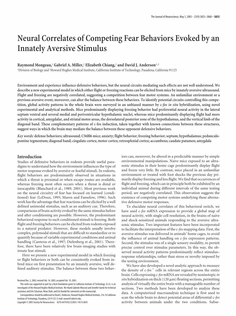
Neural Correlates of Competing Fear Behaviors Evoked by an Innately Aversive Stimulus Raymond Mongeau, 1 Gabriel A. Miller, 1 Elizabeth Chiang, 1 and David J. Anderson 1,2 1 Division of Biology and 2 Howard Hughes Medical Institute, California Institute of Technology, Pasadena, California 91125 Environment and experience influence defensive behaviors, but the neural circuits mediating such effects are not well understood. We describe a new experimental model in which either flight or freezing reactions can be elicited from mice by innately aversive ultrasound. Flight and freezing are negatively correlated, suggesting a competition between fear motor systems. An unfamiliar environment or a previous aversive event, moreover, can alter the balance between these behaviors. To identify potential circuits controlling this compe- tition, global activity patterns in the whole brain were surveyed in an unbiased manner by c-fos in situ hybridization, using novel experimental and analytical methods. Mice predominantly displaying freezing behavior had preferential neural activity in the lateral septum ventral and several medial and periventricular hypothalamic nuclei, whereas mice predominantly displaying flight had more activity in cortical, amygdalar, and striatal motor areas, the dorsolateral posterior zone of the hypothalamus, and the vertical limb of the diagonal band. These complementary patterns of c-fos induction, taken together with known connections between these structures, suggest ways in which the brain may mediate the balance between these opponent defensive behaviors. Key words: defense behaviors; ultrasound; C56Bl6 mice; anxiety; flight behavior; freezing behavior; septum; hypothalamus; pedunculo- pontine tegmentum; diagonal band; cingulate cortex; motor cortex; retrosplenial cortex; accumbens; caudate putamen; amygdala Introduction Studies of defensive behaviors in rodents provide useful para- digms to understand how the environment influences the type of motor response evoked by aversive or fearful stimuli. In rodents, flight behaviors are predominantly observed in situations in which a threat is proximal or when escape routes are available, whereas freezing most often occurs when a threat is distal or inescapable (Blanchard et al., 1989, 2001). Most previous work on the neural circuitry of fear has focused on learned (condi- tioned) fear (LeDoux, 1995; Maren and Fanselow, 1996). Such work has the advantage that fear reactions can be elicited by a well defined unimodal stimulus, such as an auditory cue. Therefore, comparisons of brain activity evoked by the same stimulus before and after conditioning are possible. However, the predominant behavioral response to such conditioned stimuli is freezing. Both flight and freezing behaviors can be elicited from rodents exposed to a natural predator. However, these models usually involve complex, polymodal stimuli that are difficult to standardize or to quantify because of variable experimental conditions and animal handling (Canteras et al., 1997; Dielenberg et al., 2001). There- fore, there have been relatively few brain-imaging studies with innate fear stimuli. Here we present a new experimental model in which freezing or flight behaviors or both can be consistently evoked from in- bred mice on first presentation by an innately aversive, well de- fined auditory stimulus. The balance between these two behav- iors can, moreover, be altered in a predictable manner by simple environmental manipulations. Naive mice exposed to an ultra- sonic stimulus in their home cage predominantly display flight and freeze very little. By contrast, mice placed in an unfamiliar environment or treated with foot shocks the previous day pri- marily display freezing and less flight. We find that occurrences of flight and freezing, which can in principle both be exhibited by an individual animal during different intervals of the same testing period, are negatively correlated. This observation suggests the existence of competing motor systems underlying these alterna- tive defensive motor responses. To elucidate neural correlates of this behavioral switch, we have used c-fos mRNA expression to provide a global map of neural activity, with single-cell resolution, in the brains of naive and shock-sensitized animals responding to the aversive ultra- sonic stimulus. Two important features have been incorporated to facilitate the interpretation of the c-fos mapping data. First, the aversive stimulus was delivered in animals’ home cages, to avoid the influence of animal handling on c-fos expression patterns. Second, the stimulus was of a single sensory modality, to permit precise control over stimulus parameters. In this way, the ob- served neural activity patterns predominantly reflect stimulus– response relationships, rather than stress or novelty imposed by the testing environment. We have also developed a novel analytic approach to measure the density of c-fos cells in relevant regions across the entire brain. Cells expressing c-fos mRNA are revealed by nonisotopic in situ hybridization on thick (120 m) floating sections, permitting analysis of virtually the entire brain with a manageable number of sections. Two methods have been developed to analyze these data. A computerized macroanalysis technique is first used to scan the whole brain to detect potential areas of differential c-fos activity between animals under the two conditions. Subse- Received Dec. 2, 2002; revised Feb. 14, 2003; accepted Feb. 19, 2003. This work was supported in part by a Keck foundation grant to California Institute of Technology. D.J.A. is an investigator of the Howard Hughes Medical Institute. We thank Gabriele Mosconi and Jennifer Uyeda for technical assistance and Erin Schuman, Nirao Shah, and Eric Kandel for comments on this manuscript. Correspondence should be addressed to David J. Anderson, Howard Hughes Medical Institute, 216-76 California Institute of Technology, Pasadena, CA 91125. E-mail: [email protected]. Copyright © 2003 Society for Neuroscience 0270-6474/03/233855-14$15.00/0 The Journal of Neuroscience, May 1, 2003 • 23(9):3855–3868 • 3855

Welcome message from author
This document is posted to help you gain knowledge. Please leave a comment to let me know what you think about it! Share it to your friends and learn new things together.
Transcript
Neural Correlates of Competing Fear Behaviors Evoked by anInnately Aversive Stimulus
Raymond Mongeau,1 Gabriel A. Miller,1 Elizabeth Chiang,1 and David J. Anderson1,2
1Division of Biology and 2Howard Hughes Medical Institute, California Institute of Technology, Pasadena, California 91125
Environment and experience influence defensive behaviors, but the neural circuits mediating such effects are not well understood. Wedescribe a new experimental model in which either flight or freezing reactions can be elicited from mice by innately aversive ultrasound.Flight and freezing are negatively correlated, suggesting a competition between fear motor systems. An unfamiliar environment or aprevious aversive event, moreover, can alter the balance between these behaviors. To identify potential circuits controlling this compe-tition, global activity patterns in the whole brain were surveyed in an unbiased manner by c-fos in situ hybridization, using novelexperimental and analytical methods. Mice predominantly displaying freezing behavior had preferential neural activity in the lateralseptum ventral and several medial and periventricular hypothalamic nuclei, whereas mice predominantly displaying flight had moreactivity in cortical, amygdalar, and striatal motor areas, the dorsolateral posterior zone of the hypothalamus, and the vertical limb of thediagonal band. These complementary patterns of c-fos induction, taken together with known connections between these structures,suggest ways in which the brain may mediate the balance between these opponent defensive behaviors.
Key words: defense behaviors; ultrasound; C56Bl6 mice; anxiety; flight behavior; freezing behavior; septum; hypothalamus; pedunculo-pontine tegmentum; diagonal band; cingulate cortex; motor cortex; retrosplenial cortex; accumbens; caudate putamen; amygdala
IntroductionStudies of defensive behaviors in rodents provide useful para-digms to understand how the environment influences the type ofmotor response evoked by aversive or fearful stimuli. In rodents,flight behaviors are predominantly observed in situations inwhich a threat is proximal or when escape routes are available,whereas freezing most often occurs when a threat is distal orinescapable (Blanchard et al., 1989, 2001). Most previous workon the neural circuitry of fear has focused on learned (condi-tioned) fear (LeDoux, 1995; Maren and Fanselow, 1996). Suchwork has the advantage that fear reactions can be elicited by a welldefined unimodal stimulus, such as an auditory cue. Therefore,comparisons of brain activity evoked by the same stimulus beforeand after conditioning are possible. However, the predominantbehavioral response to such conditioned stimuli is freezing. Bothflight and freezing behaviors can be elicited from rodents exposedto a natural predator. However, these models usually involvecomplex, polymodal stimuli that are difficult to standardize or toquantify because of variable experimental conditions and animalhandling (Canteras et al., 1997; Dielenberg et al., 2001). There-fore, there have been relatively few brain-imaging studies withinnate fear stimuli.
Here we present a new experimental model in which freezingor flight behaviors or both can be consistently evoked from in-bred mice on first presentation by an innately aversive, well de-fined auditory stimulus. The balance between these two behav-
iors can, moreover, be altered in a predictable manner by simpleenvironmental manipulations. Naive mice exposed to an ultra-sonic stimulus in their home cage predominantly display flightand freeze very little. By contrast, mice placed in an unfamiliarenvironment or treated with foot shocks the previous day pri-marily display freezing and less flight. We find that occurrences offlight and freezing, which can in principle both be exhibited by anindividual animal during different intervals of the same testingperiod, are negatively correlated. This observation suggests theexistence of competing motor systems underlying these alterna-tive defensive motor responses.
To elucidate neural correlates of this behavioral switch, wehave used c-fos mRNA expression to provide a global map ofneural activity, with single-cell resolution, in the brains of naiveand shock-sensitized animals responding to the aversive ultra-sonic stimulus. Two important features have been incorporatedto facilitate the interpretation of the c-fos mapping data. First, theaversive stimulus was delivered in animals’ home cages, to avoidthe influence of animal handling on c-fos expression patterns.Second, the stimulus was of a single sensory modality, to permitprecise control over stimulus parameters. In this way, the ob-served neural activity patterns predominantly reflect stimulus–response relationships, rather than stress or novelty imposed bythe testing environment.
We have also developed a novel analytic approach to measurethe density of c-fos� cells in relevant regions across the entirebrain. Cells expressing c-fos mRNA are revealed by nonisotopic insitu hybridization on thick (120 �m) floating sections, permittinganalysis of virtually the entire brain with a manageable number ofsections. Two methods have been developed to analyze thesedata. A computerized macroanalysis technique is first used toscan the whole brain to detect potential areas of differential c-fosactivity between animals under the two conditions. Subse-
Received Dec. 2, 2002; revised Feb. 14, 2003; accepted Feb. 19, 2003.This work was supported in part by a Keck foundation grant to California Institute of Technology. D.J.A. is an
investigator of the Howard Hughes Medical Institute. We thank Gabriele Mosconi and Jennifer Uyeda for technicalassistance and Erin Schuman, Nirao Shah, and Eric Kandel for comments on this manuscript.
Correspondence should be addressed to David J. Anderson, Howard Hughes Medical Institute, 216-76 CaliforniaInstitute of Technology, Pasadena, CA 91125. E-mail: [email protected] © 2003 Society for Neuroscience 0270-6474/03/233855-14$15.00/0
The Journal of Neuroscience, May 1, 2003 • 23(9):3855–3868 • 3855
quently, the densities of c-fos� cells in these regions are rigorouslyquantified using design-based stereology, a method that avoidscommon histological biases (Mayhew and Gundersen, 1996;Howard and Rose, 1998; Geuna, 2000). This approach has al-lowed the analysis of �70 different brain structures and identi-fied among them different regions that show preferential c-fosmRNA expression under conditions of either flight or freezing.These results have been used, in conjunction with known con-nectional and functional data relevant to these regions, to con-struct a heuristic circuit that may control the switch betweencompeting motile and immobile defensive behaviors.
Materials and MethodsMale C57Bl6/N mice from Harlan Sprague Dawley (San Diego, CA),aged between 6 and 12 weeks, were individually housed and maintainedon a 12 hr light/dark cycle with ad libitum access to food and water. Allmice were single-housed 2– 4 d before any experimental procedure. Onthe first day, two groups of mice were sensitized using 30 foot shocks (0.5mA, 6 sec, with an average of 1 min intertrial intervals) and were subse-quently placed back into their home cages. The behavioral apparatus(Coulbourn) used for the sensitization session consisted of four identicalchambers (175 cm 3) situated in a cabinet located in a dimly lit andisolated room. Foot shocks were delivered through rods wired to a shockgenerator and a scrambler. The second day, all groups were tested fortheir innate fear reactions to a train of ultrasonic stimuli (100 msecfrequency sweeps between 17 and 20 kHz, 85 dB, alternately ON 2 sec andthen OFF 2 sec for 1 min after a 3 min baseline period). Flight behaviortriggered during the ON periods is defined as an event of running fromone side of the cage to the other followed by behavioral arrest, whereasthe freezing behavior sampled every 4 sec during the OFF period is de-fined as complete immobility except for respiration. Cages (165 cm wide,275 cm long, and 155 cm high) were placed into a Plexiglas container,with a speaker (Optimus Bullet horn tweeter; Tandy) attached to a lid,providing additional sound insulation, inside an isolated room differentfrom the one in which the foot shocks were delivered. The ultrasonicstimulus was produced using a function generator (Telulex SG-100/A). Aportable sound pressure meter was calibrated using a microphone sensi-tive to 20 kHz ultrasound and a computer-based spectrograph.
For the feeding suppression test of anxiety, different groups of micewere single-housed, and foot shock sensitization was done as describedabove. All mice were deprived of their regular food 24 hr before the testand then brought to the standard testing environment, except that theywere not exposed to any acoustic stimulus. The latency to feed was mea-sured with a video camera from the time the pellets were placed in thecenter of the cage until the animal began to feed.
For c-fos analysis, naive and shock-sensitized mice were killed 30 minafter delivery of the ultrasonic stimulus in their home cages. For baselinec-fos expression analysis, we killed naive and sensitized mice taken di-rectly from their home cages without ultrasound exposure. Their brainswere collected, cut in 3– 4 mm coronal slabs using a block, and fixedovernight in 4% paraformaldehyde. In brief, free-floating-section in situhybridization was performed as follows: First, 120-�m-thick coronalsections were made from the tissue slabs using a vibratome. Then thesections were gently digested for 30 min using proteinase K, fixed with4% paraformaldehyde, and hybridized at 60°C overnight with a cRNAdigoxygenin-labeled probe specifically binding c-fos mRNA. The nonhy-bridized probe was washed off at 60°C and digested with RNase A at 37°Cfor 30 min. Immunohistochemistry was performed using anti-digoxygenin conjugated with alkaline phosphatase (Roche MolecularBiochemicals, Indianapolis, IN). Development was performed with analkaline phosphatase substrate generating a blue product, and the sec-tions were counterstained with nuclear fast red (Vector Laboratories,Burlingame, CA). Preliminary tests confirmed that all the reagents ade-quately penetrated the 120-�m-thick sections.
For the macroanalysis process, whole-section mosaics of high-magnification photomicrographs were assembled using a computerizedstage and a CCD camera using Neurolucida software. Cell profiles fromthe most densely stained cells were then thresholded (on the blue chan-
nel) and transformed into vectors (yellow markers) to provide a prelim-inary population estimate for stereological measurements and to identifyregions with possible changes between groups. The images from threesections were overlaid to make 360 �m virtual sections and to better viewthe cell distribution in regions of low cell density. These virtual sectionswere not necessary in cases in which the cell distribution was alreadyobvious from a microphotograph.
For stereology, the outlines of local brain areas to be counted, derivedfrom a standard mouse brain atlas (Paxinos and Franklin, 2001), weredigitally fitted at low magnification (4�) on the original specimens andmanually corrected for shrinkage and section distortion. Cell countswere performed at 40� magnification using the optical fractionator(50 � 50 � 60 �m counting bricks randomly sampled) automaticallyoperated by the StereoInvestigator software. The identity of each brainarea was confirmed using strict distance measurements from anatomicalcues in all directions that were clearly visible by virtue of the nuclear fastred counterstaining on thick sections. Importantly, brain areas werenever defined by the c-fos staining. Volumes were measured by planim-etry, and coefficients of variation of the sampling distribution for celldensity estimates were always �5% (Schaeffer’s test). Three differentanimals were analyzed from each group (naive and sensitized), and Stu-dent’s t test was used to evaluate the statistical differences in betweengroups. Detailed information about the in situ hybridization procedure,the stereological quantification, and the macroanalysis processes can befound in on-line Appendix A (available at www.jneurosci.org).
ResultsIdentification of innately aversive unimodal stimuli in miceWe initially undertook identification of unimodal sensory stim-uli that could reliably elicit robust defensive behaviors in inbredstrains of laboratory mice on first presentation. Predator odorsproduced risk assessment behaviors such as stretch–attend andinhibition of grooming, but intense fear reactions such as flight orfreezing were not observed in C57Bl6/N mice. Moreover, theintensity and duration of these olfactory stimuli were difficult tocontrol and to normalize to neutral control stimuli. Subse-quently, we experimented with ultrasonic tones in the �20 kHzrange, because rats are known to emit alarm vocalizations in thisrange and to respond to such auditory stimuli with defensivebehaviors (Blanchard et al., 1992; Cuomo et al., 1992; Beckett etal., 1996).
Preliminary studies indicated that explosive defensive re-sponses could be reliably elicited in C57Bl6/N mice using a con-tinuous 20 kHz square-pulse signal, and that this stimulus wasmore effective than a sine-wave signal. Nevertheless, we chose touse sine-wave signals, because ordinary sound pressure meterscannot detect the contribution to overall decibel levels of theharmonics generated by square-pulse signals. We found that pat-terning the ultrasonic stimulus, by introducing fast frequencysweeps between 17 and 20 kHz, was more effective in producingfear reactions than were continuous tones at any given frequencywithin this range. Our optimized stimulus consisted of a train of100 msec 17–20 kHz frequency sweeps delivered at 85 dB, alter-nately ON for 2 sec and then OFF for 2 sec, for 1 min.
The most frequent reactions elicited by this ultrasound fromnaive mice in their home cages were flight (which is defined as anevent of running from one side of the cage to the other, followedby arrest), rapid breathing, and swift circular defense behaviors.Occasionally we also observed tail rattling and jumping. Impor-tantly, none of these behaviors occurred during baseline obser-vations. In contrast, behaviors such as grooming and rearing,which frequently occurred during baseline, were markedly de-creased during presentation of the stimulus. In addition to thesechanges in motor behavior, the ultrasonic stimulus elicited a risein serum corticosterone and heart rate (measured by radioim-
3856 • J. Neurosci., May 1, 2003 • 23(9):3855–3868 Mongeau et al. • An Anxiety-Mediated Switch in Fear Behaviors
muno assay and telemetry; data not shown). These behavioral,endocrine, and autonomic responses support the inference thatthe ultrasonic stimulus elicits strong and reliable fear reactions inlaboratory mice.
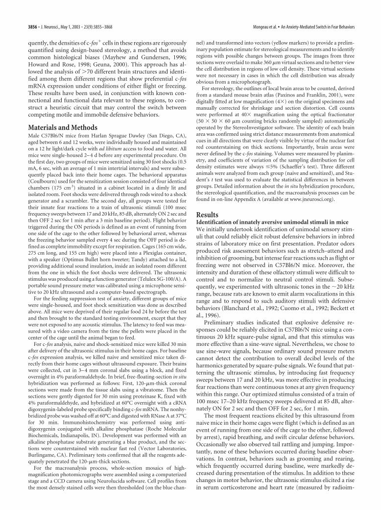
Anxiogenic manipulations cause changes in ultrasound-evoked defense behaviorsWhen mice in their home cage were exposed to the ultrasonicstimulus, frequent flight responses (six to eight events/min) wereobserved during the ON periods (Fig. 1A, home cage, white bar),but there were very few bouts of freezing (measured during theOFF periods as complete immobility except for respiration; Fig.1B). By contrast, when animals were exposed to the ultrasound inan unfamiliar cage, the frequency of flight events was reduced toapproximately half that of home cage mice (Fig. 1A, new cage,white bar), and significantly more episodes of freezing were ob-served (Fig. 1B, new cage, white bar, compare home cage). Asimilar reduction of flight and an increase in freezing were ob-served in animals tested in their home cage after being exposed 24hr earlier to a series of foot shocks (30 shocks of 0.5 mA for 6 seceach; Fig. 1A,B, home cage, black bar, compare white bars).When the foot-shocked animals were tested in a new cage, flightresponses were almost completely suppressed, and freezing wasmaximal (Fig. 1A,B, new cage, black bars). Freezing events wereseldom observed (6.7 � 3.4% of the time) in shock-sensitizedmice during exposure to a neutral auditory tone (2 kHz sine wave,1 min, 85 dB) compared with a group of mice exposed to theaversive ultrasound at the same sound pressure (54.6 � 10.6% ofthe time).
Flight was triggered primarily during the stimulus ON period,whereas freezing occurred during the OFF period. Therefore, wemeasured flight responses during the ON period and freezingresponses during the OFF period. Hence, we could theoreticallymeasure maximal flight and freezing responses from an individ-ual animal during a given experiment. Nevertheless, when thefrequency of flight and freezing to the stimulus for individualmice (n � 38) was plotted for all animals tested (collapsing bothsensitization and new cage factors), a significant negative corre-lation between the two behaviors was observed (Fig. 1C; p �0.001; r 2 � 0.6). This strong negative correlation is not simply a
trivial consequence of the fact that flight and freezing are physi-cally mutually exclusive. Bouts of freezing occupied limited in-tervals during the testing session (they rarely occurred during theON period). Therefore, there was ample time for animals thatfroze to also display flight behavior. Conversely, episodes of flightare typically interrupted by pauses (during the stimulus OFF pe-riod) during which the animal can freeze. Indeed, many individ-ual animals displayed episodes of both flight and freezing duringthe same testing period (Fig. 1C). The fact that flight occurrencesnevertheless became much less frequent as the frequency of freez-ing increased therefore suggests that these behaviors are in com-petition with one another for reasons other than simple motorincompatibility.
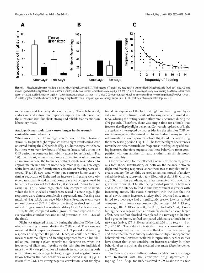
One explanation for the effect of a novel environment, previ-ous foot shock sensitization, or both on the balance betweenflight and freezing responses is that these two manipulations in-crease anxiety. To test this, we used an animal model of anxietycalled the feeding suppression task (Bodnoff et al., 1988; Gross etal., 2000). In this paradigm, mice are presented with food in agiven environment 24 hr after being food-deprived. In both ratsand mice, the latency to feed in this environment is greater withincreasing anxiety-like states. Consistent with the idea that thenovel environment increased anxiety, food-deprived mice trans-ferred to a new cage had a significantly greater latency to feedcompared with home cage controls (home cage, 110 � 19 sec;new cage, 189 � 18 sec; n � 8; p � 0.01, Student’s t test). Com-bining the new cage and sensitization factors had an even largereffect, because foot-shocked mice placed in a new cage 24 hr laterhad a greater latency to feed compared with naive animals in thenew cage (naive, 173 � 20 sec; sensitized, 230 � 14 sec; n � 6 or7; p � 0.05). These data indicate that there is a correlation be-tween manipulations that decrease flight and increase freezingand those that increase anxiety as determined by an independentbehavioral test. Consistent with this interpretation, earlier studieshave shown that shock sensitization increases anxiety in otherbehavioral tests, such as the elevated plus maze (Steenbergen etal., 1990).
We have also assessed ultrasound-induced defense after long-term treatment with the anxiolytic drug alprazolam (1mg � kg�1 � d�1, i.p., for 10 d, dissolved in 0.9% saline with a few
Figure 1. Modulation of defense reactions to an innately aversive ultrasound (USS). The frequency of flight ( A) and freezing ( B) is compared for N (white bars) and S (black bars) mice. A, S miceshowed significantly less flight than N mice (ANOVA, p � 0.01), as did mice exposed to the USS in a new cage ( p � 0.05). B, S mice showed significantly more freezing than N mice in their homecages ( p�0.01), as did mice in a new cage ( p�0.01). Data represent mean�SEM; n�5–7 mice. C, Correlation analysis with all parameters combined revealed a significant (ANOVA, p�0.001;r 2 � 0.6) negative correlation between the frequency of flight and freezing. Each point represents a single animal (n � 38). The coefficient of variation of the slope was 9%.
Mongeau et al. • An Anxiety-Mediated Switch in Fear Behaviors J. Neurosci., May 1, 2003 • 23(9):3855–3868 • 3857
drops of Tween 80, and tested in the home cage 1 hr after the lastinjection). After a control intraperitoneal saline injection (whichis a stressful manipulation like shock sensitization or placementin a new cage), mice typically display some freezing and littleflight in reaction to the aversive ultrasound. However, freezingwas significantly reduced in mice injected with alprazolam com-pared with controls (saline, 25.7 � 6.8% of the time spent freez-ing; vs alprazolam, 6.7 � 4.2% of the time; p � 0.05; n � 6). Incontrast, the frequency of flight tended to change in the oppositedirection (saline, 2.8 � 1.1 flight events; vs alprazolam, 6.8 � 2events; p � 0.11; n � 6). These data indicate that reducing anxietyincreases ultrasonic stimulus (USS)-induced flight but decreasesfreezing, supporting the idea that anxiety conversely increasesfreezing and decreases flight.
Functional imaging using quantitative analysis of c-fosmRNA-expressing cellsWe next sought to map global patterns of neuronal activity in thebrains of naive (N) and sensitized (S) mice after their first expo-sure to the ultrasonic stimulus. To do this, we used a nonisotopicin situ hybridization method with single-cell resolution to exam-ine expression of c-fos mRNA, the best characterized marker ofneuronal metabolic activity (Herdegen and Leah, 1998). Wechose to examine c-fos mRNA rather than protein because it is amore proximate indicator of c-fos expression. Induction of c-fostranscription in neurons occurs within 2–5 min in response todepolarization-induced calcium entry (Finkbeiner and Green-berg, 1998) and peaks after �30 min (Greenberg and Ziff, 1984),the time at which ultrasound-exposed animals were killed foranalysis. c-fos expression can also be induced by factors that ele-vate intracellular cAMP expression, such as monoamines or neu-ropeptides, as well as by stress hormones [glucocorticoids (forreview, see Herdegen and Leah, 1998)]. However, glucocorti-coids are not the principal influences on c-fos expression in tasksinvolving stress (Anokhin et al., 1991; Helmreich et al., 1996).
Exposure of mice to the ultrasonic stimulus in their homecages was a key feature of our paradigm, because the handling ofanimals necessary to transfer them to a new testing cage induces asubstantial number of c-fos� cells. By testing animals in theirhome cage, background levels of c-fos mRNA expression in con-trol animals were kept very low (Fig. 2A). A reflection of thespecificity of c-fos mRNA induction under these conditions isseen in the inferior colliculus (IC), which contains a tonotopicmap of frequency-responsive neurons (Ryan et al., 1988; Ehretand Fischer, 1991). Exposure to the ultrasonic stimulus inducedmassive c-fos mRNA expression in a zone of the IC correspondingto the region sensitive to frequencies in the 17–20 kHz range (Fig.2B, arrow). That the expression of c-fos reflects motor output aswell as sensory input is illustrated by its expression in the forelimbregion of the motor cortex (Li and Waters, 1991), where there wasa higher level of expression in N mice (which primarily exhibitflight) than in S mice (which primarily exhibit freezing; Fig. 2C;for quantification, see below). As expected (Castro-Alamancos etal., 1992), there was relatively little c-fos expression in regions ofmotor cortex not involved in the escape response (see supple-mental Figure S7A, nose region; supplemental Figures S1–S8 canbe found in on-line Appendix B).
To accurately quantify the induction of c-fos mRNA, we mea-sured the density of c-fos� cells in various brain regions usingunbiased stereology with the optical fractionator. This method isconsidered the most accurate way to estimate cell densities in agiven volume of brain tissue using a random sampling method.Surprisingly, however, it had not previously been used to map
c-fos expression for functional imaging studies. By performingnonisotopic in situ hybridization on free-floating thick (120 �m)sections, we could obtain a large “counting brick” (60 �m depth)with sufficient cellular resolution to estimate cell densities byoptical dissection (Fig. 2G). To control for any differentialshrinkage between sections, the volumes of brain regions sam-pled were calculated from section thickness and area measure-ments using well defined anatomical landmarks. For example, inthe forelimb motor cortex we digitally fitted the anatomicalboundaries [M1 and M2, located between anteroposterior (AP)�0.5 and �1.9 mm] from a digital brain atlas (Paxinos andFranklin, 2001) using landmarks such as the interhemisphericfissure and the corpus callosum. The measured volume for thisentire region did not differ between groups (N, 0.94 � 0.06 mm 3;S, 0.95 � 0.03 mm 3; in no case did we observe significant differ-ences in volumes between groups for the regions sampled; there-fore, the volumes are presented as a single value derived fromboth groups), but the density of c-fos� cells was found to behigher in N than in S mice (Fig. 2E), consistent with the higherlevel of motor activity in the former group.
Because it was impractical to perform stereological cell countsthrough all brain regions in each of the three animals analyzed foreach condition, we developed a method, called macroanalysis, toinitially survey large regions of the brain to identify potentialareas of differential c-fos expression and to obtain preliminaryestimates of cell density. Briefly, this method uses the Neurolu-cida software to assemble low-magnification views of entire coro-nal sections with single-cell resolution by assembling a mosaic or“virtual slice” from a series of contiguous high-magnificationfields (Fig. 2D, top panels). The positions of the most stronglystained c-fos� cells in each section are then extracted using athresholding program that identifies cell profiles on the basis oftheir color, dimension, and shape (Fig. 2D, bottom panel). Datafrom three consecutive virtual slices (360 �m) can then be over-laid to clearly reveal the cell profile distribution within a givenregion [such as the functional columns within the periaqueductalgray (PAG); Fig. 2F]. Automated counting of cell profiles couldbe performed within coarsely bounded regions for an animal ofeach group. Regions showing potential differences were then fur-ther analyzed by stereological counting in more tightly boundedregions.
Neural correlates of the switch between flight andfreezing behaviorsFor c-fos analysis, three N animals and three animals sensitized byfoot shock 24 hr previously (S) were exposed to the ultrasonicstimulus in their home cages. The behavioral data for these ani-mals confirmed our previous findings (home cage N mice: flight,7.0 � 1.2 events; freezing, 4.3 � 4.3% of the time; home-cage Smice: flight, 1.7 � 0.9 events; freezing, 60 � 14% of the time). Thefrequencies of both flight and freezing were significantly differentbetween the two groups ( p � 0.05, Student’s t test). The brain ofeach of these animals was cut into three large slabs (rostral, inter-mediate, and caudal), and each of these slabs was in turn sec-tioned at 120 �m using a vibratome for in situ hybridization.
Approximately 70 different areas or nuclei were examined.[Table 1 in on-line Appendix B (available at www.jneurosci.org)summarizes the intensity of staining observed in various regions ofthe brain in N and S mice exposed to the ultrasonic stimulus.] Ex-posure to the aversive ultrasound produced massive increases ofc-fos� cells in cortical, amygdalar, septo-hippocampal, and dience-phalic areas but less so in the basal ganglia and the brainstem. Ap-proximately 80% of the regions examined did not show signs of
3858 • J. Neurosci., May 1, 2003 • 23(9):3855–3868 Mongeau et al. • An Anxiety-Mediated Switch in Fear Behaviors
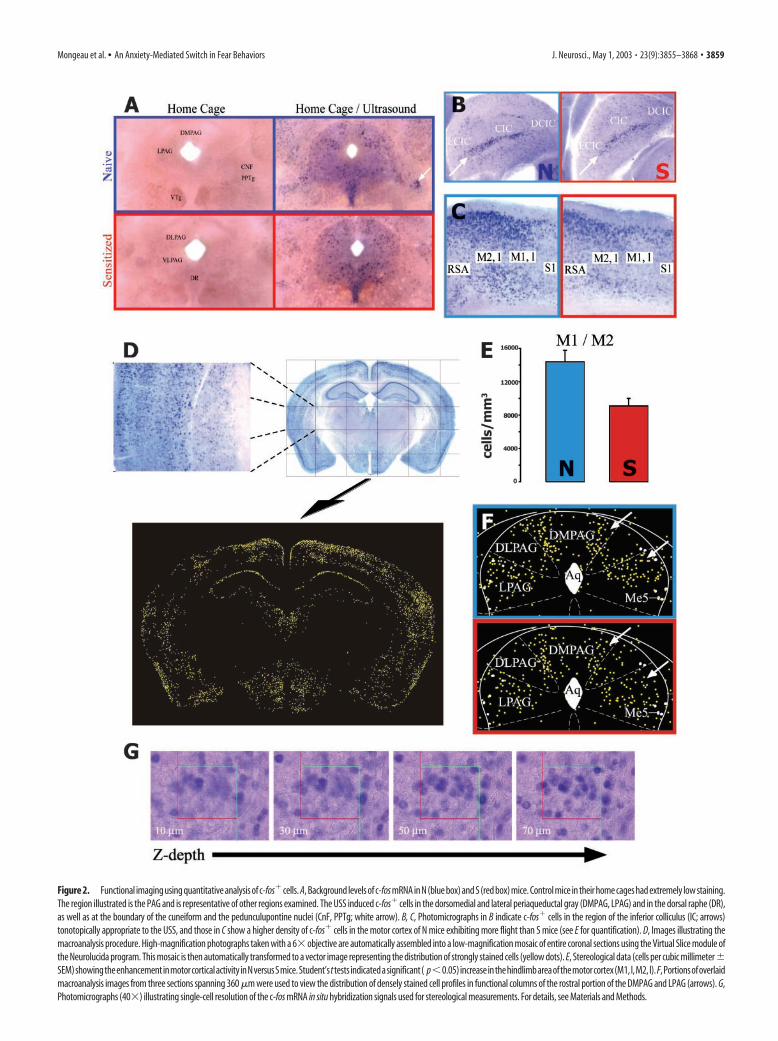
Figure 2. Functional imaging using quantitative analysis of c-fos� cells. A, Background levels of c-fos mRNA in N (blue box) and S (red box) mice. Control mice in their home cages had extremely low staining.The region illustrated is the PAG and is representative of other regions examined. The USS induced c-fos� cells in the dorsomedial and lateral periaqueductal gray (DMPAG, LPAG) and in the dorsal raphe (DR),as well as at the boundary of the cuneiform and the pedunculupontine nuclei (CnF, PPTg; white arrow). B, C, Photomicrographs in B indicate c-fos� cells in the region of the inferior colliculus (IC; arrows)tonotopically appropriate to the USS, and those in C show a higher density of c-fos� cells in the motor cortex of N mice exhibiting more flight than S mice (see E for quantification). D, Images illustrating themacroanalysis procedure. High-magnification photographs taken with a 6� objective are automatically assembled into a low-magnification mosaic of entire coronal sections using the Virtual Slice module ofthe Neurolucida program. This mosaic is then automatically transformed to a vector image representing the distribution of strongly stained cells (yellow dots). E, Stereological data (cells per cubic millimeter�SEM)showingtheenhancementinmotorcorticalactivityinNversusSmice.Student’sttests indicatedasignificant( p�0.05)increaseinthehindlimbareaofthemotorcortex(M1,l,M2,l).F,Portionsofoverlaidmacroanalysis images from three sections spanning 360 �m were used to view the distribution of densely stained cell profiles in functional columns of the rostral portion of the DMPAG and LPAG (arrows). G,Photomicrographs (40�) illustrating single-cell resolution of the c-fos mRNA in situ hybridization signals used for stereological measurements. For details, see Materials and Methods.
Mongeau et al. • An Anxiety-Mediated Switch in Fear Behaviors J. Neurosci., May 1, 2003 • 23(9):3855–3868 • 3859
differential activity. Approximately 14 regions were identified thatshowed higher levels of c-fos expression in N mice than in S mice,whereas only half as many showed higher activity in S mice (see Fig.7). Strikingly, all but one of the latter regions are clustered in thehypothalamus. Approximately eight areas showed strong activity inboth N and S mice. Below we systematically compare the detailedpatterns of activity between N and S mice, beginning with the mes-encephalon and ending with the cortex. Supplemental figures(S1–S8) can be found in on-line Appendix B (available atwww.jneurosci.org).
MesencephalonThe midbrain PAG is thought to serve as a final common pathwayfor the initiation of flight and freezing behaviors elicited by fear-ful stimuli (Bandler et al., 2000). Consistent with this, the PAGwas strongly activated in mice exposed to the ultrasonic stimulus(Fig. 2F). However, there were no significant group differences inany region of this mesencephalic structure as assessed by stereo-logical counting (Appendix B, Figs. S1, S6A). There were also noapparent differences at the level of the inferior and superior col-liculi (Figs. 2B, S3), in the median and dorsal raphe nuclei (con-taining serotonin cell bodies (Figs. S2A, S6A), and in the locuscoeruleus (containing noradrenaline cell bodies (Fig. S2C), al-though staining was strong in these areas. There were very fewc-fos� cells in the ventral tegmental area or the substantia nigra(Fig. S2B). The only area of the midbrain where we did observe adifference between N and S mice was at the lateral edges of theventral PAG, at the junction between the pedunculopontine teg-mentum (PPTg) and the cuneiform nucleus (Cnf) (Fig. 2A, arrow).Stereological cell counting in this area was performed within an ar-bitrarily defined octagonal region (Fig. S6B), because this cluster ofstaining did not fit any known boundaries in the atlas. These mea-surements confirmed a significantly higher (55%) density of c-foscells within this defined boundary in N versus S mice (N, 2594 � 312cells/mm3; S, 1795 � 23 cells/mm3; p � 0.05; between AP �4.4 and�5.1 mm; volume, 0.182 � 0.003 mm3).
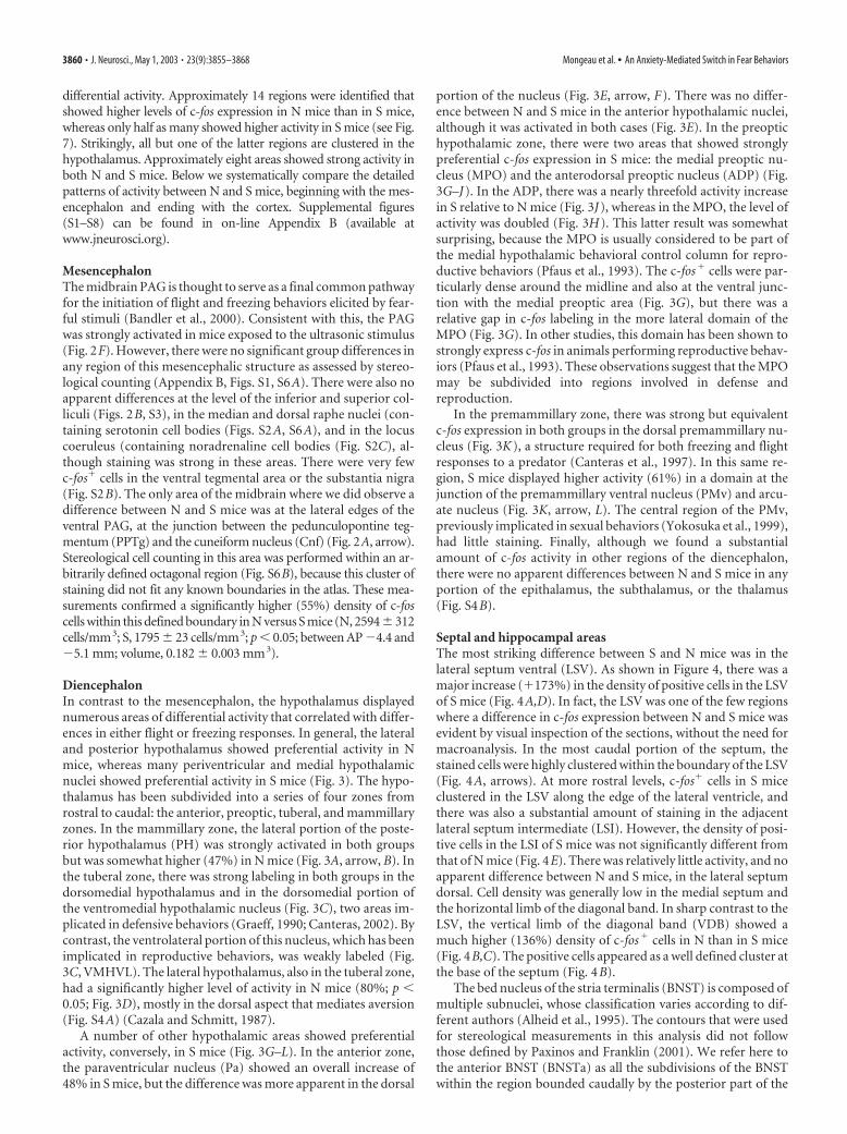
DiencephalonIn contrast to the mesencephalon, the hypothalamus displayednumerous areas of differential activity that correlated with differ-ences in either flight or freezing responses. In general, the lateraland posterior hypothalamus showed preferential activity in Nmice, whereas many periventricular and medial hypothalamicnuclei showed preferential activity in S mice (Fig. 3). The hypo-thalamus has been subdivided into a series of four zones fromrostral to caudal: the anterior, preoptic, tuberal, and mammillaryzones. In the mammillary zone, the lateral portion of the poste-rior hypothalamus (PH) was strongly activated in both groupsbut was somewhat higher (47%) in N mice (Fig. 3A, arrow, B). Inthe tuberal zone, there was strong labeling in both groups in thedorsomedial hypothalamus and in the dorsomedial portion ofthe ventromedial hypothalamic nucleus (Fig. 3C), two areas im-plicated in defensive behaviors (Graeff, 1990; Canteras, 2002). Bycontrast, the ventrolateral portion of this nucleus, which has beenimplicated in reproductive behaviors, was weakly labeled (Fig.3C, VMHVL). The lateral hypothalamus, also in the tuberal zone,had a significantly higher level of activity in N mice (80%; p �0.05; Fig. 3D), mostly in the dorsal aspect that mediates aversion(Fig. S4A) (Cazala and Schmitt, 1987).
A number of other hypothalamic areas showed preferentialactivity, conversely, in S mice (Fig. 3G–L). In the anterior zone,the paraventricular nucleus (Pa) showed an overall increase of48% in S mice, but the difference was more apparent in the dorsal
portion of the nucleus (Fig. 3E, arrow, F). There was no differ-ence between N and S mice in the anterior hypothalamic nuclei,although it was activated in both cases (Fig. 3E). In the preoptichypothalamic zone, there were two areas that showed stronglypreferential c-fos expression in S mice: the medial preoptic nu-cleus (MPO) and the anterodorsal preoptic nucleus (ADP) (Fig.3G–J). In the ADP, there was a nearly threefold activity increasein S relative to N mice (Fig. 3J), whereas in the MPO, the level ofactivity was doubled (Fig. 3H). This latter result was somewhatsurprising, because the MPO is usually considered to be part ofthe medial hypothalamic behavioral control column for repro-ductive behaviors (Pfaus et al., 1993). The c-fos� cells were par-ticularly dense around the midline and also at the ventral junc-tion with the medial preoptic area (Fig. 3G), but there was arelative gap in c-fos labeling in the more lateral domain of theMPO (Fig. 3G). In other studies, this domain has been shown tostrongly express c-fos in animals performing reproductive behav-iors (Pfaus et al., 1993). These observations suggest that the MPOmay be subdivided into regions involved in defense andreproduction.
In the premammillary zone, there was strong but equivalentc-fos expression in both groups in the dorsal premammillary nu-cleus (Fig. 3K), a structure required for both freezing and flightresponses to a predator (Canteras et al., 1997). In this same re-gion, S mice displayed higher activity (61%) in a domain at thejunction of the premammillary ventral nucleus (PMv) and arcu-ate nucleus (Fig. 3K, arrow, L). The central region of the PMv,previously implicated in sexual behaviors (Yokosuka et al., 1999),had little staining. Finally, although we found a substantialamount of c-fos activity in other regions of the diencephalon,there were no apparent differences between N and S mice in anyportion of the epithalamus, the subthalamus, or the thalamus(Fig. S4B).
Septal and hippocampal areasThe most striking difference between S and N mice was in thelateral septum ventral (LSV). As shown in Figure 4, there was amajor increase (�173%) in the density of positive cells in the LSVof S mice (Fig. 4A,D). In fact, the LSV was one of the few regionswhere a difference in c-fos expression between N and S mice wasevident by visual inspection of the sections, without the need formacroanalysis. In the most caudal portion of the septum, thestained cells were highly clustered within the boundary of the LSV(Fig. 4A, arrows). At more rostral levels, c-fos� cells in S miceclustered in the LSV along the edge of the lateral ventricle, andthere was also a substantial amount of staining in the adjacentlateral septum intermediate (LSI). However, the density of posi-tive cells in the LSI of S mice was not significantly different fromthat of N mice (Fig. 4E). There was relatively little activity, and noapparent difference between N and S mice, in the lateral septumdorsal. Cell density was generally low in the medial septum andthe horizontal limb of the diagonal band. In sharp contrast to theLSV, the vertical limb of the diagonal band (VDB) showed amuch higher (136%) density of c-fos� cells in N than in S mice(Fig. 4B,C). The positive cells appeared as a well defined cluster atthe base of the septum (Fig. 4B).
The bed nucleus of the stria terminalis (BNST) is composed ofmultiple subnuclei, whose classification varies according to dif-ferent authors (Alheid et al., 1995). The contours that were usedfor stereological measurements in this analysis did not followthose defined by Paxinos and Franklin (2001). We refer here tothe anterior BNST (BNSTa) as all the subdivisions of the BNSTwithin the region bounded caudally by the posterior part of the
3860 • J. Neurosci., May 1, 2003 • 23(9):3855–3868 Mongeau et al. • An Anxiety-Mediated Switch in Fear Behaviors
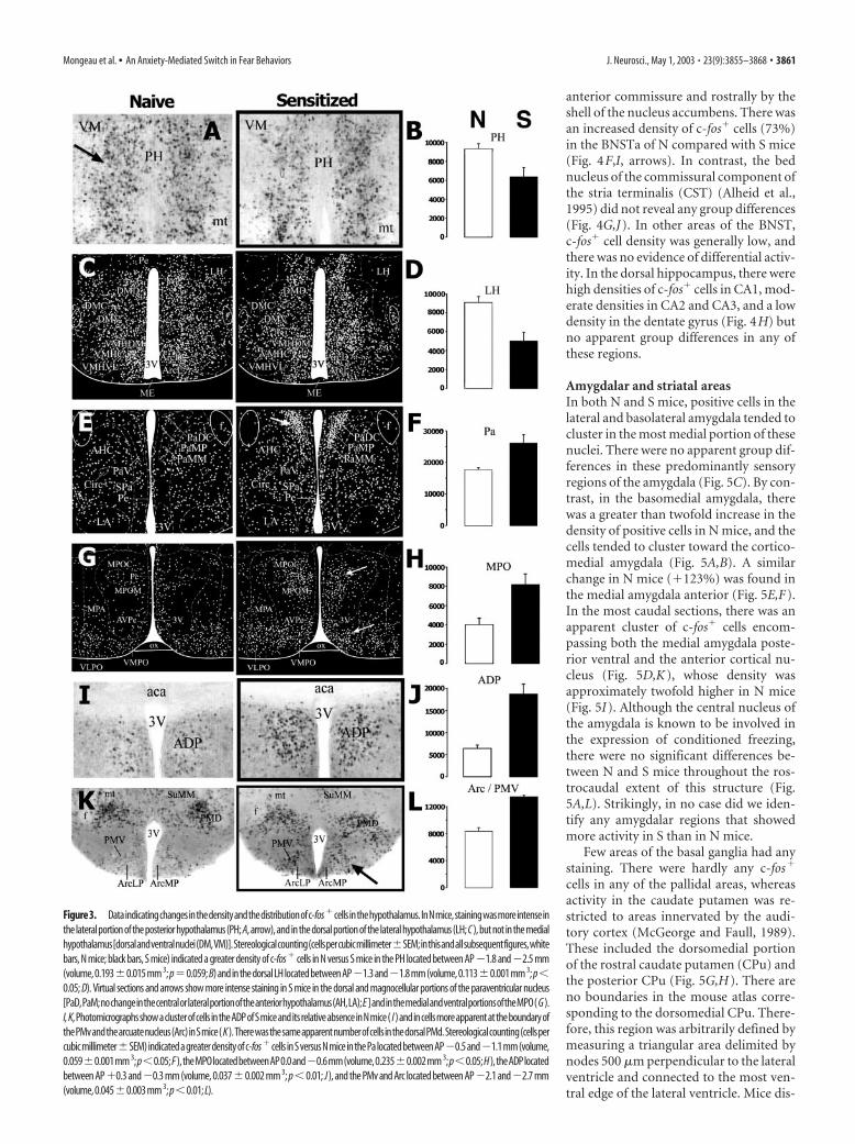
anterior commissure and rostrally by theshell of the nucleus accumbens. There wasan increased density of c-fos� cells (73%)in the BNSTa of N compared with S mice(Fig. 4F,I, arrows). In contrast, the bednucleus of the commissural component ofthe stria terminalis (CST) (Alheid et al.,1995) did not reveal any group differences(Fig. 4G,J). In other areas of the BNST,c-fos� cell density was generally low, andthere was no evidence of differential activ-ity. In the dorsal hippocampus, there werehigh densities of c-fos� cells in CA1, mod-erate densities in CA2 and CA3, and a lowdensity in the dentate gyrus (Fig. 4H) butno apparent group differences in any ofthese regions.
Amygdalar and striatal areasIn both N and S mice, positive cells in thelateral and basolateral amygdala tended tocluster in the most medial portion of thesenuclei. There were no apparent group dif-ferences in these predominantly sensoryregions of the amygdala (Fig. 5C). By con-trast, in the basomedial amygdala, therewas a greater than twofold increase in thedensity of positive cells in N mice, and thecells tended to cluster toward the cortico-medial amygdala (Fig. 5A,B). A similarchange in N mice (�123%) was found inthe medial amygdala anterior (Fig. 5E,F).In the most caudal sections, there was anapparent cluster of c-fos� cells encom-passing both the medial amygdala poste-rior ventral and the anterior cortical nu-cleus (Fig. 5D,K), whose density wasapproximately twofold higher in N mice(Fig. 5I). Although the central nucleus ofthe amygdala is known to be involved inthe expression of conditioned freezing,there were no significant differences be-tween N and S mice throughout the ros-trocaudal extent of this structure (Fig.5A,L). Strikingly, in no case did we iden-tify any amygdalar regions that showedmore activity in S than in N mice.
Few areas of the basal ganglia had anystaining. There were hardly any c-fos�
cells in any of the pallidal areas, whereasactivity in the caudate putamen was re-stricted to areas innervated by the audi-tory cortex (McGeorge and Faull, 1989).These included the dorsomedial portionof the rostral caudate putamen (CPu) andthe posterior CPu (Fig. 5G,H). There areno boundaries in the mouse atlas corre-sponding to the dorsomedial CPu. There-fore, this region was arbitrarily defined bymeasuring a triangular area delimited bynodes 500 �m perpendicular to the lateralventricle and connected to the most ven-tral edge of the lateral ventricle. Mice dis-
Figure3. Dataindicatingchangesinthedensityandthedistributionofc-fos�cellsinthehypothalamus.InNmice,stainingwasmoreintenseinthelateralportionoftheposteriorhypothalamus(PH; A,arrow),and inthe dorsalportionof thelateral hypothalamus(LH; C ), butnot inthemedialhypothalamus[dorsalandventralnuclei(DM,VM)].Stereologicalcounting(cellspercubicmillimeter�SEM;inthisandallsubsequentfigures,whitebars, N mice; black bars, S mice) indicated a greater density of c-fos� cells in N versus S mice in the PH located between AP�1.8 and�2.5 mm(volume,0.193�0.015mm 3;p�0.059;B)andinthedorsalLHlocatedbetweenAP�1.3and�1.8mm(volume,0.113�0.001mm 3;p�0.05; D). Virtual sections and arrows show more intense staining in S mice in the dorsal and magnocellular portions of the paraventricular nucleus[PaD,PaM;nochangeinthecentralorlateralportionoftheanteriorhypothalamus(AH,LA);E ]andinthemedialandventralportionsoftheMPO( G ).I,K,Photomicrographsshowaclusterofcells intheADPofSmiceanditsrelativeabsenceinNmice( I )andincellsmoreapparentattheboundaryofthePMvandthearcuatenucleus(Arc)inSmice( K ).TherewasthesameapparentnumberofcellsinthedorsalPMd.Stereologicalcounting(cellspercubicmillimeter�SEM)indicatedagreaterdensityofc-fos�cells inSversusNmiceinthePalocatedbetweenAP�0.5and�1.1mm(volume,0.059�0.001mm 3;p�0.05;F ),theMPOlocatedbetweenAP0.0and�0.6mm(volume,0.235�0.002mm 3;p�0.05;H ),theADPlocatedbetween AP�0.3 and�0.3 mm (volume, 0.037�0.002 mm 3; p�0.01; J ), and the PMv and Arc located between AP�2.1 and�2.7 mm(volume,0.045�0.003mm 3; p�0.01; L).
Mongeau et al. • An Anxiety-Mediated Switch in Fear Behaviors J. Neurosci., May 1, 2003 • 23(9):3855–3868 • 3861
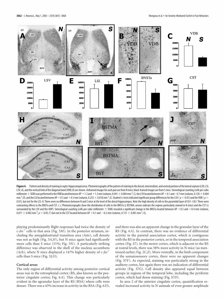
playing predominantly flight responses had twice the density ofc-fos� cells in that area (Fig. 5M). In the posterior striatum, in-cluding the amygdalostriatal transition area (Astr), cell densitywas not as high (Fig. 5A,H), but N mice again had significantlymore cells than S mice (51%; Fig. 5N). A particularly strikingdifference was observed in the shell of the nucleus accumbens(Acb), where N mice displayed a 147% higher density of c-fos�
cells than S mice (Fig. 5J,O).
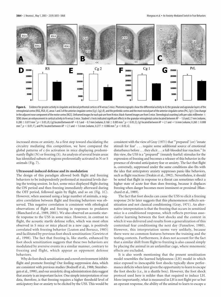
Cortical areasThe only region of differential activity among posterior corticalareas was in the retrosplenial cortex (RS, also known as the pos-terior cingulate cortex; Fig. 6A). This change was particularlyevident in the agranular layer of the RS (RSA) where cells weredenser. There was a 43% increase in activity in the RSA (Fig. 6D),
and there was also an apparent change in the granular layer of theRS (Fig. 6A). In contrast, there was no evidence of differentialactivity in the parietal association cortex, which is contiguouswith the RS in the posterior cortex, or in the temporal associationcortex (Fig. S7). In the motor cortex, which is adjacent to the RSat rostral levels, there was 58% more activity in N mice (as men-tioned earlier; Fig. 2C,E). More ventrally, in the limb componentof the somatosensory cortex, there were no apparent changes(Fig. S7F). As expected, staining was particularly strong in theauditory cortex, but again there was no indication of differentialactivity (Fig. S7G). Cell density also appeared equal betweengroups in regions of the temporal lobe, including the pyriformcortex, which had dense staining (Fig. S7D).
In area 2 of the anterior cingulate cortex, quantification re-vealed increased activity in N animals of even greater amplitude
Figure 4. Pattern and density of staining in septo-hippocampal areas. Photomicrographs of the pattern of staining in the dorsal, intermediate, and ventral portions of the lateral septum (LSD, LSI,LSV; A), and the vertical limb of the diagonal band (VDB; B) are shown. Unframed images for each pair are from N mice; black-framed images are from S mice. Stereological counting (cells per cubicmillimeter � SEM) was performed in the VDB located between AP �1.2 and �1.3 mm (volume, 0.041 � 0.004 mm 3; C), the LSV located between AP �0.1 and – 0.7 mm (volume, 0.120 � 0.004mm 3; D), and the LSI located between AP �0.5 and �0.5 mm (volume, 0.255 � 0.018 mm 3; E). Student’s t tests indicated significant group differences for the LSV ( p � 0.01) and the VDB ( p �0.01), but not for the LSI. H, There were no differences between N and S mice at the level of the dorsal hippocampus. Note the high density of cells in the pyramidal layer of CA1–CA3. There werecontrasting effects in the BNSTa and CST. I, J, Photomicrographs show the distribution of cells in the BNSTa (I; BSTMA; arrows indicate the regions particularly stained in N mice) and the CST (J;surrounded by the LSV and the ADP). Stereological counting (cells per cubic millimeter � SEM) revealed a significant change in the BNSTa located between AP �0.5 and �0.4 mm (volume,0.071 � 0.002 mm 3; p � 0.05; F ) but not in the CST located between AP �0.1 and �0.2 mm (volume, 0.131 � 0.001 mm 3; G).
3862 • J. Neurosci., May 1, 2003 • 23(9):3855–3868 Mongeau et al. • An Anxiety-Mediated Switch in Fear Behaviors
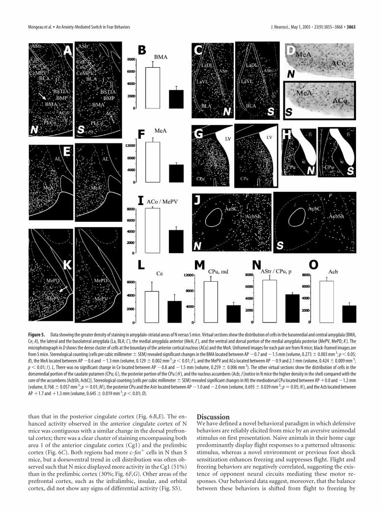
than that in the posterior cingulate cortex (Fig. 6B,E). The en-hanced activity observed in the anterior cingulate cortex of Nmice was contiguous with a similar change in the dorsal prefron-tal cortex; there was a clear cluster of staining encompassing botharea 1 of the anterior cingulate cortex (Cg1) and the prelimbiccortex (Fig. 6C). Both regions had more c-fos� cells in N than Smice, but a dorsoventral trend in cell distribution was often ob-served such that N mice displayed more activity in the Cg1 (51%)than in the prelimbic cortex (30%; Fig. 6F,G). Other areas of theprefrontal cortex, such as the infralimbic, insular, and orbitalcortex, did not show any signs of differential activity (Fig. S5).
DiscussionWe have defined a novel behavioral paradigm in which defensivebehaviors are reliably elicited from mice by an aversive unimodalstimulus on first presentation. Naive animals in their home cagepredominantly display flight responses to a patterned ultrasonicstimulus, whereas a novel environment or previous foot shocksensitization enhances freezing and suppresses flight. Flight andfreezing behaviors are negatively correlated, suggesting the exis-tence of opponent neural circuits mediating these motor re-sponses. Our behavioral data suggest, moreover, that the balancebetween these behaviors is shifted from flight to freezing by
Figure 5. Data showing the greater density of staining in amygdalo-striatal areas of N versus S mice. Virtual sections show the distribution of cells in the basomedial and central amygdala (BMA,Ce; A), the lateral and the basolateral amygdala (La, BLA; C ), the medial amygdala anterior (MeA; E ), and the ventral and dorsal portion of the medial amygdala posterior (MePV, MePD; K ). Themicrophotograph in D shows the dense cluster of cells at the boundary of the anterior cortical nucleus (ACo) and the MeA. Unframed images for each pair are from N mice; black-framed images arefrom S mice. Stereological counting (cells per cubic millimeter � SEM) revealed significant changes in the BMA located between AP �0.7 and �1.5 mm (volume, 0.273 � 0.003 mm 3; p � 0.05;B), the MeA located between AP �0.6 and �1.3 mm (volume, 0.129 � 0.002 mm 3; p � 0.01; F ), and the MePV and ACo located between AP �0.9 and 2.1 mm (volume, 0.424 � 0.009 mm 3;p � 0.01; I ). L, There was no significant change in Ce located between AP �0.8 and �1.5 mm (volume, 0.259 � 0.006 mm 3). The other virtual sections show the distribution of cells in thedorsomedial portion of the caudate putamen (CPu; G ), the posterior portion of the CPu ( H ), and the nucleus accumbens (Acb; J [notice in N mice the higher density in the shell compared with thecore of the accumbens (AcbSh, AcbC)]. Stereological counting (cells per cubic millimeter � SEM) revealed significant changes in M) the mediodorsal CPu located between AP �0.0 and �1.2 mm(volume, 0.768 � 0.057 mm 3; p � 0.01; M ), the posterior CPu and the Astr located between AP �1.0 and �2.0 mm (volume, 0.693 � 0.039 mm 3; p � 0.05; N ), and the Acb located betweenAP �1.7 and �1.3 mm (volume, 0.645 � 0.019 mm 3; p � 0.01; O).
Mongeau et al. • An Anxiety-Mediated Switch in Fear Behaviors J. Neurosci., May 1, 2003 • 23(9):3855–3868 • 3863
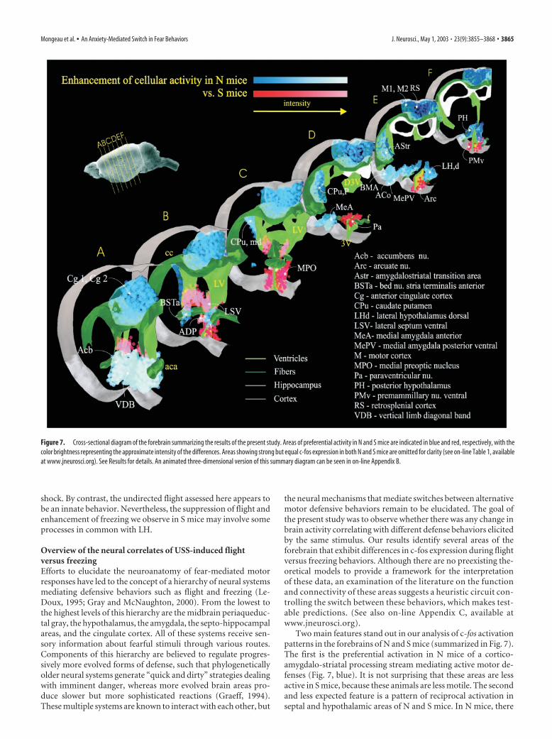
increased stress or anxiety. As a first step toward elucidating thecircuitry mediating this competition, we have compared theglobal patterns of c-fos activation in mice displaying predomi-nantly flight (N) or freezing (S). An analysis of several brain areashas identified subsets of regions preferentially activated in N or Sanimals (Fig. 7).
Ultrasound-induced defense and its modulationThe design of this paradigm allowed both flight and freezingbehaviors to be independently performed at maximal levels dur-ing the testing session. In fact, some mice displayed flight duringthe ON period and then freezing immediately afterward duringthe OFF period, followed again by flight, and so on (Fig. 1C).However, when assessed using a large number of animals, a neg-ative correlation between flight and freezing behaviors was ob-served. This negative correlation is consistent with ethologicalobservations of flight and freezing in responses to predators(Blanchard et al., 1989, 2001). We also observed an acoustic star-tle response to the USS in some mice. However, in contrast toflight, the acoustic startle defense reflex, which was most oftenobserved in S mice or mice placed in a new cage, is positivelycorrelated with freezing behavior (Leaton and Borszcz, 1985)and facilitated by previous foot shock sensitization (Gewirtz etal., 1998). The fact that freezing is, like startle, enhanced byfoot shock sensitization suggests that these two behaviors aremodulated by aversive events in a similar manner, contrary tofreezing and flight, which appear as opponent defensebehaviors.
Why do foot shock sensitization and a novel environment inhibitflight and promote freezing? Our feeding suppression data, whichare consistent with previous studies (Bodnoff et al., 1988; Steenber-gen et al., 1990), and our anxiolytic drug administration data suggestthat anxiety is an important factor. One simple interpretation of ourdata, therefore, is that freezing requires a higher threshold level ofanticipatory fear or anxiety to be elicited by the USS. This would be
consistent with the view of Gray (1971) that “‘prepared’ (or) ‘innatestimuli for fear’ . . . require some additional source of emotionaldisturbance before . . . they elicit . . . a full-blooded fear reaction.” Inthis view, the USS is a “prepared” (innately fearful) stimulus for theexpression of freezing and becomes a releaser of this behavior in thepresence of elevated anticipatory fear or anxiety. The fact that flightis, conversely, suppressed under the same conditions also fits withthe idea that anticipatory anxiety suppresses panic-like behaviors,such as flight reactions (Deakin et al., 1992). Nevertheless, it shouldbe noted that flight in response to a threat can sometimes reflect ahigher state of acute fear than does freezing, because it displacesfreezing when danger becomes more imminent or proximal (Blan-chard et al., 1989).
The fact that foot shocks can cause the USS to elicit a freezingresponse 24 hr later suggests that this phenomenon reflects sen-sitization and not classical conditioning (Gray, 1971). An alter-native interpretation is that the freezing that occurs in sensitizedmice is a conditioned response, which reflects previous asso-ciative learning between the foot shocks and the context inwhich it was delivered and results from a generalization of thiscontextual fear conditioning the next day (Fanselow, 1980).However, this interpretation seems very unlikely, becausethere were no common features between the training and thetesting contexts. Furthermore, it does not account for the factthat a similar shift from flight to freezing is also caused simplyby placing the animal in an unfamiliar cage, where mnemoniceffects are excluded.
It is also worth mentioning that the present sensitizationmodel resembles the learned helplessness (LH) model in whichmice exposed to inescapable foot shocks typically show perfor-mance deficits when later given the opportunity to learn to escapethe foot shocks (i.e., in a shuttle box). However, the foot shockprotocol used here is milder than that required to induce LH.More importantly, what is measured in LH is not flight per se butan operant response, the ability of the animal to learn to escape a
Figure 6. Evidence for greater activity in cingulate and dorsal prefrontal cortices of N versus S mice. Photomicrographs show the differential activity in A) the granular and agranular layers of theretrosplenial cortex (RSG, RSA; A), areas 1 and 2 of the anterior cingulate cortex (Cg1, Cg2; B), and the prelimbic cortex and the most rostral part of the anterior cingulate cortex (PrL, Cg1; C [no changein the adjacent nose component of the motor cortex (M2)]. Unframed images for each pair are from N mice; black-framed images are from S mice. Stereological counting (cells per cubic millimeter �SEM) shows an enhancement in cortical activity in N versus S mice. Student’s t tests indicated significant effects in the granular retrosplenial cortex located between AP �1.8 and 2.7 mm (volume,0.280�0.011 mm 3; p�0.05; D), Cg2 located between AP�0.1 and�0.7 mm (volume, 0.168�0.005 mm 3; p�0.05; E), Cg1 located between AP�2.1 and�1.6 mm (volume, 0.260�0.008mm 3; p � 0.01; F ), and PrL located between AP �2.1 and �1.6 mm (volume, 0.277 � 0.006 mm 3; p � 0.05; G).
3864 • J. Neurosci., May 1, 2003 • 23(9):3855–3868 Mongeau et al. • An Anxiety-Mediated Switch in Fear Behaviors
shock. By contrast, the undirected flight assessed here appears tobe an innate behavior. Nevertheless, the suppression of flight andenhancement of freezing we observe in S mice may involve someprocesses in common with LH.
Overview of the neural correlates of USS-induced flightversus freezingEfforts to elucidate the neuroanatomy of fear-mediated motorresponses have led to the concept of a hierarchy of neural systemsmediating defensive behaviors such as flight and freezing (Le-Doux, 1995; Gray and McNaughton, 2000). From the lowest tothe highest levels of this hierarchy are the midbrain periaqueduc-tal gray, the hypothalamus, the amygdala, the septo-hippocampalareas, and the cingulate cortex. All of these systems receive sen-sory information about fearful stimuli through various routes.Components of this hierarchy are believed to regulate progres-sively more evolved forms of defense, such that phylogeneticallyolder neural systems generate “quick and dirty” strategies dealingwith imminent danger, whereas more evolved brain areas pro-duce slower but more sophisticated reactions (Graeff, 1994).These multiple systems are known to interact with each other, but
the neural mechanisms that mediate switches between alternativemotor defensive behaviors remain to be elucidated. The goal ofthe present study was to observe whether there was any change inbrain activity correlating with different defense behaviors elicitedby the same stimulus. Our results identify several areas of theforebrain that exhibit differences in c-fos expression during flightversus freezing behaviors. Although there are no preexisting the-oretical models to provide a framework for the interpretationof these data, an examination of the literature on the functionand connectivity of these areas suggests a heuristic circuit con-trolling the switch between these behaviors, which makes test-able predictions. (See also on-line Appendix C, available atwww.jneurosci.org).
Two main features stand out in our analysis of c-fos activationpatterns in the forebrains of N and S mice (summarized in Fig. 7).The first is the preferential activation in N mice of a cortico-amygdalo-striatal processing stream mediating active motor de-fenses (Fig. 7, blue). It is not surprising that these areas are lessactive in S mice, because these animals are less motile. The secondand less expected feature is a pattern of reciprocal activation inseptal and hypothalamic areas of N and S mice. In N mice, there
Figure 7. Cross-sectional diagram of the forebrain summarizing the results of the present study. Areas of preferential activity in N and S mice are indicated in blue and red, respectively, with thecolor brightness representing the approximate intensity of the differences. Areas showing strong but equal c-fos expression in both N and S mice are omitted for clarity (see on-line Table 1, availableat www.jneurosci.org). See Results for details. An animated three-dimensional version of this summary diagram can be seen in on-line Appendix B.
Mongeau et al. • An Anxiety-Mediated Switch in Fear Behaviors J. Neurosci., May 1, 2003 • 23(9):3855–3868 • 3865
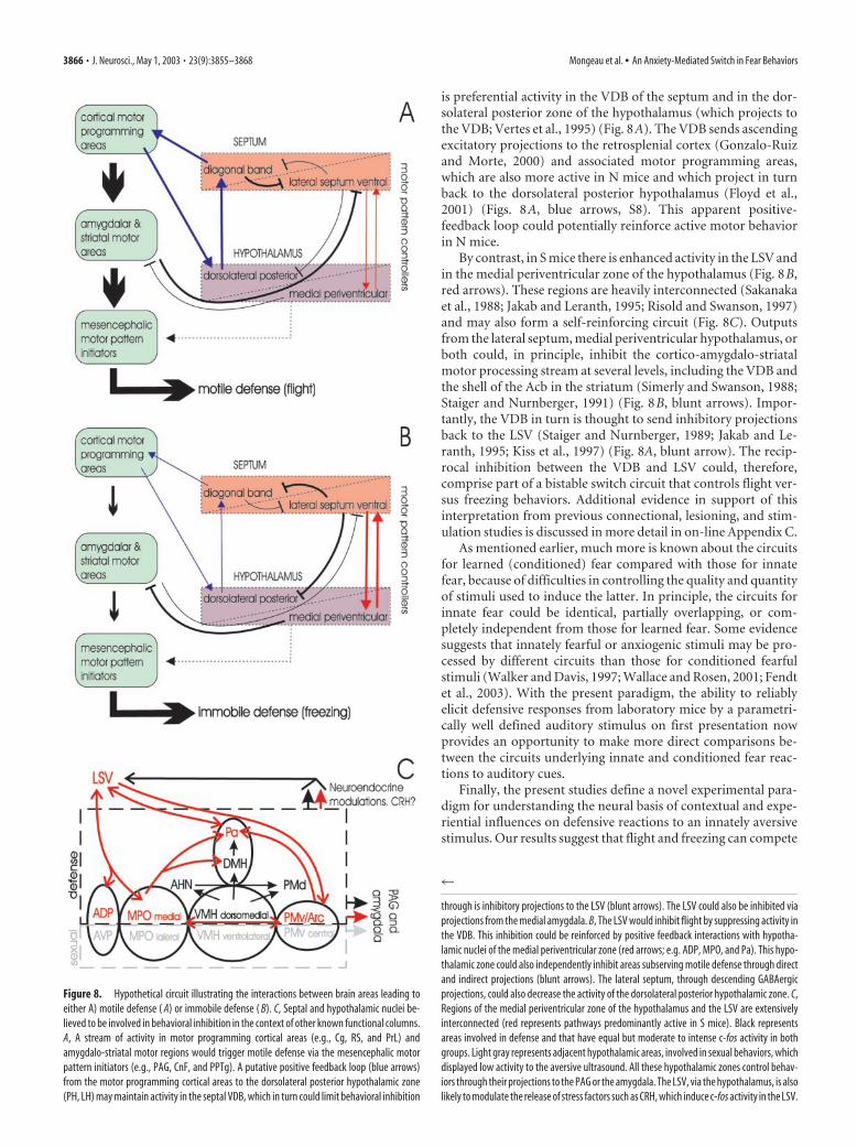
is preferential activity in the VDB of the septum and in the dor-solateral posterior zone of the hypothalamus (which projects tothe VDB; Vertes et al., 1995) (Fig. 8A). The VDB sends ascendingexcitatory projections to the retrosplenial cortex (Gonzalo-Ruizand Morte, 2000) and associated motor programming areas,which are also more active in N mice and which project in turnback to the dorsolateral posterior hypothalamus (Floyd et al.,2001) (Figs. 8A, blue arrows, S8). This apparent positive-feedback loop could potentially reinforce active motor behaviorin N mice.
By contrast, in S mice there is enhanced activity in the LSV andin the medial periventricular zone of the hypothalamus (Fig. 8B,red arrows). These regions are heavily interconnected (Sakanakaet al., 1988; Jakab and Leranth, 1995; Risold and Swanson, 1997)and may also form a self-reinforcing circuit (Fig. 8C). Outputsfrom the lateral septum, medial periventricular hypothalamus, orboth could, in principle, inhibit the cortico-amygdalo-striatalmotor processing stream at several levels, including the VDB andthe shell of the Acb in the striatum (Simerly and Swanson, 1988;Staiger and Nurnberger, 1991) (Fig. 8B, blunt arrows). Impor-tantly, the VDB in turn is thought to send inhibitory projectionsback to the LSV (Staiger and Nurnberger, 1989; Jakab and Le-ranth, 1995; Kiss et al., 1997) (Fig. 8A, blunt arrow). The recip-rocal inhibition between the VDB and LSV could, therefore,comprise part of a bistable switch circuit that controls flight ver-sus freezing behaviors. Additional evidence in support of thisinterpretation from previous connectional, lesioning, and stim-ulation studies is discussed in more detail in on-line Appendix C.
As mentioned earlier, much more is known about the circuitsfor learned (conditioned) fear compared with those for innatefear, because of difficulties in controlling the quality and quantityof stimuli used to induce the latter. In principle, the circuits forinnate fear could be identical, partially overlapping, or com-pletely independent from those for learned fear. Some evidencesuggests that innately fearful or anxiogenic stimuli may be pro-cessed by different circuits than those for conditioned fearfulstimuli (Walker and Davis, 1997; Wallace and Rosen, 2001; Fendtet al., 2003). With the present paradigm, the ability to reliablyelicit defensive responses from laboratory mice by a parametri-cally well defined auditory stimulus on first presentation nowprovides an opportunity to make more direct comparisons be-tween the circuits underlying innate and conditioned fear reac-tions to auditory cues.
Finally, the present studies define a novel experimental para-digm for understanding the neural basis of contextual and expe-riential influences on defensive reactions to an innately aversivestimulus. Our results suggest that flight and freezing can compete
Figure 8. Hypothetical circuit illustrating the interactions between brain areas leading toeither A) motile defense ( A) or immobile defense ( B). C, Septal and hypothalamic nuclei be-lieved to be involved in behavioral inhibition in the context of other known functional columns.A, A stream of activity in motor programming cortical areas (e.g., Cg, RS, and PrL) andamygdalo-striatal motor regions would trigger motile defense via the mesencephalic motorpattern initiators (e.g., PAG, CnF, and PPTg). A putative positive feedback loop (blue arrows)from the motor programming cortical areas to the dorsolateral posterior hypothalamic zone(PH, LH) may maintain activity in the septal VDB, which in turn could limit behavioral inhibition
4
through is inhibitory projections to the LSV (blunt arrows). The LSV could also be inhibited viaprojections from the medial amygdala. B, The LSV would inhibit flight by suppressing activity inthe VDB. This inhibition could be reinforced by positive feedback interactions with hypotha-lamic nuclei of the medial periventricular zone (red arrows; e.g. ADP, MPO, and Pa). This hypo-thalamic zone could also independently inhibit areas subserving motile defense through directand indirect projections (blunt arrows). The lateral septum, through descending GABAergicprojections, could also decrease the activity of the dorsolateral posterior hypothalamic zone. C,Regions of the medial periventricular zone of the hypothalamus and the LSV are extensivelyinterconnected (red represents pathways predominantly active in S mice). Black representsareas involved in defense and that have equal but moderate to intense c-fos activity in bothgroups. Light gray represents adjacent hypothalamic areas, involved in sexual behaviors, whichdisplayed low activity to the aversive ultrasound. All these hypothalamic zones control behav-iors through their projections to the PAG or the amygdala. The LSV, via the hypothalamus, is alsolikely to modulate the release of stress factors such as CRH, which induce c-fos activity in the LSV.
3866 • J. Neurosci., May 1, 2003 • 23(9):3855–3868 Mongeau et al. • An Anxiety-Mediated Switch in Fear Behaviors
for the expression of fear depending on levels of anxiety presentbefore the presentation of this stimulus. These observations inturn raise the question of where and how anxiety modifies defen-sive behavioral outputs to an aversive stimulus. The c-fos map-ping data provide a heuristic circuit for the regulation of thesecompeting behaviors by anxiety or stress, which may now betested by systematic functional perturbation experiments. Thissystem may also provide a useful model for understanding theneural substrates of human fear disorders, such as panic andanxiety, as well as for drugs used to treat them.
ReferencesAlheid GF, Olmos JS, Beltramino CA (1995) Amygdala and extended amyg-
dala. In: The rat nervous system (Paxinos G, ed), pp 495–578. San Diego:Academic.
Anokhin KV, Mileusnic R, Shamakina IY, Rose SP (1991) Effects of earlyexperience on c-fos gene expression in the chick forebrain. Brain Res544:101–107.
Bandler R, Keay KA, Floyd N, Price J (2000) Central circuits mediating pat-terned autonomic activity during active vs. passive emotional coping.Brain Res Bull 53:95–104.
Beckett SR, Aspley S, Graham M, Marsden CA (1996) Pharmacological ma-nipulation of ultrasound induced defence behaviour in the rat. Psychop-harmacology 127:384 –390.
Blanchard DC, Griebel G, Blanchard RJ (2001) Mouse defensive behaviors:pharmacological and behavioral assays for anxiety and panic. NeurosciBiobehav Rev 25:205–218.
Blanchard RJ, Blanchard DC, Hori K (1989) An ethoexperimental approachto the study of defense. In: Ethoexperimental approaches to the study ofbehavior (Blanchard RJ, ed), pp 114 –136. Dordrecht, The Netherlands:Kluwer.
Blanchard RJ, Agullana R, McGee L, Weiss S, Blanchard DC (1992) Sexdifferences in the incidence and sonographic characteristics of antipreda-tor ultrasonic cries in the laboratory rat (Rattus norvegicus). J Comp Psy-chol 106:270 –277.
Bodnoff SR, Suranyi-Cadotte B, Aitken DH, Quirion R, Meaney MJ (1988)The effects of chronic antidepressant treatment in an animal model ofanxiety. Psychopharmacology 95:298 –302.
Canteras NS (2002) The medial hypothalamic defensive system: hodologi-cal organization and functional implications. Pharmacol Biochem Behav71:481– 491.
Canteras NS, Chiavegatto S, Valle LE, Swanson LW (1997) Severe reductionof rat defensive behavior to a predator by discrete hypothalamic chemicallesions. Brain Res Bull 44:297–305.
Castro-Alamancos MA, Borrell J, Garcia-Segura LM (1992) Performance inan escape task induces fos-like immunoreactivity in a specific area of themotor cortex of the rat. Neuroscience 49:157–162.
Cazala P, Schmitt P (1987) Dorso-ventral variation in the attenuating effectof lateral hypothalamic stimulation on the switch-off response elicitedfrom the mesencephalic central gray area. Physiol Behav 40:625– 629.
Cuomo V, Cagiano R, Desalvia MA, Mazzoccoli M, Persichella M, Renna G(1992) Ultrasonic vocalization as an indicator of emotional state duringactive-avoidance learning in rats. Life Sci 50:1049 –1055.
Deakin JF, Graeff FG, Guimaraes F (1992) 5-HT receptor subtypes and themodulation of aversion. In: Central serotonin receptors and psychotropicdrugs (Marsden C, Heal DJ, eds), pp 147–174. London: Blackwell.
Dielenberg RA, Hunt GE, McGregor IS (2001) “When a rat smells a cat”: thedistribution of Fos immunoreactivity in rat brain following exposure to apredatory odor. Neuroscience 104:1085–1097.
Ehret G, Fischer R (1991) Neuronal activity and tonotopy in the auditorysystem visualized by c-fos gene expression. Brain Res 567:350 –354.
Fanselow MS (1980) Conditional and unconditional components of post-shock freezing. Pavlov J Biol Sci 15:177–182.
Fendt M, Endres T, Apfelbach R (2003) Temporary inactivation of the bednucleus of the stria terminalis but not of the amygdala blocks freezinginduced by trimethylthiazoline, a component of fox feces. J Neurosci23:23–28.
Finkbeiner S, Greenberg ME (1998) Ca2� channel-regulated neuronalgene expression. J Neurobiol 37:171–189.
Floyd NS, Price JL, Ferry AT, Keay KA, Bandler R (2001) Orbitomedialprefrontal cortical projections to hypothalamus in the rat. J Comp Neurol432:307–328.
Geuna S (2000) Appreciating the difference between design-based andmodel-based sampling strategies in quantitative morphology of the ner-vous system. J Comp Neurol 427:333–339.
Gewirtz JC, McNish KA, Davis M (1998) Lesions of the bed nucleus of thestria terminalis block sensitization of the acoustic startle reflex producedby repeated stress, but not fear-potentiated startle. Prog Neuropsycho-pharmacol Biol Psychiatry 22:625– 648.
Gonzalo-Ruiz A, Morte L (2000) Localization of amino acids, neuropep-tides and cholinergic markers in neurons of the septum-diagonal bandcomplex projecting to the retrosplenial granular cortex of the rat. BrainRes Bull 52:499 –510.
Graeff FG (1990) Brain defense systems and anxiety. In: Handbook of anx-iety: the neurobiology of anxiety (Burrows GD, Roth M, Noyes R, eds), pp307–354. Amsterdam: Elsevier.
Graeff FG (1994) Neuroanatomy and neurotransmitter regulation of defen-sive behaviors and related emotions in mammals. Braz J Med Biol Res27:811– 829.
Gray JA (1971) The psychology of fear and stress. Cambridge: CambridgeUP.
Gray JA, McNaughton N (2000) The neuropsychology of anxiety. Oxford:Oxford Medical.
Greenberg ME, Ziff EB (1984) Stimulation of 3T3 cells induces transcrip-tion of the c-fos proto-oncogene. Nature 311:433– 438.
Gross C, Santarelli L, Brunner D, Zhuang X, Hen R (2000) Altered fearcircuits in 5-HT(1A) receptor KO mice. Biol Psychiatry 48:1157–1163.
Helmreich DL, Cullinan WE, Watson SJ (1996) The effect of adrenalectomyon stress-induced c-fos mRNA expression in the rat brain. Brain Res706:137–144.
Herdegen T, Leah JD (1998) Inducible and constitutive transcription fac-tors in the mammalian nervous system: control of gene expression by Jun,Fos and Krox, and CREB/ATF proteins. Brain Res Rev 28:370 – 490.
Howard CV, Rose SP (1998) Unbiased stereology: three-dimensional mea-surement in microscopy. New York: BIOS Scientific.
Jakab RL, Leranth C (1995) Septum. In: The rat nervous system (Paxinos G,ed), pp 405– 442. San Diego: Academic.
Kiss J, Borhegyi Z, Csaky A, Szeiffert G, Leranth C (1997) Parvalbumin con-taining cells of the angular portion of the vertical limb terminate oncalbindin-immunoreactive neurons located at the border between thelateral and medial septum of the rat. Exp Brain Res 113:48 –56.
Leaton RN, Borszcz GS (1985) Potentiated startle: its relation to freezingand shock intensity in rats. J Exp Psychol 11:421– 428.
LeDoux JE (1995) Emotion: clues from the brain. Annu Rev Psychol46:209 –235.
Li CX, Waters RS (1991) Organization of the mouse motor cortex studiedby retrograde tracing and intracortical microstimulation (ICMS) map-ping. Can J Neurol Sci 18:28 –38.
Maren S, Fanselow MS (1996) The amygdala and fear conditioning: has thenut been cracked? Neuron 16:237–240.
Mayhew TM, Gundersen HJ (1996) If you assume, you can make an ass outof u and me: a decade of the dissector for stereological counting of parti-cles in 3D space. J Anat 188:1–15.
McGeorge AJ, Faull RL (1989) The organization of the projection from thecerebral cortex to the striatum in the rat. Neuroscience 29:503–537.
Paxinos G, Franklin KBJ (2001) The mouse brain in stereotaxic coordinates.Hong Kong: Academic.
Pfaus JG, Kleopoulos SP, Mobbs CV, Gibbs RB, Pfaff DW (1993) Sexualstimulation activates c-fos within estrogen-concentrating regions of thefemale rat forebrain. Brain Res 624:253–267.
Risold PY, Swanson LW (1997) Connections of the rat lateral septal com-plex. Brain Res Rev 24:115–195.
Ryan AF, Furlow Z, Woolf NK, Keithley EM (1988) The spatial representa-tion of frequency in the rat dorsal cochlear nucleus and inferior colliculus.Hear Res 36:181–189.
Sakanaka M, Magari S, Shibasaki T, Lederis K (1988) Corticotropin releasinging factor-containing afferents to the lateral septum of the rat brain. J CompNeurol 270:404–407.
Staiger JF, Nurnberger F (1989) Pattern of afferents to the lateral septum inthe guinea pig. Cell Tissue Res 257:471– 490.
Staiger JF, Nurnberger F (1991) The efferent connections of the lateral sep-
Mongeau et al. • An Anxiety-Mediated Switch in Fear Behaviors J. Neurosci., May 1, 2003 • 23(9):3855–3868 • 3867
tal nucleus in the guinea pig: intrinsic connectivity of the septum andprojections to other telencephalic areas. Cell Tissue Res 264:415– 426.
Steenbergen HL, Heinsbroek RP, Van Hest A, van de Poll NE (1990) Sex-dependent effects of inescapable shock administration on shuttlebox-escape performance and elevated plus-maze behavior. Physiol Behav48:571–576.
Simerly RB, Swanson LW (1988) Projections of the medial preoptic nu-cleus: a Phaseolus vulgaris leucoagglutinin anterograde tract-tracing studyin the rat. J Comp Neurol 270:209 –242.
Vertes RP, Crane AM, Colom LV, Bland BH (1995) Ascending projectionsof the posterior nucleus of the hypothalamus: PHA-L analysis in the rat.J Comp Neurol 359:90 –116.
Walker DL, Davis M (1997) Double dissociation between the involvementof the bed nucleus of the stria terminalis and the central nucleus of theamygdala in startle increases produced by conditioned versus uncondi-tioned fear. J Neurosci 17:9375–9383.
Wallace KJ, Rosen JB (2001) Neurotoxic lesions of the lateral nucleus ofthe amygdala decrease conditioned fear but not unconditioned fear ofa predator odor: comparison with electrolytic lesions. J Neurosci21:3619 –3627.
Yokosuka M, Matsuoka M, Ohtani-Kaneko R, Iigo M, Hara M, Hirata K,Ichikawa M (1999) Female-soiled bedding induced fos immunoreactiv-ity in the ventral part of the premammillary nucleus (PMv) of the malemouse. Physiol Behav 68:257–261.
3868 • J. Neurosci., May 1, 2003 • 23(9):3855–3868 Mongeau et al. • An Anxiety-Mediated Switch in Fear Behaviors
Related Documents