This is an Accepted Manuscript, which has been through the Royal Society of Chemistry peer review process and has been accepted for publication. Accepted Manuscripts are published online shortly after acceptance, before technical editing, formatting and proof reading. Using this free service, authors can make their results available to the community, in citable form, before we publish the edited article. We will replace this Accepted Manuscript with the edited and formatted Advance Article as soon as it is available. You can find more information about Accepted Manuscripts in the Information for Authors. Please note that technical editing may introduce minor changes to the text and/or graphics, which may alter content. The journal’s standard Terms & Conditions and the Ethical guidelines still apply. In no event shall the Royal Society of Chemistry be held responsible for any errors or omissions in this Accepted Manuscript or any consequences arising from the use of any information it contains. Accepted Manuscript Nanoscale www.rsc.org/nanoscale

Welcome message from author
This document is posted to help you gain knowledge. Please leave a comment to let me know what you think about it! Share it to your friends and learn new things together.
Transcript

This is an Accepted Manuscript, which has been through the Royal Society of Chemistry peer review process and has been accepted for publication.
Accepted Manuscripts are published online shortly after acceptance, before technical editing, formatting and proof reading. Using this free service, authors can make their results available to the community, in citable form, before we publish the edited article. We will replace this Accepted Manuscript with the edited and formatted Advance Article as soon as it is available.
You can find more information about Accepted Manuscripts in the Information for Authors.
Please note that technical editing may introduce minor changes to the text and/or graphics, which may alter content. The journal’s standard Terms & Conditions and the Ethical guidelines still apply. In no event shall the Royal Society of Chemistry be held responsible for any errors or omissions in this Accepted Manuscript or any consequences arising from the use of any information it contains.
Accepted Manuscript
Nanoscale
www.rsc.org/nanoscale

Nanoscale
ARTICLE
This journal is © The Royal Society of Chemistry 20xx J. Name., 2013, 00, 1-3 | 1
Please do not adjust margins
Please do not adjust margins
a. Departamento de Química Orgánica, Facultad de Ciencias, Universidad de Málaga, 29071, Málaga, Spain
b. Departamento de Química Física, CINBIO, Universidade de Vigo and IBIV, 36310 Vigo, Spain
c. Departamento de Quimica Inorgánica, Facultad de Ciencias, Universidad de Malaga, 29071, Málaga, Spain
†Electronic Supplementary Information (ESI) available: [details of any supplementary information available should be included here]. See DOI: 10.1039/x0xx00000x
Received 00th January 20xx,
Accepted 00th January 20xx
DOI: 10.1039/x0xx00000x
www.rsc.org/
Synthesis of Vinyl-Terminated Au Nanoprisms and Nanooctahedra Mediated by 3-Butenoic Acid: Direct Au@pNIPAM Fabrication with Improved SERS Capabilities
M. A. Casado-Rodriguez,aM. Sanchez-Molina
a, A. Lucena-Serrano,
c C. Lucena-Serrano,
a B.
Rodriguez-Gonalez,b Manuel Algarra,
c Amelia Diaz,
a M. Valpuesta,
a J. M. Lopez-Romero,
a J. Perez-
Juste,b*
R. Contreras-Caceres.a*
Here we describe the first seedless synthesis of vinyl-terminated Au nanotriangular prisms (AuNTPs) and nanooctahedra
(AuNOC) in aqueous media. This synthesis is performed by chemical reduction of chloroauric acid (HAuCl4) with 3-butenoic
acid (3BA) in presence of benzyldimethylammonium chloride (BDAC). The principal novelties of the presented method are
the use of a mixture of 3BA and BDAC, the synthesis of gold prisms and octahedra with controllable size, and the presence
of terminal double bonds on the metal surface. Initially this method produces a mixture of triangular gold nanoprisms and
octahedra, however, both morphologies are successfully separated by surfactant micelle induced depletion interaction,
reaching percentages up to ~90 %. Moreover, the alkene moieties presented on the gold surface are exploited for the
fabrication of hybrid core@shell particles. Gold octahedra and triangular prisms are easily encapsulated by free radical
polymerization of N-isopropylacrylamide (NIPAM). Finally, in order to obtain a gold core with the most number of tips,
AuNTP@pNIPAM microgels were subjected to gold core overgrowth, thus resulting in star-shaped nanoparticles
(AuSTs@pNIPAM). We use 4-aminobenzenethiol as model analyte for SERS investigations. As expected, gold cores with
tips and high curvature sites produced the highest plasmonic responses.
1. Introduction
Nowadays, the interest in the fabrication of noble metal
nanoparticles with a great variety of sizes and morphologies is
motivated by the advances in the understanding of their synthesis
and properties,1,2
as well as the possibility of being applied in a vast
number of fields such as drug delivery,3,4
DNA analysis,5,6
cancer
diagnosis8 and treatment,
9,10 immunoassay,
7 and SERS and catalytic
investigations.11,12
The properties of noble metal nanoparticles arise from the localized
surface plasmon resonance (LSPR),13,14
which remarkably depends,
among other factors, on particle size and shape.13,15
Since the first
synthesis concerning gold nanoparticles only produced spherical
shapes,16
important efforts have been performed in the
development of synthetic routes for the fabrication of non-spherical
morphologies. This interest in the synthesis of anisotropic metal
nanoparticles is because morphologies containing well-defined
angles or tips possess a more localized plasmons, thus supplying
further promising and attractive applications in the aforementioned
fields.17, 18
During the last decades, several protocols regarding the synthesis of
particles with different morphologies as triangles, rods, wires,
octahedra, decahedra, cages or stars have been reported in water
or organic media.19-25
Colloidal suspensions of Au nanoparticles are
typically prepared by reaction of a gold salt with a reducing agent,
in presence of stabilizing “capping” molecules, which play an
important role in controlling the nanoparticle morphology.22,26-
31Concerning the fabrication of Au octahedra and triangular prisms,
which are the morphologies fabricated in this work, several
protocols have been reported. For instance, Xia et al. reported the
synthesis of gold octhaedra by reducing HAuCl4 with N-vinyl
pyrrolidone in an aqueous solution in the presence of
cetyltrimethylammonium chloride (CTAC).21
Mirkin et al. fabricated
gold octahedral, in high yield, via the controlled overgrowth of
preformed seeds by Ag+-assisted, seed-mediated synthesis.
27 Liz-
Marzán et al. obtained Au nanotriangles by using CTAC-capped gold
nanoparticles as seeds, in presence of small amount of iodide ions,
and using ascorbic acid as reducing agent.32
More recently, a
seedless approach to synthesize monodisperse Au nanotriangles in
high yield (˃90%) has been reported by Zhang et al.33
Unfortunately, all these methods give rise to particles solely
stabilized by surfactants, that is, with no other functional groups on
Page 1 of 8 Nanoscale
Nan
osca
leA
ccep
ted
Man
uscr
ipt

ARTICLE Journal Name
2 | J. Name., 2012, 00, 1-3 This journal is © The Royal Society of Chemistry 20xx
Please do not adjust margins
Please do not adjust margins
the metal surface, that could be readily available to be used in
further chemical reactions.
Nowadays, a lot of research effort is devoted to the synthesis of
nanocomposite materials with a core-shell architecture. Such hybrid
systems are typically composed by a metal core encapsulated
within a polymer shell.34
These nanocomposite materials have been
demonstrated to display a better performance for sensing and
catalytic purposes compared with pure metal nanoparticles.34-37
They could also incorporate multiple functionalities and improve
their colloidal stability.38-41
However, to obtain such core-shell
morphology a surface modification step of the nanoparticles
surface is generally required. Recently, butenoic acid has been used
as reducing agent for the synthesis of spherical and octahedral Au
nanoparticles.42,43
Additionally, this molecule incorporates terminal
double bonds on the particle surface, which was exploited for the
fabrication of core@shellAu@pNIPAM hybrid nanocomposites
systems.38,42
Herein, we report a water-based seedless method for the synthesis
of vinyl-terminated triangular Au nanoprisms and nanooctahedra
with controllable size, with 3-butenoic acid acting as reducing, as
well as, shape inducing agent. Initially, two main morphologies
were obtained; triangular prisms (AuNTPs) and octahedra
(AuNOCs). We analyze the influence of the temperature and gold
salt concentration in the reaction mixture on the shape and the size
of the particles. Both morphologies were successfully separated by
surfactant micelle induced depletion interaction.44,45
Additionally,
the presence of terminal double bond on the Au nanoparticles
surface (coming from 3BA) was exploited for the fabrication of
core@shell hybrid systems by free radical polymerization of N-
isopropylacrylamide (NIPAM); including octahedra, prisms and star-
like (AuSTs@pNIPAM) gold cores. Finally, the SERS enhancement
capabilities of the different core-shell hybrids was studied using 4-
aminobenzenethiol (4ABT) as model analyte.
2. Experimental
2.1 Materials
3-Butenoic acid (3BA, 97%), cetyltrimethylammonium chloride
(CTAC, ≥98%), benzyldimethylhexadecylammonium chloride (BDAC,
≥97%) and N-isopropylacrylamide (NIPAM, 97%) and 4-
aminobenzenethiol (4ABT, 97%) were supplied by Aldrich.
HAuCl4·3H2O (≥99.9% trace metal basis) was supplied by Sigma.
N,N’-methylenebisacrylamide (BIS, ≥99.5%) was supplied by Fluka.
2,2’-Azobis(2-methylpropionamidine) dihydrochloride (AAPH, 97%)
was supplied by Acros Organics. All reactants were used without
further purification. Water was purified using a Milli-Q system
(Millipore).
2.2 Characterization Methods
UV−vis measurements of aqueous colloidal solutions were recorded
with a HP Agilent 8453 diode array spectrophotometer.
Transmission electron microscopy (TEM) images were acquired on a
JEOL JEM 1400 operating at an acceleration voltage of 80 kV.
Samples were prepared by drying a 10 μL drop of colloidal
suspension on a carbon-coated copper grid. HR-TEM images were
acquired with a JEOL JEM2010F field-emission gun transmission
electron microscope, working at 200 kV. For this specific analysis,
samples were prepared letting dry a drop of sample on TEM copper
grids coated with holey carbon thin film. Field emission scanning
electron microscopy (FESEM) images were obtained in a Helios
Nanolab 650 Dual Beam from FEI, working at acceleration voltage of
15kV, a current intensity of 0.2 nA and a tilting angle of 52o. Sample
were prepared by dropping 20 µL of an aqueous colloidal solution
onto a 1x1 cm single side polished boron-doped silicon (111) wafer
(WRS Materials). SERS spectra were measured using a confocal
Raman Microscope (CRM) alpha300R, (WITec GmbH, Ulm,
Germany). SERS signals were recorded by exciting the colloidal
solutions with a laser power of 5 mW using a 785 nm laser line. For
one Raman spectrum, between 50 and 200 single Raman spectra
with a measuring time of 0.5s were accumulated. Raman spectra
were recorded within the spectral range of 0-2500 cm-1
for Raman
shift. Samples for SERS were prepared by adding 15 µL of 4ABT 10-3
mM to 1.5 mL of each sample (AuNOC@pNIPAM,
AuNTPs@pNIPAM, AuSTs@pNIPAM, at a gold concentratio of 0.5
mM). After 1 h, allowing for thermodynamic equilibrium to be
reached, the colloidal solution was centrifugated twice, and
redispersed in 1.5 mL of water.SERS was directly recorded from
these suspensions.
2.3 Synthesis of Au nanoparticles
In a typical synthesis, 50 mL of a solution containing 0.5 mM HAuCl4
and 5 mM BDAC were introduced into a 100 mL round bottom flask
under low magnetic stirring (100 rpm). Subsequently, the solution
was heated up to 75oC, 85
oC or 95
oC. Then, 100µL of 3BA were
added into the mixture. After a suitable amount of time enough to
allow the complete reduction of HAuCl4 to Au(0) (see Table 1) the
solution was allowed to cool down at room temperature. Finally, in
order to remove the excess of 3BA and BDAC, the colloidal
dispersion containing the Au nanoparticles were centrifuged at
7500 rpm during 30 min. The supernatant was discarded and the
pellet was dispersed in 50 mL of 4mM CTAC. The same procedure
was followed for the synthesis at 1 mM and 1.5 mM HAuCl4,
keeping constant the amount of BDAC and 3BA. Scheme 1
illustrates the synthesis of Au nanoparticles mediated by 3BA.
2.4 Purification of AuNTPs and AuNOC
As mentioned in the introduction, surfactant micelle depletion-
induced flocculation was applied for the separation of gold
nanoparticles by using CTAC as surfactant. Taking into accoun that
the temperature of the syntesis will affect the final particle size (see
below), different CTAC concentrations were used in each case. We
describe in this section the separation procedure for particles
prepared at 1.5 mM HAuCl4 and at 75oC, 85
oC and 95
oC.
Synthesis at 75oC. Initially, in order to remove the bigger particles
generated during the Au synthesis, the colloidal dispersion was
centrifuged at 7500 rpm during 30 min. The supernatanat was
discarded and the precipitate was redispersed in a 5 mL vial
containing 2 mL of CTAC 100 mM. After 4 h at RT, a precipitate
(containing bigger particles) was observed at the bottom of the vial,
which was discarded (Scheme 1 separation 1). The supernatant,
containing a mixture of prisms and octahedra, was again
centrifuged at 7500 rpm during 30 min. The supernatant was
discarded and the precipitate was redispersed in a 5 mL vial
Page 2 of 8Nanoscale
Nan
osca
leA
ccep
ted
Man
uscr
ipt
Journal Name ARTICLE
This journal is © The Royal Society of Chemistry 20xx J. Name., 2013, 00, 1-3 | 3
Please do not adjust margins
Please do not adjust margins
containing 2 mL of 175 mM CTAC. After 4 h the supernatant,
containing AuNOC, was separated, and the precipitate formed at
the bottom of the vial, containing AuNTPs, was redispersed in 10
mL of 100 mM CTAC (Scheme 1, separation 2).
Synthesis at 85oC: The colloidal dispersion obtained at 85
oC was
centrifuged at 8000 rpm during 30 min. The supernatant was
discarded and the precipitate redispersed in2 mL of CTAC 125 mM
in order to promote the depletion of the bigger particles. The
supernatant was concentrated (8000 rpm, 30 minutes) and
redispersed in 2 mL of CTAC 200 mM in order to promote the
depletion of AuNTPs while AuNOC will remain in solution.
Synthesis at 95oC: The colloidal dispersion synthesized at 95
oC was
centrifuged at 8500 rpm during 30 min. The supernatant was
discarded and the precipitate redispersed in 2 mL of CTAC 200 mM
in order to promote the depletion of the bigger particles. The
supernatant was concentrated (8500 rpm, 30 minutes) and
redispersed in 2 mL of CTAC 250 mM in order to promote the
depletion of AuNTPs while AuNOC will remain in solution.
2.5 Synthesis of Au@pNIPAM and Au overgrowth
To carry out the encapsulation of gold prisms and octahedra within
pNIPAM microgels, firstly, 10 mL of each Au colloidal dispersion
([Au]≈ 5 mM) obtained after purification step was heated at 70oC
under N2 flow. Then N-isopropylacrylamide (0.1698 g, 100 mM) and
N,N’-methylenebisacrylamide (0.0234 g, 10 mM) were added under
magnetic stirring. After 15 min, the N2 flow was removed and the
polymerization was initiated by adding 2,2’-azobis(2-
methylpropionamidine) dihydrochloride (10 µL 0.1 M in water).
After 2 h at 70oC, the turbid mixture was allowed to cool down to
room temperature under stirring. Finally, to remove small
oligomers, unreacted monomers as well as gold-free microgels, the
dispersion was diluted with water (50 mL) and centrifuged (30 min
at 5500 rpm), the resulting pellet was redispersed in water. This
process was repeated 3 times.
The AuSTs@pNIPAM particles were obtained by following the same
overgrowth procedure previously reported by Liz-Marzan et al.34
To
a 10 mL growth solution containing 0.5 mM HAuCl4, 8 mM CTAB
and 4 mM ascorbic acid 0.5 mL of AuNTP@pNIPAM seed solution (1
mM in terms of gold) were added.
Scheme 1. General procedure for the synthesis of vinyl-terminated
gold nanoparticles and shape-separation by surfactant micelle
induced depletion interaction. (top) Core@shell Au@pNIPAM
fabrication and gold overgrowth (bottom).
3. Results and Discussion
3.1 Synthesis and characterization of Au nanoparticles
As mentioned in the introduction, only few methods has been
reported concerning the synthesis of gold nanoparticles with either
triangular or octahedral morphologies in aqueous media. In
addition, such synthesis procedures do not confer any surface
functionality to the as prepared nanoparticles. In order to develop a
one-step functionalization, and a highly reproducible methodology
using HAuCl4 as gold source, we used 3BA in presence of BDAC as
stabilizer. It should be noted that initially we used CTAC as
stabilizer, as in the previous mentioned methods. Unfortunately,
highly polydisperse gold nanoparticles morphologies together with
undefined morphologies were obtained (Fig S1, Supporting
Information, SI). Then, we replace CTAC for BDAC, a surfactant with
a similar structure, which contains a benzyl group instead a methyl
group in its structure. Initially, we analyzed the influence of the gold
salt concentration and the temperature in the size and shape of
gold nanoparticles (see Table 1).
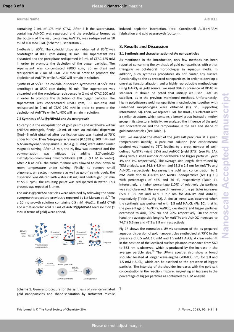
First, we analyzed the effect of the gold salt precursor at a given
temperature; initially, a precursor solution (see experimental
section) was heated to 75oC leading to a great number of well-
defined AuNTPs (yield 58%) and AuNOC (yield 37%) (see Fig 1A),
along with a small number of decahedra and bigger particles (yield
4% and 1%, respectively). The average side length, determined by
TEM analysis, was 54.8 ± 4.0 nm and 35.2 ± 2.5 nm for AuNTPs and
AuNOC, respectively. Increasing the gold salt concentration to 1
mM leads also to AuNTPs and AuNOC nanoparticles (see Fig 1B)
with percentages of 46% and 36 %, respectively (Table 1).
Interestingly, a higher percentage (10%) of relatively big particles
was also observed. The average dimension of the particles increases
65.8 ± 3.9 nm and 41.9 ± 2.7 nm for AuNTPs and AuNOC,
respectively (Table 1, Fig S2). A similar trend was observed when
the synthesis was performed with 1.5 mM HAuCl4 (Fig 1C), that is,
the percentage of AuNTPs, AuNOC, decahedra and bigger particles
decreased to 40%, 30%, 9% and 20%, respectively. On the other
hand, the average side lengths for AuNTPs and AuNOC increased to
74.7 ± 5.6 nm and 47.5 ± 3.9 nm, respectively.
Fig 1F shows the normalized UV-vis spectrum of the as prepared
aqueous dispersion of gold nanoparticles synthetized at 75oC in the
presence of 0.5 mM, 1.0 mM and 1.5 mM HAuCl4. A clear red-shift
in the position of the localized surface plasmon resonance from 569
to 583 nm is observed, which is produced by the increase in the
average particle size.45
The UV-vis spectra also show a broad
shoulder located at longer wavelengths (700-800 nm) for 1.0 and
1.5 mM HAuCl4, which can be ascribed to the presence of bigger
particles. The intensity of the shoulder increases with the gold salt
concentration in the reaction mixture, suggesting an increase in the
percentage of bigger particles as confirmed by TEM analysis.
T
Vinyl-terminated
Au nanoparticles
HAuCl4
BDAC
3BA
AuNTPs@pNIPAM
75-95oC
Separation 1 Separation 2
AuNOC@pNIPAM
CTACCTAC
NIPAM
HAuCl4
AA
AuSTs@pNIPAM
Au octahedra
Au prismsBig
particles
Page 3 of 8 Nanoscale
Nan
osca
leA
ccep
ted
Man
uscr
ipt
ARTICLE Journal Name
4 | J. Name., 2012, 00, 1-3 This journal is © The Royal Society of Chemistry 20xx
Please do not adjust margins
Please do not adjust margins
75 ºC 85 ºC 95 ºC
[HAuCl4]
0.5 mM
[HAuCl4]
1.0 mM
[HAuCl4]
1.5 mM
[HAuCl4]
0.5 mM
[HAuCl4]
0.5 mM
nm % nm % nm % nm % nm %
AuNTPs 54.8 58 65.8 46 74.7 41 44.9 55 33.3 50
AuNOC 35.2 37 41.9 36 47.5 30 34.6 41 26.6 46
Decahedra 4 8 9 3 2
Big Particles 1 10 20 1 2
Time/ min 23 15 7
We also studied the influence of the temperature on the size and
shape of the particles at a given gold salt concentration. We have
chosen 0.5 mM HAuCl4 since gave rise to the lowest contamination
of bigger particles. Overall, well-dispersed AuNTPs and AuNOC
morphologies where found in all studied cases. At 85oC, the average
side length of AuNTPs and AuNOC was 44.9 ± 3.7 and 34.6 ± 2.7
nm, respectively (see Fig S4, SI). When the reaction temperature
was raised up to 95oC, the average side length size decreased to
33.3 ± 3.0 nm and 26.6 ± 2.0 nm for AuNTPs and AuNOC,
respectively. Fig 1D and 1E show TEM images of the gold
nanoparticles synthesized at 85oC and 95
oC, respectively. This
tendency suggests that an increase in the reaction temperature led
to a higher nucleation rate leading to smaller particles. Concerning
the influence of temperature on the relative populations of AuNTPs
and AuNOC, it should be noted that the percentage of AuNTPs
decreased from 58% to 50% when the temperature was increased
from 75oC to 95
oC. On the other hand, the percentage of AuNOC
increased from 37% to 46% (see Table 1).
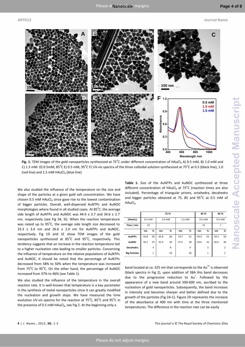
We also studied the influence of the temperature in the overall
reaction rate. It is well-known that temperature is a key parameter
in the synthesis of metal nanoparticles since it can greatly modified
the nucleation and growth steps. We have measured the time
evolution UV-vis spectra for the reaction at 75oC, 85
oC and 95
oC in
the presence of 0.5 mM HAuCl4, see Fig 2. At the beginning only a
Table 1. Size of the AuNTPs and AuNOC synthesized at three
different concentration of HAuCl4 at 75oC (reaction times are also
included). Percentage of triangular prisms, octahedra, decahedra
and bigger particles obtained at 75, 85 and 95oC at 0.5 mM of
HAuCl4.
band located at ca. 325 nm that corresponds to the Au3+
is observed
(black spectra in Fig 2), upon addition of 3BA this band decreases
due to the progressive reduction to Au+. Followed by the
appearance of a new band around 500-600 nm, ascribed to the
nucleation of gold nanoparticles. Subsequently, the band increases
in intensity and becomes sharper and better defined due to the
growth of the particles (Fig 2A-C). Figure 2D represents the increase
of the absorbance at 400 nm with time at the three mentioned
temperatures. The difference in the reaction rate can be easily
Fig. 1. TEM images of the gold nanoparticles synthesized at 75
oC under different concentration of HAuCl4 A) 0.5 mM, B) 1.0 mM and
C) 1.5 mM. D) 0.5mM, 85oC E) 0.5 mM, 95
oC F) UV-vis spectra of the three colloidal solution synthesized at 75
oC at 0.5 (black line), 1.0
(red line) and 1.5 mM HAuCl4 (blue line)
400 600 800 10000.0
0.3
0.6
0.9
1.2
Ab
so
rba
nc
e
Wavelength /nm
A B C
D
100 nm
100 nm100 nm
E)E
100 nm
100 nm
F0.5 mM
1.0 mM
1.5 mM
75 ºC 85 ºC 95 ºC
[HAuCl4] 0.5 mM 1.0 mM 1.5 mM 0.5 mM 0.5 mM
Time / min 23 15 7
nm % nm % nm % nm % nm %
AuNTPs 54.8 58 65.8 46 74.7 41 44.9 55 33.3 50
AuNOC 35.2 37 41.9 36 47.5 30 34.6 41 26.6 46
Decahedra 4 8 9 3 2
Big Particles 1 10 20 1 2
Page 4 of 8Nanoscale
Nan
osca
leA
ccep
ted
Man
uscr
ipt
Journal Name ARTICLE
This journal is © The Royal Society of Chemistry 20xx J. Name., 2013, 00, 1-3 | 5
Please do not adjust margins
Please do not adjust margins
Fig 2. UV-vis spectral evolution for the synthesis performed at 0.5
mM of HAuCl4 at A) 75oC, B) 85
oC and C) 95
oC. Evolution of the
absorbance at 400 nm for the three mentioned temperatures D).
observed; while at 75oC the absorbance reached a maximum at 23
min, when the synthesis was performed at 85oC and 95
oC the time
need to reach the maximum absorbance decreased to 15 min and 7
min, respectively. It should be noted that in all cases the final UV-vis
spectra showed a narrow localized surface plasmon band,
suggesting the absence of bigger particles.
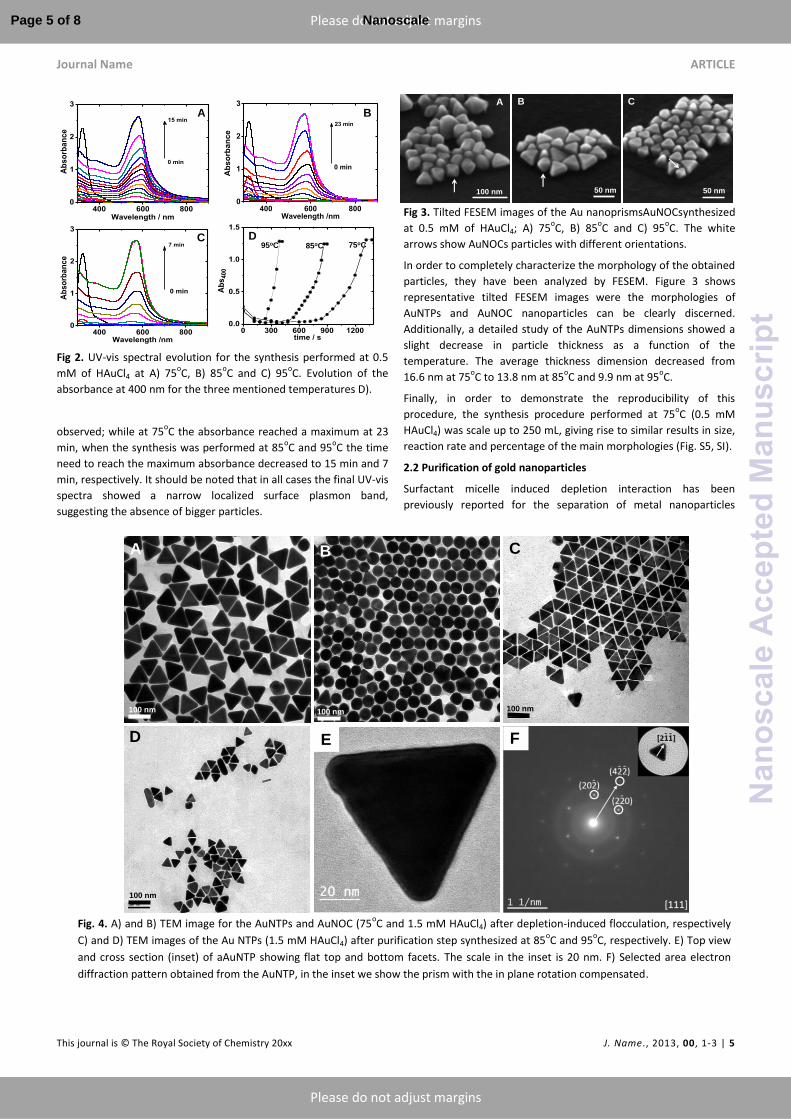
Fig 3. Tilted FESEM images of the Au nanoprismsAuNOCsynthesized
at 0.5 mM of HAuCl4; A) 75oC, B) 85
oC and C) 95
oC. The white
arrows show AuNOCs particles with different orientations.
In order to completely characterize the morphology of the obtained
particles, they have been analyzed by FESEM. Figure 3 shows
representative tilted FESEM images were the morphologies of
AuNTPs and AuNOC nanoparticles can be clearly discerned.
Additionally, a detailed study of the AuNTPs dimensions showed a
slight decrease in particle thickness as a function of the
temperature. The average thickness dimension decreased from
16.6 nm at 75oC to 13.8 nm at 85
oC and 9.9 nm at 95
oC.
Finally, in order to demonstrate the reproducibility of this
procedure, the synthesis procedure performed at 75oC (0.5 mM
HAuCl4) was scale up to 250 mL, giving rise to similar results in size,
reaction rate and percentage of the main morphologies (Fig. S5, SI).
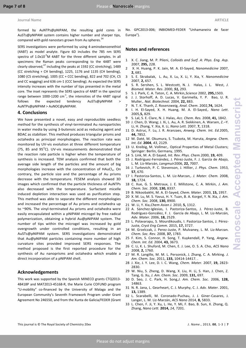
2.2 Purification of gold nanoparticles
Surfactant micelle induced depletion interaction has been
previously reported for the separation of metal nanoparticles
Fig. 4. A) and B) TEM image for the AuNTPs and AuNOC (75
oC and 1.5 mM HAuCl4) after depletion-induced flocculation, respectively
C) and D) TEM images of the Au NTPs (1.5 mM HAuCl4) after purification step synthesized at 85oC and 95
oC, respectively. E) Top view
and cross section (inset) of aAuNTP showing flat top and bottom facets. The scale in the inset is 20 nm. F) Selected area electron
diffraction pattern obtained from the AuNTP, in the inset we show the prism with the in plane rotation compensated.
A
100 nm
20 nm
B
100 nm 100 nm
C
D
100 nm
FE
400 600 8000
1
2
3
23 min
Ab
so
rban
ce
Wavelength /nm
0 min
400 600 8000
1
2
3
15 min
Ab
so
rba
nc
e
Wavelength / nm
0 min
400 600 8000
1
2
3
7 min
Ab
so
rban
ce
Wavelength /nm
0 min
0 300 600 900 12000.0
0.5
1.0
1.5
A
bs
400
time / s
95oC 85oC 75oC
A B
C D
100 nm 50 nm 50 nm
A B C
Page 5 of 8 Nanoscale
Nan
osca
leA
ccep
ted
Man
uscr
ipt

ARTICLE Journal Name
6 | J. Name., 2012, 00, 1-3 This journal is © The Royal Society of Chemistry 20xx
Please do not adjust margins
Please do not adjust margins
containing different sizes and shapes. This method is based on the
depletion interactions between particles in presence of micelles
produced by a surfactant.45
We carried out the separation process
for samples prepared with 1.5 mM of HAuCl4 at the three
mentioned temperatures. Initially, bigger particles were flocculated
with 100 mM CTAC, as it was confirmed by the TEM images and UV-
vis spectrum of the redispersed precipitate (Fig. S6, SI). The
remaining supernatant, mainly containing AuNTPs and AuNOC, was
subjected to the same separation procedure but with 175 mM
CTAC. After this second depletion process the precipitate contained
mainly AuNTPs (see Fig 4A) while the supernatant contained AuNOC
(see Fig 4B). As it can be clearly observed the percentage of either
prisms or octahedra remarkably increases compared with the
sample before purification steps (see Fig. 1C). Images at lower
magnification are shown in Fig. S7, SI. TEM images corresponding to
the AuNTPs synthesized at 85oC and 95
oC are included in Fig. 4C and
4D, respectively. As is observed, the number of prisms remarkably
increases with respect to samples before depletion. The dimension
analysis for the obtained Au prisms resulted in an averageside
length of 52.3 ± 3.5 nm, and 42.3 ± 3.2 nm for synthesis at 85 and
95oC, respectively, (Fig S8, SI) data not included in table 1. The
crystallographic structure of the AuNTPs has also been analyzed by
HR-TEM. A representative top view TEM image of an AuNTPs lying
flat on the carbon film is presented in Figure 4E. Prisms show an
almost perfect triangular shape with rounded tips. The inset shows
a cross section of one of the prism exhibiting a thickness of 43 nm
and lateral side facets. Selected area electron diffraction (SAED)
pattern, included in Figure 4F, displays a regular reciprocal net that
corresponds with the [111] zone axis of the gold crystalline
structure. In the inset we show an AuNTP with the in plane rotation
compensated; from that we have found the [211] type directions as
the crystalline directions parallel to the prisms tips.
The purification process was also monitored by UV-vis
spectroscopy. Figure 5 represents the normalized UV-vis spectra of
the Au colloidal solutions of the samples prepared at 75 oC and 1.5
mM of HAuCl4, before and after depletion induced separation
processes. Initially the spectrum exhibits a maximum plasmon band
at 583 nm (black line), and the aqueous colloidal solution displayed
a purple color, inset in Fig 5. After purification, the normalized UV-
vis spectrum of the isolated AuNOC displays a narrow plasmon band
at 554 nm (red line) and the aqueous colloidal solution (included in
the inset) shows an intense red color. The normalized UV-vis
spectrum of the AuNTPs shows a plasmon band at 602 nm (blue
line), and the aqueous colloidal solution displayed an intense blue
color.
3.3 Synthesis of ofAu@pNIPAM particles. SERS investigations
As it was mentioned, 3BA supplies vinyl-functionalization onto gold
nanoparticles surface.42
It is important to remark that double bond
terminated systems are nowadays used for different purposes; for
example, double bond terminated specimens have been recently
incorporated on H-Si surface by photoactivated hydrosilylation
reaction,46
and vinyl-finctionalized metal nanoparticles have been
encapsulated with a pNIPAM, styrene or silica shell. To this aim, the
purified samples containing AuNTPs and AuNOC were performed to
free radical polymerization in presence of N-isopropylacrylamide
and and N,N’-methylenebisacrylamide. Figs 6A and 6B show
representative TEM images of the obtained core@shell
AuNTPs@pNIPAM and AuNOC@pNIPAM nanocomposites. Both,
AuNTPs and AuNOC are homogeneously coated by a pNIPAM shell.
After that, in order to increase the number of tips within the
microgel, AuNTPs@pNIPAM particles were used as seeds for an Au
core overgrowth, under the same conditions previously reported
for Au@pNIPAMparticles,34
resulting AuSTs@pNIPAM nanoparticles
(Figure 6C). It should be noted that in this case the initial seeds are
Fig. 5. Normalized UV-vis spectra for the Au nanoparticles
synthesized at 75oC and 1.5 mMof HAuCl4 before (black line)
and after depletion-induced flocculation, which includes
AuNOC (red line) and AuNTPs (blue line). The inset shows an
image of a vial containing the AuNTPs and AuNOC colloidal
solutions
400 600 800 10000.0
0.3
0.6
0.9
1.2
Ab
so
rba
nc
e
Wavelength /nm
Initial solution
Au octahedra
Au nanoprisms
Prisms OctahedraInitial
Fig. 6. RepresentaticeTEM images of differentcore@shell
hybrid systems. A) AuNTPs@pNIPAM, B) AuNOC@pNIPAM and
C) AuSTs@pNIPAM particles. D) Raman spectra of 4ABT (10−5
M) aqueous solution adsorbedon the different samples tested.
The scale in the inset is 50 nm. The inset in D highlights the
Raman shift located at 1080 cm.1
.
1000 1250 15000
30k
60k
90k
14
89
15
92
11
76
10
80
10
05
Stars
Prisms
Octahedra
Spheres
Ra
ma
n In
ten
sit
y /A
rb. U
nit
s
Raman shift /cm-1
1065 1080 10950
30k
60k
90k
Ra
ma
n I
nt.
/A
U
Raman shift /cm-1
200 nm
A B
C D
200 nm
200 nm
Page 6 of 8Nanoscale
Nan
osca
leA
ccep
ted
Man
uscr
ipt
Journal Name ARTICLE
This journal is © The Royal Society of Chemistry 20xx J. Name., 2013, 00, 1-3 | 7
Please do not adjust margins
Please do not adjust margins
formed by AuNTPs@pNIPAM, the resulting gold cores in
AuSTs@pNIPAM system contains higher number and sharper tips,
compared with gold nanostars grown from Au spheres.34
SERS investigations were performed by using 4-aminobenzenethiol
(4ABT) as model analyte. Figure 6D includes the 785 nm SERS
spectra of 1.0x10⁻⁵
M 4ABT for the three different samples. In all
specimens the Raman peaks corresponding to the 4ABT were
clearly observed,47
including the peaks at 1592 (CC stretching), 1489
(CC stretching + CH bending), 1225, 1176 and 1135 (CH bending),
1080 (CS stretching), 1005 (CC + CCC bending), 822 and 702 (CH, CS
and CC wagging) and 636 cm-1 (CCC bending). As expected the SERS
intensity increases with the number of tips presented in the metal
core. The inset represents the SERS spectra of 4ABT in the spectral
range between 1000-1200 cm-1
, the intensities of the 4ABT signal
follows the expected tendency AuSTs@pNIPAM ˃
AuNTPs@pNIPAM ˃ AuNOC@pNIPAM.
4. Conclusions
We have presented a novel, easy and reproducible seedless
method for the synthesis of vinyl-terminated Au nanoparticles
in water media by using 3-butenoic acid as reducing agent and
BDAC as stabilizer. This method produces triangular prisms and
octahedra as principal morphologies. The reaction rate was
monitored by UV-vis evolution at three different temperature
(75, 85 and 95oC). UV-vis measurements demonstrated that
the reaction rate particle is higher when the temperature of
synthesis is increased. TEM analysis confirmed that both the
average side length of the particles and the amount of big
morphologies increase with the concentration of HAuCl4. On
contrary, the particle size and the percentage of Au prisms
decrease with the temperature. FESEM analysis showed 3D
images which confirmed that the particle thickness of AuNTPs
also decreased with the temperature. Surfactant micelle
induced depletion interaction was used as purification step.
This method was able to separate the different morphologies
and increased the percentage of Au prisms and octahedra up
to ~90%. The vinyl-terminated Au prisms and octahedra were
easily encapsulated within a pNIPAM microgel by free radical
polymerization, obtaining a hybrid Au@pNIPAM system. The
number of tips within the microgel was increased by gold
overgrowth under controlled conditions, resulting in an
AuSTs@pNIPAM system. SERS investigations demonstrated
that Au@pNIPAM particles containing more number of high
curvature sites provided improved SERS responses. The
method proposed is the first reported procedure for the
synthesis of Au nanoprisms and octahedra which enable a
direct incorporation of a pNIPAM shell.
Acknowledgements
This work was supported by the Spanish MINECO grants CTQ2013-
48418P and MAT2013-45168-R, the Marie Curie COFUND program
“U-mobility” co-financed by the University of Malaga and the
European Community’s Seventh Framework Program under Grant
Agreement No 246550, and from the Xunta de Galicia/FEDER (Grant
No. GPC2013-006; INBIOMED-FEDER “Unhamaneira de facer
Europa”).
Notes and references
1 X. C. Jiang, M. P. Pileni, Colloids and Surf. A: Phys. Eng. Asp.
2007, 295, 228. 2 X. H. Huang, P. K. Jain, M. A. El-Sayed, Nanomedicine 2007,
2, 681. 3 S. E. Skrabalak, L. Au, X. Lu, X. Li, Y. Xia, Y. Nanomedicine
2007, 2, 657. 4 S. R. Sershen, S. L. Westcott, N. J. Halas, J. L. West, J.
Biomed. Mater. Res. 2000, 51, 293. 5 S. J. Park, C. A. Taton, C. A. Mirkin,Science 2002, 295,1503. 6 J. J. Storhoff, A. D. Lucas, V. Garimella, Y. P. Bao, U. R.
Muller,. Nat. Biotechnol. 2004, 22, 883. 7 N. T. K. Thanh, Z. Rosenzweig, Anal. Chem. 2002,74, 1624. 8 I. H. El-Sayed, X. H. Huang, M. A. El-Sayed,. Nano Lett.
2005,5, 829. 9 S. Lal, S. E. Clare, N. J. Halas, Acc. Chem. Res. 2008, 41, 1842. 10 J. Chen, D. Wang; J. Xi, L. Au, A. R. Siekkinen, A. Warsen, Z.–Y.
Li, H. Zhang, Y. Xia, X. Li, Nano Lett. 2007, 7, 1318. 11 D. Astruc, F. Lu, J. R. Aranzaes, Anweg. Chem. Int. Ed.2005,
44, 7852. 12 M. Daté, M. Okumura, S. Tsubota, M. Haruta, Angew. Chem.
Int. Ed. 2004, 43, 2129. 13 U. Kreibig, M. Vollmer,. Optical Properties of Metal Clusters;
Springer: Berlin, Germany, 1995. 14 S. Link, M. A. El-Sayed, Int. Rev. Phys. Chem.2000, 19, 409. 15 J. Rodríguez-Fernández, J. Pérez-Juste, F. J. García de Abajo,
L. M. Liz-Marzán, Langmuir2006, 22, 7007. 16 J. Turkevich, P. C. Stevenson, J. Hillier, J. Phys. Chem. 1953,
57, 670. 17 I. Pastoriza-Santos, L. M. Liz-Marzan,. J. Mater. Chem. 2008,
18, 1724. 18 C. Xue, G. S. Metraux, J. E. Millstone, C. A. Mirkin, J. Am.
Chem. Soc. 2008, 130, 8337. 19 B. Nikoobakht, M. A. El-Sayed, Chem. Mater. 2003, 15, 1957. 20 X. M. Lu, M. S. Yavuz, H. Y. Tuan, B. A. Korgel, Y. N. Xia, J. Am.
Chem. Soc. 2008, 130, 8900. 21 W. Li, Y. Xia,Chem Asian J. 2010, 5, 1312. 22 A. Sánchez-Iglesias, I. Pastoriza-Santos, J. Pérez-Juste, B.
Rodríguez-González, F. J. García de Abajo, L. M. Liz-Marzán, Adv. Mater. 2006, 18, 2529.
23 L. Polavarapu, S. Mourdikoudis, I. Pastoriza-Santos, J. Pérez-Juste, Cryst Eng Comm. 2015, 17, 3727.
24 M. Grzelczak, J. Perez-Juste, P. Mulvaney, L. M. Liz-Marzan. Chem. Soc. Rev. 2008, 37, 1783.
25 F. Kim, S. Connor, H. Song, T. Kuykendall, P. Yang, Angew. Chem. Int. Ed. 2004, 43, 3673.
26 C. Li, K. L. Shuford, M. Chen, E. J. Lee, O. S. A. Cho, ACS Nano 2008, 2, 1760.
27 M. R. Langille, M. M. L. Personick, J. Zhang, C. A. Mirking. J. Am. Chem. Soc. 2011, 133, 10414-14417. .
28 J. Xie, J. Y. Lee, D. I. C. Wang, Chem. Mater. 2007, 19, 2823-2830.
29 W. Niu, S. Zheng, D. Wang, X. Liu, H. Li, S. Han, J. Chen, Z. Tang, G. Xu, J. Am. Chem. Soc. 2009, 131, 697.
30 D. Seo, J. C. Park, H. Song,J. Am. Chem. Soc. 2006, 128, 14863.
31 N. R. Jana, L. Gearheart, C. J. Murphy, C. J. Adv. Mater. 2001, 13, 1389.
32 L. Scarabelli, M. Coronado-Puchau, J. J. Giner-Casares, J. Langer, L. M. Liz-Marzán, ACS Nano 2014, 8, 5833.
33 L. Chen, F. Ji, Y. Xu, L. He, Y. Mi, F. Bao, B. Sun, X. Zhang, Q. Zhang, Nano Lett. 2014, 14, 7201.
Page 7 of 8 Nanoscale
Nan
osca
leA
ccep
ted
Man
uscr
ipt
ARTICLE Journal Name
8 | J. Name., 2012, 00, 1-3 This journal is © The Royal Society of Chemistry 20xx
Please do not adjust margins
Please do not adjust margins
34 R. Contreras-Caceres, A. Sanchez-Iglesias, M. Karg, I. Pastoriza-Santos, J. Perez-Juste, J. Pacifico, T. Hellweg, A. Fernandez-Barbero, L. M. Liz-Marzan, Adv. Mater. 2008, 20, 1666.
35 Y. Lu, Y. Mei, M. Drechsler, M. Ballauff, Angew. Chem. Int. Ed.2006,45, 813.
36 R. Contreras-Caceres, S. Abalde-Cela, P. Guardia-Giros, A. Fernandez-Barbero, J. Perez-Juste, R. A. Alvarez-Puebla, L. M. Liz-Marzan, Langmuir 2011,27, 4520.
37 M. Laurenti, P. Guardia, R. Contreras-Caceres, J Perez-Juste, A. Fernandez-Barbero, E. Lopez-Cabarcos, J. Rubio-Retama, Langmuir 2011, 27, 10484.
38 R. A. Alvarez-Puebla, R. Contreras-Caceres, I. Pastoriza-Santos, J. Perez-Juste, L. M. Liz-Marzan, Angew. Chem. Int. Ed.2009, 48, 138.
39 L. Zha, Y. Zhang, W. Yang, S. Fu, Adv. Mater.2002, 14, 1090. 40 J. –H. Kim, T. R. Lee, Chem. Mater. 2004, 16, 3647. 41 J. –H. Kim, T. R. Lee, Langmuir 2007, 23, 6504. 42 R. Contreras-Caceres, I. Pastoriza-Santos, R. A. Alvarez-
Puebla, J. Perez-Juste, A. Fernandez-Barbero, L. M. Liz-Marzan. Chem. Eur. J. 2010, 16, 9462.
43 S. Gómez-Graña, C. Fernández-López, L.Polavarapu, J. Baptiste Salmon, J. Leng, I. Pastoriza-Santos, J. Pérez-Juste, Chem. Mater. 2015, 27, 8310.
44 K. Park, H. Koerner, R. A. Vaia, Nano Lett. 2010, 10, 1433. 45 N. R. Jana, Chem. Commun. 2003, 1950. 46 A. Lucena-Serrano, C. Lucena-Serrano, R. Contreras-Caceres,
A. Diaz, M. Valpuesta, C. Cai, J. M. Lopez-Romero. Appl. Surf. Sci. 2016, 360, 419.
47 K. Kim, J.-Y. Choi, H. B. Lee, K. S. Shin. J. Chem. Phys. 2011, 135, 124705.
Page 8 of 8Nanoscale
Nan
osca
leA
ccep
ted
Man
uscr
ipt
Related Documents


![Environmental Science - [ RSC ] Publishing](https://static.cupdf.com/doc/110x72/633ee048b4763fa6980cfaeb/environmental-science-rsc-publishing-1683829039.jpg)


