This is an Accepted Manuscript, which has been through the Royal Society of Chemistry peer review process and has been accepted for publication. Accepted Manuscripts are published online shortly after acceptance, before technical editing, formatting and proof reading. Using this free service, authors can make their results available to the community, in citable form, before we publish the edited article. We will replace this Accepted Manuscript with the edited and formatted Advance Article as soon as it is available. You can find more information about Accepted Manuscripts in the Information for Authors. Please note that technical editing may introduce minor changes to the text and/or graphics, which may alter content. The journal’s standard Terms & Conditions and the Ethical guidelines still apply. In no event shall the Royal Society of Chemistry be held responsible for any errors or omissions in this Accepted Manuscript or any consequences arising from the use of any information it contains. Accepted Manuscript Molecular BioSystems www.rsc.org/molecularbiosystems

Welcome message from author
This document is posted to help you gain knowledge. Please leave a comment to let me know what you think about it! Share it to your friends and learn new things together.
Transcript

This is an Accepted Manuscript, which has been through the Royal Society of Chemistry peer review process and has been accepted for publication.
Accepted Manuscripts are published online shortly after acceptance, before technical editing, formatting and proof reading. Using this free service, authors can make their results available to the community, in citable form, before we publish the edited article. We will replace this Accepted Manuscript with the edited and formatted Advance Article as soon as it is available.
You can find more information about Accepted Manuscripts in the Information for Authors.
Please note that technical editing may introduce minor changes to the text and/or graphics, which may alter content. The journal’s standard Terms & Conditions and the Ethical guidelines still apply. In no event shall the Royal Society of Chemistry be held responsible for any errors or omissions in this Accepted Manuscript or any consequences arising from the use of any information it contains.
Accepted Manuscript
Molecular BioSystems
www.rsc.org/molecularbiosystems

We revealed that human α- and β-defensins have strong anti-HCV activity in experiments on
cellular protection, neutralization, and treatment at low concentrations, whereas synthetic linear
avian defensins could reach similar anti-HCV potentials only at noticeably higher concentrations.
Page 1 of 36 Molecular BioSystems
Mol
ecul
arB
ioS
yste
ms
Acc
epte
dM
anus
crip
t
1
Virucidal activity of human α- and β-defensins against 1
hepatitis C virus genotype 4 2
Ehab H. Mattar,1 Hussein A. Almehdar,
1 Vladimir N. Uversky,
1,2,3,* and 3
Elrashdy M. Redwan1,4,
* 4
5
1 Department of Biological Sciences, Faculty of Sciences, King Abdulaziz University, 6
P.O. Box 80203, Jeddah, Saudi Arabia 7
3 Department of Molecular Medicine and USF Health Byrd Alzheimer's Research 8
Institute, Morsani College of Medicine, University of South Florida, Tampa, FL, 9
USA; 10
3 Laboratory of Structural Dynamics, Stability and Folding of Proteins, Institute of 11
Cytology, Russian Academy of Sciences, St. Petersburg, Russian Federation; 12
4 Therapeutic and Protective Proteins Laboratory, Protein Research Department, 13
Genetic Engineering and Biotechnology Research Institute, City for Scientific 14
Research and Technology Applications, New Borg EL-Arab 21934, Alexandria, 15
Egypt. 16
17
* Corresponding authors: Redwan, E.M. ([email protected]), Uversky, V.N. 18
([email protected]). 19
20
21
22
23
24
25
26
Page 2 of 36Molecular BioSystems
Mol
ecul
arB
ioS
yste
ms
Acc
epte
dM
anus
crip
t
2
27
Abstract 28
Hepatitis C virus (HCV) is the major etiological agent of human non-A and non-B 29
hepatitis affecting about 180 million people worldwide. The goal of current study was 30
to find the effective anti-HCV proteins. As a result, defensins were selected as 31
promising candidates due to their well-known anti-viral potentials and small size. We 32
conducted in vitro evaluation of two kinds of defensins (human α- and β–defensins 33
and synthetic linear avian α-defensins) using tissue culture combined with reverse 34
transcription nested PCR (RT-nested-PCR) and real-time PCR. Human α- and β-35
defensins showed strong anti-HCV activity in experiments on cellular protection, 36
neutralization, and treatment at all concentrations used (10, 20 and 50 µg). The 37
synthetic linear defensins could reach similar anti-HCV potentials only at noticeably 38
higher concentrations (250 µg) and do not show noticeable activity at 10 and 20 µg. 39
This study suggest that defensins are potent anti-HCV agents. 40
41
Keywords: Hepatitis C virus; α-defensins; β-defensins; virucidal; linear defensins; 42
intrinsic disorder; thermodynamic instability. 43
44
Introduction 45
The alternative and complementary medicine contain a number of means for HCV 46
control. Among these means are defensins, short, cationic, cysteine-rich polypeptides 47
that have pronounced biocidal activity and belong to a diverse group of antimicrobial 48
peptides found in vertebrates, invertebrates, insects, and plants.1-8
These polypeptides 49
play important roles in innate immunity against microbial and viral infections, are 50
involved in adaptive immunity, and play various roles in inflammation, wound repair, 51
expression of cytokines and chemokines, production of histamine, and enhancement 52
of antibody responses.9-11
They are also able to induce and augment antitumor 53
immunity when fused with the non-immunogenic tumor antigens.12
These 28–42 54
amino acid cationic peptides are assumed to possess a conserved fold and contain six 55
highly conserved cysteine residues, which form three pairs of intramolecular disulfide 56
Page 3 of 36 Molecular BioSystems
Mol
ecul
arB
ioS
yste
ms
Acc
epte
dM
anus
crip
t

3
bonds, specific patterns of which are well-preserved during the evolution.8, 13-15
Based 57
on their cellular origin, the spacing between the cysteine residues, and the number and 58
pattern (or topology) of their disulfide bridges, the vertebrate defensins are classified 59
as α-, β-, and γ-defensins.8, 15, 16
In mammals, barrier epithelial cells mostly generates 60
β-defensins, whereas α-defensins are mainly stored in the azurophil granules of 61
neutrophils.7 In the mouse, Paneth cells and skin produce at least 17 α-defensins, 62
whereas various epithelial cells and keratinocytes generate four β-defensins.17
63
In this study, several innate immune defense peptides and proteins of different nature 64
were analyzed for their potential activities against hepatitis C virus (HCV) using the 65
in vitro culture system. We analyzed: proteins purified from natural resources (human 66
neutrophils peptides, α-defensins 1 to 4 as mixture); recombinant proteins (human β-67
defensins 1 to 5 and 116 as mixture), and synthetic linear peptides (avian β-defensins 68
AvBD-4, AvBD-7, AvBD-12). Their antiviral activities were monitored in peripheral 69
blood mononuclear cells (PBMCs) and Huh7.5 cell line using three experimental 70
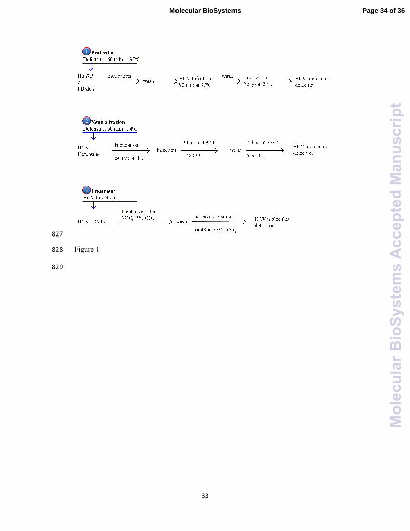
strategies depicted in Figure 1 that are based on two main methodologies for detection 71
of the viral molecules, reverse transcription nested PCR (RT-nested-PCR) and real-72
time-PCR. Throughout the study, all experiments has been run in duplicate, unless 73
otherwise mentioned. We also evaluated the total concentrations of α- and β–74
defensins in HCV-infected patients and non-infected subjects using commercial 75
ELISA kits. 76
77
Figure 1. 78
79
RESULTS 80
Cell viability and cytotoxic effects of defensins 81
First, cytotoxic effects of different defensins on PBMCs and Huh7.5 cell line were 82
studied. To this end, the PBMCs (2.5×10⁵) and Huh7.5 cells (10
5) treated for 7 days 83
with defensins at the maximal concentrations to be used in the antiviral activity 84
screening (50 and 250 µg/ml) were compared with the untreated PBMCs and Huh7.5 85
cultures. This analysis revealed that human α–defensins were not cytotoxic, whereas 86
Page 4 of 36Molecular BioSystems
Mol
ecul
arB
ioS
yste
ms
Acc
epte
dM
anus
crip
t
4
at their highest concentrations, human β–defensins and avian synthetic defensins 87
caused a slight reduction in the viability of both cells, to ~93-95% (Table 1). 88
89
Table 1. 90
91
Evaluation of the anti-HCV activity of defensins using RT-nested PCR 92
Cell protection by defensins against the entry of HCV particles 93
As shown in Figure 1, both Huh7.5 and PBMC cell cultures were treated with human 94
α- or β-defensins or avian defensins for 60 min, then washed three times with the PBS 95
buffer or fresh medium, and then infected with HCV for 90 min. The inoculated cells 96
were cultured for seven days. At all concentrations tested, α-defensins were effective 97
protectors of both cell types against the HCV attack. β-Defensins also efficiently 98
protected cells at concentrations of 20 and 50 µg/ml but completely failed to do so at 99
10 µg/ml. Synthetic linear avian β-defensins (AvBD-4, 7, and 12) failed to protect 100
both cell cultures from the HCV entry at lower concentration 10 and 20 µg/ml and 101
showed protection only at very high concentrations of 250 µg/ml (Figure 2A). Figure 102
2A shows that the HCV-related band of 174 bp was not amplified in all protected cells 103
and amplified in non-protected cells. Finally, camel lactoferrin (cLac) was used as a 104
positive control, whereas Rulc system was used as quality and reproducibility 105
indicator of the amplification system. 106
107
Figure 2. 108
109
Defensins neutralization potentials against HCV particles 110
Next, defensins were tested for their HCV neutralization potentials. To this end, they 111
were incubated with HCV-infected serum at concentrations of 10, 20, 50 and/or 250 112
µg/ml for 60 min, and these pre-incubated mixtures were used to infect Huh7.5 or 113
PBMCs cells. After incubation for 90 min the cell cultures were washed three times 114
Page 5 of 36 Molecular BioSystems
Mol
ecul
arB
ioS
yste
ms
Acc
epte
dM
anus
crip
t

5
with PBS or fresh media and the inoculated cells were cultured for seven days. Figure 115
2B shows that human α- and β-defensins were able to completely neutralize all HCV 116
particles and subsequently inhibit the viral entry into the cells at all concentrations. On 117
the other hand, the avian β-defensins failed to neutralize and block the HCV entry into 118
cells at concentrations of 10 and 20 µg/ml and were able to do so only at the highest 119
concentration of 250 µg/ml. 120
121
Effect of the intracellular treatment with defensins on HCV replication 122
Human defensins at concentrations of 10, 20, and 50 µg/ml and avian defensins at 123
concentrations of 10, 20, 250 µg/ml were investigated for their in vitro ability to 124
inhibit the viral replication inside the infected Huh7.5 and PBMCs cells. Inhibition of 125
viral replication was detected by amplification of viral non-coding RNA segments 126
using the RT-PCR technique. Human defensins at all concentration tested were able 127
to completely inhibit the HCV replication in the Huh7.5 cells and in the PBMCs 128
(Figure 2C) within 48 h. However, avian β-defensins did not inhibit HCV replication 129
at any concentrations used (10, 20, and 250 µg/ml) (Figure 2C). 130
131
Evaluation of the anti-HCV activity of defensins using real time PCR 132
In addition to the RT-nested-PCR used for detection of HCV presence in the cells, we 133
also utilized the real-time-PCR to detect and measure the HCV copy number 134
throughout all conditions analyzed in this study. 135
136
Cells protection by defensins against entry of HCV particles 137
The HCV copy number calculations revealed that at concentrations of 10, 20 and 50 138
µg/ml, human α-defensins were able to protect the PBMCs and Huh7.5 cells from 139
attack by the HCV viral particles since no HCV particles were found in these 140
experiments, indicating a relative activity of 100% for these defensins. Human β-141
defensins also offered a comparable protection for both cells types but possessed the 142
relative activity of 100% only at concentrations of 20 and 50 µg/ml, whereas at 10 143
Page 6 of 36Molecular BioSystems
Mol
ecul
arB
ioS
yste
ms
Acc
epte
dM
anus
crip
t

6
µg/ml these peptides were somewhat less potent protecting both cell types against 144
HCV entry (see Table 2). The avian defensins reached the relative activity of 100% 145
only at the concentrations of 250 µg/ml in both PBMCs and Huh7.5 cells, whereas the 146
protective effects of lower concentrations of these defensins were noticeably less 147
pronounced. 148
149
Table 2. 150
151
Neutralization potentials of defensins against HCV particles 152
Based on the HCV copy number calculations, it was clear that natural human α-153
defensins and human recombinant β-defensins were able to totally neutralize the HCV 154
particles and subsequently protect the PBMCs and Huh7.5 cells from the HCV 155
infection at all concentration studied (see Table 3). On the other hand, avian synthetic 156
β-defensins neutralized all HCV particles only being added at concentration of 250 157
µg/ml, whereas at lower concentrations they showed low neutralization activity and 158
their neutralization activity was concentration dependent. 159
160
Table 3. 161
162
Effects of intracellular treatment with defensins on HCV replication 163
Table 4 shows that according to the HCV copy number calculations, human defensins 164
were able to penetrate to the pre-infected Huh7.5 cells and PBMCs and completely 165
blocked the HCV genome replication and the subsequent assembly of viral particle at 166
all concentrations studied. However, synthetic avian β-defensins were much less 167
efficient in penetration of the infected Huh7.5 cells and PBMCs, and, consequently, 168
significant numbers of viral particles were seen at all concentrations of these 169
defensins studied in this work. The highest relative activity was achieved by 250 170
µg/ml of AvBD-12 (21% and 30.6% in infected Huh7.5 cells and PBMCs). 171
Noticeably, although low, the ability of avian β-defensins to affect HCV replication 172
Page 7 of 36 Molecular BioSystems
Mol
ecul
arB
ioS
yste
ms
Acc
epte
dM
anus
crip
t

7
was typically concentration dependent and increased with the increase in the content 173
of corresponding defensin. 174
175
Table 4. 176
177
DISCUSSION 178
Hepatitis C virus is an enveloped, single-stranded positive-sense RNA virus that 179
belongs to the Flaviviridae family. There is no insect vector or animal reservoir for 180
HCV, and the virus is acquired through person-to-person transmission by parenteral 181
routes (i.e., in a manner other than through the digestive tract).18
Before clinical 182
screening for HCV became available, infection was mainly transmitted by transfusion 183
of contaminated blood or blood products. Nowadays transmission frequently occurs 184
through the use of contaminated needles, syringes, and other instruments used for 185
injections and other skin-piercing procedures. Sexual transmission of hepatitis C 186
occurs rarely.19
187
HCV is the major cause of parenterally transmitted non-A and non-B hepatitis 188
worldwide,18
and infection with HCV is one of the leading causes of chronic liver 189
disease worldwide.20
The prevalence of HCV infection has increased during recent 190
years. It is estimated now that over 180 million people are infected with HCV world-191
wide. This means that 3% of the world's population are affected by HCV, and in some 192
countries, such as Egypt, this number reaches 15%.21
More than 70% of patients 193
infected with HCV develop chronic, if not lifelong, infection. Furthermore, persistent 194
HCV infection accounts for ~50% of serious end-stage liver diseases, such as liver 195
cirrhosis, hepatic failure, and hepatocellular carcinoma. 196
There are six major genotypes of HCV found throughout the world, with genotypes 1, 197
2, 3 and 4 being further subdivided on several sub-genotypes. Many HCV genotypes 198
are unevenly distributed, with genotypes 1 and 3 being found in most countries 199
irrespective of their economic status, and with the largest number of incidences of 200
genotypes 4 and 5 being reported in lower-income countries.22
Major clinical research 201
on antiviral therapy for chronic HCV has been conducted in Western countries23
and 202
Page 8 of 36Molecular BioSystems
Mol
ecul
arB
ioS
yste
ms
Acc
epte
dM
anus
crip
t
8
in Japan.24
Therefore, most published data deal with patients infected with HCV 203
genotypes 1, 2, and 3, and there are now articulate guidelines for the type of treatment 204
and period of antiviral treatment in such patients.25
However, there have been 205
relatively few studies that deal with the patients infected with HCV genotype 4 206
(which is highly prevalent in North Africa and the Middle East), and combination 207
therapy trials (interferon and ribavirin) for these patients did not demonstrate 208
promising efficacy.26
209
Currently, no vaccine is available to prevent HCV infection. Standard treatment with 210
interferon and ribavirin remained a gold standard of the chronic HCV remedy. This 211
therapy achieves 50% sustained virological response (SVR, which is aviremia 24 212
weeks after completion of antiviral therapy) for genotype 1 and 80% for genotype 2 213
and 3. Recent studies have shown that HCV genotype-4 patients have a response rate 214
to pegylated interferon monotherapy or combination interferon-ribavirin therapy that 215
is less favorable compared to genotypes 2 and 3, and the response failure rate of about 216
50% is observed. As pegylated interferon is expensive, standard interferon is still the 217
main therapy for HCV treatment in under-developed countries.27
Furthermore, It is 218
recognized now that the combined pegylated interferon-ribavirin therapy might have 219
severe side effects, such as haematological complications. 220
In addition to interferon and ribavirin, there are several FDA-approved anti-HCV 221
drugs. The emerging novel antivirals should optimize the treatment options, especially 222
for difficult-to-treat patients, such as those who are suffering from advanced liver 223
diseases or other co-infections, and who have poor response rates to current regimens. 224
Although the currently approved and used cocktail of anti-HCV therapy is believed to 225
cure more than 90% of infected patients, the appearance of viral resistance (due to the 226
error-prone replication of this RNA virus), the presence of non-responders or 227
treatment failure, superimposed with the adverse effects caused by the drugs in 228
addition to treatment cost, are still major limitations that must be resolved. 229
Furthermore, most entry inhibitors target host components, such as receptors or key 230
enzymes, which are required for HCV entry and definitely have high genetic barriers 231
to resistance due to their conserved nature. Therefore, these inhibitors tend to not only 232
have pan-genotypic activity against virus infection but also possess a greater risk of 233
simultaneously causing cellular toxicity.28
234
Page 9 of 36 Molecular BioSystems
Mol
ecul
arB
ioS
yste
ms
Acc
epte
dM
anus
crip
t
9
This work was dedicated for the in vitro evaluation of the antiviral potentials of 235
human α- and β-defensins against HCV. For evaluation of the efficiency of anti-HCV 236
candidate agents, three strategies were followed, cellular protection, viral particle 237
neutralization, and intracellular viral replication inhibition in two in vitro models, 238
peripheral blood mononuclear cells (PBMCs) and Huh7.5 cell line, using RT-nested-239
PCR and real-time PCR as the most accurate methodologies suitable for these 240
analyses. This analysis revealed that natural human α-defensins (HNP-1, HNP-2, 241
HNP-3, and HNP-4) and recombinant human β-defensins (1 through 5 and 116 as 242
mixture) have a relative anti-viral activity of 100% in all three experimental settings 243
(cellular protection, viral particle neutralization, and intracellular viral replication 244
inhibition) at all concentrations studied (10, 20 and 50 µg/ml). The only exception 245
from this general trend was the case of the lowest concentrations of human β-246
defensins (10 µg/ml) that could not completely protect the Huh7.5 cells, possessing a 247
relative activity of 59%. Generally, human α- and β-defensins were able to completely 248
neutralize all HCV particles added and subsequently inhibit the viral entry into the 249
Huh7.5 and PBMC cells. Furthermore, there were no markers indicating the presence 250
of the HCV amplified band or HCV particles within these cells. Different situation 251
was observed for the synthetic linear avian β-defensins (AvBD-4, AvBD-7, and 252
AvBD-12) that failed to protect both cell cultures from the HCV entry and were 253
unable to neutralize viral particle and inhibit intracellular viral replication at 254
concentration 10 and 20 µg/ml, being able to show antiviral activities at much higher 255
concentrations (250 µg/ml). It is important to note that concentrations of human α- 256
and β-defensins used in this study (10 – 50 µg/ml) were within the biologically 257
relevant range. In fact, it is known that the levels α-defensins in the human plasma 258
range from 400 ng/ml in healthy individuals to 13 µg/ml in individuals with bacterial 259
infections, and may be as high as 6 mg/ml within neutrophils.29, 30
260
Unfortunately, we could not find any systematic study on the antiviral activities of 261
human defensins against HCV. The only exception is the poster of Sherker et al. 262
presented at The International Liver Congress 2012 – 47th
Annual Meeting of the 263
European Association for the Study of the Liver.31
These authors analyzed cellular 264
protection and inhibition of the intracellular viral replication using only α-defensin 1 265
(HNP-1), whereas we looked at the cellular protection, viral particle neutralization, 266
and inhibition of the intracellular viral replication delivered by the purified mixture of 267
Page 10 of 36Molecular BioSystems
Mol
ecul
arB
ioS
yste
ms
Acc
epte
dM
anus
crip
t

10
native human α-defensins HNP-1, HNP-2, HNP-3, and HNP-4 and recombinant 268
human β-defensins (1 through 5 and 116 used as a mixture). Furthermore, the authors 269
of the previous study used the HCV cell culture (HCVcc) system in Huh7.5.1 cells 270
and HCV pseudo-particle (HCVpp) and assessed viral translation and replication with 271
specific HCV replicons.31
Whereas we used natural infection replication system with 272
native HCV genotype-4a, since this better reproduces the biology and kinetics of 273
HCV infection, where the HCV particles infect the hepatocytes and produces 274
infectious viral particles. Furthermore, the patient serum contains the whole viral 275
particle with all its quasi-species of different infectivity magnitudes whereas the 276
fabricated HCV RNA particles (HCV pseudo-particles) are usually homogenous. 277
However, despite the numerous methodological differences, the results of both studies 278
are rather similar and mutually supportive. 279
Since Sherker et al. did not analyze blocking/neutralization efficiency of defensins 280
against HCV31
and since we could not find any published work on the effects of β-281
defensins against not only HCV but any other member of the Flaviviradae family and 282
against viruses close to HCV within the genus Pestivirus, our work is the first study 283
where the direct interactions between human α- and β-defensins and HCV viral 284
particles were analyzed. In fact, we have established that the activity of α- and β-285
defensins against HCV was mediated primarily by the effect of these peptides on both 286
the target cell and the viral particles. Our results are consistent with previous reports 287
on the presence of such activity of defensins against other types of viruses. For 288
example, the ability of HNP-1–3 to directly inactivate HSV and other enveloped 289
viruses, including influenza A virus was reported, suggesting the ability of defensin to 290
destabilize viral envelopes.32
Also, the activity of HNP-1-3 defensins against HIV was 291
reported,33
and defensins were shown to inhibit infectivity of the number of enveloped 292
viruses, such as vesicular stomatitis virus (VSV), cytomegalovirus, influenza A virus 293
(IAV), sindbis virus, vaccinia virus, baculovirus, and herpes simplex virus (HSV), as 294
well as some non-enveloped viruses including human adenovirus (HAdV), 295
adenoassociated virus (AAV), and human papillomavirus (HPV).34
296
It remains unclear exactly how defensins alter host cells. In vivo, an antiviral role of 297
defensins may be manifested by affecting innate and adaptive immune responses. 298
Some defensins block viruses by up-regulating type I interferon response genes, 299
whereas β-defensins may also act as chemoattractants for T-cells, monocytes, mast 300
Page 11 of 36 Molecular BioSystems
Mol
ecul
arB
ioS
yste
ms
Acc
epte
dM
anus
crip
t
11
cells, and dendritic cells. Defensins can also activate intracellular signaling networks 301
to induce immune cell maturation, cytokine secretion, and antibody production.35
The 302
nanomolar concentrations of α-defensins are chemotactic for human monocytes and 303
immature dendritic cells.36
α-Defensins induce interleukin (IL)-8 release in vitro37
and 304
enhance the synthesis and secretion of IL-837-39
and IL-1 in airway epithelial cells and 305
primary bronchial cells.38
Other reports have shown that α-defensins are able to enter 306
the cells40
possibly by binding to the low-density lipoprotein receptor–related 307
protein/α2-macroglobulin receptor and inhibits PKCα/β by direct binding to this 308
kinase.41
In agreement with these observations, HNP-1 is among the most potent 309
inhibitory peptides of PKC.42
Therefore, one of the possible mechanisms by which 310
defensins inhibits replication of viruses involves interference with the PKC-mediated 311
inhibition of viral entry. This is in line with our finding showing that the inhibition is 312
observed when human defensins were added soon after infection. Finally, although 313
the direct membrane disruption by defensins is considered as one of the potential 314
molecular mechanisms of their anti-bacterial, anti-fungal, and anti-parasitic action43
315
such mechanism cannot be easily applied to enveloped and non-enveloped viruses. 316
Therefore, several mechanisms of action of defensins against enveloped and non-317
enveloped viruses can be proposed: 318
1. Direct distortion of the viral envelope through perturbation of the viral lipid 319
membranes. This model is not consistent with the previous reports.44
320
2. Charge-charge attraction of defensins to viruses. It is unlikely that this mechanism 321
is dominant in the antiviral activity of defensins, since this antiviral activity is 322
generally preserved at physiological salt concentrations, whereas the linearized α-323
defensins that lack a disulfide-stabilized 3-D structure are nonfunctional against all 324
viruses tested.34
Furthermore, although β-defensins are, on average, more charged 325
than α-defensins, they typically exhibit less antiviral activity, especially against non-326
enveloped viruses.34, 45
327
3. Immunomodulatory role,35
where defensins participate in activation and/or 328
enhancement of the functions of immune cells recruited to a site of viral infection.45
329
4. The ability to distort/modulate structures of viral proteins. Recent study indicated 330
that the intrinsic disorder as well as thermodynamic instability of microbial proteins 331
are the decisive characteristics of protein susceptibility to interaction with defensins.46
332
Since viral proteomes in general contain numerous intrinsically disordered proteins,47-
333
Page 12 of 36Molecular BioSystems
Mol
ecul
arB
ioS
yste
ms
Acc
epte
dM
anus
crip
t
12
50 and since many HCV proteins are intrinsically disordered,
51 it is likely that this 334
intrinsically disordered nature of HCV proteome make its proteins susceptible for 335
defensins. 336
Curiously, Figure 3 and Table 5 show that defensins themselves contain significant 337
amounts of intrinsic disorder, with human proteins being, in general, noticeably more 338
disordered than avian polypeptides. The disorderedness of these proteins is evident 339
from their high mean disorder scores (see Table 5), and from the presence of 340
disordered tails (Figure 3). Furthermore, Figure 3 shows that defensins analyzed in 341
this study can be grouped into three sets based on the peculiarities of their per-residue 342
disorder profiles obtained by PONDR®
FIT, which is a metapredictor of intrinsic 343
disorder that is known to be moderately more accurate than each of the component 344
predictors.52
This disorder-based grouping of defensins coincides with the traditional 345
classification of these proteins, suggesting that different classes of defensins are 346
characterized by class-specific peculiarities of disorder distributions. We also looked 347
at the disorder propensity of defensins by classifying them as mostly ordered or 348
disordered proteins using charge-hydropathy plot (CH-plot).53, 54
This approach is able 349
to discriminate proteins with substantial amounts of extended disorder (random coils 350
and pre-molten globules, which are located above the boundary in the corresponding 351
CH-plot) from proteins with globular conformations (molten globule-like and ordered 352
globular proteins, which are positioned below the boundary).53
Figure 3D shows that 353
human β-defensins are noticeably more disordered than human α-defensins and avian 354
β-defensins. Points corresponding to human β-defensins-3, -4 and -116 are located 355
above the boundary separating compact proteins and extended disordered proteins, 356
indicating that these three defensins are predicted to have extended disordered 357
structures. Although points corresponding to human β-defensins-1, -2, and -5 are 358
located below this boundary, they are noticeably closer to the boundary than points 359
corresponding to human α-defensins and avian β-defensins that have comparable 360
charge-hydropathy attributes (Figure 3D). 361
362
Figure 3 363
364
Table 5 365
366
Page 13 of 36 Molecular BioSystems
Mol
ecul
arB
ioS
yste
ms
Acc
epte
dM
anus
crip
t

13
In summary, we report here unique data on the ability of human native and 367
recombinant defensins to protect cellular systems from the HCV attack, to neutralize 368
viral particles, and to inhibit intracellular viral replication. These important 369
observations, taken together with the fact that the serum of HCV patients contains 370
highly elevated levels of defensins, clearly indicates that the pharmaceutical potentials 371
of human defensins cannot be ignored, especially considering their strong antiviral 372
activity combined with low molecular weight, reduced immunogenicity and 373
antigenecity, broad biocidal spectrum, and resistance to proteolysis. 374
375
MATERIALS & METHODS 376
Samples 377
Samples of the HCV-infected human serum and/or plasma used in our research 378
(without patient name(s) or medical history) were supplied by the ALBOURG clinic 379
lab (Giza, Cairo Egypt) under supervision of Prof. Ehab Eldab. Samples from ten 380
hepatitis C patients with high viremia (8.3 million copies/ml) positive for viral 381
genotype 4 antibody and confirmed by PCR, were used for the in vitro infection 382
experiments. The peripheral blood leucocytes (PBLCs) used in our study were from 383
these volunteers, to whom the goals of the experiments were explained and whose 384
informed consent was obtained in a form of oral approval. All experiments were 385
performed in compliance with the relevant laws and institutional guidelines. 386
For in vitro infection experiments, we utilized serum samples positive for the HCV 387
antibody and RNA as determined by ELISA and RT-nested PCR and genotyped as 388
genotype-4 using the method described in ref.55
389
390
391
Human subjects provided informed consent 392
393
Chemicals 394
Page 14 of 36Molecular BioSystems
Mol
ecul
arB
ioS
yste
ms
Acc
epte
dM
anus
crip
t
14
Chemicals were purchased from Fluka Biochemika (Buchs, Switzerland), Amersham 395
Pharmacia Biotech (Uppsala, Sweden), Riedel-deHaen (Germany), WINLAB (U.K.), 396
Sigma chemicals Co. (St.Louis, Mo., USA), Acros (New Jersey, USA), PARK 397
(Northampton, U.K.), Fischer Scientific (U.K.), Scharlau Chemie S.A. (Barcelona, 398
Spain), and Athen Research and Technology (Virginia, USA). 399
Agarose was from GEBCO BRL (Paisley, Scotland), RPMI-1640 cell culture media 400
were purchased from HyClone (Logan, Utah), fetal bovine serum, penicillin, 401
streptomycin, and trypsin were obtained from Sigma. Primers for PCR and MgCl2 402
were purchased from Clontech (USA); dNTP and Taq DNA polymerase were 403
purchased from Promega (Madison, WI, USA); Ready-To-Go RT-PCR beads was 404
purchased from Amersham Pharmacia Biotech (Piscataway, NJ, USA), DNA ladder 405
obtained from Promega (USA), Thiazolyl Blue Tetrazolium Bromide was purchased 406
from Sigma-Aldrich Chemie GmbH (Germany). The Huh7.5 cell line was as a gift 407
from Professor Carl Rice (USA). 408
409
Defensin proteins and peptides 410
Natural human α–defensins known as human neutrophil peptides HNP-1, HNP-2, 411
HNP-3, and HNP-4 were purified from human neutrophils. Recombinant human β-412
defensins (1-5 and 116) were produced in prokaryotic expression system. Avian β-413
defensins (AvBD-1, AvBD-2, and AvBD-3) were obtained as linear peptides via the 414
in vitro synthesis. Sequences and basic physico-chemical properties of defensins used 415
in this study are listed in Table 5. 416
417
Cell cultures of PBMCs 418
Peripheral mononuclear blood cells (PBMCs) were isolated as reported in ref.56
419
Briefly, peripheral blood samples from healthy individuals were diluted with 5 420
volumes of a freshly prepared RBC lysis buffer (38.8 mM/L NH4Cl, 2.5 mM/L 421
K2HCO3, 1 mM/L EDTA, pH 8.0), incubated at room temperature for 10 min and 422
centrifuged at 1,500 rpm and 4ºC for 5 min. The nucleated cells were precipitated in 423
the bottom of the tube. The pellet was collected and washed three times with PBS. 424
Page 15 of 36 Molecular BioSystems
Mol
ecul
arB
ioS
yste
ms
Acc
epte
dM
anus
crip
t
15
425
Assays for the analysis of cytotoxic effects of defensins 426
Throughout the current study, all in vitro experiments with the cultured tissue cells 427
were run in duplicates. The cytotoxic effect of defensins on PBMCs and Huh7.5 cells 428
was examined by the counting of viable cells after trypan blue treatment and by the 3-429
(4,5-dimethylthiazol-2-yl)-2,5-diphenyltetrazolium bromide (MTT) method. The 430
Huh7.5 cells were washed twice in RPMI-1640 media supplemented with 200 µM L-431
glutamine in 25 µL HEPES buffer (N-[2-hydroxyethyl] piperazine-N-[2-432
ethanesulphonic acid]). PBMCs cells (2.5×10⁵) and Huh7.5 (1.0×10
5) were plated in 433
three 24-well microtiter plates in duplicates and cultured in RMPI-1640 culture media 434
(RMPI-1640 supplemented media, 10% fetal bovine serum (FBS), 100 U of penicillin 435
and 100 µg streptomycin) for two days at 37ºC, 5% CO2 and 88% humidity before 436
defensins treatment, then the medium was refreshed with new RMPI-1640 437
supplemented medium containing 100 µg/ml of defensins. The cells and defensins 438
were incubated for 90 min at 37ºC and washed three times with 1 ml of PBS. The 439
cells were maintained in 1 mL of fresh medium for seven days at 37ºC, 5% CO2 and 440
88% humidity. After one week of culture, the cells were collected and suspended in 441
medium. For collection of Huh7.5 cells, the adherent cells were detached from the 442
plate using 200 µL trypsin/EDTA mixture (200 mg/L EDTA, 500 mg/L trypsin in a 443
ratio 1:250) for 1–3 minutes, the action of trypsin was stopped by the addition of 1 444
mL of RPMI culture media. The cells were scrapped and collected in a 15 ml falcon 445
tube, then washed twice by RPMI supplemented media and once by phosphate buffer 446
saline (PBS), and centrifuged at 1,000 rpm for 5 min after each wash. The pellet was 447
resuspended in 1.0 ml PBS, and then the total number of viable cells was counted 448
using the trypan blue treatment. We also examined the viability of cells which were 449
cultured for one day with medium containing 50 and 100 µg/mL of defensins. 450
The viability of the cells was also assayed by Thiazolyl Blue Tetrazolium Bromide 451
(MTT) method as following: 10×103
PBMC cells or Huh7.5 cells in 200 µL culture 452
media per well were placed in a 96-well plate. Plate was incubated at 37ºC, 5% CO2 453
and 88% humidity before defensins treatment, then the medium was refreshed with 454
new RMPI-1640 supplemented medium containing 0.100 mg/mL of defensins. The 455
cells and defensins were incubated for 90 min at 37ºC and washed three times with 456
Page 16 of 36Molecular BioSystems
Mol
ecul
arB
ioS
yste
ms
Acc
epte
dM
anus
crip
t
16
200 µl of PBS. The cells were maintained with 200 µL of fresh medium for seven 457
days at 37ºC, 5% CO2 and 88% humidity. Then, 20 µl MTT solution (5 mg of 458
Thiazolyl Blue Tetrazolium Bromide (MTT) per 1 ml PBS buffer) was added to each 459
well. Plate was placed on a shaking table, shaken at 150 rpm for 5 min, to thoroughly 460
mix the MTT into the media. Then, the plate was incubated at 37ºC, 5% CO2 and 88% 461
humidity for 5 hours to allow the MTT to metabolize. Next, aliquots of 200 µL of 462
dimethylsulfoxide (DMSO) were added to each well, and plate was placed on the 463
shaking table, shaken at 150 rpm for 5 min and then used to read optical density at 464
595 nm by ELISA reader. The viability of cells which were cultured for one day with 465
medium containing 50 and 100 µg/ml of defensins was also examined. 466
467
Antiviral activity of natural human α-defensins (HNPs), recombinant human β- 468
defensins (RHBDs), and synthetic linear avian β-defensins (ABDs) against HCV 469
The antiviral activities of human natural α-defensins (human neutrophil peptides, α-470
defensins 1 to 4 as a mixture), human recombinant β-defensins (RHBD-1, RHBD-2, 471
RHBD-3, RHBD-4, RHBD-5, and RHBD-116 as a mixture), and synthetic linear 472
avian β-defensins (AvBD-4, AvBD-7, AvBD-12) were analyzed. Their antiviral 473
activities were monitored in peripheral blood mononuclear cells (PBMCs) and Huh7.5 474
cell line using three experimental strategies, such as cellular protection, viral particle 475
neutralization, and inhibition of intracellular viral replication depicted in Figure 1, and 476
two methodologies were used for detection of the viral molecules, RT-nested-PCR 477
and real-time-PCR. 478
There is a clear difference in the action of defensins in different experiments used in 479
this study, despite the fact that there could be an overlap between the mechanisms of 480
defensin action in these three types of experimental approaches. In the “Cell 481
protection by defensins against the entry of HCV particles” approach, cells are first 482
treated with defensins and then there pre-treated cells are exposed to the virus. 483
Therefore, it is expected here that defensins act directly on cells. In the “Defensins 484
neutralization potentials against HCV particles” approach, the infected serum is first 485
treated with defensins and then this infected serum pre-treated with defensins is used 486
to infect cells. Therefore, in this approach, defensins are expected to act directly on 487
HCV particles. In the “Inhibition of intracellular viral replication” approach, HCV-488
Page 17 of 36 Molecular BioSystems
Mol
ecul
arB
ioS
yste
ms
Acc
epte
dM
anus
crip
t
17
infected cells are treated with defensins. Although one cannot exclude scenario, where 489
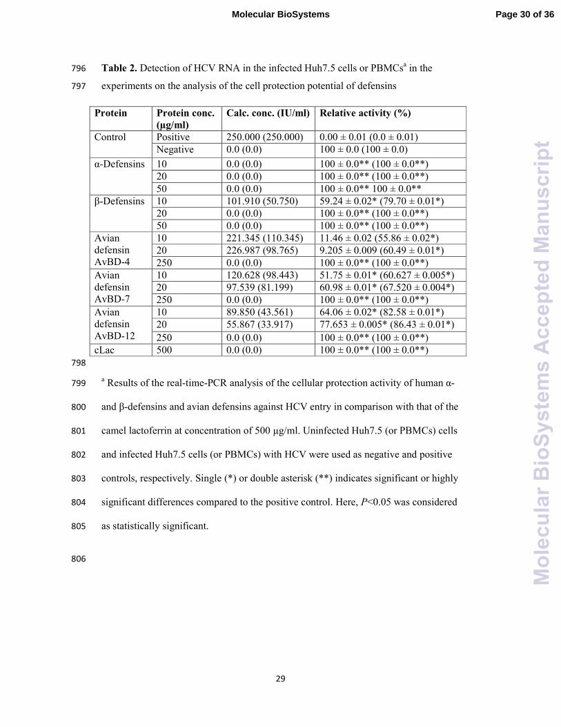
defensins can act on the HCV particles inside infected cells, there is also a possibility 490
that in this approach defensins possess effects on some cellular mechanisms and 491
pathways, potentially acting as cytokines or growth hormones. Exact mechanisms of 492
the intracellular activity of defensins are not know and this topic requires further 493
work. 494
495
Protection potential of natural human α-defensins (HNPs), recombinant human β-496
defensins (RHBDs), and synthetic linear avian β-defensins (ABDs) on HCV 497
To examine the cellular protection effects of human α-defensins (HNPs), recombinant 498
β-defensins (RHBD), and Avian β-defensins (ABD1-3), multiple parallel cultures the 499
human PBMCs (2.5×10⁵) and Huh7.5 (1.0×10
5) cells were plated in three 24-well 500
microtiter plates. HNPs, RHBDs, or ABD1-3 were added to the PBMCs and Huh7.5 501
cells in 50 mL of RPMI-1640 supplemented medium at a final concentration of 10, 502
20, or 50 µg/ml for each the above defensin peptides, then incubated for 60 min at 503
37ºC. Free defensins were removed by washing the cells three times with 1 mL of 504
cold PBS. After addition of 10 mL of medium containing 1 mL of HCV-infected 505
serum (8.3 million copies/mL, RNA G4), the cells were incubated for 90 min at 37ºC. 506
The cells were then washed three times with PBS and cultured for seven days at 37ºC, 507
5% CO2 and 88% humidity,57, 58
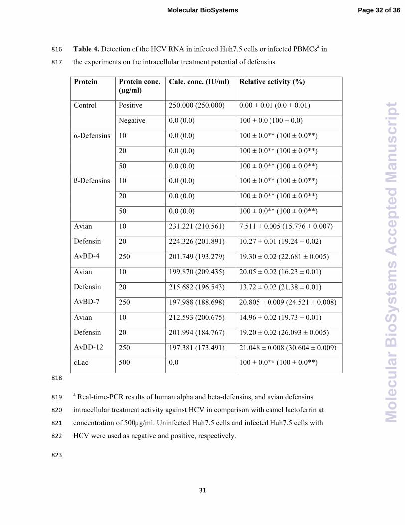
followed by total RNA extraction to use in RT-508
nested-PCR and real-time PCR.59
509
510
Neutralization potential of HNPs, RHBDs, and synthetic linear ABDs on HCV 511
To examine the neutralization effects of natural HNPs, RHBDs, or synthetic ABD1-3 512
on the HCV, one mL of infected serum (8.3 million copies/mL, RNA G4) and HNPs, 513
RHBDs, or ABD1-3 at final concentration 10, 20, or 50 µg/ml, according to refs.60, 61
514
was pre-incubated in 10 ml of medium for 1 h at 4ºC, and then the mixtures of HCV 515
and defensin peptides were added to PBMCs and Huh7.5 cells cultured as described 516
above, and incubated for 90 min at 37ºC, 5% CO2 and 88% humidity. The cells were 517
washed three times with 1 mL of PBS and further incubated for 7 days at 37ºC, 5% 518
CO2 and 88% humidity. Virus-positive cells (PBMCs (2.5×10⁵) and Huh7.5 (1.0×10
5 519
Page 18 of 36Molecular BioSystems
Mol
ecul
arB
ioS
yste
ms
Acc
epte
dM
anus
crip
t

18
infected with HCV) and virus-negative cells (PBMCs (2.5×10⁵) and Huh7.5 (1.0×10
5) 520
cells without infection) were included in the assay. The cells were washed three times 521
from debris and dead cells using RPMI-1640 supplemented media, followed by total 522
RNA extraction, 59
to use in the RT-nested-PCR and real-time PCR experiments. 523
524
525
Treatment potential of natural HNPs, RHBDs, and synthetic linear ABDs on HCV 526
Huh7.5 cells were washed twice in RPMI 1640 supplemented media. The cells 2×10⁵
527
cells/mL in RPMI 1640 culture media (RPMI-1640 supplemented media, 10% fetal 528
bovine serum (FBS); 100 U of penicillin and 100 µg streptomycin) were added to two 529
sets of 12-well plates and left to adhere for 24 h at 37ºC, 5% CO2 and 88% humidity. 530
Then, cells were infected with the HCV-infected serum (8.3 million copies/mL, RNA 531
G4) in RPMI-1640 media and incubated for 48 h at 37ºC, 5% CO2 and 88% humidity. 532
The defensins were added at concentrations of 10, 20, or 50 µg /ml. Positive Huh7.5 533
(2×10⁵) cells infected with HCV and negative Huh7.5 (2.0×10
5) cells without 534
infection were included in these experimenst. The cells were incubated for 4 days at 535
37ºC, 5% CO2 and 88% humidity. Camel lactoferrin at concentration of 0.5 mg/ml 536
was used as a positive inhibitor of HCV infection.57
The cells were washed three 537
times from debris and dead cells using RPMI-1640 supplemented media, followed by 538
total RNA extraction,59
to be used in the RT-nested-PCR and real-time PCR analyses. 539
540
Extraction of RNA from PBMCs and Huh7.5 cells 541
RNA was isolated from PBMCs and Huh7.5 cells as reported in ref.56
Briefly, cells 542
from different experiment were precipitated by centrifugation at 1,500 rpm for 5 min 543
at 4ºC and washed thoroughly with PBS or basal media to remove adherent viral 544
particles before lysis in 4 mol/L guanidinium isothiocyanate containing 25 mM 545
sodium citrate, 0.5% sarcosyl, 100 mM β-mercaptoethanol, and 100 µL sodium 546
acetate. The lysed cells were centrifuged on a microcentrifuge (Heraeus Sepatech, 547
Germany) at 12,000 rpm for 10 min at 4ºC. The aqueous layer was collected and 548
mixed with equal volume of isopropanol. After incubation at -20ºC overnight, RNA 549
Page 19 of 36 Molecular BioSystems
Mol
ecul
arB
ioS
yste
ms
Acc
epte
dM
anus
crip
t
19
was precipitated by centrifugation at 12,000 rpm for 30 min at 4ºC and the 550
precipitated RNA was washed twice with 70% ethanol. 551
RNA for the internal controls was synthesized as described in ref.59
Briefly, RNA 552
encoding Renilla luciferase (Rluc) was used as an internal control to monitor the 553
efficiency of RT-nested PCR. The pRL-TK plasmid vector encoding Rluc was 554
linearized by cutting at the Xba I site and then used as a template for in vitro 555
transcription with T7 RNA polymerase.62
The synthesized RNA was treated with 556
DNase and purified using an RNaeasy Mini kit. 557
558
Detection of HCV by RT-nested-PCR 559
Reverse transcription-nested PCR was carried out according to ref.,56
with few 560
modifications. The complimentary DNA (cDNA) and the first PCR reaction of the 561
nested PCR detection system for the HCV and rluc RNA were performed in a 50 µL 562
volume single-step reaction using the Ready-To-Go RT-PCR beads (Amersham 563
Pharmacia Biotech, Pis-cataway, NJ, USA), 400 ng of total RNA, 10 µM of the 564
reverse primer 1CH (for plus strand), 10 µM of the forward primer 2CH (for minus 565
strand), and 10 µM of reverse primer P2. The test was incubated at 42ºC for 60 min 566
and denatured at 98ºC for 10 min. Amplification of the highly conserved 5’-UTR 567
sequences was done using two PCR rounds with two pairs of nested primers 568
(Clontech, USA). First round amplification was done in 50 µL reaction mixture 569
containing 10 µM of each of 2CH forward primer and P2 reverse primer, 0.2 mM/L of 570
dNTP, 5 µL of RT reaction mixture as template and 2 U of Taq DNA polymerase 571
(Promega, Madison, USA) in a 1×buffer supplied with the enzyme. The thermal 572
cycling protocol was as follows: 1 min at 94ºC, 1 min at 55ºC, and 1 min at 72ºC for 573
30 cycles. The second round amplification was done similar to the first round, except 574
for use of the nested reverse primer D2 and forward primer F2 at 10 µM each. A 575
fragment of 174 bp was identified in positive samples. Primer sequences were as 576
follows: 577
1CH: 5’-GGTGCACGGTCTACGAGACCTC-3’; 578
2CH: 5’-AACTACTGTCTTCACGCAGAA-3’; 579
P2: 5’-TGCTCATGGTGCACGGTCTA-3’; 580
Page 20 of 36Molecular BioSystems
Mol
ecul
arB
ioS
yste
ms
Acc
epte
dM
anus
crip
t
20
D2: 5’- ACTCGGCTAGCAGTCTCGCG-3’; 581
F2: 5’-GTGCAG CCTCCAGGACCC-3’. 582
To avoid the reduction of the efficiency of HCV amplification reaction, cDNA was 583
amplified with 5:1 HCV-to-Rluc primer concentrations in the first and second rounds 584
of PCR. To control false detection of negative-strand HCV RNA55, 56
and known 585
variations in PCR efficiency, specific control assays and rigorous standardization of 586
the reaction were employed. Specific control assays were included: 587
(1) cDNA synthesis without RNA templates to exclude product contamination; 588
(2) cDNA synthesis without RTase to exclude Taq polymerase and RTase activity; 589
(3) cDNA synthesis and PCR step done with only the reverse or forward primer to 590
confirm no contamination from mixed primers. 591
These controls were consistently negative. In addition, cDNA synthesis was carried 592
out using only one primer followed by heat inactivation of RTase activity at 95ºC for 593
1 h, in an attempt to diminish false detection of negative-strand prior to the addition of 594
the second primer. Amplified DNA (174 bp for HCV and 376 bp for Rluc) were 595
detected by staining with ethidium bromide after separation on a 3% agarose gel 596
electrophoresis.57, 63
597
598
Quantification of HCV loads by real-time PCR 599
Briefly, HCV RNA was extracted from PBMCs and Huh7.5 cells as described above. 600
Amplification of HCV RNA in samples and standards was measured by SYBR Green 601
kit with two-step PCR, where the RNA is first reverse-transcribed into cDNA using 602
1CH, 2CH and P2 primers, then the second step takes place with D2 and F2 primers. 603
An aliquot of the reverse transcription reaction is then used for analysis of viral load 604
using the Rotor-Gene real time PCR machine and the report was generated by Rotor- 605
Gene Q Series Software 1.7 (Build 94) Copyright© 2008 Corbett Life Science, a 606
QIAGEN. As described previously in refs.57, 58
the relative activity (%) was calculated 607
as [(A) count of positive control – (B) count of tested protein]/(A) count of positive 608
control × 100%.57, 63
609
610
Page 21 of 36 Molecular BioSystems
Mol
ecul
arB
ioS
yste
ms
Acc
epte
dM
anus
crip
t

21
Statistical analysis 611
Throughout the study, all experiments has been run in duplicate, unless otherwise 612
mentioned. Raw results were presented as mean ± SD. The data obtained were 613
analyzed using the unpaired t test data. P values of <0.05 were considered to be 614
statistically significant. 615
616
Evaluation of intrinsic disorder propensity 617
The per-residue intrinsic disorder propensities of human and avian defensins analyzed 618
in this study (see Table 5) were evaluated by the PONDR®
FIT metapredictor, which 619
is one of the most accurate disorder predictors.52
Charge-hydropathy plot, represents 620
an approach for classification of an entire protein as mostly ordered or disordered.53, 54
621
Here, a protein is presented as a point within the charge-hydropathy phase space with 622
the coordinates of this point being parameters calculated from the amino acid 623
sequence, absolute mean net charge <R> and mean hydropathy <H>. This CH-plot 624
represents the charge-hydrophacy phase space, where ordered and disordered proteins 625
occupy two different areas and can be separated by a boundary line, <R> = 2.785<H> 626
- 1.151, with ordered and intrinsically disordered proteins being located below and 627
above this boundary, respectively.54
628
629
Author Contributions 630
EHM and HAA collected and analyzed data, contributed to discussion, and 631
participated in writing of the manuscript. VNU conducted computational analysis, 632
contributed to discussion, and wrote, reviewed and edited the manuscript. EMR 633
conceived the idea, supervised the project, organized and analyzed data, contributed 634
to discussion, and wrote the manuscript. 635
636
Competing Financial Interests 637
The authors declare no competing financial interests. 638
Page 22 of 36Molecular BioSystems
Mol
ecul
arB
ioS
yste
ms
Acc
epte
dM
anus
crip
t
22
639
640
Page 23 of 36 Molecular BioSystems
Mol
ecul
arB
ioS
yste
ms
Acc
epte
dM
anus
crip
t
23
REFERENCES 641
1. A. Izadpanah and R. L. Gallo, Journal of the American Academy of 642
Dermatology, 2005, 52, 381-392. 643
2. C. Beisswenger and R. Bals, Current Protein & Peptide Science, 2005, 6, 255-644
264. 645
3. K. A. Brogden, Nature Reviews Microbiology, 2005, 3, 238-250. 646
4. Y. J. Gordon, E. G. Romanowski and A. M. McDermott, Current Eye 647
Research, 2005, 30, 505-515. 648
5. J. Gordon, E. Romanowski, K. Yates and A. McDermott, Antiviral Research, 649
2005, 65, A96-A96. 650
6. O. Toke, Biopolymers, 2005, 80, 717-735. 651
7. M. E. Selsted and A. J. Ouellette, Nature Immunology, 2005, 6, 551-557. 652
8. M. Pazgier, D. M. Hoover, D. Yang, W. Lu and J. Lubkowski, Cellular and 653
Molecular Life Sciences, 2006, 63, 1294-1313. 654
9. T. Ganz, Nat Rev Immunol, 2003, 3, 710-720. 655
10. T. Ganz, C R Biol, 2004, 327, 539-549. 656
11. T. Ganz, Comb Chem High Throughput Screen, 2005, 8, 209-217. 657
12. D. Yang, A. Biragyn, L. W. Kwak and J. J. Oppenheim, Trends Immunol, 658
2002, 23, 291-296. 659
13. E. B. Mallow, A. Harris, N. Salzman, J. P. Russell, R. J. DeBerardinis, E. 660
Ruchelli and C. L. Bevins, J Biol Chem, 1996, 271, 4038-4045. 661
14. A. J. Ouellette and J. C. Lualdi, J Biol Chem, 1990, 265, 9831-9837. 662
15. M. E. Selsted, Y. Q. Tang, W. L. Morris, P. A. McGuire, M. J. Novotny, W. 663
Smith, A. H. Henschen and J. S. Cullor, J Biol Chem, 1993, 268, 6641-6648. 664
16. Y. Q. Tang, J. Yuan, G. Osapay, K. Osapay, D. Tran, C. J. Miller, A. J. 665
Ouellette and M. E. Selsted, Science, 1999, 286, 498-502. 666
17. U. S. Sudheendra, V. Dhople, A. Datta, R. K. Kar, C. E. Shelburne, A. Bhunia 667
and A. Ramamoorthy, Eur J Med Chem, 2015, 91, 91-99. 668
18. C. M. Rice, in Fields Virology, eds. B. N. Fields, D. M. Knipe and P. M. 669
Howley, Lippincott-Raven, Philadelphia, 3rd edn., 1996, pp. 931-956. 670
19. E. Hajiani, R. Masjedizadeh, J. Hashemi, M. Azmi and T. Rajabi, World J 671
Gastroenterol, 2006, 12, 7025-7028. 672
20. N. Kato, M. Hijikata, Y. Ootsuyama, M. Nakagawa, S. Ohkoshi, T. Sugimura 673
and K. Shimotohno, Proc Natl Acad Sci U S A, 1990, 87, 9524-9528. 674
21. D. A. Saleh, F. Shebl, M. Abdel-Hamid, S. Narooz, N. Mikhail, M. El-675
Batanony, S. El-Kafrawy, M. El-Daly, S. Sharaf, M. Hashem, S. El-Kamary, 676
L. S. Magder, S. K. Stoszek and G. T. Strickland, Trans R Soc Trop Med Hyg, 677
2008, 102, 921-928. 678
22. J. P. Messina, I. Humphreys, A. Flaxman, A. Brown, G. S. Cooke, O. G. 679
Pybus and E. Barnes, Hepatology, 2015, 61, 77-87. 680
23. T. Poynard, P. Marcellin, S. S. Lee, C. Niederau, G. S. Minuk, G. Ideo, V. 681
Bain, J. Heathcote, S. Zeuzem, C. Trepo and J. Albrecht, Lancet, 1998, 352, 682
1426-1432. 683
24. T. Okanoue, Y. Itoh, M. Minami, S. Sakamoto, K. Yasui, M. Sakamoto, K. 684
Nishioji, Y. Murakami and K. Kashima, J Hepatol, 1999, 30, 653-659. 685
25. J. C. Booth, J. O'Grady, J. Neuberger, L. Thr Royal College of Physicians of 686
and G. the British Society of, Gut, 2001, 49 Suppl 1, I1-21. 687
26. T. Y. Abdel-Ghaffar, M. M. Sira and S. El Naghi, World J Hepatol, 2015, 7, 688
2792-2810. 689
Page 24 of 36Molecular BioSystems
Mol
ecul
arB
ioS
yste
ms
Acc
epte
dM
anus
crip
t
24
27. S. Munir, S. Saleem, M. Idrees, A. Tariq, S. Butt, B. Rauff, A. Hussain, S. 690
Badar, M. Naudhani, Z. Fatima, M. Ali, L. Ali, M. Akram, M. Aftab, B. 691
Khubaib and Z. Awan, Virol J, 2010, 7, 296. 692
28. X. J. Qian, Y. Z. Zhu, P. Zhao and Z. T. Qi, Emerg Microbes Infect, 2016, 5, 693
e3. 694
29. T. Ihi, M. Nakazato, H. Mukae and S. Matsukura, Clin Infect Dis, 1997, 25, 695
1134-1140. 696
30. A. V. Panyutich, E. A. Panyutich, V. A. Krapivin, E. A. Baturevich and T. 697
Ganz, J Lab Clin Med, 1993, 122, 202-207. 698
31. A. R. Sherker, V. Cherepanov, Z. Alvandi, I. McGilvray, H. Zhang and J. J. 699
Feld, J Hepatol 2012, 56, S340. 700
32. K. A. Daher, M. E. Selsted and R. I. Lehrer, J Virol, 1986, 60, 1068-1074. 701
33. S. Sinha, N. Cheshenko, R. I. Lehrer and B. C. Herold, Antimicrob Agents 702
Chemother, 2003, 47, 494-500. 703
34. S. S. Wilson, M. E. Wiens and J. G. Smith, J Mol Biol, 2013, 425, 4965-4980. 704
35. E. H. Mattar, H. A. Almehdar, H. A. Yacoub, V. N. Uversky and E. M. 705
Redwan, Cytokine Growth Factor Rev, 2015, DOI: 706
10.1016/j.cytogfr.2015.11.002. 707
36. D. Yang, Q. Chen, O. Chertov and J. J. Oppenheim, J Leukoc Biol, 2000, 68, 708
9-14. 709
37. S. Van Wetering, S. P. Mannesse-Lazeroms, M. A. Van Sterkenburg, M. R. 710
Daha, J. H. Dijkman and P. S. Hiemstra, Am J Physiol, 1997, 272, L888-896. 711
38. N. Sakamoto, H. Mukae, T. Fujii, H. Ishii, S. Yoshioka, T. Kakugawa, K. 712
Sugiyama, Y. Mizuta, J. Kadota, M. Nakazato and S. Kohno, Am J Physiol 713
Lung Cell Mol Physiol, 2005, 288, L508-513. 714
39. A. A. Khine, L. Del Sorbo, R. Vaschetto, S. Voglis, E. Tullis, A. S. Slutsky, 715
G. P. Downey and H. Zhang, Blood, 2006, 107, 2936-2942. 716
40. L. Zhang, W. Yu, T. He, J. Yu, R. E. Caffrey, E. A. Dalmasso, S. Fu, T. Pham, 717
J. Mei, J. J. Ho, W. Zhang, P. Lopez and D. D. Ho, Science, 2002, 298, 995-718
1000. 719
41. T. Nassar, S. Akkawi, R. Bar-Shavit, A. Haj-Yehia, K. Bdeir, A. B. Al-Mehdi, 720
M. Tarshis and A. A. Higazi, Blood, 2002, 100, 4026-4032. 721
42. P. A. Charp, W. G. Rice, R. L. Raynor, E. Reimund, J. M. Kinkade, Jr., T. 722
Ganz, M. E. Selsted, R. I. Lehrer and J. F. Kuo, Biochem Pharmacol, 1988, 723
37, 951-956. 724
43. K. A. Brogden, Nat Rev Microbiol, 2005, 3, 238-250. 725
44. G. Fujii, M. E. Selsted and D. Eisenberg, Protein Sci, 1993, 2, 1301-1312. 726
45. J. G. Smith and G. R. Nemerow, Cell Host Microbe, 2008, 3, 11-19. 727
46. E. Kudryashova, R. Quintyn, S. Seveau, W. Lu, V. H. Wysocki and D. S. 728
Kudryashov, Immunity, 2014, 41, 709-721. 729
47. B. Xue, R. W. Williams, C. J. Oldfield, G. K. Goh, A. K. Dunker and V. N. 730
Uversky, Protein Pept Lett, 2010, 17, 932-951. 731
48. B. Xue, A. K. Dunker and V. N. Uversky, J Biomol Struct Dyn, 2012, 30, 137-732
149. 733
49. B. Xue, D. Blocquel, J. Habchi, A. V. Uversky, L. Kurgan, V. N. Uversky and 734
S. Longhi, Chem Rev, 2014, 114, 6880-6911. 735
50. Z. Peng, J. Yan, X. Fan, M. J. Mizianty, B. Xue, K. Wang, G. Hu, V. N. 736
Uversky and L. Kurgan, Cell Mol Life Sci, 2015, 72, 137-151. 737
51. X. Fan, B. Xue, P. T. Dolan, D. J. LaCount, L. Kurgan and V. N. Uversky, 738
Mol Biosyst, 2014, 10, 1345-1363. 739
Page 25 of 36 Molecular BioSystems
Mol
ecul
arB
ioS
yste
ms
Acc
epte
dM
anus
crip
t

25
52. B. Xue, R. L. Dunbrack, R. W. Williams, A. K. Dunker and V. N. Uversky, 740
Biochim Biophys Acta, 2010, 1804, 996-1010. 741
53. C. J. Oldfield, Y. Cheng, M. S. Cortese, C. J. Brown, V. N. Uversky and A. K. 742
Dunker, Biochemistry, 2005, 44, 1989-2000. 743
54. V. N. Uversky, J. R. Gillespie and A. L. Fink, Proteins, 2000, 41, 415-427. 744
55. O. Ohno, M. Mizokami, R. R. Wu, M. G. Saleh, K. Ohba, E. Orito, M. 745
Mukaide, R. Williams and J. Y. Lau, J Clin Microbiol, 1997, 35, 201-207. 746
56. H. F. Lohr, B. Goergen, K. H. Meyer zum Buschenfelde and G. Gerken, J 747
Med Virol, 1995, 46, 314-320. 748
57. E. M. El-Fakharany, L. Sanchez, H. A. Al-Mehdar and E. M. Redwan, Virol J, 749
2013, 10, 199. 750
58. E. M. Redwan, E. M. El-Fakharany, V. N. Uversky and M. H. Linjawi, BMC 751
Complement Altern Med, 2014, 14, 219. 752
59. M. Ikeda, A. Nozaki, K. Sugiyama, T. Tanaka, A. Naganuma, K. Tanaka, H. 753
Sekihara, K. Shimotohno, M. Saito and N. Kato, Virus Res, 2000, 66, 51-63. 754
60. R. M. Redwan el and A. Tabll, J Immunoassay Immunochem, 2007, 28, 267-755
277. 756
61. E. M. El-Fakharany, B. M. Haroun, T. B. Ng and E. R. Redwan, Protein Pept 757
Lett, 2010, 17, 1031-1039. 758
62. N. Kato, O. Yokosuka, K. Hosoda, Y. Ito, M. Ohto and M. Omata, 759
Hepatology, 1993, 18, 16-20. 760
63. Z. Liao, J. Dong, X. Hu, T. Wang, C. Wan, X. Li, L. Li, L. Guo, D. Xu and F. 761
Wen, Peptides, 2012, 38, 350-356. 762
763
764
Page 26 of 36Molecular BioSystems
Mol
ecul
arB
ioS
yste
ms
Acc
epte
dM
anus
crip
t
26
Figure Legends 765
Figure 1. Schematic of synchronized infectivity assays of different defensins 766
scenarios. 767
Figure 2. A. Huh7.5 cell line (left side) and PBMCs (right side) protection from 768
HCV infection by defensins. The cells were incubated with the defensins and then 769
exposed to HCV particles. B. Neutralization potentials of defensins against HCV 770
particles on HuH 7.5 cell line (left side) and PBMCs (right side). Different types and 771
concentrations defensins were incubated with HCV particles then used for HuH7.5 or 772
PBMCs infection. C. Effect of intracellular treatment with defensins on HCV 773
replication in Huh7.5 cell line (left side) and PBMCs (right side). HCV infected 774
Huh7.5 cell line or PBMCs were treated with different type and concentrations of 775
defensins. In all plots, lane 1 pointed the DNA leader, lanes 2 and 3 show negative 776
(non-infected Huh7.5 or PBMC) and positive (infected Huh7.5 or PBMC) control 777
samples, respectively, lanes 4-6 show the effects of human natural α–defensins (a) and 778
human recombinant β-defensin (b) at concentrations 50, 20, 10 µg/ml. Avian β-779
defensins AvBD-4 (c), AvBD-7 (d), AvBD-12 (e) concentration 250 µg/ml (lanes 4), 780
20 µg/ml (lane 5), and 10 µg/ml (lane 6). Right arrow heads pointed Rulc internal 781
control (upper) and HCV amplified fragment 174 bp (lower). Lane 7 contain the result 782
of camel lactoferrin (cLac) at 500 µg/ml as positive control. 783
Figure 3. Intrinsic disorder status of defensins analyzed in this study. The intrinsic 784
disorder propensities of human α-defensins (HNP-1, HNP-2, HNP-3, and HNP-4) (A), 785
β-defensins (RHBD-1, RHBD-2, RHBD-3, RHBD-4, RHBD-5, and RHBD-116) (B), 786
and avian β-defensins (AvBD-4, AvBD-7, and AvBD-4) (C) were evaluated by one of 787
the more accurate per-residue meta-predictors of disorder, PONDR®
FIT. In pot B, all 788
Page 27 of 36 Molecular BioSystems
Mol
ecul
arB
ioS
yste
ms
Acc
epte
dM
anus
crip
t
27
sequences of the human β-defensins were aligned to have their first Cys residue at the 789
position 40. The predispositions of these proteins to be ordered or disordered as a 790
whole were evaluated using a binary disorder predictor CH-plot (D). 791
792
Page 28 of 36Molecular BioSystems
Mol
ecul
arB
ioS
yste
ms
Acc
epte
dM
anus
crip
t

28
Table 1. Cell viability by MTT method. 793
PBMCs viability % Huh7.5 cells viability %
50µg/ml 250µg/ml 50µg/ml 250µg/ml
Control 100 100 100 100
α-defensin 99 99 99 99
β-defensin 95 94 94 94
AvBD-4
AvBD-7
AvBD-12
95
94
95
95
95
94
94
94
95
94
95
93
794
795
Page 29 of 36 Molecular BioSystems
Mol
ecul
arB
ioS
yste
ms
Acc
epte
dM
anus
crip
t
29
Table 2. Detection of HCV RNA in the infected Huh7.5 cells or PBMCsa in the 796
experiments on the analysis of the cell protection potential of defensins 797
Protein Protein conc.
(µg/ml)
Calc. conc. (IU/ml) Relative activity (%)
Control Positive 250.000 (250.000) 0.00 ± 0.01 (0.0 ± 0.01)
Negative 0.0 (0.0) 100 ± 0.0 (100 ± 0.0)
α-Defensins 10 0.0 (0.0) 100 ± 0.0** (100 ± 0.0**)
20 0.0 (0.0) 100 ± 0.0** (100 ± 0.0**)
50 0.0 (0.0) 100 ± 0.0** 100 ± 0.0**
β-Defensins 10 101.910 (50.750) 59.24 ± 0.02* (79.70 ± 0.01*)
20 0.0 (0.0) 100 ± 0.0** (100 ± 0.0**)
50 0.0 (0.0) 100 ± 0.0** (100 ± 0.0**)
Avian
defensin
AvBD-4
10 221.345 (110.345) 11.46 ± 0.02 (55.86 ± 0.02*)
20 226.987 (98.765) 9.205 ± 0.009 (60.49 ± 0.01*)
250 0.0 (0.0) 100 ± 0.0** (100 ± 0.0**)
Avian
defensin
AvBD-7
10 120.628 (98.443) 51.75 ± 0.01* (60.627 ± 0.005*)
20 97.539 (81.199) 60.98 ± 0.01* (67.520 ± 0.004*)
250 0.0 (0.0) 100 ± 0.0** (100 ± 0.0**)
Avian
defensin
AvBD-12
10 89.850 (43.561) 64.06 ± 0.02* (82.58 ± 0.01*)
20 55.867 (33.917) 77.653 ± 0.005* (86.43 ± 0.01*)
250 0.0 (0.0) 100 ± 0.0** (100 ± 0.0**)
cLac 500 0.0 (0.0) 100 ± 0.0** (100 ± 0.0**)
798
a Results of the real-time-PCR analysis of the cellular protection activity of human α- 799
and β-defensins and avian defensins against HCV entry in comparison with that of the 800
camel lactoferrin at concentration of 500 µg/ml. Uninfected Huh7.5 (or PBMCs) cells 801
and infected Huh7.5 cells (or PBMCs) with HCV were used as negative and positive 802
controls, respectively. Single (*) or double asterisk (**) indicates significant or highly 803
significant differences compared to the positive control. Here, P<0.05 was considered 804
as statistically significant. 805
806
Page 30 of 36Molecular BioSystems
Mol
ecul
arB
ioS
yste
ms
Acc
epte
dM
anus
crip
t

30
Table 3. Detection of HCV RNA in the infected Huh7.5 cells or PBMCsa in the 807
experiments on the analysis of the neutralization potential of defensins 808
Protein Protein conc.
(µg/ml)
Calc. conc.
(IU/ml) Relative activity (%)
Control Positive 250.000 (250.000) 0.00 ± 0.01 (0.0 ± 0.01)
Negative 0.0 (0.0) 100 ± 0.0 (100 ± 0.0)
α-Defensins
10 0.0 (0.0) 100 ± 0.0** (100 ± 0.0**)
20 0.0 (0.0) 100 ± 0.0** (100 ± 0.0**)
50 0.0 (0.0) 100 ± 0.0** (100 ± 0.0**)
ß-Defensins
10 0.0 (0.0) 100 ± 0.0** (100 ± 0.0**)
20 0.0 (0.0) 100 ± 0.0** (100 ± 0.0**)
50 0.0 (0.0) 100 ± 0.0** (100 ± 0.0**)
Avian
Defensin
AvBD-4
10 201.561 (210.440) 17.376 ± 0.008 (15.82 ± 0.01)
20 196.478 (196.967) 21.409 ± 0.003 (21.21 ± 0.02)
250 0.0 (0.0) 100 ± 0.0** (100 ± 0.0**)
Avian
Defensin
AvBD-7
10 211.231 (215.883) 15.51 ± 0.02 (13.647 ± 0.005)
20 167.698 (157.765) 32.921 ± 0.007 (36.894 ± 0.009)
250 0.0 (0.0) 100 ± 0.0** (100 ± 0.0**)
Avian
Defensin
AvBD-12
10 187.911 (181.561) 24.8356 ± 0.006 (27.3756 ± 0.004)
20 157.778 (150.671) 36.8888 ±0.006 (39.7316 ± 0.014)
250 0.0 (0.0) 100 ± 0.0** (100 ± 0.0**)
cLac 500 0.0 (0.0) 100 ± 0.0** (100 ± 0.0**)
809
a Results of the real-time-PCR analysis of the neutralizing activity of human α- and β-810
defensins and avian defensins against HCV in comparison with that of camel 811
lactoferrin at concentration of 500 µg/ml. Uninfected Huh7.5 cells (or PBMCs) and 812
infected Huh7.5 cells (or PBMCs) with HCV were used as negative and positive, 813
respectively. 814
815
Page 31 of 36 Molecular BioSystems
Mol
ecul
arB
ioS
yste
ms
Acc
epte
dM
anus
crip
t
31
Table 4. Detection of the HCV RNA in infected Huh7.5 cells or infected PBMCsa in 816
the experiments on the intracellular treatment potential of defensins 817
Protein Protein conc.
(µg/ml)
Calc. conc. (IU/ml) Relative activity (%)
Control Positive 250.000 (250.000) 0.00 ± 0.01 (0.0 ± 0.01)
Negative 0.0 (0.0) 100 ± 0.0 (100 ± 0.0)
α-Defensins 10 0.0 (0.0) 100 ± 0.0** (100 ± 0.0**)
20 0.0 (0.0) 100 ± 0.0** (100 ± 0.0**)
50 0.0 (0.0) 100 ± 0.0** (100 ± 0.0**)
ß-Defensins 10 0.0 (0.0) 100 ± 0.0** (100 ± 0.0**)
20 0.0 (0.0) 100 ± 0.0** (100 ± 0.0**)
50 0.0 (0.0) 100 ± 0.0** (100 ± 0.0**)
Avian
Defensin
AvBD-4
10 231.221 (210.561) 7.511 ± 0.005 (15.776 ± 0.007)
20 224.326 (201.891) 10.27 ± 0.01 (19.24 ± 0.02)
250 201.749 (193.279) 19.30 ± 0.02 (22.681 ± 0.005)
Avian
Defensin
AvBD-7
10 199.870 (209.435) 20.05 ± 0.02 (16.23 ± 0.01)
20 215.682 (196.543) 13.72 ± 0.02 (21.38 ± 0.01)
250 197.988 (188.698) 20.805 ± 0.009 (24.521 ± 0.008)
Avian
Defensin
AvBD-12
10 212.593 (200.675) 14.96 ± 0.02 (19.73 ± 0.01)
20 201.994 (184.767) 19.20 ± 0.02 (26.093 ± 0.005)
250 197.381 (173.491) 21.048 ± 0.008 (30.604 ± 0.009)
cLac 500 0.0 100 ± 0.0** (100 ± 0.0**)
818
a Real-time-PCR results of human alpha and beta-defensins, and avian defensins 819
intracellular treatment activity against HCV in comparison with camel lactoferrin at 820
concentration of 500µg/ml. Uninfected Huh7.5 cells and infected Huh7.5 cells with 821
HCV were used as negative and positive, respectively. 822
823
Page 32 of 36Molecular BioSystems
Mol
ecul
arB
ioS
yste
ms
Acc
epte
dM
anus
crip
t

32
Table 5. Basic physico-chemical properties and amino acid sequences of defensins 824
used in this study 825
Name pI Molecular
weight
Mean
disorder
score
Sequence
Human α-defensins (human neutrophil polypeptides, HNPs)
HNP-1 8.68 3448.09 0.45±0.26 ACYCRIPACI AGERRYGTCI YQGRLWAFCC
HNP-2 8.67 3377.01 0.47±0.26 CYCRIPACIA GERRYGTCIY QGRLWAFCC
HNP-3 8.67 3377.01 0.47±0.26 CYCRIPACIA GERRYGTCIY QGRLWAFCC
HNP-4 8.70 3830.55 0.40±0.28 VCSCRLVFCR RTELRVGNCL IGGVSFTYCC
TRVD
Recombinant human β-defensins (RHBDs)
RHBD-1 8.87 3934.57 0.40±0.28 DHYNCVSSGG QCLYSACPIF TKIQGTCYRG
KAKCCK
RHBD-2 9.30 4334.24 0.38±0.26 GIGDPVTCLK SGAICHPVFC PRRYKQIGTC
GLPGTKCCKK P
RHBD-3 10.08 5161.20 0.52±0.27 GIINTLQKYY CRVRGGRCAV LSCLPKEEQI
GKCSTRGRKC CRRKK
RHBD-4 9.27 5988.91 0.60±0.30 EFELDRICGY GTARCRKKCR SQEYRIGRCP
NTYACCLRKW DESLLNRTKP
RHBD-5 8.26 5783.67 0.63±0.20 GLDFSQPFPS GEFAVCESCK LGRGKCRKEC
LENEKPDGNC RLNFLCCRQR I
RHBD-116 8.58 11509.77 0.60±0.36
MGSSHHHHHH SSGLVPRGSH MGSGLFRSHN
GKSREPWNPC ELYQGMCRNA CREYEIQYLT
CPNDQKCCLK LSVKITSSKN VKEDYDSNSN
LSVTNSSSYS HI
RHBD-116
without
His-Tag
8.71 11544.33 0.44±0.34
MSVMKPCLMT IAILMILAQK TPGGLFRSHN
GKSREPWNPC ELYQGMCRNA CREYEIQYLT
CPNDQKCCLK LSVKITSSKN VKEDYDSNSN
LSVTNSSSYS HI
Avian synthetic β-defensins
AvBD-4 8.24 7130.49 0.26±0.22
MKILCLLFAV LLFLFQAAPG SADPLFPDTV
ACRTQGNFCR AGACPPTFTI SGQCHGGLLN
CCAKIPAQ
AvBD-7 8.65 7278.51 0.33±0.21
MRILFFLVAV LFFLFQAAPA YSQEDADTLA
CRQSHGSCSF VACRAPSVDI GTCRGGKLKC
CKWAPSS
AvBD-12 9.50 7162.61 0.25±0.18
MKILCFFIVL LFVAVHGAVG FSRSPRYHMQ
CGYRGTFCTP GKCPHGNAYL GLCRPKYSCC
RWL
826
Page 33 of 36 Molecular BioSystems
Mol
ecul
arB
ioS
yste
ms
Acc
epte
dM
anus
crip
t
33
827
Figure 1 828
829
Page 34 of 36Molecular BioSystems
Mol
ecul
arB
ioS
yste
ms
Acc
epte
dM
anus
crip
t

34
830
Figure 2 831
Page 35 of 36 Molecular BioSystems
Mol
ecul
arB
ioS
yste
ms
Acc
epte
dM
anus
crip
t

35
832
Figure 3 833
Page 36 of 36Molecular BioSystems
Mol
ecul
arB
ioS
yste
ms
Acc
epte
dM
anus
crip
t
Related Documents


![Environmental Science - [ RSC ] Publishing](https://static.cupdf.com/doc/110x72/633ee048b4763fa6980cfaeb/environmental-science-rsc-publishing-1683829039.jpg)
