This is an Accepted Manuscript, which has been through the Royal Society of Chemistry peer review process and has been accepted for publication. Accepted Manuscripts are published online shortly after acceptance, before technical editing, formatting and proof reading. Using this free service, authors can make their results available to the community, in citable form, before we publish the edited article. We will replace this Accepted Manuscript with the edited and formatted Advance Article as soon as it is available. You can find more information about Accepted Manuscripts in the Information for Authors. Please note that technical editing may introduce minor changes to the text and/or graphics, which may alter content. The journal’s standard Terms & Conditions and the Ethical guidelines still apply. In no event shall the Royal Society of Chemistry be held responsible for any errors or omissions in this Accepted Manuscript or any consequences arising from the use of any information it contains. Accepted Manuscript Toxicology Research www.rsc.org/ toxicology

Welcome message from author
This document is posted to help you gain knowledge. Please leave a comment to let me know what you think about it! Share it to your friends and learn new things together.
Transcript
This is an Accepted Manuscript, which has been through the Royal Society of Chemistry peer review process and has been accepted for publication.
Accepted Manuscripts are published online shortly after acceptance, before technical editing, formatting and proof reading. Using this free service, authors can make their results available to the community, in citable form, before we publish the edited article. We will replace this Accepted Manuscript with the edited and formatted Advance Article as soon as it is available.
You can find more information about Accepted Manuscripts in the Information for Authors.
Please note that technical editing may introduce minor changes to the text and/or graphics, which may alter content. The journal’s standard Terms & Conditions and the Ethical guidelines still apply. In no event shall the Royal Society of Chemistry be held responsible for any errors or omissions in this Accepted Manuscript or any consequences arising from the use of any information it contains.
Accepted Manuscript
Toxicology Research
www.rsc.org/toxicology
Toxicology Research
ARTICLE
This journal is © The Royal Society of Chemistry 2013 J. Name., 2013, 00, 1-3 | 1
Cite this: DOI: 10.1039/x0xx00000x
Received 00th January 2012,
Accepted 00th January 2012
DOI: 10.1039/x0xx00000x
www.rsc.org/
Failure of pesticides to alter migration of cancerous
and non-cancerous breast cell lines in vitro
A.M Jesionowski, S.M. Gabriel, J.D. Rich, and J.R. Schroedera
Organochlorine pesticides are routinely used in agricultural processes across the United
States. Compared to surrounding areas, Illinois ranks as one of the highest users of triazine
herbicides due to corn and soybean production. These pesticides have been detected in
dietary sources and drinking water, thus leading to risks to human health. With conflicting
reports as to whether pesticides play a role in tumor metastasis, we examined the migration
rate for cancerous (MCF-7 and MDA-MB-231) and normal (MCF-10A) breast cells after
exposure to six different pesticides using an in vitro scratch assay. Physiological
concentrations of two insecticides (chlorpyrifos and resmethrin) and four herbicides
(acetochlor, atrazine, cyanazine, and simazine) were applied to the cells for up to 72 hours
and the ability of treated cells to regrow over a wounded area was assessed in 24-hour
increments. Interestingly, significant differences in recovery after exposure to these
compounds were not observed for any pesticide tested. However, reductions in recovery
percentages were observed when comparing pesticide exposure to 17β-estradiol, a known
trophic hormone for many breast cancers, in a cell type-dependent manner. Thus, although
statistically significant increases in migration could be observed after estrogen exposure,
short-term exposure to pesticides did not increase cell migration in this wound assay.
Introduction
Breast cancer is one of the most common forms of cancer in women,
and is responsible for thousands of deaths annually 1, 2. While
approximately half of these cancers have unknown etiology, there
are several risk factors which can increase the chance of cancer
development; these include repeated exposure to hormone
replacement therapy and environmental toxins 3-9. Elevated levels of
estrogenic compounds have been strongly correlated with an
increased risk of tumorigenesis 10-13.
Environmental chemicals have often been thought to influence
cancer initiation and metastasis 5, 14. Over the last several decades,
the role of pesticides in cancer development and progression has
drawn increasing attention. While evidence indicates that
organochlorine herbicides such as atrazine may cause the
development of reproductive cancers 13, 15, conflicting studies lead
questions to the toxicity of the compounds and the impact they may
have on cancerous cells, especially in the estrogen receptor α (ERα)-
positive MCF-7 cell line 16, 17. However, a definitive separation
between the effect upon migration and proliferation is not always
seen; Pestana et al highlighted this difference in a comparison of the
effect of organochlorines on ERα positive and negative cell lines 17.
One explanation for reported differences in proliferation within the
literature could be due to the specific compounds assayed, as not all
organochlorine pesticides share the same chemical conformation and
Notes and references a Department of Biology, Millikin University, Decatur, IL 62522, USA.E-mail: [email protected]
may interact with ERα differently 18-20. This results in a wide variety
of proliferation outcomes that is dependent upon the compound and
concentration tested, as well as the presence of nuclear hormone
receptors such as ERα and androgen receptor (AR) within the cell
lines examined 21-24. These conflicting studies fuel a debate over the
mechanism of action for pesticide action. While some studies have
indicated potent activities of these compounds on steroid, nuclear, or
even G- protein coupled receptors 25-28, others have indicated either
no interaction or antagonism instead 29, 30.
Epidemiological evidence based on surveying rural communities has
suggested that the increased incidence of reproductive cancers may
be due to direct human exposure to these pesticides 31. Exposure to
agricultural pesticides and the potential risks thereto are a concern in
central Illinois, where recent assessments of pesticide usage
indicated that over twenty-seven million pounds of herbicides and
1.3 million pounds of insecticides are applied annually for corn
production 32, 33. This leads to runoff and detectable levels of these
compound in the watershed and soil in application areas, which then
in turn contaminate local drinking water sources 34-38. Although there
have been several studies regarding the effects of atrazine 26, 30, 39-41,
there are several other compounds that deserve further investigation.
To assess the effect of pesticide exposure on breast cancer cell
growth, we performed an in vitro wound assay 42. In this assay, a
thin line of cells was removed from a monolayer culture and
regrowth through the line was monitored for up to 72 hours. For our
study, cells were treated with one of four herbicides or two
insecticides commonly used in Illinois 43. Acetochlor, 2-chloro-N-
(ethyoxymethyl)-N-(2-ethyl-6-methylphenyl)acetamide, is a
Page 1 of 6 Toxicology Research
Toxi
colo
gyR
esea
rch
Acc
epte
dM
anus
crip
t
ARTICLE Journal Name
2 | J. Name., 2012, 00, 1-3 This journal is © The Royal Society of Chemistry 2012
component of several commercial herbicides 44 and has been shown
to be an endocrine disruptor 45. Acetochlor has been detected at high
levels in the urine of farmers who utilize the compound, especially
during times of application 46. Three related organochlorine
compounds were also selected: atrazine (2-chloro-4-ethylamino-6-
isopropylamino-s-triazine), cyanazine (2-(4-chloro-6-ethylamino-
1,3,5-triazin-2-ylamino)-2-methylpropionitrile, and simazine (6-
chloror-N2,N4-diethyl-1,3,5-triazine-2,4-diamine). These
compounds have all been shown to exhibit some mutagenicity or
carcinogenicity 45, 47, 48. Cyanazine has previously been applied in
excess of 20 million pounds annually, yet was removed from the
market by 2002 49. Cyanazine is a Group C possible human
carcinogen as dietary exposure has increased the incidence of
mammary tumors and triggered mutagenesis in murine lymphoma 36.
While banned in the United States, it is still used throughout Africa,
Europe, Central Asia, and South America. Two insecticides,
chlorpyrifos (O,O-diethyl O-3,5,6-trichloror-2-pyridyl
phosphorothioate) and resmethrin (5-benzyl-2-furylmethyl (1RS)-
cis,trans-2,2-dimethyl-3-(2-methylprop-1-enyl)cyclopropane-
carboxylate), were also chosen as they have been shown to influence
gravidity in mammals 45, 50. Although other studies have focused
upon large doses of these organochlorine herbicides 44, occasionally
several thousand-fold above the safe level designated by the U.S.
Environmental Protection Agency (EPA)51, we have felt it is
pertinent to study physiologically relevant doses that fall within the
levels found in the environment.
Experimental
Chemicals The following chemicals were obtained from Sigma-
Aldrich (St. Louis, MO): dimethylsulfoxide (DMSO), Minimum
Essential Medium Eagle (MEM), cholera toxin, acetochlor (CAS
34256-82-1), atrazine (CAS 1912-24-9), chlorpyriphos (CAS 2921-
88-2), cyanazine (CAS 21725-46-2), resmethrin (CAS 10453-86-8),
and simazine (CAS 122-34-9). All compounds were analytical grade
quality with a minimum purity of 94%. 17β-estradiol was purchased
from Cayman Chemical Company (Ann Arbor, MI). MEM Richter's
Modification was obtained from Hyclone (Logan, UT). HBSS,
Leibovitz's L-15 Medium, MEGS supplement, epidermal growth
factor, horse serum, and DMEM-F12 media were purchased from
Life Technologies (Grand Island, NY). Calf serum and fetal bovine
serum were purchased from PAA Laboratories (Dartmouth, MA).
Penicillin-Streptomycin solution and MEM without phenol red were
purchased from Cellgro (Manassas, VA). Gentamycin sulfate was
obtained from Teknova (Hollister, CA). MCF-7, MDA-MB-231, and
MCF-10A cell stocks (ATCC, Manassas, VA) were obtained from
current cultures in the lab of Dr. Ann M. Nardulli (University of
Illinois, Urbana, IL).
Cell culture MCF-7 cells were maintained in a closed flask at 37oC
in phenol red-containing Modified Eagle’s Media (MEM)
supplemented with 5% calf serum and antibiotics (50 IU/mL
penicillin, 50 µg/mL streptomycin, and 5 µg/mL gentamycin
sulfate). Forty-eight hours prior to plating, cells were changed to
phenol red-containing MEM supplemented with 5% calf serum and
antibiotics and in a humidified, 5% CO2 environment at 37oC.
Twenty-four hours prior to plating, cells were transferred to phenol
red-free MEM supplemented with 5% charcoal-dextran stripped calf
serum and antibiotics in a humidified, 5% CO2 environment at 37oC.
MDA-MB-231 cells were maintained in a closed flask at 37oC in
Leibovitz’s L-15 medium supplemented with 10% fetal bovine
serum and antibiotics. Cells were transferred to phenol red-
containing MEM and phenol red-free MEM at forty-eight and
twenty- four hours prior to plating, as described above for MCF-7
cells. MCF-10A cells were maintained in DMEM/F12 media
supplemented with 5% horse serum, MEGS supplement, and
antibiotics in a humidified, 5% CO2 environment at 37oC.
Wound assay The wound assay was based on that published by
Liang, et al 42. When cells reached ninety percent confluency, they
were plated evenly into 12-well plates in maintenance (MCF-10A
cells) or phenol-red free MEM (MCF-7 and MDA-MB-231 cells).
Cells were allowed to adhere for twenty-four hours prior to the
initiation of the assay. Adhered cells were artificially wounded by
removing the cells in a single line from the bottom of the well using
the end of a 200 µl disposable pipet tip. Dislodged cells were
removed with an HBSS wash and were treated in maintenance
(MCF-10A cells) or phenol red-free MEM (MCF-7 and MDA-MB-
231 cells) containing 50 nM pesticide (atrazine, acetochlor,
chlorpyrifos, cyanazine, resmethrin, or simazine), 50 nM 17β-
estradiol, or DMSO vehicle control at 37oC in a humidified, 5% CO2
environment. To prevent depletion of nutrients, media including
estradiol, DMSO or the pesticides were replaced after 48 hours. Two
images of the wounded area were taken per well in non-overlapping
areas every twenty-four hours for three days using a Panasonic
Lumix DMC-LZ5 camera (Panasonic, Secaucus, NJ). Acquired
images were analyzed using ImageJ software v1.45s (NIH,
Bethesda, MD). Data from four independent experiments with
treatments repeated in triplicate internally were compiled, and
statistical analysis was performed using a repeated measures analysis
of variance through SPSS v21.0 software (IBM, Armonk, NY).
Results and discussion
To assess cell migration of cancerous and non-cancerous breast cells,
an in vitro wound assay was performed. This method is based on the
observation that after creation of an artificial gap on a confluent
monolayer of cells, the cells on the edge of that gap will migrate
until new cell-cell contacts are established. Thus, a monolayer of
cells was subjected to an artificial wound by mechanical removal of
the cells. Following wound induction, cells were exposed to
pesticide, and regrowth into the wounded area was monitored for up
to 72 hours. In previous work, we examined the effects of between
10 and 10000 nM of each of these compounds, and found that there
was little effect on cell viability 52; thus, we felt that any effect we
observed would be due to changes in migration rather than changes
in cell number.
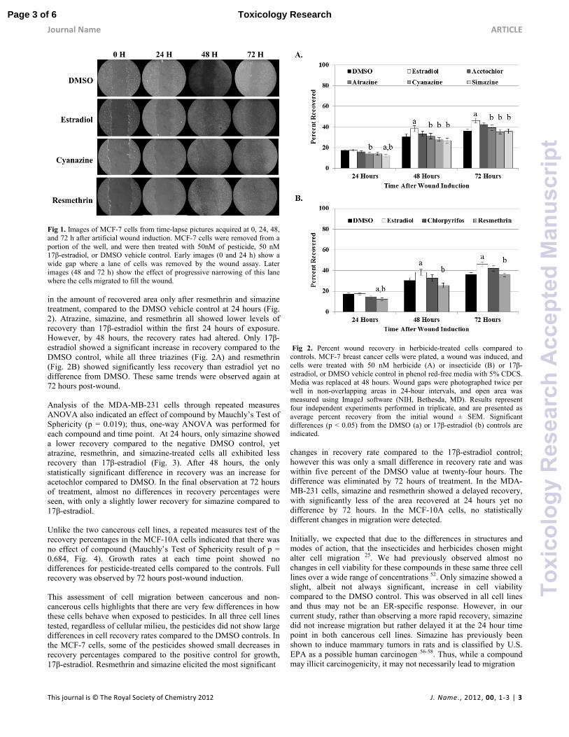
During the 72 hours post-wound induction, the cells began to
migrate across the cleared area (Fig. 1). The primary difference
between cell-populated areas and the wound is texture. The
wounded area is clear while the edges of the wound are speckled
with cells (Fig. 1). As a control for growth, cells were also exposed
to 17β-estradiol. This compound typically results in increased
growth of ER-containing cell lines, such as MCF-7 which is ERα
positive, yet do not affect the growth of MDA-MB-231 or MCF-10A
cells which are considered ER-negative 52-55. The amount of clear
area was calculated, and the area for the 0 hour time point was
normalized to 100% open, or 0% recovered, within each independent
experiment. Recovery percentages were subjected to repeated
measures ANOVA. Based upon initial between-subjects tests, cell-
dependent differences were observed and cell lines were re-analyzed
separately for statistical differences in recovery.
Subsequent repeated measures ANOVA of the MCF-7 data indicated
a significant effect of compound by Mauchly’s Test of Sphericity (p
< 0.000), thus one-way ANOVA was performed for each compound
at each time point. Post-hoc effects indicated a significant decrease
Page 2 of 6Toxicology Research
Toxi
colo
gyR
esea
rch
Acc
epte
dM
anus
crip
t
Journal Name ARTICLE
This journal is © The Royal Society of Chemistry 2012 J. Name., 2012, 00, 1-3 | 3
Fig 1. Images of MCF-7 cells from time-lapse pictures acquired at 0, 24, 48,
and 72 h after artificial wound induction. MCF-7 cells were removed from a
portion of the well, and were then treated with 50nM of pesticide, 50 nM 17β-estradiol, or DMSO vehicle control. Early images (0 and 24 h) show a
wide gap where a lane of cells was removed by the wound assay. Later
images (48 and 72 h) show the effect of progressive narrowing of this lane where the cells migrated to fill the wound.
in the amount of recovered area only after resmethrin and simazine
treatment, compared to the DMSO vehicle control at 24 hours (Fig.
2). Atrazine, simazine, and resmethrin all showed lower levels of
recovery than 17β-estradiol within the first 24 hours of exposure.
However, by 48 hours, the recovery rates had altered. Only 17β-
estradiol showed a significant increase in recovery compared to the
DMSO control, while all three triazines (Fig. 2A) and resmethrin
(Fig. 2B) showed significantly less recovery than estradiol yet no
difference from DMSO. These same trends were observed again at
72 hours post-wound.
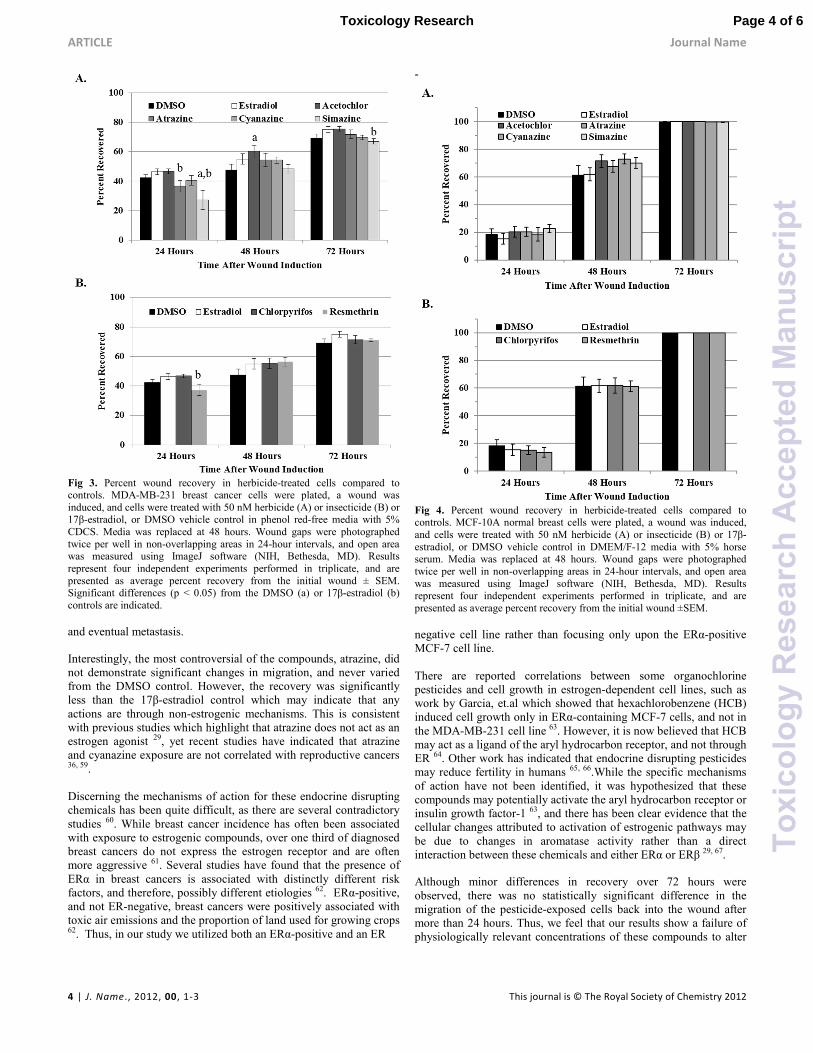
Analysis of the MDA-MB-231 cells through repeated measures
ANOVA also indicated an effect of compound by Mauchly’s Test of
Sphericity (p = 0.019); thus, one-way ANOVA was performed for
each compound and time point. At 24 hours, only simazine showed
a lower recovery compared to the negative DMSO control, yet
atrazine, resmethrin, and simazine-treated cells all exhibited less
recovery than 17β-estradiol (Fig. 3). After 48 hours, the only
statistically significant difference in recovery was an increase for
acetochlor compared to DMSO. In the final observation at 72 hours
of treatment, almost no differences in recovery percentages were
seen, with only a slightly lower recovery for simazine compared to
17β-estradiol.
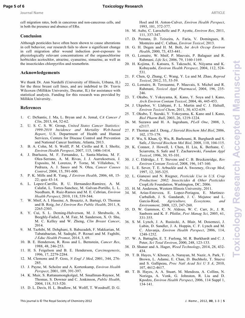
Unlike the two cancerous cell lines, a repeated measures test of the
recovery percentages in the MCF-10A cells indicated that there was
no effect of compound (Mauchly’s Test of Sphericity result of p =
0.684, Fig. 4). Growth rates at each time point showed no
differences for pesticide-treated cells compared to the controls. Full
recovery was observed by 72 hours post-wound induction.
This assessment of cell migration between cancerous and non-
cancerous cells highlights that there are very few differences in how
these cells behave when exposed to pesticides. In all three cell lines
tested, regardless of cellular milieu, the pesticides did not show large
differences in cell recovery rates compared to the DMSO controls. In
the MCF-7 cells, some of the pesticides showed small decreases in
recovery percentages compared to the positive control for growth,
17β-estradiol. Resmethrin and simazine elicited the most significant
Fig 2. Percent wound recovery in herbicide-treated cells compared to controls. MCF-7 breast cancer cells were plated, a wound was induced, and
cells were treated with 50 nM herbicide (A) or insecticide (B) or 17β-
estradiol, or DMSO vehicle control in phenol red-free media with 5% CDCS. Media was replaced at 48 hours. Wound gaps were photographed twice per
well in non-overlapping areas in 24-hour intervals, and open area was
measured using ImageJ software (NIH, Bethesda, MD). Results represent four independent experiments performed in triplicate, and are presented as
average percent recovery from the initial wound ± SEM. Significant
differences (p < 0.05) from the DMSO (a) or 17β-estradiol (b) controls are indicated.
changes in recovery rate compared to the 17β-estradiol control;
however this was only a small difference in recovery rate and was
within five percent of the DMSO value at twenty-four hours. The
difference was eliminated by 72 hours of treatment. In the MDA-
MB-231 cells, simazine and resmethrin showed a delayed recovery,
with significantly less of the area recovered at 24 hours yet no
difference by 72 hours. In the MCF-10A cells, no statistically
different changes in migration were detected.
Initially, we expected that due to the differences in structures and
modes of action, that the insecticides and herbicides chosen might
alter cell migration 25. We had previously observed almost no
changes in cell viability for these compounds in these same three cell
lines over a wide range of concentrations 52. Only simazine showed a
slight, albeit not always significant, increase in cell viability
compared to the DMSO control. This was observed in all cell lines
and thus may not be an ER-specific response. However, in our
current study, rather than observing a more rapid recovery, simazine
did not increase migration but rather delayed it at the 24 hour time
point in both cancerous cell lines. Simazine has previously been
shown to induce mammary tumors in rats and is classified by U.S.
EPA as a possible human carcinogen 56-58. Thus, while a compound
may illicit carcinogenicity, it may not necessarily lead to migration
Page 3 of 6 Toxicology Research
Toxi
colo
gyR
esea
rch
Acc
epte
dM
anus
crip
t
ARTICLE Journal Name
4 | J. Name., 2012, 00, 1-3 This journal is © The Royal Society of Chemistry 2012
Fig 3. Percent wound recovery in herbicide-treated cells compared to controls. MDA-MB-231 breast cancer cells were plated, a wound was
induced, and cells were treated with 50 nM herbicide (A) or insecticide (B) or 17β-estradiol, or DMSO vehicle control in phenol red-free media with 5%
CDCS. Media was replaced at 48 hours. Wound gaps were photographed
twice per well in non-overlapping areas in 24-hour intervals, and open area was measured using ImageJ software (NIH, Bethesda, MD). Results
represent four independent experiments performed in triplicate, and are
presented as average percent recovery from the initial wound ± SEM. Significant differences (p < 0.05) from the DMSO (a) or 17β-estradiol (b)
controls are indicated.
and eventual metastasis.
Interestingly, the most controversial of the compounds, atrazine, did
not demonstrate significant changes in migration, and never varied
from the DMSO control. However, the recovery was significantly
less than the 17β-estradiol control which may indicate that any
actions are through non-estrogenic mechanisms. This is consistent
with previous studies which highlight that atrazine does not act as an
estrogen agonist 29, yet recent studies have indicated that atrazine
and cyanazine exposure are not correlated with reproductive cancers 36, 59.
Discerning the mechanisms of action for these endocrine disrupting
chemicals has been quite difficult, as there are several contradictory
studies 60. While breast cancer incidence has often been associated
with exposure to estrogenic compounds, over one third of diagnosed
breast cancers do not express the estrogen receptor and are often
more aggressive 61. Several studies have found that the presence of
ERα in breast cancers is associated with distinctly different risk
factors, and therefore, possibly different etiologies 62. ERα-positive,
and not ER-negative, breast cancers were positively associated with
toxic air emissions and the proportion of land used for growing crops 62. Thus, in our study we utilized both an ERα-positive and an ER
-
Fig 4. Percent wound recovery in herbicide-treated cells compared to controls. MCF-10A normal breast cells were plated, a wound was induced,
and cells were treated with 50 nM herbicide (A) or insecticide (B) or 17β-
estradiol, or DMSO vehicle control in DMEM/F-12 media with 5% horse serum. Media was replaced at 48 hours. Wound gaps were photographed
twice per well in non-overlapping areas in 24-hour intervals, and open area
was measured using ImageJ software (NIH, Bethesda, MD). Results represent four independent experiments performed in triplicate, and are
presented as average percent recovery from the initial wound ±SEM.
negative cell line rather than focusing only upon the ERα-positive
MCF-7 cell line.
There are reported correlations between some organochlorine
pesticides and cell growth in estrogen-dependent cell lines, such as
work by Garcia, et.al which showed that hexachlorobenzene (HCB)
induced cell growth only in ERα-containing MCF-7 cells, and not in
the MDA-MB-231 cell line 63. However, it is now believed that HCB
may act as a ligand of the aryl hydrocarbon receptor, and not through
ER 64. Other work has indicated that endocrine disrupting pesticides
may reduce fertility in humans 65, 66.While the specific mechanisms
of action have not been identified, it was hypothesized that these
compounds may potentially activate the aryl hydrocarbon receptor or
insulin growth factor-1 63, and there has been clear evidence that the
cellular changes attributed to activation of estrogenic pathways may
be due to changes in aromatase activity rather than a direct
interaction between these chemicals and either ERα or ERβ 29, 67.
Although minor differences in recovery over 72 hours were
observed, there was no statistically significant difference in the
migration of the pesticide-exposed cells back into the wound after
more than 24 hours. Thus, we feel that our results show a failure of
physiologically relevant concentrations of these compounds to alter
Page 4 of 6Toxicology Research
Toxi
colo
gyR
esea
rch
Acc
epte
dM
anus
crip
t
Journal Name ARTICLE
This journal is © The Royal Society of Chemistry 2012 J. Name., 2012, 00, 1-3 | 5
cell migration rates, both in cancerous and non-cancerous cells, and
in both the presence and absence of ERα.
Conclusion
Although pesticides have often been shown to cause alterations
in cell behavior, our research fails to show a significant change
in cell migration after wound induction post-exposure to
physiologically relevant concentrations of the organochlorine
herbicides acetochlor, atrazine, cyanazine, simazine, as well as
the insecticides chlorpyrifos and resmethrin.
Acknowledgements
We thank Dr. Ann Nardulli (University of Illinois, Urbana, IL)
for the three breast cell lines, and are indebted to Dr. Travis
Wilcoxen (Millikin University, Decatur, IL) for assistance with
statistical analysis. Funding for this research was provided by
Millikin University.
References
1. C. DeSantis, J. Ma, L. Bryan and A. Jemal, CA Cancer J
Clin, 2013, 64, 52-62.
2. U. S. C. S. W. Group, United States Cancer Statistics:
1999-2010 Incidence and Mortality Web-based
Report, U.S. Department of Health and Human
Services, Centers for Disease Control and Prevention
and National Cancer Institute, Atlanta, 2013.
3. B. A. Cohn, M. S. Wolff, P. M. Cirillo and R. I. Sholtz,
Environ Health Perspect, 2007, 115, 1406-1414.
4. J. Ibarluzea, M. F. Fernandez, L. Santa-Marina, M. F.
Olea-Serrano, A. M. Rivas, J. J. Aurrekoetxea, J.
Exposito, M. Lorenzo, P. Torne, M. Villalobos, V.
Pedraza, A. J. Sasco and N. Olea, Cancer Causes
Control, 2004, 15, 591-600.
5. P. K. Mills and R. Yang, J Environ Health, 2006, 68, 15-
22; quiz 43-14.
6. L. Lopez-Carrillo, R. U. Hernandez-Ramirez, A. M.
Calafat, L. Torres-Sanchez, M. Galvan-Portillo, L. L.
Needham, R. Ruiz-Ramos and M. E. Cebrian, Environ
Health Perspect, 2010, 118, 539-544.
7. W. Mnif, A. I. Hassine, A. Bouaziz, A. Bartegi, O. Thomas
and B. Roig, Int J Environ Res Public Health, 2011, 8,
2265-2303.
8. Y. Cui, S. L. Deming-Halverson, M. J. Shrubsole, A.
Beeghly-Fadiel, A. M. Fair, M. Sanderson, X. O. Shu,
M. C. Kelley and W. Zheng, Clin Breast Cancer,
2014.
9. M. Tazhibi, M. Dehghani, S. Babazadeh, F. Makkarian, M.
Tabatabaeian, M. Sadeghi, P. Rezaei and M. Faghihi,
J Educ Health Promot, 2014, 3, 69.
10. B. E. Henderson, R. Ross and L. Bernstein, Cancer Res,
1988, 48, 246-253.
11. H. S. Feigelson and B. E. Henderson, Carcinogenesis,
1996, 17, 2279-2284.
12. M. Clemons and P. Goss, N Engl J Med, 2001, 344, 276-
285.
13. J. Payne, M. Scholze and A. Kortenkamp, Environ Health
Perspect, 2001, 109, 391-397.
14. K. Muir, S. Rattanamongkolgul, M. Smallman-Raynor, M.
Thomas, S. Downer and C. Jenkinson, Public Health,
2004, 118, 513-520.
15. D. L. Davis, H. L. Bradlow, M. Wolff, T. Woodruff, D. G.
Hoel and H. Anton-Culver, Environ Health Perspect,
1993, 101, 372-377.
16. M. Aube, C. Larochelle and P. Ayotte, Environ Res, 2011,
111, 337-347.
17. D. Pestana, D. Teixeira, A. Faria, V. Domingues, R.
Monteiro and C. Calhau, Environ Toxicol, 2013.
18. G. H. Degen and H. M. Bolt, Int Arch Occup Environ
Health, 2000, 73, 433-441.
19. G. Lemaire, W. Mnif, P. Mauvais, P. Balaguer and R.
Rahmani, Life Sci, 2006, 79, 1160-1169.
20. H. Kojima, E. Katsura, S. Takeuchi, K. Niiyama and K.
Kobayashi, Environ Health Perspect, 2004, 112, 524-
531.
21. F. Chen, Q. Zhang, C. Wang, Y. Lu and M. Zhao, Reprod
Toxicol, 2012, 33, 53-59.
22. G. Lemaire, B. Terouanne, P. Mauvais, S. Michel and R.
Rahmani, Toxicol Appl Pharmacol, 2004, 196, 235-
246.
23. T. Okubo, Y. Yokoyama, K. Kano, Y. Soya and I. Kano,
Arch Environ Contam Toxicol, 2004, 46, 445-453.
24. J. Ukpebor, V. Llabjani, F. L. Martin and C. J. Halsall,
Environ Toxicol Chem, 2011, 30, 632-639.
25. T. Okubo, T. Suzuki, Y. Yokoyama, K. Kano and I. Kano,
Biol Pharm Bull, 2003, 26, 1219-1224.
26. M. Suzawa and H. A. Ingraham, PLoS One, 2008, 3,
e2117.
27. P. Thomas and J. Dong, J Steroid Biochem Mol Biol, 2006,
102, 175-179.
28. F. Wu, S. Khan, Q. Wu, R. Barhoumi, R. Burghardt and S.
Safe, J Steroid Biochem Mol Biol, 2008, 110, 104-115.
29. K. Connor, J. Howell, I. Chen, H. Liu, K. Berhane, C.
Sciarretta, S. Safe and T. Zacharewski, Fundam Appl
Toxicol, 1996, 30, 93-101.
30. J. C. Eldridge, J. T. Stevens and C. B. Breckenridge, Rev
Environ Contam Toxicol, 2008, 196, 147-160.
31. L. E. Sever, T. E. Arbuckle and A. Sweeney, Occup Med,
1997, 12, 305-325.
32. L. Gianessi and N. Riegner, Pesticide Use in U.S. Crop
Production: 2002; Insecticides & Other Pesticides
CropLife Foundation, Washington, DC, 2006.
33. H. M. Anderson, Western Illinois University, 2011.
34. M. Arias-Esteveza, E. Lopez-Periagoa, E. Martinez-
Carballob, J. S. Gandarab, J. C. Mejutoc and L.
Garcia-Riod, Agriculture, Ecosytems, and
Environment, 2008, 123, 247-260.
35. D. W. Gammon, C. N. Aldous, W. C. Carr, Jr., J. R.
Sanborn and K. F. Pfeifer, Pest Manag Sci, 2005, 61,
331-355.
36. S. M. Lynch, J. A. Rusiecki, A. Blair, M. Dosemeci, J.
Lubin, D. Sandler, J. A. Hoppin, C. F. Lynch and M.
C. Alavanja, Environ Health Perspect, 2006, 114,
1248-1252.
37. W. A. Battaglin, E. T. Furlong, M. R. Burkhardt and C. J.
Peter, Sci Total Environ, 2000, 248, 123-133.
38. D. Shaner and A. Hager, Weed Technology, 2014, 28, 432-
434.
39. T. B. Hayes, V. Khoury, A. Narayan, M. Nazir, A. Park, T.
Brown, L. Adame, E. Chan, D. Buchholz, T. Stueve
and S. Gallipeau, Proc Natl Acad Sci U S A, 2010,
107, 4612-4617.
40. T. B. Hayes, A. A. Stuart, M. Mendoza, A. Collins, N.
Noriega, A. Vonk, G. Johnston, R. Liu and D.
Kpodzo, Environ Health Perspect, 2006, 114 Suppl 1,
134-141.
Page 5 of 6 Toxicology Research
Toxi
colo
gyR
esea
rch
Acc
epte
dM
anus
crip
t

ARTICLE Journal Name
6 | J. Name., 2012, 00, 1-3 This journal is © The Royal Society of Chemistry 2012
41. J. L. Rayner, C. Wood and S. E. Fenton, Toxicol Appl
Pharmacol, 2004, 195, 23-34.
42. C. C. Liang, A. Y. Park and J. L. Guan, Nat Protoc, 2007,
2, 329-333.
43. C. P. R. Institute, National Pesticide Use Database, HTTP://WWW.CROPLIFEFOUNDATION.ORG/%5CCPRI_NPUD20
02.HTM, Accessed November 11, 2011, 2011.
44. A. L. Rayburn, D. D. Moody and J. L. Freeman, Bull
Environ Contam Toxicol, 2005, 75, 691-698.
45. W. J. Hayes and E. R. Laws, Handbook of pesticide
toxicology, Academic Press, San Diego, 1991.
46. D. B. Barr, C. J. Hines, A. O. Olsson, J. A. Deddens, R.
Bravo, C. A. Striley, J. Norrgran and L. L. Needham,
J Expo Sci Environ Epidemiol, 2007, 17, 559-566.
47. U. S. E. P. Agency, ed. U. S. E. P. A. O. o. D. Water,
Washington, D.C., Editon edn., 1988.
48. U. S. E. P. Agency, ed. U. S. E. P. A. O. o. D. Water,
Washington, D.C., Editon edn., 1988.
49. M. T. Meyer, E. M. Thurman and D. A. Goolsby, J
Environ Qual, 2001, 30, 1836-1843.
50. U. S. E. P. Agency, ed. U. S. E. P. Agency, Editon edn.,
1983.
51. U. S. EPA, 1999.
52. J. D. Rich, S. M. Gabriel and J. R. Schultz-Norton, ISRN
Toxicol, 2012, 2012, 232461.
53. J. R. Schultz, L. N. Petz and A. M. Nardulli, The Journal
of Biological Chemistry, 2005, 280, 347-354.
54. J. R. Schultz-Norton, V. A. Gabisi, Y. S. Ziegler, I. X.
McLeod, J. R. Yates and A. M. Nardulli, Nucleic
Acids Res, 2007, 35, 5028-5038.
55. J. R. Schultz-Norton, W. H. McDonald, J. R. Yates and A.
M. Nardulli, Mol Endocrinol, 2006, 20, 1982-1995.
56. A. M. Fan and G. V. Alexeeff, Public Health Goal for
Simazine in Drinking Water, 2001.
57. L. Jowa and R. Howd, Journal of Environmental Science
and Health, 2011, 29, 91-144.
58. M. Silva and P. Iyer, Birth Defects Res B Dev Reprod
Toxicol, 2014, 101, 308-324.
59. J. W. Simpkins, J. A. Swenberg, N. Weiss, D. Brusick, J.
C. Eldridge, J. T. Stevens, R. J. Handa, R. C. Hovey,
T. M. Plant, T. P. Pastoor and C. B. Breckenridge,
Toxicol Sci, 2011, 123, 441-459.
60. S. De Coster and N. van Larebeke, J Environ Public
Health, 2012, 2012, 713696.
61. M. S. Sheikh, M. Garcia, P. Pujol, J. A. Fontana and H.
Rochefort, Invasion Metastasis, 1994, 14, 329-336.
62. S. St-Hilaire, R. Mandal, A. Commendador, S. Mannel and
D. Derryberry, Int J Health Geogr, 2011, 10, 32.
63. M. A. Garcia, D. Pena, L. Alvarez, C. Cocca, C. Pontillo,
R. Bergoc, D. K. de Pisarev and A. Randi, Toxicol
Lett, 2010, 192, 195-205.
64. C. A. Pontillo, P. Rojas, F. Chiappini, G. Sequeira, C.
Cocca, M. Crocci, L. Colombo, C. Lanari, D. Kleiman
de Pisarev and A. Randi, Toxicol Appl Pharmacol,
2013, 268, 331-342.
65. S. H. Swan, C. Brazil, E. Z. Drobnis, F. Liu, R. L. Kruse,
M. Hatch, J. B. Redmon, C. Wang and J. W.
Overstreet, Environ Health Perspect, 2003, 111, 414-
420.
66. S. H. Swan, R. L. Kruse, F. Liu, D. B. Barr, E. Z. Drobnis,
J. B. Redmon, C. Wang, C. Brazil and J. W.
Overstreet, Environ Health Perspect, 2003, 111, 1478-
1484.
67. M. K. Tennant, D. S. Hill, J. C. Eldridge, L. T. Wetzel, C.
B. Breckenridge and J. T. Stevens, J Toxicol Environ
Health, 1994, 43, 197-211.
Page 6 of 6Toxicology Research
Toxi
colo
gyR
esea
rch
Acc
epte
dM
anus
crip
t
Related Documents