Citation: Brar, A.; Majumder, S.; Navarro, M.Z.; Benoit-Biancamano, M.-O.; Ronholm, J.; George, S. Nanoparticle-Enabled Combination Therapy Showed Superior Activity against Multi-Drug Resistant Bacterial Pathogens in Comparison to Free Drugs. Nanomaterials 2022, 12, 2179. https://doi.org/10.3390/ nano12132179 Academic Editors: Monika Mortimer and Anne Kahru Received: 16 May 2022 Accepted: 22 June 2022 Published: 24 June 2022 Publisher’s Note: MDPI stays neutral with regard to jurisdictional claims in published maps and institutional affil- iations. Copyright: © 2022 by the authors. Licensee MDPI, Basel, Switzerland. This article is an open access article distributed under the terms and conditions of the Creative Commons Attribution (CC BY) license (https:// creativecommons.org/licenses/by/ 4.0/). nanomaterials Article Nanoparticle-Enabled Combination Therapy Showed Superior Activity against Multi-Drug Resistant Bacterial Pathogens in Comparison to Free Drugs Amarpreet Brar 1,2 , Satwik Majumder 1,2 , Maria Zardon Navarro 2,3 , Marie-Odile Benoit-Biancamano 2,3 , Jennifer Ronholm 1,4 and Saji George 1,2, * 1 Department of Food Science and Agricultural Chemistry, Macdonald Campus, McGill University, 21111 Lakeshore, Ste Anne de Bellevue, QC H9X 3V9, Canada; [email protected] (A.B.); [email protected] (S.M.); [email protected] (J.R.) 2 Swine and Poultry Infectious Diseases Research Center (CRIPA), Faculté de Médecine Vétérinaire, Université de Montréal, 3200 Sicotte, Saint-Hyacinthe, QC J2S 2M2, Canada; [email protected] (M.Z.N.); [email protected] (M.-O.B.-B.) 3 Research Group on Infectious Diseases in Production Animals (GREMIP), Department of Pathology and Microbiology, Faculté de Médecine Vétérinaire, Université de Montréal, 3200 Sicotte, Saint-Hyacinthe, QC J2S 2M2, Canada 4 Department of Animal Science, Macdonald Campus, McGill University, 2111 Lakeshore, Ste Anne de Bellevue, QC H9X 3V9, Canada * Correspondence: [email protected]; Tel.: +1-514-398-7920; Fax: +1-514-398-7990 Abstract: The emergence of multidrug-resistant (MDR) bacterial pathogens in farm animals and their zoonotic spread is a concern to both animal agriculture and public health. Apart from antimicrobial resistance (AMR), bacterial pathogens from the genera of Salmonella and Staphylococcus take refuge inside host cells, thereby demanding intervention strategies that can eliminate intracellular MDR pathogens. In this study, seven clinical isolates of Salmonella and Staphylococcus from swine farms were characterized for antibiotic (n = 24) resistance, resistance mechanisms, and virulence characteristics. All isolates showed resistance to one or more antibiotics and S. enterica ser. Typhimurium isolate had the highest resistance to the panel of antibiotics tested. Major resistance mechanisms identified were efflux pump and beta-lactamase enzyme activities. Staphylococcus isolates showed complete hemoly- sis and strong biofilm formation, while Salmonella isolates caused partial hemolysis, but showed no or weak biofilm formation. MDR isolates of S. aureus M12 and S. enterica ser. Typhimurium bacteria were subsequently tested against combinations of antibiotics and potentiating adjuvants for improved antibacterial efficacy using a checkerboard assay, and their fractional inhibitory concentration index (FICI) was calculated. A combination of chitosan and silica nanoparticles containing tetracycline (TET) and efflux pump inhibitor chlorpromazine (CPZ), respectively, was characterized for physicochemical properties and effectiveness against MDR Salmonella enterica ser. Typhimurium isolate. This combina- tion of nano-encapsulated drugs improved the antibacterial efficacy by inhibiting AMR mechanisms (efflux activity, beta-lactamase enzyme activity, and hydrogen sulfide (H 2 S) production) and reducing intracellular pathogen load by 83.02 ± 14.35%. In conclusion, this study sheds light on the promising applicability of nanoparticle-enabled combination therapy to combat multidrug-resistant pathogens encountered in animal agriculture. Keywords: multi-drug resistance; combination therapy; nanoparticle; antimicrobial resistance; Staphylococcus; Salmonella; animal agriculture; pathogen 1. Introduction The prevalence of pathogenic microorganisms in food animals and their possible zoonotic spread through direct contact or the food chain pose public health concerns [1,2]. Common bacterial pathogens infecting farmed animals can cause various infections, often Nanomaterials 2022, 12, 2179. https://doi.org/10.3390/nano12132179 https://www.mdpi.com/journal/nanomaterials

Welcome message from author
This document is posted to help you gain knowledge. Please leave a comment to let me know what you think about it! Share it to your friends and learn new things together.
Transcript
Citation: Brar, A.; Majumder, S.;
Navarro, M.Z.; Benoit-Biancamano,
M.-O.; Ronholm, J.; George, S.
Nanoparticle-Enabled Combination
Therapy Showed Superior Activity
against Multi-Drug Resistant
Bacterial Pathogens in Comparison to
Free Drugs. Nanomaterials 2022, 12,
2179. https://doi.org/10.3390/
nano12132179
Academic Editors: Monika Mortimer
and Anne Kahru
Received: 16 May 2022
Accepted: 22 June 2022
Published: 24 June 2022
Publisher’s Note: MDPI stays neutral
with regard to jurisdictional claims in
published maps and institutional affil-
iations.
Copyright: © 2022 by the authors.
Licensee MDPI, Basel, Switzerland.
This article is an open access article
distributed under the terms and
conditions of the Creative Commons
Attribution (CC BY) license (https://
creativecommons.org/licenses/by/
4.0/).
nanomaterials
Article
Nanoparticle-Enabled Combination Therapy Showed SuperiorActivity against Multi-Drug Resistant Bacterial Pathogens inComparison to Free DrugsAmarpreet Brar 1,2, Satwik Majumder 1,2 , Maria Zardon Navarro 2,3, Marie-Odile Benoit-Biancamano 2,3,Jennifer Ronholm 1,4 and Saji George 1,2,*
1 Department of Food Science and Agricultural Chemistry, Macdonald Campus, McGill University,21111 Lakeshore, Ste Anne de Bellevue, QC H9X 3V9, Canada; [email protected] (A.B.);[email protected] (S.M.); [email protected] (J.R.)
2 Swine and Poultry Infectious Diseases Research Center (CRIPA), Faculté de Médecine Vétérinaire,Université de Montréal, 3200 Sicotte, Saint-Hyacinthe, QC J2S 2M2, Canada;[email protected] (M.Z.N.); [email protected] (M.-O.B.-B.)
3 Research Group on Infectious Diseases in Production Animals (GREMIP), Department of Pathology andMicrobiology, Faculté de Médecine Vétérinaire, Université de Montréal, 3200 Sicotte, Saint-Hyacinthe,QC J2S 2M2, Canada
4 Department of Animal Science, Macdonald Campus, McGill University, 2111 Lakeshore,Ste Anne de Bellevue, QC H9X 3V9, Canada
* Correspondence: [email protected]; Tel.: +1-514-398-7920; Fax: +1-514-398-7990
Abstract: The emergence of multidrug-resistant (MDR) bacterial pathogens in farm animals and theirzoonotic spread is a concern to both animal agriculture and public health. Apart from antimicrobialresistance (AMR), bacterial pathogens from the genera of Salmonella and Staphylococcus take refugeinside host cells, thereby demanding intervention strategies that can eliminate intracellular MDRpathogens. In this study, seven clinical isolates of Salmonella and Staphylococcus from swine farms werecharacterized for antibiotic (n = 24) resistance, resistance mechanisms, and virulence characteristics.All isolates showed resistance to one or more antibiotics and S. enterica ser. Typhimurium isolate hadthe highest resistance to the panel of antibiotics tested. Major resistance mechanisms identified wereefflux pump and beta-lactamase enzyme activities. Staphylococcus isolates showed complete hemoly-sis and strong biofilm formation, while Salmonella isolates caused partial hemolysis, but showed no orweak biofilm formation. MDR isolates of S. aureus M12 and S. enterica ser. Typhimurium bacteria weresubsequently tested against combinations of antibiotics and potentiating adjuvants for improvedantibacterial efficacy using a checkerboard assay, and their fractional inhibitory concentration index(FICI) was calculated. A combination of chitosan and silica nanoparticles containing tetracycline (TET)and efflux pump inhibitor chlorpromazine (CPZ), respectively, was characterized for physicochemicalproperties and effectiveness against MDR Salmonella enterica ser. Typhimurium isolate. This combina-tion of nano-encapsulated drugs improved the antibacterial efficacy by inhibiting AMR mechanisms(efflux activity, beta-lactamase enzyme activity, and hydrogen sulfide (H2S) production) and reducingintracellular pathogen load by 83.02 ± 14.35%. In conclusion, this study sheds light on the promisingapplicability of nanoparticle-enabled combination therapy to combat multidrug-resistant pathogensencountered in animal agriculture.
Keywords: multi-drug resistance; combination therapy; nanoparticle; antimicrobial resistance;Staphylococcus; Salmonella; animal agriculture; pathogen
1. Introduction
The prevalence of pathogenic microorganisms in food animals and their possiblezoonotic spread through direct contact or the food chain pose public health concerns [1,2].Common bacterial pathogens infecting farmed animals can cause various infections, often
Nanomaterials 2022, 12, 2179. https://doi.org/10.3390/nano12132179 https://www.mdpi.com/journal/nanomaterials
Nanomaterials 2022, 12, 2179 2 of 16
requiring treatment with antibiotics. In addition, antibiotics are used as prophylactics andas antimicrobial growth promoters (AGPs) although the farm use of antimicrobials reservedfor infection control in humans has been restricted in the recent past in countries such asCanada [3,4]. Nonetheless, the use of antibiotics in animals is an important driver of theemergence and spread of antimicrobial-resistant (AMR) bacterial human infections [1,5,6].AMR pathogens in animals and their zoonotic potential necessitates the development ofalternate therapeutics for disease management of multi-drug resistant (MDR) pathogens [7].
Combining antibiotics with potentiating adjuvants is one such solution that can en-hance the potency of antibiotics and increase the sensitization of bacteria to commonly usedantibiotics. For instance, adjuvants such as efflux pump inhibitors have shown to subduethe resistance mechanisms of bacterial cells and promote the action of antibiotics andsilver [8,9]. Combination therapy—a treatment modality of using a combination of drugsis successful in boosting the effectiveness of antibiotics [10,11]. However, combinationtherapy with free forms of drugs lacks target specificity, control over release kinetics, andhas a shorter half-life in the body [12,13]. These drawbacks highlight the urgent need todevise alternate modes of drug delivery systems.
Recent developments in nanoparticle-based drug delivery have provided numerouspromising opportunities in terms of longer circulation time, controlled drug release, abilityto target specific tissue sites, and to deliver more than one drug at the same time [14–16].In addition, nanoparticle-based drug delivery systems can be custom-designed to meetdesired properties. For instance, chitosan nanoparticles are mucoadhesive, biodegradable,positively charged, and easily surface-modifiable to target specific sites inside the body,thus making chitosan nanoparticles (Ch NPs) a promising candidate for drug delivery [17].Silica nanoparticles (Si NPs) have complimentary advantageous features such as high drugloading capacity, biocompatibility, and surface modification possibilities [18,19].
In this study, Staphylococcus and Salmonella isolates from Quebec-based animal farmswere studied for AMR and major AMR mechanisms such as efflux pump activity and beta-lactamase enzyme activity were identified. In addition, virulence characteristics such asbiofilm formation, and hemolysis relevant for bacterial survival inside the host body werealso investigated. The effectiveness of combining antibiotics with adjuvant molecules wastested in vitro to identify optimal combinations for neutralizing MDR strains. We furthershowed that delivering identified combinations of drug molecules using nanoparticlescould enhance antibacterial efficiency against MDR strains evading human epithelial cells.
2. Materials and Methods2.1. Isolates and Chemicals
Seven pathogenic bacterial isolates from Quebec farm animals, submitted for diag-nostic purposes to the Complexe de diagnostic et d’épidémiosurveillance vétérinaires duQuébec (CDEVQ), Saint-Hyacinthe, QC, Canada, with diseases such as exudative epider-mitis, diarrhea, or septicemia, were selected for this study. These isolates consisted ofS. enterica serovar Choleraesuis, S. enterica serovar Dublin, S. enterica serovar Typhimurium,Staphylococcus aureus M12, S. aureus M17, S. hyicus M43, and S. hyicus M48. S. aureusATCC 25923 and E. coli ATCC 25922 (Oxoid company, Nepean, ON, Canada) were usedas quality control (QC) strains. The sodium salt of resazurin, Silica, mesostructured HMS(wormhole) (Catalog number 541036-5G) with pore size 2–4 nm, and ethidium bromide(EtBr) were purchased from Sigma-Aldrich, Canada. Chitosan (85% deacylation) wasobtained from Alfa Aesar (Thermo Fisher Scientific, Tewksbury, MA, USA). Tetracycline(TET) and chlorpromazine (CPZ) were purchased from Bio Basic, ON, Canada. Nitrocefindye was purchased from Abcam, Cambridge, United Kingdom.
2.2. Antimicrobial Susceptibility Determination
The susceptibility of bacterial isolates to 24 antibiotics (Oxoid, Thermo Fischer Sci-entific, Nepean, ON, Canada) (Supplementary Information Table S1) was determined by
Nanomaterials 2022, 12, 2179 3 of 16
the Kirby–Bauer disk diffusion method according to Clinical and Laboratory StandardsInstitutes Guidelines [20,21], and QC strains were used as controls.
2.3. Testing Combinations of Antibiotics and Adjuvants
Antibiotics of ampicillin, chloramphenicol, TET, and trimethoprim were tested for ad-ditive or synergistic effects, as they are commonly used in food animals and humans [22,23].The selected adjuvants consisted of an efflux pump inhibitor (EPI) (Chlorpromazine-CPZ),a beta-lactamase inhibitor (tazobactam), and essential oils (EOs) (thymol EO, cinnamon EO,and oregano EO) with different modes of action on bacterial cells. The composition of EOsused in this study was reported previously [24].
MICs of the antibiotics and adjuvants were determined against S. enterica ser. Ty-phimurium and S. aureus M12 using the microtiter broth dilution method. A checkerboardassay was used to categorize the type of antimicrobial effect of the tested combinations [25].Antibiotics were diluted column-wise, and the adjuvants were diluted row-wise in a 96-wellplate. Each well contained a unique concentration of drug and adjuvant to which 10 µL ofcells (0.5 McFarland) were added. The 96-well plates were incubated at 37 C overnight.Cell viability was checked using a resazurin assay (Supplementary Section S1) [26]. Thewell with no growth of bacteria (baseline reading for fluorescence) was considered fordetermining the MIC. The fractional inhibitory concentration index (FICI) was calculatedaccording to the following formula.
FICA =A
MICA; FICB =
BMICB
(1)
FICI = FICA + FICB (2)
where A and B are the MICs of components A and B in combination, respectively. FICIvalues FICI ≤ 0.5, 0.5 < FICI ≤ 1, 1 < FICI ≤ 4, and FICI > 4 were categorized as synergistic,additive, indifferent and antagonistic, respectively.
2.4. Synthesis of Drug-Loaded Nanoparticles and Combination Drug (CMD)
TET-loaded Ch NPs (Ch-TET NPs) were synthesized using the ionic gelation methodusing TPP, as reported earlier [27]. Accordingly, chitosan (50 mg) was dissolved in 25 mLof 1% (v/v) acetic acid and stirred for 48 h. The pH was adjusted to 4.5–4.6 using 1.0 NNaOH and the suspension was sonicated for 10 min. Subsequently, 3.4 mL of the 1% (w/v)TET solution was added under constant stirring. TPP was added dropwise to the resultingsolution where the chitosan:TPP ratio was 3:1. The resulting suspension was sonicatedfor 10 min and stirred for another hour to obtain Ch-TET NPs. The resulting NPs werecollected by centrifuging the suspension at 11,000× g for 12 min and discarding the super-natant. Sedimented particles were washed twice with de-ionized (DI) water and were thenresuspended in DI water.
For the synthesis of CPZ-loaded Si NPs (Si-CPZ NPs), 40 mg of mesoporous Si NPswere dispersed in 10 mL of DI water. CPZ was dissolved in DI water to obtain 1% (w/v)solution, 4 mL of which was added dropwise to the Si NPs suspension. The suspensionmixture was stirred for 24 h, and then freeze-dried to obtain a white powder containingSi-CPZ NPs. Finally, CMD was obtained by combining Ch-TET NPs and Si-CPZ NPs in a1:1 ratio in DI water.
2.5. Determination of Size and Surface Charge
Dynamic light scattering (DLS) (NanoBrook Omni instrument, Brookhaven’s, Holtsville,NY, USA) was used to measure the surface charge and hydrodynamic size of particles dis-persed in DI water (50 µg/mL). The particle suspension was loaded into a pre-rinsed foldedcapillary cell and a voltage of 100 V was applied for the measurement of zeta potential.
Nanomaterials 2022, 12, 2179 4 of 16
2.6. Scanning Electron Microscopy (SEM) Observation
Particle morphology was examined by SEM (FEI Quanta 450 Field Emission ScanningElectron Microscope, Phillips, Austin, TX, USA). Nanoparticle samples were prepared byserial dehydration in increasing concentrations of absolute ethanol in DI water (30%, 50%,70%, and 90%) for 10 min each and resuspended in 100% ethanol at a concentration of50 µg/mL. A small amount (10 µL) of the sample was dropped onto a copper grid andallowed to dry at room temperature. These particles were then coated with platinum beforeacquiring SEM images.
2.7. FTIR Spectra of Synthesized Particles
Attenuated total reflectance-Fourier transform infrared (ATR-FTIR) (ALPHA-P, Bruker,Billerica, MA, USA) was used to analyze the surface functional groups of the particles [28].For this, 5 µL of the nanoparticle suspension was dropped on the ATR probe and allowedto dry. FTIR spectra were obtained using a wavelength range of 600–4000 cm−1, with aresolution of 4 cm−1, and 32 scans. The obtained data were analyzed using the OMNIC8.2.0.387 software.
2.8. Determination of Encapsulation Efficiency (EE)
Antibiotic-loaded-nanoparticles were dispersed in DI water (1 mg/mL). The suspen-sions were sonicated for 30 min, followed by centrifugation at 40,000× g for 40 min toremove loaded antibiotics from respective nanoparticle carriers. The supernatant was usedto quantify the amount of TET and CPZ loaded by measuring the absorbance using aspectrophotometer (GenessysTM 10S, Thermo Fisher, Tewksbury, MA, USA) at 285 nm and305 nm wavelengths, respectively [29]. The total amount of antibiotic-loaded nanoparti-cles was calculated from a standard curve prepared using known concentrations of TETand CPZ.
2.9. Determination of Release Behavior
The release behavior of loaded nanoparticles was studied by dispersing nano-encapsulateddrug samples in PBS at pH 4.3 (representing the gastric pH) and at pH 6.3 (representingthe intestinal pH of swine). The samples were centrifuged at 2000× g for 3 min and thesupernatant was removed every 24 h for seven days. The absorbance (285 nm and 305 nm,respectively, for TET and CPZ) of the supernatant was measured as a function of time usinga spectrophotometer (GenessysTM 10S, Thermo Fisher, Tewksbury, MA, USA) to determinethe time bound release of TET and CPZ released.
2.10. Antibacterial Activity of Nano-Encapsulated Antibiotic and/or Efflux Pump Inhibitor
Antibacterial efficiency of Ch-TET NPs, Si-CPZ NPs, and CMD was determined againstMDR bacteria- S. enterica ser. Typhimurium and S. aureus M12. Test compounds/particleswere subjected to twofold serial dilution in a sterile 96-well plate. Bacterial cultures(10 µL) adjusted to 0.5 McFarland standard (obtained from Mueller–Hinton Broth (MHB)inoculated with a single colony of bacteria and incubated overnight at 37 C) were addedto the wells. The plates were incubated at 37 C for 18 h under mild shaking. Subsequently,bacterial viability was monitored using a resazurin assay. S. aureus ATCC 25923 and E. coliATCC 25922 were used as quality control strains.
3. Effect of Nano-Encapsulated Drugs on Bacterial Resistance Mechanisms3.1. Beta-Lactamase Inhibition Assay
The beta-lactamase inhibition was tested as mentioned previously [7]. Briefly, thebacterial isolates (100 µL at 1.0 McFarland standard) were exposed to sub-lethal concentra-tions of test compounds (MIC × 1
3 ), and were incubated for 18 h at 37 C. Subsequently,the cells were adjusted to McFarland 1.0, centrifuged at 5000× g for 10 min, and washedwith sodium phosphate buffer (pH 7.0). Cell pellets obtained were suspended in freshsodium phosphate buffer and were sonicated for 5 min on ice. The cell-free extract con-
Nanomaterials 2022, 12, 2179 5 of 16
taining beta-lactamase enzyme was obtained by centrifuging the suspension at 16,000× gfor 30 min and collecting the supernatant. Nitrocefin (a chromogenic cephalosporin) wasused as the substrate to measure beta-lactamase enzyme activity. For this, 15 µL of thenitrocefin solution (0.5 mg/mL in 5% Dimethylsulfoxide) was mixed with 30 µL of thecell-free extract and the volume was adjusted to 100 µL using sodium phosphate buffersolution in a 96-well plate. The absorbance was measured using the plate reader in kineticmode at 490 nm for 15 min.
3.2. H2S Production Inhibition Assay
The ability of test samples to inhibit the production of hydrogen sulfide (H2S) wasmeasured as reported previously [30]. Bacterial isolates were adjusted to McFarland 0.5and introduced to sub-lethal concentrations of samples and ampicillin (MIC × 1
3 ) andincubated for 18 h in MHB medium with 20 mM L-cysteine. Bacterial cells were thencollected by centrifugation at 5000× g for 10 min and washed twice with MHB + L-cysteinemedium. The cells were adjusted to 0.5 McFarland and left to incubate for 30 min; 40 µLof the cell suspensions were added to 80 µL of the distilled water (pH 9.6 + 0.1 mM di-ethylenetriaminepentaacetic acid) in a microtiter well. Forty µL of the solution (17.1 mMN,N-dimethyl-p-phenylenediamine sulfate salt, and 37.0 mM FeCl3 in 6 M HCl) was addedimmediately. This method measures the H2S produced during bacterial growth in the broth.The absorbance was immediately detected in kinetic mode at 668 nm for 45 min using theplate-reader (Spectramax i3x, Molecular Devices, San Jose, CA, USA).
3.3. Efflux Pump Inhibition Assay
The ability of test samples to inhibit the efflux activity of the bacterial isolates wasmeasured as mentioned previously with minor modifications [31]. The overnight cultureof bacterial cells was washed and adjusted to McFarland 1.0. The cells were treated withsub-lethal concentrations (MIC × 1
3 ) of the samples and CPZ, vortexed and incubatedfor 30 min. The sub-lethal concentration of EtBr (MIC × 1
3 ) was added to the bacterialsamples and 150 µL of the suspension was added to the individual wells of a 96-well plate.EtBr accumulation inside the bacterial cells was monitored (Figure S6) and followed bywashing the cells with PBS and re-energizing with the addition of 10 µL of glucose (finalconcentration 0.04% w/v). Efflux activity was then determined by monitoring fluorescence(530/590 nm) for 60 min using a plate reader.
3.4. Determination of Antibacterial Property of CMD against Intracellular S. enterica ser. Typhimurium
The antimicrobial effects of nanoparticle-encapsulated drugs against intracellularpathogens were determined as previously described [32]. Caco-2 cells (ATCC, Manassas,VA, USA) were selected as the in vitro model of the intestinal epithelium. Cells werecultured in a 96-well plate (2 × 10⁴ cells per well) until confluency and were infected withfreshly cultured S. enterica ser. Typhimurium in MHB (Multiplicity of index = 1). After anhour of incubation, the supernatant media containing residual extracellular bacteria wasremoved and the cells were washed 2 times with PBS at 4 C. The cells were then incubatedwith 100 µL of 200 µg/mL of gentamicin for 30 min to eliminate the extracellular bacteria.Gentamicin suspension was removed subsequently, and the cells were washed 3× with PBSat 4 C. The cells were incubated with Gibco Dulbecco’s Modified Eagle Medium (DMEM)(Thermofisher, Tewksbury, MA, USA) for 4 h to establish the intracellular infection. Next,the NPs containing TET and/or CPZ were added to the cultures and incubated for 12 h.After 12 h, the cells were washed 3× with PBS at 4 C to remove the extracellular bacteria.Subsequently, Caco-2 cells were lysed using 100 µL of 0.5% Triton-X and the lysate wasserially diluted and plated on Mueller–Hinton agar plates. Infected cells with no treatmentwere used as negative control and infected cells treated with ciprofloxacin (20 µg/mL) wereused as a positive control. The number of colonies was counted after incubation for 24 hat 37 C to determine viable S. enterica ser. Typhimurium counts using the drop culturemethod [33].
Nanomaterials 2022, 12, 2179 6 of 16
3.5. Statistical Analysis
Experiments were performed in triplicates. The data are expressed as the meanvalue ± standard deviation (SD). T-test was performed and p-value ≤ 0.05 was consideredsignificantly different.
4. Results4.1. Antimicrobial Susceptibility
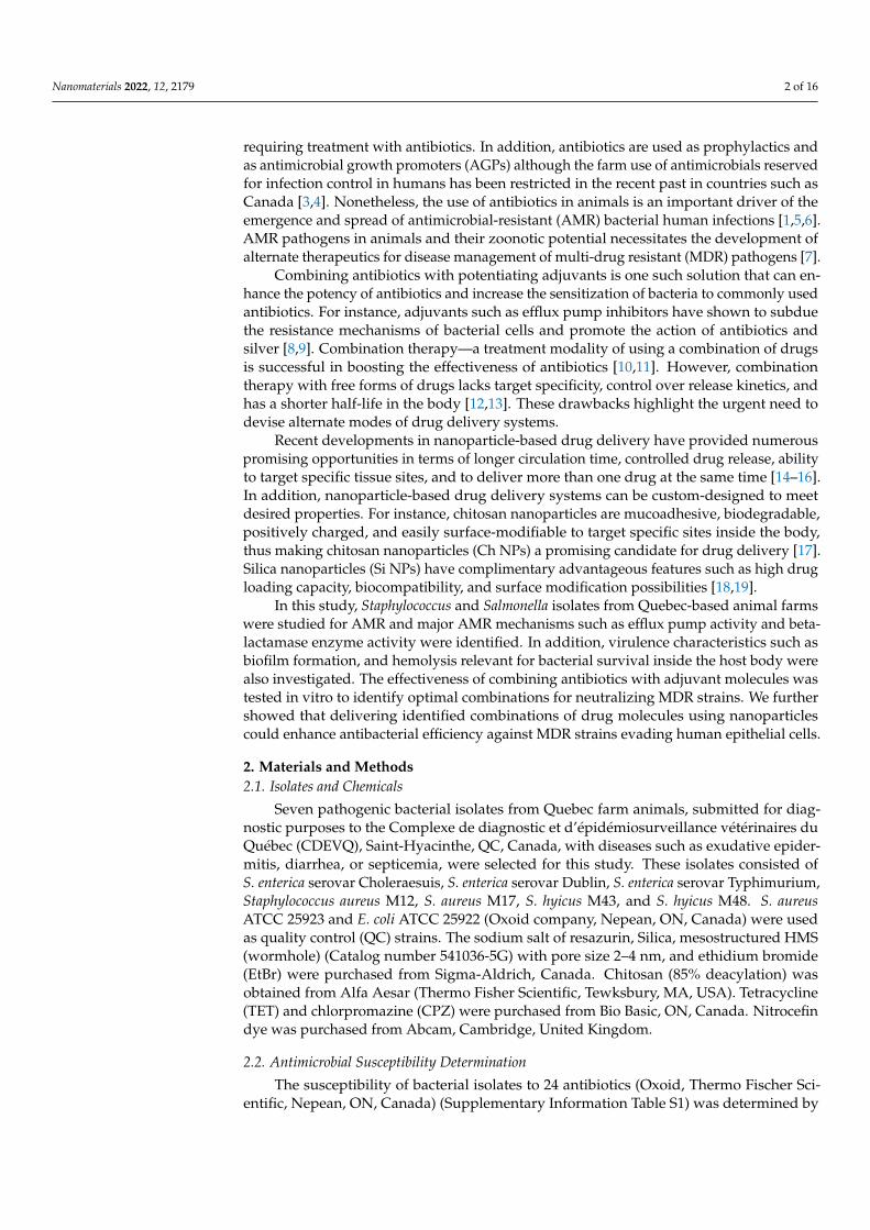
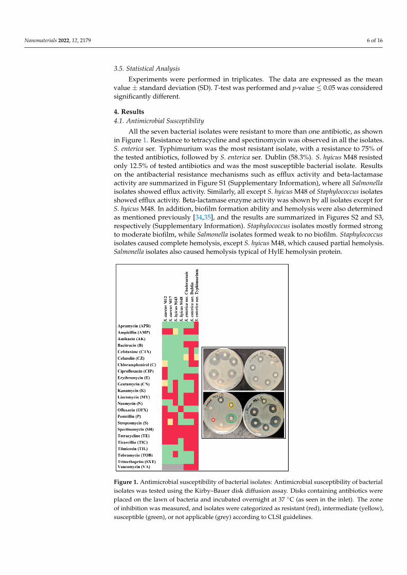
All the seven bacterial isolates were resistant to more than one antibiotic, as shownin Figure 1. Resistance to tetracycline and spectinomycin was observed in all the isolates.S. enterica ser. Typhimurium was the most resistant isolate, with a resistance to 75% ofthe tested antibiotics, followed by S. enterica ser. Dublin (58.3%). S. hyicus M48 resistedonly 12.5% of tested antibiotics and was the most susceptible bacterial isolate. Resultson the antibacterial resistance mechanisms such as efflux activity and beta-lactamaseactivity are summarized in Figure S1 (Supplementary Information), where all Salmonellaisolates showed efflux activity. Similarly, all except S. hyicus M48 of Staphylococcus isolatesshowed efflux activity. Beta-lactamase enzyme activity was shown by all isolates except forS. hyicus M48. In addition, biofilm formation ability and hemolysis were also determinedas mentioned previously [34,35], and the results are summarized in Figures S2 and S3,respectively (Supplementary Information). Staphylococcus isolates mostly formed strongto moderate biofilm, while Salmonella isolates formed weak to no biofilm. Staphylococcusisolates caused complete hemolysis, except S. hyicus M48, which caused partial hemolysis.Salmonella isolates also caused hemolysis typical of HylE hemolysin protein.
Nanomaterials 2022, 11, x FOR PEER REVIEW 7 of 17
Figure 1. Antimicrobial susceptibility of bacterial isolates: Antimicrobial susceptibility of bacterial isolates was tested using the Kirby–Bauer disk diffusion assay. Disks containing antibiotics were placed on the lawn of bacteria and incubated overnight at 37 °C (as seen in the inlet). The zone of inhibition was measured, and isolates were categorized as resistant (red), intermediate (yellow), susceptible (green), or not applicable (grey) according to CLSI guidelines.
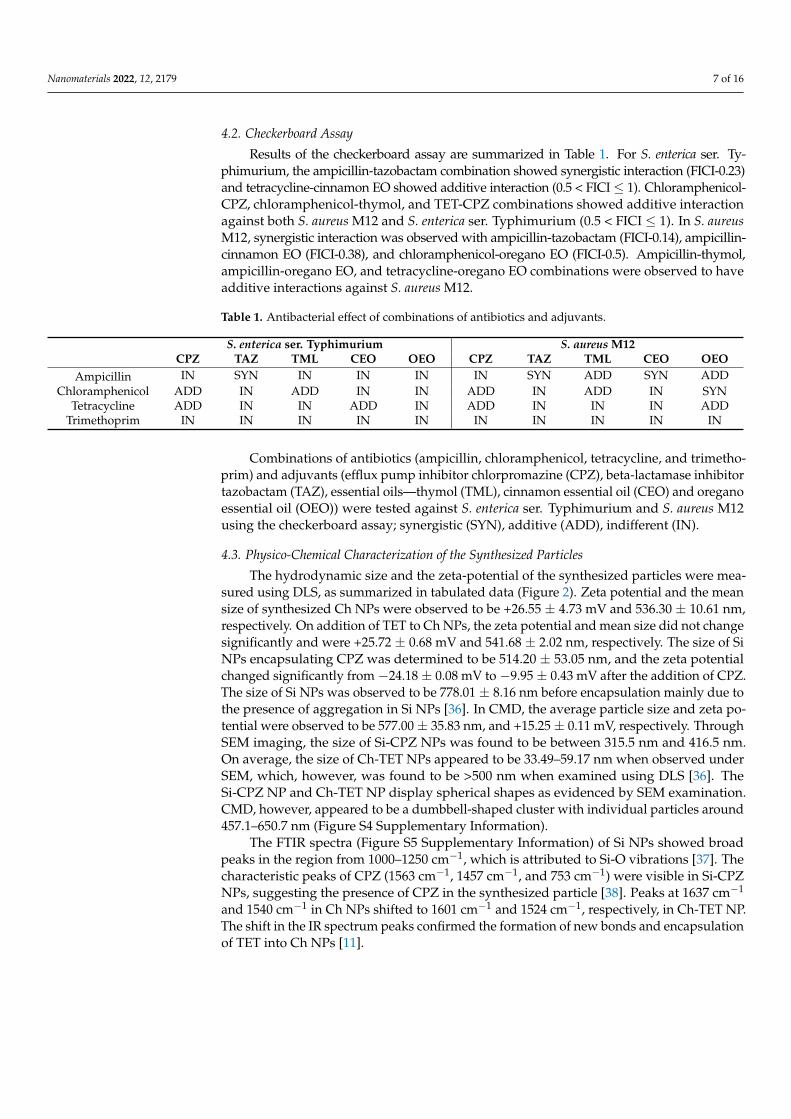
4.2. Checkerboard Assay Results of the checkerboard assay are summarized in Table 1. For S. enterica ser.
Typhimurium, the ampicillin-tazobactam combination showed synergistic interaction (FICI-0.23) and tetracycline-cinnamon EO showed additive interaction (0.5<FICI≤1). Chlo-ramphenicol-CPZ, chloramphenicol-thymol, and TET-CPZ combinations showed addi-tive interaction against both S. aureus M12 and S. enterica ser. Typhimurium (0.5<FICI≤1). In S. aureus M12, synergistic interaction was observed with ampicillin-tazobactam (FICI-0.14), ampicillin-cinnamon EO (FICI-0.38), and chloramphenicol-oregano EO (FICI-0.5). Ampicillin-thymol, ampicillin-oregano EO, and tetracycline-oregano EO combinations were observed to have additive interactions against S. aureus M12.
Table 1. Antibacterial effect of combinations of antibiotics and adjuvants.
S. enterica ser. Typhimurium S. aureus M12 CPZ TAZ TML CEO OEO CPZ TAZ TML CEO OEO
Ampicillin IN SYN IN IN IN IN SYN ADD SYN ADD Chloramphenicol ADD IN ADD IN IN ADD IN ADD IN SYN
Tetracycline ADD IN IN ADD IN ADD IN IN IN ADD Trimethoprim IN IN IN IN IN IN IN IN IN IN
Figure 1. Antimicrobial susceptibility of bacterial isolates: Antimicrobial susceptibility of bacterialisolates was tested using the Kirby–Bauer disk diffusion assay. Disks containing antibiotics wereplaced on the lawn of bacteria and incubated overnight at 37 C (as seen in the inlet). The zoneof inhibition was measured, and isolates were categorized as resistant (red), intermediate (yellow),susceptible (green), or not applicable (grey) according to CLSI guidelines.
Nanomaterials 2022, 12, 2179 7 of 16
4.2. Checkerboard Assay
Results of the checkerboard assay are summarized in Table 1. For S. enterica ser. Ty-phimurium, the ampicillin-tazobactam combination showed synergistic interaction (FICI-0.23)and tetracycline-cinnamon EO showed additive interaction (0.5 < FICI ≤ 1). Chloramphenicol-CPZ, chloramphenicol-thymol, and TET-CPZ combinations showed additive interactionagainst both S. aureus M12 and S. enterica ser. Typhimurium (0.5 < FICI ≤ 1). In S. aureusM12, synergistic interaction was observed with ampicillin-tazobactam (FICI-0.14), ampicillin-cinnamon EO (FICI-0.38), and chloramphenicol-oregano EO (FICI-0.5). Ampicillin-thymol,ampicillin-oregano EO, and tetracycline-oregano EO combinations were observed to haveadditive interactions against S. aureus M12.
Table 1. Antibacterial effect of combinations of antibiotics and adjuvants.
S. enterica ser. Typhimurium S. aureus M12CPZ TAZ TML CEO OEO CPZ TAZ TML CEO OEO
Ampicillin IN SYN IN IN IN IN SYN ADD SYN ADDChloramphenicol ADD IN ADD IN IN ADD IN ADD IN SYN
Tetracycline ADD IN IN ADD IN ADD IN IN IN ADDTrimethoprim IN IN IN IN IN IN IN IN IN IN
Combinations of antibiotics (ampicillin, chloramphenicol, tetracycline, and trimetho-prim) and adjuvants (efflux pump inhibitor chlorpromazine (CPZ), beta-lactamase inhibitortazobactam (TAZ), essential oils—thymol (TML), cinnamon essential oil (CEO) and oreganoessential oil (OEO)) were tested against S. enterica ser. Typhimurium and S. aureus M12using the checkerboard assay; synergistic (SYN), additive (ADD), indifferent (IN).
4.3. Physico-Chemical Characterization of the Synthesized Particles
The hydrodynamic size and the zeta-potential of the synthesized particles were mea-sured using DLS, as summarized in tabulated data (Figure 2). Zeta potential and the meansize of synthesized Ch NPs were observed to be +26.55 ± 4.73 mV and 536.30 ± 10.61 nm,respectively. On addition of TET to Ch NPs, the zeta potential and mean size did not changesignificantly and were +25.72 ± 0.68 mV and 541.68 ± 2.02 nm, respectively. The size of SiNPs encapsulating CPZ was determined to be 514.20 ± 53.05 nm, and the zeta potentialchanged significantly from −24.18 ± 0.08 mV to −9.95 ± 0.43 mV after the addition of CPZ.The size of Si NPs was observed to be 778.01 ± 8.16 nm before encapsulation mainly due tothe presence of aggregation in Si NPs [36]. In CMD, the average particle size and zeta po-tential were observed to be 577.00 ± 35.83 nm, and +15.25 ± 0.11 mV, respectively. ThroughSEM imaging, the size of Si-CPZ NPs was found to be between 315.5 nm and 416.5 nm.On average, the size of Ch-TET NPs appeared to be 33.49–59.17 nm when observed underSEM, which, however, was found to be >500 nm when examined using DLS [36]. TheSi-CPZ NP and Ch-TET NP display spherical shapes as evidenced by SEM examination.CMD, however, appeared to be a dumbbell-shaped cluster with individual particles around457.1–650.7 nm (Figure S4 Supplementary Information).
The FTIR spectra (Figure S5 Supplementary Information) of Si NPs showed broadpeaks in the region from 1000–1250 cm−1, which is attributed to Si-O vibrations [37]. Thecharacteristic peaks of CPZ (1563 cm−1, 1457 cm−1, and 753 cm−1) were visible in Si-CPZNPs, suggesting the presence of CPZ in the synthesized particle [38]. Peaks at 1637 cm−1
and 1540 cm−1 in Ch NPs shifted to 1601 cm−1 and 1524 cm−1, respectively, in Ch-TET NP.The shift in the IR spectrum peaks confirmed the formation of new bonds and encapsulationof TET into Ch NPs [11].

Nanomaterials 2022, 12, 2179 8 of 16
Nanomaterials 2022, 11, x FOR PEER REVIEW 8 of 17
Combinations of antibiotics (ampicillin, chloramphenicol, tetracycline, and trimethoprim) and ad-juvants (efflux pump inhibitor chlorpromazine (CPZ), beta-lactamase inhibitor tazobactam (TAZ), essential oils—thymol (TML), cinnamon essential oil (CEO) and oregano essential oil (OEO)) were tested against S. enterica ser. Typhimurium and S. aureus M12 using the checkerboard assay; syner-gistic (SYN), additive (ADD), indifferent (IN).
4.3. Physico-Chemical Characterization of the Synthesized Particles The hydrodynamic size and the zeta-potential of the synthesized particles were
measured using DLS, as summarized in tabulated data (Figure 2). Zeta potential and the mean size of synthesized Ch NPs were observed to be +26.55 ± 4.73 mV and 536.30 ± 10.61 nm, respectively. On addition of TET to Ch NPs, the zeta potential and mean size did not change significantly and were +25.72 ± 0.68 mV and 541.68 ± 2.02 nm, respectively. The size of Si NPs encapsulating CPZ was determined to be 514.20 ± 53.05 nm, and the zeta potential changed significantly from −24.18 ± 0.08 mV to −9.95 ± 0.43 mV after the addition of CPZ. The size of Si NPs was observed to be 778.01 ± 8.16 nm before encapsulation mainly due to the presence of aggregation in Si NPs [36]. In CMD, the average particle size and zeta potential were observed to be 577.00 ± 35.83 nm, and +15.25 ± 0.11 mV, re-spectively. Through SEM imaging, the size of Si-CPZ NPs was found to be between 315.5 nm and 416.5 nm. On average, the size of Ch-TET NPs appeared to be 33.49–59.17 nm when observed under SEM, which, however, was found to be >500 nm when examined using DLS [36]. The Si-CPZ NP and Ch-TET NP display spherical shapes as evidenced by SEM examination. CMD, however, appeared to be a dumbbell-shaped cluster with indi-vidual particles around 457.1–650.7 nm (Figure S4 supplementary information).
Figure 2. Characterization of particles. (a) Tabulated data showing zeta-potential, and the size of the particles measured using dynamic light scattering (DLS). (b) FTIR spectrum of Ch-TET NP and Si-CPZ NP. (c) Nanoparticles were platinum-coated and imaged using Scanning Electron Microscopy (SEM).
The FTIR spectra (Figure S5 supplementary information) of Si NPs showed broad peaks in the region from 1000–1250 cm−1, which is attributed to Si-O vibrations [37]. The
Figure 2. Characterization of particles. (a) Tabulated data showing zeta-potential, and the size of theparticles measured using dynamic light scattering (DLS). (b) FTIR spectrum of Ch-TET NP and Si-CPZNP. (c) Nanoparticles were platinum-coated and imaged using Scanning Electron Microscopy (SEM).
4.4. Encapsulation Efficiency and Release Behaviour
The EE was measured spectrophotometrically by measuring the absorbance of thesupernatant at 285 nm for TET and 305 nm for CPZ. EE of TET in Ch NPs was determinedto be 23.82 ± 0.16% and EE of CPZ in mesoporous Si NPs was 44.77 ± 0.12%. The releaseof TET from Ch NPs and of CPZ was measured by monitoring the absorbance of thesupernatant over seven days (Figure 3). It was found that the release was faster for bothTET and CPZ when the pH of suspending solution was 4.4. On day 1, 67.48 ± 1.57% ofTET was released from Ch NPs and 81.49 ± 0.29% of CPZ was released from Si NPs atpH 4.4. At pH 6.3, 52.31 ± 0.12% of TET and 61.95 ± 0.91% of CPZ were released on day 1.Following the initial burst release, the release was relatively slower over the next six days.
Nanomaterials 2022, 11, x FOR PEER REVIEW 9 of 17
characteristic peaks of CPZ (1563 cm−1, 1457 cm−1, and 753 cm−1) were visible in Si-CPZ NPs, suggesting the presence of CPZ in the synthesized particle [38]. Peaks at 1637 cm−1
and 1540 cm−1 in Ch NPs shifted to 1601 cm−1 and 1524 cm−1, respectively, in Ch-TET NP. The shift in the IR spectrum peaks confirmed the formation of new bonds and encapsula-tion of TET into Ch NPs [11].
4.4. Encapsulation Efficiency and Release Behaviour The EE was measured spectrophotometrically by measuring the absorbance of the
supernatant at 285 nm for TET and 305 nm for CPZ. EE of TET in Ch NPs was determined to be 23.82 ± 0.16% and EE of CPZ in mesoporous Si NPs was 44.77 ± 0.12%. The release of TET from Ch NPs and of CPZ was measured by monitoring the absorbance of the su-pernatant over seven days (Figure 3). It was found that the release was faster for both TET and CPZ when the pH of suspending solution was 4.4. On day 1, 67.48 ± 1.57% of TET was released from Ch NPs and 81.49 ± 0.29% of CPZ was released from Si NPs at pH 4.4. At pH 6.3, 52.31 ± 0.12% of TET and 61.95 ± 0.91% of CPZ were released on day 1. Following the initial burst release, the release was relatively slower over the next six days.
Figure 3. Release kinetics of drugs from nanoparticles. Release kinetics of tetracycline (λ = 285 nm) and chlorpromazine (λ = 305 nm) from chitosan nanoparticles and silica nanoparticles were meas-ured, respectively, using a spectrophotometer.
4.5. Antibacterial Efficacy The MIC of the synthesized particles and the CMD were determined against S. enter-
ica ser. Typhimurium and S. aureus M12 and the results are summarized in Table 2. For S. enterica ser. Typhimurium, the MIC of Ch-TET NP was determined to be 1000 µg/mL, and the MIC of Si-CPZ NP was 500 µg/mL. MIC of CMD was 250 µg/mL, whereas the actual concentration of TET was only 29.77 ± 0.20 µg/mL. A decrease in MIC was also observed against S. aureus M12 when exposed to CMD in comparison with other free drugs or par-ticles containing a single drug. Accordingly, the MIC of Ch-TET NP was 500 µg/mL, and that of Si-CPZ NP was measured to be 250 µg/mL. However, the MIC of CMD against S. aureus was 125 µg/mL, whereas the concentration of TET was 14.89 ± 0.10 µg/mL.
Table 2. Minimum inhibitory concentration (µg/mL) of Ch-TET NP, Si-CPZ NP and CMD.
Si-CPZ NP (µg/mL) Ch-TET NP (µg/mL) CMD (µg/mL) E. coli ATCC 25922 62.5 7.82 3.90
S. aureus ATCC 25923 125 3.90 1.95 S. enterica ser. Typhimurium 500 1000 250
S. aureus M12 250 500 125
Figure 3. Release kinetics of drugs from nanoparticles. Release kinetics of tetracycline (λ = 285 nm)and chlorpromazine (λ = 305 nm) from chitosan nanoparticles and silica nanoparticles were measured,respectively, using a spectrophotometer.
Nanomaterials 2022, 12, 2179 9 of 16
4.5. Antibacterial Efficacy
The MIC of the synthesized particles and the CMD were determined against S. entericaser. Typhimurium and S. aureus M12 and the results are summarized in Table 2. ForS. enterica ser. Typhimurium, the MIC of Ch-TET NP was determined to be 1000 µg/mL,and the MIC of Si-CPZ NP was 500 µg/mL. MIC of CMD was 250 µg/mL, whereas theactual concentration of TET was only 29.77 ± 0.20 µg/mL. A decrease in MIC was alsoobserved against S. aureus M12 when exposed to CMD in comparison with other free drugsor particles containing a single drug. Accordingly, the MIC of Ch-TET NP was 500 µg/mL,and that of Si-CPZ NP was measured to be 250 µg/mL. However, the MIC of CMD againstS. aureus was 125 µg/mL, whereas the concentration of TET was 14.89 ± 0.10 µg/mL.
Table 2. Minimum inhibitory concentration (µg/mL) of Ch-TET NP, Si-CPZ NP and CMD.
Si-CPZ NP (µg/mL) Ch-TET NP (µg/mL) CMD (µg/mL)
E. coli ATCC 25922 62.5 7.82 3.90S. aureus ATCC 25923 125 3.90 1.95
S. enterica ser.Typhimurium 500 1000 250
S. aureus M12 250 500 125
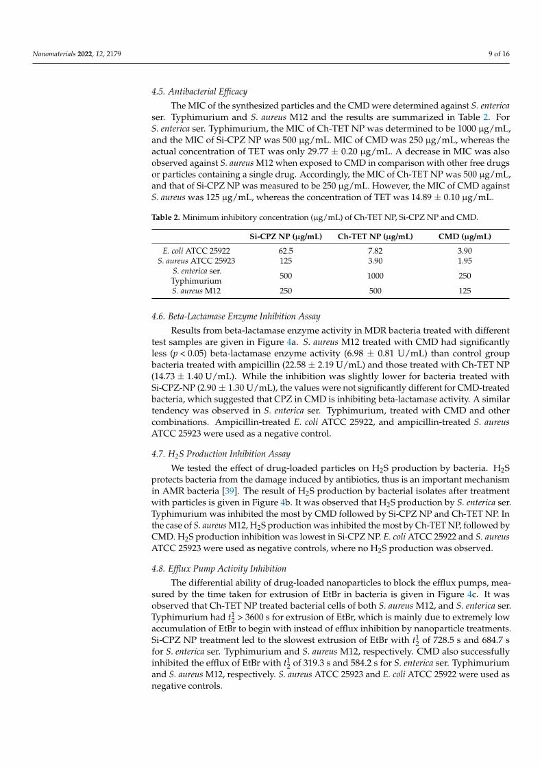
4.6. Beta-Lactamase Enzyme Inhibition Assay
Results from beta-lactamase enzyme activity in MDR bacteria treated with differenttest samples are given in Figure 4a. S. aureus M12 treated with CMD had significantlyless (p < 0.05) beta-lactamase enzyme activity (6.98 ± 0.81 U/mL) than control groupbacteria treated with ampicillin (22.58 ± 2.19 U/mL) and those treated with Ch-TET NP(14.73 ± 1.40 U/mL). While the inhibition was slightly lower for bacteria treated withSi-CPZ-NP (2.90 ± 1.30 U/mL), the values were not significantly different for CMD-treatedbacteria, which suggested that CPZ in CMD is inhibiting beta-lactamase activity. A similartendency was observed in S. enterica ser. Typhimurium, treated with CMD and othercombinations. Ampicillin-treated E. coli ATCC 25922, and ampicillin-treated S. aureusATCC 25923 were used as a negative control.
4.7. H2S Production Inhibition Assay
We tested the effect of drug-loaded particles on H2S production by bacteria. H2Sprotects bacteria from the damage induced by antibiotics, thus is an important mechanismin AMR bacteria [39]. The result of H2S production by bacterial isolates after treatmentwith particles is given in Figure 4b. It was observed that H2S production by S. enterica ser.Typhimurium was inhibited the most by CMD followed by Si-CPZ NP and Ch-TET NP. Inthe case of S. aureus M12, H2S production was inhibited the most by Ch-TET NP, followed byCMD. H2S production inhibition was lowest in Si-CPZ NP. E. coli ATCC 25922 and S. aureusATCC 25923 were used as negative controls, where no H2S production was observed.
4.8. Efflux Pump Activity Inhibition
The differential ability of drug-loaded nanoparticles to block the efflux pumps, mea-sured by the time taken for extrusion of EtBr in bacteria is given in Figure 4c. It wasobserved that Ch-TET NP treated bacterial cells of both S. aureus M12, and S. enterica ser.Typhimurium had t1
2 > 3600 s for extrusion of EtBr, which is mainly due to extremely lowaccumulation of EtBr to begin with instead of efflux inhibition by nanoparticle treatments.Si-CPZ NP treatment led to the slowest extrusion of EtBr with t1
2 of 728.5 s and 684.7 sfor S. enterica ser. Typhimurium and S. aureus M12, respectively. CMD also successfullyinhibited the efflux of EtBr with t1
2 of 319.3 s and 584.2 s for S. enterica ser. Typhimuriumand S. aureus M12, respectively. S. aureus ATCC 25923 and E. coli ATCC 25922 were used asnegative controls.
Nanomaterials 2022, 12, 2179 10 of 16
Nanomaterials 2022, 11, x FOR PEER REVIEW 10 of 17
4.6. Beta-Lactamase Enzyme Inhibition Assay Results from beta-lactamase enzyme activity in MDR bacteria treated with different
test samples are given in Figure 4a. S. aureus M12 treated with CMD had significantly less (p < 0.05) beta-lactamase enzyme activity (6.98 ± 0.81 U/mL) than control group bacteria treated with ampicillin (22.58 ± 2.19 U/mL) and those treated with Ch-TET NP (14.73 ± 1.40 U/mL). While the inhibition was slightly lower for bacteria treated with Si-CPZ-NP (2.90 ± 1.30 U/mL), the values were not significantly different for CMD-treated bacteria, which suggested that CPZ in CMD is inhibiting beta-lactamase activity. A similar ten-dency was observed in S. enterica ser. Typhimurium, treated with CMD and other combi-nations. Ampicillin-treated E. coli ATCC 25922, and ampicillin-treated S. aureus ATCC 25923 were used as a negative control.
4.7. H2S Production Inhibition Assay We tested the effect of drug-loaded particles on H2S production by bacteria. H2S pro-
tects bacteria from the damage induced by antibiotics, thus is an important mechanism in AMR bacteria [39]. The result of H2S production by bacterial isolates after treatment with particles is given in Figure 4b. It was observed that H2S production by S. enterica ser. Typhimurium was inhibited the most by CMD followed by Si-CPZ NP and Ch-TET NP. In the case of S. aureus M12, H2S production was inhibited the most by Ch-TET NP, fol-lowed by CMD. H2S production inhibition was lowest in Si-CPZ NP. E. coli ATCC 25922 and S. aureus ATCC 25923 were used as negative controls, where no H2S production was observed.
Figure 4. Effect of synthesized nanoparticles on resistance mechanisms. (a) Inhibition of beta-lac-tamase enzyme in bacteria treated with synthesized nanoparticles measured using nitrocefin assay, (b) H2S production of bacterial strains was measured after treatment with synthesized particles, and (c) EtBr extrusion (efflux) from bacterial cells after treatment with nanoparticle treatments was de-termined by measuring fluorescence.
4.8. Efflux Pump Activity Inhibition
Figure 4. Effect of synthesized nanoparticles on resistance mechanisms. (a) Inhibition of beta-lactamase enzyme in bacteria treated with synthesized nanoparticles measured using nitrocefin assay,(b) H2S production of bacterial strains was measured after treatment with synthesized particles,and (c) EtBr extrusion (efflux) from bacterial cells after treatment with nanoparticle treatments wasdetermined by measuring fluorescence.
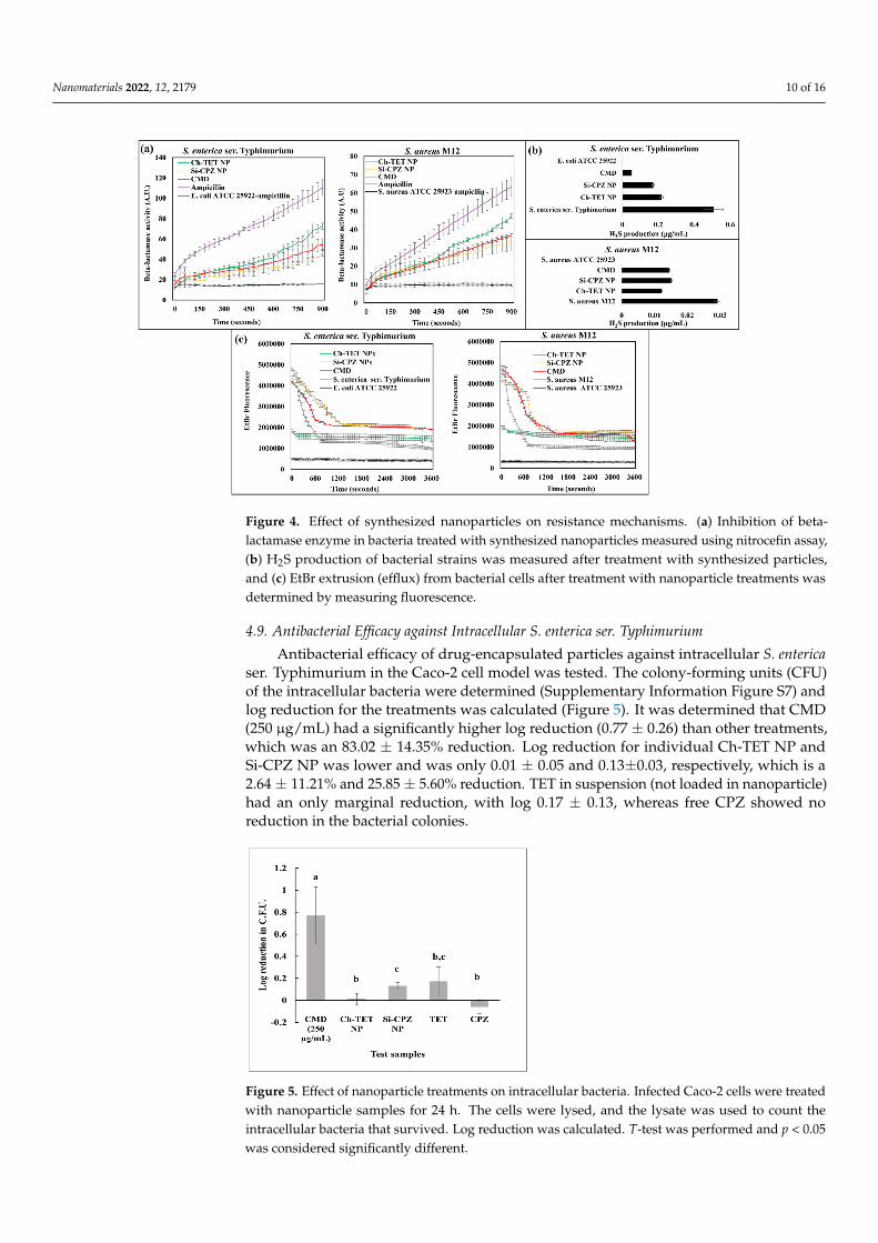
4.9. Antibacterial Efficacy against Intracellular S. enterica ser. Typhimurium
Antibacterial efficacy of drug-encapsulated particles against intracellular S. entericaser. Typhimurium in the Caco-2 cell model was tested. The colony-forming units (CFU)of the intracellular bacteria were determined (Supplementary Information Figure S7) andlog reduction for the treatments was calculated (Figure 5). It was determined that CMD(250 µg/mL) had a significantly higher log reduction (0.77 ± 0.26) than other treatments,which was an 83.02 ± 14.35% reduction. Log reduction for individual Ch-TET NP andSi-CPZ NP was lower and was only 0.01 ± 0.05 and 0.13±0.03, respectively, which is a2.64 ± 11.21% and 25.85 ± 5.60% reduction. TET in suspension (not loaded in nanoparticle)had an only marginal reduction, with log 0.17 ± 0.13, whereas free CPZ showed noreduction in the bacterial colonies.
Nanomaterials 2022, 11, x FOR PEER REVIEW 11 of 17
The differential ability of drug-loaded nanoparticles to block the efflux pumps, meas-ured by the time taken for extrusion of EtBr in bacteria is given in Figure 4c. It was ob-served that Ch-TET NP treated bacterial cells of both S. aureus M12, and S. enterica ser. Typhimurium had 𝑡 > 3600 s for extrusion of EtBr, which is mainly due to extremely low accumulation of EtBr to begin with instead of efflux inhibition by nanoparticle treat-ments. Si-CPZ NP treatment led to the slowest extrusion of EtBr with 𝑡 of 728.5 s and 684.7 sec for S. enterica ser. Typhimurium and S. aureus M12, respectively. CMD also suc-cessfully inhibited the efflux of EtBr with 𝑡 of 319.3 s and 584.2 s for S. enterica ser. Typhi-murium and S. aureus M12, respectively. S. aureus ATCC 25923 and E. coli ATCC 25922 were used as negative controls.
4.9. Antibacterial Efficacy Against Intracellular S. enterica ser. Typhimurium Antibacterial efficacy of drug-encapsulated particles against intracellular S. enterica
ser. Typhimurium in the Caco-2 cell model was tested. The colony-forming units (CFU) of the intracellular bacteria were determined (Supplementary information Figure S7) and log reduction for the treatments was calculated (Figure 5). It was determined that CMD (250 µg/mL) had a significantly higher log reduction (0.77 ± 0.26) than other treatments, which was an 83.02 ± 14.35% reduction. Log reduction for individual Ch-TET NP and Si-CPZ NP was lower and was only 0.01 ± 0.05 and 0.13±0.03, respectively, which is a 2.64 ± 11.21% and 25.85 ± 5.60% reduction. TET in suspension (not loaded in nanoparticle) had an only marginal reduction, with log 0.17 ± 0.13, whereas free CPZ showed no reduction in the bacterial colonies.
Figure 5. Effect of nanoparticle treatments on intracellular bacteria. Infected Caco-2 cells were treated with nanoparticle samples for 24 h. The cells were lysed, and the lysate was used to count the intracellular bacteria that survived. Log reduction was calculated. T-test was performed and p < 0.05 was considered significantly different.
5. Discussion Seven pathogen isolates containing Salmonella and Staphylococcus from animal farms
were characterized to gain insight into the antibiotic resistance, mechanisms of resistance, and virulence factors for custom design of an antibacterial strategy. Based on the AMR and virulence characteristics, a nano-enabled combination therapy containing TET as an antibiotic and CPZ as an adjuvant was developed and tested successively against MDR S. enterica ser. Typhimurium and S. aureus M12.
The antimicrobial susceptibility profile showed that most of the pathogens were re-sistant to TET, ampicillin, lincomycin, and penicillin, which include some of the most widely used antibiotics in agriculture [40]. MDR bacteria use multiple mechanisms to
Figure 5. Effect of nanoparticle treatments on intracellular bacteria. Infected Caco-2 cells were treatedwith nanoparticle samples for 24 h. The cells were lysed, and the lysate was used to count theintracellular bacteria that survived. Log reduction was calculated. T-test was performed and p < 0.05was considered significantly different.
Nanomaterials 2022, 12, 2179 11 of 16
5. Discussion
Seven pathogen isolates containing Salmonella and Staphylococcus from animal farmswere characterized to gain insight into the antibiotic resistance, mechanisms of resistance,and virulence factors for custom design of an antibacterial strategy. Based on the AMRand virulence characteristics, a nano-enabled combination therapy containing TET as anantibiotic and CPZ as an adjuvant was developed and tested successively against MDRS. enterica ser. Typhimurium and S. aureus M12.
The antimicrobial susceptibility profile showed that most of the pathogens were resis-tant to TET, ampicillin, lincomycin, and penicillin, which include some of the most widelyused antibiotics in agriculture [40]. MDR bacteria use multiple mechanisms to overcome theaction of antimicrobials, such as active efflux or enzymatic degradation of the antimicrobial,mutation of an antimicrobial target site, and reduced cell wall permeability [41–43]. InSalmonella, the most common efflux pump system, belonging to the RND family is theAcrAB-TolC, responsible for resistance to various antibiotics [44]. Likewise, we observedefflux activity in the tested Salmonella isolates, which might explain their strong resistanceto several tested antibiotics (Supplementary Section S2; Figure S1a). Similarly, the actionof chromosome-mediated efflux pumps such as Nor and plasmid-mediated Qac effluxpumps [45] are reported to extrude biocides and antibiotics, explaining the efflux activityfound in Staphylococcus isolates in our study (Supplementary Section S2; Figure S1b).
Another major mechanism of resistance is the action of the beta-lactamase enzymewhich hydrolyzes the beta-lactam ring. All tested bacterial isolates, except for S. hyicus M48were positive for beta-lactamase enzyme activity. Beta-lactamase enzyme activity coded byblaZ genes has been reported in AMR Staphylococcus strains [44], while that in Salmonellahas been associated with genes of blaTEM, blaCTX-M, blaSHV, blaOXA-1, and blaCMY-2 [44]. H2Sproduction in bacteria is vital for protection against antibiotic-induced damage and oxida-tive stress. It is responsible for sequestering Fe+2 ions that can damage the bacterial DNAthrough the Fenton reaction. In addition, it stimulates the activities of antioxidant enzymesas well as superoxide dismutase. Reduction in H2S production is thus an indicator ofsubduing the general mechanism of resistance in bacteria [39]. Hemolysis and biofilm for-mation were investigated because they are two key virulence factors promoting pathogensurvival in animals [46,47]. Tested Staphylococcus isolates were strong biofilm formers, whileSalmonella isolates formed very weak or no biofilms (Supplementary Section S4). In addi-tion, Staphylococcus isolates mostly manifested complete hemolysis (beta-hemolysis) [46],while Salmonella isolates showed hemolysis typical of HylE hemolysin protein (Supplemen-tary Section S5), a pore-forming protein that contributes to the systemic pathogenesis ofSalmonella [48]. The observation of the prevalence of multi-drug resistance and virulencecharacteristics in farm isolates highlights the need to develop alternate field-deployable an-tibacterial strategies. Combination therapy, wherein more than one antibiotic, or antibioticwith effector molecules are combined, has been deployed for the clinical management ofinfectious diseases in animals and humans [49,50].
We screened various combinations of antibiotics and effector molecules based onresults from bacterial profiling for AMR and virulence characteristics. Synergism observedin the ampicillin-tazobactam combination could be ascribed to the action of tazobactamon the beta-lactamase enzyme, which was highly prevalent in both isolates [42]. Similarly,the synergistic interaction of the ampicillin-cinnamon EO combination could be attributedto membrane disruption by the EO, allowing easy passage of ampicillin into the cells. Inaddition, ampicillin and cinnamon EO share a common mechanism of action and worktogether to disrupt the bacterial cell membrane [43]. Yet another synergism was observed inthe oregano EO-chloramphenicol combination against S. aureus M12. Oregano EO consistsmainly of thymol and carvacrol, which affects the respiratory and energy metabolism inaddition to membrane disruption [43]. This can damage the pH homeostasis and furtherlead to disruption of LmrS efflux pumps, which confer resistance to chloramphenicol,thus increasing the susceptibility to chloramphenicol [45]. CPZ interferes with substratebinding and thus inhibits AcrB-mediated efflux in Salmonella [51]. In Staphylococcus, CPZ
Nanomaterials 2022, 12, 2179 12 of 16
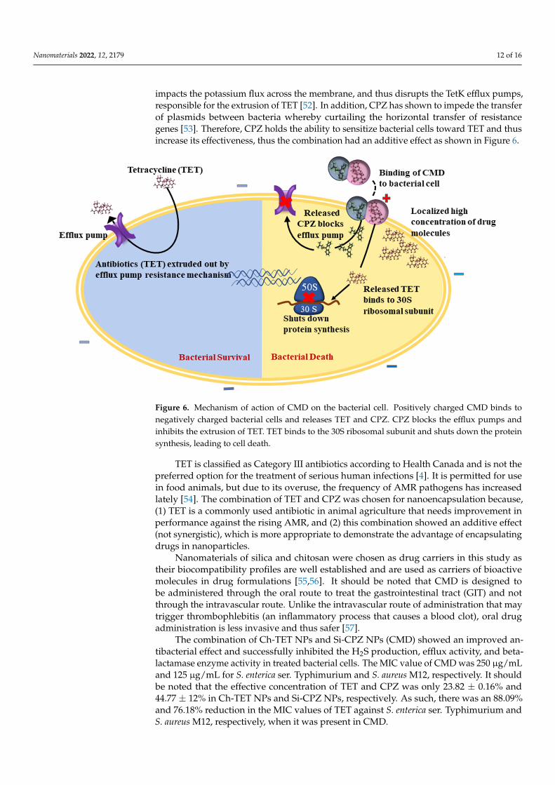
impacts the potassium flux across the membrane, and thus disrupts the TetK efflux pumps,responsible for the extrusion of TET [52]. In addition, CPZ has shown to impede the transferof plasmids between bacteria whereby curtailing the horizontal transfer of resistancegenes [53]. Therefore, CPZ holds the ability to sensitize bacterial cells toward TET and thusincrease its effectiveness, thus the combination had an additive effect as shown in Figure 6.
Nanomaterials 2022, 11, x FOR PEER REVIEW 13 of 17
Figure 6. Mechanism of action of CMD on the bacterial cell. Positively charged CMD binds to neg-atively charged bacterial cells and releases TET and CPZ. CPZ blocks the efflux pumps and inhibits the extrusion of TET. TET binds to the 30S ribosomal subunit and shuts down the protein synthesis, leading to cell death.
TET is classified as Category III antibiotics according to Health Canada and is not the preferred option for the treatment of serious human infections [4]. It is permitted for use in food animals, but due to its overuse, the frequency of AMR pathogens has increased lately [54]. The combination of TET and CPZ was chosen for nanoencapsulation because, (1) TET is a commonly used antibiotic in animal agriculture that needs improvement in performance against the rising AMR, and (2) this combination showed an additive effect (not synergistic), which is more appropriate to demonstrate the advantage of encapsulat-ing drugs in nanoparticles.
Nanomaterials of silica and chitosan were chosen as drug carriers in this study as their biocompatibility profiles are well established and are used as carriers of bioactive molecules in drug formulations [55,56]. It should be noted that CMD is designed to be administered through the oral route to treat the gastrointestinal tract (GIT) and not through the intravascular route. Unlike the intravascular route of administration that may trigger thrombophlebitis (an inflammatory process that causes a blood clot), oral drug administration is less invasive and thus safer [57].
The combination of Ch-TET NPs and Si-CPZ NPs (CMD) showed an improved anti-bacterial effect and successfully inhibited the H2S production, efflux activity, and beta-lactamase enzyme activity in treated bacterial cells. The MIC value of CMD was 250 µg/mL and 125 µg/mL for S. enterica ser. Typhimurium and S. aureus M12, respectively. It should be noted that the effective concentration of TET and CPZ was only 23.82 ± 0.16% and 44.77 ± 12% in Ch-TET NPs and Si-CPZ NPs, respectively. As such, there was an 88.09% and 76.18% reduction in the MIC values of TET against S. enterica ser. Typhi-murium and S. aureus M12, respectively, when it was present in CMD.
The enhanced antibacterial efficacy of the combinations of drugs encapsulated into nanoparticles can be attributed to two main factors: (1) the additive effect between TET and CPZ, as observed from checkerboard assay result, and (2) the improved localized de-livery of drugs to bacterial cells. Apart from its inhibitory action on efflux activity, Si-CPZ, NP, and CMD were noted to significantly lower the production of the beta-lactamase en-zyme activity in both S. enterica ser. Typhimurium and S. aureus M12. This observation is in line with the reported inhibitory action of CPZ on the expression of bla and mecA genes [58]. The combined effect shown in suspension culture may not be achieved if free drugs are administered to the human or animal body. This is because the therapeutic efficacy of
Figure 6. Mechanism of action of CMD on the bacterial cell. Positively charged CMD binds tonegatively charged bacterial cells and releases TET and CPZ. CPZ blocks the efflux pumps andinhibits the extrusion of TET. TET binds to the 30S ribosomal subunit and shuts down the proteinsynthesis, leading to cell death.
TET is classified as Category III antibiotics according to Health Canada and is not thepreferred option for the treatment of serious human infections [4]. It is permitted for usein food animals, but due to its overuse, the frequency of AMR pathogens has increasedlately [54]. The combination of TET and CPZ was chosen for nanoencapsulation because,(1) TET is a commonly used antibiotic in animal agriculture that needs improvement inperformance against the rising AMR, and (2) this combination showed an additive effect(not synergistic), which is more appropriate to demonstrate the advantage of encapsulatingdrugs in nanoparticles.
Nanomaterials of silica and chitosan were chosen as drug carriers in this study astheir biocompatibility profiles are well established and are used as carriers of bioactivemolecules in drug formulations [55,56]. It should be noted that CMD is designed tobe administered through the oral route to treat the gastrointestinal tract (GIT) and notthrough the intravascular route. Unlike the intravascular route of administration that maytrigger thrombophlebitis (an inflammatory process that causes a blood clot), oral drugadministration is less invasive and thus safer [57].
The combination of Ch-TET NPs and Si-CPZ NPs (CMD) showed an improved an-tibacterial effect and successfully inhibited the H2S production, efflux activity, and beta-lactamase enzyme activity in treated bacterial cells. The MIC value of CMD was 250 µg/mLand 125 µg/mL for S. enterica ser. Typhimurium and S. aureus M12, respectively. It shouldbe noted that the effective concentration of TET and CPZ was only 23.82 ± 0.16% and44.77 ± 12% in Ch-TET NPs and Si-CPZ NPs, respectively. As such, there was an 88.09%and 76.18% reduction in the MIC values of TET against S. enterica ser. Typhimurium andS. aureus M12, respectively, when it was present in CMD.
Nanomaterials 2022, 12, 2179 13 of 16
The enhanced antibacterial efficacy of the combinations of drugs encapsulated intonanoparticles can be attributed to two main factors: (1) the additive effect between TET andCPZ, as observed from checkerboard assay result, and (2) the improved localized deliveryof drugs to bacterial cells. Apart from its inhibitory action on efflux activity, Si-CPZ, NP,and CMD were noted to significantly lower the production of the beta-lactamase enzymeactivity in both S. enterica ser. Typhimurium and S. aureus M12. This observation is in linewith the reported inhibitory action of CPZ on the expression of bla and mecA genes [58].The combined effect shown in suspension culture may not be achieved if free drugs areadministered to the human or animal body. This is because the therapeutic efficacy ofsystemically or orally administered free antibiotics can be hindered due to various obstacles:limited bioavailability, poor target specificity, faster degradation, and body clearance [59].Nanoparticle-based drug delivery systems offer the opportunity of enhancing the efficacy ofantibiotics while minimizing the side effects [60]. Further, the ability of CMD to significantlyreduce the number of intracellular MDR S. enterica ser. Typhimurium points towards thesuperior properties of nanoparticle drug carriers to target bacteria taking refuge inside hostcells. CMD had a positive surface charge, which is beneficial for oral administration ofdrugs as the positive charge of CMD could penetrate the mucous layer on the intestinalepithelial cells, the growth site of bacteria causing gastrointestinal diseases in animals [61].In short, the superior performance of CMD stems from the additive action of antibiotic(TET) and effector molecule (CPZ) and delivery of effective concentrations of drugs to thebacteria-laden cells achieved through nanoparticles.
The ability of CMD to reduce intracellular pathogens also opens new pathways forpreserving and enhancing the antibacterial efficacy of various commonly used antibioticsin food animals, to which various pathogens have developed resistance. Thus, the nano-enabled combination therapy extends the usability of category III antibiotics in eliminatingtougher-to-treat infections, thereby slowing down the need to rely on last-resort antibioticsfor treating animal infections. While this effect was exemplified by combinations of TETand CPZ, we believe similar strategies could extend the usability and effectiveness of manyother antibiotics permitted for use in food animals.
Supplementary Materials: The following supporting information can be downloaded at: https://www.mdpi.com/article/10.3390/nano12132179/s1, Figure S1: Efflux and beta-lactamase enzyme activity:Efflux pump activity in Salmonella (A) and Staphylococcus (B) isolates was assessed using EtBr asthe substrate. Extrusion of EtBr was measured in kinetic mode by measuring the fluorescence at530-590 nm (excitation/emission) after energizing the cells. (C) Tabular data showing beta-lactamaseenzyme activity in Staphylococcus and Salmonella isolates.; Figure S2: Biofilm formation assay: Biofilmformation of Staphylococcus and Salmonella isolates from animal farms measured using crystal violetassay after 24 h (light grey) and 48 h (dark grey). Inlet shows the image of one of the platesused for biofilm assay. A significant difference in biofilm formation ability (p < 0.05) is markedas ‘*’.; Figure S3: Hemolysis assay: Bacterial isolates were streaked onto tryptic soy agar platescontaining 5% defibrinated sheep blood and incubated overnight. Hemolysis activity was assessedbased on characteristic translucency around bacterial colonies. (A) Tabular data summarizing thetype of hemolysis activity in tested isolates. (B) S. aureus M12 produced translucent halo aroundthe cultured colonies – beta-hemolysis, S. enterica ser. Typhimurium showed hemolysis (greendiscoloration).; Figure S4: SEM images of Ch–TET NPs, Si–CPZ NPs, and CMD with markeddimensions; Figure S5: FTIR spectra of Si NP, CPZ, Ch NP, and TET; Figure S6: EtBr accumulation inbacterial cells. Accumulation of EtBr in S. enterica ser. Typhimurium (A) and S. aureus M12 (B) treatedwith synthesized nanoparticles determined by measuring fluorescence (530/590) nm.; Figure S7:Determination of CFU/mL. Image of plates used to determine CFU/mL of lysates of infected Caco–2cells treated with nanoparticle sample ; Table S1: Antibiotics and their corresponding concentrationsin the disks used in the determination of antimicrobial susceptibility of tested isolates
Nanomaterials 2022, 12, 2179 14 of 16
Author Contributions: A.B.: Data curation, formal analysis, investigation, methodology, software,validation, visualization, writing—original draft, and writing—review and editing. S.M.: Formalanalysis, methodology, validation, visualization, and writing—review, and editing. M.Z.N.: Datacuration, validation, and visualization. M.-O.B.-B.: Resources, validation, visualization, and writingreview and editing. J.R.: Resources, validation, and writing review and editing. S.G.: Conceptualiza-tion, funding acquisition, investigation, project administration, resources, supervision, validation,and writing review and editing. All authors have read and agreed to the published version ofthe manuscript.
Funding: This work was supported by the Fonds de recherche du Québec—Nature et technologies(FRQNT) strategic cluster nRS-170946, by Swine and Poultry Infectious Diseases Research Center(CRIPA), and by Canada Research Chair. Amarpreet Brar acknowledges student aid from CRIPAsupported by the FRQNT nRS-170946.
Institutional Review Board Statement: Not applicable.
Informed Consent Statement: Not applicable.
Data Availability Statement: Data available upon request.
Acknowledgments: We would like to thank Xiaonan Lu and Julie-Hélène Fairbrother for providingthe bacterial isolates.
Conflicts of Interest: Authors declare no conflict of interest.
References1. Martin, M.J.; Thottathil, S.E.; Newman, T.B. Antibiotics Overuse in Animal Agriculture: A Call to Action for Health Care Providers.
Am. J. Public Health 2015, 105, 2409–2410. [CrossRef] [PubMed]2. Martins, S.B.; Häsler, B.; Rushton, J. Economic aspects of zoonoses: Impact of zoonoses on the food industry. In Zoonoses-Infections
Affecting Humans and Animals; Springer: Berlin/Heidelberg, Germany, 2015; pp. 1107–1126.3. Canadian Pork Council. New Rules for the Access and Use of Veterinary Drugs. Available online: https://www.cpc-ccp.com/
uploads/userfiles/files/F-21%20B%20New%20rules%20access%20use%20of%20vet%20drugs%20web(1).pdf (accessed on 7February 2022).
4. Health Canada. Categorization of Antimicrobial Drugs Based on Importance in Human Medicine. 2009. Available on-line: https://www.canada.ca/en/health-canada/services/drugs-health-products/veterinary-drugs/antimicrobial-resistance/categorization-antimicrobial-drugs-based-importance-human-medicine.html (accessed on 7 February 2022).
5. Khanna, T.; Friendship, R.; Dewey, C.; Weese, J.S. Methicillin resistant Staphylococcus aureus colonization in pigs and pig farmers.Vet. Microbiol. 2008, 128, 298–303. [CrossRef] [PubMed]
6. Park, S.; Ronholm, J. Staphylococcus aureus in Agriculture: Lessons in evolution from a multispecies pathogen. Clin. Microbiol. Rev.2021, 34, e00120–e00182. [CrossRef] [PubMed]
7. Majumder, S.; Jung, D.; Ronholm, J.; George, S. Prevalence and mechanisms of antibiotic resistance in Escherichia coli isolated frommastitic dairy cattle in Canada. BMC Microbiol. 2021, 21, 222.
8. Ferrer-Espada, R.; Shahrour, H.; Pitts, B.; Stewart, P.S.; Sánchez-Gómez, S.; Martínez-de-Tejada, G. A permeability-increasingdrug synergizes with bacterial efflux pump inhibitors and restores susceptibility to antibiotics in multi-drug resistantPseudomonas aeruginosa strains. Sci. Rep. 2019, 9, 3452. [CrossRef]
9. George, S.; Tay, I.; Phue, W.H.; Gardner, H.; Sukumaran, B. Enhancing the bioavailability of silver through nanotechnologyapproaches could overcome efflux pump mediated silver resistance in methicillin resistant Staphylococcus aureus. J. Biomed.Nanotechnol. 2019, 15, 2216–2228. [CrossRef]
10. Elbi, S.; Nimal, T.; Rajan, V.; Baranwal, G.; Biswas, R.; Jayakumar, R.; Sathianarayanan, S. Fucoidan coated ciprofloxacin loadedchitosan nanoparticles for the treatment of intracellular and biofilm infections of Salmonella. Colloids Surf. B Biointerfaces 2017,160, 40–47.
11. El-Alfy, E.A.; El-Bisi, M.K.; Taha, G.M.; Ibrahim, H.M. Preparation of biocompatible chitosan nanoparticles loaded by tetracycline,gentamycin and ciprofloxacin as novel drug delivery system for improvement the antibacterial properties of cellulose basedfabrics. Int. J. Biol. Macromol. 2020, 161, 1247–1260. [CrossRef]
12. Prausnitz, M.R.; Mitragotri, S.; Langer, R. Current status and future potential of transdermal drug delivery. Nat. Rev. Drug Discov.2004, 3, 115–124. [CrossRef]
13. Raza, A.; Sime, F.B.; Cabot, P.J.; Maqbool, F.; Roberts, J.A.; Falconer, J.R. Solid nanoparticles for oral antimicrobial drug delivery:A review. Drug Discov. Today 2019, 24, 858–866. [CrossRef]
14. Amreddy, N.; Babu, A.; Muralidharan, R.; Panneerselvam, J.; Srivastava, A.; Ahmed, R.; Mehta, M.; Munshi, A.; Ramesh, R.Recent advances in nanoparticle-based cancer drug and gene delivery. Adv. Cancer Res. 2018, 137, 115–170. [PubMed]
Nanomaterials 2022, 12, 2179 15 of 16
15. Menina, S.; Eisenbeis, J.; Kamal, M.A.M.; Koch, M.; Bischoff, M.; Gordon, S.; Loretz, B.; Lehr, C.M. Bioinspired liposomes for oral delivery ofcolistin to combat intracellular infections by Salmonella enterica. Adv. Healthc. Mater. 2019, 8, 1900564. [CrossRef] [PubMed]
16. Babos, G.; Biró, E.; Meiczinger, M.; Feczkó, T. Dual drug delivery of sorafenib and doxorubicin from PLGA and PEG-PLGApolymeric nanoparticles. Polymers 2018, 10, 895. [CrossRef]
17. Coutinho, A.J.; Lima, S.A.C.; Afonso, C.M.; Reis, S. Mucoadhesive and pH responsive fucoidan-chitosan nanoparticles for theoral delivery of methotrexate. Int. J. Biol. Macromol. 2020, 158, 180–188. [CrossRef]
18. Castillo, R.R.; Colilla, M.; Vallet-Regí, M. Advances in mesoporous silica-based nanocarriers for co-delivery and combinationtherapy against cancer. Expert Opin. Drug Deliv. 2017, 14, 229–243. [CrossRef]
19. Xia, T.; Kovochich, M.; Liong, M.; Meng, H.; Kabehie, S.; George, S.; Zink, J.I.; Nel, A.E. Polyethyleneimine Coating Enhances theCellular Uptake of Mesoporous Silica Nanoparticles and Allows Safe Delivery of siRNA and DNA Constructs. ACS Nano 2009, 3,3273–3286. [CrossRef] [PubMed]
20. Hudzicki, J. Kirby-Bauer Disk Diffusion Susceptibility Test Protocol; American Society for Microbiology: Washington, DC,USA, 2009. Available online: https://asm.org/getattachment/2594ce26-bd44-47f6-8287-0657aa9185ad/Kirby-Bauer-Disk-DiffusionSusceptibility-Test-Protocol-pdf.pdf (accessed on 14 March 2021).
21. Clinical Laboratory Standards Institute. Performance Standards for Antimicrobial Susceptibility Testing; Clinical Laboratory StandardsInstitute: Washington, DC, USA, 2021. Available online: https://clsi.org/standards/products/microbiology/documents/m02/(accessed on 24 October 2020).
22. World Health Organization. Critically Important Antimicrobials for Human Medicine: Ranking of Antimicrobial Agents for RiskManagement of Antimicrobial Resistance Due to Non-Human Use; World Health Organization: Geneva, Switzerland, 2017. Availableonline: https://apps.who.int/iris/bitstream/handle/10665/255027/978?sequence=1 (accessed on 20 October 2020).
23. Agriculture & Horticulture Development Board (AHDB). Antibiotic Usage Data in Pigs from the Electronic Medicines Book (eMB);AHDB: London, UK, 2020. Available online: https://projectblue.blob.core.windows.net/media/Default/Pork/Documents/eMB%20data%202020%20confidential.pdf (accessed on 20 October 2020).
24. Bagheri, L.; Khodaei, N.; Salmieri, S.; Karboune, S.; Lacroix, M. Correlation between chemical composition and antimicrobialproperties of essential oils against most common food pathogens and spoilers: In-vitro efficacy and predictive modelling. Microb.Pathog. 2020, 147, 104212. [CrossRef] [PubMed]
25. Orhan, G.; Bayram, A.; Zer, Y.; Balci, I. Synergy tests by E test and checkerboard methods of antimicrobial combinations againstBrucella melitensis. J. Clin. Microbiol. 2005, 43, 140. [CrossRef]
26. George, S.; Teo, L.L.; Majumder, S.; Chew, W.L.; Khoo, G.H. Low levels of silver in food packaging materials may have no functionaladvantage, instead enhance microbial spoilage of food through hormetic effect. Food Control 2020, 123, 107768. [CrossRef]
27. Anitha, A.; Rani, V.D.; Krishna, R.; Sreeja, V.; Selvamurugan, N.; Nair, S.; Tamura, H.; Jayakumar, R. Synthesis, characterization,cytotoxicity and antibacterial studies of chitosan, O-carboxymethyl and N, O-carboxymethyl chitosan nanoparticles. Carbohydr.Polym. 2009, 78, 672–677. [CrossRef]
28. Phue, W.H.; Bahadi, M.; Dynes, J.J.; Wang, J.; Kuppili, V.S.C.; Ismail, A.; Hameed, A.; George, S. Protein–biomolecule interactionsplay a major role in shaping corona proteome: Studies on milk interacted dietary particles. Nanoscale 2021, 13, 13353–13367.[CrossRef] [PubMed]
29. Price, R.R.; Gaber, B.P.; Lvov, Y. In-vitro release characteristics of tetracycline HCl, khellin and nicotinamide adenine dineculeotidefrom halloysite; a cylindrical mineral. J. Microencapsul. 2001, 18, 713–722. [PubMed]
30. Basic, A.; Blomqvist, S.; Carlén, A.; Dahlen, G. Estimation of bacterial hydrogen sulfide production in vitro. J. Oral Microbiol. 2015,7, 28166. [CrossRef]
31. Paixão, L.; Rodrigues, L.; Couto, I.; Martins, M.; Fernandes, P.; de Carvalho, C.C.C.R.; Monteiro, G.A.; Sansonetty, F.; Amaral, L.;Viveiros, M. Fluorometric determination of ethidium bromide efflux kinetics in Escherichia coli. J. Biol. Eng. 2009, 3, 18. [CrossRef]
32. Xie, S.; Yang, F.; Tao, Y.; Chen, D.; Qu, W.; Huang, L.; Liu, Z.; Pan, Y.; Yuan, Z. Enhanced intracellular delivery and antibacterialefficacy of enrofloxacin-loaded docosanoic acid solid lipid nanoparticles against intracellular Salmonella. Sci. Rep. 2017, 7, 41104.[CrossRef] [PubMed]
33. Shao, L.; Majumder, S.; Liu, Z.; Xu, K.; Dai, R.; George, S. Light activation of gold nanorods but not gold nanospheres enhanceantibacterial effect through photodynamic and photothermal mechanisms. J. Photochem. Photobiol. B Biol. 2022, 231, 112450.[CrossRef] [PubMed]
34. Xu, Z.; Liang, Y.; Lin, S.; Chen, D.; Li, B.; Li, L.; Deng, Y. Crystal violet and XTT assays on Staphylococcus aureus biofilmquantification. Curr. Microbiol. 2016, 73, 474–482. [CrossRef] [PubMed]
35. Buxton, R. Blood agar plates and hemolysis protocols. Am. Soc. Microbiol. 2005, 1–9. Available online: https://asm.org/Protocols/Blood-Agar-Plates-and-Hemolysis-Protocols#:~:text=Blood%20Agar%20Plates%20and%20Hemolysis%20Protocols,-Download%20the%20Protocol&text=Many%20species%20of%20bacteria%20produce,based%20on%20their%20hemolytic%20properties (accessed on 20 February 2021).
36. Bałdyga, J.; Tyl, G.; Bouaifi, M. Aggregation efficiency of amorphous silica nanoparticles. Chem. Eng. Technol. 2019, 42, 1717–1724. [CrossRef]37. Datt, A.; El-Maazawi, I.; Larsen, S.C. Aspirin loading and release from MCM-41 functionalized with aminopropyl groups via co-
condensation or postsynthesis modification methods. J. Phys. Chem. C 2012, 116, 18358–18366. [CrossRef]38. Govender, T.; Choonara, Y.E.; Kumar, P.; Du Toit, L.C.; Modi, G.; Naidoo, D.; Pillay, V. A Novel melt-dispersion technique for
simplistic preparation of chlorpromazine-loaded polycaprolactone nanocapsules. Polymers 2015, 7, 1145–1176. [CrossRef]
Nanomaterials 2022, 12, 2179 16 of 16
39. Toliver-Kinsky, T.; Cui, W.; Törö, G.; Lee, S.-J.; Shatalin, K.; Nudler, E.; Szabo, C. H2S, a bacterial defense mechanism against thehost immune response. Infect. Immun. 2019, 87, e00218–e00272. [CrossRef] [PubMed]
40. Rajic, A.; Reid-Smith, R.; Deckert, A.E.; Dewey, C.E.; McEwen, S.A. Reported antibiotic use in 90 swine farms in Alberta. Can.Vet. J. 2006, 47, 446. [PubMed]
41. Nishino, K.; Latifi, T.; Groisman, E.A. Virulence and drug resistance roles of multidrug efflux systems of Salmonella enterica serovarTyphimurium. Mol. Microbiol. 2006, 59, 126–141. [CrossRef]
42. Shahid, M.; Sobia, F.; Singh, A.; Malik, A.; Khan, H.M.; Jonas, D.; Hawkey, P.M. Beta-lactams and Beta-lactamase-inhibitors incurrent- or potential-clinical practice: A comprehensive update. Crit. Rev. Microbiol. 2009, 35, 81–108. [CrossRef]
43. Yap, P.S.X.; Lim, S.H.E.; Hu, C.P.; Yiap, B.C. Combination of essential oils and antibiotics reduce antibiotic resistance in plasmid-conferred multidrug resistant bacteria. Phytomedicine 2013, 20, 710–713. [CrossRef]
44. Uddin, M.J.; Ahn, J. Characterization of β-lactamase-and efflux pump-mediated multiple antibiotic resistance in SalmonellaTyphimurium. Food Sci. Biotechnol. 2018, 27, 921–928. [CrossRef]
45. Floyd, J.L.; Smith, K.P.; Kumar, S.H.; Floyd, J.T.; Varela, M.F. LmrS is a multidrug efflux pump of the major facilitator superfamilyfrom Staphylococcus aureus. Antimicrob. Agents Chemother. 2010, 54, 5406. [CrossRef]
46. Dinges, M.M.; Orwin, P.M.; Schlievert, P.M. Exotoxins of Staphylococcus aureus. Clin. Microbiol. Rev. 2000, 13, 16–34. [CrossRef]47. George, S.; Basrani, B.; Kishen, A. Possibilities of Gutta-Percha–centered Infection in Endodontically Treated Teeth: An In Vitro
Study. J. Endod. 2010, 36, 1241–1244. [CrossRef]48. Jajere, S.M. A review of Salmonella enterica with particular focus on the pathogenicity and virulence factors, host specificity and
antimicrobial resistance including multidrug resistance. Vet. World 2019, 12, 504. [CrossRef] [PubMed]49. Mushtaq, S.; Garello, P.; Vickers, A.; Woodford, N.; Livermore, D.M. Cefepime/tazobactam compared with other tazobactam
combinations against problem Gram-negative bacteria. Int. J. Antimicrob. Agents 2021, 57, 106318. [CrossRef] [PubMed]50. Blanco, P.; Sanz-García, F.; Hernando-Amado, S.; Martínez, J.L.; Alcalde-Rico, M. The development of efflux pump inhibitors to
treat Gram-negative infections. Expert Opin. Drug Discov. 2018, 13, 919–931. [CrossRef]51. Grimsey, E.M.; Fais, C.; Marshall, R.L.; Ricci, V.; Ciusa, M.L.; Stone, J.W.; Ivens, A.; Malloci, G.; Ruggerone, P.; Vargiu, A.V.
Chlorpromazine and amitriptyline are substrates and inhibitors of the AcrB multidrug efflux pump. MBio 2020, 11, e00420–e00465.[CrossRef] [PubMed]
52. Kaatz, G.W.; Moudgal, V.V.; Seo, S.M.; Kristiansen, J.E. Phenothiazines and thioxanthenes inhibit multidrug efflux pump activityin Staphylococcus aureus. Antimicrob. Agents Chemother. 2003, 47, 719–726. [CrossRef] [PubMed]
53. Buckner, M.M.; Ciusa, M.L.; Meek, R.W.; Moorey, A.R.; McCallum, G.E.; Prentice, E.L.; Reid, J.P.; Alderwick, L.J.; Di Maio, A.;Piddock, L.J. HIV drugs inhibit transfer of plasmids carrying extended-spectrum β-lactamase and carbapenemase genes. Mbio2020, 11, e03319–e03355. [CrossRef] [PubMed]
54. Varga, C.; Guerin, M.T.; Brash, M.L.; Slavic, D.; Boerlin, P.; Susta, L. Antimicrobial resistance in Campylobacter jejuni and Campylobacter coliisolated from small poultry flocks in Ontario, Canada: A two-year surveillance study. PLoS ONE 2019, 14, e0221429. [CrossRef]
55. Garg, U.; Chauhan, S.; Nagaich, U.; Jain, N. Current advances in chitosan nanoparticles based drug delivery and targeting. Adv.Pharm. Bull. 2019, 9, 195. [CrossRef]
56. Li, Z.; Zhang, Y.; Feng, N. Mesoporous silica nanoparticles: Synthesis, classification, drug loading, pharmacokinetics, biocompati-bility, and application in drug delivery. Expert Opin. Drug Deliv. 2019, 16, 219–237. [CrossRef]
57. McCarthy, K.; Avent, M. Oral or intravenous antibiotics? Aust. Prescr. 2020, 43, 45. [CrossRef]58. Kong, R.; Kang, O.-H.; Seo, Y.-S.; Mun, S.-H.; Zhou, T.; Shin, D.-W.; Kwon, D.-Y. The inhibition effect of Chlorpromazine against
the β-lactam resistance of MRSA. Asian Pac. J. Trop. Med. 2016, 9, 542–546. [CrossRef] [PubMed]59. Patra, J.K.; Das, G.; Fraceto, L.F.; Campos, E.V.R.; del Pilar Rodriguez-Torres, M.; Acosta-Torres, L.S.; Diaz-Torres, L.A.; Grillo, R.;
Swamy, M.K.; Sharma, S. Nano based drug delivery systems: Recent developments and future prospects. J. Nanobiotechnol. 2018,16, 71. [CrossRef] [PubMed]
60. Yeh, Y.-C.; Huang, T.-H.; Yang, S.-C.; Chen, C.-C.; Fang, J.-Y. Nano-based drug delivery or targeting to eradicate bacteria forinfection mitigation: A review of recent advances. Front. Chem. 2020, 8, 286. [CrossRef] [PubMed]
61. Van der Lubben, I.M.; Verhoef, J.C.; Borchard, G.; Junginger, H.E. Chitosan and its derivatives in mucosal drug and vaccinedelivery. Eur. J. Pharm. Sci. 2001, 14, 201–207. [CrossRef]
Related Documents