Drug Discovery Today Volume 11, Numbers 15/16 August 2006 REVIEWS Modulating TNF-a signaling with natural products Atish T. Paul, Vikrantsinh M. Gohil and Kamlesh K. Bhutani Department of Natural Products, National Institute of Pharmaceutical Education and Research (NIPER), Sector-67, S.A.S. Nagar, Punjab 160062, India Natural products have been, and continue to be, a major source of pharmacologically active substances from which drugs can be developed. Currently, tumor necrosis factor-a (TNF-a) inhibitors from natural origins are being advanced for the treatment of inflammatory disorders. Elevated TNF-a synthesis has been associated with the development of diabetes, septic shock, tumorigenesis, rheumatoid arthritis, psoriatic arthritis and inflammatory bowel disease. Currently, only protein-based drugs are available for the clinical inhibition of TNF-a activity. Small-molecule drugs that can regulate TNF-a levels or activity might provide a cost-effective alternative to protein-based therapeutics. This review briefly highlights the physiological and pathological roles of TNF-a, and covers those natural compounds capable of interfering with TNF-a activity. Since the identification of tumor necrosis factor-a (TNF-a) as a major proinflammatory cytokine, which regulates inflammation and related disorders, two decades ago, there have been enormous research efforts related to defining its precise biological action, judging by the volume of published articles on the subject, which number in the thousands. Unfortunately, there has not been a concomitant explosion in the number of drugs that have been developed and approved for the treatment of diseases mediated by TNF-a, and those that are available are protein-based and, hence, there are significant cost implications for widespread clinical use. TNF-a was identified in the mid-1970s by Lloyd Old and col- leagues [1] as an endotoxin-induced serum factor that caused the necrosis of certain murine tumors in vivo. This biological phenom- enon had actually been observed in the latter half of the 19th century when heat-killed bacteria, or products derived from them, were used to induce tumor regression in patients with inoperable neoplastic diseases [2]. These bacterial products (TNF-a and TNF-b) were first isolated in 1984, and research over the past two decades has identified a large superfamily of TNF ligands and receptors. TNF-a: structure, biosynthesis and receptors TNF-a is a homotrimeric protein encoded within the MHC. It was first identified as a 17 kDa secreted protein, but subsequent research showed that it exists as a transmembrane protein with a molecular weight of 27 kDa in its uncleaved form [3]. TNF-a mediates its diverse biologic effects through two distinct receptors known as TNF-a receptor type 1 (TNFR1; also known as p60, p55 and CD120a) and TNF-a receptor type II (TNFR2; also known as p80, p75 and CD120b), with apparent molecular masses of 55– 60 kDa and 75–80 kDa, respectively. Stimulated macrophages pro- duce 27 kDa TNF-a, which can either bind directly to TNFR1 and TNFR2 receptors through cell-to-cell contact or undergo cleavage and bind to these receptors in its soluble form. Structurally, it resembles a jelly roll, a feature it shares with viral coat proteins, and it has been hypothesized that all these proteins originated from a common ancestor cell [4]. TNF-a only has 36% amino acid sequence homology with TNF-b – also known as lymphotoxin (LT) [5]. Although the sequence homology at the amino acid level is low, the tertiary structures of the two proteins are remarkably similar and both bind to TNF-a receptors. TNFR1 expression is constitutive on all nucleated cells, whereas TNFR2 is primarily restricted to cells of hematopoietic lineage. TNF-a signal transduction TNF-a signaling involves various pathways and signaling molecules, and this makes it an interesting and complex process to investigate (Figure 1). Binding of TNF-a to TNFR1 initiates a cascade of events involving the activation of a series of mitogen-activated protein Reviews POST SCREEN Corresponding author: Bhutani, K.K. ([email protected]) 1359-6446/06/$ - see front matter ß 2006 Elsevier Ltd. All rights reserved. doi:10.1016/j.drudis.2006.06.002 www.drugdiscoverytoday.com 725

Welcome message from author
This document is posted to help you gain knowledge. Please leave a comment to let me know what you think about it! Share it to your friends and learn new things together.
Transcript
Reviews�POSTSCREEN
Drug Discovery Today � Volume 11, Numbers 15/16 �August 2006 REVIEWS
Modulating TNF-a signaling with naturalproductsAtish T. Paul, Vikrantsinh M. Gohil and Kamlesh K. Bhutani
Department of Natural Products, National Institute of Pharmaceutical Education and Research (NIPER), Sector-67, S.A.S. Nagar, Punjab 160062, India
Natural products have been, and continue to be, a major source of pharmacologically active substances
from which drugs can be developed. Currently, tumor necrosis factor-a (TNF-a) inhibitors from natural
origins are being advanced for the treatment of inflammatory disorders. Elevated TNF-a synthesis has
been associated with the development of diabetes, septic shock, tumorigenesis, rheumatoid arthritis,
psoriatic arthritis and inflammatory bowel disease. Currently, only protein-based drugs are available for
the clinical inhibition of TNF-a activity. Small-molecule drugs that can regulate TNF-a levels or activity
might provide a cost-effective alternative to protein-based therapeutics. This review briefly highlights
the physiological and pathological roles of TNF-a, and covers those natural compounds capable of
interfering with TNF-a activity.
Since the identification of tumor necrosis factor-a (TNF-a) as a
major proinflammatory cytokine, which regulates inflammation
and related disorders, two decades ago, there have been enormous
research efforts related to defining its precise biological action,
judging by the volume of published articles on the subject, which
number in the thousands. Unfortunately, there has not been a
concomitant explosion in the number of drugs that have been
developed and approved for the treatment of diseases mediated by
TNF-a, and those that are available are protein-based and, hence,
there are significant cost implications for widespread clinical use.
TNF-a was identified in the mid-1970s by Lloyd Old and col-
leagues [1] as an endotoxin-induced serum factor that caused the
necrosis of certain murine tumors in vivo. This biological phenom-
enon had actually been observed in the latter half of the 19th
century when heat-killed bacteria, or products derived from them,
were used to induce tumor regression in patients with inoperable
neoplastic diseases [2]. These bacterial products (TNF-a and TNF-b)
were first isolated in 1984, and research over the past two decades
has identified a large superfamily of TNF ligands and receptors.
TNF-a: structure, biosynthesis and receptorsTNF-a is a homotrimeric protein encoded within the MHC. It was
first identified as a 17 kDa secreted protein, but subsequent
Corresponding author: Bhutani, K.K. ([email protected])
1359-6446/06/$ - see front matter � 2006 Elsevier Ltd. All rights reserved. doi:10.1016/j.drudis.2006.06.002
research showed that it exists as a transmembrane protein with
a molecular weight of 27 kDa in its uncleaved form [3]. TNF-a
mediates its diverse biologic effects through two distinct receptors
known as TNF-a receptor type 1 (TNFR1; also known as p60, p55
and CD120a) and TNF-a receptor type II (TNFR2; also known as
p80, p75 and CD120b), with apparent molecular masses of 55–
60 kDa and 75–80 kDa, respectively. Stimulated macrophages pro-
duce 27 kDa TNF-a, which can either bind directly to TNFR1 and
TNFR2 receptors through cell-to-cell contact or undergo cleavage
and bind to these receptors in its soluble form. Structurally, it
resembles a jelly roll, a feature it shares with viral coat proteins,
and it has been hypothesized that all these proteins originated
from a common ancestor cell [4]. TNF-a only has 36% amino acid
sequence homology with TNF-b – also known as lymphotoxin (LT)
[5]. Although the sequence homology at the amino acid level is
low, the tertiary structures of the two proteins are remarkably
similar and both bind to TNF-a receptors. TNFR1 expression is
constitutive on all nucleated cells, whereas TNFR2 is primarily
restricted to cells of hematopoietic lineage.
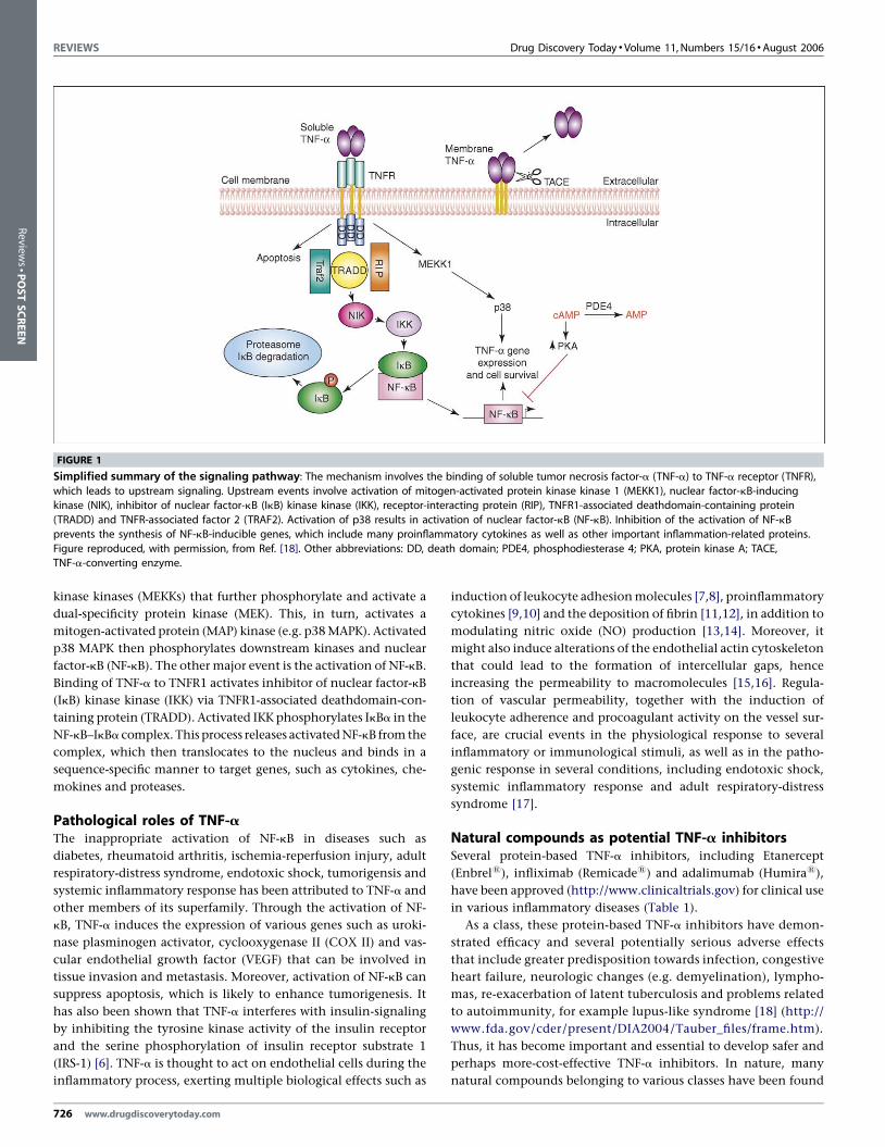
TNF-a signal transductionTNF-a signaling involves various pathwaysand signalingmolecules,
and this makes it an interesting and complex process to investigate
(Figure 1). Binding of TNF-a to TNFR1 initiates a cascade of events
involving the activation of a series of mitogen-activated protein
www.drugdiscoverytoday.com 725
REVIEWS Drug Discovery Today �Volume 11, Numbers 15/16 �August 2006
FIGURE 1
Simplified summary of the signaling pathway: The mechanism involves the binding of soluble tumor necrosis factor-a (TNF-a) to TNF-a receptor (TNFR),
which leads to upstream signaling. Upstream events involve activation of mitogen-activated protein kinase kinase 1 (MEKK1), nuclear factor-kB-inducingkinase (NIK), inhibitor of nuclear factor-kB (IkB) kinase kinase (IKK), receptor-interacting protein (RIP), TNFR1-associated deathdomain-containing protein
(TRADD) and TNFR-associated factor 2 (TRAF2). Activation of p38 results in activation of nuclear factor-kB (NF-kB). Inhibition of the activation of NF-kB
prevents the synthesis of NF-kB-inducible genes, which include many proinflammatory cytokines as well as other important inflammation-related proteins.
Figure reproduced, with permission, from Ref. [18]. Other abbreviations: DD, death domain; PDE4, phosphodiesterase 4; PKA, protein kinase A; TACE,TNF-a-converting enzyme.
Review
s�P
OSTSCREEN
kinase kinases (MEKKs) that further phosphorylate and activate a
dual-specificity protein kinase (MEK). This, in turn, activates a
mitogen-activated protein (MAP) kinase (e.g. p38 MAPK). Activated
p38 MAPK then phosphorylates downstream kinases and nuclear
factor-kB (NF-kB). The other major event is the activation of NF-kB.
Binding of TNF-a to TNFR1 activates inhibitor of nuclear factor-kB
(IkB) kinase kinase (IKK) via TNFR1-associated deathdomain-con-
taining protein (TRADD). Activated IKK phosphorylates IkBa in the
NF-kB–IkBa complex. This process releases activated NF-kB from the
complex, which then translocates to the nucleus and binds in a
sequence-specific manner to target genes, such as cytokines, che-
mokines and proteases.
Pathological roles of TNF-aThe inappropriate activation of NF-kB in diseases such as
diabetes, rheumatoid arthritis, ischemia-reperfusion injury, adult
respiratory-distress syndrome, endotoxic shock, tumorigensis and
systemic inflammatory response has been attributed to TNF-a and
other members of its superfamily. Through the activation of NF-
kB, TNF-a induces the expression of various genes such as uroki-
nase plasminogen activator, cyclooxygenase II (COX II) and vas-
cular endothelial growth factor (VEGF) that can be involved in
tissue invasion and metastasis. Moreover, activation of NF-kB can
suppress apoptosis, which is likely to enhance tumorigenesis. It
has also been shown that TNF-a interferes with insulin-signaling
by inhibiting the tyrosine kinase activity of the insulin receptor
and the serine phosphorylation of insulin receptor substrate 1
(IRS-1) [6]. TNF-a is thought to act on endothelial cells during the
inflammatory process, exerting multiple biological effects such as
726 www.drugdiscoverytoday.com
induction of leukocyte adhesion molecules [7,8], proinflammatory
cytokines [9,10] and the deposition of fibrin [11,12], in addition to
modulating nitric oxide (NO) production [13,14]. Moreover, it
might also induce alterations of the endothelial actin cytoskeleton
that could lead to the formation of intercellular gaps, hence
increasing the permeability to macromolecules [15,16]. Regula-
tion of vascular permeability, together with the induction of
leukocyte adherence and procoagulant activity on the vessel sur-
face, are crucial events in the physiological response to several
inflammatory or immunological stimuli, as well as in the patho-
genic response in several conditions, including endotoxic shock,
systemic inflammatory response and adult respiratory-distress
syndrome [17].
Natural compounds as potential TNF-a inhibitorsSeveral protein-based TNF-a inhibitors, including Etanercept
(Enbrel1), infliximab (Remicade1) and adalimumab (Humira1),
have been approved (http://www.clinicaltrials.gov) for clinical use
in various inflammatory diseases (Table 1).
As a class, these protein-based TNF-a inhibitors have demon-
strated efficacy and several potentially serious adverse effects
that include greater predisposition towards infection, congestive
heart failure, neurologic changes (e.g. demyelination), lympho-
mas, re-exacerbation of latent tuberculosis and problems related
to autoimmunity, for example lupus-like syndrome [18] (http://
www.fda.gov/cder/present/DIA2004/Tauber_files/frame.htm).
Thus, it has become important and essential to develop safer and
perhaps more-cost-effective TNF-a inhibitors. In nature, many
natural compounds belonging to various classes have been found
Drug Discovery Today � Volume 11, Numbers 15/16 �August 2006 REVIEWS
TABLE 1
Current status of some of the protein-based tumor necrosisfactor-a (TNF-a) inhibitorsa
Drugs Indications Status
Etanercept Asthma Phase IIAnkylosing spondylitis Phase IV
Infliximab Ankylosing spondylitis Approved
Crohn’s disease Approved
Dermatomyositis Phase II
Polymyositis Phase II
Adalimumab Psoriatic arthritis Phase IV
TNFR:Fc Uveitis Phase III
Arthritis
Juvenile rheumatoid arthritis
Golimumab Ankylosing spondylitis Phase IIIa Data source: http://www.clinicaltrials.gov.
Reviews�POSTSCREEN
to reduce TNF-a levels. These natural compounds (Figures 2 and 3;
Table 2) have been found to interfere with various proinflamma-
tory mediators and upstream targets, such as NF-kB and other
signaling molecules, involved in TNF-a expression and, thus,
could provide an alternative means of treating inflammatory
disease by modulating production, rather than activity, of TNF-a.
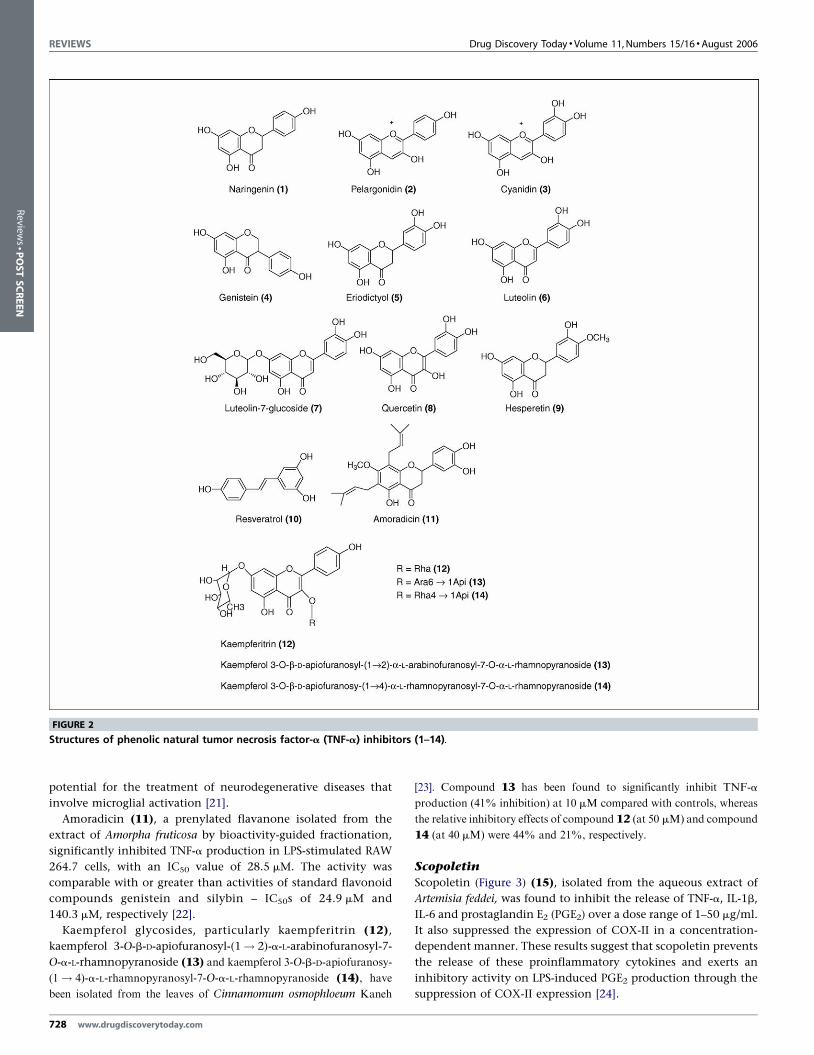
Polyphenolic modulators of TNF-a actionFlavonoids are naturally occurring polyphenolic compounds
(Figure 2) found throughout the plant kingdom. Flavonoids possess
a wide-range of biological activities (e.g. in cancer, as antioxidants)
in addition to their anti-inflammatory properties. It has been
observed that flavones, flavonols and chalcones are potent inhibi-
tors of the production of TNF-a. Flavanones naringenin (1),
anthocyanidin, pelargonidin (2) and cyanidin (3) exhibit moderate
TABLE 2
Natural compounds interfering with proinflammatory mediators an
Mediators and targets Compounds that
reduce synthesis
Compounds
that reducerelease
Tumor necrosis factor-a (TNF-a) 1,2,3,4,11,12,13,14,16,20,21,22,23,25,26,28,30
4,6,7,8,9,10,15,17,18,19,22,25,33
Interleukin-1b (IL-1b) 16,28,30 15
IL-6 20,30 4,6,7,8,15
Nitric oxide (NO) 21,22,23,25,29,30 25
Nuclear factor-kB (NF-kB)
Inducible nitric oxide synthase (iNOS) 33
Cyclooxygenase-II (COX-II)
c-fos and/or c-jun 32
p38 Mitogen-activated kinase (MAPK)
Prostaglandin E2 (PGE2) 21,25 15,25
c-Jun amino-terminal kinase (JNK) 32
Peroxisome proliferator-
ctivated receptor-g (PPARg)
a See Figures 2 and 3 for compound structures, listed here by their corresponding numbers.
inhibitory activity. By contrast, genistein (4), an isoflavone, possesses
weak inhibitory properties, whereas eriodictyol (5), another flavanone,
was found to be inactive. Furthermore, it was found that the double
bond between carbons 2 and 3, as well as the ketone group at position 4,
of flavonoids might be necessary for a potent TNF-a inhibitory activity
[19].
However, eriodictyol (5), which was previously found to be
inactive in the inhibition of TNF-a synthesis, was found to be
capable of inhibiting TNF-a release. Luteolin (6), luteolin-7-
glucoside (7), quercetin (8) and the isoflavonoid genistein all inhibited
lipopolysaccharide (LPS)-stimulated TNF-a and interleukin-6 (IL-6)
release, in RAW 264.7 cells. Hesperetin (9), however, only inhibited
TNF-a release. Luteolin and quercetin were the most potent at
inhibiting cytokine production, with IC50s <1 mM and <5 mM,
respectively, for TNF-a release. Pretreatment of the cells with luteolin
was found to attenuate LPS-induced tyrosine phosphorylation of
various proteins. Moreover, luteolin has been found to inhibit LPS-
induced phosphorylation of Akt. Treatment of macrophages with LPS
resulted in increased IkBa phosphorylation and reduced levels of IkBa.
Treating cells with luteolin abolished the effects of LPS on IkBa. In
addition, luteolin also inhibited protein tyrosine phosphorylation, NF-
kB-mediated gene expression and proinflammatory cytokine produc-
tion in murine macrophages [20].
Resveratrol (10), an antioxidant phytoalexin from grapes, has
been reported to exert anti-inflammatory activities on macro-
phages. Exposure of cultured rat cortical microglia and a mouse
microglial cell line (N9) to LPS enhanced release of TNF-a and NO
from both cell types, a phenomenon that was significantly
inhibited by resveratrol. Resveratrol appears to suppress the LPS-
induced degradation of IkBa, expression of inducible NO
synthase (iNOS) and phosphorylation of p38 MAPKs in N9
microglial cells. Thus, resveratrol demonstrates a potent suppres-
sive effect on proinflammatory responses of microglia, suggesting
d upstream targets through different mechanismsa
Compounds
thatsuppress
activation
Compounds
that inhibitphosphorylation
Compounds
that inhibitexpression
Compounds
that activateexpression
20,31,33
31
16,20,24,25,29,31
10,30,31,33
15,21,31
8,32 10
8,32 8
27
www.drugdiscoverytoday.com 727
REVIEWS Drug Discovery Today �Volume 11, Numbers 15/16 �August 2006
FIGURE 2
Structures of phenolic natural tumor necrosis factor-a (TNF-a) inhibitors (1–14).
Review
s�P
OSTSCREEN
potential for the treatment of neurodegenerative diseases that
involve microglial activation [21].
Amoradicin (11), a prenylated flavanone isolated from the
extract of Amorpha fruticosa by bioactivity-guided fractionation,
significantly inhibited TNF-a production in LPS-stimulated RAW
264.7 cells, with an IC50 value of 28.5 mM. The activity was
comparable with or greater than activities of standard flavonoid
compounds genistein and silybin – IC50s of 24.9 mM and
140.3 mM, respectively [22].
Kaempferol glycosides, particularly kaempferitrin (12),
kaempferol 3-O-b-D-apiofuranosyl-(1! 2)-a-L-arabinofuranosyl-7-
O-a-L-rhamnopyranoside (13) and kaempferol 3-O-b-D-apiofuranosy-
(1! 4)-a-L-rhamnopyranosyl-7-O-a-L-rhamnopyranoside (14), have
been isolated from the leaves of Cinnamomum osmophloeum Kaneh
728 www.drugdiscoverytoday.com
[23]. Compound 13 has been found to significantly inhibit TNF-a
production (41% inhibition) at 10 mM compared with controls, whereas
the relative inhibitory effects of compound 12 (at 50 mM) and compound
14 (at 40 mM) were 44% and 21%, respectively.
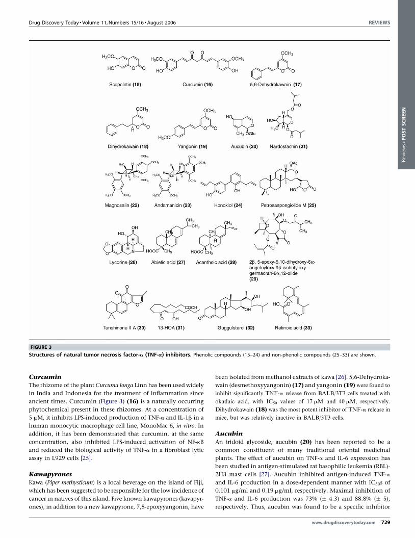
Scopoletin
Scopoletin (Figure 3) (15), isolated from the aqueous extract of
Artemisia feddei, was found to inhibit the release of TNF-a, IL-1b,
IL-6 and prostaglandin E2 (PGE2) over a dose range of 1–50 mg/ml.
It also suppressed the expression of COX-II in a concentration-
dependent manner. These results suggest that scopoletin prevents
the release of these proinflammatory cytokines and exerts an
inhibitory activity on LPS-induced PGE2 production through the
suppression of COX-II expression [24].
Drug Discovery Today � Volume 11, Numbers 15/16 �August 2006 REVIEWS
FIGURE 3
Structures of natural tumor necrosis factor-a (TNF-a) inhibitors. Phenolic compounds (15–24) and non-phenolic compounds (25–33) are shown.
Reviews�POSTSCREEN
Curcumin
The rhizome of the plant Curcuma longa Linn has been used widely
in India and Indonesia for the treatment of inflammation since
ancient times. Curcumin (Figure 3) (16) is a naturally occurring
phytochemical present in these rhizomes. At a concentration of
5 mM, it inhibits LPS-induced production of TNF-a and IL-1b in a
human monocytic macrophage cell line, MonoMac 6, in vitro. In
addition, it has been demonstrated that curcumin, at the same
concentration, also inhibited LPS-induced activation of NF-kB
and reduced the biological activity of TNF-a in a fibroblast lytic
assay in L929 cells [25].
Kawapyrones
Kawa (Piper methysticum) is a local beverage on the island of Fiji,
which has been suggested to be responsible for the low incidence of
cancer in natives of this island. Five known kawapyrones (kavapyr-
ones), in addition to a new kawapyrone, 7,8-epoxyyangonin, have
been isolated from methanol extracts of kawa [26]. 5,6-Dehydroka-
wain (desmethoxyyangonin) (17) and yangonin (19) were found to
inhibit significantly TNF-a release from BALB/3T3 cells treated with
okadaic acid, with IC50 values of 17 mM and 40 mM, respectively.
Dihydrokawain (18) was the most potent inhibitor of TNF-a release in
mice, but was relatively inactive in BALB/3T3 cells.
Aucubin
An iridoid glycoside, aucubin (20) has been reported to be a
common constituent of many traditional oriental medicinal
plants. The effect of aucubin on TNF-a and IL-6 expression has
been studied in antigen-stimulated rat basophilic leukemia (RBL)-
2H3 mast cells [27]. Aucubin inhibited antigen-induced TNF-a
and IL-6 production in a dose-dependent manner with IC50s of
0.101 mg/ml and 0.19 mg/ml, respectively. Maximal inhibition of
TNF-a and IL-6 production was 73% (� 4.3) and 88.8% (� 5),
respectively. Thus, aucubin was found to be a specific inhibitor
www.drugdiscoverytoday.com 729
REVIEWS Drug Discovery Today �Volume 11, Numbers 15/16 �August 2006
Review
s�P
OSTSCREEN
of NF-kB activation in mast cells, which might explain
its beneficial effect in the treatment of chronic allergic inflam-
matory diseases.
Nardostachin
Nardostachin (21), an iridoid isolated from Patrinia saniculaefolia,
has been found to inhibit the production of NO and TNF-a in a
dose-dependent manner, with IC50 values of 12.3 mM and
16.2 mM, respectively. In addition, this compound has been
shown to reduce expressed COX-II protein levels and PGE2
production in LPS-stimulated macrophages [28].
Magnosalin and andamanicin
Two neolignans, magnosalin (22) [1b,2a,3b,4a-1,2-dimethyl-3,4-
bis-(2,4,5-trimethoxyphenyl)-cyclobutane] and andamanicin
(23) [1a,2b,3b,4a-1,2-dimethyl-3,4-bis-(2,4,5-trimethoxyphenyl)-
cyclobutane], isolated from the leaves of Perilla frutescens, inhibited
NO synthases (IC50s = 5.9 mM and 53.5 mM, respectively) and TNF-a
in LPS-activated RAW 264.7 cells [29]. Compounds 22 and 23 have
also been tested for their ability to reduce TNF-a activity and TNF-a
levels in cell-culture media, determined in the L929 cell cytotoxicity assay.
Administration of compound 22 (10 mM) to cells produced a relative
cytotoxicity (26%), compared with LPS controls. This would suggest that
compound 22 decreased TNF-a release from activated cells and, hence,
reduced cytotoxicity against L929 cells. The inhibition of TNF-a
production by compound 22 was greater than compound 23, which
gave 84% of control cytotoxicity at a concentration of 10 mM.
Honokiol
Honokiol (24) is a lignan isolated from Magnolia officinalis that
has been shown to suppress NF-kB activation and NF-kB-regulated
gene expression through the inhibition of IKKs. Honokiol has
been found to inhibit the production of NF-kB-regulated
inflammatory and carcinogenic gene products, including
matrix metalloproteinase-9 (MMP-9), TNF-a, IL-8, intercellular
adhesion molecule 1 (ICAM-1) and monocyte chemotactic
protein-1 (MCP-1) [30].
Petrosaspongiolide M
Petrosaspongiolide M (25), a marine metabolite from the
Caledonian marine sponge Petrosaspongia nigra, reduced the
production of nitrite, PGE2 and TNF-a in a mouse air-pouch
model of inflammation [31]. It was found to be a potent inhibitor
of the NF-kB pathway at a concentration of 1 mM. Petrosaspon-
giolide M potently inhibited the release of nitrite, PGE2 and
TNF-a in a concentration-dependent manner.
EGb 761
EGb 761 [a standardized extract of Gingko biloba containing 24%
flavonoid glycosides of mainly rutin and quercetin and 6% unique
terpenes (3% bilobalide and 3% ginkgolides A, B and C)] and
quercetin, its aglycone component, have selective effects on
TNF-a and the MAPK cascade. Although both EGb 761 and quer-
cetin (8) inhibit TNF-a secretion in LPS-stimulated RAW 264.7
macrophages, the results have suggested that quercetin is unique
in its ability to inhibit TNF-a transcription by inhibiting
phosphorylation and activation of c-Jun amino-terminal kinase
(JNK)/stress-activated protein kinase (SAPK); therefore suppres-
730 www.drugdiscoverytoday.com
sing activation of the transcription factor AP-1. EGb 761 was
found to diminish LPS-induced NF-kB transcriptional activity
slightly, but it had no effect on TNF-a transcription. EGb 761 and
quercetin can also inhibit TNF-a production at the post-
transcriptional level. ERK1/2 and p38 MAPK activities, which
are important in the post-transcriptional regulation of TNF-a
mRNA, can also be inhibited by EGb 761 and quercetin [32].
Other chemical classes capable of modulating TNF-aactionAlkaloidsLycorine (26) and lycoricidinol inhibited TNF-a production from
murine macrophages stimulated with LPS (with IC50 values of
0.2 mg/ml and 0.002 mg/ml, respectively). Lycorine and lycorici-
dinol have also been reported to inhibit protein biosynthesis – at
1 mg/ml and 0.008 mg/ml. Although the inhibition of TNF-a
production by lycoricidinol was mainly caused by an overall,
non-selective inhibition of protein biosynthesis, lycorine was
capable of inhibiting TNF-a production at lower concentrations
than those required to inhibit overall protein synthesis in
macrophages. These data suggest that inhibition of TNF-a
production by lycorine and lycoricidinol is not necessarily just
caused by the inhibition of protein translation, at least at lower
concentrations [33].
TerpenesAbietic acid (27) suppresses the expression of genes involved in
inflammation, such as TNF-a and COX-II, in activated macro-
phages. At a concentration of 50 mM it inhibits TNF-a (16.3%)
and COX-II (75.6%) protein expression through the activation of
peroxisome proliferator-activated receptor-g (PPARg). Also, it was
found to regulate the expression of PPARg target genes including
aP2, LPL, and FAT/CD36 in 3T3-L1 adipocytes or RAW 264.7
macrophages [34].
Acanthoic acid (28) is (�)-pimara-9(11),15-dien-19-oic acid, a
pimaradiene diterpene isolated from the Korean medicinal plant
Acanthopanax koreanum [35]. Acanthoic acid (0.1–10.0 mg/ml)
inhibited the production of IL-1b and TNF-a by up to 90% in
human monocytes and macrophages stimulated with silica, but
the production of IL-6 was not inhibited at all. At these
concentrations there were no cytotoxic effects on human
monocytes and macrophages.
The compound 2b,5-epoxy-5,10-dihydroxy-6a-angeloyloxy-9b-
isobutyloxy-germacran-8a,12-olide (29), another terpene identi-
fied as sesquiterpene lactone from Carpesium divaricatum, also
decreased NO production in LPS–IFN-g-stimulated RAW 264.7
cells in a concentration-dependent manner, with an IC50 of
�2.16 mM; however it was found to have no direct effect on the
iNOS activity of fully LPS–IFN-g-stimulated RAW 264.7 cells.
Treating cells with compound 29 resulted in reduced levels of iNOS
protein and mRNA. These effects appeared to be caused by inhibition
of NF-kB activation through a mechanism involving the concomitant
stabilization of the NF-kB–IkBa complex, followed by a reduction in
nuclear translocation of the p65 subunit of the NF-kB–IkBa complex
[36].
Tanshinone II A (30), a diterpene isolated from Salvia
miltiorrhiza root, inhibited the production of TNF-a, IL-1b and
IL-6 in activated RAW 264.7 cells in a dose-dependent manner
Drug Discovery Today � Volume 11, Numbers 15/16 �August 2006 REVIEWS
Reviews�POSTSCREEN
(0.34–34.0 mM). It also inhibited the expression of iNOS (3.4–
34.0 mM) in a dose-dependent manner, and NO was also inhibited
with an IC50 of 20 mM [37].
Fatty acids and their derivativesThe functionally novel fatty acid (�)-13-hydroxy-10-oxo-trans-11-
octadecenoic acid (13-HOA) (31), derived from linoleic acid (LA)
by corn and rice lipoxygenase (LOX), markedly attenuates the
expression of proinflammatory genes in LPS-stimulated macro-
phages via a blockade of the NF-kB and AP-1 pathways [38]. At
higher concentrations, 10-ODO, 13-HOA and 9-HOA exhibited
profound suppressive effects on the expression of iNOS, COX-II,
IL-6 and TNF-a, with the following sensitivity: COX-II > IL-
6 > iNOS > TNF-a. The ability of 13-HOA to attenuate highly the
expression of proinflammatory genes such as COX-II, iNOS, TNF-
a and IL-6 makes it an important lead for the development of
TNF-a inhibitors.
SterolsThe anti-inflammatory properties of Commiphora mukul gum have
been known since ancient times, and it has been used in various
traditional systems, including Ayurveda and traditional Chinese
medicine (TCM). It has been possible to demonstrate downregula-
tion of inflammatory mediators such as interferon-g (IFN-g), IL-12,
TNF-a, IL-1b and NO following administration of an ethyl acetate
extract of the gum [39]. Guggulsterol (32), isolated from this gum
extract, did not inhibit MAP kinase (ERK), but it could reduce c-fos
and c-jun mRNA levels in phorbol 12-myristate 13-acetate (PMA)-
stimulated cells. This reduction in c-fos and c-jun levels, taken
together with the inhibition of MAPK activation, provides a
possible mechanism by which crude ethyl acetate extracts and
purified guggulsterol might exert their actions.
RetinoidsRetinoic acid (33), an active metabolite of vitamin A, attenuated
TNF-a (29–97%) and iNOS (61–96%) mRNA expression in
microglia exposed to either b-amyloid peptide (Ab) or LPS, in a
dose-dependent manner (0.1–10.0 mM). The inhibition of TNF-a
and iNOS mRNA expression in activated microglia, induced by
retinoic acid (33), was accompanied by a concomitant reduction in
the release of iNOS and TNF-a [40].
ConclusionsThe clinically approved protein-based TNF-a inhibitors are cap-
able of reducing TNF-a activity, but can have serious side effects.
The recently reported side effects of the blockbuster COX-II
inhibitor (COXIB) series of non-steroidal anti-inflammatory
drugs (NSAIDS) (http://www.fda.gov/cder/drug/infopage/
COX2/default.htm), combined with other disadvantages of these
protein-based anti-TNF-a drugs, has been a driver for the natural-
product chemist to search for and develop alternatives. Low-
molecular-weight natural compounds can have many advan-
tages, not least cost-effectiveness (compared with current pro-
tein-based drugs) and route of administration. Many natural
compounds, particularly the phenolics, terpenes and, to a lesser
extent, alkaloids, have been found to inhibit the upstream
signaling molecules that are involved in TNF-a expression. To
increase the number of leads from the natural-compound libraries
for TNF-a modulating activities, there is a need to develop HTS
protocols. Further, for better understanding of natural-compound
library SAR, extensive in silico studies can and should be carried out.
Thus, drugs derived from natural-compound leads, either alone or
in combination (synergistically) might provide an alternative
approach for the treatment of inflammatory diseases via modula-
tion of the TNF-a signaling pathway.
References
1 Carswell, E.A. et al. (1975) An endotoxin-induced serum factor that causes necrosis
of tumors. Proc. Natl. Acad. Sci. U. S. A. 72, 3666–3670
2 Wiemann, B. and Starnes, C.O. (1994) Coley’s toxins, tumor necrosis factor and
cancer research: a historical perspective. Pharmacol. Ther. 64, 529–564
3 Perez, C. et al. (1990) A nonsecretable cell surface mutant of tumor necrosis factor
(TNF) kills by cell-to-cell contact. Cell 63, 251–258
4 Jones, E.Y. et al. (1989) Structure of tumour necrosis factor. Nature 338, 225–228
5 Meager, A. (1991) Cytokines.. Prentice Hall
6 Hotamisligil, G.S. et al. (1994) Tumor necrosis factor alpha inhibits signaling from
the insulin receptor. Proc. Natl. Acad. Sci. U. S. A. 91, 4854–4858
7 Pober, J.S. et al. (1987) Activation of cultured human endothelial cells by
recombinant lymphotoxin: comparison with tumor necrosis factor and interleukin
1 species. J. Immunol. 138, 3319–3324
8 Slowik, M.R. et al. (1993) Tumor necrosis factor activates human endothelial cells
through the p55 tumor necrosis factor receptor but the p75 receptor contributes to
activation at low tumor necrosis factor concentration. Am. J. Pathol. 143, 1724–1730
9 Jirik, F.R. et al. (1989) Bacterial lipopolysaccharide and inflammatory mediators
augment IL-6 secretion by human endothelial cells. J. Immunol. 142, 144–147
10 Nawroth, P.P. et al. (1986) Tumor necrosis factor/cachectin interacts with
endothelial cell receptors to induce release of interleukin 1. J. Exp. Med. 163, 1363–
1375
11 Nawroth, P. et al. (1988) Tumor necrosis factor/cachectin-induced intravascular
fibrin formation in meth A fibrosarcomas. J. Exp. Med. 168, 637–647
12 Nawroth, P.P. and Stern, D.M. (1986) Modulation of endothelial cell hemostatic
properties by tumor necrosis factor. J. Exp. Med. 163, 740–745
13 Estrada, C. et al. (1992) Nitric oxide mediates tumor necrosis factor-alpha
cytotoxicity in endothelial cells. Biochem. Biophys. Res. Commun. 186, 475–482
14 Yoshizumi, M. et al. (1993) Tumor necrosis factor downregulates an endothelial
nitric oxide synthase mRNA by shortening its half-life. Circ. Res. 73, 205–209
15 Brett, J. et al. (1989) Tumor necrosis factor/cachectin increases permeability of
endothelial cell monolayers by a mechanism involving regulatory G proteins. J. Exp.
Med. 169, 1977–1991
16 Goldblum, S.E. and Sun, W.L. (1990) Tumor necrosis factor-alpha augments
pulmonary arterial transendothelial albumin flux in vitro. Am. J. Physiol. 258, L57–L67
17 Tracey, K.J. and Cerami, A. (1994) Tumor necrosis factor: a pleiotropic cytokine and
therapeutic target. Annu. Rev. Med. 45, 491–503
18 Palladino, M.A. et al. (2003) Anti-TNF-alpha therapies: the next generation. Nat. Rev.
Drug Discov. 2, 736–746
19 Herath, H.M. et al. (2003) Inhibitory effect of some flavonoids on tumor necrosis
factor-alpha production in lipopolysaccharide-stimulated mouse macrophage cell
line J774.1. J. Med. Food 6, 365–370
20 Xagorari, A. et al. (2001) Luteolin inhibits an endotoxin-stimulated
phosphorylation cascade and proinflammatory cytokine production in
macrophages. J. Pharmacol. Exp. Ther. 296, 181–187
21 Bi, X.L. et al. (2005) Resveratrol inhibits nitric oxide and TNF-alpha production by
lipopolysaccharide-activated microglia. Int. Immunopharmacol. 5, 185–193
22 Cho, J.Y. et al. (2000) Inhibitor of tumor necrosis factor-alpha production in
lipopolysaccharide-stimulated RAW264.7 cells from Amorpha fruticosa. J.
Ethnopharmacol. 70, 127–133
23 Fang, S.H. et al. (2005) Inhibitory effects of flavonol glycosides from Cinnamomum
osmophloeum on inflammatory mediators in LPS/IFN-gamma-activated murine
macrophages. Bioorg. Med. Chem. 13, 2381–2388
24 Kim, H.J. et al. (2004) Scopoletin suppresses pro-inflammatory cytokines and PGE2
from LPS-stimulated cell line, RAW 264.7 cells. Fitoterapia 75, 261–266
www.drugdiscoverytoday.com 731
REVIEWS Drug Discovery Today �Volume 11, Numbers 15/16 �August 2006
Review
s�P
OSTSCREEN
25 Chan, M.M. (1995) Inhibition of tumor necrosis factor by curcumin, a
phytochemical. Biochem. Pharmacol. 49, 1551–1556
26 Hashimoto, T. et al. (2003) Isolation and synthesis of TNF-alpha release inhibitors
from Fijian kawa (Piper methysticum). Phytomedicine 10, 309–317
27 Jeong, H.J. et al. (2002) Inhibition of TNF-alpha and IL-6 production by Aucubin
through blockade of NF-kappaB activation RBL-2H3 mast cells. Cytokine 18, 252–259
28 Ju, H.K. et al. (2003) Inhibitory effects of nardostachin on nitric oxide,
prostaglandin E2, and tumor necrosis factor-alpha production in
lipopolysaccharide activated macrophages. Biol. Pharm. Bull. 26, 1375–1378
29 Ryu, J.H. et al. (2002) Two neolignans from Perilla frutescens and their inhibition of
nitric oxide synthase and tumor necrosis factor-alpha expression in murine
macrophage cell line RAW 264.7. Bioorg. Med. Chem. Lett. 12, 649–651
30 Tse, A.K. et al. (2005) Honokiol inhibits TNF-alpha-stimulated NF-kappaB activation
and NF-kappaB-regulated gene expression through suppression of IKK activation.
Biochem. Pharmacol. 70, 1443–1457
31 Posadas, I. et al. (2003) Inhibition of the NF-kappaB signaling pathway mediates the
anti-inflammatory effects of petrosaspongiolide M. Biochem. Pharmacol. 65, 887–895
32 Wadsworth, T.L. et al. (2001) Effects of Ginkgo biloba extract (EGb 761) and
quercetin on lipopolysaccharide-induced signaling pathways involved in the
release of tumor necrosis factor-alpha. Biochem. Pharmacol. 62, 963–974
33 Yui, S. et al. (2001) Inhibition effect of amaryllidaceae alkaloids, lycorine and
lycoricidinol on macrophage TNF- a production. Yakugaku Zasshi 121, 167–171
The ScienceDire
ScienceDirect’s extensive and unique full-text collection co
The Lancet, Cell, Tetrahedron and the full suite of Trends
With ScienceDirect, the research process is enhanced w
all on a single, intu
The rapid growth of the ScienceDirect collection is a result of the
addition to the Backfiles - heritage collections in a number o
digitize all of Elsevier’s journals back to volume one, iss
journal collection on ScienceDirect. Also available online fo
containing more than 12,000 articles that highlight impor
For more information, visit
732 www.drugdiscoverytoday.com
34 Takahashi, N. et al. (2003) Abietic acid activates peroxisome proliferator-activated
receptor-gamma (PPARgamma) in RAW264.7 macrophages and 3T3-L1 adipocytes
to regulate gene expression involved in inflammation and lipid metabolism. FEBS
Lett. 550, 190–194
35 Kang, H.S. et al. (1996) Suppression of interleukin-1 and tumor necrosis factor-alpha
production by acanthoic acid, (�)-pimara-9(11),15-dien-19-oic acid, and it
antifibrotic effects in vivo. Cell. Immunol. 170, 212–221
36 Kim, E.J. et al. (2001) Suppression by a sesquiterpene lactone from Carpesium
divaricatum of inducible nitric oxide synthase by inhibiting nuclear factor-kappaB
activation. Biochem. Pharmacol. 61, 903–910
37 Jang, S.I. et al. (2003) Tanshinone IIA from Salvia miltiorrhiza inhibits inducible
nitric oxide synthase expression and production of TNF-alpha, IL-1beta and IL-6 in
activated RAW 264.7 cells. Planta Med. 69, 1057–1059
38 Murakami, A. et al. (2005) New class of linoleic acid metabolites biosynthesized by
corn and rice lipoxygenases: suppression of proinflammatory mediator expression
via attenuation of MAPK- and Akt-, but not PPARgamma-, dependent pathways in
stimulated macrophages. Biochem. Pharmacol. 70, 1330–1342
39 Manjula, N. et al. (2006) Inhibition of MAP kinases by crude extract and pure
compound isolated from Commiphora mukul leads to down regulation of TNF-alpha,
IL-1beta and IL-2. Int. Immunopharmacol. 6, 122–132
40 Dheen, S.T. et al. (2005) Retinoic acid inhibits expression of TNF-alpha and iNOS in
activated rat microglia. Glia 50, 21–31
ct collection
vers more than 1900 journals, including titles such as
, Current Opinion and Drug Discovery Today journals.
ith unsurpassed searching and linking functionality,
itive interface.
integration of several prestigious publications and the ongoing
f disciplines. The latest step in this ambitious project to
ue one, is the addition of the highly cited Cell Press
r the first time are six Cell titles’ long-awaited Backfiles,
tant historic developments in the field of life sciences.
www.sciencedirect.com
Related Documents