A Genome-Wide RNA Interference Screen Identifies Caspase 4 as a Factor Required for Tumor Necrosis Factor Alpha Signaling Dorothee Nickles, Christina Falschlehner, Marie Metzig, and Michael Boutros German Cancer Research Center (DKFZ), Division of Signaling and Functional Genomics, and Heidelberg University, Department of Cell and Molecular Biology, Medical Faculty Mannheim, Heidelberg, Germany Tumor necrosis factor alpha (TNF-) is a potent inflammatory cytokine secreted upon cellular stress as well as immunological stimuli and is implicated in the pathology of inflammatory diseases and cancer. The therapeutic potential of modifying TNF- pathway activity has been realized in several diseases, and antagonists of TNF- have reached clinical applications. While much progress in the understanding of signaling downstream of the TNF- receptor complex has been made, the compendium of fac- tors required for signal transduction is still not complete. In order to find novel regulators of proinflammatory signaling in- duced by TNF-, we conducted a genome-wide small interfering RNA screen in human cells. We identified several new candidate modulators of TNF- signaling, which were confirmed in independent experiments. Specifically, we show that caspase 4 is re- quired for the induction of NF-B activity, while it appears to be dispensable for the activation of the Jun N-terminal protein kinase signaling branch. Taken together, our experiments identify caspase 4 as a novel regulator of TNF--induced NF-B sig- naling that is required for the activation of IB kinase. We further provide the genome-wide RNA interference data set as a com- pendium in a format compliant with minimum information about an interfering RNA experiment (MAIRE). I nflammation is essential for an efficient innate immune re- sponse, helping to alert the body to potential intruders and en- abling immune cells to access the site of an infection. However, when inflammatory processes become chronic or systemic, tissue damage and diseases can arise (e.g., Crohn’s disease or psoriasis) (12, 30). The cytokine tumor necrosis factor alpha (TNF-) is the major mediator of inflammation (4). TNF- can bind to both TNF- receptor 1 (TNFR1) and TNFR2. Upon binding of TNF- to TNFR1, it induces an intracellular signaling cascade that can in- duce either inflammation or apoptosis, depending on the cell type. Molecularly, the ligand-receptor complex first recruits TRADD and TRAF2/5, followed by cellular inhibitors of apoptosis protein (cIAPs). cIAPs are responsible for forming K63- and K11-linked ubiquitin chains on RIP1 (23, 29, 55, 58). These lead to the recruit- ment of the linear ubiquitin chain assembly complex (LUBAC) and the linear ubiquitination of RIP1, NEMO, and possibly other components (59). The ubiquitin chains on RIP1 allow binding of further signaling factors, leading to the activation of NF-B (through IB kinase [IKK]) and AP-1 (through mitogen-acti- vated protein kinase/Jun N-terminal protein kinase [JNK]) tran- scription factors (59). Recently, mass spectrometric analysis revealed that LUBAC is an essential regulator of TNF- receptor complex ubiquitination (19, 24). In addition, RNA interference (RNAi) screens identified several novel TNF- signaling components, including the cylin- dromatosis tumor suppressor (CYLD) (13) in human cells and IAP2 and akirins as conserved modulators of TNF--like signal- ing pathways in Drosophila (20, 22). Yet, to date, no RNAi screen for TNF--induced activation of NF-B covering the complete human genome has been reported (13, 16, 17, 36, 41, 65). Here, we present the results of a functional genomic screen with the aim to identify novel regulators of TNF- signaling. We established a quantitative assay to measure NF-B signaling activ- ity after TNF- stimulation and screened a genome-wide small interfering RNA (siRNA) library in human cells. This approach identified several novel candidates that were confirmed with inde- pendent siRNAs and in independent cell lines. Specifically, we focused on caspase 4 (CASP4), which is required for robust acti- vation of NF-B. Transcriptional profiling showed that CASP4 is required for the expression of endogenous NF-B target genes. We used epistasis analysis to map the role of CASP4 upstream of or at the level of IKK activation. Taken together, our experiments identified CASP4 as a novel positive regulator of TNF--induced NF-B signaling. Furthermore, we provide the full RNAi screen- ing data set as a resource for further exploration. MATERIALS AND METHODS Cell lines and reagents. Human embryonic kidney 293T (HEK293T), HeLa, and HepG2 cells were kindly provided by C. Niehrs (DKFZ) and T. Dick (DKFZ). Cells were cultured in Dulbecco’s modified Eagle’s medium (DMEM; Gibco) supplemented with 10% fetal calf serum (FCS; Gibco). TNF- was obtained from Biosource. The sequences of the siRNAs used are listed in Table S1 supplemental material. Plasmids. In order to monitor NF-B transcriptional activity, a cell- based dual-luciferase assay in HEK293T cells was established. As a path- way-specific reporter, an NF-B-dependent firefly luciferase (FL) expres- sion plasmid (4-4-FL) was cloned. Eight NF-B binding sites (8 5=-GG ACTTTCC-3=, in concordance with the degenerate NF-B binding site Received 20 December 2011 Returned for modification 13 January 2012 Accepted 12 June 2012 Published ahead of print 25 June 2012 Address correspondence to Michael Boutros, [email protected]. Present address: Dorothee Nickles, Department of Neurology, University of California San Francisco, San Francisco, California, USA; Marie Metzig, Heidelberg University, Institute of Pathology, Heidelberg, Germany. Supplemental material for this article may be found at http://mcb.asm.org/. Copyright © 2012, American Society for Microbiology. All Rights Reserved. doi:10.1128/MCB.06739-11 The authors have paid a fee to allow immediate free access to this article. 3372 mcb.asm.org Molecular and Cellular Biology p. 3372–3381 September 2012 Volume 32 Number 17 Downloaded from https://journals.asm.org/journal/mcb on 02 January 2022 by 77.87.152.177.

Welcome message from author
This document is posted to help you gain knowledge. Please leave a comment to let me know what you think about it! Share it to your friends and learn new things together.
Transcript
A Genome-Wide RNA Interference Screen Identifies Caspase 4 as aFactor Required for Tumor Necrosis Factor Alpha Signaling
Dorothee Nickles, Christina Falschlehner, Marie Metzig, and Michael Boutros
German Cancer Research Center (DKFZ), Division of Signaling and Functional Genomics, and Heidelberg University, Department of Cell and Molecular Biology, MedicalFaculty Mannheim, Heidelberg, Germany
Tumor necrosis factor alpha (TNF-�) is a potent inflammatory cytokine secreted upon cellular stress as well as immunologicalstimuli and is implicated in the pathology of inflammatory diseases and cancer. The therapeutic potential of modifying TNF-�pathway activity has been realized in several diseases, and antagonists of TNF-� have reached clinical applications. While muchprogress in the understanding of signaling downstream of the TNF-� receptor complex has been made, the compendium of fac-tors required for signal transduction is still not complete. In order to find novel regulators of proinflammatory signaling in-duced by TNF-�, we conducted a genome-wide small interfering RNA screen in human cells. We identified several new candidatemodulators of TNF-� signaling, which were confirmed in independent experiments. Specifically, we show that caspase 4 is re-quired for the induction of NF-�B activity, while it appears to be dispensable for the activation of the Jun N-terminal proteinkinase signaling branch. Taken together, our experiments identify caspase 4 as a novel regulator of TNF-�-induced NF-�B sig-naling that is required for the activation of I�B kinase. We further provide the genome-wide RNA interference data set as a com-pendium in a format compliant with minimum information about an interfering RNA experiment (MAIRE).
Inflammation is essential for an efficient innate immune re-sponse, helping to alert the body to potential intruders and en-
abling immune cells to access the site of an infection. However,when inflammatory processes become chronic or systemic, tissuedamage and diseases can arise (e.g., Crohn’s disease or psoriasis)(12, 30).
The cytokine tumor necrosis factor alpha (TNF-�) is the majormediator of inflammation (4). TNF-� can bind to both TNF-�receptor 1 (TNFR1) and TNFR2. Upon binding of TNF-� toTNFR1, it induces an intracellular signaling cascade that can in-duce either inflammation or apoptosis, depending on the cell type.Molecularly, the ligand-receptor complex first recruits TRADDand TRAF2/5, followed by cellular inhibitors of apoptosis protein(cIAPs). cIAPs are responsible for forming K63- and K11-linkedubiquitin chains on RIP1 (23, 29, 55, 58). These lead to the recruit-ment of the linear ubiquitin chain assembly complex (LUBAC)and the linear ubiquitination of RIP1, NEMO, and possibly othercomponents (59). The ubiquitin chains on RIP1 allow binding offurther signaling factors, leading to the activation of NF-�B(through I�B kinase [IKK]) and AP-1 (through mitogen-acti-vated protein kinase/Jun N-terminal protein kinase [JNK]) tran-scription factors (59).
Recently, mass spectrometric analysis revealed that LUBAC isan essential regulator of TNF-� receptor complex ubiquitination(19, 24). In addition, RNA interference (RNAi) screens identifiedseveral novel TNF-� signaling components, including the cylin-dromatosis tumor suppressor (CYLD) (13) in human cells andIAP2 and akirins as conserved modulators of TNF-�-like signal-ing pathways in Drosophila (20, 22). Yet, to date, no RNAi screenfor TNF-�-induced activation of NF-�B covering the completehuman genome has been reported (13, 16, 17, 36, 41, 65).
Here, we present the results of a functional genomic screenwith the aim to identify novel regulators of TNF-� signaling. Weestablished a quantitative assay to measure NF-�B signaling activ-ity after TNF-� stimulation and screened a genome-wide smallinterfering RNA (siRNA) library in human cells. This approach
identified several novel candidates that were confirmed with inde-pendent siRNAs and in independent cell lines. Specifically, wefocused on caspase 4 (CASP4), which is required for robust acti-vation of NF-�B. Transcriptional profiling showed that CASP4 isrequired for the expression of endogenous NF-�B target genes.We used epistasis analysis to map the role of CASP4 upstream ofor at the level of IKK activation. Taken together, our experimentsidentified CASP4 as a novel positive regulator of TNF-�-inducedNF-�B signaling. Furthermore, we provide the full RNAi screen-ing data set as a resource for further exploration.
MATERIALS AND METHODSCell lines and reagents. Human embryonic kidney 293T (HEK293T),HeLa, and HepG2 cells were kindly provided by C. Niehrs (DKFZ) and T.Dick (DKFZ). Cells were cultured in Dulbecco’s modified Eagle’s medium(DMEM; Gibco) supplemented with 10% fetal calf serum (FCS; Gibco).TNF-� was obtained from Biosource. The sequences of the siRNAs usedare listed in Table S1 supplemental material.
Plasmids. In order to monitor NF-�B transcriptional activity, a cell-based dual-luciferase assay in HEK293T cells was established. As a path-way-specific reporter, an NF-�B-dependent firefly luciferase (FL) expres-sion plasmid (4-4-FL) was cloned. Eight NF-�B binding sites (8� 5=-GGACTTTCC-3=, in concordance with the degenerate NF-�B binding site
Received 20 December 2011 Returned for modification 13 January 2012Accepted 12 June 2012
Published ahead of print 25 June 2012
Address correspondence to Michael Boutros, [email protected].
Present address: Dorothee Nickles, Department of Neurology, University ofCalifornia San Francisco, San Francisco, California, USA; Marie Metzig, HeidelbergUniversity, Institute of Pathology, Heidelberg, Germany.
Supplemental material for this article may be found at http://mcb.asm.org/.
Copyright © 2012, American Society for Microbiology. All Rights Reserved.
doi:10.1128/MCB.06739-11
The authors have paid a fee to allow immediate free access to this article.
3372 mcb.asm.org Molecular and Cellular Biology p. 3372–3381 September 2012 Volume 32 Number 17
Dow
nloa
ded
from
http
s://j
ourn
als.
asm
.org
/jour
nal/m
cb o
n 02
Jan
uary
202
2 by
77.
87.1
52.1
77.
5=-GGGRNWYYCC-3=, where G stands for a purine base, N denotes anybase, W is an adenine or thymine, Y denotes a pyrimidine base [15, 35,51]) were fused upstream of the firefly luciferase gene (pGL3 promoter,Promega). A constitutively expressed Renilla luciferase (RL) driven fromthe �-actin promoter (act-RL) served as a coreporter for transfectionefficiency and cell viability. The �-actin promoter, an approximately1,100-bp sequence 5= of the transcription start site of the �-actin gene, wasamplified by PCR and then ligated into pRL null (Promega). Results ofproof-of-principle experiments are shown in Fig. S1A to C in the supple-mental material. The JUN reporter plasmid was kindly provided by P.Krammer (DKFZ) (38).
Transfections and luciferase assays. siRNAs (final concentration, 50nM) were transfected by reverse transfection using DharmaFECT1 trans-fection reagent (Dharmacon/Thermo Fisher Scientific) following themanufacturer’s instructions. After 24 h, plasmids were transfected usingTransIT transfection reagent (Mirrus), and then plasmid expression wasallowed for 48 h. Sixteen hours (HEK293T) or 24 h (HepG2) prior to theluciferase readout, cells were stimulated with 20 ng (HEK293T) or 50 ng(HepG2) per ml TNF-� (final concentrations). For overexpression stud-ies, HEK293T or HepG2 cells were reverse transfected with siRNAs usingthe DharmaFECT1 reagent (Dharmacon/Thermo Fisher Scientific) fol-lowing the manufacturer’s instructions. Twenty-four hours later, cellswere transfected with plasmids pEF-DEST51-Caspase4 (based on thepEF-DEST51 gateway vector; Invitrogen) or empty control using TransITtransfection reagent (Mirrus), allowing plasmid expression for 48 h. Cellswere either left untreated or treated with TNF-� (20 ng/ml) for 2 h priorto the luciferase readout. Calcein readout was used for normalization(21).
Primary genome-wide screening. Genome-wide RNAi experimentswere performed with a synthetic siRNA library (Qiagen) in HEK293Tcells. siRNA sequences were annotated using the RefSeq database, release27. siRNA concentrations of 20 nM were used. In each well of a 384-wellplate, 0.05 �l DharmaFECT1 was gently mixed with 4.95 �l RPMI and theplate was incubated for 10 min at room temperature before another 10 �lRPMI was added. This mix was then pipetted to 5 �l 200 nM (Qiagen)siRNA (in siRNA buffer), followed by another incubation for 30 min atroom temperature. Cells (4 � 103) in 30 �l (antibiotic-free) culture me-dium (DMEM plus 10% FCS) were seeded on top of the siRNAs, and theplates were briefly spun (Centrifuge 5804; Eppendorf) to collect all liquidat the bottom of the wells and then kept in the incubator (Binder) for 3days to allow protein depletion. siRNAs against the receptor TNFRSF1A(siTNFR1) and RelA (siRelA), i.e., siRNAs that diminish activation ofNF-�B transcriptional activity, served as positive controls. An example ofthe performance of screening of replicates is shown in Fig. S2 in the sup-plemental material. The complete data set is available in Tables S2 and S3in the supplemental material.
Secondary screening. Screening results were validated by making useof deconvoluted siRNA pools (50 nM; siGENOME set of four upgrade;Dharmacon/Thermo Fisher Scientific) and siRNA pools picked from thegenome-scale siRNA library from Qiagen (20 nM). For further experi-ments, siRNAs were purchased from Dharmacon/Thermo Fisher Scien-tific and used at a concentration of 50 nM.
ELISA. For enzyme-linked immunosorbent assay (ELISA), cells werereverse transfected with siRNAs in a 384-well format. The interleukin-8(IL-8) concentration in cell culture supernatants was assessed using ahuman CXCL8/IL-8 ELISA kit (R&D) and by following the manufactur-er’s instructions exactly. Measurements were carried out in technical du-plicates in a 96-well format, pooling eight equally treated wells. HEK293Tcells were treated with 20 ng/ml TNF-� for 16 h, while HepG2 cells werestimulated with 25 ng/ml TNF-� for 24 h. The absorbance was measuredwith a Mithras LB940 plate reader (Berthold Technologies), with the filterset at 450 nm (for proof of principle, see Fig. S1D in the supplementalmaterial).
Expression profiling and qRT-PCR. Total RNA was extracted usingan RNeasy kit (Qiagen).
(i) Expression profiling. RNA quality was assessed by measuringmRNA depletion mediated by respective siRNAs and TNF-�-mediatedinduction of target genes by quantitative real time (qRT)-PCR beforeRNA was handed over to the DKFZ Genomics and Proteomics Core Fa-cilities. There, RNA was hybridized onto Illumina Human Sentrix-8chips.
(ii) qRT-PCR. One microgram of RNA was reverse transcribed usingSuperScript III reverse transcriptase (Invitrogen). cDNA correspondingto 110 ng of starting RNA was used for two technical replicates. qRT-PCRwas performed in a 384-well format using a Universal Probe Library sys-tem (Roche) and a LightCycler 480 system (Roche). HRPT1 was used asthe housekeeping gene. The sequences of the qRT-PCR primers are givenin Table S4 in the supplemental material.
Antibodies. (i) Antibiodies for immunoblotting. The anti-�-tubulinantibody was purchased from Sigma (antibody DM1A); the anti-phos-pho-I�B-� antibody (antibody 9246), the total I�B-� antibody (antibody9242), the anti-phospho-JNK antibody (antibody 4668), and the totalJNK antibody (antibody 9252) were purchased from Cell Signaling; thecaspase 4 antibody was purchased from Santa Cruz (antibody sc-1229);the �-actin antibody (antibody AC-15) was from Abcam; and secondaryantibodies were purchased from Southern Biotech (anti-mouse IgG1) andGE Healthcare (anti-mouse antibody).
(ii) Antibodies for fluorescence-activated cell sorting (FACS). Theanti-TNFRSF1A antibody (antibody MABTNFR1-B1) was purchasedfrom BD Pharmingen, ubiquitinylated anti-mouse antibody [F(ab=)2 IgG(H�L)] was from Southern Biotech, and streptavidin-phycoerythrin(PE) was from BD. The isotype control antibody was kindly provided byH. Walczak (Imperial College, London, United Kingdom).
Immunoblotting. For immunoblotting experiments, HEK293T,HeLa, or HepG2 cells were reverse transfected with siRNAs in a 24-well or12-well format. After 2 days of siRNA treatment, cells were starved for 3 to4 h by exchanging the culture medium with serum-free DMEM. Then,medium was exchanged for culture medium or 50 ng/ml TNF-�-contain-ing medium to stimulate cells for either 0, 5, 10, or 20 min. After stimu-lation, cells were trypsinized and lysed for 30 min in 70 �l lysis buffer (1�phosphate-buffered saline [PBS], 10% Triton X-100, protease and phos-phatase inhibitors). Protein concentration was determined using a bicin-choninic acid assay (Pierce), following the manufacturer’s instructions.Proteins were separated on 10% NuPage bis-Tris nongradient gels(Novex; Invitrogen) in 1� MOPS (morpholinepropanesulfonic acid)buffer (50 mM Tris base, 50 mM MOPS, 1.025 mM EDTA, 69.3 mM SDS)and transferred onto nitrocellulose membranes by wet transfer (XCell IIblot module; Invitrogen) in 2� transfer buffer (0.5 M bis Tris, 0.5 Mbicine, 0.02 M EDTA). Membranes were blocked with 5% nonfat dry milkin Tris-buffered saline (TBS)–Tween (TBS-T; 0.137 M NaCl, 0.02 M Tris-HCl, pH 7.8) before being probed with antibodies.
FACS. HeLa cells were transfected with siRNAs in a 6-well format.After 2 days of incubation to allow protein depletion, cells were scrapedoff the wells. Cell pellets were resuspended in 50 �l FACS buffer (4% FCSin 1� PBS) and stained with primary and secondary antibodies for 30 mineach on ice. Then, cells were stained for 20 min with streptavidin-PEbefore being analyzed using a FACSArray apparatus (BD). An unspecificantibody of the same isotype served as a control.
Computational analyses. (i) Screening analysis. Screening data wereanalyzed using the R/Bioconductor software package cellHTS2 (11). Datawere log2 transformed and shorth normalized. Then, the ratio of the ex-perimental reporter (firefly luciferase) to the invariant coreporter (Renillaluciferase) value was determined in order to exclude possible artifactssuch as cell death affecting both experimental and invariant reportergenes. Replicate measurements were averaged before a z score for eachsiRNA pool was calculated. The z score is a statistical means to expresshow many standard deviations that an observation is above or below themean of all measurements. Here, a negative z score reflects a reduction insignaling pathway activity; a positive z score reflects an increase.
Caspase 4 Regulates TNF-� Signaling to NF-�B
September 2012 Volume 32 Number 17 mcb.asm.org 3373
Dow
nloa
ded
from
http
s://j
ourn
als.
asm
.org
/jour
nal/m
cb o
n 02
Jan
uary
202
2 by
77.
87.1
52.1
77.

(ii) Microarray analysis. Microarray data were read into R using thepackage beadarray (18). After quality control of the chips using the Rpackage arrayQualityMetrics (32), data were normalized using the vsnmethod (27, 28). Then, two different TNF-�-induced target gene expres-sion signatures were defined: (i) genes altered after 2 h of TNF-� treat-ment and (ii) genes altered after 8 h of TNF-� treatment compared to geneexpression by unstimulated cells. In order to define a TNF-�-inducedgene expression signature, first, genes differentially expressed in TNF-�-stimulated cells that were treated with the negative-control siRNA weredetermined. Moderated t statistics for these differentially expressed geneswere calculated using the empirical Bayes method (54). Only those genesthat passed a false discovery rate (FDR; calculated by the Benjamini andHochberg method) with a maximum of 0.1 were considered in the geneexpression signature and were reported to be TNF-� target genes (http://bioinfo.lifl.fr/NF-KB/). Differential gene expression was assessed usingthe package limma (54). Figure S3 in the supplemental material shows theresult of unsupervised hierarchical clustering of all samples by TNF-�target genes, demonstrating that expression of these genes discriminatesuntreated from TNF-�-treated samples.
Microarray data accession number. The microarray data from thepresent study have been submitted to ArrayExpress and can be foundunder accession no. E-MTAB-900.
RESULTSA genome-wide RNAi screen identifies novel components ofTNF-� signaling. The application of TNF-� to many vertebratecell types leads to the rapid induction of NF-�B target genes, in-cluding proinflammatory cytokines and signaling regulators (26).In order to quantitatively measure NF-�B activity in a high-throughput format, we constructed a sensitive reporter gene con-struct by fusing eight NF-�B binding sites to a firefly luciferasegene (Fig. 1A). In addition, we generated a constitutively ex-pressed Renilla luciferase coreporter based on the �-actin pro-moter to normalize reporter gene activity for variation in trans-fection efficiency and changes in cell viability. We tested thesereporters in HEK293T cells and showed a robust 16-fold induc-tion after TNF-� stimulation sufficient for high-throughput ex-periments (see Fig. S1B in the supplemental material).
Next, we tested known components of the TNF-� signalingpathway for their ability to reduce relative reporter gene activity.As shown in Fig. 1B, siRNAs targeting TNFR1, but not TNFR2, ledto a �70% reduction in relative reporter activity, indicating thatTNF-� signaling in HEK293T cells is primarily transmittedthrough TNFR1. Similarly, knockdown of RelA led to a strongreduction in relative reporter activity (Fig. 1C; see Fig. S1C in thesupplemental material). These experiments validated the NF-�Breporter for use in high-throughput screening experiments.
Next, we screened a genome-wide siRNA library (Qiagen) inHEK293T cells for novel regulators of TNF-� signaling pathwayactivity. In brief, HEK293T cells were reverse transfected with 20nM siRNAs prealiquoted in 384-well plates. At 24 h after siRNAtransfection, cells were transfected with reporter plasmids, fol-lowed by another incubation for 48 h before cell lysis. At 56 h, cellswere treated with 20 ng/ml TNF-� for 16 h. After addition of thelysis buffer, cell lysates were frozen at �80°C to ensure constantlytimed stimulation conditions across the whole screen. Cell lysateswere thawed, and luciferase levels were assessed. Each 384-wellplate contained positive controls (siTNFR1, siRelA) and negativecontrols (siRNA against LRP5 [siLRP5]) to monitor transfectionefficiency and cell viability across the screen (see the work flow inFig. 1D and Materials and Methods). The performance of thescreen was evaluated by assessing the separation of measurements
for negative and positive controls. As shown in Fig. 1E, the datawere of good quality (9), with measurements for the negative con-trol (siLRP5) clearly separated from those for the strongest posi-tive control (siTNFR1). A quantile-quantile (QQ) plot (Fig. 1F)indicated that the screen recovered several candidate modifiers ofTNF-� signaling.
Computational analysis of the genome-wide RNAi screen.Overall, more than 40,000 experimental data points were col-lected. In order to normalize for experimental variation and ex-clude cell viability modifiers, we performed a stepwise analysis andfiltering process (9, 11). First, raw screening data were analyzedusing the R/Bioconductor software package cellHTS2 (11). Datawere log2 transformed and shorth normalized before the ratio ofthe experimental reporter (FL) to the invariant coreporter (RL)value was determined. Phenotypes of screened siRNAs were ex-pressed as z scores, with a negative z score reflecting a reduction insignaling pathway activity and a positive z score reflecting an in-crease. The full data set, including raw and processed data, is avail-able as supplemental material in a form compliant with minimuminformation about an RNAi experiment (MIARE) (see Table S3 inthe supplemental material).
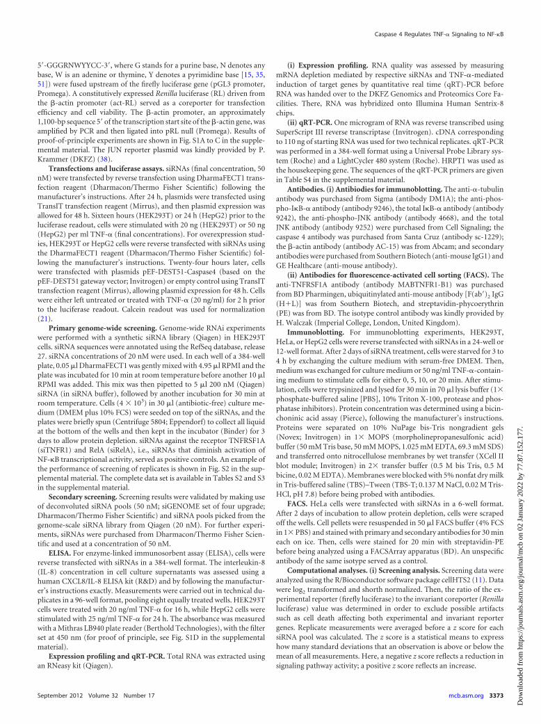
To identify the strongest and most robust modifiers, we ap-plied stringent thresholds: (i) reduction in FL levels by at least 50%compared to the mean of the experiment and (ii) a concomitantreduction of RL expression of not more than 30%. We furtherrequired a z-score cutoff of ��2.5. In this way, we recovered 49potential positive regulators of TNF-� signaling, among them theknown pathway components TNFRSF1A (TNFR1) and RelA.Nineteen candidates were selected for secondary validation exper-iments, on the basis of research in the literature (e.g., prior reportson a role in inflammation or immune signaling), the presence ofsignaling-related protein domains, and protein-protein interac-tions (Fig. 2A).
To exclude phenotypes caused by primary screening artifacts(Fig. 2A), we repeated the original screening assay to reproducescreening results (Fig. 2B, secondary screening). In addition, wealso employed an ELISA detecting protein levels of the endoge-nous TNF-� target gene IL-8 for independent phenotypic valida-tion. To exclude siRNA off-target effects, follow-up experimentswere performed with several individual siRNAs targeting the samegene. Five candidates (MEN1, ROBO4, LRRC59/PRO1855,PTK9, and CASP4) that showed a robust reduction both in inde-pendent assays and with independent siRNAs were tested in asecond cell line (Fig. 2A). Among the top five candidates, CASP4displayed one of the most robust phenotypes in all assays (seeTables S5 and S6 in the supplemental material).
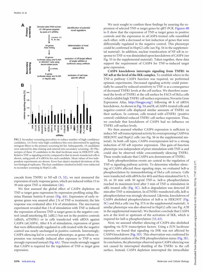
CASP4 is required for TNF-� signaling. CASP4 belongs to thefamily of inflammatory caspases, fulfilling functions in both apop-tosis (endoplasmic reticulum [ER] stress induced) and inflamma-tion (Toll-like receptor 4 [TLR4] signaling and inflammasome)(62). In the secondary assays, silencing of CASP4 resulted in an50% reduction in TNF-� signaling activity in both the luciferasereporter assay and the IL-8 ELISA in HEK293T cells (Fig. 3A andB) and HepG2 cells (Fig. 3C and D). These phenotypes were con-sistent with efficient siRNA-mediated depletion of CASP4 (Fig. 3Eand F).
For both the luciferase assays and ELISAs, cells had beentreated with TNF-� for 16 h to monitor the NF-�B transcriptionalactivity induced by extended TNF-� signaling. To investigate apossible role for CASP4 in the early, feedback loop-independent
Nickles et al.
3374 mcb.asm.org Molecular and Cellular Biology
Dow
nloa
ded
from
http
s://j
ourn
als.
asm
.org
/jour
nal/m
cb o
n 02
Jan
uary
202
2 by
77.
87.1
52.1
77.

FIG 1 Genome-wide RNAi screens for TNF-�-induced NF-�B signaling using a cell-based assay in HEK293T cells. (A) The screening procedure is based on adual-luciferase reporter system. TNF-� signaling is monitored using an NF-�B binding-dependent firefly luciferase construct. A constitutively expressed coreporter(Renilla luciferase) monitors transfection efficiency and cell viability. (B) TNF-� signaling in HEK293T cells was mediated by TNFRSF1A (TNFR1). Knockdown ofTNFRSF1B (TNFR2) did not decrease relative signaling pathway activity (compared to that for the mock-infected control). Cells were transfected with four individualsiRNAs (1 to 4) or with pools of four siRNAs (pool, siLRP5). R1 � R2, double knockdown of both receptors (siRNA pools); siLRP5, negative control. One representativeresult of two independent experiments is shown. (C) Phenotypes of the siRNA controls that were used in the RNAi screens. Induction of TNF-� signaling and, thus,NF-�B transcriptional activity leads to an approximately 16-fold induction of firefly luciferase expression. siTNFR1 and siRelA strongly reduced NF-�B reporter activityand served as positive controls. siLRP5 did not target a known component of the TNF-� pathway and was used as a negative control. siRNA against Con (siCon) slightlyinduced the pathway and was therefore not further used. Pools of four siRNAs were used. One representative result of at least 3 independent experiments is shown. (D)Summary of the screening approach. HEK293T cells were reverse transfected with siRNAs prealiquoted in 384-well plates. Twenty-four hours after siRNA transfection,cells were transfected with reporter plasmids. After 32 h, cells were treated with 20 ng/ml TNF-� for 16 h before luciferase expression was assessed. (E) Distribution ofnormalized luciferase intensities of controls. The Z= factor is a statistical means for expressing the separation of the distribution of luciferase expression values measuredfor positive controls (yellow, siTNFR1; red, siRelA) or negative controls (blue, siLRP5). Z= factors from 0 to 0.5 depict acceptable assays; those from 0.5 to 1 depict verygood assays (9, 64). (F) Quantile-quantile plot of normally distributed quantiles against actual pathway screening result quantiles. A fit to a normal distribution isrepresented by the solid line. siRNAs in the tails represent RNAi reagents with significant phenotypes. The dashed line depicts where the z-score cutoff in the individualscreening analysis was set to define hits (z score � �2.5).
Caspase 4 Regulates TNF-� Signaling to NF-�B
September 2012 Volume 32 Number 17 mcb.asm.org 3375
Dow
nloa
ded
from
http
s://j
ourn
als.
asm
.org
/jour
nal/m
cb o
n 02
Jan
uary
202
2 by
77.
87.1
52.1
77.
cascade from TNFR1 to NF-�B (5, 52), we next measured theexpression of early response genes, which are induced within 15 to30 min upon TNF-� stimulation (26).
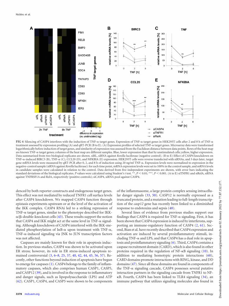
We first assessed the global effect of CASP4 depletion onTNF-� target gene expression by expression profiling using Illu-mina Human Sentrix-8 arrays (Fig. 4A). Expression of early re-sponse genes was assayed after 2 h of TNF-� treatment; the lateresponse was evaluated after 8 h of stimulation. The microarrayexperiment revealed that 2 h of stimulation with TNF-� inducedthe expression of known TNF-� target genes in the negative con-trol (small interfering RL [siRL]) but not in the positive controls(siRelA, siTNFR1) or in cells transfected with siRNA againstCASP4 (siCASP4). After 8 h of stimulation, expression of genesthat were differentially regulated in cells treated with the negativecontrol was nearly unchanged in positive controls. Interestingly,CASP4 silencing led to a reversed phenotype, as genes whose ex-pression was normally induced after exposure to TNF-� werestrongly repressed instead (Fig. 4A). These results strongly suggestthat CASP4 is required for the regulation of TNF-� target geneexpression.
We next sought to confirm these findings by assaying the ex-pression of selected TNF-� target genes by qRT-PCR. Figures 4Bto E show that the expression of TNF-� target genes in positivecontrols and the expression in siCASP4-treated cells resembledeach other, with a decreased or lost induction of genes that weredifferentially regulated in the negative control. This phenotypecould be confirmed in HepG2 cells (see Fig. S4 in the supplemen-tal material). In addition, nuclear translocation of NF-�B in re-sponse to TNF-� was diminished upon knockdown of CASP4 (seeFig. S5 in the supplemental material). Taken together, these datasupport the requirement of CASP4 for TNF-�-induced targetgene expression.
CASP4 knockdown interrupts signaling from TNFR1 toNF-�B at the level of the IKK complex. To establish where in theTNF-� pathway CASP4 function was required, we performedepistasis experiments. Decreased signaling activity could poten-tially be caused by reduced sensitivity to TNF-� as a consequenceof decreased TNFR1 levels at the cell surface. We therefore exam-ined the levels of TNFR1 at the cell surface by FACS of HeLa cells(which exhibit high TNFR1 cell surface expression; Novartis GeneExpression Atlas, http://biogps.org/) following 48 h of siRNAknockdown. As shown in Fig. 5A and B, siCASP4-treated cells andnegative-control cells displayed similar amounts of TNFR1 ontheir surfaces. In contrast, cells treated with siTNFR1 (positivecontrol) exhibited reduced TNFR1 cell surface expression. Thus,we conclude that knockdown of CASP4 had no influence onTNFR1 cell surface levels.
We then assessed whether CASP4 expression is sufficient toinduce NF-�B transcriptional activity by overexpressing CASP4 inHEK293T and HepG2 cells (see Fig. S6 in the supplemental ma-terial). In both cell types, CASP4 overexpression led to a stronginduction of NF-�B reporter expression. This gain-of-functionphenotype was independent of prior stimulation with TNF-� andcould also be observed while concomitantly silencing TNFR1.These results indicate that CASP4 acts downstream of TNFR1.
Early phosphorylation events are central to the regulation ofTNF-� signaling pathway activity. To determine whether silenc-ing of CASP4 affected these signaling steps, we evaluated I�B-�phosphorylation by immunoblotting of HeLa cell extracts. Cellswere transfected with siRNAs for 48 h and then stimulated for 0, 5,10, or 20 min with 50 ng/ml TNF-�. I�B-� phosphorylationreached its maximum level after 5 min of TNF-� stimulation insiRL-treated cells (Fig. 5C). I�B-� degradation was detected 20min after TNF-� stimulation. In siTNFR1-transfected cells, I�B-�phosphorylation was strongly decreased. Likewise, knockdown ofCASP4 abolished phosphorylation of I�B-� in HEK293T (Fig.5C) and HeLa cells (see Fig. S7A in the supplemental material). Asimilar phenotype was also observed in HepG2 cells (see Fig. A7Bin the supplemental material). We therefore conclude that CASP4acts at the level or upstream of the activation of IKK, which isrequired for I�B-� phosphorylation (53, 63).
Next, we assessed whether silencing of CASP4 also abolishedsignaling via JUN transcription factors. Using a JUN luciferasereporter, we found that signaling via JNK was not affected byCASP4 knockdown (Fig. 5D). This observation was confirmed byimmunoblot analysis of the JNK phosphorylation status (Fig. 5E).In conclusion, the phenotype observed upon CASP4 silencing wasnot caused by interrupted shuttling of the TNFR1 to the cellsurface. Instead, CASP4 depletion interrupted the intracellular
FIG 2 Secondary screening procedure to reduce number of high-confidencecandidates. (A) Forty-nine high-confidence hits were determined by applyingstringent filters to the primary screening hit list. Subsequently, 19 candidateswere selected by data mining and entered into secondary screening. (B) Phe-notypes of these 19 candidates in the dual-luciferase assay in HEK293T cells.Relative TNF-� signaling activity compared to that in mock-transfected cells isshown, using pools of 4 siRNAs for each candidate. Mean values of two inde-pendent experiments are shown. Error bars depict standard deviations of thetwo biological replicates. The four candidates (marked in bold) were subjectedto secondary screening in HepG2 cells.
Nickles et al.
3376 mcb.asm.org Molecular and Cellular Biology
Dow
nloa
ded
from
http
s://j
ourn
als.
asm
.org
/jour
nal/m
cb o
n 02
Jan
uary
202
2 by
77.
87.1
52.1
77.
TNF-� signaling cascade, specifically, the signaling branch acti-vating NF-�B transcription factors.
DISCUSSION
Because of its role in numerous pathologies, different strategieshave been pursued to characterize the TNF-� signaling cascadeand find potential new intervention points (49). Here we reportthe first genome-wide mammalian screen for TNF-�-mediatedactivation of NF-�B, which expands the candidate lists of previ-
ously targeted RNAi screens (13, 16, 36, 41, 65) to a genome-widecompendium of putative modulators of TNF-� signaling modu-lators, including known pathway components such as TNFR1 andRelA. Our genetic screening strategy also encompassed an exten-sive follow-up analysis using a range of secondary screening ex-periments and identified five high-confidence candidates. Weparticularly focused on the role of CASP4 in TNF-� signaling. Ourexperiments confirmed that silencing of CASP4 led to reducedTNF-� signaling pathway activity in multiple cell lines, as evi-
FIG 3 CASP4 is required for TNF-� signaling. (A to D) HEK293T (A and B) or HepG2 (C and D) cells were reverse transfected with siRNAs, and thedual-luciferase assay (A and C) and IL-8 ELISA (B and D) were performed. Depicted is relative signaling activity, i.e., FL/RL (luciferase assay) or relative IL-8secretion (ELISA) compared to mock-transfected cells (medium; HEK293T) or negative-control transfected cells (HepG2). RIP1, TNFR1, and RelA, siRNA poolsagainst RIP1, TNFRSF1A, and RelA, respectively (positive controls). Mean values of two independent experiments are shown. Error bars depict standarddeviations of the two biological replicates. ctrl, mean of negative siRNA controls. (E) siRNA-mediated mRNA depletion assessed by qRT-PCR in HEK293T cells.Relative CASP4 mRNA levels compared to those in cells transfected with the negative control are shown. Error bars depict standard deviations of the two technicalreplicates. Data representative of 3 independent experiments are shown. (F) siRNA-mediated protein depletion assessed by immunoblotting in HeLa cells.Staining for �-tubulin served as a loading control. Representative data of 4 independent experiments are shown.
Caspase 4 Regulates TNF-� Signaling to NF-�B
September 2012 Volume 32 Number 17 mcb.asm.org 3377
Dow
nloa
ded
from
http
s://j
ourn
als.
asm
.org
/jour
nal/m
cb o
n 02
Jan
uary
202
2 by
77.
87.1
52.1
77.
denced by both reporter constructs and endogenous target genes.This effect was not mediated by reduced TNFR1 cell surface levelsafter CASP4 knockdown. We mapped CASP4 function throughepistasis experiments upstream or at the level of the activation ofthe IKK complex. CASP4 RNAi led to a striking repression ofTNF-� target genes, similar to the phenotype described for IKK-�/�-double-knockout cells (43). These results support the notionthat CASP4 and IKK might act at the same level in TNF-� signal-ing. Although knockdown of CASP4 interfered with the IKK-me-diated phosphorylation of I�B-� upon treatment with TNF-�,TNF-�-induced signaling via JNK to JUN transcription factorswas not affected.
Caspases are mainly known for their role in apoptosis induc-tion. In previous studies, CASP4 was shown to be activated uponER stress; however, its role in ER stress-induced apoptosis re-mained controversial (3, 6–8, 25, 37, 40, 42, 44, 45, 56, 57). Re-cently, other functions beyond induction of apoptosis have begunto emerge for caspases (1). CASP4 belongs to the family of inflam-matory caspases, which also comprises human CASP1, CASP5,and CASP12 (39), and is involved in the response to inflammatoryand danger signals, such as lipopolysaccharide (LPS) and ATP(62). CASP1, CASP4, and CASP5 were shown to be components
of the inflammasome, a large protein complex sensing intracellu-lar danger signals (33, 50). CASP12 is normally expressed as atruncated protein, and a mutation leading to full-length transcrip-tion of the casp12 gene has recently been linked to a diminishedresponse to endotoxins and sepsis (48).
Several lines of evidence from previous studies support ourfindings that CASP4 is required for TNF-� signaling. First, it hasbeen shown that CASP4 expression is induced by interferons, sup-porting an immune-regulatory function of CASP4 (2, 61). Sec-ond, Bian et al. have recently described that CASP4 expression andactivation are induced by several proinflammatory stimuli, in-cluding TNF-� and LPS, and that CASP4 has a dual role in apop-tosis and proinflammatory signaling (6). Third, CASP4 contains acaspase recruitment domain (CARD), which is also found in otherproteins required in the regulation of NF-�B signaling (10). Inaddition to mediating homotypic protein interactions (60),CARD domains promote interactions with RING, kinase, and DDdomains (47). Since all these domains are found in components ofthe TNF-� signaling cascade, CASP4 possesses several putativeinteraction partners in the signaling cascade from TNFR1 to NF-�B. Fourth, CASP4 has been linked to TLR4 signaling (34), animmune pathway that utilizes signaling molecules also found in
FIG 4 Silencing of CASP4 interferes with the induction of TNF-� target genes. Expression of TNF-� target genes in HEK293T cells after 2 and 8 h of TNF-�treatment assessed by expression profiling (A) and qRT-PCR (B to E). (A) Expression profiles of selected TNF-� target genes. Microarray data were transformedlogarithmically before induction of target genes, and similarity of expression was assessed from the Euclidean distance between data points. Rows of the heat mapare known TNF-� target genes; columns of the heat map are different samples. Blue, lower expression than that by unstimulated cells; yellow, higher expression.Data summarized from two biological replicates are shown. siRL, siRNA against Renilla luciferase (negative control). (B to E) Effect of CASP4 knockdown onTNF-�-induced BIRC3 (B), TNF-� (C), CCL20 (D), and NFKBIA (E) expression. HEK293T cells were reverse transfected with siRNAs, and 3 days later, targetgene mRNA levels were measured by qRT-PCR after 0, 2, and 8 h of induction using 20 ng/ml TNF-�. Expression levels were normalized to expression in thenegative-control sample (siRNA against Renilla luciferase): for each time point, mRNA expression levels were set to 100% in the control sample, and mRNA levelsin candidate samples were calculated in relation to the control. Data derived from five independent experiments are shown, with error bars indicating thestandard deviations of the biological replicates. P values were calculated using Student’s t test. **, P 0.01; ***, P 0.001. (A to E) siTNFR1 and siRelA, siRNAagainst TNFRSF1A and RelA, respectively (positive controls); siCASP4, siRNA pool against CASP4.
Nickles et al.
3378 mcb.asm.org Molecular and Cellular Biology
Dow
nloa
ded
from
http
s://j
ourn
als.
asm
.org
/jour
nal/m
cb o
n 02
Jan
uary
202
2 by
77.
87.1
52.1
77.

FIG 5 Silencing of CASP4 interferes with direct signaling from TNFR1 to the IKK but not with TNFR1 cell surface expression or TNF-�-induced activation ofJNK. (A) Knockdown of CASP4 does not affect TNFR1 cell surface expression. HeLa cells were transfected with control and candidate siRNAs and were stainedfor cell surface TNFR1. TNFR1 was detected using a specific antibody labeled with PE. PE staining was assessed by FACS. The histograms represent the intensityof the TNFR1-PE stain (x axis) dependent on cell number (y axis). An overlay of all samples is shown. Blue curve, siCASP4-treated cells; black curve,siTNFR1-treated cells; red curve, siRL-treated cells. Data shown are representative of two independent experiments. (B) Quantification of the results from panelA. PE-positive and -negative cells were quantified as a percentage of all analyzed cells. For quantification, two populations, PE positive and PE negative, weredefined on the basis of the fact that siRL-treated cells were stained with the isotype-matched control antibody (isotype control). For each sample, the percentageof cells in either of the two populations was calculated. Control antibody-stained cells showed background staining. Anti-TNFR1 staining in siTNFR1-transfectedcells was decreased to background, while cells treated with siRNAs against CASP4 or the negative control exhibited similar staining. (C) Knockdown of CASP4abolishes I�B-� phosphorylation upon TNF-� stimulation in HEK293T cells. HEK293T cells were transfected with siRNA pools, before I�B-� phosphorylationupon TNF-� treatment (50 ng/ml) was assessed by immunoblotting. �-Actin served as a loading control. p-I�B-�, phospho-I�B-�. (D) Knockdown of CASP4does not affect TNF-�-induced activation of JNK signaling. HEK293T cells were reverse transfected with siRNAs, and a dual-luciferase assay was performed,using a JUN-dependent firefly luciferase as a pathway-specific reporter and the �-actin promoter-driven Renilla luciferase as a coreporter. Depicted is relativesignaling activity, i.e., FL/RL compared to negative-control transfected cells. ctrls, mean of negative siRNA controls; RelA, siRNA pools against RelA (positivecontrol). Mean values of two independent experiments are shown. Error bars depict standard deviations of the two biological replicates. (E) Phosphorylation ofJNK is unaffected by siCASP4 treatment. HeLa cells were transfected with siRNA pools, before JNK phosphorylation upon TNF-� treatment (25 ng/ml) wasassessed by immunoblotting. �-Actin served as a loading control. Data shown are representative of two independent experiments. p-JNK, phospho-JNK. (A andE) siRL, siRNA pool against Renilla luciferase (negative control); siTNFR1, siRNA pool against TNFRSF1A (positive control); siCASP4, siRNA pools againstcaspase 4. (F) Model of CASP4’s role in TNF-� signaling. Epistasis experiments showed that CASP4 is needed for the activation of IKK; hence, CASP4 is probablylocated at TNFR complex I (TNFR-C). At TNFR-C, two different branches of signaling are initiated: (i) both TRAF2 and TRAF5 together with RIP1 are requiredfor the activation of IKK and, finally, NF-�B; (ii) TRAF2 alone is sufficient to mediate the activation of JNK signaling activating transcription factors such as JUN.Knockdown of CASP4 does not affect this second signaling branch but affects signaling only to NF-�B.
Caspase 4 Regulates TNF-� Signaling to NF-�B
September 2012 Volume 32 Number 17 mcb.asm.org 3379
Dow
nloa
ded
from
http
s://j
ourn
als.
asm
.org
/jour
nal/m
cb o
n 02
Jan
uary
202
2 by
77.
87.1
52.1
77.
the TNF-� pathway, such as DD proteins and members of theTRAF family. This suggests that CASP4 might be another proteinshared between TNF-� and TLR signaling. Finally, a caspase 4(caspase 11)-knockout mouse which is resistant to LPS-inducedsepsis has recently been reported (33). While there is currently nomechanistic explanation for the in vivo phenotype, it is temptingto speculate that LPS-induced inflammatory feedback loops viaTNF-� are constrained in CASP4-knockout animals.
CASP4 could fulfill its function in TNF-� signaling both as aprotease (31) and as a scaffold protein. Both mechanisms havebeen described to play a role for TNF-�-induced activation ofNF-�B for other caspases and organisms. Recently, the Drosophilaorthologue of CASP8, DREDD, was shown to cleave DrosophilaIMD (46). This cleavage enabled the interaction of components ofthe TNFR complex and thereby the activation of IKK and NF-�B(46). However, a scaffold function of CASP8 has also been de-scribed to be required for TNF-� signaling to NF-�B (14).
According to our results, depletion of CASP4 does not interferewith JNK activation. Thus, we propose that CASP4’s mode ofaction in TNF-� signaling is probably located in the signalingbranch required for the activation of IKK and, finally, NF-�B (Fig.5F). CASP4 could act as either a protease or a scaffold molecule atthe TNFR complex. However, further studies will be necessary tocharacterize its molecular function.
ACKNOWLEDGMENTS
We thank Dierk Ingelfinger and Tobias Haas for providing protocols aswell as Bernhard Korn and Tamara Fries of the DKFZ Genomics andProteomics Core Facilities for microarrays. We are grateful to WolfgangHuber and Simon Anders for their support during microarray analysis.We thank Nadège Pelte, Anan Ragab, Iris Augustin, and Thomas Sand-mann for technical advice and for critical reading of the manuscript. Weare also grateful to Grainne Kerr for bioinformatics support.
D.N. was supported by a Ph.D. fellowship from the German CancerResearch Center (DKFZ). M.M. received a fellowship from the Studien-stiftung and the I. Behr Foundation. Work in the laboratory of M.B. hasbeen supported by the Deutsche Forschungsgemeinschaft, the HelmholtzAlliance for Systems Biology, and the BMBF/EU Network ERASysBio�.
REFERENCES1. Abraham MC, Shaham S. 2004. Death without caspases, caspases without
death. Trends Cell Biol. 14:184 –193.2. Ahn EY, Pan G, Vickers SM, McDonald JM. 2002. IFN-gamma upregu-
lates apoptosis-related molecules and enhances Fas-mediated apoptosis inhuman cholangiocarcinoma. Int. J. Cancer 100:445– 451.
3. Ariazi EA, et al. 2011. Estrogen induces apoptosis in estrogen depriva-tion-resistant breast cancer through stress responses as identified by globalgene expression across time. Proc. Natl. Acad. Sci. U. S. A. 108:18879 –18886.
4. Baud V, Karin M. 2001. Signal transduction by tumor necrosis factor andits relatives. Trends Cell Biol. 11:372–377.
5. Beutler B, Milsark IW, Cerami AC. 1985. Passive immunization againstcachectin/tumor necrosis factor protects mice from lethal effect of endo-toxin. Science 229:869 – 871.
6. Bian ZM, Elner SG, Elner VM. 2009. Dual involvement of caspase-4 ininflammatory and ER stress-induced apoptotic responses in human reti-nal pigment epithelial cells. Invest. Ophthalmol. Vis. Sci. 50:6006 – 6014.
7. Binet F, Chiasson S, Girard D. 2010. Arsenic trioxide induces endoplas-mic reticulum stress-related events in neutrophils. Int. Immunopharma-col. 10:508 –512.
8. Binet F, Chiasson S, Girard D. 2010. Evidence that endoplasmic reticu-lum (ER) stress and caspase-4 activation occur in human neutrophils.Biochem. Biophys. Res. Commun. 391:18 –23.
9. Birmingham A, et al. 2009. Statistical methods for analysis of high-throughput RNA interference screens. Nat. Methods 6:569 –575.
10. Bouchier-Hayes L, Martin SJ. 2002. CARD games in apoptosis and im-munity. EMBO Rep. 3:616 – 621.
11. Boutros M, Bras LP, Huber W. 2006. Analysis of cell-based RNAiscreens. Genome Biol. 7:R66. doi:10.1186/gb-2006-7-7-r66.
12. Bradley JR. 2008. TNF-mediated inflammatory disease. J. Pathol. 214:149 –160.
13. Brummelkamp TR, Nijman SM, Dirac AM, Bernards R. 2003. Loss ofthe cylindromatosis tumour suppressor inhibits apoptosis by activatingNF-kappaB. Nature 424:797– 801.
14. Chaudhary PM, et al. 2000. Activation of the NF-kappaB pathway bycaspase 8 and its homologs. Oncogene 19:4451– 4460.
15. Chen FE, Ghosh G. 1999. Regulation of DNA binding by Rel/NF-kappaBtranscription factors: structural views. Oncogene 18:6845– 6852.
16. Chew J, et al. 2009. WIP1 phosphatase is a negative regulator of NF-kappaB signalling. Nat. Cell Biol. 11:659 – 666.
17. Choudhary S, et al. 2011. High throughput short interfering RNA(siRNA) screening of the human kinome identifies novel kinases control-ling the canonical nuclear factor-kappaB (NF-kappaB) activation path-way. J. Biol. Chem. 286:37187–37195.
18. Dunning MJ, Smith ML, Ritchie ME, Tavare S. 2007. beadarray: Rclasses and methods for Illumina bead-based data. Bioinformatics 23:2183–2184.
19. Gerlach B, et al. 2011. Linear ubiquitination prevents inflammation andregulates immune signalling. Nature 471:591–596.
20. Gesellchen V, Kuttenkeuler D, Steckel M, Pelte N, Boutros M. 2005. AnRNA interference screen identifies inhibitor of apoptosis protein 2 as aregulator of innate immune signalling in Drosophila. EMBO Rep. 6:979 –984.
21. Gilbert DF, et al. 2011. A novel multiplex cell viability assay for high-throughput RNAi screening. PLoS One 6:e28338. doi:10.1371/journal.pone.0028338.
22. Goto A, et al. 2008. Akirins are highly conserved nuclear proteins re-quired for NF-kappaB-dependent gene expression in Drosophila andmice. Nat. Immunol. 9:97–104.
23. Grell M, Wajant H, Zimmermann G, Scheurich P. 1998. The type 1receptor (CD120a) is the high-affinity receptor for soluble tumor necrosisfactor. Proc. Natl. Acad. Sci. U. S. A. 95:570 –575.
24. Haas TL, et al. 2009. Recruitment of the linear ubiquitin chain assemblycomplex stabilizes the TNF-R1 signaling complex and is required forTNF-mediated gene induction. Mol. Cell 36:831– 844.
25. Hitomi J, et al. 2004. Involvement of caspase-4 in endoplasmic reticulumstress-induced apoptosis and Abeta-induced cell death. J. Cell Biol. 165:347–356.
26. Hoffmann A, Levchenko A, Scott ML, Baltimore D. 2002. The IkappaB-NF-kappaB signaling module: temporal control and selective gene activa-tion. Science 298:1241–1245.
27. Huber W, von Heydebreck A, Sueltmann H, Poustka A, Vingron M.2003. Parameter estimation for the calibration and variance stabilizationof microarray data. Stat. Appl. Genet. Mol. Biol. 2:Article 3.
28. Huber W, von Heydebreck A, Sultmann H, Poustka A, Vingron M.2002. Variance stabilization applied to microarray data calibration and tothe quantification of differential expression. Bioinformatics 18(Suppl 1):S96 –S104.
29. Jiang Y, Woronicz JD, Liu W, Goeddel DV. 1999. Prevention of consti-tutive TNF receptor 1 signaling by silencer of death domains. Science283:543–546.
30. Karin M, Greten FR. 2005. NF-kappaB: linking inflammation and im-munity to cancer development and progression. Nat. Rev. Immunol.5:749 –759.
31. Karki P, Dahal GR, Park IS. 2007. Both dimerization and interdomainprocessing are essential for caspase-4 activation. Biochem. Biophys. Res.Commun. 356:1056 –1061.
32. Kauffmann A, Gentleman R, Huber W. 2009. arrayQualityMetrics—abioconductor package for quality assessment of microarray data. Bioin-formatics 25:415– 416.
33. Kayagaki N, et al. 2011. Non-canonical inflammasome activation targetscaspase-11. Nature 479:117–121.
34. Lakshmanan U, Porter AG. 2007. Caspase-4 interacts with TNF receptor-associated factor 6 and mediates lipopolysaccharide-induced NF-kappaB-dependent production of IL-8 and CC chemokine ligand 4 (macrophage-inflammatory protein-1). J. Immunol. 179:8480 – 8490.
35. Le Bail O, Schmidt-Ullrich R, Israel A. 1993. Promoter analysis of thegene encoding the I kappa B-alpha/MAD3 inhibitor of NF-kappa B: pos-
Nickles et al.
3380 mcb.asm.org Molecular and Cellular Biology
Dow
nloa
ded
from
http
s://j
ourn
als.
asm
.org
/jour
nal/m
cb o
n 02
Jan
uary
202
2 by
77.
87.1
52.1
77.
itive regulation by members of the rel/NF-kappa B family. EMBO J. 12:5043–5049.
36. Li S, Wang L, Berman MA, Zhang Y, Dorf ME. 2006. RNAi screen inmouse astrocytes identifies phosphatases that regulate NF-kappaB signal-ing. Mol. Cell 24:497–509.
37. Lin R, Sun Y, Li C, Xie C, Wang S. 2007. Identification of differentiallyexpressed genes in human lymphoblastoid cells exposed to irradiation andsuppression of radiation-induced apoptosis with antisense oligonucleo-tides against caspase-4. Oligonucleotides 17:314 –326.
38. Li-Weber M, Giaisi M, Baumann S, Treiber MK, Krammer PH. 2002.The anti-inflammatory sesquiterpene lactone parthenolide suppressesCD95-mediated activation-induced-cell-death in T-cells. Cell Death Dif-fer. 9:1256 –1265.
39. Martinon F, Tschopp J. 2004. Inflammatory caspases: linking an intra-cellular innate immune system to autoinflammatory diseases. Cell 117:561–574.
40. Matsuzaki S, Hiratsuka T, Kuwahara R, Katayama T, Tohyama M.2010. Caspase-4 is partially cleaved by calpain via the impairment of Ca2�homeostasis under the ER stress. Neurochem. Int. 56:352–356.
41. Metzig M, et al. 2011. An RNAi screen identifies USP2 as a factor requiredfor TNF-alpha-induced NF-kappaB signaling. Int. J. Cancer 129:607– 618.
42. Miller SD, et al. 2007. Tauroursodeoxycholic acid inhibits apoptosisinduced by Z alpha-1 antitrypsin via inhibition of Bad. Hepatology 46:496 –503.
43. Molestina RE, Sinai AP. 2005. Host and parasite-derived IKK activitiesdirect distinct temporal phases of NF-kappaB activation and target geneexpression following Toxoplasma gondii infection. J. Cell Sci. 118:5785–5796.
44. Mulugeta S, et al. 2007. Misfolded BRICHOS SP-C mutant proteinsinduce apoptosis via caspase-4- and cytochrome c-related mechanisms.Am. J. Physiol. Lung Cell. Mol. Physiol. 293:L720 –L729.
45. Oda T, Kosuge Y, Arakawa M, Ishige K, Ito Y. 2008. Distinct mechanismof cell death is responsible for tunicamycin-induced ER stress in SK-N-SHand SH-SY5Y cells. Neurosci. Res. 60:29 –39.
46. Paquette N, et al. 2010. Caspase-mediated cleavage, IAP binding, andubiquitination: linking three mechanisms crucial for Drosophila NF-kappaB signaling. Mol. Cell 37:172–182.
47. Reed JC, Doctor KS, Godzik A. 2004. The domains of apoptosis: agenomics perspective. Sci. STKE 2004:re9. doi:10.1126/stke.2392004re9.
48. Saleh M, et al. 2004. Differential modulation of endotoxin responsivenessby human caspase-12 polymorphisms. Nature 429:75–79.
49. Scheinfeld N. 2004. A comprehensive review and evaluation of the sideeffects of the tumor necrosis factor alpha blockers etanercept, infliximaband adalimumab. J. Dermatolog. Treat. 15:280 –294.
50. Schroder K, Tschopp J. 2010. The inflammasomes. Cell 140:821– 832.51. Sen R, Baltimore D. 1986. Inducibility of kappa immunoglobulin en-
hancer-binding protein Nf-kappa B by a posttranslational mechanism.Cell 47:921–928.
52. Shakhov AN, Collart MA, Vassalli P, Nedospasov SA, Jongeneel CV.1990. Kappa B-type enhancers are involved in lipopolysaccharide-mediated transcriptional activation of the tumor necrosis factor alphagene in primary macrophages. J. Exp. Med. 171:35– 47.
53. Sizemore N, Lerner N, Dombrowski N, Sakurai H, Stark GR. 2002.Distinct roles of the Ikappa B kinase alpha and beta subunits in liberatingnuclear factor kappa B (NF-kappa B) from Ikappa B and in phosphory-lating the p65 subunit of NF-kappa B. J. Biol. Chem. 277:3863–3869.
54. Smyth GK. 2004. Linear models and empirical Bayes methods for assess-ing differential expression in microarray experiments. Stat. Appl. Genet.Mol. Biol. 3:Article 3.
55. Stanger BZ, Leder P, Lee TH, Kim E, Seed B. 1995. RIP: a novel proteincontaining a death domain that interacts with Fas/APO-1 (CD95) in yeastand causes cell death. Cell 81:513–523.
56. Suzuki M, Endo M, Shinohara F, Echigo S, Rikiishi H. 2009. Enhance-ment of cisplatin cytotoxicity by SAHA involves endoplasmic reticulumstress-mediated apoptosis in oral squamous cell carcinoma cells. CancerChemother. Pharmacol. 64:1115–1122.
57. Szaraz P, Banhegyi G, Benedetti A. 2010. Altered redox state of luminalpyridine nucleotides facilitates the sensitivity towards oxidative injury andleads to endoplasmic reticulum stress dependent autophagy in HepG2cells. Int. J. Biochem. Cell Biol. 42:157–166.
58. Ting AT, Pimentel-Muinos FX, Seed B. 1996. RIP mediates tumornecrosis factor receptor 1 activation of NF-kappaB but not Fas/APO-1-initiated apoptosis. EMBO J. 15:6189 – 6196.
59. Walczak H. 2011. TNF and ubiquitin at the crossroads of gene activation,cell death, inflammation, and cancer. Immunol. Rev. 244:9 –28.
60. Weber CH, Vincenz C. 2001. The death domain superfamily: a tale of twointerfaces? Trends Biochem. Sci. 26:475– 481.
61. Yamamoto-Yamaguchi Y, Okabe-Kado J, Kasukabe T, Honma Y. 2003.Induction of apoptosis by combined treatment with differentiation-inducing agents and interferon-alpha in human lung cancer cells. Anti-cancer Res. 23:2537–2547.
62. Yazdi AS, Guarda G, D’Ombrain MC, Drexler SK. 2010. Inflammatorycaspases in innate immunity and inflammation. J. Innate Immun. 2:228 –237.
63. Zandi E, Rothwarf DM, Delhase M, Hayakawa M, Karin M. 1997. TheIkappaB kinase complex (IKK) contains two kinase subunits, IKKalphaand IKKbeta, necessary for IkappaB phosphorylation and NF-kappaB ac-tivation. Cell 91:243–252.
64. Zhang JH, Chung TD, Oldenburg KR. 1999. A simple statistical param-eter for use in evaluation and validation of high throughput screeningassays. J. Biomol. Screen. 4:67–73.
65. Zheng L, et al. 2004. An approach to genomewide screens of expressedsmall interfering RNAs in mammalian cells. Proc. Natl. Acad. Sci. U. S. A.101:135–140.
Caspase 4 Regulates TNF-� Signaling to NF-�B
September 2012 Volume 32 Number 17 mcb.asm.org 3381
Dow
nloa
ded
from
http
s://j
ourn
als.
asm
.org
/jour
nal/m
cb o
n 02
Jan
uary
202
2 by
77.
87.1
52.1
77.
Related Documents