JOURNAL OF INTERFERON & CYTOKINE RESEARCH 26:719–729 (2006) © Mary Ann Liebert, Inc. MKK3/6-p38 MAPK Signaling Is Required for IL-1 and TNF--Induced RANKL Expression in Bone Marrow Stromal Cells CARLOS ROSSA, JR., 1, * KATHRYN EHMANN, 2, * MIN LIU, 3 CHETAN PATIL, 2,3 and KEITH L. KIRKWOOD 3 ABSTRACT Coupled bone turnover is directed by the expression of receptor-activated NF-B ligand (RANKL) and its decoy receptor, osteoprotegerin (OPG). Proinflammatory cytokines, such as interleukin-1 (IL-1) and tu- mor necrosis factor- (TNF-) induce RANKL expression in bone marrow stromal cells. Here, we report that IL-1 and TNF--induced RANKL requires p38 mitogen-activating protein kinase (MAPK) pathway acti- vation for maximal expression. Real-time PCR was used to assess the p38 contribution toward IL-1 and TNF--induced RANKL mRNA expression. Steady-state RANKL RNA levels were increased approximately 17-fold by IL-1 treatment and subsequently reduced 70%–90% when p38 MAPK was inhibited with SB203580. RANKL mRNA stability data indicated that p38 MAPK did not alter the rate of mRNA decay in IL-1-induced cells. Using a RANKL-luciferase cell line receptor containing a 120-kB segment of the 5 flank- ing region of the RANKL gene, reporter expression was stimulated 4–5-fold by IL-1 or TNF- treatment. IL-1-induced RANKL reporter expression was completely blocked with specific p38 inhibitors as well as dominant negative mutant constructs of MAPK kinase-3 and -6. In addition, blocking p38 signaling in bone marrow stromal cells partially inhibited IL-1 and TNF--induced osteoclastogenesis in vitro. Results from these studies indicate that p38 MAPK is a major signaling pathway involved in IL-1 and TNF--induced RANKL expression in bone marrow stromal cells. 719 INTRODUCTION E XCESSIVE OSTEOCLAST-MEDIATED bone resorption is a common feature of chronic inflammatory processes, for ex- ample, periodonitis, rheumatoid arthritis, and failing joint pros- theses. Several lines of evidence indicate that cytokine net- working and crosstalk between stromal/osteoblastic cells and monocyte/osteoclast progenitor cells dictate cellular responses involved in bone remodeling. Several mediators of bone re- sorption and remodeling have been identified in response to such proinflammatory cytokines, as interleukin-1 (IL-1), bac- terial lipopolysaccharide (LPS), and other bone-resorptive agents. Bone marrow stromal cells respond to these agents and directly or indirectly secrete various cytokines that activate re- ceptor activator of NF-B ligand (RANKL), resulting in en- hanced osteoclastogenesis. 1,2 Bone marrow stromal cell-derived RANKL is essential for osteoclastic differentiation of monocyte precursor cells into ma- ture multinucleated osteoclasts in the presence of macrophage colony-stimulating factor (M-CSF). 3,4 RANKL is highly ex- pressed on the surface of bone marrow stromal cells and pre- osteoblasts in the areas of excessive osteolysis and trabecular bone remodeling. 5 RANKL expression is upregulated in os- teoblasts or stromal cells by IL-1, IL-6 (in the presence of soluble IL-6 receptor), tumor necrosis factor- (TNF-), pros- taglandin E 2 (PGE 2 ), parathyroid hormone (PTH), 1,25-dihy- droxyvitamin D 3 , and others. 1,6–8 Through interactions with its cognate receptor (RANK) on osteoclast progenitor cells, RANKL induces differentiation and maturation of osteoclasts. Balancing this system is the decoy receptor, osteoprotegerin (OPG), a soluble member of the tumor necrosis family capable of binding to RANKL, thus decreasing RANKL functional ac- 1 Department of Diagnosis and Surgery, State University of Sao Paulo (UNESP), Araraquara, SP, Brazil. 2 Department of Oral Biology, State University of New York at Buffalo, Buffalo, NY 14214. 3 Department of Periodontics and Oral Medicine, University of Michigan, Ann Arbor, MI 48109. *These authors contributed equally to this work.

Welcome message from author
This document is posted to help you gain knowledge. Please leave a comment to let me know what you think about it! Share it to your friends and learn new things together.
Transcript
JOURNAL OF INTERFERON & CYTOKINE RESEARCH 26:719–729 (2006)© Mary Ann Liebert, Inc.
MKK3/6-p38 MAPK Signaling Is Required for IL-1� andTNF-�-Induced RANKL Expression in
Bone Marrow Stromal Cells
CARLOS ROSSA, JR.,1,* KATHRYN EHMANN,2,* MIN LIU,3
CHETAN PATIL,2,3 and KEITH L. KIRKWOOD3
ABSTRACT
Coupled bone turnover is directed by the expression of receptor-activated NF-�B ligand (RANKL) and itsdecoy receptor, osteoprotegerin (OPG). Proinflammatory cytokines, such as interleukin-1� (IL-1�) and tu-mor necrosis factor-� (TNF-�) induce RANKL expression in bone marrow stromal cells. Here, we report thatIL-1� and TNF-�-induced RANKL requires p38 mitogen-activating protein kinase (MAPK) pathway acti-vation for maximal expression. Real-time PCR was used to assess the p38 contribution toward IL-1� andTNF-�-induced RANKL mRNA expression. Steady-state RANKL RNA levels were increased approximately17-fold by IL-1� treatment and subsequently reduced �70%–90% when p38 MAPK was inhibited withSB203580. RANKL mRNA stability data indicated that p38 MAPK did not alter the rate of mRNA decay inIL-1�-induced cells. Using a RANKL-luciferase cell line receptor containing a 120-kB segment of the 5� flank-ing region of the RANKL gene, reporter expression was stimulated 4–5-fold by IL-1� or TNF-� treatment.IL-1�-induced RANKL reporter expression was completely blocked with specific p38 inhibitors as well asdominant negative mutant constructs of MAPK kinase-3 and -6. In addition, blocking p38 signaling in bonemarrow stromal cells partially inhibited IL-1� and TNF-�-induced osteoclastogenesis in vitro. Results fromthese studies indicate that p38 MAPK is a major signaling pathway involved in IL-1� and TNF-�-inducedRANKL expression in bone marrow stromal cells.
719
INTRODUCTION
EXCESSIVE OSTEOCLAST-MEDIATED bone resorption is a common feature of chronic inflammatory processes, for ex-
ample, periodonitis, rheumatoid arthritis, and failing joint pros-theses. Several lines of evidence indicate that cytokine net-working and crosstalk between stromal/osteoblastic cells andmonocyte/osteoclast progenitor cells dictate cellular responsesinvolved in bone remodeling. Several mediators of bone re-sorption and remodeling have been identified in response tosuch proinflammatory cytokines, as interleukin-1 (IL-1), bac-terial lipopolysaccharide (LPS), and other bone-resorptiveagents. Bone marrow stromal cells respond to these agents anddirectly or indirectly secrete various cytokines that activate re-ceptor activator of NF-�B ligand (RANKL), resulting in en-hanced osteoclastogenesis.1,2
Bone marrow stromal cell-derived RANKL is essential forosteoclastic differentiation of monocyte precursor cells into ma-ture multinucleated osteoclasts in the presence of macrophagecolony-stimulating factor (M-CSF).3,4 RANKL is highly ex-pressed on the surface of bone marrow stromal cells and pre-osteoblasts in the areas of excessive osteolysis and trabecularbone remodeling.5 RANKL expression is upregulated in os-teoblasts or stromal cells by IL-1�, IL-6 (in the presence of soluble IL-6 receptor), tumor necrosis factor-� (TNF-�), pros-taglandin E2 (PGE2), parathyroid hormone (PTH), 1,25-dihy-droxyvitamin D3, and others.1,6–8 Through interactions with itscognate receptor (RANK) on osteoclast progenitor cells,RANKL induces differentiation and maturation of osteoclasts.Balancing this system is the decoy receptor, osteoprotegerin(OPG), a soluble member of the tumor necrosis family capableof binding to RANKL, thus decreasing RANKL functional ac-
1Department of Diagnosis and Surgery, State University of Sao Paulo (UNESP), Araraquara, SP, Brazil.2Department of Oral Biology, State University of New York at Buffalo, Buffalo, NY 14214.3Department of Periodontics and Oral Medicine, University of Michigan, Ann Arbor, MI 48109.*These authors contributed equally to this work.
tivity.9 It is the ratio of RANKL/OPG that eventually deter-mines bone turnover.10
Proinflammatory cytokines stimulate multiple intracellularsignaling pathways, including the p38 mitogen-activating pro-tein kinase (MAPK) pathways, NF-�B pathway, extracellularsignal-regulated kinase (ERK) pathway, and stress-activatedprotein kinase/c-Jun N-terminal kinase (SAPK/JNK) pathway(SAPK/JNK).11,12 Although much attention has been focusedon the NF-�B pathway of cytokine gene activation, more re-cent study has addressed JNK and p38 MAPK during inflam-mation and environmental stress-induced signaling.13 The p38kinases constitute a distinct MAPK subfamily that plays a rolein adaptation, homeostasis, and stress responses.14 Of the iden-tified four splice variants, p38� MAPK is the best character-ized and perhaps the most relevant kinase involved in inflam-matory responses.15,16 The essential role of p38� MAPK inRANKL-induced osteoclastogenesis has been elucidated inmacrophage/osteoclast precursor cells,17 although the role ofp38 regulation in stromal/osteoblastic-derived RANKL expres-sion has been addressed only recently.7,18,19 In this study, therole of p38 signaling was investigated during cytokine-inducedRANKL expression in bone marrow stromal cells. Our resultsindicate that MKK3/6-p38 signaling is necessary for IL-1�-in-duced RANKL expression involving a transcriptional mecha-nism that does not require the proximal RANKL promoter rel-ative to the transcriptional start site.
MATERIALS AND METHODS
Tissue culture of murine ST-2, UAMS-32P cells
ST-2 bone marrow stromal cells were obtained from Riken(Wako, Japan). UAMS-32P bone marrow stromal cells were ob-tained from Dr. Charles O’Brien (University of Arkansas).UAMS-32P cell lines express RANKL in response to1,25(OH)2D3 and constitutively express M-CSF.20 Cells werecultured in �-MEM medium (Life Technologies, Gaithersburg,MD) supplemented with 10% fetal bovine serum (FBS) (Sigma,St. Louis, MO). Cells were routinely serum starved in 0.3% FBS-containing medium for 6–8 h prior to cytokine stimulation.
Real-time PCR analysis
Total RNA (5 �g) from ST-2 and UAMS-32P cells was iso-lated using Trizol reagent (Invitrogen, Carlsbad, CA) and usedfor cDNA synthesis with oligo(dT) 12-18 primers and Super-script II (RNAse H) (Invitrogen) in reverse transcription (RT)reactions. Real-time PCR was performed with 2 �L of the RTpreparation using Taq polymerase (Invitrogen) and primers spe-cific for mouse RANKL and GAPDH cDNAs. RANKL andGAPDH were amplified for 50 cycles using the BioRad’sSYBR Green Supermix kit (Bio-Rad, Hercules, CA) followingthe manufacturer’s instructions. Real-time PCR primer se-quences were as follows: RANKL, forward: 5�-agcaacg-gaaaactaagggt-3�, reverse: 5�-cccccaacatttatggaata-3�; GAPDH,forward: 5�-caaagccagagtccttcaga-3�, and reverse: 5�-gatg-gtcttggtccttagcc-3�. Threshold values were assigned by the iCycler program (Bio-Rad) and used with amplification effi-ciencies to calculate RANKL expression. Q-gene (www.biotechniques.com/softlib/qgene.html) quantitative software
was used to analyze gene expression based on cycle thresholdvalues normalized to GAPDH expression. Real-time PCR prod-ucts were also verified through electrophoresis on 2% agarosegel and visualized using ethidium bromide staining to verifyauthenticity.
mRNA stability experiments
To determine the relative role of p38 MAPK on IL-1�-in-duced RANKL mRNA stability, the rate of RANKL mRNA de-cay was determined in the presence and absence of the p38 in-hibitor, SB203580. Actinomycin D (ActD) was used in theexperiments to arrest transcription as described previously.21
Briefly, ST-2 and UAMS-32P cells were pretreated withSB203580 (2 �M) for 45 min, then treated with IL-1� for 16–18h. Subsequently, ActD (2 �M) was added to prevent furthertranscription. Total RNA was harvested and analyzed by real-time RT-PCR as described 0–8 h post-ActD addition.
RT-PCR analysis
Total RNA (5 �g) from ST-2 and UAMS-32P cells was iso-lated using Trizol reagent and used for cDNA synthesis witholigo(dT) 12-18 primers and Superscript II (RNAse H) in RTreactions. RT product (2 �L) was used as a template for PCRamplification of RANKL and GAPDH gene products. Primerssequences used were: RANKL [AF_019048, forward: 5�-cagcactcactgcttttatagaatcc-3�, reverse: 5�-agctgaagatagtctgtag-gtacgc-3�; GAPDH [NM_002046], forward: 5�-caccatggagaag-gccgggg 3�, reverse: 5�-gacggacacattggggtag-3�. RT-PCRproducts were separated and analyzed by gel electrophoresis.Resulting images were captured using the Gel-Doc (Bio-Rad)imaging system equipped with UV light and a gel scanner. PCRresults were quantitated using Bio-Rad’s PhosphoImager sys-tem and Quantity One software to assess relative differences.
Immunoblot analysis
UAMS-32P cells were exposed to SB203580 (5 �M) for�30 min, then stimulated with IL-1� (5 ng/mL) for 72 h. Cellswere rinsed with ice-cold phosphate-buffered saline (PBS), thenlysed in SDS-PAGE buffer (Bio-Rad). Protein concentrationswere measured by Bradford’s method (Bio-Rad). Each sample(10 �g) was electrophoresed on 10% denatured SDS-PAGEgels and electrotransferred to nitrocellulose membranes (Bio-Rad). For detection of membrane-bound RANKL expression,whole cell lysates were probed with 0.2 �g/mL rabbit antimouseRANKL IgG (R&D Systems, Minneapolis, MN) and detectedusing horseradish peroxidase (HRP)-conjugated secondary an-tibodies and LumniGlo (Cell Signaling Technologies, Beverly,MA) chemiluminescence detection. Anti-GAPDH antibody(Chemicon, Temecula, CA) was used to verify even proteinloading transferred to membranes.
RANKL-luciferase reporter cell line
RANKL-luc cells (derived from UAMS-32P cells) weregenerated through stable transfection of a bacterial artificialchromosome (BAC) clone containing the entire exonic/in-tronic sequence of the murine RANKL gene along with the5� flanking region (�120 kb). The 3�-UTR of the RANKLgene was removed in the BAC clone and replaced with a lu-
ROSSA ET AL.720
ciferase reporter (gift from C. O’Brien, University ofArkansas22). The dual luciferase assay kit (Promega, Madi-son, WI) was used to measure RANKL-luc activity in totalprotein normalized samples (LMaxII, Molecular Devices,Menlo Park, CA). Luciferase activity was measured in re-sponse to constructs harboring upstream constitutively active
genes encoding MKK6b and MKK3b23 (a gift from Bhat,University of South Carolina). In other studies, MKK6 andMKK3 dominate negative (dn) constructs (a gift from J. Han,Scripps Institute) were transfected, and IL-1� or TNF-�was added 24 h posttransfection to stimulate RANKL-luccells.
p38 REQUIRED FOR IL-1-INDUCED RANKL EXPRESSION 721
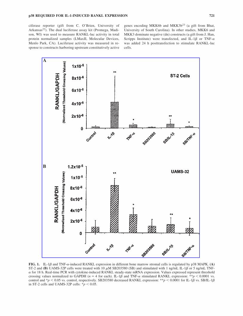
FIG. 1. IL-1� and TNF-�-induced RANKL expression in different bone marrow stromal cells is regulated by p38 MAPK. (A)ST-2 and (B) UAMS-32P cells were treated with 10 �M SB203580 (SB) and stimulated with 1 ng/mL IL-1� or 5 ng/mL TNF-� for 18 h. Real-time PCR with cytokine-induced RANKL steady-state mRNA expression. Values expressed represent thresholdcrossing values normalized to GAPDH (n � 4 for each). IL-1� and TNF-� stimulated RANKL expression: **p � 0.0001 vs.control and *p � 0.05 vs. control, respectively. SB203580 decreased RANKL expression: **p � 0.0001 for IL-1� vs. SB/IL-1�in ST-2 cells and UAMS-32P cells: *p � 0.05.
A
B
Coculture assays
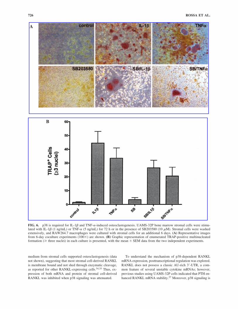
UAMS-32P bone marrow stromal cells (1 � 104) were cul-tured in 6-well dishes for 24 h, deinduced for 6 h in 0.3% serumcontaining medium, and stimulated for 72 h as described for im-munoblot analysis. Following the 72-h incubation period, stromalcells were washed extensively with PBS, and RAW264.7 macro-phages were added at 2 � 104 cells/well and cocultured with ad-herent stromal cells in �-MEM containing 10% serum for 6 days,with the medium replaced on day 3. For control cultures,RAW264.7 cells only were cultured with or without RANKL (50ng/mL). Following coculture, cells were cytochemically stainedfor tartrate-resistant acid phosphatase (TRAP), using a commer-cially available kit (Sigma). TRAP-positive multinucleated cellscontaining more than three nuclei were identified as osteoclastsand counted by light microscopy. All experiments were carriedout three times in triplicate measurements. Images of cultured cellswere digitally captured using a Nikon TS100 inverted scope andNikon 5.1 megapixel camera (Nikon, Tokyo, Japan).
Statistical analysis
Pairwise comparisons between experimental groups wereperformed using the Student’s t-test with Welch’s correctionfor unequal variances or one-way ANOVA analysis where in-dicated. Significance level was set at 5% unless otherwisenoted. All calculations were performed using Prism 4 software(GraphPad, Inc., San Diego, CA).
RESULTS
SB203580 inhibits IL-1� and TNF�-induced RANKL mRNA steady-state levels in bone marrow stromal cells
Real-time PCR was used to quantitate stromal cell-derivedRANKL expression. By comparison of normalized threshold
crossing values (Fig. 1), results indicate that steady-state RNAlevels were increased approximately 17-fold by IL-1� treatment(p � 0.0001) and subsequently reduced 70%–90% by pretreat-ment with SB203580 (Fig. 1) (p � 0.0001 in ST-2 cells andp � 0.05 in UMAS-32P cells). Inhibition of TNF-�-stimulatedRANKL expression was observed following addition of a p38inhibitor; however, the level of stimulation with TNF-� wasconsistently less than the stimulation observed with IL-1� inboth ST-2 and UAMS-32P cells (p � 0.05 vs. control).
Inhibition of p38 MAPK decreases IL-1� and TNF-�-induced membrane-bound RANKL expression
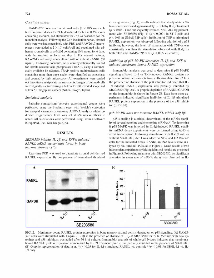
Immunoblot analysis was used to determine if p38 MAPKsignaling affected IL-1 or TNF-induced RANKL protein ex-pression. Whole cell extracts from cells stimulated for 72 h inthe presence or absence of the p38 inhibitor indicated that IL-1�-induced RANKL expression was partially inhibited bySB203580 (Fig. 2A). A graphic depiction of RANKL:GAPDHon the immunoblot is shown in Figure 2B. Data from three ex-periments indicated significant inhibition of IL-1�-stimulatedRANKL protein expression in the presence of the p38 inhibi-tor (p � 0.01).
p38 MAPK does not increase RANKL mRNA half-life
p38 signaling is a critical determinant of the mRNA stabil-ity of several cytokine and chemokine mRNAs.24 To determineif p38 MAPK was involved in IL-1�-induced RANKL stabil-ity, mRNA decay experiments were performed using ActD toarrest transcription. Following stimulation with IL-1� with orwithout SB203580, ActD was added to ST-2 and UAMS-32Pcells for the indicated times. RANKL mRNA levels were ana-lyzed by real-time RT-PCR, as in Figure 1. Mean results of twoindependent experiments yielding identical results are presentedin Figure 3. Following treatment with SB203580, no significantalteration in mean rate of mRNA decay was observed in IL-
ROSSA ET AL.722
FIG. 2. Membrane-bound RANKL protein expression in bone marrow stromal cells is dependent on p38 signaling. (A) UAMS-32P cells were stimulated with 1 ng/mL IL-1� in the presence or absence of 10 �M SB203580 for 72 h. Medium with new cy-tokines and p38 inhibitors was added after 36 h of culture. Immunoblot analysis of whole cell lysates indicates that membrane-bound RANKL protein expression is increased by IL-1� treatment (lane 2) but partially inhibited in the presence of SB203580.(B) Graphic representation of data in A. *p � 0.05 for IL-1�-stimulated RANKL vs. control; **p � 0.01 for SB/IL-1� vs. IL-1� only.
A B
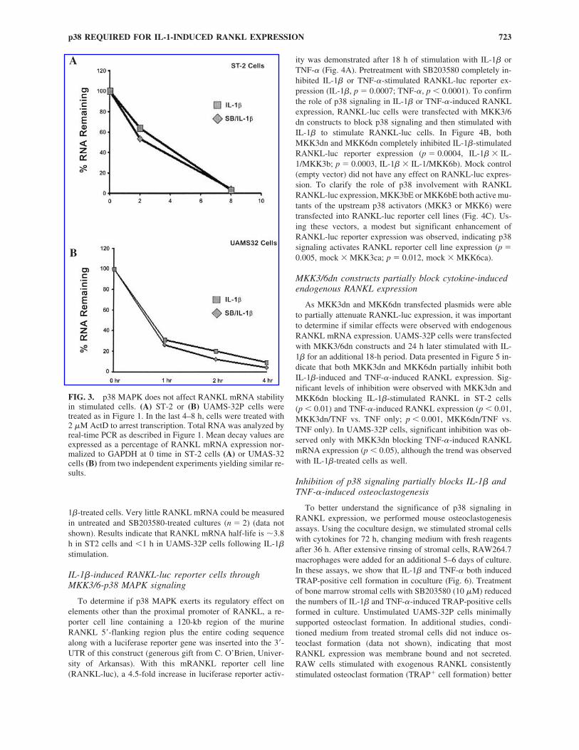
1�-treated cells. Very little RANKL mRNA could be measuredin untreated and SB203580-treated cultures (n � 2) (data notshown). Results indicate that RANKL mRNA half-life is �3.8h in ST2 cells and �1 h in UAMS-32P cells following IL-1�stimulation.
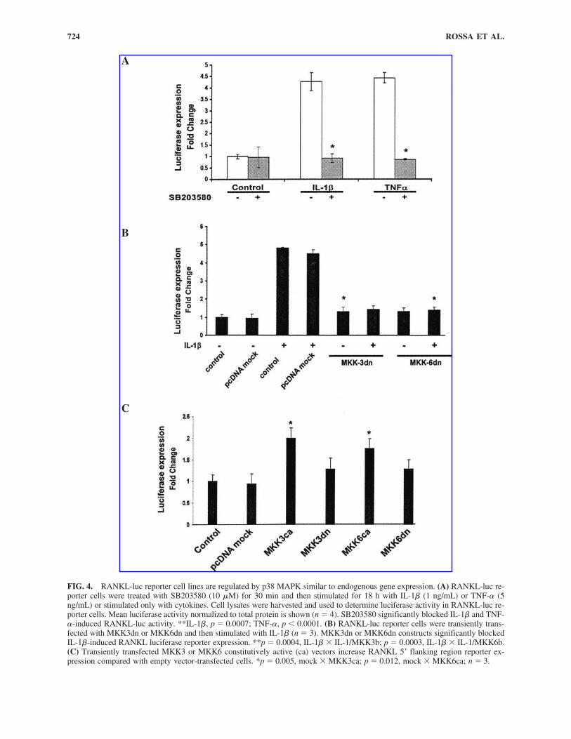
IL-1�-induced RANKL-luc reporter cells throughMKK3/6-p38 MAPK signaling
To determine if p38 MAPK exerts its regulatory effect onelements other than the proximal promoter of RANKL, a re-porter cell line containing a 120-kb region of the murineRANKL 5�-flanking region plus the entire coding sequencealong with a luciferase reporter gene was inserted into the 3�-UTR of this construct (generous gift from C. O’Brien, Univer-sity of Arkansas). With this mRANKL reporter cell line(RANKL-luc), a 4.5-fold increase in luciferase reporter activ-
ity was demonstrated after 18 h of stimulation with IL-1� orTNF-� (Fig. 4A). Pretreatment with SB203580 completely in-hibited IL-1� or TNF-�-stimulated RANKL-luc reporter ex-pression (IL-1�, p � 0.0007; TNF-�, p � 0.0001). To confirmthe role of p38 signaling in IL-1� or TNF-�-induced RANKLexpression, RANKL-luc cells were transfected with MKK3/6dn constructs to block p38 signaling and then stimulated withIL-1� to stimulate RANKL-luc cells. In Figure 4B, bothMKK3dn and MKK6dn completely inhibited IL-1�-stimulatedRANKL-luc reporter expression (p � 0.0004, IL-1� � IL-1/MKK3b; p � 0.0003, IL-1� � IL-1/MKK6b). Mock control(empty vector) did not have any effect on RANKL-luc expres-sion. To clarify the role of p38 involvement with RANKLRANKL-luc expression, MKK3bE or MKK6bE both active mu-tants of the upstream p38 activators (MKK3 or MKK6) weretransfected into RANKL-luc reporter cell lines (Fig. 4C). Us-ing these vectors, a modest but significant enhancement ofRANKL-luc reporter expression was observed, indicating p38signaling activates RANKL reporter cell line expression (p �0.005, mock � MKK3ca; p � 0.012, mock � MKK6ca).
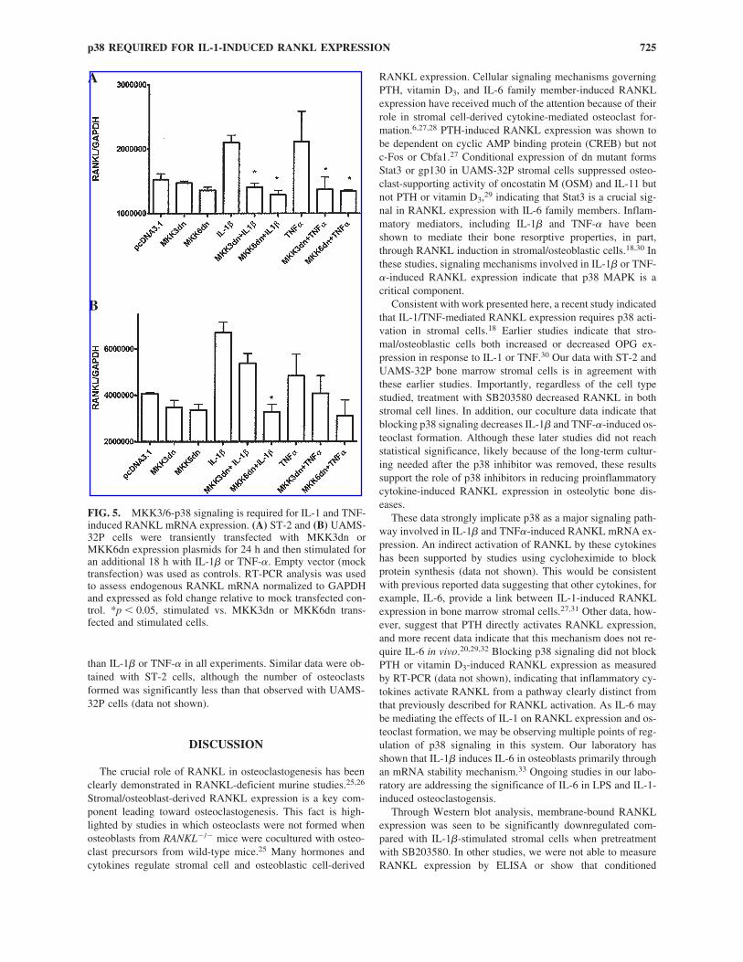
MKK3/6dn constructs partially block cytokine-inducedendogenous RANKL expression
As MKK3dn and MKK6dn transfected plasmids were ableto partially attenuate RANKL-luc expression, it was importantto determine if similar effects were observed with endogenousRANKL mRNA expression. UAMS-32P cells were transfectedwith MKK3/6dn constructs and 24 h later stimulated with IL-1� for an additional 18-h period. Data presented in Figure 5 in-dicate that both MKK3dn and MKK6dn partially inhibit bothIL-1�-induced and TNF-�-induced RANKL expression. Sig-nificant levels of inhibition were observed with MKK3dn andMKK6dn blocking IL-1�-stimulated RANKL in ST-2 cells(p � 0.01) and TNF-�-induced RANKL expression (p � 0.01,MKK3dn/TNF vs. TNF only; p � 0.001, MKK6dn/TNF vs.TNF only). In UAMS-32P cells, significant inhibition was ob-served only with MKK3dn blocking TNF-�-induced RANKLmRNA expression (p � 0.05), although the trend was observedwith IL-1�-treated cells as well.
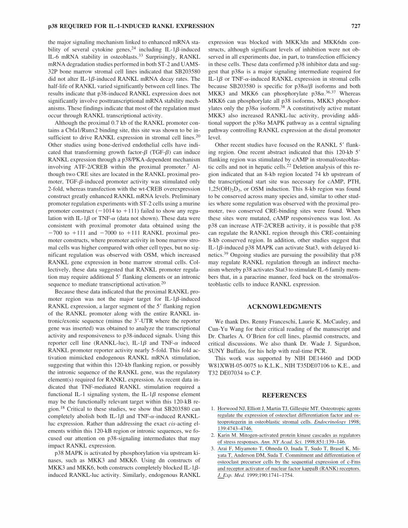
Inhibition of p38 signaling partially blocks IL-1� andTNF-�-induced osteoclastogenesis
To better understand the significance of p38 signaling inRANKL expression, we performed mouse osteoclastogenesisassays. Using the coculture design, we stimulated stromal cellswith cytokines for 72 h, changing medium with fresh reagentsafter 36 h. After extensive rinsing of stromal cells, RAW264.7macrophages were added for an additional 5–6 days of culture.In these assays, we show that IL-1� and TNF-� both inducedTRAP-positive cell formation in coculture (Fig. 6). Treatmentof bone marrow stromal cells with SB203580 (10 �M) reducedthe numbers of IL-1� and TNF-�-induced TRAP-positive cellsformed in culture. Unstimulated UAMS-32P cells minimallysupported osteoclast formation. In additional studies, condi-tioned medium from treated stromal cells did not induce os-teoclast formation (data not shown), indicating that mostRANKL expression was membrane bound and not secreted.RAW cells stimulated with exogenous RANKL consistentlystimulated osteoclast formation (TRAP� cell formation) better
p38 REQUIRED FOR IL-1-INDUCED RANKL EXPRESSION 723
FIG. 3. p38 MAPK does not affect RANKL mRNA stabilityin stimulated cells. (A) ST-2 or (B) UAMS-32P cells weretreated as in Figure 1. In the last 4–8 h, cells were treated with2 �M ActD to arrest transcription. Total RNA was analyzed byreal-time PCR as described in Figure 1. Mean decay values areexpressed as a percentage of RANKL mRNA expression nor-malized to GAPDH at 0 time in ST-2 cells (A) or UMAS-32cells (B) from two independent experiments yielding similar re-sults.
A
B
ROSSA ET AL.724
FIG. 4. RANKL-luc reporter cell lines are regulated by p38 MAPK similar to endogenous gene expression. (A) RANKL-luc re-porter cells were treated with SB203580 (10 �M) for 30 min and then stimulated for 18 h with IL-1� (1 ng/mL) or TNF-� (5ng/mL) or stimulated only with cytokines. Cell lysates were harvested and used to determine luciferase activity in RANKL-luc re-porter cells. Mean luciferase activity normalized to total protein is shown (n � 4). SB203580 significantly blocked IL-1� and TNF-�-induced RANKL-luc activity. **IL-1�, p � 0.0007; TNF-�, p � 0.0001. (B) RANKL-luc reporter cells were transiently trans-fected with MKK3dn or MKK6dn and then stimulated with IL-1� (n � 3). MKK3dn or MKK6dn constructs significantly blockedIL-1�-induced RANKL luciferase reporter expression. **p � 0.0004, IL-1� � IL-1/MKK3b; p � 0.0003, IL-1� � IL-1/MKK6b.(C) Transiently transfected MKK3 or MKK6 constitutively active (ca) vectors increase RANKL 5� flanking region reporter ex-pression compared with empty vector-transfected cells. *p � 0.005, mock � MKK3ca; p � 0.012, mock � MKK6ca; n � 3.
A
B
C
than IL-1� or TNF-� in all experiments. Similar data were ob-tained with ST-2 cells, although the number of osteoclastsformed was significantly less than that observed with UAMS-32P cells (data not shown).
DISCUSSION
The crucial role of RANKL in osteoclastogenesis has beenclearly demonstrated in RANKL-deficient murine studies.25,26
Stromal/osteoblast-derived RANKL expression is a key com-ponent leading toward osteoclastogenesis. This fact is high-lighted by studies in which osteoclasts were not formed whenosteoblasts from RANKL�/� mice were cocultured with osteo-clast precursors from wild-type mice.25 Many hormones andcytokines regulate stromal cell and osteoblastic cell-derived
RANKL expression. Cellular signaling mechanisms governingPTH, vitamin D3, and IL-6 family member-induced RANKLexpression have received much of the attention because of theirrole in stromal cell-derived cytokine-mediated osteoclast for-mation.6,27,28 PTH-induced RANKL expression was shown tobe dependent on cyclic AMP binding protein (CREB) but notc-Fos or Cbfa1.27 Conditional expression of dn mutant formsStat3 or gp130 in UAMS-32P stromal cells suppressed osteo-clast-supporting activity of oncostatin M (OSM) and IL-11 butnot PTH or vitamin D3,29 indicating that Stat3 is a crucial sig-nal in RANKL expression with IL-6 family members. Inflam-matory mediators, including IL-1� and TNF-� have beenshown to mediate their bone resorptive properties, in part,through RANKL induction in stromal/osteoblastic cells.18,30 Inthese studies, signaling mechanisms involved in IL-1� or TNF-�-induced RANKL expression indicate that p38 MAPK is acritical component.
Consistent with work presented here, a recent study indicatedthat IL-1/TNF-mediated RANKL expression requires p38 acti-vation in stromal cells.18 Earlier studies indicate that stro-mal/osteoblastic cells both increased or decreased OPG ex-pression in response to IL-1 or TNF.30 Our data with ST-2 andUAMS-32P bone marrow stromal cells is in agreement withthese earlier studies. Importantly, regardless of the cell typestudied, treatment with SB203580 decreased RANKL in bothstromal cell lines. In addition, our coculture data indicate thatblocking p38 signaling decreases IL-1� and TNF-�-induced os-teoclast formation. Although these later studies did not reachstatistical significance, likely because of the long-term cultur-ing needed after the p38 inhibitor was removed, these resultssupport the role of p38 inhibitors in reducing proinflammatorycytokine-induced RANKL expression in osteolytic bone dis-eases.
These data strongly implicate p38 as a major signaling path-way involved in IL-1� and TNF�-induced RANKL mRNA ex-pression. An indirect activation of RANKL by these cytokineshas been supported by studies using cycloheximide to blockprotein synthesis (data not shown). This would be consistentwith previous reported data suggesting that other cytokines, forexample, IL-6, provide a link between IL-1-induced RANKLexpression in bone marrow stromal cells.27,31 Other data, how-ever, suggest that PTH directly activates RANKL expression,and more recent data indicate that this mechanism does not re-quire IL-6 in vivo.20,29,32 Blocking p38 signaling did not blockPTH or vitamin D3-induced RANKL expression as measuredby RT-PCR (data not shown), indicating that inflammatory cy-tokines activate RANKL from a pathway clearly distinct fromthat previously described for RANKL activation. As IL-6 maybe mediating the effects of IL-1 on RANKL expression and os-teoclast formation, we may be observing multiple points of reg-ulation of p38 signaling in this system. Our laboratory hasshown that IL-1� induces IL-6 in osteoblasts primarily throughan mRNA stability mechanism.33 Ongoing studies in our labo-ratory are addressing the significance of IL-6 in LPS and IL-1-induced osteoclastogensis.
Through Western blot analysis, membrane-bound RANKLexpression was seen to be significantly downregulated com-pared with IL-1�-stimulated stromal cells when pretreatmentwith SB203580. In other studies, we were not able to measureRANKL expression by ELISA or show that conditioned
p38 REQUIRED FOR IL-1-INDUCED RANKL EXPRESSION 725
FIG. 5. MKK3/6-p38 signaling is required for IL-1 and TNF-induced RANKL mRNA expression. (A) ST-2 and (B) UAMS-32P cells were transiently transfected with MKK3dn orMKK6dn expression plasmids for 24 h and then stimulated foran additional 18 h with IL-1� or TNF-�. Empty vector (mocktransfection) was used as controls. RT-PCR analysis was usedto assess endogenous RANKL mRNA normalized to GAPDHand expressed as fold change relative to mock transfected con-trol. *p � 0.05, stimulated vs. MKK3dn or MKK6dn trans-fected and stimulated cells.
A
B
medium from stromal cells supported osteoclastogenesis (datanot shown), suggesting that most stromal cell-derived RANKLis membrane bound and not shed through enzymatic cleavage,as reported for other RANKL-expressing cells.34,35 Thus, ex-pression of both mRNA and protein of stromal cell-derivedRANKL was inhibited when p38 signaling was attenuated.
To understand the mechanism of p38-dependent RANKLmRNA expression, posttranscriptional regulation was explored.RANKL does not possess a classic AU-rich 3�-UTR, a com-mon feature of several unstable cytokine mRNAs; however,previous studies using UAMS-32P cells indicated that PTH en-hanced RANKL mRNA stability.27 Moreover, p38 signaling is
ROSSA ET AL.726
FIG. 6. p38 is required for IL-1� and TNF-�-induced osteoclastogenesis. UAMS-32P bone marrow stromal cells were stimu-lated with IL-1� (1 ng/mL) or TNF-� (5 ng/mL) for 72 h or in the presence of SB203580 (10 �M). Stromal cells were washedextensively, and RAW264.7 macrophages were cultured with stromal cells for an additional 6 days. (A) Representative imagesfrom 6-day coculture experiments (100�) are shown. (B) Graphic representation of enumerated TRAP-positive multinucleatedformation (� three nuclei) in each culture is presented, with the mean SEM data from the two independent experiments.
A
B
the major signaling mechanism linked to enhanced mRNA sta-bility of several cytokine genes,24 including IL-1�-induced IL-6 mRNA stability in osteoblasts.33 Surprisingly, RANKLmRNA degradation studies performed in both ST-2 and UAMS-32P bone marrow stromal cell lines indicated that SB203580did not alter IL-1�-induced RANKL mRNA decay rates. Thehalf-life of RANKL varied significantly between cell lines. Theresults indicate that p38-induced RANKL expression does notsignificantly involve posttranscriptional mRNA stability mech-anisms. These findings indicate that most of the regulation mustoccur through RANKL transcriptional activity.
Although the proximal 0.7 kb of the RANKL promoter con-tains a Cbfa1/Runx2 binding site, this site was shown to be in-sufficient to drive RANKL expression in stromal cell lines.20
Other studies using bone-derived endothelial cells have indi-cated that transforming growth factor-� (TGF-�) can induceRANKL expression through a p38/PKA-dependent mechanisminvolving ATF-2/CREB within the proximal promoter.7 Al-though two CRE sites are located in the RANKL proximal pro-moter, TGF-�-induced promoter activity was stimulated only2-fold, whereas transfection with the wt-CREB overexpressionconstruct greatly enhanced RANKL mRNA levels. Preliminarypromoter regulation experiments with ST-2 cells using a murinepromoter construct (�1014 to �111) failed to show any regu-lation with IL-1� or TNF-� (data not shown). These data wereconsistent with proximal promoter data obtained using the�700 to �111 and �7000 to �111 RANKL proximal pro-moter constructs, where promoter activity in bone marrow stro-mal cells was higher compared with other cell types, but no sig-nificant regulation was observed with OSM, which increasedRANKL gene expression in bone marrow stromal cells. Col-lectively, these data suggested that RANKL promoter regula-tion may require additional 5� flanking elements or an intronicsequence to mediate transcriptional activation.20
Because these data indicated that the proximal RANKL pro-moter region was not the major target for IL-1�-inducedRANKL expression, a larger segment of the 5� flanking regionof the RANKL promoter along with the entire RANKL in-tronic/exonic sequence (minus the 3�-UTR where the reportergene was inserted) was obtained to analyze the transcriptionalactivity and responsiveness to p38-induced signals. Using thisreporter cell line (RANKL-luc), IL-1� and TNF-� inducedRANKL promoter reporter activity nearly 5-fold. This fold ac-tivation mimicked endogenous RANKL mRNA stimulation,suggesting that within this 120-kb flanking region, or possiblythe intronic sequence of the RANKL gene, was the regulatoryelement(s) required for RANKL expression. As recent data in-dicated that TNF-mediated RANKL stimulation required afunctional IL-1 signaling system, the IL-1� response elementmay be the functionally relevant target within this 120-kB re-gion.18 Critical to these studies, we show that SB203580 cancompletely abolish both IL-1� and TNF-�-induced RANKL-luc expression. Rather than addressing the exact cis-acting el-ements within this 120-kB region or intronic sequences, we fo-cused our attention on p38-signaling intermediates that mayimpact RANKL expression.
p38 MAPK is activated by phosphorylation via upstream ki-nases, such as MKK3 and MKK6. Using dn constructs ofMKK3 and MKK6, both constructs completely blocked IL-1�-induced RANKL-luc activity. Similarly, endogenous RANKL
expression was blocked with MKK3dn and MKK6dn con-structs, although significant levels of inhibition were not ob-served in all experiments due, in part, to transfection efficiencyin these cells. These data confirmed p38 inhibitor data and sug-gest that p38� is a major signaling intermediate required forIL-1� or TNF-�-induced RANKL expression in stromal cellsbecause SB203580 is specific for p38�/� isoforms and bothMKK3 and MKK6 can phosphorylate p38�.36,37 WhereasMKK6 can phosphorylate all p38 isoforms, MKK3 phosphor-ylates only the p38� isoform.38 A constitutively active mutantMKK3 also increased RANKL-luc activity, providing addi-tional support the p38� MAPK pathway as a central signalingpathway controlling RANKL expression at the distal promoterlevel.
Other recent studies have focused on the RANKL 5� flank-ing region. One recent abstract indicated that this 120-kb 5�flanking region was stimulated by cAMP in stromal/osteoblas-tic cells and not in hepatic cells.22 Deletion analysis of this re-gion indicated that an 8-kb region located 74 kb upstream ofthe transcriptional start site was necessary for cAMP, PTH,1,25(OH)2D3, or OSM induction. This 8-kb region was foundto be conserved across many species and, similar to other stud-ies where some regulation was observed with the proximal pro-moter, two conserved CRE-binding sites were found. Whenthese sites were mutated, cAMP responsiveness was lost. Asp38 can increase ATF-2/CREB activity, it is possible that p38can regulate the RANKL region through this CRE-containing8-kb conserved region. In addition, other studies suggest thatIL-1�-induced p38 MAPK can activate Stat3, with delayed ki-netics.39 Ongoing studies are pursuing the possibility that p38may regulate RANKL regulation through an indirect mecha-nism whereby p38 activates Stat3 to stimulate IL-6 family mem-bers that, in a paracrine manner, feed back on the stromal/os-teoblastic cells to induce RANKL expression.
ACKNOWLEDGMENTS
We thank Drs. Renny Franceschi, Laurie K. McCauley, andCun-Yu Wang for their critical reading of the manuscript andDr. Charles A. O’Brien for cell lines, plasmid constructs, andcritical discussions. We also thank Dr. Wade J. Sigurdson,SUNY Buffalo, for his help with real-time PCR.
This work was supported by NIH DE14460 and DODW81XWH-05-0075 to K.L.K., NIH T35DE07106 to K.E., andT32 DE07034 to C.P.
REFERENCES
1. Horwood NJ, Elliott J, Martin TJ, Gillespie MT. Osteotropic agentsregulate the expression of osteoclast differentiation factor and os-teoprotegerin in osteoblastic stromal cells. Endocrinology 1998;139:4743–4746.
2. Karin M. Mitogen-activated protein kinase cascades as regulatorsof stress responses. Ann. NY Acad. Sci. 1998;851:139–146.
3. Arai F, Miyamoto T, Ohneda O, Inada T, Sudo T, Brasel K, Mi-yata T, Anderson DM, Suda T. Commitment and differentiation ofosteoclast precursor cells by the sequential expression of c-Fmsand receptor activator of nuclear factor kappaB (RANK) receptors.J. Exp. Med. 1999;190:1741–1754.
p38 REQUIRED FOR IL-1-INDUCED RANKL EXPRESSION 727
4. Filvaroff E, Derynck R. Bone remodelling: a signalling system forosteoclast regulation. Curr. Biol. 1998;8:R679–682.
5. Ikeda T, Utsuyama M, Hirokawa K. Expression profiles of recep-tor activator of nuclear factor kappaB ligand, receptor activator ofnuclear factor kappaB, and osteoprotegerin messenger RNA in agedand ovariectomized rat bones. J. Bone Miner. Res. 2001;16:1416–1425.
6. Palmqvist P, Persson E, Conaway HH, Lerner UH. IL-6, leukemiainhibitory factor, and oncostatin M stimulate bone resorption andregulate the expression of receptor activator of NF-kappa B ligand,osteoprotegerin, and receptor activator of NF-kappa B in mousecalvariae. J. Immunol. 2002;169:3353–3362.
7. Ishida A, Fujita N, Kitazawa R, Tsuruo T. Transforming growthfactor-beta induces expression of receptor activator of NF-kappa Bligand in vascular endothelial cells derived from bone. J. Biol.Chem. 2002;277:26217–26224.
8. Hofbauer LC, Heufelder AE. Role of receptor activator of nuclearfactor-kappaB ligand and osteoprotegerin in bone cell biology. J.Mol. Med. 2001;79:243–253.
9. Simonet WS, Lacey DL, Dunstan CR, Kelley M, Chang MS, LuthyR, Nguyen HQ, Wooden S, Bennett L, Boone T, Shimamoto G,DeRose M, Elliott R, Colombero A, Tan HL, Trail G, Sullivan J,Davy E, Bucay N, Renshaw-Gegg L, Hughes TM, Hill D, PattisonW, Campbell P, Boyle WJ, et al. Osteoprotegerin: a novel secretedprotein involved in the regulation of bone density. Cell 1997;89:309–319.
10. Thomas GP, Baker SU, Eisman JA, Gardiner EM. ChangingRANKL/OPG mRNA expression in differentiating murine primaryosteoblasts. J. Endocrinol. 2001;170:451–460.
11. Gonzalez GA, Montminy MR. Cyclie AMP stimulates somatostatingene transcription by phosphorylation of CREB at serine 133. Cell1989;59:675–680.
12. Haskill S, Beg AA, Tompkins SM, Morris JS, Yurochko AD,Sampson-Johannes A, Mondal K, Ralph P, Baldwin AS Jr. Char-acterization of an immediate-early gene induced in adherent mono-cytes that encodes I kappa B-like activity. Cell 1991;65:1281–1289.
13. Schett G, Tohidast-Akrad M, Smolen JS, Schmid BJ, Steiner CW,Bitzan P, Zenz P, Redlich K, Xu Q, Steiner G. Activation, differ-ential localization, and regulation of the stress-activated protein ki-nases, extracellular signal-regulated kinase, c-JUN N-terminal ki-nase, and p38 mitogen-activated protein kinase, in synovial tissueand cells in rheumatoid arthritis. Arthritis Rheum. 2004;43:2501–2512.
14. Mansky KC, Sankar U, Han J, Ostrowski MC. Microphthalmiatranscription factor is a target of the p38 MAPK pathway in re-sponse to receptor activator of NF-kappa B ligand signaling. J. Biol.Chem. 2002;277:11077–11083.
15. Branger J, van den Blink B, Weijer S, Madwed J, Bos CL, GuptaA, Yong CL, Polmar SH, Olszyna DP, Hack CE, van Deventer SJ,Peppelenbosch MP, van der Poll T. Anti-inflammatory effects ofa p38 mitogen-activated protein kinase inhibitor during human en-dotoxemia. J. Immunol. 2002;168:4070–4077.
16. Kumar S, Boehm J, Lee JC. p38 MAP kinases: key signalling mol-ecules as therapeutic targets for inflammatory diseases. Nat. Rev.Drug Discov. 2003;2:717–726.
17. Matsumoto M, Sudo T, Saito T, Osada H, Tsujimoto M. Involve-ment of p38 mitogen-activated protein kinase signaling pathway inosteoclastogenesis mediated by receptor activator of NF-kappa Bligand (RANKL). J. Biol. Chem. 2000;275:31155–31161.
18. Wei S, Kitaura H, Zhou P, Ross FP, Teitelbaum SL. IL-1 medi-ates TNF-induced osteoclastogenesis. J. Clin. Invest. 2005;115:282–290.
19. Dai JC, He P, Chen X, Greenfield EM. TNFalpha and PTH utilizedistinct mechanisms to induce IL-6 and RANKL expression withmarkedly different kinetics. Bone 2006;38:509–520.
20. O’Brien CA, Kern B, Gubrij I, Karsenty G, Manolagas SC. Cbfa1does not regulate RANKL gene activity in stromal/osteoblasticcells. Bone 2002;30:453–462.
21. Kirkwood K, Martin T, Andreadis ST, Kim YJ. Chemically mod-ified tetracyclines selectively inhibit IL-6 expression in osteoblastsby decreasing mRNA stability. Biochem. Pharmacol. 2003;66:1809–1819.
22. Fu Q, Manolagas SC, O’Brien CA. Parathyroid hormone controlsreceptor activator of NF-�B ligand gene expression via a distanttranscriptional enhancer. Mol. Cell Biol. 2006;26:6453–6468.
23. Bhat NR, Feinstein DL, Shen Q, Bhat AN. p38 MAPK-mediatedtranscriptional activation of inducible nitric-oxide synthase in glialcells. Roles of nuclear factors, nuclear factor kappa B, cAMP re-sponse element-binding protein, CCAAT/enhancer-binding pro-tein-beta, and activating transcription factor-2. J. Biol. Chem.2002;277:29584–29592.
24. Tebo J, Der S, Frevel M, Khabar KS, Williams BR, Hamilton TA.Heterogeneity in control of mRNA stability by AU-rich elements.J. Biol. Chem. 2003;278:12085–12093.
25. Kong YY, Yoshida H, Sarosi I, Tan HL, Timms E, Capparelli C,Morony S, Oliveira-dos-Santos AJ, Van G, Itie A, Khoo W, Wake-ham A, Dunstan CR, Lacey DL, Mak TW, Boyle WJ, PenningerJM. OPGL is a key regulator of osteoclastogenesis, lymphocyte de-velopment and lymph-node organogenesis. Nature 1999;397:315–323.
26. Kong YY, Boyle WJ, Penninger JM. Osteoprotegerin ligand: acommon link between osteoclastogenesis, lymph node formationand lymphocyte development. Immunol. Cell. Biol. 1999;77:188–193.
27. Fu Q, Jilka RL, Manolagas SC, O’Brien CA. Parathyroid hormonestimulates receptor activator of NFkappa B ligand and inhibits os-teoprotegerin expression via protein kinase A activation of cAMP-response element-binding protein. J. Biol. Chem. 2002;277:48868–48875.
28. Kitazawa S, Kajimoto K, Kondo T, Kitazawa R. Vitamin D3 sup-ports osteoclastogenesis via functional vitamin D response elementof human RANKL gene promoter. J. Cell. Biochem. 2003;89:771–777.
29. O’Brien CA, Gubrij I, Lin SC, Saylors RL, Manolagas SC. Stat3activation in stromal/osteoblastic cells is required for induction ofthe receptor activator of NF-kappaB ligand and stimulation of os-teoclastogenesis by gp130-utilizing cytokines or interleukin-1 butnot 1,25-dihydroxyvitamin D3 or parathyroid hormone. J. Biol.Chem. 1999;274:19301–19308.
30. Hofbauer LC, Lacey DL, Dunstan CR, Spelsberg TC, Riggs BL,Khosla S. Interleukin-1beta and tumor necrosis factor-alpha, butnot interleukin-6, stimulate osteoprotegerin ligand gene expressionin human osteoblastic cells. Bone 1999;25:255–259.
31. Devlin RD, Reddy SV, Savino R, Ciliberto G, Roodman GD. IL-6 mediates the effects of IL-1 or TNF, but not PTHrP or 1,25(OH)2D3, on osteoclast-like cell formation in normal human bone marrow cultures. J. Bone Miner. Res. 1998;13:393–399.
32. O’Brien CA, Jilka RL, Fu Q, Stewart S, Weinstein RS, Manola-gas SC. IL-6 is not required for parathyroid hormone stimulationof RANKL expression, osteoclast formation, and bone loss in mice.Am. J. Physiol. Endocrinol. Metab. 2005;289:E784–E793.
33. Patil C, Zhu X, Rossa C Jr, Kim YJ, Kirkwood KL. p38 MAPKregulates IL-1beta induced IL-6 expression through mRNA stabil-ity in osteoblasts. Immunol. Invest. 2004;33:213–233.
34. Horiki M, Nakase T, Myoui A, Sugano N, Nishii T, Tomita T,Miyaji T, Yoshikawa H. Localization of RANKL in osteolytic tis-sue around a loosened joint prosthesis. J. Bone Miner. Metab.2004;22:346–351.
35. Miyamoto N, Higuchi Y, Mori K, Ito M, Tsurudome M, Nishio M,
ROSSA ET AL.728
Yamada H, Sudo A, Kato K, Uchida A, Ito Y. Human osteosar-coma-derived cell lines produce soluble factor(s) that induces dif-ferentiation of blood monocytes to osteoclast-like cells. Int. Im-munopharmacol. 2002;2:25–38.
36. Cuenda A, Cohen P, Buee-Scherrer V, Goedert M. Activation ofstress-activated protein kinase-3 (SAPK3) by cytokines and cellu-lar stresses is mediated via SAPKK3 (MKK6): comparison of thespecificities of SAPK3 and SAPK2 (RK/p38). EMBO J. 1997;16:295–305.
37. Kumar S, McDonnell PC, Gum RJ, Hand AT, Lee JC, Young PR.Novel homologues of CSBP/p38 MAP kinase: activation, substratespecificity and sensitivity to inhibition by pyridinyl imidazoles.Biochem. Biophys. Res. Commun. 1997;235:533–538.
38. Shi Y, Gaestel M. In the cellular garden of forking paths: how p38MAPKs signal for downstream assistance. Biol. Chem. 2002;383:1519–1536.
39. Ng DC, Long CS, Bogoyevitch MA. A role for the extracellularsignal-regulated kinase and p38 mitogen-activated protein kinases
in interleukin-1 beta-stimulated delayed signal transducer and ac-tivator of transcription 3 activation, atrial natriuretic factor ex-pression, and cardiac myocyte morphology. J. Biol. Chem. 2001;276:29490–29498.
Address reprint requests or correspondence to:Dr. Keith L. Kirkwood
Department of Periodontics and Oral MedicineUniversity of Michigan
1011 N. University AvenueAnn Arbor, MI 48109-1078
Tel: (734) 763-7120Fax: (734) 763-5503
E-mail: [email protected]
Received 10 February 2006/Accepted 20 April 2006
p38 REQUIRED FOR IL-1-INDUCED RANKL EXPRESSION 729
This article has been cited by:
1. Chetan S Patil, Min Liu, Wenpu Zhao, Derek D Coatney, Fei Li, Elizabeth A VanTubergen, Nisha J D'Silva, Keith L Kirkwood.2008. Targeting mRNA Stability Arrests Inflammatory Bone Loss. Molecular Therapy 16:10, 1657-1664. [CrossRef]
2. Kageyama Yasunori, Takahashi Masaaki, Nagafusa Tetsuyuki, Kobayashi Hayato, Nagano Akira. 2008. Reduction of urinary levelsof pyridinoline and deoxypyridinoline and serum levels of soluble receptor activator of NF-kappaB ligand by etanercept in patientswith rheumatoid arthritis. Clinical Rheumatology 27:9, 1093-1101. [CrossRef]
3. Y. Hiruma, N. Kurihara, M. A. Subler, H. Zhou, C. S. Boykin, H. Zhang, S. Ishizuka, D. W. Dempster, G. D. Roodman,J. J. Windle. 2008. A SQSTM1/p62 mutation linked to Paget's disease increases the osteoclastogenic potential of the bonemicroenvironment. Human Molecular Genetics 17:23, 3708-3719. [CrossRef]
4. Yuan Li, Xiaonan Zhang, Biao Zhu, Zhanggang Xue. 2008. Desflurane Preconditioning Inhibits Endothelial Nuclear Factor-??-BActivation by Targeting the Proximal End of Tumor Necrosis Factor-?? Signaling. Anesthesia & Analgesia 106:5, 1473-1479.[CrossRef]
5. Kumaran Sundaram, Santhosh K. Mani, Kazuyuki Kitatani, Kongming Wu, Richard G. Pestell, Sakamuri V. Reddy. 2008. DACH1negatively regulates the human RANK ligand gene expression in stromal/preosteoblast cells. Journal of Cellular Biochemistry 103:6,1747-1759. [CrossRef]
6. C. Rossa, M. Liu, K. L. Kirkwood. 2008. A dominant function of p38 mitogen-activated protein kinase signaling in receptoractivator of nuclear factor-κB ligand expression and osteoclastogenesis induction by Aggregatibacter actinomycetemcomitans andEscherichia coli lipopolysaccharide. Journal of Periodontal Research 43:2, 201-211. [CrossRef]
7. G D Roodman. 2008. Treatment strategies for bone disease. Bone Marrow Transplantation 40:12, 1139-1146. [CrossRef]8. Jill E. Rogers, Fei Li, Derek D. Coatney, Jodie Otremba, Jaclynn M. Kriegl, Andrew A. Protter, Linda S. Higgins, Satyanarayana
Medicherla, Keith L. Kirkwood. 2007. A p38 Mitogen-Activated Protein Kinase Inhibitor Arrests Active Alveolar Bone Loss ina Rat Periodontitis Model. Journal of Periodontology 78:10, 1992-1998. [CrossRef]
9. Jill E. Rogers, Fei Li, Derek D. Coatney, Carlos Rossa, Paul Bronson, Jaclynn M. Krieder, William V. Giannobile, KeithL. Kirkwood. 2007. Actinobacillus actinomycetemcomitans Lipopolysaccharide-Mediated Experimental Bone Loss Model forAggressive Periodontitis. Journal of Periodontology 78:3, 550-558. [CrossRef]
Related Documents