A RANKL G278R mutation causing osteopetrosis identifies a functional amino acid essential for trimer assembly in RANKL and TNF Eleni Douni 1,2, ∗ , Vagelis Rinotas 1 , Eleni Makrinou 1 , Jochen Zwerina 3 , Josef M. Penninger 4 , Elias Eliopoulos 2 , Georg Schett 3 and George Kollias 1 1 Institute of Immunology, Biomedical Sciences Research Center ‘Alexander Fleming’, Vari 16672, Greece, 2 Department of Agricultural Biotechnology, Agricultural University of Athens, Athens 11855, Greece, 3 Department of Internal Medicine 3, Rheumatology and Immunology, University of Erlangen-Nuremberg, Erlangen D-91054, Germany and 4 Institute of Molecular Biotechnology of the Austrian Academy of Sciences, Vienna A-1030, Austria Received September 6, 2011; Revised and Accepted October 29, 2011 Receptor activator of nuclear factor-kB ligand (RANKL), a trimeric tumor necrosis factor (TNF) superfamily member, is the central mediator of osteoclast formation and bone resorption. Functional mutations in RANKL lead to human autosomal recessive osteopetrosis (ARO), whereas RANKL overexpression has been implicated in the pathogenesis of bone degenerative diseases such as osteoporosis. Following a for- ward genetics approach using N-ethyl-N-nitrosourea (ENU)-mediated random mutagenesis, we generated a novel mouse model of ARO caused by a new loss-of-function allele of Rankl with a glycine-to-arginine mutation at codon 278 (G278R) at the extracellular inner hydrophobic F b-strand of RANKL. Mutant mice develop severe osteopetrosis similar to Rankl-deficient mice, whereas exogenous administration of recombinant RANKL restores osteoclast formation in vivo. We show that RANKL G278R monomers fail to assemble into homotrimers, are unable to bind and activate the RANK receptor and interact with wild-type RANKL exerting a dominant-negative effect on its trimerization and function in vitro. Since G278 is highly conserved within the TNF superfamily, we identified that a similar substitution in TNF, G122R, also abrogated trimerization, binding to TNF receptor and consequently impaired TNF biological activity. Notably, SPD304, a potent small-molecule inhibitor of TNF trimerization that interacts with G122, also inhibited RANKL activity, suggesting analogous inhibitory mechanisms. Our results provide a new disease model for ARO and identify a functional amino acid in the TNF-like core domain essential for trimer formation both in RANKL and in TNF that could be considered a novel potential target for inhi- biting their biological activities. INTRODUCTION Bone remodeling is a constant process that functions through the synthesis of bone matrix by osteoblasts and the coordinate bone resorption by osteoclasts (1,2). Normally, osteoblastic and osteoclastic activities are balanced so that skeletal integ- rity is preserved. Perturbations in bone remodeling can result in skeletal abnormalities such as osteopetrosis and osteopor- osis which are characterized by excessive or decreased bone mass, due to impaired or enhanced osteoclast activity. Recep- tor activator of nuclear factor-kB ligand (RANKL) is the primary mediator of osteoclast-induced bone resorption (3) and belongs to the tumor necrosis factor (TNF) superfamily (4,5) that is characterized by homotrimerization. It is a type II transmembrane protein that consists of a short N-terminal cytoplasmic domain and a conserved extracellular TNF-like core domain forming an antiparallel b-sheet that is predicted to assemble into a trimer required for receptor activation ∗ To whom correspondence should be addressed at: Biomedical Sciences Research Center ‘Alexander Fleming’, 34 Fleming str., 16672 Vari, Greece. Tel: +30 2109656310; Fax: +30 2109656563; Email: douni@fleming.gr # The Author 2011. Published by Oxford University Press. All rights reserved. For Permissions, please email: [email protected] Human Molecular Genetics, 2012, Vol. 21, No. 4 784–798 doi:10.1093/hmg/ddr510 Advance Access published on November 7, 2011 by guest on February 19, 2013 http://hmg.oxfordjournals.org/ Downloaded from

Welcome message from author
This document is posted to help you gain knowledge. Please leave a comment to let me know what you think about it! Share it to your friends and learn new things together.
Transcript

A RANKL G278R mutation causing osteopetrosisidentifies a functional amino acid essentialfor trimer assembly in RANKL and TNF
Eleni Douni1,2,∗, Vagelis Rinotas1, Eleni Makrinou1, Jochen Zwerina3, Josef M. Penninger4,
Elias Eliopoulos2, Georg Schett3 and George Kollias1
1Institute of Immunology, Biomedical Sciences Research Center ‘Alexander Fleming’, Vari 16672, Greece,2Department of Agricultural Biotechnology, Agricultural University of Athens, Athens 11855, Greece, 3Department of
Internal Medicine 3, Rheumatology and Immunology, University of Erlangen-Nuremberg, Erlangen D-91054, Germany
and 4Institute of Molecular Biotechnology of the Austrian Academy of Sciences, Vienna A-1030, Austria
Received September 6, 2011; Revised and Accepted October 29, 2011
Receptor activator of nuclear factor-kB ligand (RANKL), a trimeric tumor necrosis factor (TNF) superfamilymember, is the central mediator of osteoclast formation and bone resorption. Functional mutations inRANKL lead to human autosomal recessive osteopetrosis (ARO), whereas RANKL overexpression hasbeen implicated in the pathogenesis of bone degenerative diseases such as osteoporosis. Following a for-ward genetics approach using N-ethyl-N-nitrosourea (ENU)-mediated random mutagenesis, we generated anovel mouse model of ARO caused by a new loss-of-function allele of Rankl with a glycine-to-argininemutation at codon 278 (G278R) at the extracellular inner hydrophobic F b-strand of RANKL. Mutantmice develop severe osteopetrosis similar to Rankl-deficient mice, whereas exogenous administrationof recombinant RANKL restores osteoclast formation in vivo. We show that RANKLG278R monomers failto assemble into homotrimers, are unable to bind and activate the RANK receptor and interact withwild-type RANKL exerting a dominant-negative effect on its trimerization and function in vitro. SinceG278 is highly conserved within the TNF superfamily, we identified that a similar substitution in TNF,G122R, also abrogated trimerization, binding to TNF receptor and consequently impaired TNF biologicalactivity. Notably, SPD304, a potent small-molecule inhibitor of TNF trimerization that interacts withG122, also inhibited RANKL activity, suggesting analogous inhibitory mechanisms. Our results providea new disease model for ARO and identify a functional amino acid in the TNF-like core domain essentialfor trimer formation both in RANKL and in TNF that could be considered a novel potential target for inhi-biting their biological activities.
INTRODUCTION
Bone remodeling is a constant process that functions throughthe synthesis of bone matrix by osteoblasts and the coordinatebone resorption by osteoclasts (1,2). Normally, osteoblasticand osteoclastic activities are balanced so that skeletal integ-rity is preserved. Perturbations in bone remodeling can resultin skeletal abnormalities such as osteopetrosis and osteopor-osis which are characterized by excessive or decreased bone
mass, due to impaired or enhanced osteoclast activity. Recep-tor activator of nuclear factor-kB ligand (RANKL) is theprimary mediator of osteoclast-induced bone resorption (3)and belongs to the tumor necrosis factor (TNF) superfamily(4,5) that is characterized by homotrimerization. It is a typeII transmembrane protein that consists of a short N-terminalcytoplasmic domain and a conserved extracellular TNF-likecore domain forming an antiparallel b-sheet that is predictedto assemble into a trimer required for receptor activation
∗To whom correspondence should be addressed at: Biomedical Sciences Research Center ‘Alexander Fleming’, 34 Fleming str., 16672 Vari, Greece.Tel: +30 2109656310; Fax: +30 2109656563; Email: [email protected]
# The Author 2011. Published by Oxford University Press. All rights reserved.For Permissions, please email: [email protected]
Human Molecular Genetics, 2012, Vol. 21, No. 4 784–798doi:10.1093/hmg/ddr510Advance Access published on November 7, 2011
by guest on February 19, 2013http://hm
g.oxfordjournals.org/D
ownloaded from

(6,7). Soluble RANKL is generated either by proteolytic pro-cessing of the transmembrane form or by alternative splicing(8,9). RANKL is expressed on activated T lymphocytes (4,5)as well as on stromal cells (10,11) and binds as a trimerto its receptor RANK that is expressed on the surface ofosteoclast precursors and mature osteoclasts. RANKL andRANK form a heterohexameric complex with a receptormolecule bound along each of the three clefts formed byneighboring monomers of the RANKL homotrimer. This inter-action is necessary for osteoclast differentiation, activity andsurvival (10,12), which subsequently lead to bone resorption.Osteoprotegerin (OPG), a decoy receptor of RANKL, inhibitsthe binding of RANKL to RANK and thereby limits osteoclas-togenesis (11). Genetic ablations of either RANKL (13,14)or RANK (15,16) result in severe osteopetrosis due tocomplete lack of osteoclast formation, demonstrating thatRANKL and RANK are indispensable for osteoclastogenesis,whereas absence of OPG causes increased osteoclasto-genesis and osteopenia (17). Although RANKL is bestknown for its role in bone resorption, it also plays multipleroles in the immune system (4,5,13,18,19), in mammarygland development during pregnancy (20), thermoregulation(21), cancer metastasis (22) and hormone-derived breastdevelopment (23).
As a result of its effects on bone metabolism, RANKL isconsidered a major therapeutic target for the suppression ofbone resorption in bone metabolic diseases such as osteopor-osis, rheumatoid arthritis and cancer metastasis (24). Conse-quently, recombinant proteins that inhibit RANKL–RANKinteractions, such as RANK-Fc, Fc-OPG and anti-RANKLantibodies, have been developed as agents against osteopor-osis. Indeed, clinical trials with denosumab, a fully humanmonoclonal antibody against RANKL, showed an increasedbone mass and reduced incidence of fractures in postmenopau-sal women with osteoporosis (25) and in prostate cancerpatients receiving androgen-deprivation therapy (26). Thisantibody has been recently approved in the USA and EU forthe treatment of patients with osteoporosis and in prostatecancer patients undergoing hormonal ablation therapy.However, inhibitors that target formation of functionalRANKL trimers have not been reported yet.
Additionally, a variety of loss-of-function mutations loca-lized within the extracellular domain of RANKL havebeen recently reported in children with autosomal recessiveosteopetrosis (ARO) (OMIM 602642), an incurable raregenetic disease (27). However, the molecular mechanisms bywhich the identified mutations cause RANKL inactivationand osteopetrosis have not been fully resolved. Characteriza-tion of functional RANKL mutations existing either inhumans or in animal models is a powerful approach for theelucidation of the molecular basis of ARO as well as for thepotential design of novel RANKL inhibitors. The aim of thisstudy was to characterize a novel loss-of-function point muta-tion in the Rankl gene that causes osteopetrosis in mice, inorder to define the underlying molecular mechanismthat results in ARO pathogenesis, investigate whether asimilar loss-of-function mechanism exists in other TNFfamily members such as TNF and examine whether the iden-tified amino acid could serve as a potential target for RANKLinhibition.
RESULTS
Generation of a novel ENU-induced mouse model of severeosteopetrosis
The toothless (tles) phenotype was identified as a recessivetrait in which complete failure of tooth eruption was detectedin N-ethyl-N-nitrosourea (ENU)-mutagenized G3 mice in bothsexes (Supplementary Material, Fig. S1A). Mutant mice dis-played also growth retardation, and lymphoid aberrations char-acterized by thymic hypoplasia, enlarged spleens and absenceof lymph nodes. Additionally, these mice displayed earlylethality, where 60% of the tles/tles mice died by theseventh week of age (Supplementary Material, Fig. S1B).Since failure of tooth eruption is a typical finding in osteope-trosis, we performed extensive histological analysis of thetibiae and femurs in 4–6-week-old tles/tles mice and wild-type (WT) control littermates. Staining of long bones withvon Kossa (Fig. 1A), as well as with hematoxylin/eosin(Fig. 1B), revealed severe osteopetrosis in mutant mice,whereas staining with tartrate-resistant acid phosphatase(TRAP), an enzyme that is highly expressed in osteoclasts,showed that tles/tles mice completely lacked TRAP-positive(TRAP+) multinucleated osteoclasts (Fig. 1B).
Failure of osteoclast formation can result either from anintrinsic defect in osteoclast differentiation or from animpaired crosstalk between osteoclasts and osteoblasts/stromal cells (28,29). To discriminate these possibilities, weperformed ex vivo osteoclastogenesis assays using hematopoi-etic progenitor cells isolated from bone marrow (BM) orspleens that can differentiate into TRAP+ mature multinu-cleated osteoclasts in the presence of macrophagecolony-stimulating factor (M-CSF) and RANKL (13). Cul-tures of BM cells and splenocytes from either WT or tles/tles mice differentiated into TRAP+ multinucleated osteo-clasts (Fig. 1C and Supplementary Material, Fig. S2A), indi-cating that the intrinsic osteoclast differentiation process isnot defective in the tles/tles mice. To determine whether osteo-blasts isolated from the tles/tles mice can support osteoclasto-genesis, we established ex vivo co-culture assays betweenprimary osteoblast cultures and hematopoietic progenitorsfrom BM or spleens in the presence of 1,25(OH)2 vitaminD3 and prostaglandin E2 (PGE2) (30). Osteoblasts from WTmice supported osteoclast formation in progenitors isolatedeither from WT or from tles/tles mice, whereas osteoblastsderived from tles/tles mice were inadequate to crosstalk withhematopoietic progenitors and direct their differentiationtowards osteoclasts (Fig. 1D and Supplementary Material,Fig. S2B). These results demonstrate a defective crosstalkbetween osteoclast precursors and osteoblasts that could bepossibly caused by a critical factor missing from the osteo-blasts of tles/tles mice.
tles is a missense mutation in the Rankl gene
The entire genome of 124 F2 animals (62 affected and 62normal control siblings) was scanned with a collection of 71polymorphic markers. Initial screening of 20 animals (10affected and 10 normal siblings) established linkage to distalchromosome 14. Fine mapping of the locus based on 248meioses confirmed linkage to 14qD3 at 44 cM, between
Human Molecular Genetics, 2012, Vol. 21, No. 4 785
by guest on February 19, 2013http://hm
g.oxfordjournals.org/D
ownloaded from

single-nucleotide polymorphisms (SNPs) rs13482262 andrs30965774, with a logarithm of odds score of 33.8 and aP-value of 8.80912e242 (Fig. 2A).
Screening of the region for candidate genes indicated thepresence of the Rankl gene and sequencing of its codingregion identified within exon 5 a single base transition ofguanine to adenine (GenBank NM_011613.3), resulting in aglycine (G) to arginine (R) substitution at position 278(G278R) (NP_035743) (Fig. 2B). G278 is located at the hydro-phobic F b-strand of the monomer that is part of the innerA’AHCF b-sheet involved in intersubunit association andtrimer assembly (6,7). Thus, the G278R substitution is likelyto interrupt trimerization of the RANKL monomers due tosteric clashes and positive charge introduction (Fig. 2C and D).G278 residue is highly conserved among various TNF super-family members, including TNF, CD40L, TRAIL, BAFFand APRIL (Fig. 2E).
Genetic confirmation of the RANKL G278R mutation
Toconfirmthat theRANKLG278Rsubstitutioncauses theosteo-petrotic phenotype developed in the tles/tles (Rankltles/tles)mice, we performed genetic complementation by generatingRankl2/tles compound heterozygous mice through intercrossesbetween heterozygous Rankl+/tles mice and heterozygousRankl null mice (Rankl+/2) (13). Rankl2/tles mice (n ¼ 6)exhibited severe osteopetrosis characterized by failure oftooth eruption, high bone mass and absence of osteoclastscomparable with the phenotype developed in Rankltles/tles
and Rankl2/2 mice (Fig. 3A). These results verify that the
G-to-A transition is a loss-of-function mutation that resultsin severe osteopetrosis in the Rankltles/tles mice.
Three-dimensional microstructural analyses using high-resolution microcomputed tomography confirmed severeosteopetrosis in Rankltles/tles mice (Fig. 3B), which wasfurther validated using bone histomorphometric analysis(Fig. 3C). Rankltles/tles mice develop severe osteopetrosissimilar to Rankl2/2 mice, also indicating that the mutantprotein is inactive. Interestingly, Rankl+/tles mice are notosteopetrotic and exhibit bone parameters similar to those ofWT control mice and Rankl+/2 mice (Fig. 3C).
To verify whether administration of recombinant RANKLrestores osteoclast formation in vivo, Rankltles/tles mice weretreated from day 13 of age for a period of 14 days withdaily subcutaneous injections of recombinant murineRANKL at 150 mg/kg. A massive formation of TRAP+cells was identified both in trabecular and in cortical bonesof RANKL-treated Rankltles/tles mice, indicating that exogen-ous RANKL efficiently restores osteoclast formation in vivo(Fig. 3D). These results confirm that administration of recom-binant RANKL might be considered for the therapy of humanRANKL-mediated ARO (27).
G278R impairs RANKL trimerization and binding toRANK
G278R substitution allows normal RANKL gene expressionand protein production (Supplementary Material, Fig. S3).Since G278 resides at the subunit interfaces in the trimer, itmay alter trimer formation. To determine whether G278Raffects trimer assembly, recombinant soluble WT RANKLand RANKLG278R fused at the N-terminus with glutathioneS-transferase (GST) were produced and characterized bio-chemically. Previous studies have shown that the GSTmoiety does not impact on RANKL function (31,32),whereas it enhances the formation of multimers due to thenatural tendency of GST to dimerize. RANKL multimerswere detected in WT GST-RANKL, but not inGST-RANKLG278R, using both monoclonal and polyclonalantibodies against murine RANKL or polyclonal antibodiesagainst GST in native polyacrylamide gels (Fig. 4A). Instead, alower molecular weight band was detected exclusively inGST-RANKLG278R using polyclonal antibodies againstRANKL or GST, which corresponds most probable toGST-RANKLG278R monomers. In addition, both antibodiesimmunoreacted with high molecular weight GST-RANKLG278R
complexes, indicating protein aggregation. The failure ofGST-RANKLG278R detection by the monoclonal antibody innative conditions could be explained by the modification ofthe RANKLG278R structure so that the specific epitopes wereeither destroyed or masked. However, both GST-RANKLand GST-RANKLG278R were identified by both monoclonaland polyclonal antibodies against RANKL in SDS-reducedconditions (Fig. 4A).
The inability of the soluble RANKLG278R protein to formtrimers was then verified using chemical crosslinking(Fig. 4B). GST was removed from the RANKL protein, withproteolytic cleavage of GST-RANKL bound on glutathionebeads. Even though, soluble WT RANKL was released effi-ciently from the beads, the majority of the soluble
Figure 1. Severe osteopetrosis in tles/tles mice. (A) Representative vonKossa-stained proximal tibia sections are shown for 4-week-old WT andtles/tles mice (n ¼ 6). (B) Representative serial sections of distal femursstained with hematoxylin/eosin (H/E) and hematoxylin/TRAP (H/TRAP)(n ¼ 6). (C) TRAP staining of osteoclast cultures derived from BM cells orsplenocytes (SP) treated with M-CSF and RANKL. (D) TRAP staining ofcocultures between BM cells and primary calvarial osteoblasts (OB) in thepresence of 1,25(OH)2 vitamin D3 and PGE2. Representative data of threeexperiments performed in triplicate. Bars: (A) 200 mm; (B, C and D) 100 mm.
786 Human Molecular Genetics, 2012, Vol. 21, No. 4
by guest on February 19, 2013http://hm
g.oxfordjournals.org/D
ownloaded from

RANKLG278R protein remained bound on the beads after di-gestion (data not shown). This phenomenon indicatesincreased hydrophobicity of the RANKLG278R protein due tothe formation of hydrophobic protein–protein interactions.Chemical crosslinking of soluble WT RANKL showed atrimer form in addition to a dimer and a monomer form,whereas without crosslinker only monomers were detected(Fig. 4B) (33). In contrast, crosslinking of the releasedsoluble RANKLG278R protein revealed only the monomerform and a high molecular weight ‘aggregate’ form.
To verify that RANKLG278R cannot form trimers in eukary-otic cells, HEK 293FT cells were transiently transfected withexpression vectors of the full-length WT or RANKLG278R
fused to FLAG or Myc tag at the C-terminus (Fig. 4C).Similar to the analysis of recombinant RANKL proteins,trimer formation was detected only in WT RANKL-Myc butnot in RANKLG278R-Myc. Co-transfection of WT RANKL-FLAG with either WT RANKL-Myc or RANKLG278R-Mycrevealed the presence of trimer formation only in cells coex-pressing both WT forms (Fig. 4C). These results indicate
Figure 2. Mapping, identification and representation of the tles mutation. (A) Based on genome-wide genetic analysis, the causal mutation was mapped tochromosome 14. (B) DNA sequencing of the Rankl gene in WT (+/+) control, +/tles heterozygous and tles/tles homozygous mice revealed that the mutationcorresponds to a G-to-A transition (asterisk) causing a glycine-to-arginine substitution at residue 278. (C) Ribbon diagram of the RANKL trimer viewed downthe 3-fold symmetry axis represents a trimer consisting of two WT monomers containing G278 (orange) and one monomer containing the G278R mutated residue(yellow). (D) Space-filling diagram of the RANKL monomer viewed towards the trimer interface, with the mutation G278R (yellow chickenwire) in place.Hydrophobic amino acids are colored purple, polar in green and charged (+/2) in blue/red, respectively. (E) The sequence of the extracellular F b-strandof the murine RANKL is aligned to those of human TNF family cytokines RANKL, TNF, CD40L, TRAIL, BAFF, APRIL and LTa. The degree of homologycorrelates with gray scaling, 0–50% conservation (no color), 50–70% (gray), 70–90% (dark gray), .90% (black). The asterisk indicates G278.
Human Molecular Genetics, 2012, Vol. 21, No. 4 787
by guest on February 19, 2013http://hm
g.oxfordjournals.org/D
ownloaded from

that RANKLG278R not only fails to form trimers but also inhi-bits WT RANKL trimerization.
Soluble RANKL (8,9) was detected in supernatants of HEK293FT cells transfected with WT RANKL-FLAG and WTRANKL-Myc or co-transfected with both WT forms but notin supernatants of cells transfected with RANKLG278R-Mycor co-transfected with WT RANKL-FLAG (Fig. 4D). Theseresults support a failure of trimer assembly in cells expressingRANKLG278R or coexpressing RANKLG278R and WT RANKL,
as the specific antibodies recognize epitopes on RANKLtrimers which are not formed in the latter cases.
To investigate whether RANKLG278R interacts with WTRANKL, immunoprecipitation was performed. Lysates ofHEK 293FT cells transfected with WT RANKL-FLAG inthe presence of either WT RANKL-Myc or RANKLG278R-Myc were immunoprecipitated with an anti-Myc antibody,and the immunoprecipitates were assayed for the presence ofthe FLAG epitope by immunoblot (Fig. 4E). WT RANKL-
Figure 3. Genetic confirmation of the RANKL G278R mutation. (A) Serial sections of tibiae from 3-week-old Rankl2/tles compound heterozygous mice werestained with hematoxylin and TRAP (H/TRAP). Bar: 100 mm. (B) Representative femur trabecular areas from Rankl+/+, Rankl2/2 and Rankltles/tles micescanned with microCT (n ¼ 6 per group). (C) Histomorphometric analysis of structural bone parameters of femurs from Rankl+/+ (n ¼ 12), Rankl+/2 (n ¼6), Rankl2/2 (n ¼ 6), Rankl+/tles (n ¼ 6) and Rankltles/tles (n ¼ 6) littermate mice at 4 weeks of age. BV/TV, bone volume/total volume; NOc/T.Ar, numberof osteoclasts/total area; NOc/B.Pm, number of osteoclasts/bone perimeter/mm; Tr.Th, trabecular thickness (mm); Tr.N, trabecular number/mm; Tr.S, trabecularseparation/mm3. ∗∗∗P , 0.0001 and ∗∗P , 0.001 when Rankl2/2 and Rankltles/tles mice were compared with the rest groups. (D) Osteoclast formation is restoredby recombinant RANKL administration. Daily subcutaneous injections of recombinant RANKL at 150 mg/kg in Rankltles/tles mice (n ¼ 4) induce formation ofTRAP+ cells in trabecular bones. Representative TRAP staining of distal femur sections are shown. Scale bar: 50 mm.
788 Human Molecular Genetics, 2012, Vol. 21, No. 4
by guest on February 19, 2013http://hm
g.oxfordjournals.org/D
ownloaded from

FLAG coimmunoprecipitated with either WT RANKL-Myc orRANKLG278R-Myc, indicating that WT RANKL interacts withRANKLG278R.
To examine whether RANKLG278R binds to the RANKreceptor, serial dilutions of murine RANK-Fc were incubatedwith immobilized WT GST-RANKL, GST- RANKLG278R orGST (Fig. 4F). RANK-Fc interacted with GST-RANKL in adose-dependent manner, but not with GST-RANKLG278R orGST. This result shows that the binding affinity ofGST-RANKLG278R for RANK-Fc was completely abolished,as a result of its inability to form trimers. Collectively, these
results indicate that G278R substitution is critically involved inthe abrogation of RANKL trimer formation and subsequentlyreceptor binding.
RANKLG278R lacks biological activity and possesses adominant-negative effect
To confirm that RANKLG278R is inactive and to test whether itinterferes with the ability of WT RANKL to induce ex vivoosteoclast formation, BM cells were treated with 25 ng/mlM-CSF and 50 ng/ml GST-RANKL for 5 days in the presence
Figure 4. RANKLG278R fails to trimerize and bind to RANK but interacts with WT RANKL. (A) Recombinant WT GST-RANKL and GST-RANKLG278R wereresolved either on native or on SDS-reduced polyacrylamide gel electrophoresis (PAGE) and detected by western blotting using monoclonal (mono) or poly-clonal (poly) antibodies against RANKL or against GST. (B) Soluble WT RANKL and RANKLG278R proteins were crosslinked with DSS (+) or PBS (2),run on 12% SDS–PAGE and detected by western blot using an anti-RANKL polyclonal antibody. (C) HEK 293FT cells were transfected with full-lengthWT RANKL-FLAG, WT RANKL-Myc and/or RANKLG278R-Myc. Lysates were analyzed in native gels followed by western blot using an anti-Myc antibody.The protein input was determined in SDS–PAGE and western blotting using antibodies against FLAG, Myc and actin. (D) The levels of soluble RANKL werequantified in supernatants of transfected HEK 293FT cells displayed in (C). Data are shown as mean+SEM of three experiments in duplicate. ∗∗∗P , 0.0001when compared with WT RANKL-expressing cells. (E) Lysates of transfected HEK 293FT cells were immunoprecipitated with a Myc-specific antibody, andimmunoblotted with an anti-FLAG antibody. The protein input was determined in western blots using antibodies against FLAG, Myc and actin. A representativefigure of three independent experiments is shown for western blots. (F) Different concentrations of RANK-Fc were added to plates coated with either WTGST-RANKL, GST-RANKLG278R or GST, and the binding was monitored by fluorescence detection of PE-conjugated goat anti-human IgG. Data areshown as mean+SEM of three experiments performed in duplicate.
Human Molecular Genetics, 2012, Vol. 21, No. 4 789
by guest on February 19, 2013http://hm
g.oxfordjournals.org/D
ownloaded from

or absence of GST-RANKLG278R at different concentrationsfrom 12.5 to 100 ng/ml (Fig. 5A–C). It is shown that WTGST-RANKL (ratio 1:0) induces formation of TRAP+ giantosteoclasts. Instead, RANKLG278R lacks biological activity asGST-RANKLG278R failed to induce formation of TRAP+ cells(ratio 0:1). Complete inhibition in the formation of multinu-cleated TRAP+ osteoclasts was noticed when the concentrationof WT GST-RANKL was half of that of GST-RANKLG278R
(ratio 1:2). Incubation of WT GST-RANKL with GST-RANKLG278R at equal molar 1:1 concentrations impaired theformation of TRAP+ giant multinucleated cells, whereassmall-sized TRAP+ cells with low numbers of nuclei werestill formed. However, TRAP+ giant multinucleated cellswere formed at a 2:1 ratio, which were morphologicallysmaller and exhibited less multinucleation when comparedwith osteoclasts formed in the presence of WT GST-RANKLexclusively (ratio 1:0). Formation of giant osteoclast-like cellswas evident when WT GST-RANKL was mixed withGST-RANKLG278R at a ratio of 4:1 or higher. Incubation ofWT GST-RANKL with GST at similar concentrations(12.5–100 ng/ml) did not affect the formation of osteoclasts.These results indicate that the RANKLG278R variant lacks bio-logical activity and possesses a dominant-negative effect onWT RANKL function.
However, a dominant-negative effect does not apply in het-erozygous mutant mice. Since heterozygous mice are expected
to produce a 1(WT):1(RANKLG278R) protein ratio, we investi-gated the interactions between WT RANKL andRANKLG278R at equimolar amounts, after preincubation for1 h, crosslinking with disuccinimidyl suberate (DSS) and ana-lysis in SDS–PAGE (Supplementary Material, Fig. S4). Inter-estingly, a dramatic increase in the intensity of RANKLtrimers, dimers and monomers was observed, which could beexplained by an interaction of RANKLG278R with WTRANKL and the formation of trimeric heterocomplexes (2WT:1 RANKLG278R). Exchange of one WT monomer in homo-trimers with one RANKLG278R monomer is expected to increasethe amount of WT monomers which could further bind togetherto form homodimers, as this constitutes a dynamic process. Theincrease in RANKL trimers could be attributed to the presenceof both intact WT RANKL homotrimers and newly formed het-erotrimers with RANKLG278R (2 WT:1 RANKLG278R), whichaccording to our in silico binding analyses are likely functional.Therefore, we postulate that, in heterozygous mice, adequatelevels of functional RANKL trimers are formed enablingproper osteoclastogenesis, which could explain the absence ofa dominant-negative effect in heterozygous animals.
G122R substitution abrogates TNF activity
Glycine at codon 278 of RANKL is highly conserved amongvarious members of the TNF superfamily (Fig. 2E). Thus,
Figure 5. Dose-dependent suppression of RANKL-induced osteoclast formation by RANKLG278R. (A) Representative TRAP stain of osteoclast cultures fromWT BM cells treated with M-CSF and GST-RANKL in the absence (1:0) or presence of GST-RANKLG278R at various concentrations including 100 ng/ml (1:2),50 ng/ml (1:1), 25 ng/ml (2:1) or 12.5 ng/ml (4:1). Bar: 100 mm. (B) The number of TRAP+ multinucleated (three or more nuclei) cells was calculated per well(24-well plate). (C) The nuclei number in TRAP+ multinucleated cells was also calculated. Data are shown as mean+SEM of three experiments in duplicate.Each group was compared with that of GST-RANKL (1:0) (∗∗P , 0.001, ∗∗∗P , 0.0001).
790 Human Molecular Genetics, 2012, Vol. 21, No. 4
by guest on February 19, 2013http://hm
g.oxfordjournals.org/D
ownloaded from
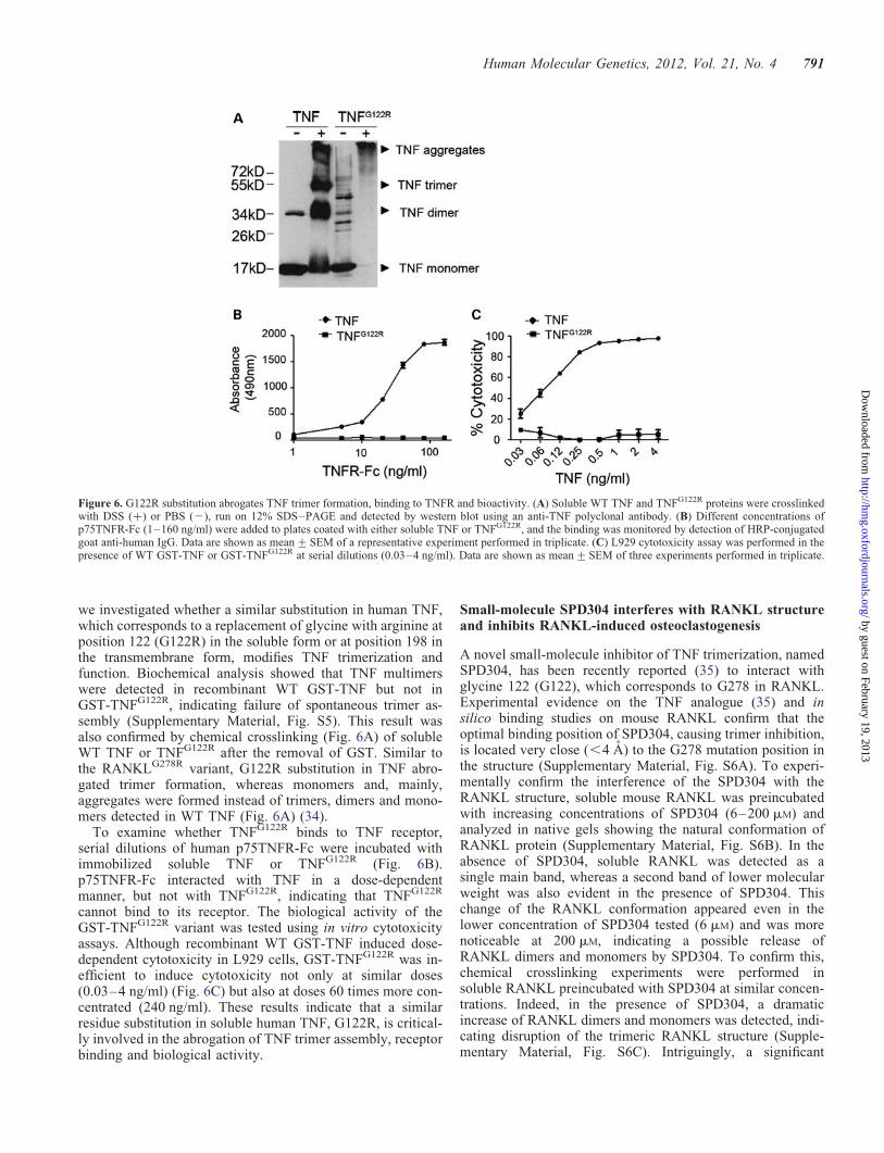
we investigated whether a similar substitution in human TNF,which corresponds to a replacement of glycine with arginine atposition 122 (G122R) in the soluble form or at position 198 inthe transmembrane form, modifies TNF trimerization andfunction. Biochemical analysis showed that TNF multimerswere detected in recombinant WT GST-TNF but not inGST-TNFG122R, indicating failure of spontaneous trimer as-sembly (Supplementary Material, Fig. S5). This result wasalso confirmed by chemical crosslinking (Fig. 6A) of solubleWT TNF or TNFG122R after the removal of GST. Similar tothe RANKLG278R variant, G122R substitution in TNF abro-gated trimer formation, whereas monomers and, mainly,aggregates were formed instead of trimers, dimers and mono-mers detected in WT TNF (Fig. 6A) (34).
To examine whether TNFG122R binds to TNF receptor,serial dilutions of human p75TNFR-Fc were incubated withimmobilized soluble TNF or TNFG122R (Fig. 6B).p75TNFR-Fc interacted with TNF in a dose-dependentmanner, but not with TNFG122R, indicating that TNFG122R
cannot bind to its receptor. The biological activity of theGST-TNFG122R variant was tested using in vitro cytotoxicityassays. Although recombinant WT GST-TNF induced dose-dependent cytotoxicity in L929 cells, GST-TNFG122R was in-efficient to induce cytotoxicity not only at similar doses(0.03–4 ng/ml) (Fig. 6C) but also at doses 60 times more con-centrated (240 ng/ml). These results indicate that a similarresidue substitution in soluble human TNF, G122R, is critical-ly involved in the abrogation of TNF trimer assembly, receptorbinding and biological activity.
Small-molecule SPD304 interferes with RANKL structureand inhibits RANKL-induced osteoclastogenesis
A novel small-molecule inhibitor of TNF trimerization, namedSPD304, has been recently reported (35) to interact withglycine 122 (G122), which corresponds to G278 in RANKL.Experimental evidence on the TNF analogue (35) and insilico binding studies on mouse RANKL confirm that theoptimal binding position of SPD304, causing trimer inhibition,is located very close (,4 A) to the G278 mutation position inthe structure (Supplementary Material, Fig. S6A). To experi-mentally confirm the interference of the SPD304 with theRANKL structure, soluble mouse RANKL was preincubatedwith increasing concentrations of SPD304 (6–200 mM) andanalyzed in native gels showing the natural conformation ofRANKL protein (Supplementary Material, Fig. S6B). In theabsence of SPD304, soluble RANKL was detected as asingle main band, whereas a second band of lower molecularweight was also evident in the presence of SPD304. Thischange of the RANKL conformation appeared even in thelower concentration of SPD304 tested (6 mM) and was morenoticeable at 200 mM, indicating a possible release ofRANKL dimers and monomers by SPD304. To confirm this,chemical crosslinking experiments were performed insoluble RANKL preincubated with SPD304 at similar concen-trations. Indeed, in the presence of SPD304, a dramaticincrease of RANKL dimers and monomers was detected, indi-cating disruption of the trimeric RANKL structure (Supple-mentary Material, Fig. S6C). Intriguingly, a significant
Figure 6. G122R substitution abrogates TNF trimer formation, binding to TNFR and bioactivity. (A) Soluble WT TNF and TNFG122R proteins were crosslinkedwith DSS (+) or PBS (2), run on 12% SDS–PAGE and detected by western blot using an anti-TNF polyclonal antibody. (B) Different concentrations ofp75TNFR-Fc (1–160 ng/ml) were added to plates coated with either soluble TNF or TNFG122R, and the binding was monitored by detection of HRP-conjugatedgoat anti-human IgG. Data are shown as mean+SEM of a representative experiment performed in triplicate. (C) L929 cytotoxicity assay was performed in thepresence of WT GST-TNF or GST-TNFG122R at serial dilutions (0.03–4 ng/ml). Data are shown as mean+SEM of three experiments performed in triplicate.
Human Molecular Genetics, 2012, Vol. 21, No. 4 791
by guest on February 19, 2013http://hm
g.oxfordjournals.org/D
ownloaded from

increase in the intensity of the band corresponding to RANKLtrimers was also noticed. This could reflect a possible con-formational alteration in the structure of RANKL trimers com-plexed with SPD304 that lowers the threshold required for thedetection of RANKL trimeric molecules by the polyclonalanti-RANKL antibody, enabling the detection of moreRANKL molecules.
To investigate whether SPD304 can inhibit RANKL func-tion in osteoclastogenesis assays, BM cells were treated with25 ng/ml M-CSF and 80 ng/ml GST-RANKL in the presenceof SPD304 at different concentrations ranging from 0.25 to2 mM. SPD304 at 1 mM attenuated both the number and thesize of TRAP+ multinucleated cells, whereas at 2 mM the for-mation of multinuclear TRAP+ osteoclast was completelyinhibited (Fig. 7A–C). SPD304 did not display any cell tox-icity at the indicated doses. Collectively, these resultsprovide supporting evidence that SPD304 modifies the con-formation of the RANKL trimeric structure and inhibitsRANKL-mediated osteoclastogenesis.
DISCUSSION
Human osteopetroses are a heterogeneous group of bone re-modeling disorders characterized by an increase in bonedensity due to a defect in osteoclastic bone resorption. Func-tional mutations in RANKL have been identified in a newform of osteoclast-poor ARO that could not be cured by hem-atopoietic stem cell transplantation. Three loss-of-functionRANKL mutations have been identified in ARO, M199K,del145-177AA and V277WfX5 (27); the single amino acid
substitution, M199K, is located within a highly conserveddomain, the deletion 145–177 removes b-strand A and halfof the AA′ loop which could abolish the interaction withRANK, whereas the frameshift deletion V277WfX5 and theresultant loss of strand F, which is important for the trimeriza-tion of RANKL, could abrogate the conformation and thebinding activity (36). Interestingly, these RANKL mutationswere localized within the extracellular bioactive TNF-likecore domain, whereas the underlying molecular mechanismsleading to ARO pathogenesis remain unknown. Characteriza-tion of functional RANKL mutations derived either in humansor in animal models constitutes a powerful approach for theelucidation of the molecular basis of ARO as well as for thepotential design of novel RANKL inhibitors.
Following a forward genetics approach using ENU-mediated random mutagenesis, we identified a novel AROmodel (tles), characterized by defective tooth eruption, com-plete lack in osteoclasts, increased bone mass and absenceof lymph nodes. In this study, we demonstrate that the tlesosteopetrotic phenotype is caused by a missense point muta-tion in the Rankl gene that corresponds to a single aminoacid substitution from glycine to arginine (G278R) at theextracellular TNF-like core domain of RANKL. Since theskeletal phenotype of the Rankl mutant mice (Rankltles/tles) issimilar to the existing Rankl knockout models (13,14), ourresults indicate that a single amino acid change is sufficientto cause a loss-of function Rankl allele and subsequentlyARO in vivo. However, the Rankltles/tles mice differ from theRankl knockout mice at the molecular level. Even thoughRankltles/tles mice produce physiological levels of a non-functional RANKL protein, in Rankl knockout mice there isno production of either mRNA or RANKL protein. This isdue to the fact that the Rankl null alleles have been highly dis-rupted by the incorporation of the neomycin selection cassetteas well as by the deletion of large protein regions. Instead, themutant allele contains a point mutation in a coding region thatcauses a single amino acid substitution, mimicking naturalgenetic variants. The Rankltles/tles osteopetrotic model is thefirst reported animal model that carries a functional mutationin the Rankl gene and closely resembles RANKL-mediatedhuman ARO, as in both cases the RANKL protein is producedbut is inactive due to disruptive mutations at the extracellularbioactive TNF-like core domain, unlike the Rankl null alleles(13,14). G278R substitution fully abrogates RANKL functionby impairing trimer assembly, providing a possible mechanis-tic explanation for some forms of human RANKL-mediatedARO. Even though, an equivalent substitution for G278 hasnot been reported in ARO patients so far, such amino acid isdeleted at the frameshift deletion V277WfX5, which is pre-dicted to cause loss of the bG, bH and bF strands ofRANKL, important for its trimerization (27). It is thereforelikely that the loss of trimerization we have described in thisstudy may represent a pathogenetic mechanism driving AROpathology. Thus, in addition to the use of Rankl knockoutmodels for preclinical studies, our newly described AROmodel may also be useful in the validation of new therapeuticapproaches in ARO due to the presence, as in humans, of amutated RANKL variant. Indeed, administration of recombin-ant soluble RANKL completely rescues the osteoclast defectin vivo, indicating that treatment with recombinant RANKL
Figure 7. SPD304 inhibits RANKL-induced osteoclastogenesis. (A) Repre-sentative TRAP stain of osteoclast cultures from WT BM cells treated withM-CSF and GST-RANKL in the presence of 0.25–2 mM SPD304. Bar:100 mm. (B) The number of TRAP+ multinucleated (three or more nuclei)cells was quantitated per well (48-well plate). (C) The nuclei number inTRAP+ multinucleated cells was also calculated. Data are shown asmean+SEM of three experiments performed in duplicate. The effect ofSPD304 on osteoclast formation was compared with that of untreated cells(∗P , 0.05, ∗∗P , 0.001, ∗∗∗P , 0.0001).
792 Human Molecular Genetics, 2012, Vol. 21, No. 4
by guest on February 19, 2013http://hm
g.oxfordjournals.org/D
ownloaded from

might overcome failure of osteoclast formation and osteope-trosis in ARO. Future comparative studies could showwhether the effectiveness of novel therapeutic approaches issimilar in both ARO mouse models, Rankltles/tles and Ranklknockout mice, which differ at the protein level.
At the molecular level, our data demonstrate that a singleamino acid substitution in the core of TNF-like trimeric struc-ture, G278R, completely inactivates RANKL function by inhi-biting its trimerization. Even though, it has been previouslyproposed that RANKL trimerization involves intersubunitinteractions among 43 residues, scattered mainly within the10 highly conserved b-strands of each monomer (6), it isshown here for first time that a single amino acid substitutionis sufficient to completely disrupt trimer assembly. G278 is ori-ginally shown here to constitute one of the critical amino acidinvolved in RANKL function, providing new opportunitiesfor the rational design of novel RANKL inhibitors. So far, theidentification of functional RANKL residues has been basedon predictions made on the crystal structure of RANKL/RANK (36,37). Such studies have been exclusively concen-trated in amino acids interacting with the RANK receptorsuch as the Glu225, Arg222 and Asp299 residues (37), wheretheir substitution leads to a dramatic decrease on binding toRANK and subsequent inability to promote osteoclast forma-tion. Our forward genetics approach identifies and characterizesa critical amino acid substitution that results in protein inactiva-tion and subsequently to osteopetrosis in vivo.
From our constructed model of the RANKLG278R proteinbased on the known crystal structure (6,7), it is apparent thatRANKLG278R mutation may not affect the formation andfolding of the monomer but will most definitely affect the for-mation of a trimer assembly. The RANKLG278R mutation islocated at the hydrophobic F b-strand (Fig. 2C), which is100% conserved between human and mouse RANKL. The Fb-strand is part of the inner A’AHCF b-sheet that is involvedin intersubunit association. The introduction of a positivecharge as well as a long side chain is expected to disrupt thehydrophobic interface and create steric hindrances causingpacking inefficiencies (Fig. 2D). Our biochemical analysison recombinant soluble RANKL has revealed that functionaltrimers or multimers are not detected for the RANKLG278R
protein, confirming our structure-based prediction regardingthe trimerization inability of RANKLG278R. Instead, ourstudies reveal the presence of monomers as well as the forma-tion of RANKLG278R aggregates that could be explained bythe exposure of hydrophobic regions previously buried intrimer interfaces. Such hydrophobic surfaces have a naturaltendency to stick together forming non-functional proteinaggregates. Since formation of a functional RANKL trimeris prerequisite for receptor binding, RANKLG278R is unableto bind and activate RANK that is required for the stimulationof the downstream signaling cascades, leading to osteoclastdifferentiation, activation and survival.
Interestingly, the G278 residue of RANKL is highly con-served in various members of the TNF superfamily includingTNF, CD40L, TRAIL, BAFF and APRIL, suggesting a similarinvolvement in homotrimer assembly. Notably, a similar sub-stitution of this conserved glycine residue by valine at position227 has also been detected in the CD40L gene in patients withX-linked hyper IgM syndrome (38). However, it has not been
experimentally proven that glycine 227 is involved in ligandtrimerization. Therefore, we investigated whether a similarsubstitution in another TNF family member results also inan inability in the formation of trimers. TNF, the prototypeof the TNF superfamily, has a central role in the pathogenesisof chronic inflammatory and autoimmune disorders (39), andTNF inhibitors were the first biologic therapeutics approvedfor the treatment of rheumatoid arthritis and inflammatorybowel disease. Our present data confirm that a similarresidue substitution in soluble TNF, G122R, abrogates TNFtrimer formation, binding to the p75TNF receptor and bio-activity. Therefore, the identified conserved glycine withinthe TNF-like core domain seems to be critically involved intrimer formation and could be exploited for the design ofnovel specific inhibitors against TNF superfamily members.
Recombinant RANKLG278R protein has a dominant-negative effect in ex vivo osteoclastogenesis assays whichdepends on the ratio between WT and RANKLG278R proteins.According to our in vitro experimental data and proposedstructural model, functional trimers can be formed when theratio of WT to RANKLG278R protein is 3:0 (WT homotrimers)or 2:1 (no dominant negative), whereas there is no activity atthe ratio of 0:3 (only RANKLG278R) or 1:2 (dominant nega-tive), as the latter are proposed to lack trimerizationpotency. The formation of a heterotrimer with one mutantmonomer may still be possible without exerting a dominant-negative effect, since two out of three hydrophobic interfacesbetween the monomers are still intact and hold the trimer as-sembly (Fig. 2C). However, the introduction of two or threemutant monomers in the trimer is expected to introduceextra charge repulsions as well as additional steric hindrancesleading to the failure of functional trimer assembly.The absence of a dominant-negative effect in heterozygousRankltles/+ mice in vivo can be explained by the stoichiometrybetween WT and RANKLG278R in RANKL trimers. AsRANKL spontaneously forms trimers, WT andRANKLG278R proteins can interact and form either2(WT):1(RANKLG278R) trimers or 1(WT):2(RANKLG278R)trimers. Even though heterozygous mice are expected toproduce a 1 (WT):1(RANKLG278R) protein ratio, due to thetrimeric RANKL nature these RANKL variants can stillform functional WT homotrimers or 2(WT):1(RANKLG278R)trimers, which are shown in vitro to lead to osteoclastogenesis(Fig. 5). Indeed, our biochemical analyses show that at equi-molar ratio, soluble WT RANKL and RANKLG278R interact,increasing the levels of trimeric RANKL molecules (Supple-mentary Material, Fig. S4). Such trimers could correspond toboth intact WT RANKL homotrimers and the newly formedheterotrimers with RANKLG278R (2WT:1RANKLG278R),which according to our in silico binding analyses are likelyfunctional. Therefore, we postulate that, in heterozygousmice, adequate levels of functional RANKL trimers areformed enabling proper osteoclastogenesis, which couldexplain why a dominant-negative effect does not apply inthe heterozygous animals. Instead, complete blockade ofRANKL trimer formation in vivo would require at least twotimes higher levels of the RANKLG278R protein so that thestoichiometry favors the dominant-negative effect by1(WT):2(RANKLG278R), which is not probable in heterozy-gous mice, but may be achieved pharmacologically.
Human Molecular Genetics, 2012, Vol. 21, No. 4 793
by guest on February 19, 2013http://hm
g.oxfordjournals.org/D
ownloaded from

RANKL is a key therapeutic target in bone-loss diseasessuch as osteoporosis, rheumatoid arthritis and bone metastasis.Recombinant proteins that inhibit RANKL–RANK interac-tions, such as RANK-Fc, Fc-OPG and anti-RANKL anti-bodies, have been effectively used for the treatment ofosteolytic bone diseases (25,40,41). However, the use oflarge macromolecules as therapeutic agents can be hinderedby drawbacks, including antigenicity, poor bioavailabilityand high cost. A variety of novel antiresorptive agents,either small peptides or peptidomimetics based on the struc-ture of RANKL and RANK, have been recently developedthat mainly antagonize either the RANKL–RANK interaction(36,37,42) or the interaction of RANK with signaling adaptormolecules (43). Such inhibitors have been developed as alter-native agents to overcome the disadvantages of using macro-molecules. The identification of G278 as a critical residuefunctionally involved in RANKL trimer assembly points tothe potential use of novel RANKL inhibitors, such as peptidesor small molecules that are designed to interact with suchresidue in order to inhibit trimerization. It has been previouslyshown that trimer assembly within the TNF ligand family con-stitutes a dynamic process where subunits can be exchanged(44). This phenomenon could explain the dominant-negativeeffect exerted by the RANKLG278R variant. Additionally, ithas been recently demonstrated that the disruption of tightlypreassociated homotrimeric proteins, such as TNF, is feasiblethrough inhibitors that accelerate subunit dissociation.SPD304, the strongest small-molecule TNF antagonistreported to date, promotes the dissociation of TNF trimers(35) by displacing one of the three TNF subunits andforming a complex with the remaining two that are unableto bind to and stimulate TNF receptors. Among the 16 inter-acting residues, 7 are localized in the F b-strand, includingG122, which corresponds to G278 in RANKL. Our resultsdemonstrate that SPD304 effectively inhibits RANKL-inducedex vivo osteoclast formation, suggesting a commonality ofmechanism to that of TNF inhibition. This effect might beexplained by interference of SPD304 with the hydrophobicinterfaces of RANKL monomers, including the G278residue. Indeed, in silico binding studies on mouse RANKLconfirm that the optimal binding position of SPD304,causing trimer inhibition, is located very close (,4 A) to theG278 mutation position (35). Therefore, both the G278R mu-tation and SPD304 acting on the same location of the trimer’smonomer–monomer interface are likely to cause an equiva-lent inhibition effect to the trimer formation. Our experimentalresults provide supporting evidence that SPD304 interfereswith the trimeric RANKL structure promoting release ofdimers and monomers similar to the mechanism reported forTNF (35). However, SPD304 contains a potentially toxic3-substituted indole moiety that produces reactive intermedi-ates which possibly cause toxicities by covalently binding tonucleophilic residues of protein and/or DNA (45). Furthermodifications of SPD304 will allow the development ofmore potent chemical agents that abrogate trimerization withhigher specificity to TNF and/or RANKL and less toxicitythat can be applied for therapeutic purposes. Especially forRANKL, the development of small-molecule inhibitorscould offer accessibility to tissues such as brain whereRANKL is highly expressed and is involved in important
biological processes like thermoregulation (21). In thisaspect, our results provide a molecular basis for the rationaldesign and evaluation of novel RANKL inhibitors, either pep-tides and/or small molecules that target trimerization as anovel strategic approach for impairing RANKL bioactivity.Collectively, our results reveal a functional amino acid criticalfor ligand trimerization and bioactivity within the TNF ligandsuperfamily which could be used as a target for designingnovel inhibitors that specifically inhibit trimer assembly andsubsequently function of RANKL, TNF or other TNF super-family members.
MATERIALS AND METHODS
Mouse husbandry
The Rankl2/2 mice have been previously reported (13). DBA/2J mice were purchased from the Jackson Laboratories. Micewere maintained and bred under specific pathogen-free condi-tions in the animal facility of Biomedical Sciences ResearchCenter (BSRC) ‘Alexander Fleming’. All animal procedureswere approved and carried out in strict accordance with theguidelines of the Institutional Animal Care and Use Commit-tee of BSRC ‘Alexander Fleming’ and in accordance with theHellenic License for Animal Experimentation at the BSRC‘Alexander Fleming’.
ENU mutagenesis
G0 males of a mixed C57BL/6Jx129S6 background weretreated with ENU (Sigma-Aldrich, Inc.) administered inthree weekly doses at 100 mg/kg of body weight (46,47).Each G0 mouse was crossed to WT C57BL/6Jx129S6 femalesto produce G1 males that were further mated with WT femalesto produce G2 daughters that were subsequently backcrossedwith the G1 parent to generate G3 progeny (47–49). ENU muta-genesis was performed at BSRC ‘Alexander Fleming’.
Mapping and sequencing
Heterozygous +/tles animals were outcrossed with DBA/2Jmice and the F1 offspring were intercrossed to generate theF2 progeny harboring the recessive tles mutation. F2progeny were screened for osteopetrosis and used for geneticanalysis. A total of 71 polymorphic markers, includingsimple sequence-length polymorphisms (SSLPs) and SNPs,were used for genome-wide linkage analysis. SSLPs wereresolved on 4% agarose gels, whereas SNPs were identifiedby pyrosequencing using the Pyromark ID instrument(Biotage AB). A standard genome scan was conducted usingR/qtl (The R Foundation for Statistical Computing, version2.8.0) (50). Log-likelihood linkage for single-trait analysiswas established by non-parametric interval mapping of abinary model (diseased versus healthy control siblings), on124 F2 animals in total, computed at 1 cM increments overthe entire genome. Sequencing was carried out as a serviceby MWG Biotech AG.
794 Human Molecular Genetics, 2012, Vol. 21, No. 4
by guest on February 19, 2013http://hm
g.oxfordjournals.org/D
ownloaded from

Crystal structure and molecular modeling
The RANKL homotrimer structure was obtained from theProtein Data Bank (PDB; code 1S55) (www.rcsb.org/pdb/).Molecular models for the G278R mutant homo- and heterotri-mers were built using Modeller v9.4 (51) and tested forpacking inconsistencies and atomic clashes using theprogram QUANTA-CHARM (Molecular Simulations, Inc.,San Diego, CA, USA) (52).
Histopathological analysis
Femurs and tibiae were fixed in 4% PFA for 6 h, decalcified in13% EDTA and embedded in paraffin. Sections of 5 mm thick-ness were stained with hematoxylin/eosin. Osteoclasts werestained for TRAP activity using a leukocyte acid phosphatase(TRAP) kit (Sigma-Aldrich).
Ex vivo osteoclast formation
BM cells were collected after flushing out of femurs and tibiae,subjected to gradient purification using ficoll-paque (GE Health-care), plated in 24-well plates at a density of 5 × 10⁵ cells perwell and cultured in aMEM medium (GIBCO) containing10% fetal bovine serum supplemented with 40 ng/mlRANKL (R&D Systems) and 25 ng/ml M-CSF (R&DSystems) for 5 days. Similarly, splenocytes were collected,plated in 24-well plates at a density of 106 cells per welland cultured in the presence of recombinant RANKL andM-CSF for 6 days. GST-RANKLG278R was preincubatedwith WT GST-RANKL at room temperature for 20 min,prior to the stimulation of the BM cell cultures, in order toenable exchange of the RANKL variants and heterotrimerformation. Small-molecule SPD304 (Sigma-Aldrich) was pre-incubated with 80 ng/ml GST-RANKL at various concentra-tions from 0.25 to 2 mM in aMEM medium for 1 h at roomtemperature and then added to culture. Osteoclasts werestained for TRAP activity.
Osteoblasts were isolated from calvariae of 10-day-oldmice, using a sequential collagenase/dispase digestion proced-ure, were plated in 24-well plates at a density of 4 × 10⁴ cellsper well and cultured overnight in aMEM medium with 10%FBS. BM cells or splenocytes were collected, cultured with10 ng/ml M-CSF overnight, subjected to gradient centrifuga-tion and co-cultured with osteoblasts at a density of 5 × 10⁵
(BM cells) and 2 × 10⁶ (splenocytes) in aMEM medium sup-plemented with 1,25(OH)2 vitamin D3 (10 nM) and PGE2(1 mM) for 6 days.
Bone histomorphometry
Left femurs were fixed in 4% formalin and embedded inmethylmethacrylate resin (Technovit; Heraeus Kulzer, Wehr-heim, Germany) using standard procedures.Four-micrometer-thick sections were prepared with a Jungmicrotome (Jung, Heidelberg, Germany) and stained withvon Kossa stain and toluidine blue. Standard bone histomor-phometric measures were analyzed using a Zeiss Axioskop 2microscope (Zeiss, Marburg, Germany) equipped with anOsteomeasure image analysis system.
MicroCT imaging
MicroCT images were acquired on a vivaCT40 (ScancoMedical, Bassersdorf, Switzerland). The scanner generates acone beam at 5 mm spot size and operates at 50 keV.Images of femurs from WT, Rankl2/2, Rank+/2, Rankltles/
tles and Rankl+/tles mice were acquired.
Quantification of soluble RANKL
The levels of soluble mouse RANKL were quantitated using acommercial ELISA kit (R&D).
Expression and purification of GST-RANKL andGST-TNF
The extracellular domains of RANKL, RANKLG278R, TNFand TNFG122R were expressed in Escherichia coli as aGST-fusion protein. Briefly, a cDNA encoding the core ecto-domain of murine RANKL residues 158–316, with or withoutthe G278R substitution, was cloned into pGEX-6P-1 (GEHealthcare Life Sciences) downstream of GST. For the gener-ation of recombinant GST-TNF, a cDNA encoding the extra-cellular domain of human TNF from valine 77 to leucine 233was also cloned into pGEX-6P-1. The G122R substitution wasintroduced by a two-step overlapping PCR approach. Follow-ing IPTG-mediated (100 mM) induction of protein expression,BL21cells were lysed by sonication, and incubated withglutathione-sepharose beads. The GST-fused proteins werereleased from the affinity matrix by competitive elution with50 mM glutathione (Sigma-Aldrich).
Purification of soluble RANKL and TNF
After the capture of GST-RANKL or GST-TNF on glutathionebeads, soluble RANKL or TNF was eluted by cleavage ofbeads with PreScission Protease (GE healthcare) for overnightat 48C.
Protein crosslinking assay
The chemical crosslinking reagent DSS (Sigma) was used toexamine the trimeric property of RANKL and TNF (33).50 mM of DSS was prepared as a stock solution in dimethylsulfoxide. RANKL or TNF proteins at a final concentrationof 0.1 mM in PBS buffer (pH 7.5) were mixed with 1 mM
DSS (the molar ratio of DSS is 10:1). The crosslinking reac-tions were carried out for 1 h at room temperature and termi-nated with 50 mM Tris (pH 7.5) for 30 min. Proteins fromreaction mixtures were separated on 12% SDS–PAGE, fol-lowed by staining with Coomassie blue R-250 or proceededin western blot.
Generation of C-terminus-tagged full-length WT andRANKLG278R
The full-length mouse WT or RANKLG278R cDNA constructsencoded residues 1–316 without a stop codon. A Myc-taggedRANKL expression vector was constructed by inserting full-length RANKL into the pcDNA3.1/myc-His A MCS vector
Human Molecular Genetics, 2012, Vol. 21, No. 4 795
by guest on February 19, 2013http://hm
g.oxfordjournals.org/D
ownloaded from

(Invitrogen). FLAG-tagged RANKL was created by subclon-ing full-length RANKL into the p3XFLAG-CMV-14 expres-sion vector (Sigma-Aldrich).
Transient 293 transfection assays
HEK 293FT cells were transfected with 1 mg of plasmid DNAusing TransIt-293 transfection reagent (Mirus, Madison,WI, USA). After 48 h, transfected cells were harvested inPBS and the half quantity was diluted in equal volume of2× Laemmli sample buffer, and analyzed in 12% acrylamidedenatured gels. The remaining cells were lysed by sonication,centrifuged and analyzed in 8% native acrylamide gels.
Western blot
Recombinant proteins or lysates were resolved either on 8%native acrylamide gels or on 12% SDS denatured acrylamidegels. RANKL was detected by western blotting using eithera monoclonal (clone IK22/5, eBioscience) or a polyclonal(R&D Systems) anti-RANKL antibody, whereas for GSTdetection a rabbit polyclonal anti-GST antibody was used.Human TNF was detected using a rabbit polyclonalanti-TNF antibody provided by Professor Wim Buurman(Maastricht University). Moreover, antibodies against Myc(rabbit polyclonal, Santa Cruz Biotechnology), FLAG (M2,Sigma) and actin (goat polyclonal, Santa Cruz Biotechnology)were also used.
Immunoprecipitation
HEK 293FT cells were harvested 48 h after transient transfec-tion, lysed and incubated with an anti-Myc antibody.Anti-Myc immunocomplexes were precipitated with proteinA/G Sepharose (Santa Cruz Biotechnology). Protein com-plexes were resolved by SDS–PAGE and immunoblottedwith an anti-FLAG antibody.
Binding assay of GST-RANKLG278R to RANK
Nunc plates were coated with recombinant WT GST-RANKL,GST-RANKLG278R or GST at 3 mg/ml and after blocking with1% BSA were incubated with increasing amount ofrecombinant mouse RANK-Fc (R&D Systems). RANKbinding was detected with a phycoerythrin (PE)-conjugatedgoat anti-human IgG (Fc) (SouthernBiotech, Birmingham,AL, USA) that was measured (539–573 nm) with the fluores-cent plate reader TECAN infinite M200.
Binding assay of TNFG122R to TNFR
Nunc plates were coated with recombinant soluble TNF orTNFG122R at 3 mg/ml and incubated with increasing amountof recombinant human p75TNFR-Fc (Wyeth). TNFR bindingwas detected with a horseradish peroxidase (HRP)-conjugatedgoat anti-human IgG (Fc) (SouthernBiotech) usingo-phenylenediamine substrate (Thermo Scientific Pierce),which was measured at 490 nm.
In vivo administration of soluble RANKL
Recombinant soluble RANKL was produced after the diges-tion of the GST-RANKL protein with prescission protease(GE Healthcare) for the removal of GST. Mice were treatedfrom day 13 of age for a period of 14 days with subcutaneousinjections of 150 mg/kg soluble RANKL.
Statistical analysis
Statistical analysis was performed on the Prism software, usingone-way ANOVA with Tukey’s multiple comparison test. Allvalues are reported as the mean+ standard error of the mean(SEM). All P-values ,0.05 were considered significant.
SUPPLEMENTARY MATERIAL
Supplementary Material is available at HMG online.
ACKNOWLEDGEMENTS
We thank George Alexakos, Nikos Giannakas and SpirosLalos for expert technical assistance in genetic analysis,animal breeding and histology, respectively.
Conflict of Interest statement. None declared.
FUNDING
This work was supported by the European Commission (grantnumber MUGEN LSHG-CT-2005-005203 to E.D. and G.K.,MASTERSWITCH HEALTH-F2-2008-223404 to G.S. andG.K.); the Hellenic Ministry for Development (grant number04AKMON72 to E.D. and G.K.); the Public Benefit Founda-tion John S. Latsis (grant number 2010-16 to E.D.) and theDeutsche Forschungsgemeinschaft (grant number IMMUNO-BONE SPP1408 to G.S.).
REFERENCES
1. Karsenty, G. and Wagner, E.F. (2002) Reaching a genetic and molecularunderstanding of skeletal development. Dev. Cell, 2, 389–406.
2. Boyle, W.J., Simonet, W.S. and Lacey, D.L. (2003) Osteoclastdifferentiation and activation. Nature, 423, 337–342.
3. Fuller, K., Wong, B., Fox, S., Choi, Y. and Chambers, T.J. (1998)TRANCE is necessary and sufficient for osteoblast-mediated activation ofbone resorption in osteoclasts. J. Exp. Med., 188, 997–1001.
4. Anderson, D.M., Maraskovsky, E., Billingsley, W.L., Dougall, W.C.,Tometsko, M.E., Roux, E.R., Teepe, M.C., DuBose, R.F., Cosman, D. andGalibert, L. (1997) A homologue of the TNF receptor and its ligandenhance T-cell growth and dendritic-cell function. Nature, 390, 175–179.
5. Wong, B.R., Rho, J., Arron, J., Robinson, E., Orlinick, J., Chao, M.,Kalachikov, S., Cayani, E., Bartlett, F.S. III, Frankel, W.N. et al. (1997)TRANCE is a novel ligand of the tumor necrosis factor receptor familythat activates c-Jun N-terminal kinase in T cells. J. Biol. Chem., 272,25190–25194.
6. Lam, J., Nelson, C.A., Ross, F.P., Teitelbaum, S.L. and Fremont, D.H.(2001) Crystal structure of the TRANCE/RANKL cytokine revealsdeterminants of receptor-ligand specificity. J. Clin. Invest., 108, 971–979.
7. Ito, S., Wakabayashi, K., Ubukata, O., Hayashi, S., Okada, F. and Hata, T.(2002) Crystal structure of the extracellular domain of mouse RANKligand at 2.2-A resolution. J. Biol. Chem., 277, 6631–6636.
8. Hikita, A., Yana, I., Wakeyama, H., Nakamura, M., Kadono, Y., Oshima,Y., Nakamura, K., Seiki, M. and Tanaka, S. (2006) Negative regulation of
796 Human Molecular Genetics, 2012, Vol. 21, No. 4
by guest on February 19, 2013http://hm
g.oxfordjournals.org/D
ownloaded from

osteoclastogenesis by ectodomain shedding of receptor activator ofNF-kappaB ligand. J. Biol. Chem., 281, 36846–36855.
9. Ikeda, T., Kasai, M., Utsuyama, M. and Hirokawa, K. (2001)Determination of three isoforms of the receptor activator of nuclearfactor-kappaB ligand and their differential expression in bone and thymus.Endocrinology, 142, 1419–1426.
10. Lacey, D.L., Timms, E., Tan, H.L., Kelley, M.J., Dunstan, C.R., Burgess,T., Elliott, R., Colombero, A., Elliott, G., Scully, S. et al. (1998)Osteoprotegerin ligand is a cytokine that regulates osteoclastdifferentiation and activation. Cell, 93, 165–176.
11. Yasuda, H., Shima, N., Nakagawa, N., Yamaguchi, K., Kinosaki, M.,Mochizuki, S., Tomoyasu, A., Yano, K., Goto, M., Murakami, A. et al.
(1998) Osteoclast differentiation factor is a ligand for osteoprotegerin/osteoclastogenesis-inhibitory factor and is identical to TRANCE/RANKL.Proc. Natl Acad. Sci. USA, 95, 3597–3602.
12. Lacey, D.L., Tan, H.L., Lu, J., Kaufman, S., Van, G., Qiu, W., Rattan, A.,Scully, S., Fletcher, F., Juan, T. et al. (2000) Osteoprotegerin ligandmodulates murine osteoclast survival in vitro and in vivo. Am. J. Pathol.,157, 435–448.
13. Kong, Y.Y., Yoshida, H., Sarosi, I., Tan, H.L., Timms, E., Capparelli, C.,Morony, S., Oliveira-dos-Santos, A.J., Van, G., Itie, A. et al. (1999)OPGL is a key regulator of osteoclastogenesis, lymphocyte developmentand lymph-node organogenesis. Nature, 397, 315–323.
14. Kim, N., Odgren, P.R., Kim, D.K., Marks, S.C. Jr and Choi, Y. (2000)Diverse roles of the tumor necrosis factor family member TRANCE inskeletal physiology revealed by TRANCE deficiency and partial rescue bya lymphocyte-expressed TRANCE transgene. Proc. Natl Acad. Sci. USA,97, 10905–10910.
15. Dougall, W.C., Glaccum, M., Charrier, K., Rohrbach, K., Brasel, K., DeSmedt, T., Daro, E., Smith, J., Tometsko, M.E., Maliszewski, C.R. et al.
(1999) RANK is essential for osteoclast and lymph node development.Genes Dev., 13, 2412–2424.
16. Li, J., Sarosi, I., Yan, X.Q., Morony, S., Capparelli, C., Tan, H.L.,McCabe, S., Elliott, R., Scully, S., Van, G. et al. (2000) RANK is theintrinsic hematopoietic cell surface receptor that controlsosteoclastogenesis and regulation of bone mass and calcium metabolism.Proc. Natl Acad. Sci. USA, 97, 1566–1571.
17. Bucay, N., Sarosi, I., Dunstan, C.R., Morony, S., Tarpley, J., Capparelli,C., Scully, S., Tan, H.L., Xu, W., Lacey, D.L. et al. (1998)Osteoprotegerin-deficient mice develop early onset osteoporosis andarterial calcification. Genes Dev., 12, 1260–1268.
18. Rossi, S.W., Kim, M.Y., Leibbrandt, A., Parnell, S.M., Jenkinson, W.E.,Glanville, S.H., McConnell, F.M., Scott, H.S., Penninger, J.M., Jenkinson,E.J. et al. (2007) RANK signals from CD4(+)3(-) inducer cells regulatedevelopment of Aire-expressing epithelial cells in the thymic medulla.J. Exp. Med., 204, 1267–1272.
19. Takayanagi, H. (2007) Osteoimmunology: shared mechanisms andcrosstalk between the immune and bone systems. Nat. Rev. Immunol., 7,292–304.
20. Fata, J.E., Kong, Y.Y., Li, J., Sasaki, T., Irie-Sasaki, J., Moorehead, R.A.,Elliott, R., Scully, S., Voura, E.B., Lacey, D.L. et al. (2000) Theosteoclast differentiation factor osteoprotegerin-ligand is essential formammary gland development. Cell, 103, 41–50.
21. Hanada, R., Leibbrandt, A., Hanada, T., Kitaoka, S., Furuyashiki, T.,Fujihara, H., Trichereau, J., Paolino, M., Qadri, F., Plehm, R. et al. (2009)Central control of fever and female body temperature by RANKL/RANK.Nature, 462, 505–509.
22. Jones, D.H., Nakashima, T., Sanchez, O.H., Kozieradzki, I., Komarova,S.V., Sarosi, I., Morony, S., Rubin, E., Sarao, R., Hojilla, C.V. et al.(2006) Regulation of cancer cell migration and bone metastasis byRANKL. Nature, 440, 692–696.
23. Schramek, D., Leibbrandt, A., Sigl, V., Kenner, L., Pospisilik, J.A., Lee,H.J., Hanada, R., Joshi, P.A., Aliprantis, A., Glimcher, L. et al. (2010)Osteoclast differentiation factor RANKL controls development ofprogestin-driven mammary cancer. Nature, 468, 98–102.
24. Leibbrandt, A. and Penninger, J.M. (2009) RANK(L) as a key target forcontrolling bone loss. Adv. Exp. Med. Biol., 647, 130–145.
25. Cummings, S.R., San Martin, J., McClung, M.R., Siris, E.S., Eastell, R.,Reid, I.R., Delmas, P., Zoog, H.B., Austin, M., Wang, A. et al. (2009)Denosumab for prevention of fractures in postmenopausal women withosteoporosis. N. Engl. J. Med., 361, 756–765.
26. Smith, M.R., Egerdie, B., Hernandez Toriz, N., Feldman, R., Tammela,T.L., Saad, F., Heracek, J., Szwedowski, M., Ke, C., Kupic, A. et al.
(2009) Denosumab in men receiving androgen-deprivation therapy forprostate cancer. N. Engl. J. Med., 361, 745–755.
27. Sobacchi, C., Frattini, A., Guerrini, M.M., Abinun, M., Pangrazio, A.,Susani, L., Bredius, R., Mancini, G., Cant, A., Bishop, N. et al. (2007)Osteoclast-poor human osteopetrosis due to mutations in the geneencoding RANKL. Nat. Genet., 39, 960–962.
28. Grigoriadis, A.E., Wang, Z.Q., Cecchini, M.G., Hofstetter, W., Felix, R.,Fleisch, H.A. and Wagner, E.F. (1994) c-Fos: a key regulator ofosteoclast-macrophage lineage determination and bone remodeling.Science, 266, 443–448.
29. Yoshida, H., Hayashi, S., Kunisada, T., Ogawa, M., Nishikawa, S.,Okamura, H., Sudo, T. and Shultz, L.D. (1990) The murine mutationosteopetrosis is in the coding region of the macrophage colony stimulatingfactor gene. Nature, 345, 442–444.
30. Suda, T., Ueno, Y., Fujii, K. and Shinki, T. (2003) Vitamin D and bone.J. Cell Biochem., 88, 259–266.
31. Xu, J., Tan, J.W., Huang, L., Gao, X.H., Laird, R., Liu, D., Wysocki, S.and Zheng, M.H. (2000) Cloning, sequencing, and functionalcharacterization of the rat homologue of receptor activator of NF-kappaBligand. J. Bone Miner. Res., 15, 2178–2186.
32. Cheng, T., Pavlos, N.J., Wang, C., Tan, J.W., Lin, J.M., Cornish, J.,Zheng, M.H. and Xu, J. (2009) Mutations within the TNF-like coredomain of RANKL impair osteoclast differentiation and activation. Mol.Endocrinol., 23, 35–46.
33. Zhang, S., Liu, C., Huang, P., Zhou, S., Ren, J., Kitamura, Y., Tang, P.,Bi, Z. and Gao, B. (2009) The affinity of human RANK binding to itsligand RANKL. Arch. Biochem. Biophys., 487, 49–53.
34. Zhang, X.M., Weber, I. and Chen, M.J. (1992) Site-directed mutationalanalysis of human tumor necrosis factor-alpha receptor binding site andstructure-functional relationship. J. Biol. Chem., 267, 24069–24075.
35. He, M.M., Smith, A.S., Oslob, J.D., Flanagan, W.M., Braisted, A.C.,Whitty, A., Cancilla, M.T., Wang, J., Lugovskoy, A.A., Yoburn, J.C.et al. (2005) Small-molecule inhibition of TNF-alpha. Science, 310,
1022–1025.36. Ta, H.M., Nguyen, G.T., Jin, H.M., Choi, J., Park, H., Kim, N., Hwang,
H.Y. and Kim, K.K. (2010) Structure-based development of a receptoractivator of nuclear factor-kappaB ligand (RANKL) inhibitor peptideand molecular basis for osteopetrosis. Proc. Natl Acad. Sci. USA, 107,20281–20286.
37. Liu, C., Walter, T.S., Huang, P., Zhang, S., Zhu, X., Wu, Y., Wedderburn,L.R., Tang, P., Owens, R.J., Stuart, D.I. et al. (2010) Structural andfunctional insights of RANKL-RANK interaction and signaling.J. Immunol., 184, 6910–6919.
38. Seyama, K., Nonoyama, S., Gangsaas, I., Hollenbaugh, D., Pabst, H.F.,Aruffo, A. and Ochs, H.D. (1998) Mutations of the CD40 ligand gene andits effect on CD40 ligand expression in patients with X-linked hyper IgMsyndrome. Blood, 92, 2421–2434.
39. Douni, E., Armaka, M., Kontoyiannis, D.L. and Kollias, G. (2007)Functional genetic and genomic analysis of modeled arthritis. Adv. Exp.
Med. Biol., 602, 33–42.40. Feeley, B.T., Liu, N.Q., Conduah, A.H., Krenek, L., Roth, K., Dougall,
W.C., Huard, J., Dubinett, S. and Lieberman, J.R. (2006) Mixedmetastatic lung cancer lesions in bone are inhibited by nogginoverexpression and Rank:Fc administration. J. Bone Miner. Res., 21,1571–1580.
41. Body, J.J., Greipp, P., Coleman, R.E., Facon, T., Geurs, F., Fermand, J.P.,Harousseau, J.L., Lipton, A., Mariette, X., Williams, C.D. et al. (2003) Aphase I study of AMGN-0007, a recombinant osteoprotegerin construct, inpatients with multiple myeloma or breast carcinoma related bonemetastases. Cancer, 97, 887–892.
42. Aoki, K., Saito, H., Itzstein, C., Ishiguro, M., Shibata, T., Blanque, R.,Mian, A.H., Takahashi, M., Suzuki, Y., Yoshimatsu, M. et al. (2006) ATNF receptor loop peptide mimic blocks RANK ligand-induced signaling,bone resorption, and bone loss. J. Clin. Invest., 116, 1525–1534.
43. Poblenz, A.T., Jacoby, J.J., Singh, S. and Darnay, B.G. (2007) Inhibitionof RANKL-mediated osteoclast differentiation by selective TRAF6 decoypeptides. Biochem. Biophys. Res. Commun., 359, 510–515.
44. Ameloot, P., Declercq, W., Fiers, W., Vandenabeele, P. and Brouckaert,P. (2001) Heterotrimers formed by tumor necrosis factors of differentspecies or muteins. J. Biol. Chem., 276, 27098–27103.
45. Sun, H. and Yost, G.S. (2008) Metabolic activation of a novel3-substituted indole-containing TNF-alpha inhibitor: dehydrogenation andinactivation of CYP3A4. Chem. Res. Toxicol., 21, 374–385.
Human Molecular Genetics, 2012, Vol. 21, No. 4 797
by guest on February 19, 2013http://hm
g.oxfordjournals.org/D
ownloaded from

46. Justice, M.J., Carpenter, D.A., Favor, J., Neuhauser-Klaus, A., Hrabe deAngelis, M., Soewarto, D., Moser, A., Cordes, S., Miller, D., Chapman, V.et al. (2000) Effects of ENU dosage on mouse strains. Mamm. Genome,11, 484–488.
47. Hrabe de Angelis, M.H., Flaswinkel, H., Fuchs, H., Rathkolb, B.,Soewarto, D., Marschall, S., Heffner, S., Pargent, W., Wuensch, K., Jung,M. et al. (2000) Genome-wide, large-scale production of mutant mice byENU mutagenesis. Nat. Genet., 25, 444–447.
48. Nelms, K.A. and Goodnow, C.C. (2001) Genome-wide ENU mutagenesisto reveal immune regulators. Immunity, 15, 409–418.
49. Georgel, P., Du, X., Hoebe, K. and Beutler, B. (2008) ENU mutagenesisin mice. Methods Mol. Biol., 415, 1–16.
50. Broman, K.W., Wu, H., Sen, S. and Churchill, G.A. (2003) R/qtl: QTLmapping in experimental crosses. Bioinformatics, 19, 889–890.
51. Sali, A. and Blundell, T.L. (1993) Comparative protein modelling bysatisfaction of spatial restraints. J. Mol. Biol., 234, 779–815.
52. Brooks, B.R., Brooks, C.L. III, Mackerell, A.D. Jr, Nilsson, L., Petrella,R.J., Roux, B., Won, Y., Archontis, G., Bartels, C., Boresch, S. et al.(2009) CHARMM: the biomolecular simulation program. J. Comput.Chem., 30, 1545–1614.
798 Human Molecular Genetics, 2012, Vol. 21, No. 4
by guest on February 19, 2013http://hm
g.oxfordjournals.org/D
ownloaded from
Related Documents