Colloids and Surfaces B: Biointerfaces 82 (2011) 397–403 Contents lists available at ScienceDirect Colloids and Surfaces B: Biointerfaces journal homepage: www.elsevier.com/locate/colsurfb Microscopic and spectroscopic evaluation of novel PLGA–chitosan Nanoplexes as an ocular delivery system Gaurav K. Jain a,∗ , Shadab A. Pathan a , Sohail Akhter a , Nirmal Jayabalan b , Sushma Talegaonkar a , Roop K. Khar a , Farhan J. Ahmad a a Department of Pharmaceutics, F/O Pharmacy, Hamdard University, Hamdard Nagar, New Delhi 110062, India b Department of Ocular Pharmacology and Pharmacy, Dr Rajender Prasad Centre for Ophthalmic Sciences, All India Institute of Medical Science, New Delhi 110029, India article info Article history: Received 2 June 2010 Received in revised form 15 August 2010 Accepted 13 September 2010 Available online 18 September 2010 Keywords: Confocal microscopy Chitosan Nanoplexes Ocular drug delivery PLGA abstract The interaction of PLGA–chitosan Nanoplexes with ocular mucosa was investigated ex vivo and in vivo to assess their potential as ocular delivery system. Fluorescent Rhodamine Nanoplexes (Rd-Nanoplexes) were prepared by ionotropic gelation method. The size and morphology of Nanoplexes was investigated by TEM, SEM and PCS. The corneal retention, uptake and penetration of Nanoplexes were analyzed by spectrofluorimetry and confocal microscopy. Corneas from Rd-Nanoplexes-treated rabbits were eval- uated for the in vivo uptake and ocular tolerance. The Nanoplexes prepared were round with a mean diameter of 115.6 ± 17 nm and the encapsulation efficiency of Rd was 59.4 ± 2.5%. Data from ex vivo and in vivo studies showed that the amounts of Rd in the cornea were significantly higher for Nanoplexes than for a control Rd solution, these amounts being fairly constant for up to 24 h. Confocal microscopy of the corneas revealed paracellular and transcellular uptake of the Nanoplexes. The uptake mechanism postulated was adsorptive-mediated endocytosis and opening of the tight junctions between epithelial cells. No alteration was microscopically observed after ocular surface exposure to Nanoplexes. Taken together, these data demonstrate that Nanoplexes are potentially useful as ocular drug carriers. © 2010 Elsevier B.V. All rights reserved. 1. Introduction Topical instillation represents the most convenient route of ocu- lar drug delivery. However, this route is impeded by poor ocular bioavailability (<5%), mainly attributed to low corneal permeabil- ity of drugs, tear turn over, and drug elimination via conjunctiva and sclera [1]. Efforts to enhance ocular bioavailability from topical instillation have been accomplished either using prodrug design, permeation enhancing formulations, longer residence formula- tions or nano-sized formulations [2–4]. Nano-sized formulations have been evaluated as ocular drug delivery systems to enhance the absorption of therapeutic drugs, improve bioavailability, reduce systemic side effects, and sustain intraocular drug levels [5–17]. The use of polymeric nanoparticles is an attractive strategy to enhance the ocular bioavailability of topically administered drugs because they offer unique features while preserving the ease of delivery in liquid form [9–17]. Polymeric nanoparticles have been utilized to improve the corneal and conjunctival penetration of therapeutic drugs and peptides, sustain drug levels and reduce systemic side effects [9–17]. PLGA, a copolymer of poly (d,l-lactide- co-glycolide), is an ideal biodegradable polymer for nanoparticle ∗ Corresponding author. Tel.: +91 09 811127909; fax: +91 11 26059663. E-mail address: [email protected] (G.K. Jain). formulation due to its biocompatibility, safety, regulatory approval and wide use [12–17]. PLGA nanoparticles are well tolerated in animal models and their potentialities in ophthalmology are well documented [12–17]. In fact, several experiments have shown that PLGA nanoparticle-entrapped drugs have improved ocular bioavailability [15–17]. However, the short residence time of these nanosystems represents a limitation in their therapeutic use [18]. On the other hand, the potential of chitosan for topical ocular drug delivery is particularly promising due to its unique proper- ties such as mucoadhesion, tolerability, biodegradability and ability to enhance the paracellular transport of drugs [19–22]. Chitosan nanoparticles and nanocapsules have shown interesting properties and promising results as drug carriers across the ocular mucosa. Although chitosan nanoparticles reside on the ocular surface for a prolonged period, they lack proper control over drug release owing to rapid drug diffusion. Some previous reports demonstrated improved performance with chitosan-coated nanocapsules [23]. However, to achieve therapeutic concentrations in eye the dosage form must reside in the cul-de-sac for prolonged period and the entrapped drug moiety should be released from the nanopar- ticles at an appropriate rate. Consequently, the purpose of our work is to design nanoparticulate system consisting of complexes of release-controlling anionic polymer – PLGA and mucoadhe- sive cationic polymer – chitosan. Our hypothesis was that this PLGA–chitosan nanocomplex (Nanoplex), which presumably could 0927-7765/$ – see front matter © 2010 Elsevier B.V. All rights reserved. doi:10.1016/j.colsurfb.2010.09.010

Welcome message from author
This document is posted to help you gain knowledge. Please leave a comment to let me know what you think about it! Share it to your friends and learn new things together.
Transcript
Ma
GRa
b
a
ARRAA
KCCNOP
1
lbiaipthtsTebdutsc
0d
Colloids and Surfaces B: Biointerfaces 82 (2011) 397–403
Contents lists available at ScienceDirect
Colloids and Surfaces B: Biointerfaces
journa l homepage: www.e lsev ier .com/ locate /co lsur fb
icroscopic and spectroscopic evaluation of novel PLGA–chitosan Nanoplexes asn ocular delivery system
aurav K. Jaina,∗, Shadab A. Pathana, Sohail Akhtera, Nirmal Jayabalanb, Sushma Talegaonkara,oop K. Khara, Farhan J. Ahmada
Department of Pharmaceutics, F/O Pharmacy, Hamdard University, Hamdard Nagar, New Delhi 110062, IndiaDepartment of Ocular Pharmacology and Pharmacy, Dr Rajender Prasad Centre for Ophthalmic Sciences, All India Institute of Medical Science, New Delhi 110029, India
r t i c l e i n f o
rticle history:eceived 2 June 2010eceived in revised form 15 August 2010ccepted 13 September 2010vailable online 18 September 2010
eywords:
a b s t r a c t
The interaction of PLGA–chitosan Nanoplexes with ocular mucosa was investigated ex vivo and in vivoto assess their potential as ocular delivery system. Fluorescent Rhodamine Nanoplexes (Rd-Nanoplexes)were prepared by ionotropic gelation method. The size and morphology of Nanoplexes was investigatedby TEM, SEM and PCS. The corneal retention, uptake and penetration of Nanoplexes were analyzed byspectrofluorimetry and confocal microscopy. Corneas from Rd-Nanoplexes-treated rabbits were eval-uated for the in vivo uptake and ocular tolerance. The Nanoplexes prepared were round with a mean
onfocal microscopyhitosananoplexescular drug deliveryLGA
diameter of 115.6 ± 17 nm and the encapsulation efficiency of Rd was 59.4 ± 2.5%. Data from ex vivo andin vivo studies showed that the amounts of Rd in the cornea were significantly higher for Nanoplexesthan for a control Rd solution, these amounts being fairly constant for up to 24 h. Confocal microscopyof the corneas revealed paracellular and transcellular uptake of the Nanoplexes. The uptake mechanismpostulated was adsorptive-mediated endocytosis and opening of the tight junctions between epithelial
icroonstr
cells. No alteration was mtogether, these data dem
. Introduction
Topical instillation represents the most convenient route of ocu-ar drug delivery. However, this route is impeded by poor ocularioavailability (<5%), mainly attributed to low corneal permeabil-
ty of drugs, tear turn over, and drug elimination via conjunctivand sclera [1]. Efforts to enhance ocular bioavailability from topicalnstillation have been accomplished either using prodrug design,ermeation enhancing formulations, longer residence formula-ions or nano-sized formulations [2–4]. Nano-sized formulationsave been evaluated as ocular drug delivery systems to enhancehe absorption of therapeutic drugs, improve bioavailability, reduceystemic side effects, and sustain intraocular drug levels [5–17].he use of polymeric nanoparticles is an attractive strategy tonhance the ocular bioavailability of topically administered drugsecause they offer unique features while preserving the ease ofelivery in liquid form [9–17]. Polymeric nanoparticles have been
tilized to improve the corneal and conjunctival penetration ofherapeutic drugs and peptides, sustain drug levels and reduceystemic side effects [9–17]. PLGA, a copolymer of poly (d,l-lactide-o-glycolide), is an ideal biodegradable polymer for nanoparticle∗ Corresponding author. Tel.: +91 09 811127909; fax: +91 11 26059663.E-mail address: [email protected] (G.K. Jain).
927-7765/$ – see front matter © 2010 Elsevier B.V. All rights reserved.oi:10.1016/j.colsurfb.2010.09.010
scopically observed after ocular surface exposure to Nanoplexes. Takenate that Nanoplexes are potentially useful as ocular drug carriers.
© 2010 Elsevier B.V. All rights reserved.
formulation due to its biocompatibility, safety, regulatory approvaland wide use [12–17]. PLGA nanoparticles are well tolerated inanimal models and their potentialities in ophthalmology are welldocumented [12–17]. In fact, several experiments have shownthat PLGA nanoparticle-entrapped drugs have improved ocularbioavailability [15–17]. However, the short residence time of thesenanosystems represents a limitation in their therapeutic use [18].On the other hand, the potential of chitosan for topical oculardrug delivery is particularly promising due to its unique proper-ties such as mucoadhesion, tolerability, biodegradability and abilityto enhance the paracellular transport of drugs [19–22]. Chitosannanoparticles and nanocapsules have shown interesting propertiesand promising results as drug carriers across the ocular mucosa.Although chitosan nanoparticles reside on the ocular surface fora prolonged period, they lack proper control over drug releaseowing to rapid drug diffusion. Some previous reports demonstratedimproved performance with chitosan-coated nanocapsules [23].
However, to achieve therapeutic concentrations in eye thedosage form must reside in the cul-de-sac for prolonged period andthe entrapped drug moiety should be released from the nanopar-
ticles at an appropriate rate. Consequently, the purpose of ourwork is to design nanoparticulate system consisting of complexesof release-controlling anionic polymer – PLGA and mucoadhe-sive cationic polymer – chitosan. Our hypothesis was that thisPLGA–chitosan nanocomplex (Nanoplex), which presumably could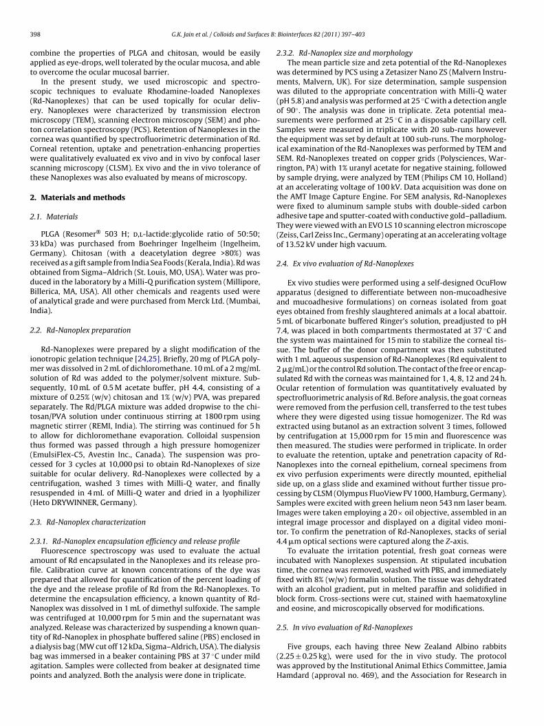
3 ces B:
cat
s(emtcCwst
2
2
3GrodBoI
2
imssmstmtt(cscr(
2
2
afiptdNwatabap
98 G.K. Jain et al. / Colloids and Surfa
ombine the properties of PLGA and chitosan, would be easilypplied as eye-drops, well tolerated by the ocular mucosa, and ableo overcome the ocular mucosal barrier.
In the present study, we used microscopic and spectro-copic techniques to evaluate Rhodamine-loaded NanoplexesRd-Nanoplexes) that can be used topically for ocular deliv-ry. Nanoplexes were characterized by transmission electronicroscopy (TEM), scanning electron microscopy (SEM) and pho-
on correlation spectroscopy (PCS). Retention of Nanoplexes in theornea was quantified by spectrofluorimetric determination of Rd.orneal retention, uptake and penetration-enhancing propertiesere qualitatively evaluated ex vivo and in vivo by confocal laser
canning microscopy (CLSM). Ex vivo and the in vivo tolerance ofhese Nanoplexes was also evaluated by means of microscopy.
. Materials and methods
.1. Materials
PLGA (Resomer® 503 H; d,l-lactide:glycolide ratio of 50:50;3 kDa) was purchased from Boehringer Ingelheim (Ingelheim,ermany). Chitosan (with a deacetylation degree >80%) was
eceived as a gift sample from India Sea Foods (Kerala, India). Rd wasbtained from Sigma–Aldrich (St. Louis, MO, USA). Water was pro-uced in the laboratory by a Milli-Q purification system (Millipore,illerica, MA, USA). All other chemicals and reagents used weref analytical grade and were purchased from Merck Ltd. (Mumbai,ndia).
.2. Rd-Nanoplex preparation
Rd-Nanoplexes were prepared by a slight modification of theonotropic gelation technique [24,25]. Briefly, 20 mg of PLGA poly-
er was dissolved in 2 mL of dichloromethane. 10 mL of a 2 mg/mLolution of Rd was added to the polymer/solvent mixture. Sub-equently, 10 mL of 0.5 M acetate buffer, pH 4.4, consisting of aixture of 0.25% (w/v) chitosan and 1% (w/v) PVA, was prepared
eparately. The Rd/PLGA mixture was added dropwise to the chi-osan/PVA solution under continuous stirring at 1800 rpm using
agnetic stirrer (REMI, India). The stirring was continued for 5 ho allow for dichloromethane evaporation. Colloidal suspensionhus formed was passed through a high pressure homogenizerEmulsiFlex-C5, Avestin Inc., Canada). The suspension was pro-essed for 3 cycles at 10,000 psi to obtain Rd-Nanoplexes of sizeuitable for ocular delivery. Rd-Nanoplexes were collected by aentrifugation, washed 3 times with Milli-Q water, and finallyesuspended in 4 mL of Milli-Q water and dried in a lyophilizerHeto DRYWINNER, Germany).
.3. Rd-Nanoplex characterization
.3.1. Rd-Nanoplex encapsulation efficiency and release profileFluorescence spectroscopy was used to evaluate the actual
mount of Rd encapsulated in the Nanoplexes and its release pro-le. Calibration curve at known concentrations of the dye wasrepared that allowed for quantification of the percent loading ofhe dye and the release profile of Rd from the Rd-Nanoplexes. Toetermine the encapsulation efficiency, a known quantity of Rd-anoplex was dissolved in 1 mL of dimethyl sulfoxide. The sampleas centrifuged at 10,000 rpm for 5 min and the supernatant was
nalyzed. Release was characterized by suspending a known quan-
ity of Rd-Nanoplex in phosphate buffered saline (PBS) enclosed indialysis bag (MW cut off 12 kDa, Sigma–Aldrich, USA). The dialysisag was immersed in a beaker containing PBS at 37 ◦C under mildgitation. Samples were collected from beaker at designated timeoints and analyzed. Both the analysis were done in triplicate.Biointerfaces 82 (2011) 397–403
2.3.2. Rd-Nanoplex size and morphologyThe mean particle size and zeta potential of the Rd-Nanoplexes
was determined by PCS using a Zetasizer Nano ZS (Malvern Instru-ments, Malvern, UK). For size determination, sample suspensionwas diluted to the appropriate concentration with Milli-Q water(pH 5.8) and analysis was performed at 25 ◦C with a detection angleof 90◦. The analysis was done in triplicate. Zeta potential mea-surements were performed at 25 ◦C in a disposable capillary cell.Samples were measured in triplicate with 20 sub-runs howeverthe equipment was set by default at 100 sub-runs. The morpholog-ical examination of the Rd-Nanoplexes was performed by TEM andSEM. Rd-Nanoplexes treated on copper grids (Polysciences, War-rington, PA) with 1% uranyl acetate for negative staining, followedby sample drying, were analyzed by TEM (Philips CM 10, Holland)at an accelerating voltage of 100 kV. Data acquisition was done onthe AMT Image Capture Engine. For SEM analysis, Rd-Nanoplexeswere fixed to aluminum sample stubs with double-sided carbonadhesive tape and sputter-coated with conductive gold–palladium.They were viewed with an EVO LS 10 scanning electron microscope(Zeiss, Carl Zeiss Inc., Germany) operating at an accelerating voltageof 13.52 kV under high vacuum.
2.4. Ex vivo evaluation of Rd-Nanoplexes
Ex vivo studies were performed using a self-designed OcuFlowapparatus (designed to differentiate between non-mucoadhesiveand mucoadhesive formulations) on corneas isolated from goateyes obtained from freshly slaughtered animals at a local abattoir.5 mL of bicarbonate buffered Ringer’s solution, preadjusted to pH7.4, was placed in both compartments thermostated at 37 ◦C andthe system was maintained for 15 min to stabilize the corneal tis-sue. The buffer of the donor compartment was then substitutedwith 1 mL aqueous suspension of Rd-Nanoplexes (Rd equivalent to2 �g/mL) or the control Rd solution. The contact of the free or encap-sulated Rd with the corneas was maintained for 1, 4, 8, 12 and 24 h.Ocular retention of formulation was quantitatively evaluated byspectrofluorimetric analysis of Rd. Before analysis, the goat corneaswere removed from the perfusion cell, transferred to the test tubeswhere they were digested using tissue homogenizer. The Rd wasextracted using butanol as an extraction solvent 3 times, followedby centrifugation at 15,000 rpm for 15 min and fluorescence wasthen measured. The studies were performed in triplicate. In orderto evaluate the retention, uptake and penetration capacity of Rd-Nanoplexes into the corneal epithelium, corneal specimens fromex vivo perfusion experiments were directly mounted, epithelialside up, on a glass slide and examined without further tissue pro-cessing by CLSM (Olympus FluoView FV 1000, Hamburg, Germany).Samples were excited with green helium neon 543 nm laser beam.Images were taken employing a 20× oil objective, assembled in anintegral image processor and displayed on a digital video moni-tor. To confirm the penetration of Rd-Nanoplexes, stacks of serial4.4 �m optical sections were captured along the Z-axis.
To evaluate the irritation potential, fresh goat corneas wereincubated with Nanoplexes suspension. At stipulated incubationtime, the cornea was removed, washed with PBS, and immediatelyfixed with 8% (w/w) formalin solution. The tissue was dehydratedwith an alcohol gradient, put in melted paraffin and solidified inblock form. Cross-sections were cut, stained with haematoxylineand eosine, and microscopically observed for modifications.
2.5. In vivo evaluation of Rd-Nanoplexes
Five groups, each having three New Zealand Albino rabbits(2.25 ± 0.25 kg), were used for the in vivo study. The protocolwas approved by the Institutional Animal Ethics Committee, JamiaHamdard (approval no. 469), and the Association for Research in
G.K. Jain et al. / Colloids and Surfaces B: Biointerfaces 82 (2011) 397–403 399
) Tran
Vmltvfiruvfe
2
scmc
3
3
dawlpapu(wqDooE
sctNv
formation was kept at pH 4.4. Lower pH values were not tried sinceit is known that PLGA undergoes degradation at low pH values,while at higher pH values Nanoplexes were not formed due to pHdependent solubility of chitosan. Particle size analysis of the opti-
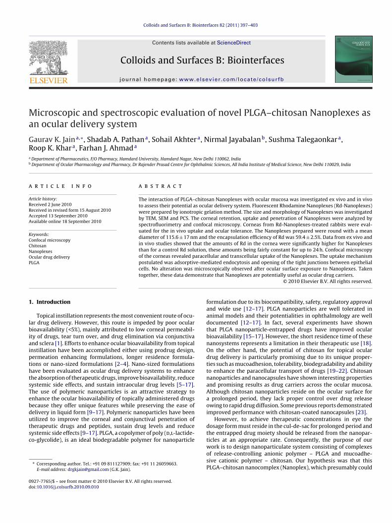
Fig. 1. Representative electron micrographs of Rd-Nanoplexes. (A
ision and Ophthalmology (ARVO) guidelines for usage of ani-als for ophthalmic use were followed. A weighed amount of
yophilized Rd-Nanoplexes was dispersed in PBS and administeredo the cul-de-sac of conscious rabbits in order to quantify their inivo interaction with the cornea. Aqueous solution of Rd as controlormulation was also administered. The animals were maintainedn an upright position using restraining boxes. They were then sac-ificed at 1, 2, 4, 8, and 24 h after the instillation of Rd-Nanoplexessing an overdose of sodium pentobarbital given through intra-enous route. The eyes were enucleated and corneal specimens,reshly excised, were observed for Rd-Nanoplexes retention, pen-tration and irritation potential as described in Section 2.4.
.6. Statistical analysis
Data were expressed as mean ± standard deviation (SD). Thetatistical significance of the differences between Nanoplexes andontrols at each time point was analyzed by paired t-test (Sig-aStat program; Jandel Scientific, Version 1.0). Differences were
onsidered to be significant when p < 0.05.
. Results and discussion
.1. Rd-Nanoplexes preparation and characterization
Our aim was to evaluate PLGA–chitosan Nanoplexes as ocularelivery systems by fluorescence spectroscopy and CLSM. In ourttempt to evaluate the interaction of PLGA–chitosan Nanoplexesith the ocular surface, the first step was the development of Rd-
oaded PLGA–chitosan Nanoplexes. Influence of type of organichase solvent, PLGA concentration in the organic phase, chitosannd PVA concentration in the aqueous phase and the aqueous phaseH on Rd-Nanoplexes size and encapsulation efficiency was eval-ated and optimized. Nanoplexes formed with dichloromethaneDCM) as solvent had lower particle size compared to that formedith ethyl acetate (EA). This was attributed to low thermodynamic
uality of DCM for PLGA and subsequently low viscosity of PLGA-CM solution. Further, low encapsulation efficiency of Rd wasbserved with EA, which favours Rd partitioning from inner aque-us phase to the organic phase, owing to higher water solubility ofA (8.7 wt%) compared to DCM (1.32 wt%).
The size of the Nanoplex system depends upon the net shear
tress available for droplet breakdown. Increasing, either the PLGAoncentration above 1% in the organic phase or chitosan concen-ration above 0.25% in the aqueous phase resulted in increasedanoplex size. Increased polymer concentration led to increasediscous forces which resist droplet break down. At high PLGA con-smission electron micrograph; (B) scanning electron micrograph.
centrations or at relatively low PVA concentrations, the amountof PVA is insufficient to stabilize the emulsion droplets and thusresulted in bimodal distributions. Nanoplexes with minimum par-ticle size were obtained when PVA was used at a concentrationof 1%. Above 1% PVA concentration, the viscosity of the aqueousphase was increased and the net shear stress available for dropletbreakdown was reduced. The aqueous phase pH during Nanoplex
Fig. 2. Rhodamine (Rd) amount transported across the cornea after the instillationof Rd-Nanoplexes (�) and Rd-solution (�) during (A) ex vivo studies (n = 3); (B) invivo studies (n = 3).
400 G.K. Jain et al. / Colloids and Surfaces B: Biointerfaces 82 (2011) 397–403
F ake ofe cells (r the a
mwaceNa
3
wpftwpfsTRcme
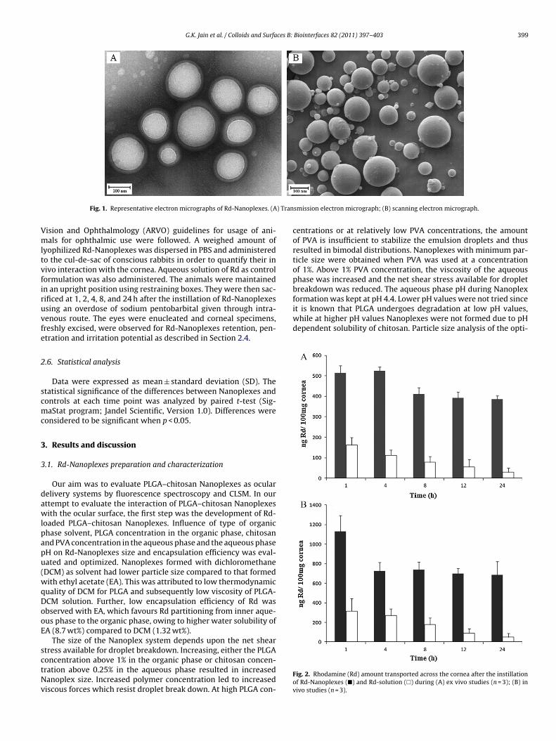
ig. 3. Confocal images of goat corneal epithelium (ex vivo studies) showing uptpithelium at depths of 20 and 60 �m are provided. Nanoplexes are seen within theeferences to color in this figure legend, the reader is referred to the web version of
ized formulation revealed an average diameter of 115.6 ± 17 nmith a zeta potential of +32.5 mV. Representative TEM (Fig. 1A)
nd SEM (Fig. 1B) micrographs illustrated that Nanoplexes had aharacteristic round shape and were monodisperse. Encapsulationfficiency of Rd was 59.4% (w/w). The release of Rd from the Rd-anoplexes was sustained over a period of 48 h, and thus it is andequate fluorescent marker.
.2. Corneal retention of Nanoplexes – quantitative evaluation
In order to estimate the corneal retention of the Nanoplexes,e evaluated the Rd content in cornea at different time pointsost-incubation. Fig. 2 shows the concentration of Rd in the corneaollowing instillation of the Rd-Nanoplexes and Rd solution as con-rol. These results showed that the behaviour of the Rd-Nanoplexesas remarkably different from that of Rd solution. Rd-Nanoplexesrovided greater concentrations of Rd than Rd solution. The dif-erences in Rd concentrations for the Rd-Nanoplexes and the Rdolution were statistically significant at all time points (p < 0.05).
he results (Fig. 2) also indicated that following instillation of thed-Nanoplexes, the concentration of Rd in cornea remained fairlyonstant for up to 24 h. The reason for this could be due to theechanism of interaction of PLGA and chitosan with the cornealpithelium. The proposed mechanism of interaction of PLGA with
Rd-Nanoplexes after 1 and 24 h of incubation. Colour overlay images of cornealyellow circles) and in between the cells (yellow squares). (For interpretation of the
rticle.)
the cornea is adsorptive-mediated endocytosis [26,27] and that forchitosan is electrostatic interaction and mucoadhesion [28–30], allbeing susceptible to saturation. Further, in the non-physiologicalex vivo study there was no reconstruction of the mucin layer andit is possible to assume a saturation of interaction sites for Rd-Nanoplexes. In contrast, the levels associated with the Rd solutiondecreased gradually with time. In other words, the Nanoplexes hadbetter retention and more persistent interaction with the ocularsurface compared to solution. The prolonged ocular retention ofthe Nanoplexes compared to solution is in good agreement witha previous work that showed prolonged corneal retention of col-loidal particles [5]. The results obtained ex vivo (Fig. 2A) were quitesimilar to those obtained in vivo (Fig. 2B). Further, the difference inRd content observed ex vivo and in vivo might be due to interplay ofa number of factors including difference in dose applied, tear turnover, blinking latency and pressure applied by the eyelids.
3.3. Corneal uptake and penetration of Nanoplexes – qualitativeevaluation
In order to elucidate the disposition of Nanoplexes in the cornea,we examined cross-sections of the cornea by CLSM. The confocalimages of different cross-sections of the goat cornea (ex vivo study)exposed to the Rd-Nanoplexes are shown in Fig. 3. Qualitative
G.K. Jain et al. / Colloids and Surfaces B: Biointerfaces 82 (2011) 397–403 401
F take oe cells (r the a
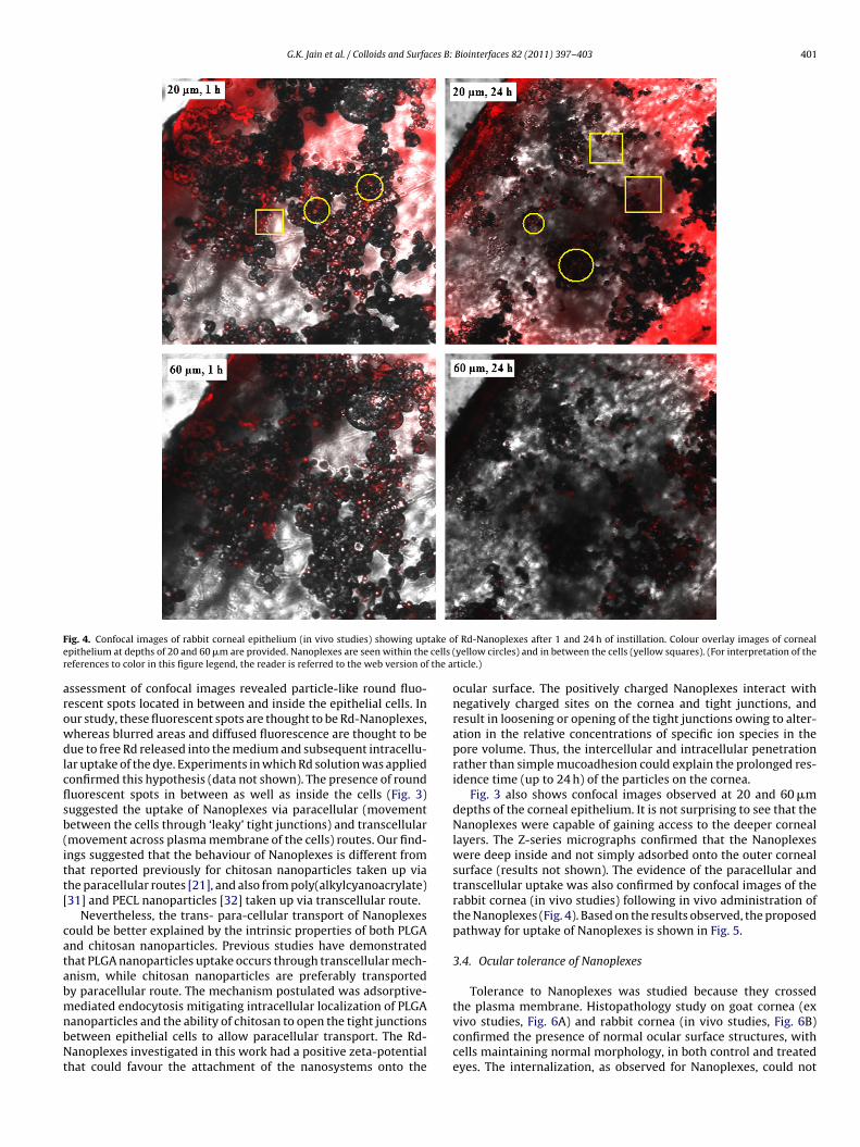
arowdlcflsb(itt[
catabmnbNt
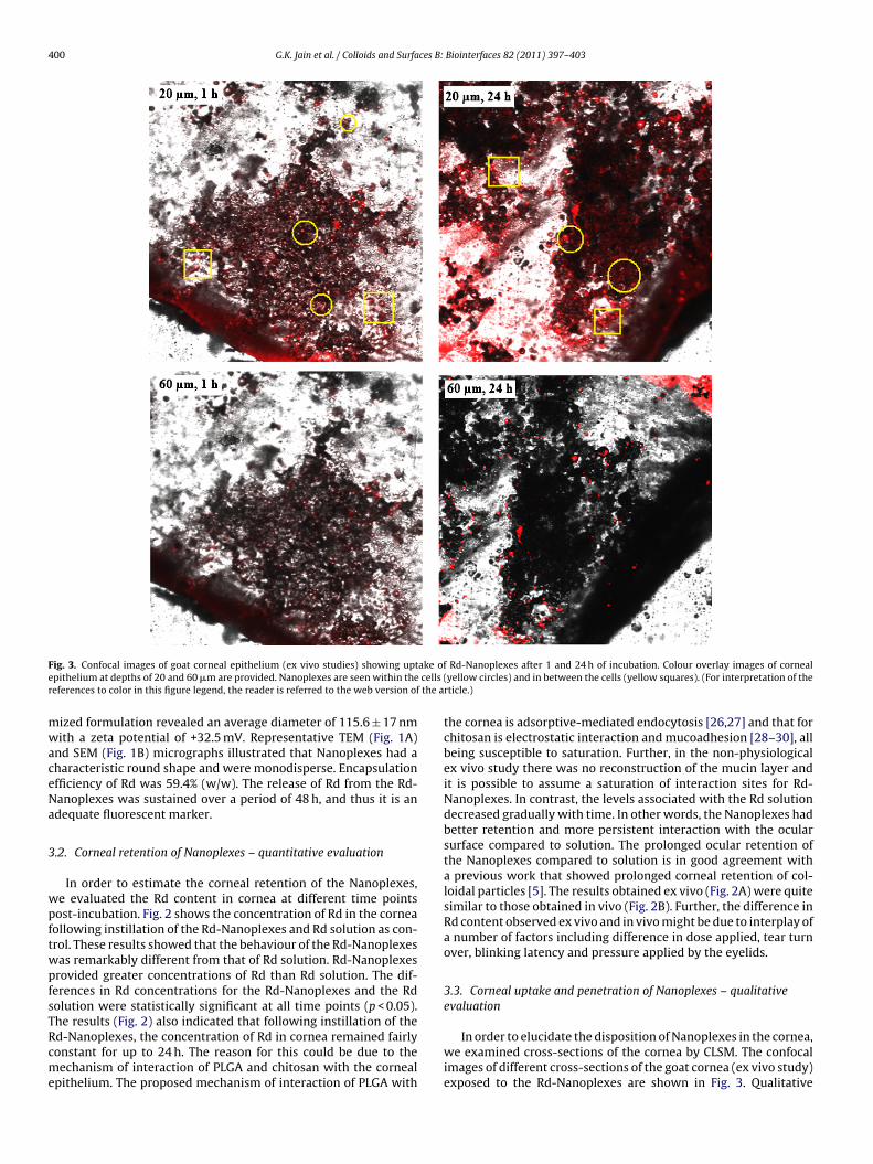
ig. 4. Confocal images of rabbit corneal epithelium (in vivo studies) showing uppithelium at depths of 20 and 60 �m are provided. Nanoplexes are seen within theeferences to color in this figure legend, the reader is referred to the web version of
ssessment of confocal images revealed particle-like round fluo-escent spots located in between and inside the epithelial cells. Inur study, these fluorescent spots are thought to be Rd-Nanoplexes,hereas blurred areas and diffused fluorescence are thought to beue to free Rd released into the medium and subsequent intracellu-
ar uptake of the dye. Experiments in which Rd solution was appliedonfirmed this hypothesis (data not shown). The presence of rounduorescent spots in between as well as inside the cells (Fig. 3)uggested the uptake of Nanoplexes via paracellular (movementetween the cells through ‘leaky’ tight junctions) and transcellularmovement across plasma membrane of the cells) routes. Our find-ngs suggested that the behaviour of Nanoplexes is different fromhat reported previously for chitosan nanoparticles taken up viahe paracellular routes [21], and also from poly(alkylcyanoacrylate)31] and PECL nanoparticles [32] taken up via transcellular route.
Nevertheless, the trans- para-cellular transport of Nanoplexesould be better explained by the intrinsic properties of both PLGAnd chitosan nanoparticles. Previous studies have demonstratedhat PLGA nanoparticles uptake occurs through transcellular mech-nism, while chitosan nanoparticles are preferably transportedy paracellular route. The mechanism postulated was adsorptive-
ediated endocytosis mitigating intracellular localization of PLGAanoparticles and the ability of chitosan to open the tight junctionsetween epithelial cells to allow paracellular transport. The Rd-anoplexes investigated in this work had a positive zeta-potential
hat could favour the attachment of the nanosystems onto the
f Rd-Nanoplexes after 1 and 24 h of instillation. Colour overlay images of cornealyellow circles) and in between the cells (yellow squares). (For interpretation of the
rticle.)
ocular surface. The positively charged Nanoplexes interact withnegatively charged sites on the cornea and tight junctions, andresult in loosening or opening of the tight junctions owing to alter-ation in the relative concentrations of specific ion species in thepore volume. Thus, the intercellular and intracellular penetrationrather than simple mucoadhesion could explain the prolonged res-idence time (up to 24 h) of the particles on the cornea.
Fig. 3 also shows confocal images observed at 20 and 60 �mdepths of the corneal epithelium. It is not surprising to see that theNanoplexes were capable of gaining access to the deeper corneallayers. The Z-series micrographs confirmed that the Nanoplexeswere deep inside and not simply adsorbed onto the outer cornealsurface (results not shown). The evidence of the paracellular andtranscellular uptake was also confirmed by confocal images of therabbit cornea (in vivo studies) following in vivo administration ofthe Nanoplexes (Fig. 4). Based on the results observed, the proposedpathway for uptake of Nanoplexes is shown in Fig. 5.
3.4. Ocular tolerance of Nanoplexes
Tolerance to Nanoplexes was studied because they crossed
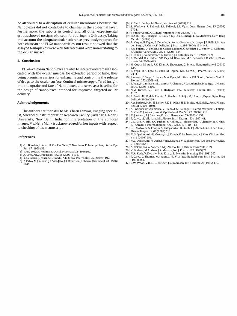
the plasma membrane. Histopathology study on goat cornea (exvivo studies, Fig. 6A) and rabbit cornea (in vivo studies, Fig. 6B)confirmed the presence of normal ocular surface structures, withcells maintaining normal morphology, in both control and treatedeyes. The internalization, as observed for Nanoplexes, could not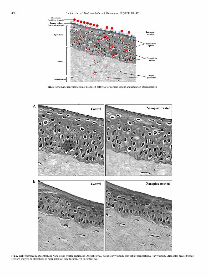
402 G.K. Jain et al. / Colloids and Surfaces B: Biointerfaces 82 (2011) 397–403
Fig. 5. Schematic representation of proposed pathway for corneal uptake and retention of Nanoplexes.
Fig. 6. Light microscopy of control and Nanoplexes treated sections of (A) goat corneal tissue (ex vivo study); (B) rabbit corneal tissue (in vivo study). Nanoplex-treated tissuesections showed no alterations in morphological details compared to control eyes.
ces B:
bNFgibat
4
cboitd
A
iUit
R
[
[
[[
[
[
[
[
[
[
[
[
[[[
[
[
[
G.K. Jain et al. / Colloids and Surfa
e attributed to a disruption of cellular membranes because theanoplexes did not contribute to changes in the epidermal layer.urthermore, the rabbits in control and all other experimentalroups showed no signs of discomfort during the 24 h assay. Takingnto account the adequate ocular tolerance previously reported foroth chitosan and PLGA nanoparticles, our results showed that thessayed Nanoplexes were well tolerated and were non-irritating tohe ocular surface.
. Conclusion
PLGA–chitosan Nanoplexes are able to interact and remain asso-iated with the ocular mucosa for extended period of time, thuseing promising carriers for enhancing and controlling the releasef drugs to the ocular surface. Confocal microscopy offered insightnto the uptake and fate of Nanoplexes, and serve as a baseline forhe design of Nanoplexes intended for improved, targeted ocularelivery.
cknowledgements
The authors are thankful to Ms. Charu Tanwar, Imaging special-st, Advanced Instrumentation Research Facility, Jawaharlal Nehruniversity, New Delhi, India for interpretation of the confocal
mages. Ms. Neha Malik is acknowledged for her inputs with respecto checking of the manuscript.
eferences
[1] C.L. Bourlais, L. Acar, H. Zia, P.A. Sado, T. Needham, R. Leverge, Prog. Retin. Eye
Res. 17 (1998) 33.[2] V.H.L. Lee, J.R. Robinson, J. Ocul. Pharmacol. 2 (1986) 67.[3] A. Urtti, Adv. Drug Deliv. Rev. 58 (2006) 1131.[4] R. Gaudana, J. Jwala, S.H. Boddu, A.K. Mitra, Pharm. Res. 26 (2009) 1197.[5] P. Calvo, M.J. Alonso, J.L. Vila-Jato, J.R. Robinson, J. Pharm. Pharmacol. 48 (1996)
1147.
[[[[
[
Biointerfaces 82 (2011) 397–403 403
[6] X. Cai, S. Conley, M. Naash, Vis. Res. 48 (2008) 319.[7] S. Wadhwa, R. Paliwal, S.R. Paliwal, S.P. Vyas, Curr. Pharm. Des. 15 (2009)
2724.[8] J. Vandervoort, A. Ludwig, Nanomedicine 2 (2007) 11.[9] H.Z. Bu, H.J. Gukasyan, L. Goulet, X.J. Lou, C. Xiang, T. Koudriakova, Curr. Drug
Metab. 8 (2007) 91.10] A. Vargas, B. Pegaz, E. Debefve, Y. Konan-Kouakou, N. Lange, J.P. Ballini, H. van
den Bergh, R. Gurny, F. Delie, Int. J. Pharm. 286 (2004) 131–145.11] R.A. Bejjani, D. BenEzra, H. Cohen, J. Rieger, C. Andrieu, J.C. Jeanny, G. Gollomb,
F.F. Behar-Cohen, Mol. Vis. 11 (2005) 124.12] K. Dillen, J. Vandervoort, A. Ludwig, J. Contr. Release 101 (2005) 369.13] B. Mandal, K.K. Halder, S.K. Dey, M. Bhoumik, M.C. Debnath, L.K. Ghosh, Phar-
mazie 64 (2009) 445.14] H. Gupta, M. Aqil, R.K. Khar, A. Bhatnagar, G. Mittal, Nanomedicine 6 (2010)
324.15] E. Vega, M.A. Egea, O. Valls, M. Espina, M.L. García, J. Pharm. Sci. 95 (2006)
2393.16] J. Araújo, E. Vega, C. Lopes, M.A. Egea, M.L. Garcia, E.B. Souto, Colloids Surf. B:
Biointerf. 72 (2009) 48.17] E. Vega, F. Gamisans, M.L. García, A. Chauvet, F. Lacoulonche, M.A. Egea, J. Pharm.
Sci. 97 (2008) 5306.18] N.M. Davies, S.J. Farr, J. Hadgraft, I.W. Kellaway, Pharm. Res. 9 (1992)
1137.19] P. Paolicelli, M. dela Fuente, A. Sánchez, B. Seijo, M.J. Alonso, Expert Opin. Drug
Deliv. 6 (2009) 239.20] A.A. Badawi, H.M. El-Laithy, R.K. El Qidra, H. El Mofty, M. El dally, Arch. Pharm.
Res. 31 (2008) 1040.21] A. Enríquez de Salamanca, Y. Diebold, M. Calonge, C. García-Vazquez, S. Callejo,
A. Vila, M.J. Alonso, Invest. Ophthalmol. Vis. Sci. 47 (2006) 1416.22] M.J. Alonso, A.J. Sánchez, Pharm. Pharmacol. 55 (2003) 1451.23] P. Calvo, J.L. Vila-Jato, M.J. Alonso, Int. J. Pharm. 153 (1997) 41.24] G.K. Jain, N. Jain, S.A. Pathan, S. Akhter, S. Talegaonkar, P. Chander, R.K. Khar,
F.J. Ahmad, J. Pharm. Biomed. Anal. 52 (2010) 110–113.25] S.K. Motwani, S. Chopra, S. Talegaonkar, K. Kohli, F.J. Ahmad, R.K. Khar, Eur. J.
Pharm. Biopharm. 68 (2008) 513.26] M.G. Qaddoumi, H.J. Gukasyan, J. Davda, V. Labhasetwar, K.J. Kim, V.H. Lee, Mol.
Vis. 9 (2003) 559.27] M.G. Qaddoumi, H. Ueda, J. Yang, J. Davda, V. Labhasetwar, V.H. Lee, Pharm. Res.
21 (2004) 641.
28] A. DeCampos, A. Sanchez, M.J. Alonso, Int. J. Pharm. 224 (2001) 159.29] V. Dodane, M.A. Khan, J.R. Merwin, Int. J. Pharm. 182 (1999) 21.30] M.A. Koch, V. Dodane, M.A. Khan, J.R. Merwin, Scanning 20 (1998) 262.31] P. Calvo, C. Thomas, M.J. Alonso, J.L. Vila-Jato, J.R. Robinson, Int. J. Pharm. 103(1994) 283.32] R.W. Wood, V.H. Li, R. Kreuter, J.R. Robinson, Int. J. Pharm. 23 (1985) 175.
Related Documents