In vivo uptake and acute immune response to orally administered chitosan and PEG coated PLGA nanoparticles B. Semete a, ⁎ ,1 , L.I.J. Booysen a,b,1 , L. Kalombo a , J.D. Venter c , L. Katata a , B. Ramalapa a , J.A. Verschoor d , H. Swai a a Council for Scientific and Industrial Research, Polymers and Composites, P O Box 395 Pretoria, 0001, South Africa b Department of Pharmaceutics, North-West University, Potchefstroom Campus, Potchefstroom, 2520, South Africa c South African Medical Research Council, TB laboratory, Pretoria, 0001, South Africa d Department of Biochemistry, University of Pretoria, Pretoria, 0001, South Africa abstract article info Article history: Received 2 June 2010 Revised 31 August 2010 Accepted 3 September 2010 Available online 17 September 2010 Keywords: PLGA nanoparticles Inflammation Cytokines Nanoparticulate drug delivery systems offer great promise in addressing challenges of drug toxicity, poor bioavailability and non-specificity for a number of drugs. Much progress has been reported for nano drug delivery systems for intravenous administration, however very little is known about the effects of orally administered nanoparticles. Furthermore, the development of nanoparticulate systems necessitates a thorough understanding of the biological response post exposure. This study aimed to elucidate the in vivo uptake of chitosan and polyethylene glycol (PEG) coated Poly, DL, lactic-co-glycolic Acid (PLGA) nanoparticles and the immunological response within 24 h of oral and peritoneal administration. These PLGA nanoparticles were administered orally and peritoneally to female Balb/C mice, they were taken up by macrophages of the peritoneum. When these particles were fluorescently labelled, intracellular localisation was observed. The expression of pro-inflammatory cytokines IL-2, IL-6, IL-12p70 and TNF-α in plasma and peritoneal lavage was found to remain at low concentration in PLGA nanoparticles treated mice as well as ZnO nanoparticles during the 24 hour period. However, these were significantly increased in lipopolysaccharide (LPS) treated mice. Of these pro-inflammatory cytokines, IL-6 and IL-12p70 were produced at the highest concentration in the positive control group. The anti-inflammatory cytokines IL-10 and chemokines INF-γ, IL-4, IL-5 remained at normal levels in PLGA treated mice. IL-10 and INF-γ were significantly increased in LPS treated mice. MCP-1 was found to be significantly produced in all groups in the first hours, except the saline treated mice. These results provide the first report to detail the induction of cytokine production by PLGA nanoparticles engineered for oral applications. © 2010 Elsevier Inc. All rights reserved. Introduction Nanoparticles have to date been extensively used for various applications including drug delivery (Liversidge and Cundy, 1995; Duncan, 2005), tissue engineering (Langer, 2000) and imaging (Bruchez, 2005). Their physiochemical properties including their small size and large surface area have led to these advances. In drug delivery, they have been reported to significantly improve the bioavailability of drugs and minimise drug toxicity (Bawarski et al., 2008; Farokhzad and Langer, 2006; Langer, 2000), thus leading to more efficient therapies. In drug delivery, the nano size range of particles is the ‘holy grail’ of efficient drug delivery, facilitating efficient uptake of the drugs via various uptake mechanisms (Jones et al., 2003). Intracellular uptake of the drugs is not very efficient with conventional formulations, albeit its necessity, primarily for drugs against intracellular microorganism. This shortfall is addressed by nanoparticulate drug delivery systems, where increased intracellular concentrations of drugs are observed when the drugs were nanoencapsulated (Kisich et al., 2007). The first cellular targets for nanoparticles are macrophages and dendritic cells (DC), which are professional antigen presenting cells that are at the fore front of the body's defence system. After engulfing foreign material, they mature to become active antigen presenting cells expressing specific maturation markers such as CD11c and MOMA-2 and others (Noti and Reinemann, 1995). In addition, when these cells are activated, they produce cytokines such as interleukin (IL)-1, IL-6, IL-8, IL-10, IL-18 and tumor necrosis factor alpha (TNF-α) and chemokines that attract other inflammatory cells to the site of inflammation (Anderson et al., 2008). Since nanoparticles are foreign, their uptake may result in the release of the pro-inflammatory cytokines (Chang, 2010; Lee et al., 2009). The immunogenicity of synthetic polymers highly depended Toxicology and Applied Pharmacology 249 (2010) 158–165 ⁎ Corresponding author. Fax: + 27 12 841 3553. E-mail address: [email protected] (B. Semete). 1 These authors contributed equally to this work. 0041-008X/$ – see front matter © 2010 Elsevier Inc. All rights reserved. doi:10.1016/j.taap.2010.09.002 Contents lists available at ScienceDirect Toxicology and Applied Pharmacology journal homepage: www.elsevier.com/locate/ytaap

Welcome message from author
This document is posted to help you gain knowledge. Please leave a comment to let me know what you think about it! Share it to your friends and learn new things together.
Transcript
Toxicology and Applied Pharmacology 249 (2010) 158–165
Contents lists available at ScienceDirect
Toxicology and Applied Pharmacology
j ourna l homepage: www.e lsev ie r.com/ locate /ytaap
In vivo uptake and acute immune response to orally administered chitosan and PEGcoated PLGA nanoparticles
B. Semete a,⁎,1, L.I.J. Booysen a,b,1, L. Kalombo a, J.D. Venter c, L. Katata a, B. Ramalapa a,J.A. Verschoor d, H. Swai a
a Council for Scientific and Industrial Research, Polymers and Composites, P O Box 395 Pretoria, 0001, South Africab Department of Pharmaceutics, North-West University, Potchefstroom Campus, Potchefstroom, 2520, South Africac South African Medical Research Council, TB laboratory, Pretoria, 0001, South Africad Department of Biochemistry, University of Pretoria, Pretoria, 0001, South Africa
⁎ Corresponding author. Fax: +27 12 841 3553.E-mail address: [email protected] (B. Semete).
1 These authors contributed equally to this work.
0041-008X/$ – see front matter © 2010 Elsevier Inc. Adoi:10.1016/j.taap.2010.09.002
a b s t r a c t
a r t i c l e i n f oArticle history:Received 2 June 2010Revised 31 August 2010Accepted 3 September 2010Available online 17 September 2010
Keywords:PLGA nanoparticlesInflammationCytokines
Nanoparticulate drug delivery systems offer great promise in addressing challenges of drug toxicity, poorbioavailability and non-specificity for a number of drugs. Much progress has been reported for nano drugdelivery systems for intravenous administration, however very little is known about the effects of orallyadministered nanoparticles. Furthermore, the development of nanoparticulate systems necessitates athorough understanding of the biological response post exposure. This study aimed to elucidate the in vivouptake of chitosan and polyethylene glycol (PEG) coated Poly, DL, lactic-co-glycolic Acid (PLGA) nanoparticlesand the immunological response within 24 h of oral and peritoneal administration. These PLGA nanoparticleswere administered orally and peritoneally to female Balb/C mice, they were taken up by macrophages of theperitoneum. When these particles were fluorescently labelled, intracellular localisation was observed. Theexpression of pro-inflammatory cytokines IL-2, IL-6, IL-12p70 and TNF-α in plasma and peritoneal lavage wasfound to remain at low concentration in PLGA nanoparticles treated mice as well as ZnO nanoparticles duringthe 24 hour period. However, these were significantly increased in lipopolysaccharide (LPS) treated mice. Ofthese pro-inflammatory cytokines, IL-6 and IL-12p70 were produced at the highest concentration in thepositive control group. The anti-inflammatory cytokines IL-10 and chemokines INF-γ, IL-4, IL-5 remained atnormal levels in PLGA treated mice. IL-10 and INF-γ were significantly increased in LPS treated mice. MCP-1was found to be significantly produced in all groups in the first hours, except the saline treated mice. Theseresults provide the first report to detail the induction of cytokine production by PLGA nanoparticlesengineered for oral applications.
ll rights reserved.
© 2010 Elsevier Inc. All rights reserved.
Introduction
Nanoparticles have to date been extensively used for variousapplications including drug delivery (Liversidge and Cundy, 1995;Duncan, 2005), tissue engineering (Langer, 2000) and imaging(Bruchez, 2005). Their physiochemical properties including theirsmall size and large surface area have led to these advances. In drugdelivery, they have been reported to significantly improve thebioavailability of drugs and minimise drug toxicity (Bawarski et al.,2008; Farokhzad and Langer, 2006; Langer, 2000), thus leading tomore efficient therapies.
In drug delivery, the nano size range of particles is the ‘holy grail’ ofefficient drug delivery, facilitating efficient uptake of the drugs viavarious uptakemechanisms (Jones et al., 2003). Intracellular uptake of
the drugs is not very efficient with conventional formulations, albeitits necessity, primarily for drugs against intracellular microorganism.This shortfall is addressed by nanoparticulate drug delivery systems,where increased intracellular concentrations of drugs are observedwhen the drugs were nanoencapsulated (Kisich et al., 2007). The firstcellular targets for nanoparticles are macrophages and dendritic cells(DC), which are professional antigen presenting cells that are at thefore front of the body's defence system. After engulfing foreignmaterial, they mature to become active antigen presenting cellsexpressing specific maturation markers such as CD11c and MOMA-2and others (Noti and Reinemann, 1995). In addition, when these cellsare activated, they produce cytokines such as interleukin (IL)-1, IL-6,IL-8, IL-10, IL-18 and tumor necrosis factor alpha (TNF-α) andchemokines that attract other inflammatory cells to the site ofinflammation (Anderson et al., 2008).
Since nanoparticles are foreign, their uptake may result in therelease of the pro-inflammatory cytokines (Chang, 2010; Lee et al.,2009). The immunogenicity of synthetic polymers highly depended
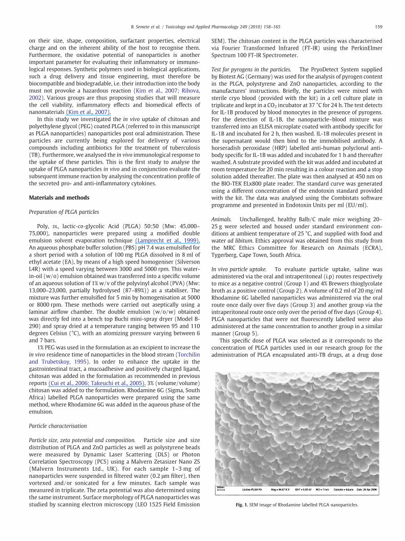
Fig. 1. SEM image of Rhodamine labelled PLGA nanoparticles.
159B. Semete et al. / Toxicology and Applied Pharmacology 249 (2010) 158–165
on their size, shape, composition, surfactant properties, electricalcharge and on the inherent ability of the host to recognise them.Furthermore, the oxidative potential of nanoparticles is anotherimportant parameter for evaluating their inflammatory or immuno-logical responses. Synthetic polymers used in biological applications,such a drug delivery and tissue engineering, must therefore bebiocompatible and biodegradable, i.e. their introduction into the bodymust not provoke a hazardous reaction (Kim et al., 2007; Rihova,2002). Various groups are thus proposing studies that will measurethe cell viability, inflammatory effects and biomedical effects ofnanomaterials (Kim et al., 2007).
In this study we investigated the in vivo uptake of chitosan andpolyethylene glycol (PEG) coated PLGA (referred to in this manuscriptas PLGA nanoparticles) nanoparticles post oral administration. Theseparticles are currently being explored for delivery of variouscompounds including antibiotics for the treatment of tuberculosis(TB). Furthermore, we analysed the in vivo immunological response tothe uptake of these particles. This is the first study to analyse theuptake of PLGA nanoparticles in vivo and in conjunction evaluate thesubsequent immune reaction by analysing the concentration profile ofthe secreted pro- and anti-inflammatory cytokines.
Materials and methods
Preparation of PLGA particles
Poly, DL, lactic-co-glycolic Acid (PLGA) 50:50 (Mw: 45,000–75,000), nanoparticles were prepared using a modified doubleemulsion solvent evaporation technique (Lamprecht et al., 1999).An aqueous phosphate buffer solution (PBS) pH 7.4 was emulsified fora short period with a solution of 100 mg PLGA dissolved in 8 ml ofethyl acetate (EA), by means of a high speed homogeniser (SilversonL4R) with a speed varying between 3000 and 5000 rpm. This water-in-oil (w/o) emulsion obtained was transferred into a specific volumeof an aqueous solution of 1% w/v of the polyvinyl alcohol (PVA) (Mw:13,000–23,000, partially hydrolysed (87–89%)) as a stabiliser. Themixture was further emulsified for 5 min by homogenisation at 5000or 8000 rpm. These methods were carried out aseptically using alaminar airflow chamber. The double emulsion (w/o/w) obtainedwas directly fed into a bench top Buchi mini-spray dryer (Model B-290) and spray dried at a temperature ranging between 95 and 110degrees Celsius (°C), with an atomizing pressure varying between 6and 7 bars.
1% PEG was used in the formulation as an excipient to increase thein vivo residence time of nanoparticles in the blood stream (Torchilinand Trubetskoy, 1995). In order to enhance the uptake in thegastrointestinal tract, a mucoadhesive and positively charged ligand,chitosan was added in the formulation as recommended in previousreports (Cui et al., 2006; Takeuchi et al., 2005). 3% (volume/volume)chitosan was added to the formulation. Rhodamine 6G (Sigma, SouthAfrica) labelled PLGA nanoparticles were prepared using the samemethod, where Rhodamine 6G was added in the aqueous phase of theemulsion.
Particle characterisation
Particle size, zeta potential and composition. Particle size and sizedistribution of PLGA and ZnO particles as well as polystyrene beadswere measured by Dynamic Laser Scattering (DLS) or PhotonCorrelation Spectroscopy (PCS) using a Malvern Zetasizer Nano ZS(Malvern Instruments Ltd., UK). For each sample 1–3 mg ofnanoparticles were suspended in filtered water (0.2 μm filter), thenvortexed and/or sonicated for a few minutes. Each sample wasmeasured in triplicate. The zeta potential was also determined usingthe same instrument. Surfacemorphology of PLGA nanoparticles wasstudied by scanning electron microscopy (LEO 1525 Field Emission
SEM). The chitosan content in the PLGA particles was characterisedvia Fourier Transformed Infrared (FT-IR) using the PerkinElmerSpectrum 100 FT-IR Spectrometer.
Test for pyrogens in the particles. The PryoDetect System suppliedby Biotest AG (Germany) was used for the analysis of pyrogen contentin the PLGA, polystyrene and ZnO nanoparticles, according to themanufactures' instructions. Briefly, the particles were mixed withsterile cryo blood (provided with the kit) in a cell culture plate intriplicate and kept in a CO2 incubator at 37 °C for 24 h. The test detectsfor IL-1B produced by blood monocytes in the presence of pyrogens.For the detection of IL-1B, the nanoparticle–blood mixture wastransferred into an ELISA microplate coated with antibody specific forIL-1B and incubated for 2 h, then washed. IL-1B molecules present inthe supernatant would then bind to the immobilised antibody. Ahorseradish peroxidase (HRP) labelled anti-human polyclonal anti-body specific for IL-1B was added and incubated for 1 h and thereafterwashed. A substrate providedwith the kit was added and incubated atroom temperature for 20 min resulting in a colour reaction and a stopsolution added thereafter. The plate was then analysed at 450 nm onthe BIO-TEK ELx800 plate reader. The standard curve was generatedusing a different concentration of the endotoxin standard providedwith the kit. The data was analysed using the Combistats softwareprogramme and presented in Endotoxin Units per ml (EU/ml).
Animals. Unchallenged, healthy Balb/C male mice weighing 20–25 g were selected and housed under standard environment con-ditions at ambient temperature of 25 °C, and supplied with food andwater ad libitum. Ethics approval was obtained from this study fromthe MRC Ethics Committee for Research on Animals (ECRA),Tygerberg, Cape Town, South Africa.
In vivo particle uptake. To evaluate particle uptake, saline wasadministered via the oral and intraperitoneal (i.p) routes respectivelyto mice as a negative control (Group 1) and 4% Brewers thioglycolatebroth as a positive control (Group 2). A volume of 0.2 ml of 20 mg/mlRhodamine 6G labelled nanoparticles was administered via the oralroute once daily over five days (Group 3) and another group via theintraperitoneal route once only over the period of five days (Group 4).PLGA nanoparticles that were not fluorescently labelled were alsoadministered at the same concentration to another group in a similarmanner (Group 5).
This specific dose of PLGA was selected as it corresponds to theconcentration of PLGA particles used in our research group for theadministration of PLGA encapsulated anti-TB drugs, at a drug dose
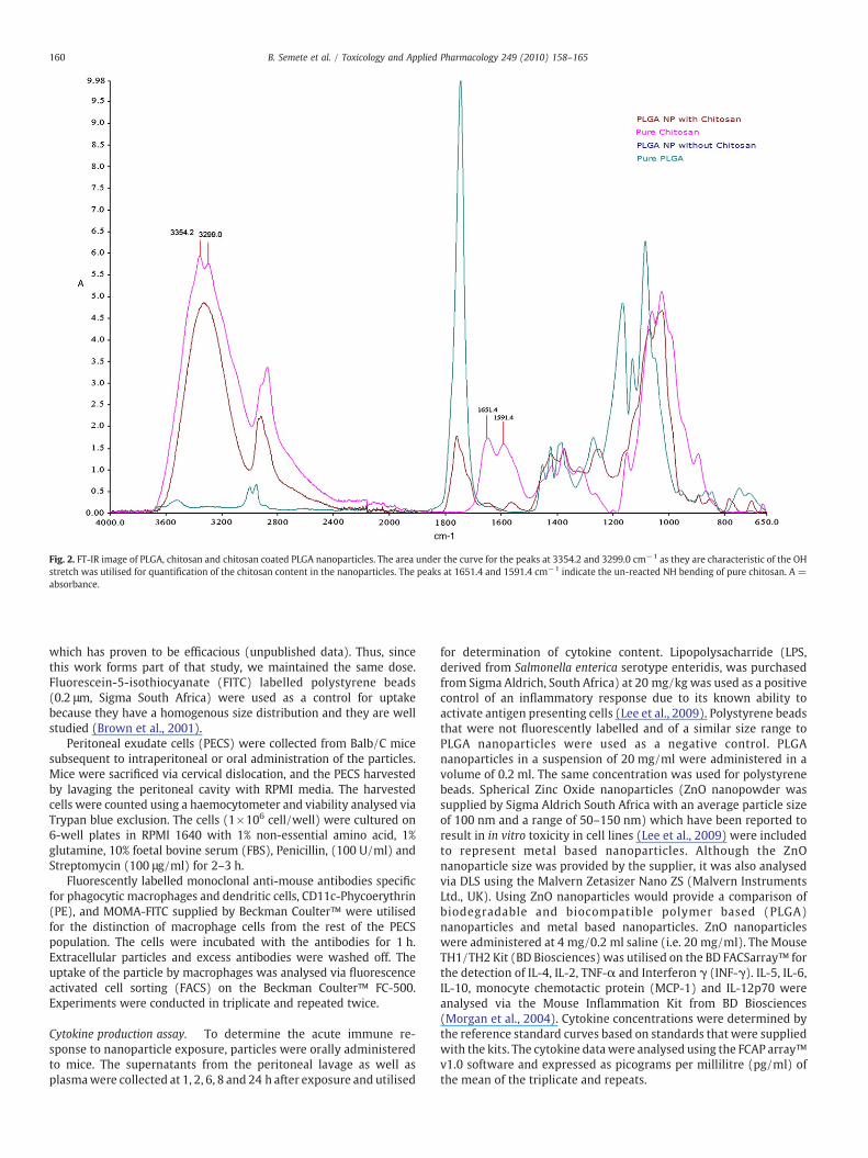
Fig. 2. FT-IR image of PLGA, chitosan and chitosan coated PLGA nanoparticles. The area under the curve for the peaks at 3354.2 and 3299.0 cm−1 as they are characteristic of the OHstretch was utilised for quantification of the chitosan content in the nanoparticles. The peaks at 1651.4 and 1591.4 cm−1 indicate the un-reacted NH bending of pure chitosan. A =absorbance.
160 B. Semete et al. / Toxicology and Applied Pharmacology 249 (2010) 158–165
which has proven to be efficacious (unpublished data). Thus, sincethis work forms part of that study, we maintained the same dose.Fluorescein-5-isothiocyanate (FITC) labelled polystyrene beads(0.2 μm, Sigma South Africa) were used as a control for uptakebecause they have a homogenous size distribution and they are wellstudied (Brown et al., 2001).
Peritoneal exudate cells (PECS) were collected from Balb/C micesubsequent to intraperitoneal or oral administration of the particles.Mice were sacrificed via cervical dislocation, and the PECS harvestedby lavaging the peritoneal cavity with RPMI media. The harvestedcells were counted using a haemocytometer and viability analysed viaTrypan blue exclusion. The cells (1×10⁶ cell/well) were cultured on6-well plates in RPMI 1640 with 1% non-essential amino acid, 1%glutamine, 10% foetal bovine serum (FBS), Penicillin, (100 U/ml) andStreptomycin (100 μg/ml) for 2–3 h.
Fluorescently labelled monoclonal anti-mouse antibodies specificfor phagocytic macrophages and dendritic cells, CD11c-Phycoerythrin(PE), and MOMA-FITC supplied by Beckman Coulter™ were utilisedfor the distinction of macrophage cells from the rest of the PECSpopulation. The cells were incubated with the antibodies for 1 h.Extracellular particles and excess antibodies were washed off. Theuptake of the particle by macrophages was analysed via fluorescenceactivated cell sorting (FACS) on the Beckman Coulter™ FC-500.Experiments were conducted in triplicate and repeated twice.
Cytokine production assay. To determine the acute immune re-sponse to nanoparticle exposure, particles were orally administeredto mice. The supernatants from the peritoneal lavage as well asplasmawere collected at 1, 2, 6, 8 and 24 h after exposure and utilised
for determination of cytokine content. Lipopolysacharride (LPS,derived from Salmonella enterica serotype enteridis, was purchasedfrom Sigma Aldrich, South Africa) at 20 mg/kg was used as a positivecontrol of an inflammatory response due to its known ability toactivate antigen presenting cells (Lee et al., 2009). Polystyrene beadsthat were not fluorescently labelled and of a similar size range toPLGA nanoparticles were used as a negative control. PLGAnanoparticles in a suspension of 20 mg/ml were administered in avolume of 0.2 ml. The same concentration was used for polystyrenebeads. Spherical Zinc Oxide nanoparticles (ZnO nanopowder wassupplied by Sigma Aldrich South Africa with an average particle sizeof 100 nm and a range of 50–150 nm) which have been reported toresult in in vitro toxicity in cell lines (Lee et al., 2009) were includedto represent metal based nanoparticles. Although the ZnOnanoparticle size was provided by the supplier, it was also analysedvia DLS using the Malvern Zetasizer Nano ZS (Malvern InstrumentsLtd., UK). Using ZnO nanoparticles would provide a comparison ofbiodegradable and biocompatible polymer based (PLGA)nanoparticles and metal based nanoparticles. ZnO nanoparticleswere administered at 4 mg/0.2 ml saline (i.e. 20 mg/ml). The MouseTH1/TH2 Kit (BD Biosciences) was utilised on the BD FACSarray™ forthe detection of IL-4, IL-2, TNF-α and Interferon γ (INF-γ). IL-5, IL-6,IL-10, monocyte chemotactic protein (MCP-1) and IL-12p70 wereanalysed via the Mouse Inflammation Kit from BD Biosciences(Morgan et al., 2004). Cytokine concentrations were determined bythe reference standard curves based on standards that were suppliedwith the kits. The cytokine data were analysed using the FCAP array™v1.0 software and expressed as picograms per millilitre (pg/ml) ofthe mean of the triplicate and repeats.
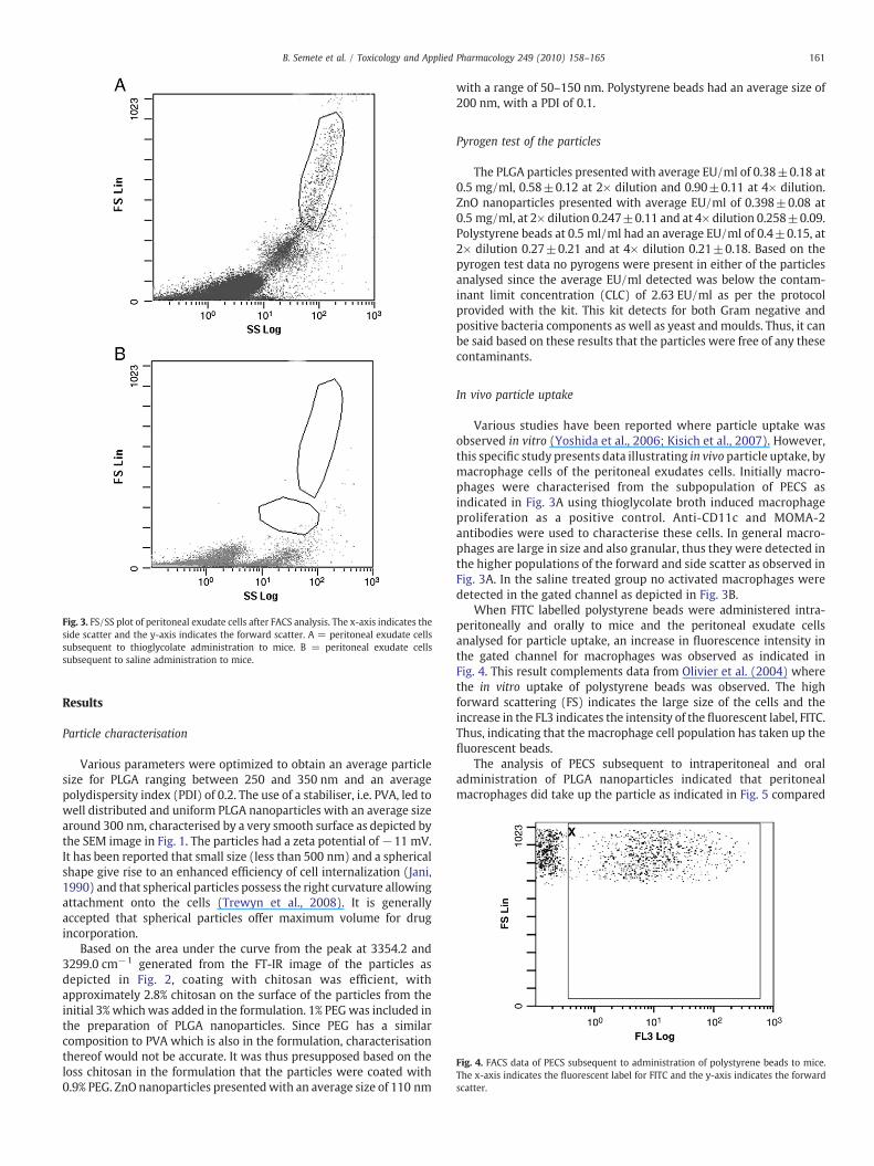
Fig. 4. FACS data of PECS subsequent to administration of polystyrene beads to mice.The x-axis indicates the fluorescent label for FITC and the y-axis indicates the forwardscatter.
Fig. 3. FS/SS plot of peritoneal exudate cells after FACS analysis. The x-axis indicates theside scatter and the y-axis indicates the forward scatter. A = peritoneal exudate cellssubsequent to thioglycolate administration to mice. B = peritoneal exudate cellssubsequent to saline administration to mice.
161B. Semete et al. / Toxicology and Applied Pharmacology 249 (2010) 158–165
Results
Particle characterisation
Various parameters were optimized to obtain an average particlesize for PLGA ranging between 250 and 350 nm and an averagepolydispersity index (PDI) of 0.2. The use of a stabiliser, i.e. PVA, led towell distributed and uniform PLGA nanoparticles with an average sizearound 300 nm, characterised by a very smooth surface as depicted bythe SEM image in Fig. 1. The particles had a zeta potential of−11 mV.It has been reported that small size (less than 500 nm) and a sphericalshape give rise to an enhanced efficiency of cell internalization (Jani,1990) and that spherical particles possess the right curvature allowingattachment onto the cells (Trewyn et al., 2008). It is generallyaccepted that spherical particles offer maximum volume for drugincorporation.
Based on the area under the curve from the peak at 3354.2 and3299.0 cm−1 generated from the FT-IR image of the particles asdepicted in Fig. 2, coating with chitosan was efficient, withapproximately 2.8% chitosan on the surface of the particles from theinitial 3% which was added in the formulation. 1% PEGwas included inthe preparation of PLGA nanoparticles. Since PEG has a similarcomposition to PVA which is also in the formulation, characterisationthereof would not be accurate. It was thus presupposed based on theloss chitosan in the formulation that the particles were coated with0.9% PEG. ZnO nanoparticles presentedwith an average size of 110 nm
with a range of 50–150 nm. Polystyrene beads had an average size of200 nm, with a PDI of 0.1.
Pyrogen test of the particles
The PLGA particles presented with average EU/ml of 0.38±0.18 at0.5 mg/ml, 0.58±0.12 at 2× dilution and 0.90±0.11 at 4× dilution.ZnO nanoparticles presented with average EU/ml of 0.398±0.08 at0.5 mg/ml, at 2× dilution 0.247±0.11 and at 4× dilution 0.258±0.09.Polystyrene beads at 0.5 ml/ml had an average EU/ml of 0.4±0.15, at2× dilution 0.27±0.21 and at 4× dilution 0.21±0.18. Based on thepyrogen test data no pyrogens were present in either of the particlesanalysed since the average EU/ml detected was below the contam-inant limit concentration (CLC) of 2.63 EU/ml as per the protocolprovided with the kit. This kit detects for both Gram negative andpositive bacteria components as well as yeast andmoulds. Thus, it canbe said based on these results that the particles were free of any thesecontaminants.
In vivo particle uptake
Various studies have been reported where particle uptake wasobserved in vitro (Yoshida et al., 2006; Kisich et al., 2007). However,this specific study presents data illustrating in vivo particle uptake, bymacrophage cells of the peritoneal exudates cells. Initially macro-phages were characterised from the subpopulation of PECS asindicated in Fig. 3A using thioglycolate broth induced macrophageproliferation as a positive control. Anti-CD11c and MOMA-2antibodies were used to characterise these cells. In general macro-phages are large in size and also granular, thus they were detected inthe higher populations of the forward and side scatter as observed inFig. 3A. In the saline treated group no activated macrophages weredetected in the gated channel as depicted in Fig. 3B.
When FITC labelled polystyrene beads were administered intra-peritoneally and orally to mice and the peritoneal exudate cellsanalysed for particle uptake, an increase in fluorescence intensity inthe gated channel for macrophages was observed as indicated inFig. 4. This result complements data from Olivier et al. (2004) wherethe in vitro uptake of polystyrene beads was observed. The highforward scattering (FS) indicates the large size of the cells and theincrease in the FL3 indicates the intensity of the fluorescent label, FITC.Thus, indicating that the macrophage cell population has taken up thefluorescent beads.
The analysis of PECS subsequent to intraperitoneal and oraladministration of PLGA nanoparticles indicated that peritonealmacrophages did take up the particle as indicated in Fig. 5 compared
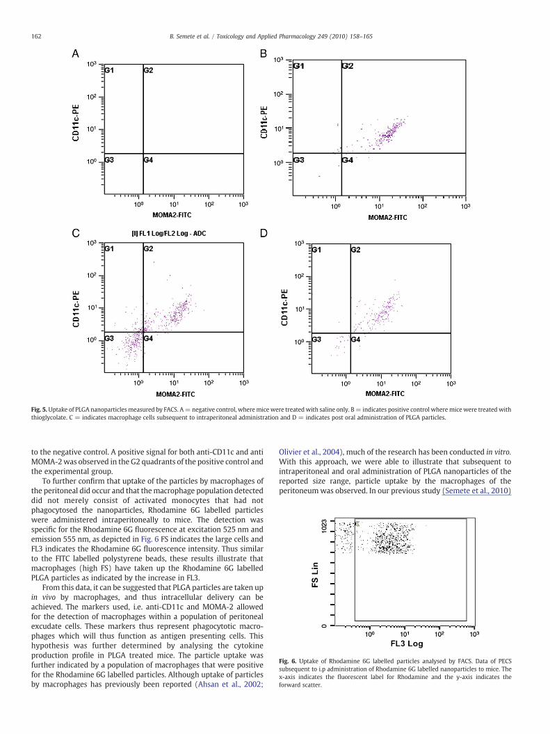
Fig. 6. Uptake of Rhodamine 6G labelled particles analysed by FACS. Data of PECSsubsequent to i.p administration of Rhodamine 6G labelled nanoparticles to mice. Thex-axis indicates the fluorescent label for Rhodamine and the y-axis indicates theforward scatter.
Fig. 5.Uptake of PLGA nanoparticles measured by FACS. A= negative control, wheremice were treated with saline only. B= indicates positive control where mice were treated withthioglycolate. C = indicates macrophage cells subsequent to intraperitoneal administration and D = indicates post oral administration of PLGA particles.
162 B. Semete et al. / Toxicology and Applied Pharmacology 249 (2010) 158–165
to the negative control. A positive signal for both anti-CD11c and antiMOMA-2was observed in the G2 quadrants of the positive control andthe experimental group.
To further confirm that uptake of the particles by macrophages ofthe peritoneal did occur and that themacrophage population detecteddid not merely consist of activated monocytes that had notphagocytosed the nanoparticles, Rhodamine 6G labelled particleswere administered intraperitoneally to mice. The detection wasspecific for the Rhodamine 6G fluorescence at excitation 525 nm andemission 555 nm, as depicted in Fig. 6 FS indicates the large cells andFL3 indicates the Rhodamine 6G fluorescence intensity. Thus similarto the FITC labelled polystyrene beads, these results illustrate thatmacrophages (high FS) have taken up the Rhodamine 6G labelledPLGA particles as indicated by the increase in FL3.
From this data, it can be suggested that PLGA particles are taken upin vivo by macrophages, and thus intracellular delivery can beachieved. The markers used, i.e. anti-CD11c and MOMA-2 allowedfor the detection of macrophages within a population of peritonealexcudate cells. These markers thus represent phagocytotic macro-phages which will thus function as antigen presenting cells. Thishypothesis was further determined by analysing the cytokineproduction profile in PLGA treated mice. The particle uptake wasfurther indicated by a population of macrophages that were positivefor the Rhodamine 6G labelled particles. Although uptake of particlesby macrophages has previously been reported (Ahsan et al., 2002;
Olivier et al., 2004), much of the research has been conducted in vitro.With this approach, we were able to illustrate that subsequent tointraperitoneal and oral administration of PLGA nanoparticles of thereported size range, particle uptake by the macrophages of theperitoneumwas observed. In our previous study (Semete et al., 2010)
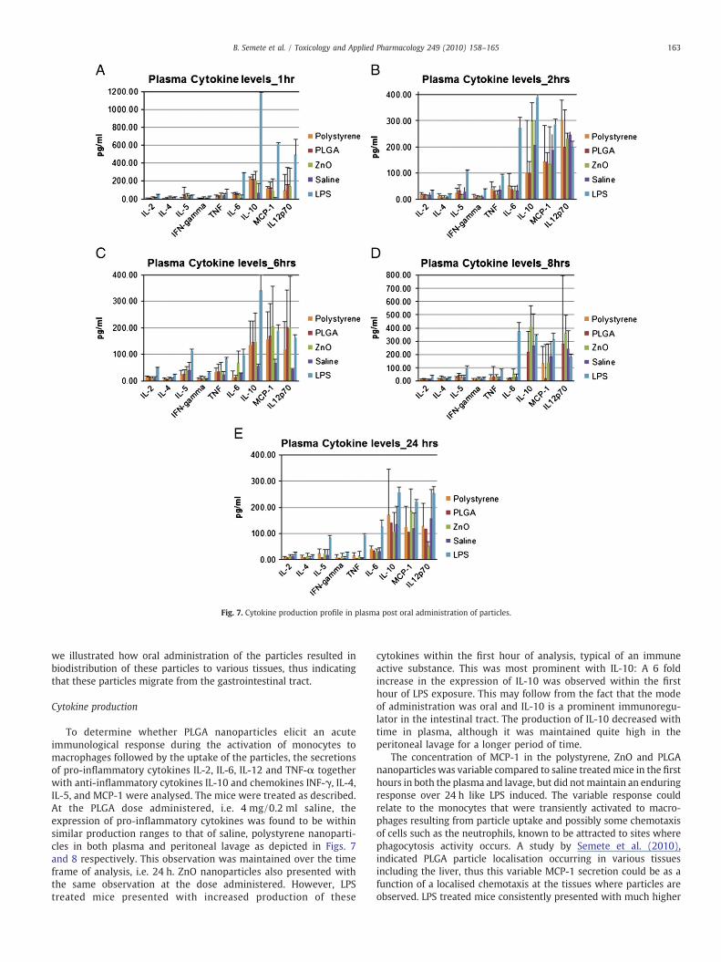
Fig. 7. Cytokine production profile in plasma post oral administration of particles.
163B. Semete et al. / Toxicology and Applied Pharmacology 249 (2010) 158–165
we illustrated how oral administration of the particles resulted inbiodistribution of these particles to various tissues, thus indicatingthat these particles migrate from the gastrointestinal tract.
Cytokine production
To determine whether PLGA nanoparticles elicit an acuteimmunological response during the activation of monocytes tomacrophages followed by the uptake of the particles, the secretionsof pro-inflammatory cytokines IL-2, IL-6, IL-12 and TNF-α togetherwith anti-inflammatory cytokines IL-10 and chemokines INF-γ, IL-4,IL-5, and MCP-1 were analysed. The mice were treated as described.At the PLGA dose administered, i.e. 4 mg/0.2 ml saline, theexpression of pro-inflammatory cytokines was found to be withinsimilar production ranges to that of saline, polystyrene nanoparti-cles in both plasma and peritoneal lavage as depicted in Figs. 7and 8 respectively. This observation was maintained over the timeframe of analysis, i.e. 24 h. ZnO nanoparticles also presented withthe same observation at the dose administered. However, LPStreated mice presented with increased production of these
cytokines within the first hour of analysis, typical of an immuneactive substance. This was most prominent with IL-10: A 6 foldincrease in the expression of IL-10 was observed within the firsthour of LPS exposure. This may follow from the fact that the modeof administration was oral and IL-10 is a prominent immunoregu-lator in the intestinal tract. The production of IL-10 decreased withtime in plasma, although it was maintained quite high in theperitoneal lavage for a longer period of time.
The concentration of MCP-1 in the polystyrene, ZnO and PLGAnanoparticles was variable compared to saline treatedmice in the firsthours in both the plasma and lavage, but did notmaintain an enduringresponse over 24 h like LPS induced. The variable response couldrelate to the monocytes that were transiently activated to macro-phages resulting from particle uptake and possibly some chemotaxisof cells such as the neutrophils, known to be attracted to sites wherephagocytosis activity occurs. A study by Semete et al. (2010),indicated PLGA particle localisation occurring in various tissuesincluding the liver, thus this variable MCP-1 secretion could be as afunction of a localised chemotaxis at the tissues where particles areobserved. LPS treated mice consistently presented with much higher
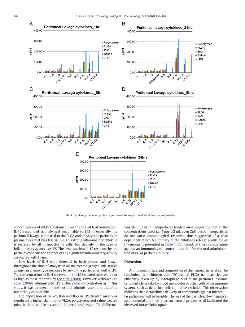
Fig. 8. Cytokine production profile in peritoneal lavage post oral administration of particles.
164 B. Semete et al. / Toxicology and Applied Pharmacology 249 (2010) 158–165
concentrations of MCP-1 sustained over the full 24 h of observation.IL-12 responded strongly and sustainable to LPS in especially theperitoneal lavage, compared to the PLGA and polystyrene particles. Inplasma this effect was less visible. This strong inflammatory cytokineis secreted by all phagocytosing cells, but strongly in the case ofinflammatory agents like LPS. The low, transient IL-12 response by theparticles confirms the absence of any significant inflammatory activityassociated with them.
Low levels of IL-4 were detected in both plasma and lavagethroughout the time of analysis in all the treated groups. This arguesagainst an allergic type response by any of the particles as well as LPS.The concentrations of IL-4 observed in the LPS treated mice were notas high as those reported by Lee et al. (2009). However, although Leeet al. (2009) administered LPS at the same concentration as in thisstudy, it was by injection and not oral administration and thereforenot strictly comparable.
The expression of TNF-α, IL-6 and IL-5 in LPS treated mice wassignificantly higher than that of PLGA, polystyrene and saline treatedmice, both in the plasma and in the peritoneal lavage. The difference
was also noted in nanoparticles treated mice suggesting that at theconcentrations used i.e. 4 mg/0.2 ml, even ZnO based nanoparticlesdo not cause immunological response, thus suggestive of a dosedependent effect. A summary of the cytokines release profile for allthe groups is presented in Table 1. Combined, all these results argueagainst an immunological contra-indication for the oral administra-tion of PLGA particles in mice.
Discussion
At this specific size and composition of the nanoparticles, it can beconcluded that chitosan and PEG coated PLGA nanoparticles areefficiently taken up by macrophage cells of the peritoneal exudatecells. Particle uptake by blood monocytes or other cells of the immunesystems such as dendritric cells cannot be excluded. This observationindicates that intracellular delivery of compounds against intracellu-lar pathogens will be feasible. The size of the particles, (low negative)zeta potential and their physicochemical properties all facilitated theobserved intracellular uptake.
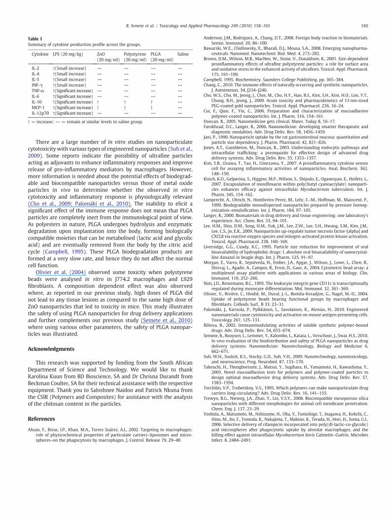
Table 1Summary of cytokine production profile across the groups.
Cytokine LPS (20 mg/kg) ZnO(20 mg/ml)
Polystyrene(20 mg/ml)
PLGA(20 mg/ml)
Saline
IL-2 ↑(Small increase) ↔ ↔ ↔ ↔IL-4 ↑(Small increase) ↔ ↔ ↔ ↔IL-5 ↑(Small increase) ↔ ↔ ↔ ↔INF-γ ↑(Small increase) ↔ ↔ ↔ ↔TNF-α ↑(Significant increase) ↔ ↔ ↔ ↔IL-6 ↑(Significant increase) ↔ ↔ ↔ ↔IL-10 ↑(Significant increase) ↑ ↑ ↑ ↔MCP-1 ↑(Significant increase) ↑ ↑ ↑ ↔IL-12p70 ↑(Significant increase) ↔ ↔ ↔ ↔
↑ = increase; ↔ = remain at similar levels to saline group.
165B. Semete et al. / Toxicology and Applied Pharmacology 249 (2010) 158–165
There are a large number of in vitro studies on nanoparticulatecytotoxicity with various types of engineered nanoparticles (Suh et al.,2009). Some reports indicate the possibility of ultrafine particlesacting as adjuvants to enhance inflammatory responses and improverelease of pro-inflammatory mediators by macrophages. However,more information is needed about the potential effects of biodegrad-able and biocompatible nanoparticles versus those of metal oxideparticles in vivo to determine whether the observed in vitrocytotoxicity and inflammatory response is physiologically relevant(Cho et al., 2009; Palomäki et al., 2010). The inability to elicit asignificant effect of the immune response does not mean that PLGAparticles are completely inert from the immunological point of view.As polyesters in nature, PLGA undergoes hydrolysis and enzymaticdegradation upon implantation into the body, forming biologicallycompatible moieties that can be metabolised (lactic acid and glycolicacid) and are eventually removed from the body by the citric acidcycle (Campbell, 1995). These PLGA biodegradation products areformed at a very slow rate, and hence they do not affect the normalcell function.
Olivier et al. (2004) observed some toxicity when polystyrenebeads were analysed in vitro in J774.2 macrophages and L929fibroblasts. A composition dependent effect was also observedwhere, as reported in our previous study, high doses of PLGA didnot lead to any tissue lesions as compared to the same high dose ofZnO nanoparticles that led to toxicity in mice. This study illustratesthe safety of using PLGA nanoparticles for drug delivery applicationsand further complements our previous study (Semete et al., 2010)where using various other parameters, the safety of PLGA nanopar-ticles was illustrated.
Acknowledgments
This research was supported by funding from the South AfricanDepartment of Science and Technology. We would like to thankKarolina Kuun from BD Bioscience, SA and Dr Chrisna Durandt fromBeckman Coulter, SA for their technical assistance with the respectiveequipment. Thank you to Saloshnee Naidoo and Patrick Nkuna fromthe CSIR (Polymers and Composites) for assistance with the analysisof the chitosan content in the particles.
References
Ahsan, F., Rivas, I.P., Khan, M.A., Torres Suárez, A.I., 2002. Targeting to macrophages:role of physicochemical properties of particulate carriers–liposomes and micro-spheres–on the phagocytosis by macrophages. J. Control. Release 79, 29–40.
Anderson, J.M., Rodriguez, A., Chang, D.T., 2008. Foreign body reaction to biomaterials.Semin. Immunol. 20, 86–100.
Bawarski, W.E., Chidlowsky, E., Bharali, D.J., Mousa, S.A., 2008. Emerging nanopharma-ceuticals. Nanomed. Nanotechnol. Biol. Med. 4, 273–282.
Brown, D.M., Wilson, M.R., MacNee, W., Stone, V., Donaldson, K., 2001. Size-dependentproinflammatory effects of ultrafine polystyrene particles: a role for surface areaand oxidative stress in the enhanced activity of ultrafines. Toxicol. Appl. Pharmacol.175, 191–199.
Campbell, 1995. Biochemistry. Saunders College Publishing, pp. 365–384.Chang, C., 2010. The immune effects of naturally occurring and synthetic nanoparticles.
J. Autoimmun. 34, J234–J246.Cho, W.S., Cho, M., Jeong, J., Choi, M., Cho, H.Y., Han, B.S., Kim, S.H., Kim, H.O., Lim, Y.T.,
Chung, B.H., Jeong, J., 2009. Acute toxicity and pharmacokinetics of 13 nm-sizedPEG-coated gold nanoparticles. Toxicol. Appl. Pharmacol. 236, 16–24.
Cui, F., Qian, F., Yin, C., 2006. Preparation and characterization of mucoadhesivepolymer-coated nanoparticles. Int. J. Pharm. 316, 154–161.
Duncan, R., 2005. Nanomedicine gets clinical. Mater. Today 8, 16–17.Farokhzad, O.C., Langer, R., 2006. Nanomedicine: developing smarter therapeutic and
diagnostic modalities. Adv. Drug Deliv. Rev. 58, 1456–1459.Jani, P., 1990. Nanoparticle uptake by the rat gastrointestinal mucosa: quantitation and
particle size dependency. J. Pharm. Pharmacol. 42, 821–826.Jones, A.T., Gumbleton, M., Duncan, R., 2003. Understanding endocytic pathways and
intracellular trafficking: a prerequisite for effective design of advanced drugdelivery systems. Adv. Drug Deliv. Rev. 55, 1353–1357.
Kim, S.B., Ozawa, T., Tao, H., Umezawa, Y., 2007. A proinflammatory cytokine sensorcell for assaying inflammatory activities of nanoparticles. Anal. Biochem. 362,148–150.
Kisich, K.O., Gelperina, S., Higgins, M.P., Wilson, S., Shipulo, E., Oganesyan, E., Heifets, L.,2007. Encapsulation of moxifloxacin within poly(butyl cyanoacrylate) nanoparti-cles enhances efficacy against intracellular Mycobacterium tuberculosis. Int. J.Pharm. 345, 154–162.
Lamprecht, A., Ubrich, N., Hombreiro Perez, M., Lehr, C.-M., Hoffman, M., Maincent, P.,1999. Biodegradable monodispersed nanoparticles prepared by pressure homog-enization–emulsification. Int. J. Pharm. 184, 97–105.
Langer, R., 2000. Biomaterials in drug delivery and tissue engineering: one laboratory'sexperience. Acc. Chem. Res. 33, 94–101.
Lee, H.M., Shin, D.M., Song, H.M., Yuk, J.M., Lee, Z.W., Lee, S.H., Hwang, S.M., Kim, J.M.,Lee, C.S., Jo, E.K., 2009. Nanoparticles up-regulate tumor necrosis factor-[alpha] andCXCL8 via reactive oxygen species andmitogen-activated protein kinase activation.Toxicol. Appl. Pharmacol. 238, 160–169.
Liversidge, G.G., Cundy, K.C., 1995. Particle size reduction for improvement of oralbioavailability of hydrophobic drugs: I. absolute oral bioavailability of nanocrystal-line danazol in beagle dogs. Int. J. Pharm. 125, 91–97.
Morgan, E., Varro, R., Sepulveda, H., Ember, J.A., Apgar, J., Wilson, J., Lowe, L., Chen, R.,Shivraj, L., Agadir, A., Campos, R., Ernst, D., Gaur, A., 2004. Cytometric bead array: amultiplexed assay platform with applications in various areas of biology. Clin.Immunol. 110, 252–266.
Noti, J.D., Reinemann, B.C., 1995. The leukocyte integrin gene CD11c is transcriptionallyregulated during monocyte differentiation. Mol. Immunol. 32, 361–369.
Olivier, V., Rivière, C., Hindié, M., Duval, J.-L., Bomila-Koradjim, G., Nagel, M.-D., 2004.Uptake of polystyrene beads bearing functional groups by macrophages andfibroblasts. Colloids Surf., B 33, 23–31.
Palomäki, J., Karisola, P., Pylkkänen, L., Savolainen, K., Alenius, H., 2010. Engineerednanomaterials cause cytotoxicity and activation on mouse antigen presenting cells.Toxicology 267, 125–131.
Rihova, B., 2002. Immunomodulating activities of soluble synthetic polymer-bounddrugs. Adv. Drug Deliv. Rev. 54, 653–674.
Semete, B., Booysen, L., Lemmer, Y., Kalombo, L., Katata, L., Verschoor, J., Swai, H.S., 2010.In vivo evaluation of the biodistribution and safety of PLGA nanoparticles as drugdelivery systems. Nanomedicine: Nanotechnology, Biology and Medicine 6,662–671.
Suh, W.H., Suslick, K.S., Stucky, G.D., Suh, Y.H., 2009. Nanotechnology, nanotoxicology,and neuroscience. Prog. Neurobiol. 87, 133–170.
Takeuchi, H., Thongborisute, J., Matsui, Y., Sugihara, H., Yamamoto, H., Kawashima, Y.,2005. Novel mucoadhesion tests for polymers and polymer-coated particles todesign optimal mucoadhesive drug delivery systems. Adv. Drug Deliv. Rev. 57,1583–1594.
Torchilin, V.P., Trubetskoy, V.S., 1995. Which polymers can make nanoparticulate drugcarriers long-circulating? Adv. Drug Deliv. Rev. 16, 141–155.
Trewyn, B.G., Nieweg, J.A., Zhao, Y., Lin, V.S.Y., 2008. Biocompatible mesoporous silicananoparticles with different morphologies for animal cell membrane penetration.Chem. Eng. J. 137, 23–29.
Yoshida, A., Matumoto, M., Hshizume, H., Oba, Y., Tomishige, T., Inagawa, H., Kohchi, C.,Hino, M., Ito, F., Tomoda, K., Nakajima, T., Makino, K., Terada, H., Hori, H., Soma, G.I.,2006. Selective delivery of rifampicin incorporated into poly(dl-lactic-co-glycolic)acid microspheres after phagocytotic uptake by alveolar macrophages, and thekilling effect against intracellular Mycobacterium bovis Calmette–Guérin. MicrobesInfect. 8, 2484–2491.
Related Documents