RESEARCH PAPER The Development and Mechanism Studies of Cationic Chitosan-Modified Biodegradable PLGA Nanoparticles for Efficient siRNA Drug Delivery Xudong Yuan & Bruhal A. Shah & Naimesh K. Kotadia & Jian Li & Hua Gu & Zhiqian Wu Received: 26 November 2009 / Accepted: 24 February 2010 / Published online: 23 March 2010 # Springer Science+Business Media, LLC 2010 ABSTRACT Purpose In order to improve siRNA delivery for possible clinical applications, we developed biodegradable chitosan- modified poly(D,L-lactide-co-glycolide) (CHT-PLGA) nanopar- ticles with positive surface charge, high siRNA loading, high transfection efficiency and low toxicity. Methods CHT-PLGA nanoparticles were prepared, and siRNA was loaded by emulsion evaporation method with poly(vinyl alcohol) (PVA) as emulsifier. siRNA loading efficiency, particle size, and Zeta potential of nanoparticles were measured. Gel retardation and protection assays were conducted to determine the loading and binding of siRNA in the formulation. Cell transfection was performed to study in vitro siRNA silencing efficiency. XTT assay was used to evaluate the cytotoxicity. Results It was found that the nanoparticle diameter and positive Zeta potential increase as the chitosan coating concentration increases. CHT-PLGA nanoparticles showed excellent siRNA binding ability and effective protection of oligos from RNase degradation. siRNA-loaded nanoparticles were successfully delivered into the HEK 293 T cell line, and the silencing of green fluorescence protein (GFP) expression was observed using fluorescent microscopy and flow cytom- etry. In addition, the cytotoxicity assay revealed that CHT- PLGA nanoparticles had relatively low cytotoxicity. Conclusion This study suggests that biodegradable cationic CHT-PLGA nanoparticles possess great potential for efficient and safer siRNA delivery in future clinical applications. KEY WORDS chitosan . nanoparticles . nanotechnology . poly (D,L-lactide-co-glycolide) (PLGA) . siRNA delivery INTRODUCTION Gene therapy refers to specifically reducing or silencing the expression of deleterious genes in the targeted cells or organs (1,2); therefore, it is a powerful approach for the treatment of a wide range of diseases, including cancer, by producing bioactive agents or stopping abnormal cell functions, such as genetic disorders or uncontrollable proliferation of cells (3). Recently, short interfering RNA (siRNA) has been used to regulate gene expression in mammalian cells through RNA interference (RNAi) (4). RNAi was first demonstrated in mammalian cells by Elbashir et al.(5). Discovery of the RNAi mechanism by Fire, Mello and coworkers in the late 1990s provided new direction and gave renewed promise to the field of gene therapy (6). RNAi is a simple and rapid method of silencing gene expression in a wide range of organisms by double-stranded 21– 23-nucleotides RNA duplex (siRNA) that contains 5′- phosphate and 3′- hydroxyl termini with two nucleotide overhangs (7). After cleavage of long double-stranded RNA (dsRNA) into short fragments of siRNA, these oligos are then separated into single strands and incorporated into a X. Yuan (*) : B. A. Shah : N. K. Kotadia : Z. Wu Division of Pharmaceutical Sciences, Arnold & Marie Schwartz College of Pharmacy, Long Island University, 75 DeKalb Avenue, Brooklyn, New York 11201-5497, USA e-mail: [email protected] J. Li Nanotarget, 10054 Mesa Ridge Court, San Diego, California 92121, USA H. Gu Department of Microbiology, College of Physicians and Surgeons, Columbia University, 701 West 168th Street, New York City, New York 10032, USA Pharm Res (2010) 27:1285–1295 DOI 10.1007/s11095-010-0103-0

Welcome message from author
This document is posted to help you gain knowledge. Please leave a comment to let me know what you think about it! Share it to your friends and learn new things together.
Transcript
RESEARCH PAPER
The Development and Mechanism Studies of CationicChitosan-Modified Biodegradable PLGA Nanoparticlesfor Efficient siRNA Drug Delivery
Xudong Yuan & Bruhal A. Shah & Naimesh K. Kotadia & Jian Li & Hua Gu & Zhiqian Wu
Received: 26 November 2009 /Accepted: 24 February 2010 /Published online: 23 March 2010# Springer Science+Business Media, LLC 2010
ABSTRACTPurpose In order to improve siRNA delivery for possibleclinical applications, we developed biodegradable chitosan-modified poly(D,L-lactide-co-glycolide) (CHT-PLGA) nanopar-ticles with positive surface charge, high siRNA loading, hightransfection efficiency and low toxicity.Methods CHT-PLGA nanoparticles were prepared, andsiRNA was loaded by emulsion evaporation method withpoly(vinyl alcohol) (PVA) as emulsifier. siRNA loading efficiency,particle size, and Zeta potential of nanoparticles weremeasured. Gel retardation and protection assays wereconducted to determine the loading and binding of siRNA inthe formulation. Cell transfection was performed to study invitro siRNA silencing efficiency. XTT assay was used to evaluatethe cytotoxicity.Results It was found that the nanoparticle diameter andpositive Zeta potential increase as the chitosan coatingconcentration increases. CHT-PLGA nanoparticles showedexcellent siRNA binding ability and effective protection ofoligos from RNase degradation. siRNA-loaded nanoparticleswere successfully delivered into the HEK 293 T cell line, and
the silencing of green fluorescence protein (GFP) expressionwas observed using fluorescent microscopy and flow cytom-etry. In addition, the cytotoxicity assay revealed that CHT-PLGA nanoparticles had relatively low cytotoxicity.Conclusion This study suggests that biodegradable cationicCHT-PLGA nanoparticles possess great potential for efficientand safer siRNA delivery in future clinical applications.
KEY WORDS chitosan . nanoparticles . nanotechnology . poly(D,L-lactide-co-glycolide) (PLGA) . siRNA delivery
INTRODUCTION
Gene therapy refers to specifically reducing or silencing theexpression of deleterious genes in the targeted cells ororgans (1,2); therefore, it is a powerful approach for thetreatment of a wide range of diseases, including cancer, byproducing bioactive agents or stopping abnormal cellfunctions, such as genetic disorders or uncontrollableproliferation of cells (3). Recently, short interfering RNA(siRNA) has been used to regulate gene expression inmammalian cells through RNA interference (RNAi) (4).RNAi was first demonstrated in mammalian cells byElbashir et al. (5). Discovery of the RNAi mechanism byFire, Mello and coworkers in the late 1990s provided newdirection and gave renewed promise to the field of genetherapy (6).
RNAi is a simple and rapid method of silencing geneexpression in a wide range of organisms by double-stranded21– 23-nucleotides RNA duplex (siRNA) that contains 5′-phosphate and 3′- hydroxyl termini with two nucleotideoverhangs (7). After cleavage of long double-stranded RNA(dsRNA) into short fragments of siRNA, these oligos arethen separated into single strands and incorporated into a
X. Yuan (*) : B. A. Shah :N. K. Kotadia : Z. WuDivision of Pharmaceutical Sciences, Arnold & Marie SchwartzCollege of Pharmacy, Long Island University,75 DeKalb Avenue,Brooklyn, New York 11201-5497, USAe-mail: [email protected]
J. LiNanotarget,10054 Mesa Ridge Court,San Diego, California 92121, USA
H. GuDepartment of Microbiology, College of Physicians and Surgeons,Columbia University,701 West 168th Street,New York City, New York 10032, USA
Pharm Res (2010) 27:1285–1295DOI 10.1007/s11095-010-0103-0
RISC complex and subsequently target messenger RNA(mRNA), where they induce cleavage, preventing it frombeing used as a template. The RNAi gene silencing techniqueis widely used in biological and medical research to probe thebiological role of a particular gene as a means to inhibitinfection by human immunodeficiency virus, poliovirus andhepatitis C virus, and to suppress cancer cell growth bysilencing virus and cancer gene expression (8–12).
Significant advancements in siRNA delivery systems willbe needed to translate this nucleic acid-based therapy intoreliable clinical therapies (13). Since cellular uptake ofsiRNA is usually very poor, and naked siRNA can berapidly degraded by RNase enzymes and excreted by thekidney, wide distribution of siRNA in the body must beprevented in order to concentrate administered siRNA atthe site of action. To overcome these challenges, there is agreat need to develop efficient siRNA delivery systems. Todate, both viral and non-viral vectors have been used todeliver siRNA. Even though viral vectors are frequentlyused due to high efficiency, their clinical applications arelimited because of immunogenicity, potential infectivity,inflammation and complicated production. Therefore, non-viral vectors, such as cationic liposomes and polymers, havebeen used to avoid these problems and overcome intracel-lular and extracellular barriers (14,15). However, com-plexes formed with cationic liposomes are large in size,thermodynamically unstable, and can be easily clearedfrom bloodstream (16,17). Compared to liposomes, poly-mers can form more stable nano-size complexes, which canprovide better protection for DNA or siRNA oligos fromnuclease degradation (18). Different types of polymers havebeen used for this purpose, such as poly(D,L-lactide-co-glycolide) (PLGA), poly(lactic acid) (PLA), poly(cyanoacry-late) (PCA), chitosan, polyethylenimine (PEI), poly(L-lysine)(PLL), dendrimers, poly(vinylimidazole) and imidazole con-taining polymers (19–23).
PLGA is one of the most promising polymeric carriers forsiRNA delivery. It has been tested for toxicity and safety inmany studies, and is currently being used in humans forresorbable sutures, bone implants, artificial organs, tissueengineering, and contraceptive implants (24–27). Polyestersin nature, PLGA polymers undergo hydrolysis uponadministration into the body, forming biologically compat-ible and metabolizable moieties (lactic acid and glycolic acid)that are eventually removed from the body by the citric acidcycle. PLGA has been successfully used to prepare siRNA-loaded nanoparticles for delivery of siRNA and silencing invitro (19). However, the loading capacity of PLGA nano-particles is not high, and the net negative charge of PLGAnanoparticles will not be favorable if higher silencingefficiency is desired. Therefore, in this project, we incorpo-rated cationic chitosan into PLGA nanopartices in order toimprove siRNA payload and its silencing efficiency.
Chitosan is a linear polysaccharide composed of β-(1–4)-linked D-Glucosamine (deacetylated unit) and N-acetylated-D-glucosamine (acetylated unit). Generally, theDDA (degree of deacetylation) of chitosan influences thecharacteristics such as charge density, solubility, crystallin-ity, and degradation of the polymer (28,29). Chitosan isbiodegradable and biocompatible and has been widelyinvestigated in the drug delivery, drug targeting, develop-ment of hemodialysis membranes and artificial skin, and inother applications. Chitosan breaks down slowly toharmless products (amino sugars), which are completelyabsorbed by the human body (30). Most studies ofchitosan/DNA complexes have used highly deacetylatedchitosan with higher oligo binding efficacy (31,32). It wasreported that chitosan could be incorporated into PLGAnanoparticles to improve delivery of small molecules, suchas paclitaxel (33), and to improve the delivery of DNA andantisense oligos (34–36). However, the delivery of siRNA,which is a very promising therapeutic in the future, bychitosan-modified PLGA nanoparticles to the cells hasrarely been studied to date (37). Therefore, in this study,we developed cationic chitosan-modified biodegradablePLGA (CHT-PLGA) nanoparticles in order to achievehigher siRNA loading, protection of oligos from degrada-tion, reduced cytotoxicity and enhanced transfectionefficiency.
MATERIALS AND METHODS
Materials
Chitosan (deacetylation degree >85%), poly (D,L-lactide-co-glycolide) (L:G molar ratio=75:25, MW 66,000–107,000),poly (vinyl alcohol) (PVA, MW 9,000–10,000, 80%hydrolyzed), Dulbecco’s phosphate-buffered saline (PBS),Dulbecco’s Modified Eagle’s medium (DMEM) high glu-cose, LB medium, Penicillin-Streptomycin antibiotics anddimethylsulfoxide (DMSO, 99.5% GC) were purchasedfrom Sigma-Aldrich (St. Louis, MO, USA). si-GFP-RNAoligo ( 5′-GCAAGCUGACCCUGAAGUUCAU-3′, 3′-GCCGUUCGACUGGGACUUCAAG-5′) was purchasedfrom Ambion (Austin, TX, USA). Lipofectamine™ 2,000,Opti-MEM-I Reduced Serum medium (1X), Fetal BovineSerum (FBS), competent E.coli DH5α cells were obtainedfrom Invitrogen (Carlsbad, CA, USA). HPLC gradedichloromethane (DCM) was purchased from EM Science(Gibbstown, NJ, USA). Plasmid extraction MIDI prep kitwas purchased from Qiagen (Valencia, CA, USA). Sodiumacetate buffer (pH 4.5) was prepared in accordance withUSP guidelines. Human embryonic kidney cells (HEK293 T) were obtained from Dr. Hua Gu’s lab at ColumbiaUniversity.
1286 Yuan et al.
Plasmid Extraction
E.coli DH5α was incubated with plasmid DNA and allowedto grow by using LB media for transformation. A freshlystreaked selective plate was taken and inoculated in aculture of LB media with antibiotics (liquid media), andthen incubated for about 10 h at 37°C on a shakerincubator. The standard protocol of Qiagen’s MIDI kit wasfollowed, and the final confirmation was performed by gelelectrophoresis. The concentration of extracted plasmidwas determined by UV spectroscopy.
Preparation of siRNA-Loaded CHT-PLGANanoparticles
For the preparation of CHT-PLGA nanoparticles, theemulsion solvent evaporation method was used. Chitosanwas dissolved in 20 ml of sodium acetate buffer (pH 4.5) atfive different concentrations (0.00, 0.01, 0.05, 0.1, and0.25% w/v). Then 40 mg of PLGA was dissolved in 2 ml ofDCM in a flask by vigorous vortex mixing. PVA solution1.0% (w/v) was prepared separately with sterilized waterand filtered using a 0.2 µm membrane filter. PVA solution(10 ml) was added to the prepared aqueous chitosansolution of different concentrations to give five samples.The final chitosan coating concentrations in the solutionswere 0.00, 0.0067, 0.033, 0.067 and 0.17% in Samples I,II, III, IV and V, respectively. The resultant polymeraqueous solution was mixed by a magnetic stirrer for 5 minat 500 rpm. The equivalent of 1,000 picomole of siRNA(50 µl) was added to the aqueous chitosan solution. PLGAsolution was poured slowly into the chitosan solution,resulting in an oil-in-water (O/W) emulsion upon constantmagnetic stirring. The emulsion was further processedusing a Misonix probe sonicator (Farmingdale, NY, USA)in pulse mode at 1 watt for 45 s. DCM was removed byovernight stirring with a magnetic stirrer at room temper-ature in a sterile hood. After 24 h, nanoparticles werecollected using an ultracentrifuge (Beckman LE-Ultracentrifuge, Fullerton, CA) at 10,000 rpm for 15 min,and the sediment (pellet) was resuspended in RNase-freedeionized water. This process was repeated twice to removeexcess amount of PVA. The control was PLGA nano-particles not modified with chitosan, which is Sample I.
Characterization of Prepared Nanoparticles
The different samples of CHT-PLGA nanoparticles werecollected and further diluted with deionized water to anappropriate concentration. The particle size and surfacecharge (Zeta potential) of nanoparticles were measured by adynamic light scattering (DLS) particle sizer–Zetasizer(Malvern Instruments, Westborough, MA, USA).
Determination of siRNA Loading Efficiency
Nanoparticles were centrifuged (Beckman LE-Ultracentrifuge,Fullerton, CA, USA) at 30,000 rpm for 20 min at 22°C aftersolvent evaporation. The loading efficiency of siRNA in theCHT-PLGA nanoparticles was obtained from the freesiRNA concentration in the supernatant recovered afterultra-centrifugation. A UV Spectrophotometer (ShimadzuUV-1,700 Pharma Spec., Columbia, MD, USA) was usedto measure absorbance at 260 nm. 1.0% of PVA solution(without siRNA) was used as a blank. A standard cali-bration was obtained by plotting UV absorbance of a serialof siRNA solutions against nominal concentrations.
Gel Retardation Assay
The binding/condensation ability of siRNA with the CHT-PLGA nanoparticles was determined by agarose gelelectrophoresis. Samples I–V of PLGA nanoparticlesmodified with different concentrations of chitosan wereused for this study. Briefly, 10 µl of the sample were mixedwith 2 µl of 6× loading dye making the final volume of12 µl. The complexes were loaded onto 1% agarose gel andrun with Tris-borate (TBE) buffer at 100 V for 45 min.Then the gel was collected and soaked with ethidiumbromide (EtBr), a staining solution, for 30 min. Finally, itwas removed and soaked in deionized water, a de-stainingsolution, for 30 min. The siRNA bands were visualized byirradiation with UV light using Kodak Gel Logic 200imaging system (Eastman Kodak Company, Rochester,NY, USA). Naked siRNA and blank PLGA nanoparticleswithout chitosan were used as the controls.
Protection Assay of siRNA
Protection assays of siRNA in CHT-PLGA nanoparticleswere carried out by electrophoresis according to a modifiedtechnique of Park et al. (38). Briefly, 0.2857 µl of RNase I (2units) was added to 4 µl of prepared nanoparticles or 0.1 µgof naked siRNA, and then incubated at 37°C with shakingfor 1 h. For inactivation of RNase I, all the samples weretreated with 4 µl of EDTA (0.25 M) for 10 min and mixedwith 1.0% sodium dodecyl sulfate (SDS), which was dissolvedin 1 M NaOH (pH 7.2–7.5), making the final volume of18 µl. The final samples were incubated for 1 h, and then gelelectrophoresis was performed in 1% agarose gel with TBEbuffer for 45 min at 100 V. Naked siRNA and PLGAnanoparticles without chitosan were used as the controls.
Cell Transfection Studies
HEK 293 T cells were cultured in DMEM supplementedwith 10% of FBS and 1% of streptomycin-penicillin
Chitosan-PLGA Nanoparticles for siRNA Delivery 1287
antibiotics. Cells were incubated at 37°C in an atmo-sphere of 5% CO2 and split using trypsin/EDTA complexsolution when 80% of confluence was achieved. Then cellswere seeded in 24-well plates at an initial density of 105
cells/well in 500 µl complete growth medium. Afterincubation for 24 h, the media were replaced with serum-and antibiotic-free DMEM medium. One-hundred µl ofprepared CHT-PLGA nanoparticles were added to thewell together with 500 µl of fresh DMEM medium. After24 h of incubation, the medium was changed withcomplete DMEM medium with FBS and antibiotics, andthe gene silencing effect was monitored under a fluores-cence microscope (Axiovert 200 M, Carl Zeiss, Göttingen,Germany). Further fluorescence quantification for eachwell was performed by using fluorescence-activated cellsorting flow cytometry (FACS) (BD LSR II, BD bioscien-ces, San Diego, CA, USA). Each transfection experimentwas carried out in triplicate. Lipofectamine 2,000 mixedwith Opti-MEM-I was used as the positive control.Student’s t-test was used to compare the transfectionefficiency of CHT-PLGA nanoparticles with that of thepositive control Lipofectamine (P<0.05).
Cytotoxicity Assay
In vitro cytotoxicity tests were conducted by following thestandard protocol of XTT assay kit from Sigma (XTTis a tetrazolium derivative 2, 3-bis [2-methoxy-4-nitro-5-sulfophenyl]-2H-tetrazolium-5-carboxyanilide inner salt).Cells were seeded in 24-well plates at an initial densityof 104 cells/well in 500 µl of growth medium andincubated for 24 h at 37°C in a humidified atmosphereof 5% CO2. After 24 h, growth medium was replacedby a fresh, antibiotic- and serum-free medium. Simul-taneously, 100 µl of various concentrations of preparednanoparticles were added to the assigned wells. After anadditional incubation for 24 h, the medium waschanged with growth medium containing reconstitutedXTT in an amount equal to 20% of the culturemedium volume. Finally, after further incubation for3 h, the content of each individual well was transferredto an appropriate size cuvette, and absorbance wasmeasured individually for each sample at wavelength of450 nm by a UV spectrophotometer. The cell viability(%) was calculated using following equation:
Cell viability %ð Þ ¼ OD450 sampleð Þ�OD450 controlð Þ
� �� 100
where OD450(sample) is the absorbance measurement froma well treated with samples and OD450(control) is theabsorbance measurement from a well treated with XTTsolution without drug.
RESULTS
Characterization of Prepared CHT-PLGANanoparticles
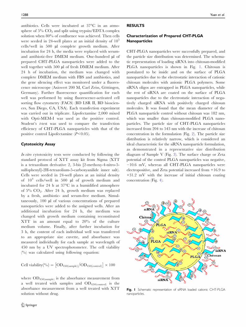
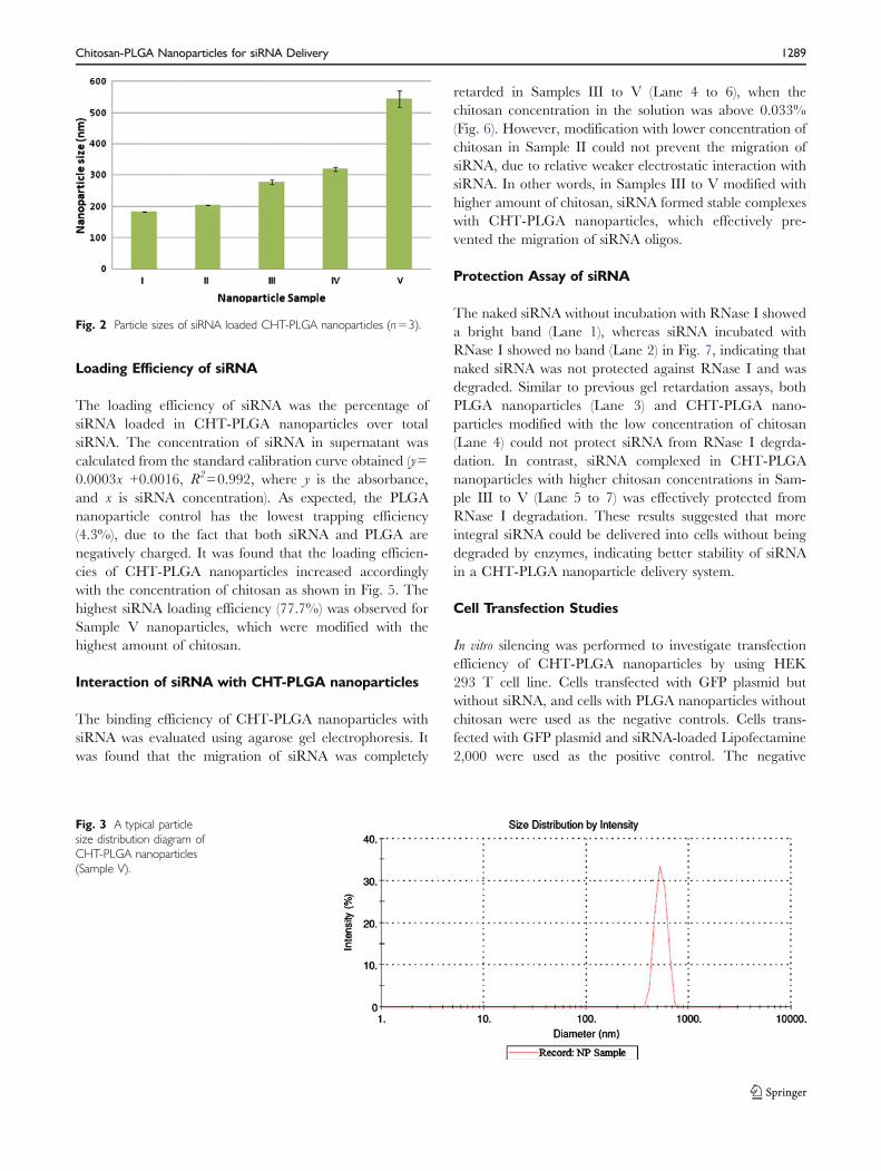
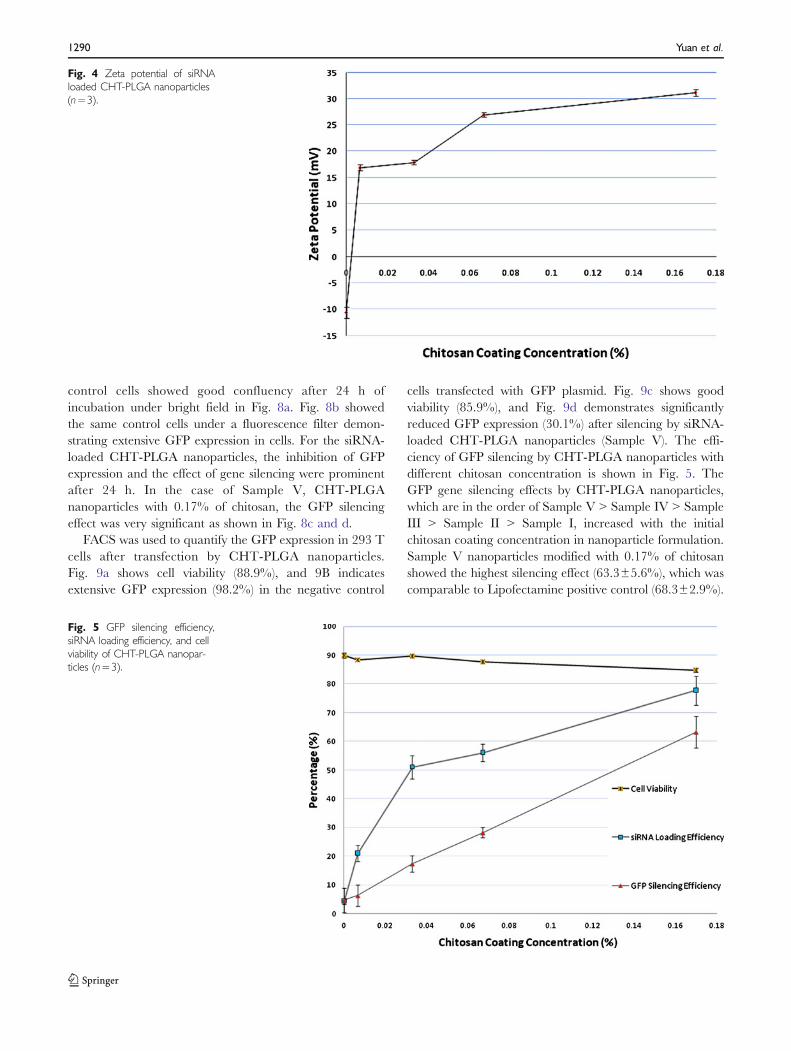
CHT-PLGA nanoparticles were successfully prepared, andthe particle size distribution was determined. The schema-tic representation of loading siRNA into chitosan-modifiedPLGA nanoparticles is shown in Fig. 1. Chitosan ispostulated to be inside and on the surface of PLGAnanoparticles due to the electrostatic interaction of cationicchitosan molecules with anionic PLGA polymers. SomesiRNA oligos are entrapped in PLGA nanoparticles, whilethe rest of siRNA are coated on the surface of PLGAnanoparticles due to the electrostatic interaction of nega-tively charged siRNA with positively charged chitosanmolecules. It was found that the mean diameter of thePLGA nanoparticle control without chitosan was 182 nm,which was smaller than chitosan-modified PLGA nano-particles. The particle size of CHT-PLGA nanoparticlesincreased from 204 to 543 nm with the increase of chitosanconcentration in the formulation (Fig. 2). The particle sizedistribution is relatively narrow, which is considered anideal characteristic for the siRNA nanoparticle formulation,as demonstrated in a representative size distributiondiagram of Sample V (Fig. 3). The surface charge or Zetapotential of the control PLGA nanoparticles was negative,−10.6 mV, whereas all CHT-PLGA nanoparticles wereelectropositive, and Zeta potential increased from +16.9 to+31.2 mV with the increase of initial chitosan coatingconcentration (Fig. 4).
Fig. 1 Schematic representation of siRNA loaded cationic CHT-PLGAnanoparticles.
1288 Yuan et al.
Loading Efficiency of siRNA
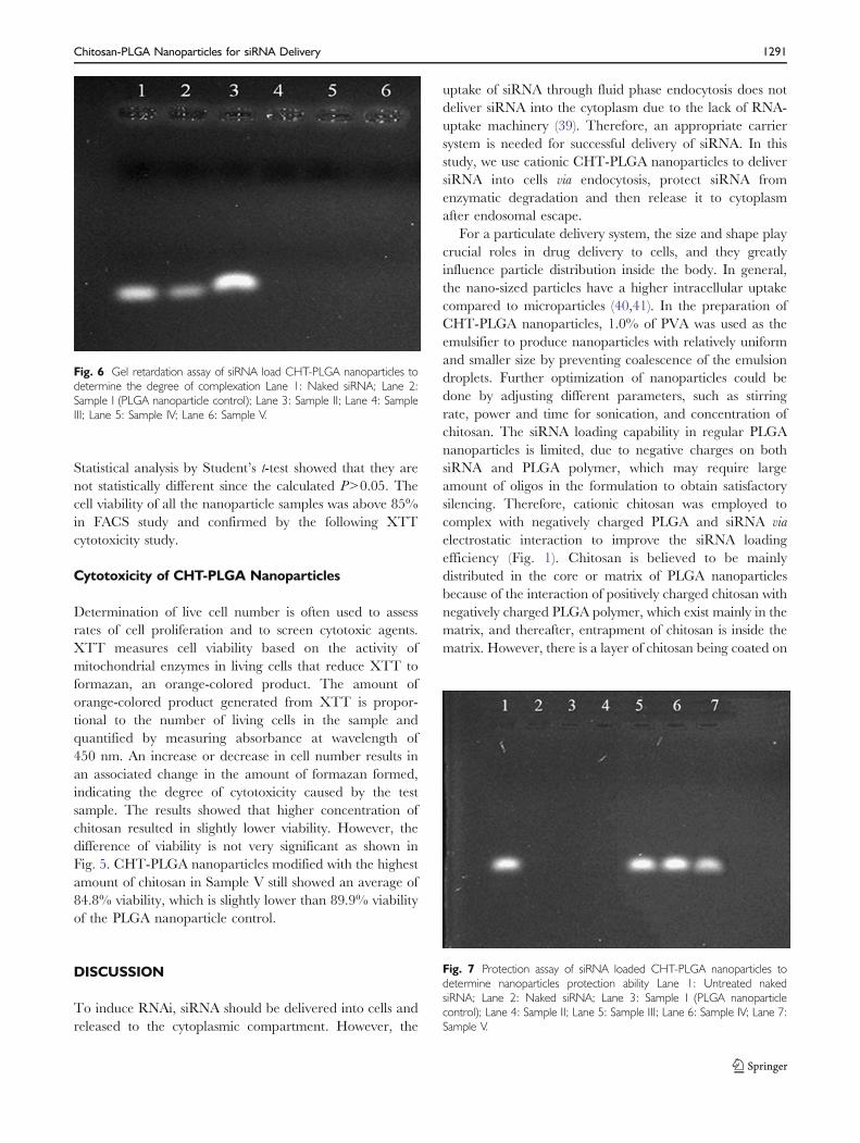
The loading efficiency of siRNA was the percentage ofsiRNA loaded in CHT-PLGA nanoparticles over totalsiRNA. The concentration of siRNA in supernatant wascalculated from the standard calibration curve obtained (y=0.0003x +0.0016, R2=0.992, where y is the absorbance,and x is siRNA concentration). As expected, the PLGAnanoparticle control has the lowest trapping efficiency(4.3%), due to the fact that both siRNA and PLGA arenegatively charged. It was found that the loading efficien-cies of CHT-PLGA nanoparticles increased accordinglywith the concentration of chitosan as shown in Fig. 5. Thehighest siRNA loading efficiency (77.7%) was observed forSample V nanoparticles, which were modified with thehighest amount of chitosan.
Interaction of siRNA with CHT-PLGA nanoparticles
The binding efficiency of CHT-PLGA nanoparticles withsiRNA was evaluated using agarose gel electrophoresis. Itwas found that the migration of siRNA was completely
retarded in Samples III to V (Lane 4 to 6), when thechitosan concentration in the solution was above 0.033%(Fig. 6). However, modification with lower concentration ofchitosan in Sample II could not prevent the migration ofsiRNA, due to relative weaker electrostatic interaction withsiRNA. In other words, in Samples III to V modified withhigher amount of chitosan, siRNA formed stable complexeswith CHT-PLGA nanoparticles, which effectively pre-vented the migration of siRNA oligos.
Protection Assay of siRNA
The naked siRNA without incubation with RNase I showeda bright band (Lane 1), whereas siRNA incubated withRNase I showed no band (Lane 2) in Fig. 7, indicating thatnaked siRNA was not protected against RNase I and wasdegraded. Similar to previous gel retardation assays, bothPLGA nanoparticles (Lane 3) and CHT-PLGA nano-particles modified with the low concentration of chitosan(Lane 4) could not protect siRNA from RNase I degrda-dation. In contrast, siRNA complexed in CHT-PLGAnanoparticles with higher chitosan concentrations in Sam-ple III to V (Lane 5 to 7) was effectively protected fromRNase I degradation. These results suggested that moreintegral siRNA could be delivered into cells without beingdegraded by enzymes, indicating better stability of siRNAin a CHT-PLGA nanoparticle delivery system.
Cell Transfection Studies
In vitro silencing was performed to investigate transfectionefficiency of CHT-PLGA nanoparticles by using HEK293 T cell line. Cells transfected with GFP plasmid butwithout siRNA, and cells with PLGA nanoparticles withoutchitosan were used as the negative controls. Cells trans-fected with GFP plasmid and siRNA-loaded Lipofectamine2,000 were used as the positive control. The negative
Fig. 2 Particle sizes of siRNA loaded CHT-PLGA nanoparticles (n=3).
Fig. 3 A typical particlesize distribution diagram ofCHT-PLGA nanoparticles(Sample V).
Chitosan-PLGA Nanoparticles for siRNA Delivery 1289
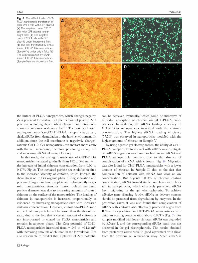
control cells showed good confluency after 24 h ofincubation under bright field in Fig. 8a. Fig. 8b showedthe same control cells under a fluorescence filter demon-strating extensive GFP expression in cells. For the siRNA-loaded CHT-PLGA nanoparticles, the inhibition of GFPexpression and the effect of gene silencing were prominentafter 24 h. In the case of Sample V, CHT-PLGAnanoparticles with 0.17% of chitosan, the GFP silencingeffect was very significant as shown in Fig. 8c and d.
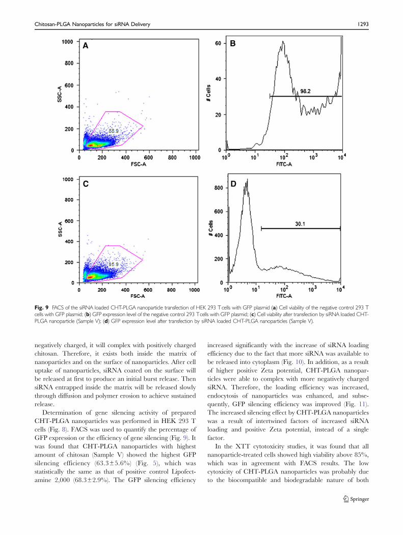
FACS was used to quantify the GFP expression in 293 Tcells after transfection by CHT-PLGA nanoparticles.Fig. 9a shows cell viability (88.9%), and 9B indicatesextensive GFP expression (98.2%) in the negative control
cells transfected with GFP plasmid. Fig. 9c shows goodviability (85.9%), and Fig. 9d demonstrates significantlyreduced GFP expression (30.1%) after silencing by siRNA-loaded CHT-PLGA nanoparticles (Sample V). The effi-ciency of GFP silencing by CHT-PLGA nanoparticles withdifferent chitosan concentration is shown in Fig. 5. TheGFP gene silencing effects by CHT-PLGA nanoparticles,which are in the order of Sample V > Sample IV > SampleIII > Sample II > Sample I, increased with the initialchitosan coating concentration in nanoparticle formulation.Sample V nanoparticles modified with 0.17% of chitosanshowed the highest silencing effect (63.3±5.6%), which wascomparable to Lipofectamine positive control (68.3±2.9%).
Fig. 5 GFP silencing efficiency,siRNA loading efficiency, and cellviability of CHT-PLGA nanopar-ticles (n=3).
Fig. 4 Zeta potential of siRNAloaded CHT-PLGA nanoparticles(n=3).
1290 Yuan et al.
Statistical analysis by Student’s t-test showed that they arenot statistically different since the calculated P>0.05. Thecell viability of all the nanoparticle samples was above 85%in FACS study and confirmed by the following XTTcytotoxicity study.
Cytotoxicity of CHT-PLGA Nanoparticles
Determination of live cell number is often used to assessrates of cell proliferation and to screen cytotoxic agents.XTT measures cell viability based on the activity ofmitochondrial enzymes in living cells that reduce XTT toformazan, an orange-colored product. The amount oforange-colored product generated from XTT is propor-tional to the number of living cells in the sample andquantified by measuring absorbance at wavelength of450 nm. An increase or decrease in cell number results inan associated change in the amount of formazan formed,indicating the degree of cytotoxicity caused by the testsample. The results showed that higher concentration ofchitosan resulted in slightly lower viability. However, thedifference of viability is not very significant as shown inFig. 5. CHT-PLGA nanoparticles modified with the highestamount of chitosan in Sample V still showed an average of84.8% viability, which is slightly lower than 89.9% viabilityof the PLGA nanoparticle control.
DISCUSSION
To induce RNAi, siRNA should be delivered into cells andreleased to the cytoplasmic compartment. However, the
uptake of siRNA through fluid phase endocytosis does notdeliver siRNA into the cytoplasm due to the lack of RNA-uptake machinery (39). Therefore, an appropriate carriersystem is needed for successful delivery of siRNA. In thisstudy, we use cationic CHT-PLGA nanoparticles to deliversiRNA into cells via endocytosis, protect siRNA fromenzymatic degradation and then release it to cytoplasmafter endosomal escape.
For a particulate delivery system, the size and shape playcrucial roles in drug delivery to cells, and they greatlyinfluence particle distribution inside the body. In general,the nano-sized particles have a higher intracellular uptakecompared to microparticles (40,41). In the preparation ofCHT-PLGA nanoparticles, 1.0% of PVA was used as theemulsifier to produce nanoparticles with relatively uniformand smaller size by preventing coalescence of the emulsiondroplets. Further optimization of nanoparticles could bedone by adjusting different parameters, such as stirringrate, power and time for sonication, and concentration ofchitosan. The siRNA loading capability in regular PLGAnanoparticles is limited, due to negative charges on bothsiRNA and PLGA polymer, which may require largeamount of oligos in the formulation to obtain satisfactorysilencing. Therefore, cationic chitosan was employed tocomplex with negatively charged PLGA and siRNA viaelectrostatic interaction to improve the siRNA loadingefficiency (Fig. 1). Chitosan is believed to be mainlydistributed in the core or matrix of PLGA nanoparticlesbecause of the interaction of positively charged chitosan withnegatively charged PLGA polymer, which exist mainly in thematrix, and thereafter, entrapment of chitosan is inside thematrix. However, there is a layer of chitosan being coated on
Fig. 6 Gel retardation assay of siRNA load CHT-PLGA nanoparticles todetermine the degree of complexation Lane 1: Naked siRNA; Lane 2:Sample I (PLGA nanoparticle control); Lane 3: Sample II; Lane 4: SampleIII; Lane 5: Sample IV; Lane 6: Sample V.
Fig. 7 Protection assay of siRNA loaded CHT-PLGA nanoparticles todetermine nanoparticles protection ability Lane 1: Untreated nakedsiRNA; Lane 2: Naked siRNA; Lane 3: Sample I (PLGA nanoparticlecontrol); Lane 4: Sample II; Lane 5: Sample III; Lane 6: Sample IV; Lane 7:Sample V.
Chitosan-PLGA Nanoparticles for siRNA Delivery 1291
the surface of PLGA nanoparticles, which changes negativeZeta potential to positive. But the increase of positive Zetapotential is not significant when chitosan concentration isabove certain range as shown in Fig. 3. The positive chitosancoating on the surface of CHT-PLGA nanoparticles can alsoshield siRNA from degradation in the harsh environment. Inaddition, since the cell membrane is negatively charged,cationic CHT-PLGA nanoparticles can interact more easilywith the cell membrane, therefore promoting endocytosisand increasing siRNA silencing efficiency.
In this study, the average particle size of CHT-PLGAnanoparticles increased gradually from 182 to 543 nm withthe increase of initial chitosan concentration from 0.00 to0.17% (Fig. 2). The increased particle size could be creditedto the increased viscosity of chitosan, which lowered theshear stress on PLGA organic phase during sonication andproduced larger emulsion droplets and subsequently largersolid nanoparticles. Another reason behind increasedparticle diameter was due to increasing amounts of coatedchitosan on the surface of the nanoparticles. The loading ofchitosan in nanoparticles is increased proportionally asevidenced by increasing nanoparticle sizes with increasedchitosan concentration. However, chitosan-to-PLGA ratioin the final nanoparticles will be lower than the theoreticalratio, due to the fact that a certain amount of chitosan isnot incorporated or coated on PLGA nanoparticles andremains in aqueous phase. The Zeta potential of CHT-PLGA nanoparticles increased from −10.6 to +31.2 mVwith increasing amounts of chitosan in the formulation. It isalso reasonable to predict that a plateau of Zeta potential
can be achieved eventually, which could be indicative ofsaturated adsorption of chitosan on CHT-PLGA nano-particles. In addition, the siRNA loading efficiency inCHT-PLGA nanoparticles increased with the chitosanconcentration. The highest siRNA loading efficiency(77.7%) was observed in nanoparticles modified with thehighest amount of chitosan in Sample V.
By using agarose gel electrophoresis, the ability of CHT-PLGA nanoparticles to interact with siRNA was investigat-ed. siRNA migration was found for both naked siRNA andPLGA nanoparticle controls, due to the absence ofcomplexation of siRNA with chitosan (Fig. 6). Migrationwas also found for CHT-PLGA nanoparticles with a loweramount of chitosan in Sample II, due to the fact thatcomplexation of chitosan with siRNA was weak at lowconcentration. But beyond 0.033% of chitosan coatingconcentration, siRNA formed stable complexes with chito-san in nanoparticles, which effectively prevented siRNAfrom migrating in the gel electrophoresis. To achieveeffective gene silencing in vivo, siRNA in delivery systemsshould be protected from degradation by enzymes. In theprotection assay, it was also found that complexation ofsiRNA with chitosan also effectively protected oligos fromRNase I degradation in CHT-PLGA nanoparticles withchitosan coating concentration above 0.033% (Fig. 7). Forsamples modified with lower chitosan, siRNA was degradedby RNase I, and the corresponding siRNA band was notobserved in the gel electrophoresis. The results obtainedfrom protection assays were in good agreement with thosefrom the previous gel retardation assay. Since siRNA is
Fig. 8 The siRNA loaded CHT-PLGA nanoparticle transfection ofHEK 293 Tcells with GFP plasmid(a) The negative control 293 Tcells with GFP plasmid underbright field; (b) The negativecontrol 293 T cells with GFPplasmid under fluorescent filter;(c) The cells transfected by siRNAloaded CHT-PLGA nanoparticles(Sample V) under bright field; (d)The cells transfected by siRNAloaded CHT-PLGA nanoparticles(Sample V) under fluorescent filter.
1292 Yuan et al.
negatively charged, it will complex with positively chargedchitosan. Therefore, it exists both inside the matrix ofnanoparticles and on the surface of nanoparticles. After celluptake of nanoparticles, siRNA coated on the surface willbe released at first to produce an initial burst release. ThensiRNA entrapped inside the matrix will be released slowlythrough diffusion and polymer erosion to achieve sustainedrelease.
Determination of gene silencing activity of preparedCHT-PLGA nanoparticles was performed in HEK 293 Tcells (Fig. 8). FACS was used to quantify the percentage ofGFP expression or the efficiency of gene silencing (Fig. 9). Itwas found that CHT-PLGA nanoparticles with highestamount of chitosan (Sample V) showed the highest GFPsilencing efficiency (63.3±5.6%) (Fig. 5), which wasstatistically the same as that of positive control Lipofect-amine 2,000 (68.3±2.9%). The GFP silencing efficiency
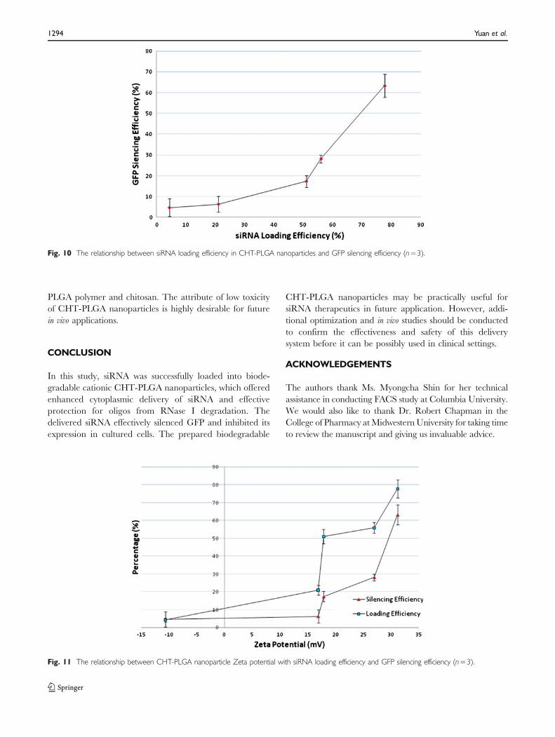
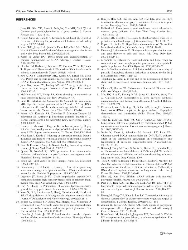
increased significantly with the increase of siRNA loadingefficiency due to the fact that more siRNA was available tobe released into cytoplasm (Fig. 10). In addition, as a resultof higher positive Zeta potential, CHT-PLGA nanopar-ticles were able to complex with more negatively chargedsiRNA. Therefore, the loading efficiency was increased,endocytosis of nanoparticles was enhanced, and subse-quently, GFP silencing efficiency was improved (Fig. 11).The increased silencing effect by CHT-PLGA nanoparticleswas a result of intertwined factors of increased siRNAloading and positive Zeta potential, instead of a singlefactor.
In the XTT cytotoxicity studies, it was found that allnanoparticle-treated cells showed high viability above 85%,which was in agreement with FACS results. The lowcytoxicity of CHT-PLGA nanoparticles was probably dueto the biocompatible and biodegradable nature of both
Fig. 9 FACS of the siRNA loaded CHT-PLGA nanoparticle transfection of HEK 293 Tcells with GFP plasmid (a) Cell viability of the negative control 293 Tcells with GFP plasmid; (b) GFP expression level of the negative control 293 Tcells with GFP plasmid; (c) Cell viability after transfection by siRNA loaded CHT-PLGA nanoparticle (Sample V); (d) GFP expression level after transfection by siRNA loaded CHT-PLGA nanoparticles (Sample V).
Chitosan-PLGA Nanoparticles for siRNA Delivery 1293
PLGA polymer and chitosan. The attribute of low toxicityof CHT-PLGA nanoparticles is highly desirable for futurein vivo applications.
CONCLUSION
In this study, siRNA was successfully loaded into biode-gradable cationic CHT-PLGA nanoparticles, which offeredenhanced cytoplasmic delivery of siRNA and effectiveprotection for oligos from RNase I degradation. Thedelivered siRNA effectively silenced GFP and inhibited itsexpression in cultured cells. The prepared biodegradable
CHT-PLGA nanoparticles may be practically useful forsiRNA therapeutics in future application. However, addi-tional optimization and in vivo studies should be conductedto confirm the effectiveness and safety of this deliverysystem before it can be possibly used in clinical settings.
ACKNOWLEDGEMENTS
The authors thank Ms. Myongcha Shin for her technicalassistance in conducting FACS study at Columbia University.We would also like to thank Dr. Robert Chapman in theCollege of Pharmacy atMidwestern University for taking timeto review the manuscript and giving us invaluable advice.
Fig. 10 The relationship between siRNA loading efficiency in CHT-PLGA nanoparticles and GFP silencing efficiency (n=3).
Fig. 11 The relationship between CHT-PLGA nanoparticle Zeta potential with siRNA loading efficiency and GFP silencing efficiency (n=3).
1294 Yuan et al.
REFERENCES
1. Jiang HL, Kim YK, Arote R, Nah JW, Cho MH, Choi YJ et al.Chitosan-graft-polyethylenimine as a gene carrier. J ControlRelease. 2007;117:273–80.
2. Relano-Gines A, Gabelle A, Lehmann S, Milhavet O, Crozet C.Gene and cell therapy for prion diseases. Infectious disorders drugtargets. 2009;9:58–68.
3. Kima T-H, Jianga H-L, Jerea D, Parka I-K, Chob M-H, Nahc J-W et al. Chemical modification of chitosan as a gene carrier in vitroand in vivo. Prog Polym Sci. 2007;32:726–53.
4. Katas H, Alpar HO. Development and characterisation ofchitosan nanoparticles for siRNA delivery. J Control Release.2006;115:216–25.
5. Elbashir SM, Harborth J, Lendeckel W, Yalcin A, Weber K, TuschlT. Duplexes of 21-nucleotide RNAs mediate RNA interference incultured mammalian cells. Nature. 2001;411:494–8.
6. Fire A, Xu S, Montgomery MK, Kostas SA, Driver SE, MelloCC. Potent and specific genetic interference by double-strandedRNA in Caenorhabditis elegans. Nature. 1998;391:806–11.
7. Jones SW, Souza PM, Lindsay MA. siRNA for gene silencing: aroute to drug target discovery. Curr Opin Pharmacol.2004;4:522–7.
8. McManusand MT, Sharp PA. Gene silencing in mammals bysmall interfering RNAs. Nat Rev. 2002;3:737–47.
9. Lima RT, Martins LM, Guimaraes JE, Sambade C, VasconcelosMH. Specific downregulation of bcl-2 and xIAP by RNAienhances the effects of chemotherapeutic agents in MCF-7 humanbreast cancer cells. Cancer Gene Ther. 2004;11:309–16.
10. Fraser AG, Kamath RS, Zipperlen P, Martinez-Campos M,Sohrmann M, Ahringer J. Functional genomic analysis of C.elegans chromosome I by systematic RNA interference. Nature.2000;408:325–30.
11. Gonczy P, Echeverri C, Oegema K, Coulson A, Jones SJ, CopleyRR et al. Functional genomic analysis of cell division in C. elegansusing RNAi of genes on chromosome III. Nature. 2000;408:331–6.
12. Nabatiyan A, Krude T. Silencing of chromatin assembly factor 1in human cells leads to cell death and loss of chromatin assemblyduring DNA synthesis. Mol Cell Biol. 2004;24:2853–62.
13. Suri SS, Fenniri H, Singh B. Nanotechnology-based drug deliverysystems. J Occup Med Toxicol. 2007;2:16.
14. Quong D, Neufeld RJ. DNA protection from extracapsularnucleases, within chitosan- or poly-L-lysine-coated alginate beads.Biotechnol Bioeng. 1998;60:124–34.
15. Smith AE. Viral vectors in gene therapy. Annu Rev Microbiol.1995;49:807–38.
16. Pinnaduwage P, Schmitt L, Huang L. Use of a quaternaryammonium detergent in liposome mediated DNA transfection ofmouse L-cells. Biochim Biophys Acta. 1989;985:33–7.
17. Legendre JY, Szoka Jr FC. Cyclic amphipathic peptide-DNAcomplexes mediate high-efficiency transfection of adherent mam-malian cells. Proc Natl Acad Sci USA. 1993;90:893–7.
18. Gao X, Huang L. Potentiation of cationic liposome-mediatedgene delivery by polycations. Biochemistry. 1996;35:1027–36.
19. Yuan X, Li L, Rathinavelu A, Hao J, Narasimhan M, He M et al.SiRNA drug delivery by biodegradable polymeric nanoparticles.Journal of Nanoscience and Nanotechnology. 2006;6:2821–8.
20. Boussif O, Lezoualc’h F, Zanta MA, Mergny MD, Scherman D,Demeneix B et al. A versatile vector for gene and oligonucleotidetransfer into cells in culture and in vivo: polyethylenimine. ProcNatl Acad Sci USA. 1995;92:7297–301.
21. Haensler J, Szoka Jr FC. Polyamidoamine cascade polymersmediate efficient transfection of cells in culture. Bioconjug Chem.1993;4:372–9.
22. Ihm JE, Han KO, Han IK, Ahn KD, Han DK, Cho CS. Hightransfection efficiency of poly(4-vinylimidazole) as a new genecarrier. Bioconjug Chem. 2003;14:707–8.
23. Rolland AP. From genes to gene medicines: recent advances innonviral gene delivery. Crit Rev Ther Drug Carrier Syst.1998;15:143–98.
24. Montag ME, Morales Jr L, Daane S. Bioabsorbables: their use inpediatric craniofacial surgery. J Craniofac Surg. 1997;8:100–2.
25. Eppley BL, Reilly M. Degradation characteristics of PLLA-PGAbone fixation devices. J Craniofac Surg. 1997;8:116–20.
26. Panyam J, Labhasetwar V. Biodegradable nanoparticles for drugand gene delivery to cells and tissue. Adv Drug Deliv Rev.2003;55:329–47.
27. Miyamoto S, Takaoka K. Bone induction and bone repair bycomposites of bone morphogenetic protein and biodegradablesynthetic polymers. Ann Chir Gynaecol. 1993;207:69–75.
28. Aiba S. Studies on chitosan: 2. Solution stability and reactivity ofpartially N-acetylated chitosan derivatives in aqueous media. Int JBiol Macromol. 1989;11:249–52.
29. Tomihata K, Ikada Y. In vitro and in vivo degradation of films ofchitin and its deacetylated derivatives. Biomaterials. 1997;18:567–75.
30. Chandy T, Sharma CP. Chitosan-as a biomaterial. Biomater ArtifCells Artif Organs. 1990;18:1–24.
31. Mao HQ, Roy K, Troung-Le VL, Janes KA, Lin KY, Wang Y etal. Chitosan-DNA nanoparticles as gene carriers: synthesis,characterization and transfection efficiency. J Control Release.2001;70:399–421.
32. Erbacher P, Zou S, Bettinger T, Steffan AM, Remy JS. Chitosan-based vector/DNA complexes for gene delivery: biophysicalcharacteristics and transfection ability. Pharm Res. 1998;15:1332–9.
33. Yang R, Yang SG, Shim WS, Cui F, Cheng G, Kim IW et al.Lung-specific delivery of paclitaxel by chitosan-modified PLGAnanoparticles via transient formation of microaggregates. J PharmSci. 2009;98:970–84.
34. Nafee N, Taetz S, Schneider M, Schaefer UF, Lehr CM.Chitosan-coated PLGA nanoparticles for DNA/RNA delivery:effect of the formulation parameters on complexation andtransfection of antisense oligonucleotides. Nanomedicine.2007;3:173–83.
35. Beisner J, Dong M, Taetz S, Nafee N, Griese EU, Schaefer U, etal. Nanoparticle mediated delivery of 2′-O-methyl-RNA leads toefficient telomerase inhibition and telomere shortening in humanlung cancer cells. Lung Cancer. 2009.
36. Taetz S, Nafee N, Beisner J, Piotrowska K, Baldes C, Murdter TEet al. The influence of chitosan content in cationic chitosan/PLGAnanoparticles on the delivery efficiency of antisense 2′-O-methyl-RNA directed against telomerase in lung cancer cells. Eur JPharm Biopharm. 2009;72:358–69.
37. Kim WJ, Kim SW. Efficient siRNA delivery with non-viralpolymeric vehicles. Pharm Res. 2009;26:657–66.
38. Park MR, Han KO, Han IK, Cho MH, Nah JW, Choi YJ et al.Degradable polyethylenimine-alt-poly(ethylene glycol) copoly-mers as novel gene carriers. J Control Release. 2005;105:367–80.
39. Huang M, Fong CW, Khor E, Lim LY. Transfection efficiency ofchitosan vectors: effect of polymer molecular weight and degree ofdeacetylation. J Control Release. 2005;106:391–406.
40. Zauner W, Farrow NA, Haines AM. In vitro uptake of polystyrenemicrospheres: effect of particle size, cell line and cell density. JControl Release. 2001;71:39–51.
41. Bivas-Benita M, Romeijn S, Junginger HE, Borchard G. PLGA-PEI nanoparticles for gene delivery to pulmonary epithelium. EurJ Pharm Biopharm. 2004;58:1–6.
Chitosan-PLGA Nanoparticles for siRNA Delivery 1295
Related Documents


![A dual targeting dendrimer-mediated siRNA delivery system …nanosized volume.[16-20] We have recently developed a series of cationic amphiphilic dendrimers[21-24] which couple the](https://static.cupdf.com/doc/110x72/5feda2b5c8964e53213a54f4/a-dual-targeting-dendrimer-mediated-sirna-delivery-system-nanosized-volume16-20.jpg)