Cationic nano-copolymers mediated IKKβ targeting siRNA inhibit the proliferation of human Tenon’s capsule fibroblasts in vitro Yongheng Duan, 1 Xipeng Guan, 2 Jian Ge, 1 Daping Quan, 2 Yehong Zhuo, 1 Hehua Ye, 1 Tingting Shao 1 (The first two authors contributed equally to this work.) 1 State Key Laboratory of Ophthalmology, Zhongshan Ophthalmic Center, Sun Yat-sen University, Guangzhou, China; 2 Institute of Polymer Science, School of Chemistry and Chemical Engineering, Sun Yat-sen University, Guangzhou, China Purpose: To synthesize a ternary cationic copolymer called CS-g-(PEI-b-mPEG) and characterize its features as a non- viral siRNA carrier; in turn, to investigate the influence of small interfering RNA (siRNA) targeting IκB kinase subunit β (IKKβ) on the proliferation of human Tenon’s capsule fibroblasts (HTFs) in vitro. Methods: First, a novel cationic copolymer composed of low molecular weight, linear poly(ethyleneimine) [PEI] blocked with polyethylene glycol (PEG) and grafted onto a chitosan (CS) molecule was synthesized. CS-g-(PEI-b-mPEG) was then compacted with 21nt siRNA at various copolymer/siRNA charge (N/P) ratios, and the resulting complexes were characterized by dynamic light scattering, gel electrophoresis, and serum incubation. Cell Titer 96 ® AQueous One Solution cell proliferation assay was used to investigate the cytotoxicity of this cationic copolymer. Second, siRNAs targeting IKKβ (IKKΒ-siRNAs) were delivered into the HTFs using CS-g-(PEI-b-mPEG) as the vehicle. Real-time reverse transcription polymerase chain reaction (RT–PCR) subsequently assessed the mRNA level of IKKβ, and western blot assay was used to determine protein expression. After IKKB-siRNA transfection, Cell Titer 96 ® AQueous One Solution cell proliferation assay was used to evaluate the proliferation of HTFs. Results: The diameter of the CS-g-(PEI-b-mPEG)/siRNA complexes tended to decrease whereas their zeta potential tended to increase as the N/P ratio increased. The CS-g-(PEI-b-mPEG) copolymer showed good siRNA binding ability and high siRNA protection capacity. Furthermore, the copolymer presented remarkable transfection efficiency and showed much less cytotoxicity than 25 kDa PEI. IKKB-siRNAs were successfully delivered into HTFs using CS-g-(PEI-b-mPEG) as a vector. As a result, the expression of IKKβ was downregulated at both the mRNA and protein levels, and the activation of nuclear factor-κB (NF-κB) in the HTFs was subsequently inhibited. Most impressively, the proliferation of HTFs was also effectively suppressed through the blocking of the NF-κB pathway. Conclusions: All the results demonstrate that CS-g-(PEI-b-mPEG) is a promising candidate for siRNA delivery, featuring excellent biocompatibility, biodegradability, and transfection efficiency. The RNA interference (RNAi) strategy using cationic copolymers as siRNA carriers will be a safe and efficient anti-scarring method following glaucoma filtration surgery. RNA interference (RNAi) was originally recognized as an evolutionary conserved defense mechanism in higher eukaryotic cells, and this system can easily and effectively inhibit the expression of one specific gene [1]. The RNAi process is mediated through small, double-stranded RNA molecules called small interfering RNA (siRNA), which specifically trigger the cleavage and subsequent degradation of their target mRNA in a sequence-dependence manner. Hence, synthesis of the protein encoded by those mRNA is prevented [2]. Recently, RNAi-mediated gene silencing has also been shown to be efficient in mammalian cells, and this has led to the increasing feasibility of RNAi technology for the therapy of certain human diseases [3]. Correspondence to: Professor Jian Ge, Zhongshan Ophthalmic Center, Sun Yat-sen University, 54 South Xianlie Road, Guangzhou, 510060, China; Phone: +86-20-87331374; FAX: +86-20-87333271; email: [email protected] The efficiency of RNAi mainly depends on the successful delivery of intact siRNA into mammalian cells. However, due to the low stability of siRNA against enzymatic degradation and low permeability across cell membranes, the efficacy of naked siRNA is insufficient. Therefore, it is necessary to develop efficient and convenient methods for siRNA delivery. Until now, viral delivery systems have been used as vectors for genes in many studies due to the advantage of high transfection efficacy, but the use of such delivery systems is limited by endogenous recombination and host immunity [4]. Moreover, since ontogenesis and mortality have been reported [5], concerns have been raised regarding the safety of using viral vectors in gene therapy trials in humans. In light of these problems, studies of alternative delivery strategies focus on non-viral systems for gene delivery. Recently, cationic copolymers have been demonstrated to be a promising non- viral vector for transfecting nucleotides into various cell types or tissues [6]. Among these cationic polymers, Molecular Vision 2008; 14:2616-2628 <http://www.molvis.org/molvis/v14/a299> Received 20 October 2008 | Accepted 5 December 2008 | Published 31 December 2008 © 2008 Molecular Vision 2616

Welcome message from author
This document is posted to help you gain knowledge. Please leave a comment to let me know what you think about it! Share it to your friends and learn new things together.
Transcript
Cationic nano-copolymers mediated IKKβ targeting siRNA inhibitthe proliferation of human Tenon’s capsule fibroblasts in vitro
Yongheng Duan,1 Xipeng Guan,2 Jian Ge,1 Daping Quan,2 Yehong Zhuo,1 Hehua Ye,1 Tingting Shao1
(The first two authors contributed equally to this work.)
1State Key Laboratory of Ophthalmology, Zhongshan Ophthalmic Center, Sun Yat-sen University, Guangzhou, China; 2Institute ofPolymer Science, School of Chemistry and Chemical Engineering, Sun Yat-sen University, Guangzhou, China
Purpose: To synthesize a ternary cationic copolymer called CS-g-(PEI-b-mPEG) and characterize its features as a non-viral siRNA carrier; in turn, to investigate the influence of small interfering RNA (siRNA) targeting IκB kinase subunitβ (IKKβ) on the proliferation of human Tenon’s capsule fibroblasts (HTFs) in vitro.Methods: First, a novel cationic copolymer composed of low molecular weight, linear poly(ethyleneimine) [PEI] blockedwith polyethylene glycol (PEG) and grafted onto a chitosan (CS) molecule was synthesized. CS-g-(PEI-b-mPEG) wasthen compacted with 21nt siRNA at various copolymer/siRNA charge (N/P) ratios, and the resulting complexes werecharacterized by dynamic light scattering, gel electrophoresis, and serum incubation. Cell Titer 96® AQueous One Solutioncell proliferation assay was used to investigate the cytotoxicity of this cationic copolymer. Second, siRNAs targetingIKKβ (IKKΒ-siRNAs) were delivered into the HTFs using CS-g-(PEI-b-mPEG) as the vehicle. Real-time reversetranscription polymerase chain reaction (RT–PCR) subsequently assessed the mRNA level of IKKβ, and western blotassay was used to determine protein expression. After IKKB-siRNA transfection, Cell Titer 96® AQueous One Solutioncell proliferation assay was used to evaluate the proliferation of HTFs.Results: The diameter of the CS-g-(PEI-b-mPEG)/siRNA complexes tended to decrease whereas their zeta potentialtended to increase as the N/P ratio increased. The CS-g-(PEI-b-mPEG) copolymer showed good siRNA binding abilityand high siRNA protection capacity. Furthermore, the copolymer presented remarkable transfection efficiency and showedmuch less cytotoxicity than 25 kDa PEI. IKKB-siRNAs were successfully delivered into HTFs using CS-g-(PEI-b-mPEG)as a vector. As a result, the expression of IKKβ was downregulated at both the mRNA and protein levels, and the activationof nuclear factor-κB (NF-κB) in the HTFs was subsequently inhibited. Most impressively, the proliferation of HTFs wasalso effectively suppressed through the blocking of the NF-κB pathway.Conclusions: All the results demonstrate that CS-g-(PEI-b-mPEG) is a promising candidate for siRNA delivery, featuringexcellent biocompatibility, biodegradability, and transfection efficiency. The RNA interference (RNAi) strategy usingcationic copolymers as siRNA carriers will be a safe and efficient anti-scarring method following glaucoma filtrationsurgery.
RNA interference (RNAi) was originally recognized asan evolutionary conserved defense mechanism in highereukaryotic cells, and this system can easily and effectivelyinhibit the expression of one specific gene [1]. The RNAiprocess is mediated through small, double-stranded RNAmolecules called small interfering RNA (siRNA), whichspecifically trigger the cleavage and subsequent degradationof their target mRNA in a sequence-dependence manner.Hence, synthesis of the protein encoded by those mRNA isprevented [2]. Recently, RNAi-mediated gene silencing hasalso been shown to be efficient in mammalian cells, and thishas led to the increasing feasibility of RNAi technology forthe therapy of certain human diseases [3].
Correspondence to: Professor Jian Ge, Zhongshan OphthalmicCenter, Sun Yat-sen University, 54 South Xianlie Road, Guangzhou,510060, China; Phone: +86-20-87331374; FAX: +86-20-87333271;email: [email protected]
The efficiency of RNAi mainly depends on the successfuldelivery of intact siRNA into mammalian cells. However, dueto the low stability of siRNA against enzymatic degradationand low permeability across cell membranes, the efficacy ofnaked siRNA is insufficient. Therefore, it is necessary todevelop efficient and convenient methods for siRNA delivery.Until now, viral delivery systems have been used as vectorsfor genes in many studies due to the advantage of hightransfection efficacy, but the use of such delivery systems islimited by endogenous recombination and host immunity [4].Moreover, since ontogenesis and mortality have been reported[5], concerns have been raised regarding the safety of usingviral vectors in gene therapy trials in humans. In light of theseproblems, studies of alternative delivery strategies focus onnon-viral systems for gene delivery. Recently, cationiccopolymers have been demonstrated to be a promising non-viral vector for transfecting nucleotides into various cell typesor tissues [6]. Among these cationic polymers,
Molecular Vision 2008; 14:2616-2628 <http://www.molvis.org/molvis/v14/a299>Received 20 October 2008 | Accepted 5 December 2008 | Published 31 December 2008
© 2008 Molecular Vision
2616
polyethylenimine (PEI) is an effective gene carrier due toPEI’s high charge density and endosomal disruption function,but it is difficult to achieve both the goals of highertransfection efficiency and lower cytotoxicity with PEIhomopolymers [7]. Various modifications of PEI have beeninvestigated to promote its nucleotide delivery ability as wellas to reduce its adverse effects on cell viability. It has beenshown that grafting PEI with nonionic hydrophilic polymerssuch as polyethylene glycol (PEG) could be an effectiveapproach for minimizing the cationic toxicity of PEI and thatthe cationic toxicity of PEI decreases as its molecular weightdecreases [8]. However, the transfection efficiency of thePEGylated, low molecular weight PEI copolymer is lowerthan that of 25 kDa PEI [9]. Chitosan (CS) is a non-toxic,biodegradable cationic polymer with relatively lowimmunogenicity and especially good macro-adhesion. CS hasbeen extensively investigated as a delivery system fortherapeutic macromolecules, nucleotides, and proteinmolecules [10]. Thus, we hypothesize that a new copolymercould be synthesized as a siRNA carrier that would have boththe efficient transfection ability of PEI and thebiocompatibility of PEG and CS.
Glaucoma is an eye disease usually associated withincreased intraocular pressure that leads to irreversiblefunctional impairment of the optic nerve. Filtration surgery toenhance the drainage of aqueous humor is one of the mosteffective therapies for glaucoma [11], but the therapy’ssuccess rate is reduced by blockage of the surgically createddrainage channel by subconjunctival scarring that may occurwith wound healing [12]. Fibroblasts located in thesubconjunctival area play a major role in scar formation afterfiltration surgery through proliferation, migration, andsynthesis of the extracellular matrix (ECM). Thus, regulatingthe biological activities of subconjunctival Tenon’s capsulefibroblasts (TCFs) during the wound healing process is amajor anti-scarring strategy for glaucoma filtration surgery[13]. Nuclear factor-κB (NF-κB) is a transcription factor thatis also a positive regulator for fibroblasts, and a proteincomplex called IκB kinase (IKK) is a critical regulator of theactivation of NF-κB [14,15]. Most studies on the role of theNF-κB pathway in the regulation of cell proliferation haveused immortal cell lines (cells capable of continuouslyrenewing themselves). In this study, we investigated whetherinhibiting the function of IKK and subsequently blocking thesignaling pathway of NF-κB could effectively manipulate theactivation and proliferation of TCFs during the scarringprocess following glaucoma filtration surgery.
In the study reported here, absorbable, low molecularweight PEI was blocked with polyethylene glycolmonomethyl ether (mPEG) and grafted onto chitosan. As aresult, a novel biodegradable copolymer, CS-g-(PEI-b-mPEG), was synthesized. The properties of the CS-g-(PEI-b-mPEG)/siRNA complexes such as particle size, zeta potential,siRNA binding and protection capacity, transfection ability,
and cytotoxicity were studied, and IKKΒ-siRNAs were thendelivered into human Tenon’s capsule fibroblasts (HTFs)using CS-g-(PEI-b-mPEG) as the vehicle. The expression ofIKKβ was detected at both the mRNA and protein levels afterthe transfection of IKKΒ-siRNAs. We also investigated theactivation of NF-κB and the proliferation of HTFs after theRNA interference process targeting IKKβ.
METHODSCell culture: HeLa (human cervix epithelial carcinoma) cellswere obtained from the American Type Culture Collection(Number CCL-2.1; ATCC, Rockville, MD) and weremaintained in Dulbecco’s modified Eagle’s medium (DMEM;Gibco, Grand Island, NY) containing 10% fetal bovine serum(FBS; HyClone, Logan, UT), 2 mM of L-glutamine, 100 IU/ml of penicillin, 100 μg/ml of streptomycin, and 25 μg/ml ofamphotericin B (all from Sigma-Aldrich, St. Louis, MO) at37 °C with 5% CO2, 95% humidified atmosphere.
Tissue explants of human Tenon’s capsule were obtainedfrom three male patients (aged 28, 39, and 62 without anytopical eye treatment) who had undergone trauma or cataractsurgery. Patients were informed of the nature and possibleconsequences of the tissue removal procedure, and written,informed consent was obtained. The tenets of the Declarationof Helsinki were followed, and approval by the Sun Yat-senUniversity Human Experimentation committee was granted.HTFs were cultured by a previously reported method [16] withsome modification, as described below. Cells weremaintained as a monolayer at 37 °C with 5% CO2, 95%humidified atmosphere in DMEM supplemented with 10%FBS, 2 mM of L-glutamine, 100 IU/ml of penicillin, 100 μg/ml of streptomycin, and 25 μg/ml of amphotericin B. Cellsbetween passages 3 and 6 were used for the followingexperiments.Synthesis and characterization of CS-g-(PEI-b-mPEG):Chitosan, (the weight-average molecular weight of chitosanwas 3.50×10⁵ [Mw=3.50×10⁵], which was measured by theviscosity method and the degree of deacetylation of chitosanwas 88%, which was determined by proton nuclear magneticresonance [1H NMR]), was purified by a solvent precipitationmethod. Polyethylene glycol monomethyl ether (mPEG, AR,Mn=2000) was purchased from Sigma-Aldrich Chemie Gmbh(Steinheim, Germany). Linear PEI (Mn=600) and branchedPEI (Mn=25,000) were obtained from Wako Pure ChemicalIndustries, Ltd. (Osaka, Japan). CS-g-(PEI-b-mPEG) wassynthesized by Jiang’s method with some modification [17],and the steps are briefly described as follows. Di-blockcopolymer, PEI-b-mPEG, was synthesized by an iminereaction, and then the periodate ion, IO4
-, was used to oxidizeCS to produce dialdehyde. A novel, comb-like copolymer,CS-g-(PEI-b-mPEG), was synthesized by an imine reactionbetween the amino groups of PEI-b-mPEG and the aldehydegroups of periodate-oxidized CS. The resultant product waspurified by dialysis against double deionized (DD) water with
Molecular Vision 2008; 14:2616-2628 <http://www.molvis.org/molvis/v14/a299> © 2008 Molecular Vision
2617
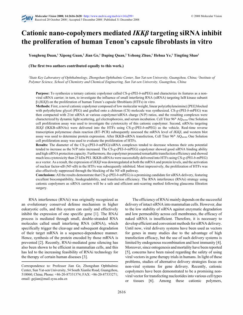
the use of Spectra/Pro2 membrane (molecular weight cut offwas 12 K; Spectrum, Houston, TX) for 72 h to remove theunreacted PEI and mPEG and then freeze-dried for another 24h. A schematic illustration of the synthesis process is shownin Figure 1, and the structure of the newly synthesized CS-g-(PEI-b-mPEG) was proved by 1H NMR and gel permeationchromatography (GPC).Preparation of CS-g-(PEI-b-mPEG)/siRNA complexes:IKKΒ-siRNA, scrambled siRNA, and fluoresceinisothiocyanate (FITC)-conjugated scrambled siRNA were allsynthesized by Ribobio Co. Ltd. (Guangzhou, China).Lyophilized siRNAs were dissolved in RNase-free H2O(pH=7.4) and incubated for 5 min at room temperature, andthe CS-g-(PEI-b-mPEG) copolymer was dissolved in serum-free DMEM at a stock concentration of 1 mg/ml.Subsequently, different concentrations of the CS-g-(PEI-b-mPEG) solution were added into dissolved siRNA to makecomplexes of various charge ratios. The charge ratio of CS-g-(PEI-b-mPEG) and siRNA was expressed as the molar ratioof the amine groups of the copolymer (representing positiveunits) to the phosphates of siRNA (representing negativeunits) called the N/P ratio. The mixture was gently vortexedfor 10 s and incubated at room temperature for 30 min to allowthe formation of CS-g-(PEI-b-mPEG)/siRNA complexes.Measurement of particle size and zeta potential: Dynamiclight scattering (DLS) with a Zetasizer Nano ZS instrument(Malvern Instruments, Worcestershire, UK) was used tomeasure the diameter and zeta potential of the CS-g-(PEI-b-mPEG)/siRNA complexes with N/P ratios of 1, 3, 5, 10, and20. The hydrodynamic diameter of the freshly prepared
complexes was measured at 25 °C with a scattering angle of90° (10 mW He-Ne Laser, 633 nm), and the zeta potential wasdetermined by the standard capillary electrophoresis cell ofZetasizer Nano ZS at position 17.0 and at 25 °C. All theaverage values were performed with the data from threeseparate measurements.Electrophoresis mobility assay: The binding degree betweenCS-g-(PEI-b-mPEG) and siRNA was determined by 4%agarose (low melting point) gel electrophoresis [10]. Thecomplexes were prepared at N/P ratios of 0 (samplescontaining siRNAs that did not compact with CS-g-(PEI-b-mPEG) were considered as N/P ratio=0), 1, 3, 5, 10, and 20as described above, and samples, each containing 0.133 μg(1×10⁻² nmol) of siRNAs, were loaded onto 4% agarose geltogether with 1:6 dilution of the loading buffer. Theelectrophoresis was performed in TBE buffer (4.5 mM of Tris-base, 1 mM of sodium EDTA, 4.5 mM of boric acid, pH=8.3)at 55 V for 1 h. To visualize the siRNA, the gel was immersedin 0.5 µg/ml of ethidium bromide solution (Sigma-Aldrich),and the fluorescence images were captured under ultraviolet(UV) illumination (Vilber Lourmat, France).Serum resistance test: The ability of CS-g-(PEI-b-mPEG) toprotect siRNA against enzymatic degradation wasinvestigated according to a previously reported method withminor modifications [10]. The CS-g-(PEI-b-mPEG)/siRNAcomplexes containing the same amount of siRNAs wereprepared at an N/P ratio of 0 (naked siRNA), 1, 3, 5, 10, and20. Subsequently, FBS was added as needed to achieve a finalconcentration of 10%. The mixtures were incubated at 37 °C,and at each determined time interval (2 h, 4 h, 8 h, 12 h, and
Figure 1. The synthesis schedule of CS-g-(PEI-b-mPEG) copolymer.
Molecular Vision 2008; 14:2616-2628 <http://www.molvis.org/molvis/v14/a299> © 2008 Molecular Vision
2618
24 h), a sample of each N/P ratio was removed and incubatedat 70 °C for 5 min to inactivate the serum enzymes. Then, 5 µlof heparin (1000 IU/ml) was added to displace the siRNAsfrom the complexes before the mixtures were loaded onto a15% polyacrylamide gel containing 7 M urea. Electrophoresiswas performed in TBE buffer at 200 V for 1 h. Afterward, gelswere stained in a 1:10,000 dilution of SYBR® Green IIfluorescent RNA dye (Molecular Probe Inc., Eugene, OR) for40 min, and Bio-Capt version 10.0 software (Vilber Lourmat,France) was used to analyze the fluorescence intensity of eachband. All experiments were performed in triplicate, and thefluorescence intensity of each band was compared with thatof the non-FBS treated siRNAs (which served as the control)on the same gel.In vitro transfection and cell viability assays: HeLa cells andHTFs were plated in six well plates with a density of 6×10⁵
cells per well and incubated for 12 h or 24 h (reaching 60%–70% confluence). The culture media were then replaced withDMEM without serum or with antibiotics 2 h beforetransfection. The CS-g-(PEI-b-mPEG) and FITC-conjugatedsiRNA complexes were prepared as described above, and theN/P ratios were performed at 0 (naked FITC-conjugatedsiRNAs), 1, 3, 5, 10, and 20. A total volume of 2 ml of serumand antibiotics-free DMEM-containing complexes was addedto each well, and the final concentration of siRNAs was 50nM. After a 6 h incubation at 37 °C, the remaining media werediscarded, and cells were trypsinized and resuspended in PBSat a density of 5×10⁵ cells/ml for flow cytometry analysis. Thetransfection efficiency was calculated by measuring thepercentage of FITC-labeled cells using a FACSAriaTM System(BD Bioscience, Oxford, UK).
Cell Titer 96® AQueous One Solution cell proliferationassay (Promega, Madison, WI) was used to evaluate cellviability [18]. HTFs were seeded in 96 well plates with aninitial density of 5×10³ cells per well and incubated for 24 h(reaching 80% confluence) before the copolymers wereadded. Then, serum-supplied DMEM were replaced by 200 µlof serum and antibiotic-free DMEM that contained variousconcentrations (1, 5, 10, 50, and 100 μg/ml) of CS-g-(PEI-b-mPEG) or 25 kDa PEI. The CS-g-(PEI-b-mPEG) or 25 kDaPEI/siRNA complexes containing 100 nM of siRNAs wereprepared at an N/P ratio of 0 (only scrambled siRNAs), 1, 3,5, 10, and 20, and their cytotoxicity was also evaluated. After24 h incubation at 37 °C, the media were replaced with freshserum and antibiotic-free DMEM that contained 20 μl of CellTiter 96® AQueous One Solution Reagent (MTS). Finally, after4 h of additional incubation, a micro-plate reader (Bio-RadLab Inc., Hercules, CA) measured the absorbance of each wellat 570 nm. Cell viability was calculated according to thefollowing equation:
where OD570(sample) represents the average absorbance of cellstreated with media that contain different concentrations ofcationic polymers or cationic polymers/siRNA complexes,and OD570(control) represents the average absorbance of cellstreated only with an equal volume of serum-free DMEM.Delivery of IKKB-siRNA into human Tenon’s capsulefibroblasts via CS-g-(PEI-b-mPEG): Two pairs of siRNAspecifically targeting IKKβ (IKKΒ-siRNA) were derivedfrom the coding sequence of the human IKKβ gene (GenBankNM_001556) and were designed using a siRNA Target Finderprogram. A BLAST search checked all the duplex sequencesand target sequences of these siRNAs to preclude sequenceswith significant similarity to other genes in the humangenome. The duplex sequences of IKKB-si1 were 5′-CCGACA UUG UGG ACU UAC AdT dT, dTd TGG CUG UAACAC CUG AAU GU-5′, and the duplex sequences of IKKB-si2 were 5′-GCU UAG AUA CCU UCA UGA AdT dT, dTdTCG AAU CUA UGG AAG UAC UU-5′. HTFs were platedin six well plates with a density of 6×10⁵ cells per well andincubated for 12 h. Subsequently, the culture media werereplaced with serum- and antibiotic-free DMEM 2 h beforetransfection. CS-g-(PEI-b-mPEG)/IKKΒ-si1 and CS-g-(PEI-b-mPEG)/IKKΒ-si2 complexes were prepared at an N/P ratioof 10 30 min before transfection, and cells were incubated withserum- and antibiotic-free DMEM that contained complexescorresponding to the determined final concentrations ofIKKΒ-si1 or IKKΒ-si2 (5, 10, 25, 50, and 100 nM) for 6 h.Then, the cells were maintained in serum-supplied DMEM foranother 24 h or 48 h before the following assays wereperformed as described below. Non-transfected HTFs wereregarded as the control, and cells were also transfected with100 nM of scrambled siRNA.Real-time reverse transcription polymerase chain reaction:Total RNA was extracted from 1×10⁵ to 2×10⁵ HTFs 24 h afterthe transfection medium was removed using the RNeasyMicro Kit (Qiagen Inc., Valencia, CA) according to themanufacturer’s protocol. The yield and purity of the RNAwere spectrophotometrically determined, and the cDNA wereprepared using the ReverAidTM First Strand cDNA SynthesisKit (Fermentas Inc., Hanover, MD). A real-time reversetranscription polymerase chain reaction (RT–PCR) procedurewas conducted according to the manufacturer’s protocol forthe SYBR® Premix Ex TaqTM Kit (Takara Biotechnology,Otsu, Shiga, Japan). Reaction participants were assembled ina 96 well optical reaction plate (Applied Biosystems, FosterCity, CA), and each well contained SYBR® Premix Ex TaqTM
(2X), 200 nM of forward primer, 200 nM of reverse primer,ROX Reference Dye (50X), and cDNA solution with a totalvolume of 20 μl. For IKKβ, the forward primer was 5′-TGTCAG TGG AAG CCC GGA TAG-3′, and the reverse primerwas 3′-AGG TTA TGT GCT TCA GCC ACC AG-5′. ThemRNA level of glyceraldehydes-3-phosphate dehydrogenase(GAPDH) was also measured in each sample as an internalcontrol. The forward primer was 5′-ATC ACC ATC TTC
Molecular Vision 2008; 14:2616-2628 <http://www.molvis.org/molvis/v14/a299> © 2008 Molecular Vision
2619
Cell viability (%)=(OD570(sample)/OD570(control) x 100
CAG GAG CGA-3′, and the reverse primer was 3′-CAG AAGTGG TGG TAC CTC TTC C-5′. Reactions were performedunder the following conditions: 10 min at 95 °C for the initialdenaturation, 40 cycles of amplification (5 s at 95 °C), andannealing for 31 s at 60 °C, using the ABI Prism 7000Sequence Detection System (Applied Biosystems). Thethreshold cycle (Ct) values were determined by ABI Prism7000 Software (Applied Biosystems) and were normalized bysubtracting the Ct GAPDH values. All experiments wereperformed in triplicate, and the relative amount of mRNA ofeach sample was calculated using the 2-ΔCt method inindividual experiments [19].
Western blot: Each group of HTFs was lysed in lysis buffer(60 mM of Tris, 2% SDS, 100 mM of 2-mercaptoethanol, and0.01% bromophenol blue) 48 h after the transfectionprocedure. An equal amount of protein (10 µg) was loaded on12% sodium dodecyl sulfate-polyacrylamide gel, andelectrophoresis was performed for 1 h. The proteins were thenelectrophoretically transferred to a polyvinylidene diflouride(PVDF) membrane (Invitrogen, Carlsbad, CA) for probingwith mouse monoclonal anti-IKKβ (BD Bioscience, San Jose,CA) and horseradish peroxidase (HRP)-conjugated goat anti-mouse IgG (Santa Cruz Biotechnology Inc., Santa Cruz, CA).Blotting signals were detected by chemiluminescencereagents using an ECL kit (Amersham Bioscience,Piscataway, NJ) following the manufacturer’s instructions.The β-actin protein amount of each sample was also measuredas an internal control.
Confocal laser scanning microscopy: HTFs prepared for theconfocal microscopy study were seeded onto preloaded glasscoverslips (18 mm×18 mm) in six well plates with a densityof 6×10⁵ cells per well and incubated for 24 h to allowadhesion. Then, 100 nM of IKKΒ-siRNA or 100 nM ofscrambled siRNA were transfected into HTFs as describedabove, and after another 24 h, the cells were stimulated with20 ng/ml of tumor necrosis factor-α (TNF-α) for 1 h. All thecoverslips were taken out 24 h after the TNF-α stimulationand were rinsed three times with PBS. The cells were thenfixed by incubation with 4% paraformaldehyde solution atroom temperature for 10 min followed by 10 minpermeabilization by 0.2% Triton X-100 (Sigma-Aldrich).After blocking the nonspecific binding with goat serum for 30min, all the samples were incubated with mouse monoclonalanti-p65 of NF-κB (1:100, Santa Cruz Biotechnology Inc.)and FITC-conjugated goat anti-mouse IgG (1:200, Santa CruzBiotechnology Inc.) at 37 °C for 1 h, both under lightexclusion. The nuclei of the cells were counterstained with2 µg/ml of DAPI (4’, 6-diamidino-2-phenylindoledihydrochloride) at room temperature for 10 min under lightexclusion. A Zeiss LSM 510 confocal laser scanning device(Zeiss, Oberkochen, Germany) was used to capture theinflorescence images. Cells not treated with either CS-g-(PEI-b-mPEG)/siRNA complexes or TNF-α were considered the
negative control, and cells treated with only 20 ng/ml of TNF-α for 1 h were measured as the positive control.Cell proliferation assay: The proliferation of HTFs wasmeasured using Cell Titer 96® AQueous One Solution cellproliferation assay. After siRNA transfection, each group ofHTFs was trypsinized and seeded in a 96 well plate with aninitial density of 5×10³ cells per well. Subsequently, cells werecultured in a serum-supplied medium for 72 h, and theabsorbance of each sample corresponding to the cell numberwas measured.Statistical analysis: All data are presented as means±standarddeviation (SD). The statistical analyses were conducted usingSPSS version 13.0 for windows (SPSS Science Inc., Chicago,IL). Statistical analysis was performed using Student’s t-testor one-way analysis of variance (ANOVA). Probability (p) ofless than 0.05 was considered significant.
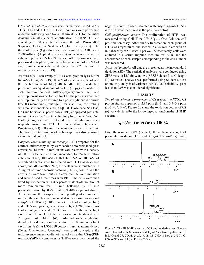
RESULTSThe physicochemical properties of CS-g-(PEI-b-mPEG): CSproton signals appeared at 2.84 ppm (H-2) and 3.3−3.8 ppm(H-3, 4, 5, 6, 6’; Figure 2B), and the oxidation degree of CS(q) was calculated by the following equation from the 1H NMRspectrum:
From the results of GPC (Table 1), the molecular weights ofperiodate oxidation CS and CS-g-(PEI-b-mPEG) were
Figure 2. The 1H NMR spectra of CS and its derivatives. Spectrawere obtained with 32 scans, and delay of 2 s between pulses. A: CSin D2O and CF3COOD at 293 K, B: CS-CHO in D2O at 293 K; C:CS-g-(PEI-b-mPEG) in D2O at 293 K.
Molecular Vision 2008; 14:2616-2628 <http://www.molvis.org/molvis/v14/a299> © 2008 Molecular Vision
2620
q=(IH2-IH2')/(IH2) x 100%
1.16×10⁴ and 2.95×10⁴, and the degree of grafted PEI-b-mPEG can be calculated by the following equation accordingto the data above:
M’W,CS-CHO is the average molecular weight of repeat unitof CS-CHO.
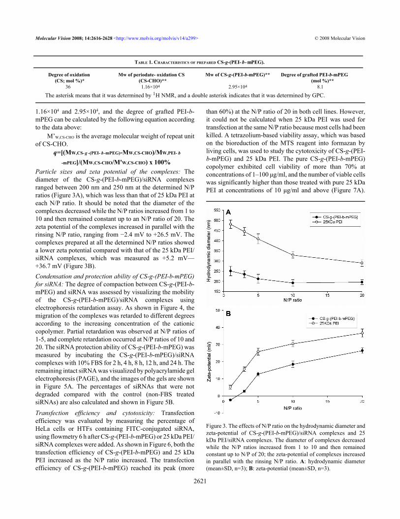
Particle sizes and zeta potential of the complexes: Thediameter of the CS-g-(PEI-b-mPEG)/siRNA complexesranged between 200 nm and 250 nm at the determined N/Pratios (Figure 3A), which was less than that of 25 kDa PEI ateach N/P ratio. It should be noted that the diameter of thecomplexes decreased while the N/P ratios increased from 1 to10 and then remained constant up to an N/P ratio of 20. Thezeta potential of the complexes increased in parallel with therinsing N/P ratio, ranging from −2.4 mV to +26.5 mV. Thecomplexes prepared at all the determined N/P ratios showeda lower zeta potential compared with that of the 25 kDa PEI/siRNA complexes, which was measured as +5.2 mV—+36.7 mV (Figure 3B).Condensation and protection ability of CS-g-(PEI-b-mPEG)for siRNA: The degree of compaction between CS-g-(PEI-b-mPEG) and siRNA was assessed by visualizing the mobilityof the CS-g-(PEI-b-mPEG)/siRNA complexes usingelectrophoresis retardation assay. As shown in Figure 4, themigration of the complexes was retarded to different degreesaccording to the increasing concentration of the cationiccopolymer. Partial retardation was observed at N/P ratios of1-5, and complete retardation occurred at N/P ratios of 10 and20. The siRNA protection ability of CS-g-(PEI-b-mPEG) wasmeasured by incubating the CS-g-(PEI-b-mPEG)/siRNAcomplexes with 10% FBS for 2 h, 4 h, 8 h, 12 h, and 24 h. Theremaining intact siRNA was visualized by polyacrylamide gelelectrophoresis (PAGE), and the images of the gels are shownin Figure 5A. The percentages of siRNAs that were notdegraded compared with the control (non-FBS treatedsiRNAs) are also calculated and shown in Figure 5B.
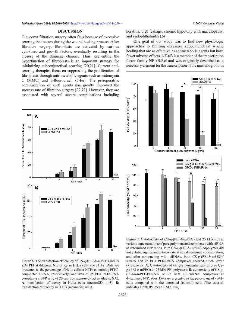
Transfection efficiency and cytotoxicity: Transfectionefficiency was evaluated by measuring the percentage ofHeLa cells or HTFs containing FITC-conjugated siRNA,using flowmetry 6 h after CS-g-(PEI-b-mPEG) or 25 kDa PEI/siRNA complexes were added. As shown in Figure 6, both thetransfection efficiency of CS-g-(PEI-b-mPEG) and 25 kDaPEI increased as the N/P ratio increased. The transfectionefficiency of CS-g-(PEI-b-mPEG) reached its peak (more
than 60%) at the N/P ratio of 20 in both cell lines. However,it could not be calculated when 25 kDa PEI was used fortransfection at the same N/P ratio because most cells had beenkilled. A tetrazolium-based viability assay, which was basedon the bioreduction of the MTS reagent into formazan byliving cells, was used to study the cytotoxicity of CS-g-(PEI-b-mPEG) and 25 kDa PEI. The pure CS-g-(PEI-b-mPEG)copolymer exhibited cell viability of more than 70% atconcentrations of 1–100 μg/ml, and the number of viable cellswas significantly higher than those treated with pure 25 kDaPEI at concentrations of 10 μg/ml and above (Figure 7A).
Figure 3. The effects of N/P ratio on the hydrodynamic diameter andzeta-potential of CS-g-(PEI-b-mPEG)/siRNA complexes and 25kDa PEI/siRNA complexes. The diameter of complexes decreasedwhile the N/P ratios increased from 1 to 10 and then remainedconstant up to N/P of 20; the zeta-potential of complexes increasedin parallel with the rinsing N/P ratio. A: hydrodynamic diameter(mean±SD, n=3); B: zeta-potential (mean±SD, n=3).
Molecular Vision 2008; 14:2616-2628 <http://www.molvis.org/molvis/v14/a299> © 2008 Molecular Vision
2621
TABLE 1. CHARACTERISTICS OF PREPARED CS-g-(PEI- b- mPEG).
Degree of oxidation(CS; mol %)*
Mw of periodate- oxidation CS(CS-CHO)**
Mw of CS-g-(PEI-b-mPEG)** Degree of grafted PEI-b-mPEG(mol %)**
36 1.16×10⁴ 2.95×10⁴ 8.1
The asterisk means that it was determined by 1H NMR, and a double asterisk indicates that it was determined by GPC.
q=[(MW,CS-g -(PEI- b -mPEG)-MW,CS-CHO)/MW,PEI- b
-mPEG]/(MW,CS-CHO/M'W,CS-CHO) x 100%
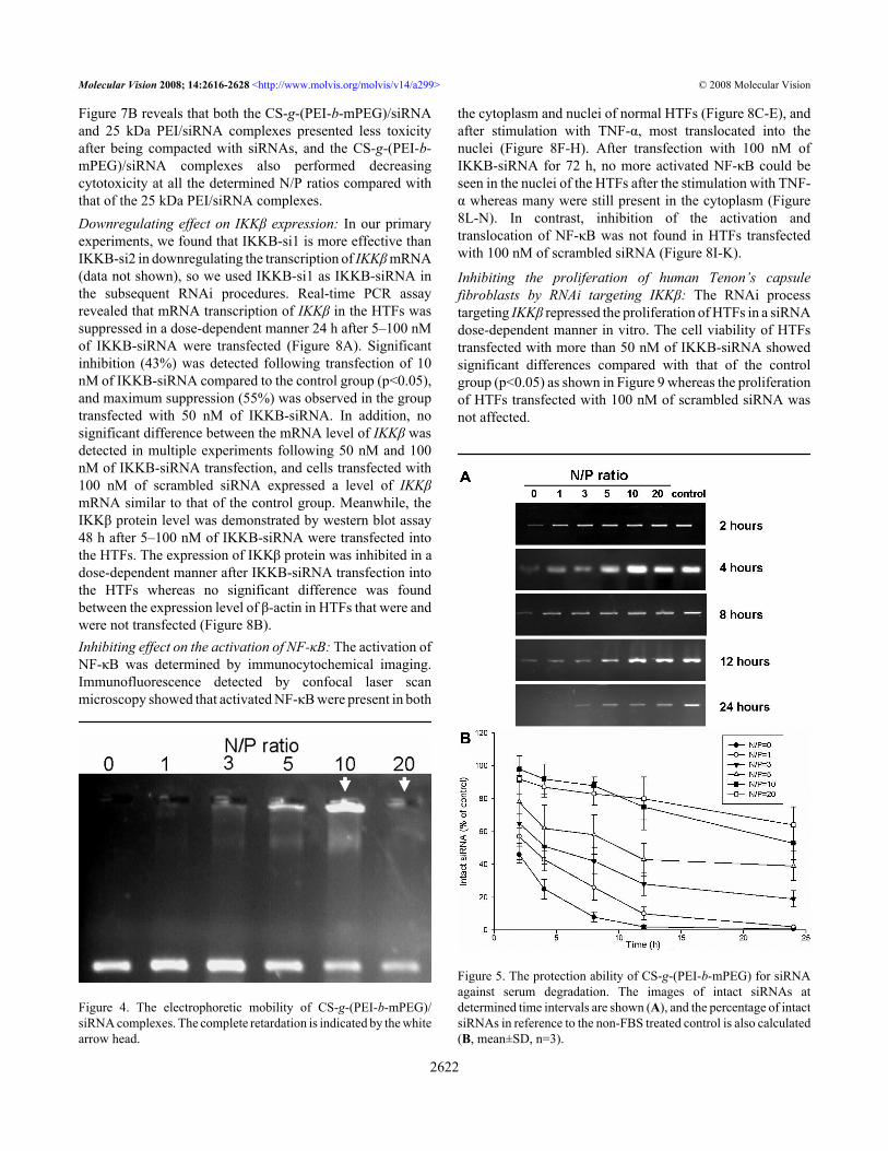
Figure 7B reveals that both the CS-g-(PEI-b-mPEG)/siRNAand 25 kDa PEI/siRNA complexes presented less toxicityafter being compacted with siRNAs, and the CS-g-(PEI-b-mPEG)/siRNA complexes also performed decreasingcytotoxicity at all the determined N/P ratios compared withthat of the 25 kDa PEI/siRNA complexes.Downregulating effect on IKKβ expression: In our primaryexperiments, we found that IKKΒ-si1 is more effective thanIKKΒ-si2 in downregulating the transcription of IKKβ mRNA(data not shown), so we used IKKΒ-si1 as IKKΒ-siRNA inthe subsequent RNAi procedures. Real-time PCR assayrevealed that mRNA transcription of IKKβ in the HTFs wassuppressed in a dose-dependent manner 24 h after 5–100 nMof IKKΒ-siRNA were transfected (Figure 8A). Significantinhibition (43%) was detected following transfection of 10nM of IKKΒ-siRNA compared to the control group (p<0.05),and maximum suppression (55%) was observed in the grouptransfected with 50 nM of IKKΒ-siRNA. In addition, nosignificant difference between the mRNA level of IKKβ wasdetected in multiple experiments following 50 nM and 100nM of IKKΒ-siRNA transfection, and cells transfected with100 nM of scrambled siRNA expressed a level of IKKβmRNA similar to that of the control group. Meanwhile, theIKKβ protein level was demonstrated by western blot assay48 h after 5–100 nM of IKKΒ-siRNA were transfected intothe HTFs. The expression of IKKβ protein was inhibited in adose-dependent manner after IKKΒ-siRNA transfection intothe HTFs whereas no significant difference was foundbetween the expression level of β-actin in HTFs that were andwere not transfected (Figure 8B).Inhibiting effect on the activation of NF-κB: The activation ofNF-κB was determined by immunocytochemical imaging.Immunofluorescence detected by confocal laser scanmicroscopy showed that activated NF-κB were present in both
Figure 4. The electrophoretic mobility of CS-g-(PEI-b-mPEG)/siRNA complexes. The complete retardation is indicated by the whitearrow head.
the cytoplasm and nuclei of normal HTFs (Figure 8C-E), andafter stimulation with TNF-α, most translocated into thenuclei (Figure 8F-H). After transfection with 100 nM ofIKKΒ-siRNA for 72 h, no more activated NF-κB could beseen in the nuclei of the HTFs after the stimulation with TNF-α whereas many were still present in the cytoplasm (Figure8L-N). In contrast, inhibition of the activation andtranslocation of NF-κB was not found in HTFs transfectedwith 100 nM of scrambled siRNA (Figure 8I-K).
Inhibiting the proliferation of human Tenon’s capsulefibroblasts by RNAi targeting IKKβ: The RNAi processtargeting IKKβ repressed the proliferation of HTFs in a siRNAdose-dependent manner in vitro. The cell viability of HTFstransfected with more than 50 nM of IKKΒ-siRNA showedsignificant differences compared with that of the controlgroup (p<0.05) as shown in Figure 9 whereas the proliferationof HTFs transfected with 100 nM of scrambled siRNA wasnot affected.
Figure 5. The protection ability of CS-g-(PEI-b-mPEG) for siRNAagainst serum degradation. The images of intact siRNAs atdetermined time intervals are shown (A), and the percentage of intactsiRNAs in reference to the non-FBS treated control is also calculated(B, mean±SD, n=3).
Molecular Vision 2008; 14:2616-2628 <http://www.molvis.org/molvis/v14/a299> © 2008 Molecular Vision
2622
DISCUSSIONGlaucoma filtration surgery often fails because of excessivescarring that occurs during the wound healing process. Afterfiltration surgery, fibroblasts are activated by variouscytokines and growth factors, eventually resulting in theclosure of the drainage channel. Thus, preventing thehyperfunction of fibroblasts is an important strategy forminimizing subconjunctival scarring [20,21]. Current anti-scarring therapies focus on suppressing the proliferation offibroblasts through anti-metabolic agents such as mitomycinC (MMC) and 5-fluorouracil (5-Fu). The perioperativeadministration of such agents has greatly improved thesuccess rate of filtration surgery [22,23]. However, they areassociated with several severe complications including
Figure 6. The transfection efficiency of CS-g-(PEI-b-mPEG) and 25kDa PEI at different N/P ratios in HeLa cells and HTFs. Data arepresented as the percentage of HeLa cells or HTFs containing FITC-conjuncted siRNA, respectively, and data of 25 kDa PEI/siRNAcomplexes at N/P ratio of 20 can’t be measured (not available, NA).A: transfection efficiency in HeLa cells (mean±SD, n=3); B:transfection efficiency in HTFs (mean±SD, n=3).
keratitis, bleb leakage, chronic hypotony with maculopathy,and endophthalmitis [24].
One goal of our study was to find new physiologicapproaches to limiting excessive subconjunctival woundhealing that are as effective as antimetabolic agents but havefewer adverse effects. NF-κB is a member of the transcriptionfactor family NF-κB/Rel and was originally described as anecessary element for the transcription of the immunoglobulin
Figure 7. Cytotoxicity of CS-g-(PEI-b-mPEG) and 25 kDa PEI atvarious concentrations of pure polymers and complexes with siRNAat determined N/P ratios. Pure CS-g-(PEI-b-mPEG) copolymer didnot exhibit significant cytotoxicity at any determined concentration,and after compacting with siRNAs, both CS-g-(PEI-b-mPEG)/siRNA and 25 kDa PEI/siRNA complexes showed much lowercytotoxicity. A: Cytotoxicity of various concentrations of pure CS-g-(PEI-b-mPEG) or 25 kDa PEI polymers; B: cytotoxicity of CS-g-(PEI-b-mPEG)/siRNA or 25 kDa PEI/siRNA complexes atdetermined N/P ratios. Data are presented as the percentage of viablecells compared with the untreated (control) cells (The asteriskindicates a p<0.05, mean ± SD, n=6).
Molecular Vision 2008; 14:2616-2628 <http://www.molvis.org/molvis/v14/a299> © 2008 Molecular Vision
2623

κL chain gene in mature B cells [25]. NF-κB has recentlyproved to be a ubiquitous factor associated with woundhealing through the factor’s ability to stimulate transcription
Figure 8. IKKΒ-siRNA inhibits the expression of IKKβ on both themRNA and protein level. A: mRNA transcription of IKKβ in HTFsassessed by real-time RT-PCR 24 h after 5-100 nM IKKΒ-siRNAwas transfected. The normalized IKKβ mRNA level of non-transfected HTFs is taken as 1.0 (the asterisk indicates a p<0.05,mean±SD, n=3). B: Protein levels of IKKβ demonstrated by westernblot. C-N: Confocal laser scanning microscopy images shows theintracellular distribution of NF-κB in HTFs. Green fluorescenceindicates the intracellular distribution of phosphated NF-κB, andblue fluorescence represents the DAPI counterstained cell nuclei.
of various genes involved in the activation of inflammationand cell proliferation [26,27]. Thus, we thought the NF-κBpathway may be an essential factor in the regulation of theproliferation of HTFs after glaucoma filtration surgery. NF-κB is bonded with an inhibitor protein, IκB, which sequestersNF-κB in an inactive form in the cytoplasm. A specific IκBkinase, IKK, is a protein complex that contains three subunits,and studies indicate that IKKβ is indispensable for theactivation of NF-κB [28]. IKK phosphorylates IκB andinitiates the inhibitor’s conjugation to ubiquitin andsubsequent degradation by proteasomes. In turn, NF-κB isactivated through release from IκB and translocates into thenuclei [29]. RNAi is commonly used as a powerful tool inbiological and biomedical research [30] and has been usedexperimentally to prevent ocular neovascularization andinflammation [31,32]. Researchers in our group havesuccessfully inhibited the proliferation of HTFs throughspecifically downregulating the expression of IKKβ using acommercial transfection reagent, LipofectamineTM 2000(Invitrogen, Carlsbad, CA), as a siRNA-delivering vector.However, this cationic liposome compound cannot be usedfor in vivo investigation in humans [33].
Therefore, another objective of our study was to designand synthesize new compounds that are non-toxic, non-immunogenic, degradable, and efficient for delivering siRNAinto HTFs. Among non-viral nucleotide carriers currentlyunder investigation, PEI is one kind of synthetic polymer witha high cationic charge density and a protonable amino groupin every third position. PEI can condense and compact DNAinto complexes, and the strong proton capacity of PEI allowsit to deliver plasmid DNA and oligonucleotides intomammalian cells, both in vitro and in vivo [34]. However,despite having effective delivery capacity, high molecular
Figure 9. The inhibition effect of blocking NF-κB pathway on theproliferation of HTFs through RNAi. Data are presented as thepercentage of viable cells compared with the untreated (control) cells(mean ± SD, n=6). An asterisk indicates that p<0.05.
Molecular Vision 2008; 14:2616-2628 <http://www.molvis.org/molvis/v14/a299> © 2008 Molecular Vision
2624
weight PEI can not be degraded by body fluids and its highcationic charge density also makes PEI toxic to cells [35].Therefore, the current trend is toward using modified lowmolecular weight PEI as a nucleotide delivery reagent, whichcombines high biocompatibility and reduced cytotoxicity[36,37]. PEG, a nonionic hydrophilic polyether, has beenwidely investigated as a synthesizing graft copolymer withPEI. The nonionic PEG chains can serve as a hydrophilic shell,which makes the new copolymer more soluble and stable, andreduces its non-specific interaction with proteins inphysiologic fluids [38,39]. One drawback of these copolymersfor in vivo application is their lack of transfection efficiency.Chitosan is the name given to a group of linear cationicpolymers of glucosamine and N-acetylglucosamine that arederived from the natural biopolymer, chitin, by alkalinedeacetylation. Chitosan has been investigated as a foodadditive or a wound dressing, and it has been recentlyconsidered to be a good candidate for gene delivery becauseof its reported biocompatibility and biodegradability and itsrelatively non-toxic nature [40].
In light of these results, we speculated that a novelcationic copolymer combined with PEI, PEG, and CS couldbe synthesized as a vehicle for delivering siRNA intomammalian cells effectively and safely. In our research, wefound that after the grafting reaction, characteristic absorptionof PEI and mPEG appeared at 2.5−3.3 ppm and 3.5 ppm in the1H NMR spectrum of the copolymer, respectively. CS protonabsorption signals (H-2, H-3, 4, 5, 6, 6’, 2.8−3.8 ppm) alsooverlapped the PEI and mPEG proton signals (Figure 2). Allof the data above prove that the designed CS-g-(PEI-b-mPEG)copolymer was successfully obtained. The molecular weightof CS-g-(PEI-b-mPEG) was 2.95×10⁴, which was relativelylarger than some reported PEI-alt-PEG copolymers [41] butsmaller than generally used PEI [37].
As indicated in previous reports, the size and shape ofnanoparticles play an important role in the delivery processand greatly influence distribution in the body [42]. It has beenreported that the nano-size of particles is a key prerequisitefor cell uptake [43]. Similar to plasmid DNA andoligonucleotides, siRNAs are taken up by cells throughendocytosis. Therefore, suitable particle size has an importantinfluence on the delivering capacity of a siRNA vector. Weinvestigated the diameter of the CS-g-(PEI-b-mPEG)/siRNAcomplexes at five different N/P charge ratios and observedthat the complexes represented particles with nanometer size(about 200 nm), which were much less than that of 25 kDaPEI. Furthermore, particle size tended to decrease as the N/Pcharge ratios of the CS-g-(PEI-b-mPEG)/siRNA complexesincreased, indicating that the CS-g-(PEI-b-mPEG) copolymercan condense siRNA into a more compact structure, mainlyowing to the net electrostatic repulsive forces betweencomplexes.
The surface charge of the CS-g-(PEI-b-mPEG)/siRNAcomplexes is also a major factor influencing transfection
efficiency. After the compaction between cationiccopolymers and siRNA, the negative charge of siRNA isneutralized and the newly assembled nanoparticles may retaina partial positive surface charge to help siRNA pass throughthe cell membrane and escape the endolysosomes. However,the excessive positive charge of PEI homopolymers maysubsequently lead to hyperpermeability of the membrane,resulting in cell death [44]. The density of the surface chargeis reflected by measured zeta potential values, and as we haveshown, an initial negative value of zeta potential (−2.4 mV)was detected when the complexes formed at an N/P ratio of1, which means that siRNA could not be completelycompacted under this condition. Then, the positive surfacecharge of the complexes exhibited an increasing trendcorresponding to the rising N/P ratio, indicating that more andmore siRNAs were compacted with CS-g-(PEI-b-mPEG) andthe negative charge was neutralized. But the zeta potential ofthe CS-g-(PEI-b-mPEG)/siRNA complexes at each N/P ratiothat we measured was still lower than that of the 25 kDa PEI/siRNA complexes. The reason for this result is probablyconsistent with the reason Petersen et al. [8] regardingcopolymer-based DNA complexes, which was that theshielding effect of neutral components on the PEI part of thecopolymer produces a relatively low zeta potential.
Condensation ability is one requirement for a siRNAcarrier. An optimal binding degree between CS-g-(PEI-b-mPEG) and siRNA can achieve more efficient deliveringcapacity. If the complexes are formed efficiently, all siRNAsare bound to the copolymers to form nanoparticles. Hence, thecomplexes become relatively large and remain immobile inthe loading well with no bands of free siRNA apparent. Weobserved that the migration of siRNA was retarded to differentdegrees in accordance with the increasing N/P ratio. Completeretardation occurs at an N/P ratio of 10, which means the CS-g-(PEI-b-mPEG)/siRNA complexes are completely formed.The complete complexes of 600 kDa PEI and siRNA cannotbe found even at an N/P ratio of 50 (data not shown), whichindicates that the condensation ability of CS-g-(PEI-b-mPEG)is better than low molecular weight PEI. It was also detectedthat the band of siRNA at an N/P ratio of 20 demonstratedmuch lower fluorescence intensity than that of the band at theN/P ratio of 10. This decrease in fluorescence was alsoobserved by other experiments executed on DNA bands [8,45]. The reason for this phenomenon is that the measuredfluorescence is attributed to the intercalated ethidium bromidein siRNA. When the condensation degree between siRNA andthe cationic copolymer gradually increases, ethidium bromidecannot intercalate with siRNA anymore. Therefore, thefluorescence intensity of the bands decreases accordingly[39].
The main hindrance to the use of RNAi as a therapeutictool for human diseases is that the unprotected dsRNA orsiRNA will be rapidly degraded by either nucleases in serumor the endosomal compartment of cells. The enzymatic
Molecular Vision 2008; 14:2616-2628 <http://www.molvis.org/molvis/v14/a299> © 2008 Molecular Vision
2625
degradation of siRNA is accompanied by a rapid decline inbiological activity and therapeutic efficiency. Therefore, thepotential of this technology as a clinical therapy methoddepends largely on the improvement of siRNA vectors’protection ability against enzymatic degradation. We foundthat after incubation in 10% FBS for 24 h, 64% of siRNA wasprotected from degradation at the N/P ratio of 20 whereas only0.7% naked siRNA remained intact. We attributed theseresults to two factors. First, PEG not only has the reportedability to stabilize the structure of nanospheres, but PEG canalso protect siRNA from being attacked by nucleases [46,47]. Second, it has been reported that chitosan can effectivelyprotect DNA from nuclease degradation [48]. The CS-g-(PEI-b-mPEG) we have synthesized has both PEG and CS elementsand can provide efficient protection for siRNA againstenzymatic degradation.
Low toxicity is also a major requirement for an siRNAdelivery system. The cytotoxicity of cationic copolymers ismainly caused by the aggregation of nanoparticles on the cellmembrane, impairing its normal function. In addition, theexcessive positive surface charge of nanoparticles may alsointerfere with critical intracellular processes of cells [44]. Ithas been shown that chitosan salts and chitosan derivativesare less toxic than PEI [49]. However, no data have beenreported regarding the cytotoxic analysis of a syntheticcationic copolymer on human Tenon’s capsule fibroblasts.Therefore, we explored the influence of differentconcentrations of CS-g-(PEI-b-mPEG) and 25 kDa PEI on thecell viability of HTFs. CS-g-(PEI-b-mPEG) showed muchless cytotoxicity than 25 kDa PEI, which is consistent withwhat had been obtained by Kim et al. [18] in tests of HeLaand HepG2 cells. The cell viabilities of the HTFs decreaseddrastically as the concentrations of 25 kDa PEI increasedwhereas the pure CS-g-(PEI-b-mPEG) copolymer did notexhibit significant cytotoxicity at any determinedconcentration. We hypothesize that the relatively lowcytotoxicity of CS-g-(PEI-b-mPEG) could be explained fromtwo aspects. First, the copolymer can be degraded into CS,PEG, and low molecular weight PEI units in cells, all of whichcan be easily eliminated by excretion pathways, thus makingthis copolymer relatively less toxic than 25 kDa PEI. Second,PEG reduces toxicity by substituting the amino groups of PEI,which are the main toxic moieties of the copolymer [50].Furthermore, after compacting with siRNA, both the CS-g-(PEI-b-mPEG)/siRNA and 25 kDa PEI/siRNA complexesshowed much lower cytotoxicity, which is due to theneutralization effect of the negative charge of siRNA on thepositive charge of the pure polymers.
The transfection efficiency of the CS-g-(PEI-b-mPEG)/siRNA complexes was assessed at various N/P ratios in HeLacells and HTFs. This is mainly because the N/P ratio is directlyrelated to the size, surface charge, compaction degree, andserum-resistant capacity of the CS-g-(PEI-b-mPEG)/siRNAcomplexes, all of which can affect delivery efficiency. At the
N/P ratio of 20, CS-g-(PEI-b-mPEG) outperformed thehighest transfection rate in both HeLa cells and HTFs, whichcan be explained by the appropriate particle size and surfacecharge as well as the excellent stability of the CS-g-(PEI-b-mPEG)/siRNA complexes. The transfection rate was a littlelower than that of 25 kDa PEI at the same N/P ratio partlybecause of the shielding effect of mPEG on the positive chargeof PEI. We also found that both CS-g-(PEI-b-mPEG) and25 kDa PEI showed a lower transfection rate in HTFs than inHeLa cells, which may be attributed to a cell line dependencyof the cationic polymer’s delivery ability [51]. From ourresults, we can conclude that PEI with low molecular weightgrafted onto CS avoids the cytotoxicity of high molecularweight PEI. Meanwhile, mPEG improves the stability of CS-g-(PEI-b-mPEG)/siRNA complexes, and as a result, the CS-g-(PEI-b-mPEG)/siRNA nanoparticles can offer a substantialgene silencing effect with minimal side effects. Moreover,many factors governing the transfection efficiency of cationiccopolymers need to be investigated in future studies such asthe presence of serum and the pH value of solution [52], andligands will be conjugated to the copolymers to achievereceptor-mediated endocytosis and potentially to target cellsor tissues.
A special siRNA targeting IKKβ gene was successfullycompacted with CS-g-(PEI-b-mPEG) and effectivelydelivered into HTFs. Subsequently, both the mRNA andprotein levels of IKKβ were suppressed, and the activation ofNF-κB was inhibited in turn. Finally, IKKΒ-siRNA-mediatedblocking of the NF-κB pathway resulted in repression of theproliferation of HTFs, and cells transfected with IKKΒ-siRNA showed growth inhibition up to 42%. All of thesefindings suggest that blocking the signaling pathway of NF-κB could be an effective way to manipulate the scar formationprocess by downregulating the proliferation of HTFs. Thisnovel method based on nanotechnology and RNAi couldrepresent a remarkable anti-scarring therapeutic approach forglaucoma filtration surgery. Follow-up studies will focus onthe in vivo application of CS-g-(PEI-b-mPEG)/IKKΒ-siRNAcomplexes.
We have reported on the synthesis and characterizationof a novel cationic copolymer, CS-g-(PEI-b-mPEG). Wefound it to have powerful siRNA binding and protectionability, relatively high transfection efficiency, and lowcytotoxicity, making CS-g-(PEI-b-mPEG) a suitable deliveryvector for transfecting siRNA into cells. We observed thatsiRNA targeting IKKβ was successfully transfected intohuman Tenon’s capsule fibroblasts in vitro, and RNAiprocesses against the expression of IKKβ can subsequentlyinhibit the activation of NF-κB and in turn, the proliferationof HTFs. Our results indicate the potential for a safe andeffective strategy for preventing scar formation afterglaucoma filtration surgery.
Molecular Vision 2008; 14:2616-2628 <http://www.molvis.org/molvis/v14/a299> © 2008 Molecular Vision
2626
ACKNOWLEDGMENTSThe authors are grateful for the financial support provided bythe National Basic Research Program of China (“973”program, Number 2007CB512207) and the special researchfoundation for doctor disciplines at colleges, the Ministry ofEducation, China (Number 20060558035). Professors Ge andQuan contributed equally to this publication and can beconsidered co- corresponding authors.
REFERENCES1. Plasterk RH. RNA silencing: the genome's immune system.
Science 2002; 296:1263-5. [PMID: 12016302]2. Hannon GJ. RNA interference. Nature 2002; 418:244-51.
[PMID: 12110901]3. Elbashir SM, Harborth J, Lendeckel W, Yalcin A, Weber K,
Tuschl T. Duplexes of 21-nucleotide RNAs mediate RNAinterference in cultured mammalian cells. Nature 2001;411:494-8. [PMID: 11373684]
4. Sun JY, Anand-Jawa V, Chatterjee S, Wong KK. Immuneresponses to adeno-associated virus and its recombinantvectors. Gene Ther 2003; 10:964-76. [PMID: 12756417]
5. Lehrman S. Virus treatment questioned after gene therapydeath. Nature 1999; 401:517-8. [PMID: 10524611]
6. Merdan T, Kopecek J, Kissel T. Prospects for cationic polymersin gene and oligonucleotide therapy against cancer. Adv DrugDeliv Rev 2002; 54:715-58. [PMID: 12204600]
7. Boussif O. Lezoualc'h F, Zanta MA, Mergny MD, Scherman D,Demeneix B, Behr JP. A versatile vector for gene andoligonucleotide transfer into cells in culture and in vivo:polyethylenimine. Proc Natl Acad Sci USA 1995;92:7297-301. [PMID: 7638184]
8. Petersen H, Fechner PM, Martin AL, Kunath K, Stolnik S,Roberts CJ, Fischer D, Davies MC, Kissel T.Polyethylenimine-graft-poly (ethylene glycol) copolymers:influence of copolymer block structure on DNAcomplexation and biological activities as gene deliverysystem. Bioconjug Chem 2002; 13:845-54. [PMID:12121141]
9. Ahn CH, Chae SY, Bae YH, Kim SW. Biodegradablepoly(ethylenimine) for plasmid DNA delivery. J ControlRelease 2002; 80:273-82. [PMID: 11943404]
10. Katas H, Alpar HO. Development and characterisation ofchitosan nanoparticles for siRNA delivery. J Control Release2006; 115:216-25. [PMID: 16959358]
11. The Advanced Glaucoma Intervention Study (AGIS):7. Therelationship between control of intraocular pressure andvisual field deterioration.The AGIS Investigators.Am JOphthalmol200013042940 [PubMed: 11024415]
12. Cordeiro MF, Chang L, Lim KS, Daniels JT, Pleass RD,Siriwardena D, Khaw PT. Modulating conjunctival woundhealing. Eye 2000; 14:536-47. [PMID: 11026984]
13. Khaw PT, Occleston NL, Schultz G, Grierson I, Sherwood MB,Larkin G. Activation and suppression of fibroblast function.Eye 1994; (Pt 2):188-95. [PMID: 7958020]
14. Kessler DJ, Duyao MP, Spicer DB, Sonenshein GE. NF-kappaB-like factors mediate interleukin 1 induction of c-myc genetranscription in fibroblasts. J Exp Med 1992; 176:787-92.[PMID: 1512542]
15. DiDonato JA, Hayakawa M, Rothwarf DM, Zandi E, Karin M.A cytokine-responsive IkappaB kinase that activates thetranscription factor NF-kappaB. Nature 1997; 388:548-54.[PMID: 9252186]
16. Khaw PT, Ward S, Porter A, Grierson I, Hitchings RA, RiceNS. The long-term effects of 5-fluorouracil and sodiumbutyrate on human Tenon's fibroblasts. Invest OphthalmolVis Sci 1992; 33:2043-52. [PMID: 1582809]
17. Jiang HL, Kim YK, Arote R, Nah JW, Cho MH, Choi YJ,Akaike T, Cho CS. Chitosan-graft-polyethylenimine as agene carrier. J Control Release 2007; 117:273-80. [PMID:17166614]
18. Kim TH, Park IK, Nah JW, Choi YJ, Cho CS. Galactosylatedchitosan/DNA nanoparticles prepared using water-solublechitosan as a gene carrier. Biomaterials 2004; 25:3783-92.[PMID: 15020154]
19. Livak KJ, Schmittgen TD. Analysis of relative gene expressiondata using real-time quantitative PCR and the 2(-Delta DeltaC (T)). Methods 2001; 25:402-8. [PMID: 11846609]
20. Khaw PT, Chang L, Wong TT, Mead A, Daniels JT, CordeiroMF. Modulation of wound healing after glaucoma surgery.Curr Opin Ophthalmol 2001; 12:143-8. [PMID: 11224722]
21. Atreides SP, Skuta GL, Reynolds AC. Wound healingmodulation in glaucoma filtering surgery. Int OphthalmolClin 2004; 44:61-106. [PMID: 15087731]
22. Smith MF, Doyle JW, Nguyen QH, Sherwood MB. Results ofintraoperative 5-fluorouracil or lower dose mitomycin-Cadministration on initial trabeculectomy surgery. J Glaucoma1997; 6:104-10. [PMID: 9098818]
23. Singh RP, Goldberg I, Mohsin M. The efficacy and safety ofintraoperative and/or postoperative 5-fluorouracil intrabeculectomy and phacotrabeculectomy. Clin ExperimentOphthalmol 2001; 29:296-302. [PMID: 11720155]
24. Sihota R, Dada T, Gupta SD, Sharma S, Arora R, Agarwal HC.Conjunctival dysfunction and mitomycin C-inducedhypotony. J Glaucoma 2000; 9:392-7. [PMID: 11039741]
25. Baeuerle PA, Baltimore D. NF-kappa B: ten years after. Cell1996; 87:13-20. [PMID: 8858144]
26. Kaltschmidt B, Kaltschmidt C, Hehner SP, Droge W, SchmitzML. Repression of NF-kappaB impairs HeLa cellproliferation by functional interference with cell cyclecheckpoint regulators. Oncogene 1999; 18:3213-25. [PMID:10359527]
27. Li M, Carpio DF, Zheng Y, Bruzzo P, Singh V, Ouaaz F,Medzhitov RM, Beg AA. An essential role of the NF-kappaB/Toll-like receptor pathway in induction of inflammatoryand tissue-repair gene expression by necrotic cells. J Immunol2001; 166:7128-35. [PMID: 11390458]
28. Zandi E, Rothwarf DM, Delhase M, Hayakawa M, Karin M.The IkappaB kinase complex (IKK) contains two kinasesubunits, IKKalpha and IKKbeta, necessary for IkappaBphosphorylation and NF-kappaB activation. Cell 1997;91:243-52. [PMID: 9346241]
29. Woronicz JD, Gao X, Cao Z, Rothe M, Goeddel DV. IkappaBkinase-beta: NF-kappaB activation and complex formationwith IkappaB kinase-alpha and NIK. Science 1997;278:866-9. [PMID: 9346485]
30. Novina CD, Sharp PA. The RNAi revolution. Nature 2004;430:161-4. [PMID: 15241403]
Molecular Vision 2008; 14:2616-2628 <http://www.molvis.org/molvis/v14/a299> © 2008 Molecular Vision
2627
http://www.ncbi.nlm.nih.gov/entrez/query.fcgi?cmd=Retrieve&db=PubMed&dopt=abstract&list_uids=7638184
http://www.ncbi.nlm.nih.gov/entrez/query.fcgi?cmd=Retrieve&db=PubMed&dopt=abstract&list_uids=7958020
http://www.ncbi.nlm.nih.gov/entrez/query.fcgi?cmd=Retrieve&db=PubMed&dopt=abstract&list_uids=1512542
http://www.ncbi.nlm.nih.gov/entrez/query.fcgi?cmd=Retrieve&db=PubMed&dopt=abstract&list_uids=1512542
http://www.ncbi.nlm.nih.gov/entrez/query.fcgi?cmd=Retrieve&db=PubMed&dopt=abstract&list_uids=9252186
http://www.ncbi.nlm.nih.gov/entrez/query.fcgi?cmd=Retrieve&db=PubMed&dopt=abstract&list_uids=9252186
http://www.ncbi.nlm.nih.gov/entrez/query.fcgi?cmd=Retrieve&db=PubMed&dopt=abstract&list_uids=1582809
http://www.ncbi.nlm.nih.gov/entrez/query.fcgi?cmd=Retrieve&db=PubMed&dopt=abstract&list_uids=9098818
http://www.ncbi.nlm.nih.gov/entrez/query.fcgi?cmd=Retrieve&db=PubMed&dopt=abstract&list_uids=8858144
http://www.ncbi.nlm.nih.gov/entrez/query.fcgi?cmd=Retrieve&db=PubMed&dopt=abstract&list_uids=9346241
31. Reich SJ, Fosnot J, Kuroki A, Tang W, Yang X, Maguire AM,Bennett J, Tolentino MJ. Small interfering RNA (siRNA)targeting VEGF effectively inhibits ocular neovascularizationin a mouse model. Mol Vis 2003; 9:210-6. [PMID: 12789138]
32. Nakamura H, Siddiqui SS, Shen X, Malik AB, Pulido JS, KumarNM, Yue BY. RNA interference targeting transforminggrowth factor-beta type II receptor suppresses ocularinflammation and fibrosis. Mol Vis 2004; 10:703-11. [PMID:15475878]
33. Huang SS, Ge J, Wang LN, Yin XB, Wei YT, Ma P. Preliminarystudy of inhibition of human Tenon’s capsule fibroblasts invitro by RNA interference targeting IKK-beta. Zhonghua YanKe Za Zhi 2005; 41:1076-81. [PMID: 16409758]
34. Kichler A, Leborgne C, Coeytaux E, Danos O.Polyethylenimine-mediated gene delivery: a mechanisticstudy. J Gene Med 2001; 3:135-44. [PMID: 11318112]
35. Moghimi SM, Symonds P, Murray JC, Hunter AC, Debska G,Szewczyk A. A two-stage poly (ethylenimine)-mediatedcytotoxicity: implications for gene transfer/therapy. Mol Ther2005; 11:990-5. [PMID: 15922971]
36. Kim YH, Park JH, Lee M, Kim YH, Park TG, Kim SW.Polyethylenimine with acid-labile linkages as abiodegradable gene carrier. J Control Release 2005;103:209-19. [PMID: 15710512]
37. Fischer D, Bieber T, Li Y, Elsasser HP, Kissel T. A novel non-viral vector for DNA delivery based on low molecular weight,branched polyethylenimine: effect of molecular weight ontransfection efficiency and cytotoxicity. Pharm Res 1999;16:1273-9. [PMID: 10468031]
38. Brus C, Petersen H, Aigner A, Czubayko F, Kissel T.Physicochemical and biological characterization ofpolyethylenimine-graft-poly (ethylene glycol) blockcopolymers as a delivery system for oligonucleotides andribozymes. Bioconjug Chem 2004; 15:677-84. [PMID:15264853]
39. Mao S, Neu M, Germershaus O, Merkel O, Sitterberg J,Bakowsky U, Kissel T. Influence of polyethylene glycol chainlength on the physicochemical and biological properties ofpoly(ethylene imine)-graft-poly(ethylene glycol) blockcopolymer/siRNA polyplexes. Bioconjug Chem 2006;17:1209-18. [PMID: 16984130]
40. Mansouri S, Cuie Y, Winnik F, Shi Q, Lavigne P, BenderdourM, Beaumont E, Fernandes JC. Characterization of folate-chitosan-DNA nanoparticles for gene therapy. Biomaterials2006; 27:2060-5. [PMID: 16202449]
41. Park MR, Han KO, Han IK, Cho MH, Nah JW, Choi YJ, ChoCS. Degradable polyethylenimine-alt-poly (ethylene glycol)
copolymers as novel gene carriers. J Control Release 2005;105:367-80. [PMID: 15936108]
42. Schiffelers RM, Woodle MC, Scaria P. Pharmaceuticalprospects for RNA interference. Pharm Res 2004; 21:1-7.[PMID: 14984251]
43. Liu G, Molas M, Grossmann GA, Pasumarthy M, Perales JC,Cooper MJ, Hanson RW. Biological properties of poly-L-lysine-DNA complexes generated by cooperative binding ofthe polycation. J Biol Chem 2001; 276:34379-87. [PMID:11438546]
44. Fischer D, Li Y, Ahlemeyer B, Krieglstein J, Kissel T. In vitrocytotoxicity testing of polycations: influence of polymerstructure on cell viability and hemolysis. Biomaterials 2003;24:1121-31. [PMID: 12527253]
45. Kim TH, Kim SI, Akaike T, Cho CS. Synergistic effect of poly(ethylenimine) on the transfection efficiency ofgalactosylated chitosan/DNA complexes. J Control Release2005; 105:354-66. [PMID: 15949861]
46. Kim SH, Jeong JH, Lee SH, Kim SW, Park TG. PEG conjugatedVEGF siRNA for anti-angiogenic gene therapy. J ControlRelease 2006; 116:123-9. [PMID: 16831481]
47. Lee SH, Kim SH, Park TG. Intracellular siRNA delivery systemusing polyelectrolyte complex micelles prepared from VEGFsiRNA-PEG conjugate and cationic fusogenic peptide.Biochem Biophys Res Commun 2007; 357:511-6. [PMID:17434451]
48. Corsi K, Chellat F, Yahia L, Fernandes JC. Mesenchymal stemcells, MG63 and HEK293 transfection using chitosan-DNAnanoparticles. Biomaterials 2003; 24:1255-64. [PMID:12527266]
49. Thanou M, Florea BI, Geldof M, Junginger HE, Borchard G.Quaternized chitosan oligomers as novel gene deliveryvectors in epithelial cell lines. Biomaterials 2002; 23:153-9.[PMID: 11762833]
50. Kostarelos K. Rational design and engineering of deliverysystems for therapeutics: biomedical exercises in colloid andsurface science. Adv Colloid Interface Sci 2003;106:147-68. [PMID: 14672846]
51. Florea BI, Meaney C, Junginger HE, Borchard G. Transfectionefficiency and toxicity of polyethylenimine in differentiatedCalu-3 and nondifferentiated COS-1 cell cultures. AAPSPharmSci 2002; 4:E12. [PMID: 12423061]
52. Sato T, Ishii T, Okahata Y. In vitro gene delivery mediated bychitosan. Effect of pH, serum, and molecular mass of chitosanon the transfection efficiency. Biomaterials 2001;22:2075-80. [PMID: 11432586]
Molecular Vision 2008; 14:2616-2628 <http://www.molvis.org/molvis/v14/a299> © 2008 Molecular Vision
The print version of this article was created on 31 December 2008. This reflects all typographical corrections and errata to thearticle through that date. Details of any changes may be found in the online version of the article.
2628
Related Documents

![Static and Dynamic Density Functional Theory and ...called copolymers. Here we consider the class of copolymers called \block copolymers" [7] while there are many kinds of copolymers.](https://static.cupdf.com/doc/110x72/5eccfbf97d791301bb64d299/static-and-dynamic-density-functional-theory-and-called-copolymers-here-we.jpg)


